Depth Of a central venous catheter tiplength of insertionguidelinefor pediatric patients
心内心电图辅助锁骨下静脉穿刺中心静脉导管定位的临床观察_翟美琴
中心静脉穿刺导管留置的关键在于将导管尖端定 位于上腔静脉近右心房口处 , 但在临床实践中 , 因导管 异位导致心律失常及测压失真极为常见 。 传统方法是 通过 X线片透视或胸片调整导管位置 , 在此过程中医 患均受 X线辐射的污染 , 且放置过程中不能实时监测 导管位置的变化 。本研究利用心内心电图引导技术观 察锁骨下静脉穿刺中心静脉导管的到位情况 , 以探讨 此项技术应用于中心静脉置管术的可行性 。 1 资料与方法 1.1 对象 选择 2006年 7月 ~ 2007年 3月我院临床 拟行中心静脉置管术的全麻成年患者 80例 , ASAⅠ ~ Ⅲ级 , 术前无心律失常 、局部感染 、凝血机制障碍及颈 肩部手术史 。 将患者随机均分为 A、B两 组 , 每组 40
临床误诊误治 2007年 10月第 20卷第 10期 ClinicalMisdiagnosis& Mistherapy, October2007, Vol.20, No.10
上肢置于体侧 , 背部垫一薄 枕 , 头低 15 ~ 30°, 穿刺点 为右锁骨下切迹下方 1 ~ 1.5 cm, 穿刺针指向胸骨上 凹紧贴锁骨进针 , 穿刺至静脉回抽到静脉血后 , 放置导 丝 , 撤出穿刺针 , 进行皮下扩张放置导管 。
ClinicalObservationofthePositionofCentralVenousCatheterAidedwiththeIntravenousECGViaSubclavianVein ZHAIMei-qin, ZHAOYan-li, LIUXiao-ming, LIUJing-jing(HebeiProvincalPeople'sHospital, Shijiazhuang050051, China) Abstract:Objective:ToevaluatethepossibilityofIVECG asasimpleandreliabletechniquetomonitoringthepositionofthe catheter.Methods:80 adultpatientsscheduledforelectivemajorsurgeriesandcentralvenouscatheterizationsundergeneralanesthesiawererandomlydividedintotwogroups, with40 casesineachgroup.Patientsallreceivedrightsubclavianveincatheterizationthroughthesupraclaricularapproachbelowrightsubclavianthyroidnotch1.0-1.5 cm.PatientsinGroupAreceivedroutine method, thedepthofthecatheter(L)=(H/10)-2;PatientsinGroupBadjustedthedepthofthecatheterbasedonthecharactersofIVECG.Thepositionofthecatheterwasreassuredbyusingposterior-anteriorpositionX-rayexaminationafteroperation. Results:Thedepthofthecatheterwas(15.1 ±1.2)cm, (13.4 ±0.9)cmrespectively;therateofsatisfactorycathetertipplacementwere80% and100% respectively.Therewereobviousstatisticalsignificancebetweenthetwogroups(P<0.05).Conclusion:ApplicationofthecatheterbasedonthecharactersofIVECGcanmonitorthevariationofthedepthofthecatheterandalso
深静脉留置导管感染的应急预案
深静脉留置导管感染的应急预案深静脉留置导管(Central Venous Catheter,CVC)在临床中广泛应用,为患者提供静脉通路,方便输液、输血、营养支持、药物给予等治疗措施。
然而,CVC使用过程中可能发生感染,引发严重的并发症。
因此,制定科学合理的应急预案,对于应对CVC感染具有重要意义。
本文将就深静脉留置导管感染的应急预案进行讨论,并提出一套应急处理措施。
一、感染的识别与评估1. 临床表现患者发热、寒战、局部红肿、疼痛等,应高度警惕CVC感染的可能性。
2. 相关检查进行血常规、C-反应蛋白等相关实验室检查,结合深静脉留置导管留置部位进行彩色超声、CT等影像学检查,以判断CVC是否存在感染。
二、应急预案的执行1. 与专业人员协商一旦怀疑CVC感染,首先要与医疗团队的专业人员进行沟通和协商,以利于制定合理的应急预案。
2. 安全隔离在确认CVC感染的情况下,立即将患者进行隔离,以防传染给他人。
3. 导管拔除及取样将CVC拔除,使用无菌技术采集导管尖端、静脉血培养和周围皮肤培养标本。
4. 适当的药物治疗根据导管相关感染引起的不同菌种选择相应的抗生素进行治疗。
5. 处理导管周围血栓如发现导管周围存在血栓形成,应及时进行抗凝治疗,并评估是否需要行介入手术。
6. 定期观察和评估在治疗过程中,定期观察患者的临床症状,实验室指标和感染征象,及时调整治疗方案。
三、预防感染的策略1. 严格遵守无菌操作在插管前应进行必要的洗手和消毒,全程采用无菌操作,减少细菌污染的机会。
2. 使用无菌敷料和盖帽CVC插管后,应使用透明无菌敷料进行覆盖并定期更换,同时保持插管口盖帽的无菌,以预防细菌感染。
3. 定期更换导管按照医疗卫生规范,推荐定期更换导管,避免长时间使用导管,减少感染的机会。
4. 药物预防根据患者的具体情况,考虑采用药物预防措施,预防感染的发生。
结语深静脉留置导管感染是医疗领域中常见的并发症之一,对于患者的治疗效果和生命安全产生重大影响。
中心静脉导管尖端罕见异位基于个案报道的文献综述
・104・LemldJjouJl2021,Jan;49(1):104-109•综述.中心静脉导管尖端罕见异位:基于个案报道的文献综述苏效添I,戈杨2,周玲林3,李青青I,张晓霞I(1.四川大学华西医院,四川成都610041:2.成都市公共卫生临床医疗中心,四川成都610066;3.四川大学望江医院,四川成都610065)[摘要]中心静脉导管(central venous catheter,CVC)被广泛用于静脉输液、血流动力学和血气监测、血液透析以及留取静脉血液标本等。
导管尖端异位是CVC常见并发症之一,异位导管通常位于直径较为粗大的静脉主干,如颈内静脉、腋静脉、锁骨下静脉以及无名静脉等。
与常见异位不同,CVC罕见异位通常位于静脉干的属支小血管内,如奇静脉、肋间上静脉、甲状腺下静脉及腰升静脉等。
罕见异位发生率低,国内外学者多以个案方式进行回顾性报道。
罕见异位的血管解剖与常见异位血管解剖有所不同,两者影像学表现也存在较大差异。
本文作者以文献回顾的方式就国内外中心静脉导管罕见异位个案报道进行汇总分析,重点探讨导管异位的血管和淋巴管的解剖学基础以及部分异位导管的影像学表现,为临床医护人员识别罕见异位提供参考。
[关键词]中心静脉导管;导管异位;血管解剖;影像学;综述[中图分类号]R472[文献标识码]A[文章编号]1671-7562(2021)01-0104-06doi:10.3969/j.issn.1671-7562.2021.01.022中心静脉导管(central venous catheter,CVC)是临床置入最多的静脉内导管之一,广泛用于静脉输液、血液透析、血流动力学监测以及留取静脉血液标本等。
导管尖端异位是CVC常见并发症,异位可引起导管功能异常,并引发一系列并发症。
CVC常见异位部位都是人体血管直径较为粗大的静脉主干,如颈内静脉、腋静脉、锁骨下静脉以及无名静脉等⑴。
从CVC应用于临床开始,就陆续有一些细小属支血管的罕见异位报道。
cvc消毒范围
cvc消毒范围
CVC消毒范围主要指的是对中央静脉导管(Central Venous Catheter,简称CVC)进行消毒的范围。
CVC消毒的主要目的是预防和控制导管相关血流感染的发生。
CVC消毒范围包括以下几个部分:
1. 皮肤消毒:在插入CVC之前,需要首先对插入点的皮肤进
行消毒。
一般采用含有酒精的消毒剂进行皮肤消毒,可以有效地清洁皮肤表面,杀灭细菌和其他病原体。
2. CVC端口消毒:在CVC使用过程中,CVC上的端口需要
经常进行消毒,以防止细菌进入导管内部。
一般使用含有酒精的消毒剂进行端口消毒。
3. CVC周围区域清洁:除了直接接触CVC的皮肤和端口外,
周围的区域也需要保持清洁。
这可以通过定期更换敷料,以及定期清洁周围皮肤来实现。
需要注意的是,CVC消毒范围是根据具体情况和医院的消毒
政策来确定的。
不同的医院和不同的操作者可能存在一些差异。
因此,在使用CVC之前,最好咨询医护人员,了解具体的消
毒要求和程序。
锁骨下静脉刺术英文版
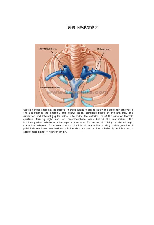
锁骨下静脉穿刺术Central venous access at the superior thoracic aperture can be safely and efficiently achieved if one understands the anatomy and follows logical principles based on the anatomy. The subclavian and internal jugular veins unite inside the anterior rim of the superior thoracic aperture, forming right and left brachiocephalic veins behind the manubrium. The brachiocephalics unite to form the superior vena cava. The second rib joining the sternal angle marks the mid-point of the vena cava and the third rib marks the caval-right atrial junction. A point between these two landmarks is the ideal position for the catheter tip and is used to approximate catheter insertion length.The heads of the clavicles extend posteriorly into the superior aperture, displacing the veins posteriorly from the sternum. The anterior scalene muscles attach to the first ribs between the subclavian veins and arteries. Because of the AP slope of the superior aperture, the arteries are more cephalad as well as more posterior than the veins. The thoracic duct rises out of the chest, arches over the left subclavian artery and enters the junction of left internal jugular and subclavian veins where it is subject to injury during left sided catheterization. The cupola of the lungs protrude into the aperture behind the vessels. The trachea and esophagus lie centrally.Both the phrenic nerves and brachial plexus nerves are subject to injury during central catheterization. The phrenic nerves descend from the cervical plexus around the lateral border of the anterior scalene muscles and enter the chest at the medial border of the first ribs just anterior to the insertion of the scalenes where they are vulnerable to injury. The brachial plexusnerves approach and encircle the axillary artery laterally and are relatively protected by their posterior position near the first rib where subclavian puncture is performed.Central access can be achieved through the subclavian vein both below and above the clavicle, but the latter route is more risky and seldom used. Access to the internal jugular vein is possible between the heads of the sternocleidomastoid muscles and posterior to the border of that muscle. These routes are suitable for acute monitoring but present logistical problems for situating long-term catheters.The subclavian vein crosses the first rib at the anterior end of the rib and lies between the anterior scalene muscle and the posterior bulge of the head of the clavicle. As can be seen in this sagittal view, the vein lies behind the medial part of the clavicle, not between the clavicleand first rib as is conventionally taught. Therefore it is not logical to aim for the sternal notch when trying to puncture the vein. The vein is compressed between the clavicle and scalene when the shoulder is displaced posteriorly and downward. Thus it is advantageous to have the shoulder in neutral position with no roll in order to keep the vein wide open.Two structures connect the medial part of the clavicle to the costal cartilage of the first rib: the costoclavicular (Halsted's) ligament and the tendon of the subclavius muscle. By aiming toward the sternal notch with the needle, one penetrates these structures and will not find the subclavian vein. One will eventually encounter periosteum which is extremely sensitive and painful to the patient. The traditional teaching of "walking" posteriorly off the periosteum in addition to being painful, blunts the needle tip and causes a ragged entry into the vein when it is finally encountered in its posterior location.Bony landmarks help one visualize the position of the axillary and subclavian portions of the vein in the AP view. Halsted's ligament at the costoclavicular junction marks the transition of axillary to subclavian vein at the lateral border of the first rib. The start of the axillary vein is beneath the skin of the medial surface of the brachium. Between these two points, the vein follows a gentle arch along the tapering cone of the upper ribs and its mid-point lies beneath the tip of the coracoid process of the scapula and the attached pectoralis minor muscle. The coracoid is easily located as the medial most bulge of the shoulder. Most students when asked to locate this landmark mistakenly palpate in the depression between coracoid and clavicle thinking it is more posterior than it actually is.Knowing the approximate course of the vein, one can plan a logical puncture site to maximize chances for entering the vein on the first try. Each false pass increases the chance of complications and results in extravasation and hematoma which make identification of the moment of puncture more difficult and compress the vein making a smaller target. The first try is the best chance for success. By puncturing skin along the imagined course of the vein at the junction of medial and lateral thirds of the clavicle one increases the probability of hitting the vein. The target point is ideally the terminal axillary vein lateral to Halsted's ligament. More central entry into the subclavian vein proper subjects the catheter to stress between the clavicle and rib with the danger of material fatigue causing the catheter to break off and embolize centrally. The point of skin puncture should take into account the length of the needle and depth of the vein at the intended puncture site to ensure adequate length.The angle of the needle in the frontal plane along the course of the vein is one of two critical angles necessary for proper three dimensional orientation. The second is the angle of the needle in cross-section. To accept the logic of this angle, one must understand the location of the subclavian vein in relation to the skin surface. The vein is posterior to the medial third of the clavicle, at least two centimeters deep to the skin surface in a thin individual. It lies on the anterior end of the first rib which diverges posteriorly from the clavicle and is thus behind the clavicle where it crosses the rib. It does not lie between the rib and clavicle. Only Halsted's ligament and subclavius tendon occupy that space.Thus a precise stereotactic puncture of the terminal axillary vein requires that the needle be elevated approximately 30 degrees from the frontal plane of the body. Combined with the angle mandated by the course of the vein in the frontal plane, the aiming point for the needle tip is actually the body of the first thoracic vertebra at the posterior margin of the superior thoracic aperture. If these criteria are followed, it is essential that steady gentle suction be maintained and advancement be stopped as soon as the front wall of the vein is punctured to avoid injuring deeper structures. Using this anatomic approach, first puncture success has been enhanced for the author and his students.。
ICU深静脉插管技术详解美国旧金山加利福尼亚大学
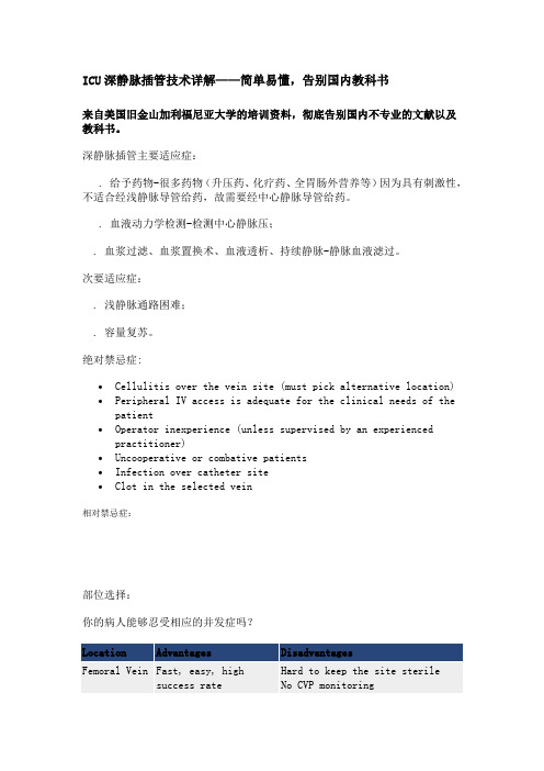
ICU深静脉插管技术详解——简单易懂,告别国内教科书来自美国旧金山加利福尼亚大学的培训资料,彻底告别国内不专业的文献以及教科书。
深静脉插管主要适应症:. 给予药物-很多药物(升压药、化疗药、全胃肠外营养等)因为具有刺激性,不适合经浅静脉导管给药,故需要经中心静脉导管给药。
. 血液动力学检测-检测中心静脉压;. 血浆过滤、血浆置换术、血液透析、持续静脉-静脉血液滤过。
次要适应症:. 浅静脉通路困难;. 容量复苏。
绝对禁忌症:∙Cellulitis over the vein site (must pick alternative location) ∙Peripheral IV access is adequate for the clinical needs of the patient∙Operator inexperience (unless supervised by an experienced practitioner)∙Uncooperative or combative patients∙Infection over catheter site∙Clot in the selected vein相对禁忌症:部位选择:你的病人能够忍受相应的并发症吗?Location Advantages DisadvantagesFemoral Vein Fast, easy, highsuccess rate Hard to keep the site sterile No CVP monitoringDoes not interfere with intubation 0% risk of pneumothorax Prevents patient mobilizationHigher rates of thrombosis than SCVHigher rates of line infectionFemoral artery puncture morefrequent than SCVInternalJugular Easy to control bleeding Pneumothorax is less common Straight shot into SVC Difficult to access if pt being intubated or withtrach or has a large neckDressings hard to maintainPoor landmarks in obese patientsCarotid puncture more frequentthan SCVHigher rates of thrombosis than SCVSubclavian VeinMost comfortable forpatientBony landmarks inobesity Higher risk for pneumothorax Compression of bleeding site difficult Long pass from skin to vein(consider in obesity)Lowest risk of thrombosisLowest risk of line infection Contraindications serious lungdisease, coagulopathy解剖:The IJ vein travels with the carotid artery; the vein typically lies anterolateral to the carotid artery. It runs under the medial portion of the upper part of the sternocleidomastoid muscle and travels under the apex of the triangle formed by the sternal and clavicular heads of the sternocleidomastoid muscle and the clavicle.The subclavian vein iseasily found in almostevery patient, and acatheter in thesubclavian vein is morecomfortable for thepatient than one placed inthe internal jugularvein.As the subclavian veincrosses behind the firstrib, it lies posterior tothe medial third of the clavicle, and has a diameter of 1-2 cm. At this point, the subclavian artery lies superior and posterior to the vein. As these vessels continue laterally, they both drop caudally to enter the axillary region. The right side is often preferred for line insertions as the dome of the pleura of the lung may extend above the first rib on the left, but rarely extends this far on the right. Insertion on the right also avoids the risk of damage to the thoracic duct on the left.ConsentAlways obtain consent prior to the procedure.Be sure to inform the patient of the reason for the procedure, the proposed benefits, its major risks and the potential management of these complications (including insertion of a chest tube, surgery or cardioversion). It is also best to walk the patient through the steps of the procedure to minimize their anxiety.Step-by-Step Procedures GuideEquipmentBefore you begin, you should be familiar with the kit. One should gather all needed materials before starting the procedure. In addition to a central venous access kit, you will need the following supplies:∙Insertion Checklist∙Sterile gloves, gown, cap, mask with face fluid shield for each member of the insertion team∙Chloraprep (if extra desired, provided in the kit)∙Large sterile drape∙Lidocaine 1% (in the kit, but it doesn’t hurt to have some extra just in case)∙Tegaderm∙Central line kit (cordis, triple or quad lumen kit)Before starting, be sure all of your materials are within reach and familiarize yourself with the kit you will be using.* Bold items are IHI guidelines and have been proven to reduce central line infections.Every day assess the line- does the site look OK, is there swelling, and is still needed? If not, take it out! Remember the riskes of line complications include mechanical (pneumothorax, hematoma, vemous perforation, catheter malposition, thoracic duct injury, arterial puncture), infections (line infection, sepsis) and thrombosis (DVT, PE) and these happen in about 5-20% of cases.Complications1.Venous Hematoma2.Arterial Dilitation3.Hemothorax4.Bleeding - arterial puncutre or injury: approximately 3% with theinternal jugular approach5.Pneumothorax: approximately 3% with the subclavian approach6.Infection: Insertion site infection, thrombophlebitis,bacteremia, sepsis, cellulitis7.Embolization of clot, air, guidewire or catheter8.Arrhythmia9.Phlebitis or thrombosis of veins10.Pericardial tamponade11.Injury to neighboring nerves (phrenic, recurrent laryngeal)12.DeathEvaluator Checklist for safe central line placement - Click Here Preventing Complications of Central Venous Catheterization- Click HereCoagulopathy and thrombocytopenia (relative goal platelets are >50k and INR >1.5, but linescan be placed if these goals are not reached depending on the case)∙Injury or previous surgery to superior vena cava (e.g., superiorvena cava syndrome)∙Complications that can belife threatening (i.e pneumothorax or bleed). Do not put asubclavian line in a patient with a coagulopathy or in patient with severe parenchymal lungdisease and respiratory failure with little respiratory reserve) ∙Coagulopathy and thrombocytopenia (relative goal plateletsare >50k and INR >1.5, but linescan be placed if these goals are not reached depending on the case) ∙Injury or previous surgery to superior vena cava (e.g., superior vena cava syndrome)∙Complications that can belife threatening (i.e pneumothorax or bleed). Do not put asubclavian line in a patient with a coagulopathy or in patient with severe parenchymal lungdisease and respiratory failure with little respiratory reserve)。
中心静脉导管centralvenouscatheter
尾部入导丝法 (蓝孔针)
29
• 7、拔出穿刺针,放入扩 张鞘,深入皮肤1-2cm作扩张
扩张时应注意进扩张器的方 向和力度,特别是与钢丝间 的传递力,手部的力度应该 由小到大,不可过猛;
30
• 退出扩张鞘,经导丝尾端放 入导管,并马上在管腔内注入 肝素,以防堵管!!! • 确定导管全段进入人体并在 最终位置后固定 • 连接其他管路和装置 • 进导管的力度大小与扩张的
• RIJV or LIJV ?
• RIJV与无名静脉和上腔静脉几乎成 一直线 • 右侧胸膜顶低于左侧 ◆ 右侧无胸导管
9
10
前路法 体位: a.去枕平卧,头转向对侧 b . 肩背部垫一薄枕,取头低位 10°~15°
C
.
穿刺侧大腿外展、外旋
30~45° 定位: a.SCM 前缘向内推开颈总 动脉,SCM中点(即 喉结/甲状软骨上缘水平) b . 颈动脉三角处触及颈总动脉, 旁开 0.5~1.0cm。 进针:针干与皮肤冠状面呈 30°~45°,针尖指向同侧乳头
管体
尖端软头
27
• 插管步骤 • 1、平卧,去枕头后仰,头 转向对侧使颈伸展, 必要时肩 背部略垫高,头低15-30°,穿刺大腿外 展、外旋30~45° • 2、确定穿刺点 • 3、进针做局麻,针干与 中线平行与皮肤呈30~45° • 4、试探针抽到静脉血后, 放入静脉穿刺针。 • 试穿刺时应注意进针方向和回血
程度有极大关系;如阻力较
大时,每次进入的长度应减 少;注意不要使钢丝打折;
31
谢 谢
32
• 171组病人因置管部位解剖复杂, 穿刺盲目性大,操作难 度大,并且周围重要脏器、组织多,容易发生严重并发症, 其操作仅限于资深医护人员操作,不宜广泛推广。因锁骨 下静脉因人而异,深度不一,一般置管长度约为(9$%, 包括游离于肌肉组织内及放置于血管内长度,导管进入血 管内具体长度难以准确把握,放置深度易发生过短及过深, 过短容易发生滑出血管外,游离于肌肉组织内,造成药液 渗于颈部锁 骨下肌肉组织,过深容易刺激心房,发生心 律失常[(] ,且动脉 伴随静脉,穿刺中易误扎入深动脉, 发生皮下血肿,且导管虽经缝线固定,但时间长后缝线易 自行断裂,病人常因睡眠中不断翻身,局部瘙痒等多种因 素不难将其从管腔拉出,造成 管身脱出[!]
床旁置入PICC导管头端位置观察

床旁置入PICC导管头端位置观察冯毕龙; 周素军; 覃慧敏; 王旭飞【期刊名称】《《护理研究》》【年(卷),期】2010(024)002【总页数】2页(P157-158)【作者】冯毕龙; 周素军; 覃慧敏; 王旭飞【作者单位】430071 武汉大学中南医院【正文语种】中文【中图分类】R472经外周静脉穿刺置入中心静脉导管(PICC)因一次置管成功率高、并发症少、操作简单安全,并能为病人提供长时间的静脉通路而在临床中得到了广泛使用。
目前,对于在何处进行置管操作尚存在不同意见。
一些专家建议床旁置入PICC,然后行胸片检查确认导管头端位置[1]。
另一些人认为,置管操作应在介入导管室X线辅助下完成,导管头端位置能够立刻得到确认,如果需要也可即时进行调整[2]。
因此,床旁置管时头端位于中心静脉部位的到位率是决定在何处置管的关键。
如果到位率高,床旁置管就具有很多优势;相反,如果到位率低,则介入导管室X线辅助下置管更具有优势。
在国内几乎所有的PICC均由经过培训的静脉治疗护士在床旁置入,迄今尚无到位率的相关报道。
本研究的目的是评价床旁置入PICC后导管头端的位置及其位于中心静脉部位的到位率。
1 对象和方法1.1 对象选取2007年5月—2008年5月因各种临床疾病需要床边置入PICC的成人病人,均于置入后即刻行胸部X线检查以确认导管头端位置。
所有病人均签署知情同意书,本研究也得到了医院医学伦理委员会的同意。
1.2 方法1.2.1 置入PICC PICC置入均在无X线透视和超声辅助下由静脉治疗护士于床边完成。
这些静脉治疗护士来自于临床各个科室,均已经过专门的PICC培训并考核合格。
本研究中使用的导管均为BD公司生产的4F或5F单腔硅胶PICC导管。
操作步骤:①选择血管。
在病人上臂中段扎止血带,选择穿刺静脉,穿刺点定位。
②预测长度,修剪导管。
取仰卧位,拟穿刺上肢外展90°,自穿刺点至右侧胸锁关节,再自胸锁关节至同侧第三肋间,两者之和即为大致进管长度,根据预测长度修剪导管。
中心静脉导管用于胸、腹腔积液引流的使用风险

RESEARCH WORK引言中心静脉导管,通常以组包类产品形式供销,其基本组成有中心静脉导管、导丝(带助推器)、扩张管、肝素帽、移动限流夹、穿刺针、无菌注射针等,主要适用范围为经外周静脉进入中心静脉系统,进行持续或间断性静脉输液、输血或血液制品、监测中心静脉压力和/或全肠外静脉营养,同时还可允许进行中心静脉压监测、采集血液样本。
导管内有多腔设计,便于同时进行以上数种程序。
近年来,中心静脉导管用于胸、腹腔积液引流的使用方法,由于操作方便、组织相容性好、患者舒适度高等优点,在医院临床逐渐普及[1-2]。
通过检索国内外中心静脉导管有关产品注册证的适用范围,无一标明可用于积液引流。
本文对江苏省2015—2018年收集到的中心静脉导管用于胸、腹腔积液引流有关的病例报告进行统计,结合对近20年有关文献资料的分析,对该种用法的风险与受益进行分析研究。
1 资料与方法1.1 资料来源2016年1月1日至2018年12月31日,江苏省药品不良反应监测中心收集到的中心静脉导管用于胸、腹腔积液引流的可疑不良事件报告。
检索中国知网数据库,时间范围1999—2018年的文献,主题选取“中心静脉导管”并中心静脉导管用于胸、腹腔积液引流的使用风险卞蓉蓉1,李尧1,赵燕2,赵敏1,马丹华1,宋杏芳11. 江苏省药品不良反应监测中心,江苏南京 210002;2. 国家药品监督管理局药品评价中心,北京 100022[摘 要] 目的 对中心静脉导管用于胸、腹腔积液引流的不良事件进行研究,通过监测数据和文献调研挖掘使用风险点,为降低该类不良事件的发生率提出控制措施和建议。
方法 对近三年江苏省收集到的58例中心静脉导管用于胸、腹腔积液引流的不良事件进行统计,对1999—2018年中国知网数据库中相关文献进行可视化分析,与辖区内生产企业进行风险会商。
结果 国内和进口的中心静脉导管均在临床上被用于胸、腹腔积液引流,预期用途有胸腔、腹腔和心包积液引流;主要产品故障表现为堵塞、折叠、压扁不能回弹等;文献研究得知,中心静脉导管广泛用于积液引流;从中心静脉导管的功能、使用部位、适用人群、结构设计等方面探讨,均不能确保安全有效。
cvc非计划拔管处理流程
cvc非计划拔管处理流程英文回答:CVC unplanned extubation refers to the accidental removal of a central venous catheter (CVC) from thepatient's body. This can lead to serious complications and requires a prompt and organized response from the healthcare team. Here is the general process for managing an unplanned CVC extubation:1. Assessment: The first step is to assess thepatient's condition. This includes checking for any signs of distress, monitoring vital signs, and assessing the site where the CVC was removed.2. Secure the Airway: If the patient is havingdifficulty breathing, it is important to secure the airway immediately. This may involve providing oxygen support or reintubating the patient if necessary.3. Control Bleeding: In some cases, the removal of the CVC may cause bleeding. It is important to control any bleeding and apply pressure to the site if needed.4. Assess the CVC Site: Once the patient is stable, the healthcare team should assess the CVC site to determine if reinsertion is possible. This may involve obtaining a chest X-ray to check the position of the CVC and ensure that itis safe to reinsert.5. Reinsertion of the CVC: If it is determined that the CVC needs to be reinserted, the healthcare team should follow proper sterile technique and guidelines for CVC insertion to minimize the risk of infection.6. Monitoring and Follow-up: After the CVC has been reinserted, the patient should be closely monitored for any signs of complications, such as infection or bleeding. The healthcare team should also provide appropriate follow-up care to ensure the patient's safety and well-being.中文回答:CVC非计划拔管是指中心静脉导管(CVC)意外从患者体内移除。
中长导管拔管操作流程
中长导管拔管操作流程Removing a central venous catheter (CVC), also known as a central venous line or central venous access device, is a common procedure that requires precision and attention to detail. 中长导管拔管操作是一个常见的程序,需要高度的精确性和细致的注意。
Before initiating the process of removing a CVC, it is crucial to ensure that the patient is adequately prepared both physically and emotionally. Central venous catheters are often used to deliver medications, blood products, or nutrition directly into the bloodstream, so their removal can be an emotional experience for the patient. 在开始拔除中长导管之前,关键是确保患者在身体和情绪上做好充分准备。
中长导管通常用于将药物、血液制品或营养液直接输送到血液中,因此对患者而言,拔管可能是一种情绪上的体验。
One important aspect of preparing the patient for CVC removal is providing clear and thorough explanations of the procedure. It is essential to inform the patient about the reasons for removing the catheter, the steps involved in the process, and any potential risks or complications that may arise. 给患者准备中长导管拔管的一个重要方面是提供清晰而全面的拔管程序解释。
中心静脉压操作流程口述

中心静脉压操作流程口述Central venous pressure (CVP) is an important parameter that reflects the fluid status and cardiac function of a patient. It is crucial in guiding fluid management and assessing the response to fluid resuscitation in critically ill patients. The measurement of CVP involves the placement of a central venous catheter, typically in the internal jugular,subclavian, or femoral vein. This procedure should be performed with caution and attention to detail to minimize the risk of complications.中心静脉压(CVP)是一个重要的参数,反映了患者的液体状态和心脏功能。
在重症患者中,引导液体管理和评估对液体复苏的反应时,CVP至关重要。
测量CVP涉及置入一根中心静脉导管,通常在颈内、锁骨下或股静脉。
这个程序应该小心地进行,并且注意细节,以减少并发症的风险。
Before carrying out the procedure, it is essential to obtain informed consent from the patient or their legal guardian. The risks, benefits, and alternatives of the central venous catheterization should be explained in a language that the patient can understand. In addition, the healthcare provider should ensure that the patient is adequatelyprepared for the procedure, which may include fasting for a certain period and obtaining relevant laboratory tests.在执行程序之前,必须从患者或其法定监护人那获得知情同意。
中心静脉导管评估方法
中心静脉导管评估方法Central venous catheter (CVC) is a crucial tool in the management of critically ill patients. 中心静脉导管(CVC)是重症患者管理中的关键工具。
It provides a direct and reliable access to the central circulatory system for various purposes such as administering medications, monitoring central venous pressure, and obtaining blood samples. 它为多种用途提供了对中心循环系统的直接可靠访问,例如给药、监测中心静脉压力和获取血液样本。
However, complications related to the use of CVCs are not uncommon and can lead to serious morbidity and mortality. 然而,与CVC使用有关的并发症并不少见,可能导致严重的发病率和死亡率。
One of the most important aspects of managing patients with CVCsis the assessment of the catheter to ensure its proper function and to detect any potential complications. 管理使用CVC的患者最重要的方面之一是对导管进行评估,以确保其正常功能,并检测任何潜在的并发症。
There are several methods for assessing CVCs, including physical examination, chest X-ray, and ultrasound. 有几种方法可用于评估CVC,包括体格检查、胸部X线和超声波检查。
小儿颈内静脉置管深度研究进展
288国际麻醉学与复苏杂志2021年3月第42卷第3期Int J Anesth Resus,March2021,Vol.42,No.3**小儿颈内静脉置管深度研究进展杨艳艳孙瑛张马忠上海交通大学医学院附属上海儿童医学中心麻醉科200127通信作者:孙瑛,Email:yingsun821@【摘要】颈内静internal jugular vein,IJV)穿刺置管是临床常用技术,用于液体或药物管理,以及CVP测定等。
随着小儿身高、体重的变化,UV导管置入的合适深度也会随之发生变化。
文章描述了IJV导管尖端理想位置,介绍了通过经食管超声心动图(transesophageal echocardiography,TEE)、经胸超声心动图(transthoracic echocardiography,TTE)、胸部X线检查(chest radiograph,CXR)和腔内心电图等方法确定导管尖端位置。
此外,还对右侧IJV置管深度、特殊人群的IJV置管深度、左侧IJV 解剖特点及置管深度进行了综述。
目前已有不少计算IJV置管深度的公式,由于儿童在发育阶段存在一定的发育差异,它对于个体的适用性仍有待临床进一步考证。
【关键词】儿童;颈内静脉;留置导管D01:10.3760/321761-20200414-00240Progress of the depth of internal jugular venous catheter in pediatric patientsYang Yanyan,Sun Ying,Zhang MazhongDepartment of Anesthesiology,Shanghai Children's Medical Center,Affiliated to School of Medicine,Shanghai Jiao Tong University f Shanghai200127,ChinaCorresponding author:Sun Ying,Email:yingsun821@【Abstract】Internal jugular vein(IJV)catheterization is a common clinical technique for fluid or drug administration,as well as for central venous pressure(CVP)measurement.The optimal depth of the IJV catheter varies with the children's height and weight. This paper describes the suitable position of the catheter tip and introduces the main methods to determine the position of the catheter tip,such as transesophageal echocardiography(TEE),transthoracic echocardiography(TTE),chest radiograph(CXR)and intracanal electrocardiogram.Besides,ways to determine the appropriate depth of IJV catheters on the right or left side and depth of catheter in special populations are reviewed in this paper.There are some formulas to calculate the depth of IJV catheters.Considering the individual diversity of children,its applicability to individuals needs further clinical examination.【Key words]Child;Internal jugular vein;Indwelling catheterDOI:10.3760/321761-20200414-00240中心静脉置管是手术室及ICU常用技术,用于获得准确的血流动力学信息,并用于血管活性药物和液体的管理等叫目前临床医师大多根据个人经验来决定置管深度,没有统一标准。
临床常见导管的评估
临床常见导管的评估导管在临床实践中被广泛应用,用于输液、引流、监测和治疗等多种目的。
对导管的评估是确保导管的安全和有效使用的重要环节。
本文将详细介绍临床常见导管的评估标准。
一、外周静脉导管(Peripheral Intravenous Catheter,简称PIVC)1. 导管插入部位:应检查导管插入部位是否有红肿、温热、压痛等炎症表现,如有异常应及时报告医生。
2. 导管固定:导管应固定在患者的皮肤上,固定材料应无松动、破损或过紧的情况。
3. 导管外观:应检查导管是否完整,无断裂、破损或脱落的情况。
4. 导管连接:导管连接部位应无渗漏、松动或脱落的情况。
5. 皮肤周围情况:应检查患者的皮肤是否有红肿、渗液、瘙痒等过敏或感染的表现。
6. 患者症状:应询问患者是否有局部疼痛、肿胀、瘙痒等不适感觉。
二、中心静脉导管(Central Venous Catheter,简称CVC)1. 导管插入部位:应检查导管插入部位是否有红肿、温热、压痛等炎症表现,如有异常应及时报告医生。
2. 导管固定:导管应固定在患者的皮肤上,固定材料应无松动、破损或过紧的情况。
3. 导管外观:应检查导管是否完整,无断裂、破损或脱落的情况。
4. 导管连接:导管连接部位应无渗漏、松动或脱落的情况。
5. 皮肤周围情况:应检查患者的皮肤是否有红肿、渗液、瘙痒等过敏或感染的表现。
6. 患者症状:应询问患者是否有局部疼痛、肿胀、瘙痒等不适感觉。
7. X线确认:CVC插入后应进行X线检查,确保导管的位置正确。
三、气管插管(Endotracheal Tube,简称ETT)1. 导管插入部位:应检查导管插入部位是否有红肿、温热、压痛等炎症表现,如有异常应及时报告医生。
2. 导管固定:导管应固定在患者的口腔或气管上,固定材料应无松动、破损或过紧的情况。
3. 导管外观:应检查导管是否完整,无断裂、破损或脱落的情况。
4. 导管连接:导管连接部位应无渗漏、松动或脱落的情况。
食管超声心动图构建右颈内中心静脉置入导管深度的简易预测公式
静脉穿刺,置入导管深度( cm)= 身高( cm) ÷10-1 5( r 2 = 0 437,P<0 01) 。 17 例患者出现心律失常,
未观察到误穿动脉、血肿形成、气胸等现象。 结论 对于我国成年患者行右颈内中心静脉置管,Peres
公式计算深度有一定局限性,利用 TEE 完成置管安全有效。 所构建的简易预测公式男女均适用,可
Corresponding author: GE Yali, Email: ge_yl@163.com
【Abstract】 Objective Placing the central venous catheter through the right internal jugular vein,
trance of the superior vena cava) . The central venous catheter was guided along the guidewire, and the
depth of the catheter was equal to the depth of the guidewire. Gender, height, weight, and BMI of patients
刺区域,皮肤常规消毒,铺无菌洞巾,体表超声探头
是关于中心静脉置管
套无菌保护套,常规检查穿刺血管,确认无血栓形
深度的一个简易预测公式,临床运用广泛,但该方
成。 在环状软骨平面,通过体表超声显露右颈内静
法是基于欧洲人群生理数据得出,应用于亚洲人群
可能 面 临 不 同 的 情 况。 经 食 管 超 声 心 动 图
(14 9±1 2) cm,P<0 01] 。 男性身高明显高于女性,体重明显重于女性,Peres 公式计算深度、置入导
中心静脉氧饱和度的监测及其应用
国际呼吸杂志2008年第28卷第6期IntJRespir,Mar.2008,V01.28,No.6中心静脉氧饱和度的监测及其应用郑伟浩萧正伦【摘要】通过中心静脉导管监测中心静脉氧饱和度(ScV0z)是一个方便、可靠的方法,它能快速反映危重患者全身氧的供需平衡的瞬时变化,能早期发现组织缺氧,且优于其它传统的血流动力学参数,是评估组织氧合充分与否的有用的间接指标,也是评估危重患者院内病死率及预后的重要指标;用ScV0。
、平均动脉压和中心静脉压来指导早期目标指导性治疗,可提高严重脓毒血症或感染性休克患者的存活率。
因此,ScV0z的监测对危重患者的评估和管理是非常重要和有用的。
【关键词】中心静脉氧饱和度;混合静脉氧饱和度;氧监测Useandme髓urementofcentralveno吣oxyg明鼢turationZHENGV%i一矗口D,XjAoZ矗P咒g—Z“咒.G锄咒gz幻“f竹s£it札£eo,Respir口tor3,Dis8口ses,旃eFirs£A,厅zi口£8dHos乡i£口zo,G“岔咒gz矗oz‘Medic口zCDzz已ge,(孰口挖gz^o“510120,Ghi挖口【Abstract】Measurementofcentralvenousoxygensaturation(ScV02)fromcentralvenouscathetersisareliableandconvenientmethodwhichcouIdrapidlywarnofacutechangeintheglobaloxygensupply/demandratioofcritica“yillpatientsandwhichissuperiortoconventionalhemodynamicparameters,anditcandetecttissuehypo妇aearly.ScV02,asausefulindirectindexofadequatetissueoxygenation,isanimportantindicatorofin-hospitalmortalityrateandprognosesforcriticallyiIlpatients.Theearly_goaldirectedtherapyguidedbyScV02,meanarterialpressureandcentralvenouspressureisabletoincreasesurvivalinthepatientswithseveresepsisorsepticshock.Therefore,measurementofScV02isveryimportantandusefulforevaluationandmanagementofcriticallyi11patients.【Keywords】Centralvenousoxygensatufation;AⅡixedvenousoxygensaturation;oXimetry通过肺动脉导管所测得的混合静脉氧饱和度(miXedvenousoxygensaturation,&02)可准确反映全身氧供需平衡,是组织氧合的一个间接指标[1≈]。
cvc中心静脉导管容积
cvc中心静脉导管容积中心静脉导管(Central venous catheter, CVC)是一种插入在体内的管状装置,用于输送液体、药物、营养物质或采集血液样本等目的。
它通常插入静脉,直接进入心脏附近的大血管,如锁骨下静脉、颈内静脉或股静脉。
CVC具有较大的导管容积,这是指导管内部可容纳液体的空间大小。
首先,CVC的导管容积是一个重要的指标,它对于安全和有效地输液非常关键。
导管容积决定了CVC内部液体的储存能力,会影响到输液的速度、血液流动的速度以及药物输送的准确性。
通常来说,CVC的导管容积应根据具体临床需求来确定,常见的导管容积选项包括1.5ml、2.5ml、3.5ml等,医生根据病情和治疗需求来选择合适的导管容积。
其次,CVC的导管容积还与导管的直径密切相关。
一般来说,导管的内径越大,导管容积也相对较大。
导管容积的大小不仅受到导管本身的内径限制,还会受到导管材质、连接器的特性以及导管内部的特殊设计等因素的影响。
例如,带有储液腔的导管会有更大的导管容积,因为储液腔能够提供额外的液体储存空间。
此外,导管容积的选择也与具体的治疗目标有关。
例如,在临床应用中,需要大量快速输液的情况下,通常会选择具有较大容积的CVC,以确保药物或液体能够快速输送到患者体内。
相反,若仅需输注少量药物或采集血液样本,容积较小的CVC则可能更为适合。
然而,在选择CVC导管容积时,也需要考虑与之相关的一些风险和问题。
过大的导管容积可能导致静脉炎症、血栓形成、导管堵塞等并发症的风险增加。
此外,患者应在使用CVC之前进行详细的检查和评估,以确定合适的导管容积,避免过度或不足。
医护人员在操作CVC时,也需要严格按照相关操作规程进行操作,以确保正确地使用和管理CVC,降低并发症风险。
综上所述,CVC的导管容积是一个重要的指标,可以影响到输液的速度、血液流动的速度以及药物输送的准确性。
导管容积的选择应根据具体的临床需求来确定,同时还应考虑并发症风险以及患者的个体差异等因素。
使用经食管超声获得右颈内静脉置管深度的经验公式
使用经食管超声获得右颈内静脉置管深度的经验公式鞠辉;孙晓晨;冯艺【摘要】目的使用经食管超声心动图(TEE)技术确定经中路右颈内静脉置管的最佳深度,结合体表标志测量和身高得出预测公式.方法选择具有术中使用TEE及中心静脉置管指征的患者74例,采用Seldinger's技术进行经中路右颈内静脉置管.TEE观察导丝尖端位于上腔静脉中下段且心包反折外,测量此时导丝置入深度作为导管的固定深度.测量体表标志,并得出导管合适深度的经验公式.结果 TEE下经中路右颈内静脉置管的固定深度为(15.1±1.7)cm.建立固定深度的预测公式为:固定深度=身高÷20+(穿刺点经右胸锁关节到胸骨角右缘的连线距离)÷2(r=0.595,r2=0.354,P<0.01).结论根据TEE确定的最佳置管深度,结合身高和体表标志可以得出以上预测公式,为今后临床预测右颈内静脉置管的合适深度提供参考.【期刊名称】《实用医学杂志》【年(卷),期】2019(035)005【总页数】4页(P772-775)【关键词】经食管超声心动图;右颈内静脉;中心静脉置管;深度【作者】鞠辉;孙晓晨;冯艺【作者单位】北京大学人民医院麻醉科北京100044;北京大学人民医院麻醉科北京100044;中日友好医院麻醉科北京100029;北京大学人民医院麻醉科北京100044【正文语种】中文中心静脉穿刺置管是围术期常见的有创操作,合适的置管深度对患者具有重要意义。
置管过深可导致血管穿破、胸腔积液、心律失常[1],甚至心脏穿孔、心包填塞等危及生命的并发症[2]。
置管过浅可导致静脉穿孔、侧孔输液或给药至血管外(胸腔、心包或纵膈)等,增加血栓风险和导管相关感染率[3-4]。
研究[5]表明,中心静脉导管尖端的合适位置位于上腔静脉(superior vena cava,SVC)与右心房的交界处或下段上腔静脉内,心包反折的上方。
此处导管尖端周围的血流量最佳,可以防止导管内血栓形成,防止血管外注药、同时避免尖端进入心包腔,刺激心房壁引起心律失常甚至心包填塞。
cvc中心静脉导管孔径
cvc中心静脉导管孔径
摘要:
一、引言
二、cvc 中心静脉导管的概念与作用
三、cvc 中心静脉导管的孔径选择
四、cvc 中心静脉导管孔径与治疗效果的关系
五、结论
正文:
【引言】
cvc 中心静脉导管是临床诊疗中常用的一种医疗器械,对于危重病人的救治具有重要意义。
然而,对于cvc 中心静脉导管的孔径选择,许多人并不十分了解。
本文将对此进行详细介绍。
【cvc 中心静脉导管的概念与作用】
cvc 中心静脉导管,全称为Central Venous Catheter,是一种自颈部或股部的静脉插入,直至心脏的导管。
它可以用于输液、输血、药物注射以及监测中心静脉压等。
【cvc 中心静脉导管的孔径选择】
cvc 中心静脉导管的孔径选择主要取决于患者的年龄、体重、病情以及治疗需求。
通常来说,成人使用的cvc 中心静脉导管的孔径多为8-12F,而儿童和新生儿则根据其体型和年龄选择6-10F 的导管。
【cvc 中心静脉导管孔径与治疗效果的关系】
选择合适的cvc 中心静脉导管孔径,不仅可以保证药物和液体的顺利输送,而且可以降低导管相关性感染的风险,提高患者的治疗效果和舒适度。
【结论】
总的来说,cvc 中心静脉导管的孔径选择是一项非常重要的临床操作,需要综合考虑患者的各种因素,选择最适合的导管。
- 1、下载文档前请自行甄别文档内容的完整性,平台不提供额外的编辑、内容补充、找答案等附加服务。
- 2、"仅部分预览"的文档,不可在线预览部分如存在完整性等问题,可反馈申请退款(可完整预览的文档不适用该条件!)。
- 3、如文档侵犯您的权益,请联系客服反馈,我们会尽快为您处理(人工客服工作时间:9:00-18:30)。
MethodsThe Ethics Committee of Seoul National University Hospital approved the study protocol,and informed parental consent was obtained in all cases.Over a 6-month period,pediatric patients scheduled for congenital heart surgery were enrolled.Patients with any extra cardiac vascular or rotational abnormality were excluded.In this study,an anterior approach to the right internal jugular vein was used.The puncture site was located medial to the sternal head of the sterno-cleidomastiod muscle,between the apex of the tri-angle and the cricoid cartilage and lateral to the carotid artery.The CVC insertion technique and the manner in which we verified catheter tips are described in our previous study (12).After advan-cing the tip of a CVC to the SVC—RA junction,under TEE monitoring,a nurse blinded to the experiment measured the length of the CVC beneath the skin using the depth indicator on the CVC.Information on patient height,age,and weight were also collected.Scatter plots of distance from skin to the SVC/RA junction and age,height and weight were made,and the linearities of these rela-tionships were examined using correlation analysis.Multiple linear regression analysis was also per-formed to identify factors capable of predicting the distance from the skin to the SVC/RA junction.Statistical significance was accepted for P -values <0.05.Statistical analyzes were performed using Sigma plot 2000and SPSS version 10.0(SPSS Inc.,Chicago,IL).ResultsA total of 60CVC cannulations were studied.Patient’s demographic characteristics are listed in Table 1.Age,height,and weight are presented as median (minimum Àmaximum)because these dis-tributions were skewed to the right.On the first post-operative chest radiograph,none of the cathe-ter tips were in the right atrium.The distance from the skin puncture site to the SVC—RA junction was more correlated with patient height (r ¼0.81)than weight (r ¼0.76)or age (r ¼0.60).Using multiple regression analysis,after adjusting for the other two variables,only patient height was significantly correlated with the distance from the skin puncture site to the SVC—RA ing a simple regression model,the lower 95%prediction interval with each patient height was obtained.A straight line was then constructed using the minimum and maximum values of the lower pre-diction limit (Fig.1).The prediction interval provides a method of predicting individual values for distance to the SVC—RA junction,whereas the confidence interval of the mean provides those of the predicted mean value.The resulting equation was:Initial length of insertion ðcm Þ¼1:7þ½0:07Âheight ðcm Þ :This formula predicts a CVC tip positioning above the RA junction at least 97.5%of the ing this equation,58CVC tips in 60patients were above the SVC—RA junction.The other two were within 1cm of the SVC—RA junction (Fig.1).DiscussionTo our knowledge,this is the second report concern-ing the depth of CVC insertion in pediatric patients.Before Andropoulos et al.(7)proposed the formula—Table 1Demographic characteristics of the study subjects Variables Median (minimum,maximum)Age (month)13.5(1.0,173.0)Height (cm)80.8(56.0,160.0)Weight (kg)10.6(4.2,46.2)Values represented are median (minimum,maximum).16141210864406080100120140160180Height (cm)D i s t a n c e f r o m s k i n t o S V C –R A J u n c t i o n (c m )Fig.1.Scatter plot of patient heights vs.distance from the catheter insertion site to the junction of the superior vena cava (SVC)and the right atrium (RA)for a right internal jugular vein (IJV)ing a simple regression model,the lower 95%predic-tion interval with each patient height was obtained.A straight line was then constructed using minimum and maximum values of the prediction lower limits.S.Z.Yoon et al.356(height in cm/10)À1for patients100cm in height,and(height in cm/10)À2for patients !100cm for correct positioning of an inserted CVC tip in pediatric patients—the length of inser-tion was decided based on personal experience for most anesthesiologists(13).For a90-cm tall patient, the length of insertion is8cm using the formula of Andropoulos et al.,which compares to8.9cm using our formula.For a110-cm tall patient,the length of insertion is9cm using Andropoulos’et al.formula, but9.4cm using our formula.These differences may be explained by the fact that they used chest radiography to identify the SVC—RA junction.With regard to the study by Aslamy et al.(9),a consider-able portion of CVC tips which were in the cardiac silhouette on the chest radiograph may have actu-ally been positioned in the SVC.Thus,the guide-lines proposed by Andropoulos et al.(7)for CVC placement can lead to an unintentional placement of a catheter tip in the mid-SVC away from the SVC —RA junction,which may cause suboptimal catheter performance.Our study has some limitations.Our formula for initial CVC insertion only applies to an anterior approach to the internal jugular vein(IJV)not to a lower approach to the IJV or subclavian vein,and our formula has not been validated for children below40or above140cm.In conclusion,our proposed formula can be used to position the catheter tip close to the right atrium and prevent unintentional placement of the cathe-ter tip into the atrium in the studied patient population.AcknowledgementsThis article was supported from departmental sources and the study was performed at Seoul National University Hospital. This article was presented,in part,at the2005Annual Meeting of the European Society of Anesthesiologists,Vienna,Austria.References1.Hayashi Y,Maruyama K,Takaki O,Yamauchi J,Ohnishi Y.Optimal placement of the CVP catheter in pediatric cardiac patients.Can J Anaesth1995;42:479—82.2.Fischer GW,Scherz RG.Neck vein catheters and pericardialtamponade.Pediatrics1973;52:868—71.3.Vessely TM.Central venous catheter tip position:a continu-ing controversy.J Vasc Interv Radiol2003;14:527—34.4.Nowlen TT,Rosenthal GL,Johnson GL,Tom DJ,Vagaro TA.Pericardial effusion and tamponade in infants with central catheters.Pediatrics2002;110:137—42.5.Yoder D.Cardiac perforation and tamponade:the deadly duoof central venous catheters.Int J Trauma Nurs2001;7:108—12.6.National Association of Vascular Access Networks.NAVANPosition Statement.J Vasc Access Devices1998;3:8—10.7.Andropoulos DB,Bent ST,Skjonsby B,Stayer SA.Theoptimal length of insertion of central venous catheters for pediatric patients.Anesth Analg2001;93:883—6.8.Kim KO,Jo JO,Kim HS,Kim CS.Positioning internal jugularvenous catheters using the right third intercostal space in children.Acta Anaesthesiol Scand2003;47:1284—6.9.Aslamy Z,Dewald CL,Heffner JE.MRI of central venousanatomy.Implications for central venous catheter insertion.Chest1998;114:820—6.10.Andropoulos DB,Stayer SA,Bent ST,Campos CJ.A con-trolled study of transesophageal echocardiography to guide central venous catheter placement in congenital heart sur-gery patients.Anesth Analg1999;89:65—70.11.Chu KS,Hsu JH,Shie S,Tang CS,Cheng KI.Accurate centralport-A catheter placement:intravenous electrocardiography and surfaces landmark technique compared by using trans-esophageal echocardiography.Anesth Analg2004;98:910—4.12.Yoon SZ,Shin JH,Hahn S,Kim HS,Kim SD,Oh AY et al.Usefulness of carina as a radiographic landmark for central venous catheter tip in pediatric patients.Br J Anaesth2005;95:514—7.13.Davies LK.Anesthesia for pediatric cardiovascular surgery.In:Kirby RR,Gravenstein N,Lobato EB,Gravenstein JS,eds.Clinical Anesthesia Practice.Philadelphia:W.B.Saunders Company,2002:1222—3.Address:Chong D.ParkDepartment of AnesthesiologySeoul National UniversityCollege of Medicine#28Yeongon-dongJongro-guSeoul110-744Koreae-mail:cdpark@snu.ac.krOptimal depth of a central venous catheter357。