Development of cell-based immunoassays to measure type I collagen in cultured fibroblasts
药物的免疫原性分析方法浅析
药物的免疫原性分析方法浅析摘要:药物的免疫原性评价是临床安全性评价的重要组成部分,药物引起的抗体反应会影响药动学、药效或毒性反应。
ADA的产生会缩短或延长药物半衰期,改变药物在体内的生物分布。
免疫原性的分析在复杂的血清基质中进行,涉及到多学科多领域。
分析方法及检测水平也存在不一致的情况,需要规范实验操作,提高检测的有效性。
免疫原性评价是药物开发阶段中安全与疗效评价的需要,也是申报提交的关键要素。
开发有效的免疫原性检测方法意义重大。
关键词:免疫原性,MRD,方法学验证正文免疫原性通常是指治疗性蛋白和/或代谢物诱发其自身或相关蛋白的免疫应答或免疫相关事件的能力。
一般分子量越大免疫反应越强。
分子量低于4000时一般不具有免疫原性,在4000到10000之间时具有弱免疫原性,分子量大于10000时具有强免疫原性。
但是也有例外要根据物质的结构决定。
免疫原性分析必须在临床前和临床阶段均开展。
具有免疫原性的物质可能诱发机体产生有害的免疫反应,甚至威胁生命。
免疫原性评价需要有效特异的检测方法。
在临床前便需要确定检测方式,在方法的设计、开发和验证中需要严格控制各环节。
临床前和临床中样本的检测需使用经过严格验证的方法进行检验。
目前应用较广泛的是平台主要包括ELISA、ECL、RIA和SPR。
检测方法一般为夹心法、间接法或直接法。
免疫原性方法学的开发要考虑多种因素,主要包括反应模式的选择、包被抗原浓度的选择、包被条件的选择、封闭条件的选择、样品稀释液的选择、最小稀释度(MRD)的选择、酶稀释液的选择、显色液时间的选择以及各条件的孵育时间及温度的选择。
每个反应步骤都会影响方法的性能。
阳性抗体的质量直接影响方法的灵敏度。
阳性抗体需要稀释在与待检样本一致的空白动物血清中。
由于基质的复杂性,在很大程度上会增加检测背景,需要优化各反应条件将背景降低。
对于背景的高低无确定的要求,但是背景越低越好,太高的背景也会影响检测灵敏度。
开发过程中要优先考虑灵敏度和耐药性,其次为精密度、选择性、特异性、稳定性。
elisa实验必看的几本书
/bbs/post/view?bid=68&id=10630144&tpg=1&ppg=1&sty=0&age=0 2008-1-25
【共享】ELISA实验的几本必备工具书 - Welcome to Forum
Page 2 of 8
freecell
且行且远且纵歌 丁香园中级VIP站友 发贴: 10037 积分: 1444 得票: 99 状态: 隐身
文字很浅,人生很深... • 【求助】刚发现怀孕,打算要孩子。会不会影响找工作?
2007-12-07 16:46
举报
第二本: ELISA and Other Solid Phase Immunoassays: Theoretical and Practical A spects 下载地址:寻找中 内容介绍:This is a quick-reference manual on practical aspects of immunoassay. Providing a theoretical and practical basis for modern applications of solid-phase i mmunoassays, this text brings together experts who have used ELISA and other assays in a variety of fields. Contributors offer step-by-step guidance on how to u se the various techniques involved in immunoassay. These techniques are extem ely useful to laboratory-based researchers and technicians working on the detecti on of allergy, the AIDS virus, autoimmunity, etc. Chapters analyze the solid-phas e supports used, the amplification systems, and the quantitation and affinity of a ntibodies and discuss the applications of assays to biology, immunology, and micr obiology. /WileyCDA/WileyTitle/productCd-0471909823.html http://www.amazon.de/Elisa-Other-Solid-Phase-Immunoassays/dp/0471909823
自组装铁蛋白在纳米疫苗领域的应用进展

生物技术进展2019年㊀第9卷㊀第3期㊀240~245CurrentBiotechnology㊀ISSN2095 ̄2341进展评述Reviews㊀收稿日期:2018 ̄12 ̄26ꎻ接受日期:2019 ̄02 ̄22㊀基金项目:国家重点研发计划项目(2017YFD0500706ꎻ2016YFD0500108)ꎻ国家自然科学基金项目(31670156)资助ꎮ㊀作者简介:魏珍珍ꎬ硕士研究生ꎬ研究方向为病毒微生物ꎮE ̄mail:646122815@qq.comꎮ∗通信作者:易咏竹ꎬ副研究员ꎬ研究方向为病毒微生物ꎮE ̄mail:Yiyongzhu@126.com自组装铁蛋白在纳米疫苗领域的应用进展魏珍珍1ꎬ㊀刘兴健2ꎬ㊀王㊀朋1ꎬ㊀张志芳2ꎬ㊀易咏竹3∗1.江苏科技大学生物技术学院ꎬ江苏镇江212003ꎻ2.中国农业科学院生物技术研究所ꎬ北京100081ꎻ3.中国农业科学院蚕业研究所ꎬ江苏镇江212018摘㊀要:自组装蛋白在真核细胞及原核细胞中是普遍存在的ꎬ其对生命体的正常运转具有重要意义ꎬ甚至关系到生命体的进化ꎮ常见的自组装蛋白包括病毒颗粒(virusparticles)㊁血清白蛋白(serumalbumin)㊁丝蛋白(silkprotein)及铁蛋白(ferritin)ꎮ其中ꎬ铁蛋白可形成粒径均一㊁生物相容性良好的纳米材料ꎬ还具有独特的理化性质ꎬ如pH敏感㊁高温耐受㊁大多数变性剂耐受ꎬ即可通过调节pH来控制铁蛋白的自组装特性ꎮ铁蛋白是存在于大多数生物体内的天然蛋白ꎬ在肿瘤的诊断成像及治疗㊁药物载体和纳米疫苗等领域具有广阔的应用前景ꎮ重点探讨了铁蛋白的仿生合成及其在纳米疫苗领域的应用进展ꎬ以期为新型动物纳米疫苗的研发提供参考ꎮ关键词:自组装蛋白ꎻ重组铁蛋白ꎻ纳米疫苗DOI:10.19586/j.2095 ̄2341.2018.0139ApplicationProgressofSelf ̄assembledFerritininNano ̄vaccineWEIZhenzhen1ꎬLIUXingjian2ꎬWANGPeng1ꎬZHANGZhifang2ꎬYIYongzhu3∗1.CollegeofBiotechnologyꎬJiangsuUniversityofScienceandTechnologyꎬJiangsuZhenjiang212003ꎬChinaꎻ2.BiotechnologyResearchInstituteꎬChineseAcademyofAgriculturalSciencesꎬBeijing100081ꎬChinaꎻ3.SericulturalResearchInstituteꎬChineseAcademyofAgriculturalSciencesꎬJiangsuZhenjiang212018ꎬChinaAbstract:Self ̄assembledproteinsareubiquitousineukaryoticandprokaryoticcellsꎬandtheyareimportantforlivingorganismstomaintainthenormaloperationꎬandevenrelatedtotheevolutionoflivingorganisms.Commonself ̄assembledproteinsincludevirusparticlesꎬserumalbuminꎬsilkproteinandferritin.Amongthemꎬferritincanformnanomaterialswithuniformparticlesizeandgoodbiocompatibility.ItalsohasuniquephysicalandchemicalpropertiesꎬsuchaspHsensitivityꎬhightemperaturetoleranceꎬandresistancetomostdenaturantsꎬsoastocontroltheself ̄assemblycharacteristicsofferritinbypHregulation.Ferritinisanaturalproteinfoundinmostlivingorganismsꎬandithasabroadapplicationprospectintumordiagnosticimagingandtherapyꎬdrugcarrierandnano ̄vaccine.Thebionicsynthesisofferritinanditsapplicationinnano ̄vaccineweremainlydiscussedinordertoprovidereferencesfortheresearchanddevelopmentofnovelanimalnano ̄vaccine.Keywords:self ̄assembledproteinꎻrecombinantferritinꎻnano ̄vaccine㊀㊀自组装蛋白在真核细胞及原核细胞中是普遍存在的ꎬ蛋白质亚基间会自发组装构成高度有序的结构ꎬ这是维持机体正常运转的保证ꎬ也是机体进化的推动力[1]ꎮ由自组装蛋白形成的纳米材料ꎬ不仅具有生物相容性良好以及粒径均一㊁稳定的特性ꎬ还在细胞成像㊁病灶检测和药物缓释等方面具有广阔的应用前景ꎮ到目前为止ꎬ研究最多的自组装蛋白纳米颗粒包括病毒颗粒(virusparticles)㊁血清白蛋白(se ̄rumalbumin)㊁丝蛋白(silkprotein)及铁蛋白(fer ̄ritin)ꎮ其中ꎬ病毒颗粒侵染宿主细胞并在宿主细胞内的自组装行为ꎬ是自然界中典型的生物纳米. All Rights Reserved.材料的形成方式ꎬ主要用于特异性检测以及病毒侵染宿主细胞的机制和路径的研究[2ꎬ3]ꎬ经基因修饰后还可用于研制借助病毒释放基因的药物等方面的研究[4]ꎻ血清白蛋白是脊椎动物血浆中含量最高的蛋白质ꎬ其分子的弹性良好ꎬ结构改变后也极易恢复ꎬ不同来源的血清白蛋白的空间构造均十分保守[5]ꎬ在药物传递系统领域拥有潜在的应用前景[6]ꎻ丝蛋白是一类线状蛋白的生物高分子材料ꎬ可抗紫外线ꎬ也可抗蛋白水解酶ꎬ其柔韧性好㊁抗疲劳度高ꎬ有着与钢材类似的张力强度ꎬ还具有良好的热㊁酸㊁碱稳定性和生物相容性ꎬ在生物材料[7]和药物载体[8]领域应用广泛ꎮ而铁蛋白是存在于大多数生物体内的天然蛋白ꎬ具有独特的理化性质:①铁蛋白对pH不耐受ꎬ较为敏感ꎬ在酸性条件(pH2.0)下铁蛋白外壳会解体成亚基ꎬ而当pH回升到生理条件(pH7.4)时ꎬ各亚基又重组形成完整的铁蛋白[9ꎬ10]ꎻ②铁蛋白的天然高级结构不受多种变性剂的影响ꎬ一般蛋白质在1~4mol/L的低浓度盐酸胍或者脲溶液中就会发生变性ꎬ而铁蛋白在6mol/L的盐酸胍或8mol/L的脲溶液中才会发生蛋白质解聚ꎬ即铁蛋白对变性剂的耐受性高[11]ꎻ③铁蛋白对高温具有较高的耐受性ꎬ大多数蛋白质在温度高于生理条件后极易变性ꎬ但铁蛋白在高温(70ħ~80ħ)时可维持10min以上不会发生变性ꎬ且其高级结构维持完好[12]ꎮ基于铁蛋白独特的理化性质ꎬ本文主要对铁蛋白的仿生合成及其在肿瘤的诊断成像及治疗㊁药物载体和纳米疫苗领域的应用进展进行了综述ꎬ阐述了天然铁蛋白的结构及修饰㊁人工制备重组铁蛋白的研究进程ꎬ分析了重组铁蛋白在各领域中的应用ꎬ以期为研发对机体无害㊁适应不同生物体的新型疫苗提供参考ꎮ1㊀铁蛋白的结构及其修饰在生命体中ꎬ天然的铁蛋白主要由水合氧化铁核和蛋白质外壳2个部分组成ꎬ其结构是高度对称的ꎬ封闭的笼形结构由24个亚基组成ꎮ哺乳动物铁蛋白外壳的分子量约为480kDaꎬ外直径约为12nmꎬ可容纳约4500个铁原子的内腔直径约为8nmꎮ哺乳动物机体中的铁蛋白外壳是由H亚基和L亚基组成的ꎬ但亚铁氧化酶活性中心(ferroxidasecenter)只存在于H亚基上[13]ꎮ许多在机体中发挥重要作用的蛋白质和辅酶的组成成分都含有铁元素ꎻ而广泛存在于机体中的铁蛋白在铁离子代谢中起着至关重要的作用ꎬ可维持铁的稳态ꎬ抵抗氧化应激ꎻ此外ꎬ铁蛋白还可以捕捉游离二价铁将其氧化并形成稳定的铁核ꎬ从而消除过量金属离子的其他毒性作用[14]ꎮ自然界中的铁蛋白都含有铁核ꎬ其组分是水铁矿(5Fe2O3 9H2O)ꎬ也可称之为全铁蛋白(ho ̄loferritin)ꎬ即铁蛋白(ferritin)ꎬ而不含铁内核的铁蛋白ꎬ称为去铁铁蛋白(apoferritin)ꎮ铁蛋白的球形中空结构有3个界面:内表面㊁外表面及亚基间接触面(图1)[15]ꎮ在对铁蛋白进行修饰改造时ꎬ其内表面可将材料包裹于铁蛋白内核ꎬ作为纳米复合材料合成的纳米反应器ꎻ外表面可连接配体ꎬ赋予铁蛋白特殊功能ꎻ亚基间接触面可通过调节溶液pH完成解聚与重组ꎬ开发铁蛋白的新功能ꎮ图1㊀可用于修饰的铁蛋白3个界面[16]Fig.1㊀Threeinterfacesofferritinthatcanbeusedformodification[16].2㊀重组铁蛋白的人工制备随着交叉学科的快速发展㊁生物学与纳米技术的联用ꎬ仿生合成铁蛋白技术也逐渐得到改善ꎮ1991年ꎬ英国巴斯大学首次合成了磁性铁蛋白ꎬ他们以天然马脾铁蛋白为模板ꎬ人工除去了水铁矿(5Fe2O3 9H2O)的天然内核ꎬ并将磁性铁核在马脾铁蛋白的空腔内合成[17]ꎬ这项工作开辟了一个新领域 仿生合成纳米颗粒ꎮ但这同样也存在着问题ꎬ在利用天然马脾铁蛋白外壳作为模板142魏珍珍ꎬ等:自组装铁蛋白在纳米疫苗领域的应用进展. All Rights Reserved.合成纳米颗粒前ꎬ首先要除去蛋白质内的天然水铁矿内核ꎬ而去核的过程需要利用可破坏蛋白质外壳的强还原剂处理铁蛋白ꎬ以致亚铁离子不能全部进入蛋白质外壳的内核中ꎬ而是吸附到蛋白质外壳的表面被氧化ꎬ从而导致合成的铁蛋白聚集[18]ꎮ天然铁蛋白的自组装特性ꎬ使得在大肠杆菌中批量表达重组铁蛋白成为可能ꎮ利用大肠杆菌表达的铁蛋白亚基可以自组装形成24聚体的铁蛋白外壳ꎬ与天然铁蛋白相比ꎬ结构一致㊁分散性好㊁粒径均一ꎬ所以在不破坏铁蛋白外壳完整性的前提下ꎬ可将大肠杆菌作为优良的模式生物来仿生合成铁蛋白纳米颗粒ꎮ2006年ꎬ美国蒙大拿州立大学首次利用大肠杆菌成功获得几乎纯的铁蛋白外壳ꎬ并以这些铁蛋白外壳为模板ꎬ仿生合成了磁性铁蛋白[19]ꎮ这种新技术不仅极大地简化了分离纯化天然铁蛋白外壳的过程ꎬ而且避免了强还原剂对蛋白质外壳的破坏ꎬ保持了蛋白质外壳良好的完整性ꎬ使得整个合成过程高效且快速ꎮ值得注意的是ꎬ虽然利用大肠杆菌可仿生合成与天然铁蛋白结构相似的铁蛋白ꎬ但是二者内核晶型不同ꎬ仿生合成铁蛋白的内核为Fe3O4ꎬ具有超顺磁性ꎬ这也是仿生合成的铁蛋白被称为磁性铁蛋白的原因ꎮ目前ꎬ已能够成功构建基于大肠杆菌的铁蛋白原核表达体系ꎬ利用IPTG诱导表达后ꎬ经过纯化㊁复性等步骤ꎬ即可获得与天然结构相同的铁蛋白纳米颗粒ꎬ其在生物医药领域具有广泛的应用前景[20]ꎮ仿生合成的铁蛋白纳米颗粒与其他纳米颗粒相比ꎬ具有以下优点:①粒径小ꎬ约为12nmꎬ有利于其在病灶组织(如肿瘤)的渗透和积累[21]ꎻ②粒径均一ꎬ在大肠杆菌中能仿生合成理想的粒径均匀且分散性良好的铁蛋白纳米颗粒ꎻ③生物相容性良好ꎬ利用大肠杆菌表达的人重组铁蛋白纳米颗粒制成的生物技术药物ꎬ应用于机体后ꎬ不易引起免疫排斥反应ꎬ对机体的毒性有较大程度的降低ꎻ④易于靶向性修饰ꎬ铁蛋白纳米颗粒在合成时可直接通过基因修饰ꎬ在外壳及亚基间接触面上修饰所需肽段等ꎬ使其成为纳米载体ꎮ此外ꎬ仿生合成的磁性铁蛋白纳米颗粒内核为Fe3O4ꎬ具有超顺磁性和过氧化物酶活性的双功能特性ꎮFe3O4的内核直径在4~7nmꎬ具有超顺磁性ꎬ使其成为潜在的MRI造影剂[22]ꎮ而我国科学家于2007年发现ꎬFe3O4磁性纳米颗粒还具有过氧化物酶的活性[23]ꎬ即在显色底物中含有H2O2时ꎬFe3O4磁性纳米颗粒可以将其催化氧化发生颜色反应ꎮ已有研究表明ꎬ铁蛋白的表达量在病变的脑组织和多种类型的肿瘤细胞中都较正常组织细胞多[24]ꎮ目前ꎬ检测脑神经退化性疾病及各种肿瘤的无创伤性的手段即为磁共振成像(magneticresonanceimagingꎬMRI)ꎬ可以对病变组织内的铁含量进行定量检测[25]ꎮ因此ꎬ仿生合成的磁性铁蛋白纳米颗粒在病灶诊断及治疗中具有巨大的应用前景(图2)ꎮ3㊀铁蛋白纳米颗粒的应用3.1㊀铁蛋白纳米颗粒在药物载体领域的应用铁蛋白纳米颗粒在药物载体领域ꎬ不仅可作为载体ꎬ同时还可作为信号分子ꎮ基于铁蛋白纳米颗粒具有的良好的生物相容性和特殊的球形空腔结构ꎬ其可成为铁氰化物㊁荧光素等各类小分子探针的理想载体ꎮ英国诺丁汉大学以无内核的铁蛋白外壳作为纳米材料的载体ꎬ系统地评估了铁蛋白包装对纳米材料稳定性及生物相容性的影响ꎮ实验结果表明ꎬ包装有探针的纳米颗粒不仅具有量子点优异的荧光性质ꎬ同时ꎬ还因为被铁蛋白包裹而降低了相应的毒性ꎻ通过进一步对铁蛋白外壳的修饰ꎬ包裹有量子点的铁蛋白纳米颗粒还可实现靶向细胞识别ꎬ并使得靶向过程可视[28]ꎬ为后期的临床诊断及病灶组织治疗提供了重要的技术支持ꎮ此外ꎬ铁蛋白也可作为信号分子ꎬ在生物传感器中利用其纳米材料的特性ꎬ双向放大电信号ꎬ构建一种电化学免疫检测方法ꎮ如利用金纳米颗粒与rGO ̄AuNPs材料修饰的玻碳电极合成AuNPs ̄Ab2 ̄Ferritin复合物ꎬ通过2次免疫反应可形成AuNPs ̄Ab2 ̄ferritin/Ag/Ab1/rGO ̄Au ̄chi/GCꎬ一种特殊的夹心免疫结构ꎬ该结构能实现检测人血浆硝化铜蓝蛋白(nitratedceruloplasmin)的目的[29]ꎮ3.2㊀铁蛋白纳米颗粒在纳米疫苗领域的应用研究人员基于铁蛋白特殊的空间结构ꎬ对其进行改造ꎬ结果表明ꎬ生物基因改造不会影响铁蛋白亚基间的自组装ꎬ而且24个亚基的基因均可进242生物技术进展CurrentBiotechnology. All Rights Reserved.图2㊀可用于靶向肿瘤并使其可视化的磁性铁蛋白纳米颗粒Fig.2㊀Magneticferritinnanoparticlesthatcanbeusedtotargetandvisualizetumors.注:A:仿生合成磁性铁蛋白[26]ꎻB:磁性铁蛋白的双功能特性ꎻC:常规免疫组化方法ꎻD:磁性铁蛋白检测肿瘤新技术[27]ꎮ行改造ꎬ这一发现使得铁蛋白纳米颗粒成为一个疫苗开发和抗原递呈的平台[30]ꎮ2006年ꎬ美国新世纪医药公司首次利用铁蛋白外壳作为呈递抗原的疫苗研发平台ꎬ在铁蛋白L亚基的N端融合表达HIV ̄1病毒的Tat肽段ꎬ利用铁蛋白的自组装特性生成融合蛋白ꎬ随后进行动物免疫实验ꎬ实验结果表明ꎬ该融合蛋白在动物机体内可激起免疫应答反应[30]ꎮ2013年ꎬ美国国家卫生研究所和过敏与传染病研究所将铁蛋白应用于流感疫苗的研发ꎬ将幽门螺杆菌铁蛋白亚基的N端与流感病毒的血凝素蛋白(hemagglutininꎬHA)基因融合ꎬ当铁蛋白自组装形成融合蛋白时ꎬ由蛋白核心向外伸出引入的血凝素HAꎬ由于铁蛋白具有三重对称轴ꎬ因而可形成8个HA突起ꎬ与流感病毒表面的突起相似(图3)[32]ꎮ将该融合蛋白纳米颗粒作为抗原进行动物免疫实验ꎬ在动物体内成功诱导了中和性抗体ꎬ达到了流感病毒疫苗的作用ꎮ同时ꎬ与传统灭活病毒疫苗相比ꎬ这种流感血凝素融合蛋白纳米颗粒在动物体内产生的中和性抗体水平高10倍以上ꎬ而且存在于铁蛋白表面的HA突起能特异性识别流感病毒HA三聚体蛋白的茎部和头部这2个高度保守的位点ꎮ此外ꎬ这种新型疫苗的免疫范围更广ꎬ能中和绝大多数同型病毒ꎮ通过基因修饰ꎬ铁蛋白自组装纳米图3㊀流感病毒HA的铁蛋白纳米颗粒的分子设计和表征[32]Fig.3㊀ThemoleculardesignandcharacterizationofferritinnanoparticlesfrominfluenzavirusHA[32].注:纳米粒子的负面染色TEM图像ꎮ1~6代表了HA尖峰在图像中的编号ꎮ342魏珍珍ꎬ等:自组装铁蛋白在纳米疫苗领域的应用进展. All Rights Reserved.颗粒还可以融合表达其他病毒抗原作为抗原递呈的制备疫苗平台ꎬ为各类动物病毒病的防治提供了较好的技术支持ꎮ目前ꎬ在制备双组分铁蛋白纳米颗粒ꎬ即同时表达多种抗原的铁蛋白纳米颗粒方面也做了尝试(图4)ꎬ纳米颗粒上的抗原多聚化可以使中和抗体响应得到改善[33]ꎮ在此研究中ꎬ设计了双组分铁蛋白变体ꎬ允许在1个颗粒上以确定的比例和几何图案黏着2种不同的抗原ꎮ双组分铁蛋白专门设计用于三聚体抗原ꎬ每个抗原接受每个颗粒图4㊀双组分铁蛋白纳米粒子的设计ꎬ用于附着不同的三聚体抗原[33]Fig.4㊀Designoftwo ̄componentferritinnanoparticlesforattachmentofdifferenttrimericantigens[33].注:单组分铁蛋白的示意图ꎮ其具有8个拷贝的三聚体抗原A(黑色)和双组分铁蛋白ꎬ每个三聚体抗原A具有4个拷贝(黑色)和B(灰色)ꎮ4个三聚体ꎬ并用来自HIV ̄1包膜(Env)和流感血凝素(HA)的抗原进行测试ꎮ用具有不同Env㊁HA或2种抗原的双组分铁蛋白颗粒对豚鼠进行免疫ꎬ引发针对各病毒的中和抗体应答ꎮ该结果证明了铁蛋白表面可展示不只1种抗原ꎬ也提供了双组分纳米颗粒自组装原理的证据ꎬ将来可作为三聚体抗原的多聚体免疫原呈递的一般技术ꎮ此研究的成功展开ꎬ为后期新型疫苗的制备开拓了新的思路ꎮ相比于直接在铁蛋白表面表达抗原ꎬ也可在铁蛋白表面或者空腔内连接衍生自卵清蛋白的抗原肽OT ̄1(SIINFEKL)或OT ̄2(ISQAVHAA ̄HAEINEAGR)ꎬ然后再将重组铁蛋白作用于树突细胞ꎬ其可启动和控制抗原特异性免疫应答ꎮ树突细胞在其中起着重要作用ꎬ即将抗原内化ꎬ再加工和呈递给原始T淋巴细胞并诱导其增殖和分化为效应细胞(图5)ꎬ导致抗原特异性靶细胞的选择性杀伤[21]ꎬ同时ꎬIFN ̄γ/IL ̄2和IL ̄10/IL ̄13细胞因子的产生可证实铁蛋白纳米疫苗会增强机体的免疫反应ꎮ基于树突细胞的铁蛋白纳米颗粒疫苗的开发已成为体内直接抗原特异性适应性免疫的非常有前景的一种方法ꎮ图5㊀携带OT肽的铁蛋白蛋白笼纳米颗粒诱导的抗原特异性T细胞增殖和随后的免疫应答[34]Fig.5㊀FerroproteinproteincagenanoparticlescarryingOTpeptideinducedantigen ̄specificTcellproliferationandsubsequentimmuneresponse[34].4㊀展望自组装蛋白广泛存在于机体中ꎬ与其他自组装蛋白相比ꎬ自组装铁蛋白具有独特的解聚与重组方式ꎬ可耐受高热和高浓度变性剂ꎬ同时其独特的高级空间结构也便于进行基因定向修饰ꎬ可在一定程度上对修饰过程实现精准控制ꎮ通过生物手段与化学方法相结合的修饰方法ꎬ如在铁蛋白表面共价连接各类大分子ꎬ可实现特异性修饰特定位点ꎬ还可赋予铁蛋白更多新的性能ꎬ铁蛋白的应用范围也被拓宽ꎻ而通过将标记蛋白与铁蛋白亚基融合表达ꎬ使融合蛋白有序的展示在铁蛋白外壳的外表面ꎬ可提高抗体或药物等目标蛋白的载量和效率ꎬ从而作为一种潜在的新型疫苗ꎮ同时ꎬ基于铁蛋白的纳米颗粒特性ꎬ其也可作为信号442生物技术进展CurrentBiotechnology. All Rights Reserved.分子在生物传感器中双向放大信号ꎬ构建电化学免疫检测方法ꎬ在疾病诊治方面具有广阔的应用前景ꎮ因而ꎬ实现铁蛋白的改造及修饰多功能化是未来研究的重要方向ꎮ不过ꎬ有关自组装铁蛋白的研究仍有以下3个方面亟待深入探究:①铁蛋白的磁学性质及生理机制ꎻ②铁蛋白表面展示融合蛋白后ꎬ其具体的作用机制及通路ꎻ③目前作为抗原载体的铁蛋白多为昆虫的铁蛋白及马脾铁蛋白ꎬ其他生物体内的铁蛋白的具体分类及差异ꎮ使用从机体提取的天然无害蛋白来生产各种疫苗是值得期待的ꎬ并且生产纳米级疫苗是近期的研究重点ꎬ利用铁蛋白表面表达单种融合抗原甚至可能是多种融合抗原来生产新型疫苗必将成为未来的研究热点ꎮ参㊀考㊀文㊀献[1]㊀BergerBꎬWaldispühlJ.Novelperspectivesonproteinstructureprediction[A].In:ProblemSolvingHandbookinComputationalBiologyandBioinformatics[M].Boston:Spring ̄erꎬ2010ꎬ179-207.[2]㊀BeecherJF.Organicmaterials:Woodꎬtreesandnanotechnology[J].Nat.Nanotechnol.ꎬ2007ꎬ2(8):466-467. [3]㊀DouglasTꎬYoungM.Host ̄guestencapsulationofmaterialsbyassembledvirusproteincages[J].Natureꎬ1998ꎬ393(6681):152-155.[4]㊀WeaverJꎬZakeriRꎬAouadiSꎬetal..Synthesisandcharacter ̄izationofquantumdot ̄polymercomposites[J].J.Mater.Chem.ꎬ2009ꎬ19(20):3198-3206.[5]㊀BeattieWGꎬDugaiczykA.Structureandevolutionofhumanα ̄fetoproteindeducedfrompartialsequenceofclonedcDNA[J].Geneꎬ1982ꎬ20(3):415-422.[6]㊀何乃普ꎬ潘素娟ꎬ王荣民.热诱导白蛋白与壳聚糖在溶液中的自组装[J].高分子学报ꎬ2015(1):61-69. [7]㊀吴蕾.丝素蛋白取向凝胶/羟基磷灰石复合支架的设计及对骨髓间充质干细胞成骨性能的调控研究[D].江苏苏州:苏州大学ꎬ硕士学位论文ꎬ2017.[8]㊀雷容.多孔丝素蛋白颗粒的制备及其作为阿霉素药物载体的研究[D].杭州:浙江理工大学ꎬ硕士学位论文ꎬ2018. [9]㊀KangSꎬOltroggeLMꎬBroomellCCꎬetal..Controlledas ̄semblyofbifunctionalchimericproteincagesandcompositionanalysisusingnoncovalentmassspectrometry[J].J.Am.Chem.Soc.ꎬ2008ꎬ130(49):16527-16529.[10]㊀王占通.基于铁蛋白纳米颗粒的诊断治疗一体化探针研究[D].福建厦门:厦门大学ꎬ博士学位论文ꎬ2017. [11]㊀SantambrogioPꎬPintoPꎬSoniaLꎬetal..Effectsofmodifica ̄tionsnearthe2 ̄ꎬ3 ̄and4 ̄foldsymmetryaxesonhumanfer ̄ritinrenaturation[J].Biochem.J.ꎬ1997ꎬ322(2):461-468. [12]㊀StefaniniSꎬCavalloSꎬWangCQꎬetal..ThermalstabilityofhorsespleenapoferritinandhumanrecombinantHapoferritin[J].Arch.Biochem.Biophys.ꎬ1996ꎬ325(1):58-64. [13]㊀StillmanTJꎬHempsteadPDꎬArtymiukPJꎬetal..Thehigh ̄resolutionX ̄raycrystallographicstructureoftheferritin(EcFt ̄nA)ofEscherichiacoliꎻcomparisonwithhumanHferritin(HuHF)andthestructuresoftheFe3+andZn2+derivatives[J].J.Mol.Biol.ꎬ2001ꎬ307(2):587-603.[14]㊀AlkhateebAAꎬConnorJR.Nuclearferritin:Anewroleforferritinincellbiology[J].BBAGeneSubjectsꎬ2010ꎬ1800(8):793-797.[15]㊀UchidaMꎬKangSꎬReichhardtCꎬetal..Theferritinsuper ̄family:Supramoleculartemplatesformaterialssynthesis[J].BBAGeneSubjectsꎬ2010ꎬ1800(8):834-845.[16]㊀胡有生ꎬ邹国林.用铁蛋白合成纳米粒子的研究进展[J].氨基酸和生物资源ꎬ2003ꎬ25(3):34-36.[17]㊀MeldrumFCꎬWadeVJꎬNimmoDLꎬetal..Synthesisofin ̄organicnanophasematerialsinsupramolecularproteincages[J].Natureꎬ1991ꎬ349(6311):684-687.[18]㊀MoskowitzBMꎬFrankelRBꎬWaltonSAꎬetal..Determina ̄tionofthepreexponentialfrequencyfactorforsuper ̄paramagneticmaghemiteparticlesinmagnetoferritin[J].J.Geophys.Res.Sol.Ea.ꎬ1997ꎬ102(B10):22671-22680. [19]㊀OkudaMꎬKobayashiYꎬSuzukiKꎬetal..Self ̄organizedinor ̄ganicnanoparticlearraysonproteinlattices[J].NanoLett.ꎬ2005ꎬ5(5):991-993.[20]㊀李志鹏ꎬ刘福航ꎬ崔奎青ꎬ等.铁蛋白Ferritin原核表达和纯化及纳米颗粒胞外自组装[J].畜牧兽医学报ꎬ2018ꎬ49(1):75-82.[21]㊀DreherMRꎬLiuWꎬMichelichCRꎬetal..Tumorvascularpermeabilityꎬaccumulationꎬandpenetrationofmacromoleculardrugcarriers[J].J.NatlCancerI.ꎬ2006ꎬ98(5):335-344. [22]㊀UchidaMꎬTerashimaMꎬCunninghamCHꎬetal..Ahumanferritinironoxidenano ̄compositemagneticresonancecontrastagent[J].Magnet.Reson.Med.ꎬ2008ꎬ60(5):1073-1081. [23]㊀阎锡蕴ꎬ高利增ꎬ聂棱ꎬ等.磁性纳米材料的新功能及新用途:中国ꎬ101037676B[P].2011-05-04.[24]㊀SabbahENꎬKadoucheJꎬEllisonDꎬetal..InvitroandinvivocomparisonofDTPA ̄andDOTA ̄conjugatedantiferritinmono ̄clonalantibodyforimagingandtherapyofpancreaticcancer[J].Nucl.Med.Biol.ꎬ2007ꎬ34(3):293-304.[25]㊀HammondKEꎬMetcalfMꎬCarvajalLꎬetal..Quantitativeinvivomagneticresonanceimagingofmultiplesclerosisat7Teslawithsensitivitytoiron[J].Ann.Neurol.ꎬ2008ꎬ64(6):707-713.[26]㊀FanKꎬCaoCꎬPanYꎬetal..Magnetoferritinnanoparticlesfortargetingandvisualizingtumourtissues[J].Nat.Nanotechnol.ꎬ2012ꎬ7(7):459-464.[27]㊀FanKꎬGaoLꎬYanX.Humanferritinfortumordetectionandtherapy[J].WIRESNanomed.Nanobiotechnol.ꎬ2013ꎬ5(4):287-298.[28]㊀TuryanskaLꎬBradshawTDꎬSharpeJꎬetal..Thebiocompati ̄bilityofapoferritin ̄encapsulatedPbSquantumdots[J].Smallꎬ2009ꎬ5(15):1738-1741.[29]㊀刘碧荣.基于纳米技术的免疫传感器在生物标志物检测中的应用[D].武汉:华中师范大学ꎬ硕士学位论文ꎬ2014. [30]㊀张婷婷.基于铁蛋白的纳米结构可控自组装与功能化[D].河南开封:河南大学ꎬ硕士学位论文ꎬ2016.[31]㊀CarterDCꎬLiCQ.Ferritinfusionproteinsforuseinvaccinesandotherapplications:USꎬ20040006001A1[P].2004-01-08. [32]㊀KanekiyoMꎬWeiCJꎬYassineHMꎬetal..Self ̄assemblinginfluenzananoparticlevaccineselicitbroadlyneutralizingH1N1antibodies[J].Natureꎬ2013ꎬ499(7456):102-106. [33]㊀GeorgievISꎬJoyceMGꎬChenREꎬetal..Two ̄componentferritinnanoparticlesformultimerizationofdiversetrimericanti ̄gens[J].ACSInfect.Dis.ꎬ2018ꎬ4(5):788-796. [34]㊀HanJAꎬKangYJꎬShinCꎬetal..Ferritinproteincagenano ̄particlesasversatileantigendeliverynanoplatformsfordendriticcell(DC) ̄basedvaccinedevelopment[J].Nanomedicineꎬ2014ꎬ10(3):561-569.542魏珍珍ꎬ等:自组装铁蛋白在纳米疫苗领域的应用进展. All Rights Reserved.。
法国生物梅里埃介绍

A world leader in in vitro diagnosticsContribute to the improvement of public health worldwide through in vitro diagnosticsAnalyzing a food, drug or air sample to monitor and confirm the quality of the production processThe Company and its Market先生Mérieux)开创了科学和工业应用领域,创建梅里埃基金会创立了生物梅里埃——所致力发展和生产用于细菌学、血清学、临床生化学和凝血方面的标准化试剂的化验所,进而又把它扩展为国际性公司¾1897: Marcel Mérieux creates the “Institut Biologique Mérieux”,The Start of the Mérieux Venture¾1917: Installation in Marcy l’Etoile9Focus on 4 pathologies:tuberculosis,diphtheria, tetanus and puerperal streptococcus9Production of tuberculin(Koch’s bacillus)9Production of sera (foot-and-mouth disease)9Microbiological analyses1911: Marcel Mérieux inBrings Industrialization to Biology¾1937: Upon his fatherMarcel’s death, CharlesMérieux took over ashead of theInstitut Mérieux¾1947: introduction of invitro culture techniquesdeveloped in well-knownuniversities around theworld (Reiks University,Prof. Frenkel, JonasSalk…)1942: Dr. Charles Mérieux at a laboratory in Marcy l’EtoileBanner for vaccination campaign in Rio(in the middle) and of which he was掌控了公司的绝大多数股份,至此BD Mérieux成为了收购麦道公司的1994增加对生物梅里埃的控股并收购TransgeneApplications & ProductsBecome the undisputed leader with full microbiology lab automationVITEK ®2 CompactDiversiLab ®VIDAS ®API ®袋装肉汤致病菌检测TEMPO 卫生指标菌群定量VITEK ®2 Compact菌株分型检测环境监测BioBall BacT/ALERT Count-Tact ®Air/DEAL ®API ®Global Customer Service& Training¾Product trainingTraining by University professorsEditions (more than 6 subjectsManufacturing & QualityFlorence (Italy)Basingstoke(UK)Durham (USA)Rio de Janeiro(Brazil)Brisbane (Australia)Saint Louis (USA)GrenobleSidney (Australia)Portland (USA)Lombard (USA)2010Tres Cantos (Spain)Shanghai (China)SitesbioMérieux La BalmebioMérieux Marcy l’EtoileIDC –St Vulbas bioMérieux CraponneMarcy l’EtoileFranceHead Office¾Site since1971¾Staff9~ 1,150 people*¾Activities9bioMérieux administration9Research9Production9Quality Control laboratory forclinical chemistry, immunoassaysand microbiology¾Products9Clinical chemistry9Immunoserology9VIDAS®reagents31。
赖氨酸作为骨架的聚合

Peptides23(2002)2091–2098Design and construction of novel molecular conjugates for signalamplification(I):conjugation of multiple horseradish peroxidase molecules to immunoglobulin via primary amineson lysine peptide chainsଝSubhash Dhawan∗Laboratory of Molecular Virology,Immunopathogenesis Section,Division of Emerging and Transfusion Transmitted Diseases,Center for Biologics Evaluation and Research,Food and Drug Administration,1401Rockville Pike(HFM-315),Rockville,MD20852-1448,USAReceived6May2002;accepted31July2002AbstractImmunoconjugates are widely used for indirect detection of analytes(such as antibodies or antigens)in a variety of immunoassays. However,the availability of functional groups such as primary amines or free sulfhydryls in an immunoglobulin molecule is the limiting factor for optimal conjugation and,therefore,determines the sensitivity of an assay.In the present study,an N-terminal bromoacetylated 20amino acid peptide containing20lysine residues was conjugated to N-succinimidyl-S-acetylthioacetate(SATA)-modified IgG or free sulfhydryl groups on2-mercaptoethylamine(2-MEA)-reduced IgG molecules via a thioether(S–CH2CONH)linkage to introduce multiple reactive primary amines per IgG.These primary amines were then covalently coupled with maleimide-activated horseradish peroxidase (HRP).The poly-HRP–antibody conjugates thus generated demonstrated greater than15-fold signal amplification upon reaction with orthophenyldiamine substrate.The poly-HRP–antibody conjugates efficiently detected human immunodeficiency virus(HIV)-1antibodies in plasma specimens with significantly higher sensitivity than conventionally prepared HRP–antibody conjugates in an HIV-1solid-phase enzyme immunoassay and Western blot analysis.The signal amplification techniques reported here could have the potential for development of highly sensitive immunodiagnostic assay systems.©2002Elsevier Science Inc.All rights reserved.Keywords:Peptide;Lysine;Signal amplification;Enzyme conjugates1.IntroductionSince the introduction of indirect enzyme immunoassays (EIA)nearly30years ago as a novel methodology to de-termine the affinity distribution of antibodies with limiting amounts of serum or culture supernatants[12,14],its appli-cation has tremendously increased.This technique has now been successfully applied to a wide range of disciplines. The major advantage of this technique is that virtually at Abbreviations:EIA,enzyme immunoassay;HIV,human immunode-ficiency virus;HTLV,human T lymphotropic virus;HRP,horseradish peroxidase;IgG,immunoglobulin;OPD,orthophenyldiamine;SATA, N-succinimidyl-S-acetylthioacetate;PBS,phosphate buffer saline;PBST, phosphate buffered saline-Tween-20ଝThis work was presented at the12th International Symposium on HIV and Emerging Infectious Diseases at Toulon,France during13–15June 2002.∗Tel.:+1-301-827-0796;fax:+1-301-480-7928.E-mail address:dhawan@(S.Dhawan).every step in an assay,it is possible to substitute the ana-lytical reagents with other desired components to achieve an optimized system.This technique,therefore,provides an enormousflexibility in customizing the assay for desired usage.In addition to common laboratory techniques used in im-munology,EIA and Western blot analyses(which are an-other form of enzyme immunoassay),are the most widely used serological tests by blood banks to detect blood-borne infections.These include human immunodeficiency virus (HIV),human T lymphotropic virus(HTLV),hepatitis,and many others[3,4,9,12,20].These infections can be transmit-ted by sexual contacts,exposure of infected blood or blood components,and by infected mother to the fetus.With an estimated36million individuals infected with HIV world-wide and nearly15,000new infections every day,HIV has become a major health concern worldwide[18].Early and accurate detection of HIV infection is,therefore,critical for the safety of the world’s blood supply.Since the discovery0196-9781/02/$–see front matter©2002Elsevier Science Inc.All rights reserved. PII:S0196-9781(02)00250-42092S.Dhawan/Peptides23(2002)2091–2098of HIV nearly two decades ago,a number of diagnostic and screening tests have been approved or licensed by the United States Food and Drug Administration to detect circulating antibodies against HIV and other infections in blood donor populations.Despite the performance of EIA systems being greatly improved by selecting conserved immunodominant antigens from various viral strains,there is an unmet need for improvement in enhancing the sensitivity of immunoas-says to detect low levels of antibodies without compromis-ing with the specificity of these tests.The secondary detector antibody–enzyme conjugates play a critical role in an indirect enzyme immunoassay [11,16,19,22–24].The quality of detector antibody–enzyme conjugates and the efficacy of conjugation are directly re-lated to assay performance.These conjugates are generally prepared by linking of an enzyme to major highly reactive functional groups such as primary amines,sulfhydryls,sug-ars and polysaccharides,lipids,and many other molecules that can be chemically derivatized[5–7].Therefore,the sensitivity of an immunoassay depends upon the specific activity of the enzyme conjugates.The extent of conjuga-tion depends upon the number of functional groups on an immunoglobulin.The present report describes the design and construction of chemically engineered poly-horseradish peroxidase(HRP)-conjugated immunoglobulins in which multiple functional primary amino groups were intro-duced by chemical manipulation.Covalent coupling of HRP via additional primary amino groups provided these immunoglobulin conjugates with increased specific activ-ity.These novel poly-enzyme–immunoglobulin conjugates were capable of amplifying signals based on low antibody binding by several-fold and,therefore,have the potential for development of highly sensitive diagnostic tools to detect antibodies against HIV and other infections with greater accuracy.2.Methods2.1.MaterialsPurified goat anti-human IgG(Fc-specific),Tween-20, orthophenyldiamine(OPD),4-chloro-1-naphthol substrate, and Sephadex G-25were purchased from Sigma Chemi-cal Co.(St.Louis,MO).Recombinant HIV-1gp41was obtained from The Binding Site Inc.(San Diego,CA). Maleimide-activated HRP,E-Z Link Maleimide-activated peroxidase kit with N-succinimidyl-S-acetylthioacetate (SATA),and FreeZyme conjugate purification kit were purchased from Pierce Chemical Co.(Rockford,IL). Immulon-1microtiter plates were purchased from Dynex Technologies Inc.(Chantilly,V A).Western blot kit was from Bio-Rad Laboratories(Redmond,W A).Amino acids with side-chain protected groups were purchased from Novabiochem(La Jolla,CA).All other reagents were of analytical grade.2.2.Synthesis of HIV-1gp41peptide and lysine polypeptide chainThe antigenic sequence A VERYLKDQQLLGIWGCS-GKLIC corresponding to amino acids585–607from the gp41transmembrane region of HIV-1envelope protein was synthesized on an ABI Model433peptide synthesizer us-ing(Fmoc)chemistry[1,17] at the CBER Facility.A lysine polypeptide chain containing20residues was synthesized on a Renin Symphony Quarted peptide synthesizer(Protein Technolo-gies Inc.,Tuscan,AZ)using Fmoc chemistry mediated by 2-(1H-benzotriazole-1-yl)-1,13,3-tetramethyluronium hexa-fluorophosphate(HBTU)activation on the Rink Amide (4,2 ,4 -dimethoxyphenyl-Fmoc-aminomethyl)phenoxyac-etamido-norleucyl-MBHA resin(0.72mmol/g resin sub-stitution)(Novabiochem,La Jolla,CA)at the Protein Resource Center,Rockefeller University,New York,NY (Protein/peptide core facility).The N-terminus of the lysine polypeptide was bromoacetylated with a bromoacetyl group as previously described[8].Following RP-HPLC purifica-tion,identity of the peptide was confirmed by amino acid compositional analysis and MALDI-TOF mass spectro-scopic analysis.The peptides were lyophilyzed and stored at−70◦C until used.2.3.Antibody modification and HRP conjugationGoat anti-human IgG(Sigma)were modified to gener-ate free sulfhydryl groups and conjugated to HRP using the EZ-Link Maleimide-activated HRP kit(Pierce Chem-icals Inc.,Rockford,IL).Briefly,0.5ml of2mg/ml IgG (1mg)solution was mixed with4l of10mg/ml SATA solution prepared in dimethyl formamide and incubated for30min at room temperature.The SATA–IgG solu-tion was then deacetylated by adding20l solution of freshly prepared hydroxylamine hydrochloride(prepared in conjugation buffer at a concentration of5mg/ml)and incubating the reaction mixture at room temperature for 2h.The deacetylated IgG derivative was separated from hydroxylamine and by-products with a desalting col-umn(Pierce)pre-equilibrated with maleimide conjugation buffer,and0.5ml fractions were collected.The fractions were monitored at280nm and protein-containing fractions were pooled.The concentration of IgG was determined by Pierce BCA protein assay.The IgG–HRP conjugate was prepared by incubating SATA-derivatized IgG with maleimide-activated HRP for1h at room temperature. HRP conjugates of IgG with reduced disulfide groups in the hinge region were prepared by reducing IgG with 2-mercaptothylamine(2-MEA)in the presence of10mM EDTA,purified by polyacrylamide6000desalting column (Pierce),prior to HRP conjugation.All conjugates were purified using Pierce’s FreeZyme conjugate purification kit to remove unbound HRP.The columnflow through was qualitatively tested for reactivity with the OPD substrate.S.Dhawan /Peptides 23(2002)2091–20982093When the final wash of the column tested negative for the reactivity with OPD substrate,the bound antibody–HRP conjugate was eluted with buffer containing EDTA.The protein-containing fractions were pooled and purified by polyacrylamide 6000desalting column to remove EDTA (Pierce).The column fractions were monitored for ab-sorbance at 280nm for protein and 403nm for HRP,and the HRP activity using OPD as substrate.The peak frac-tions containing the enzymatic activity coinciding with the absorbance at 280nm were pooled.2.4.Coupling of N-terminal bromoacetylated polylysine peptide and HRP conjugation to IgGOne-half milligram of SATA-modified goat anti-human IgG or 2-MEA-reduced IgG were incubated with 5mg of bromoacetylated lysine peptide in 0.25ml of 10mM bicarbonate buffer,pH 8for 2h under nitrogen.At the end of the incubation,unbound bromoacetylated polypep-tide was removed using a PD-10Sephadex G-25M col-umn (Amersham Biosciences AB,Uppsala,Sweden)pre-equilibrated in bicarbonate buffer.Fractions containing immunoglobulin–polylysine complex were pooled,conju-gated to HRP using the EZ-Link Plus activated peroxidase kit (Pierce),and purified using Pierce’s FreeZyme kit.The HRP–IgG conjugates containing EDTA were filtered through the Pierce acrylamide column to remove EDTA and stored at −20◦C after adding glycerol to a final con-centration of 50%.2.5.Determination of protein concentrations in immunoconjugatesTotal protein concentration in fractions containing IgG–HRP conjugates was measured by the Pierce BCA pro-tein assay.The extinction coefficient of HRP as A 403=2.1Fig.1.Chemical structure of various antibody–HRP conjugates.Symbols with solid circles represent HRP conjugation.for a 0.1%solution was used to determine HRP concen-tration in the same solution.Concentration of IgG was determined by subtracting the HRP concentration from the total protein concentration.2.6.Enzyme immunoassayNinety-six-well Immulon-2microtiter plates (Dynex Technologies Inc.,Chantilly,V A)were coated with a mixture of recombinant HIV-1gp41protein and a syn-thetic HIV-1gp41peptide at a concentration of 1g/ml in 50mM bicarbonate buffer,pH 9.6at 4◦C for 18h.The plates were washed six times with PBS pH 7.4contain-ing 0.5%Tween-20(PBST)and blocked with SuperBlock solution (Pierce)for 2h at 4◦C followed by washing six times with PBST.One hundred microliters of 1:101di-luted HIV antibody-positive control plasma specimens were then added to microwells and incubated for 2h at 37◦C.After washing the plates six times with PBST,100l of HRP-conjugated goat anti-human IgG or goat anti-human poly-HRP-conjugate solution (400ng/ml)was added and incubated for an additional period of 1h at 37◦C.At the end of incubation,plates were washed with PBST,and 100l of OPD substrate solution was added for color de-velopment at room temperature.The reaction was stopped by addition of 100l 2M sulfuric acid after 10min of in-cubation,and the absorbance of the developed color was determined spectrophotometrically at 490nm on an ELISA reader (Molecular Devices,Sunnyvale,CA).2.7.Western blot analysisWestern blot analysis of HIV antibody-positive control plasma specimens was performed using a Bio-Rad HIV-1Western blot kit according to the manufacturer’s instruc-tions,except that instead of the antibody–enzyme conjugateS.Dhawan/Peptides23(2002)2091–20982095provided in the kit,HRP-conjugated goat anti-human IgG or poly-HRP-conjugated goat anti-human IgG was used for de-tection.The blots were developed with4-chloro-1-naphthol substrate as substrate to visualize viral protein bands.3.ResultsFig.1illustrates structures of various HRP immuno-conjugates prepared by chemical modification of goat anti-human IgG.These HRP–IgG conjugates were pre-pared by coupling of maleimide-activated enzyme with amine groups of IgG selectively modified with the chem-ical cross-linker SATA or by reduction of disulfide bonds at the hinge region of the IgG molecule by2-MEA to gen-erate highly reactive free sulfhydryl groups.The sequential steps involved in the generation of these conjugates are shown in Fig.2.SATA-modified antibody was reacted with maleimide-activated HRP to produce an antibody–enzyme conjugate.This conjugation resulted in the formation of a highly stable covalent linkage between the HRP and IgG molecule(Fig.2,panel A).Similarly,HRP conjugates of IgG with reduced disulfide groups in the hinge region were prepared by reducing IgG with2-mercaptothylamine (2-MEA)in the presence of EDTA prior to conjugation with maleimide-activated enzyme(Fig.2,panel B).As evident from these reactions,the availability of functional groups (primary amines or free sulfhydryls)is the limiting factor for HRP conjugation.These conventional methods are generally used to conjugate IgG with HRP or other enzymes[5–7]. In order to increase the functional efficacy of IgG-HRP conjugates,multiple additional primary amine groups were introduced into an IgG molecule.A peptide containing20 lysine residues was synthesized,modified at the N-terminus with bromoacetyl glycylglycine prior to cleavage of the peptide from the resin,purified,and covalently coupled to SATA-modified IgG or2-MEA/EDTA-reduced IgG via a thioether(S–CH2CONH)linkage.This methodology intro-duced20additional primary amines on the IgG molecule for HRP conjugation at each original IgG site available. The poly-HRP–IgG or poly-HRP-reduced IgG conjugates thus generated could potentially have many more conjugated HRP molecules in contrast to those were prepared by con-ventional HRP–IgG conjugation(Fig.2,panels C and D, respectively).The immunoconjugates were tested for their efficiency of enzyme conjugation by reacting them with OPD sub-strate as described below.To accurately determine the func-tional activity of poly-HRP–IgG conjugates,conventionally prepared HRP–antibody conjugates(as shown in Fig.2A and B)werefirst diluted to yield low absorbance values (<0.5)at490nm upon reaction with OPD substrate.Then poly-HRP–antibody conjugates were diluted to the same IgG concentration,mixed with OPD substrate,and the color in-tensity was determined spectrophotometrically at490nm. Fig.3shows that both intact IgG and2-MEA-reducedIgG Fig. 3.Reactivity of antibody–enzyme conjugates and poly-enzyme–antibody conjugates.Conventionally prepared HRP–antibody conjugates (as shown in Fig.2A and B)were diluted to yield low absorbance val-ues(<0.5)at490nm upon reaction with OPD substrate and compared with poly-HRP–antibody conjugates diluted to the same IgG concentra-tion(400ng/ml).The color intensity was determined spectrophotometri-cally at490nm with an ELISA reader.Each experiment was performed in duplicate,and data are shown as absorbance±S.E.M.at490nm. poly-HRP conjugates(as shown in Fig.2C and D)demon-strated a15-fold higher reactivity with OPD as compared to conventionally prepared IgG conjugates.These data indicate that the reactivity of the poly-HRP–antibodies was15-fold higher than the conventional HRP–IgG conjugates.The biological function of poly-HRP–antibody conju-gates was examined by their ability to detect HIV-1anti-body by ELISA in a96-well plate coated with rgp41plus a gp41synthetic peptide as described in Section2.A panel of HIV-1antibody-positive specimens with low,medium, and high reactivity was prepared by spiking the normal plasma with known amounts of HIV antibody-positive control plasma specimens and used as internal standards to determine analytical sensitivity of the assays.The poly-HRP–antibody conjugates detected HIV-specific an-tibodies with approximately three-fold greater sensitivity than the conventional HRP-goat anti-human IgG when used as secondary immunoconjugates(Fig.4).These results were highly significant,especially for the specimens with low and medium antibody reactivities.Consistent with the ELISA results,poly-HRP–antibody conjugates efficiently detected HIV protein bands on HIV-1Western blot strips tested using a low HIV antibody-positive sample with much greater sensitivity than the conventionally prepared HRP–IgG conjugates(Fig.5).These results were highly specific,as no apparent false viral bands could be visualized with HIV-1antibody-negative specimens.4.DiscussionThe enzyme–antibody conjugates used as the secondary antibody in an indirect immunoassay play an important2096S.Dhawan /Peptides 23(2002)2091–2098Fig.4.Detection of anti-HIV antibodies by antibody–enzyme conjugates and poly-enzyme–antibody conjugates.Ninety-six-well plates coated with HIV antigens were incubated with a panel of HIV antibody with low,medium,and high reactivity,and bound antibodies were detected by EIA using antibody–enzyme and poly-enzyme–antibody conjugates.Panel A,Conjugate A;Panel B,Conjugate B;Panel C,Conjugate C;and Panel D,Conjugate D.Each experiment was performed in duplicate,and data are shown as absorbance ±S .E .M .at 490nm.role in determining the sensitivity of an assay.The present report describes the design and construction of novel poly-enzyme–antibody conjugates that have the ability to amplify detection signal and thereby substantially enhance the sensitivity of an immunoassay.Signal amplification by poly-HRP–antibody conjugates was due to higher number of enzyme molecules attached per IgG as compared to conven-tionally prepared enzyme conjugates.This enhanced specific activity of the chemically engineered poly-HRP–antibody conjugates enabled amplification of signal in specimens with low reactivity that were nearly undetectable by con-ventionally prepared HRP–antibody conjugates.Conjugation of enzymes or other tracers to a protein is primarily mediated by reactive groups of amino acids such as the primary amine on lysine and arginine or the free sulfhydryl on cysteine residues [5–7].Therefore,the num-ber of these functional groups in a protein is the limiting factor for optimal conjugation of enzymes or other desired labels.The more of these reactive groups that are present on a protein,the higher the specific activity of the con-jugate can be.This was the basic concept for generating poly-enzyme–antibody conjugates described in this report.A number of lysine residues were chemically introduced in the form of a polypeptide chain selectively modified to contain a bromoacetyl linkage on its N-terminus.The high affinity of bromine toward the sulfhydryl group enabled the attachment of the lysine chain only from one end,leaving the functional primary amine groups on the linear polypep-tide easily accessible for reaction with HRP.Importantly,as evident by the functional data shown in Figs.4and 5,chemical modification of IgG molecules by the addition of polylysine peptides did not alter their ability to interact with antibodies attached to antigens on the solid-phase.Although the use of polylysine has been previously reported for other biological applications ([2,15,21],and references therein),to my knowledge,this is the first report describing its use in a signal amplification technique.Selection of lysine for in-troducing additional functional amines on an IgG molecule was made primarily because of two reasons.First,lysine is a highly charged amino acid that contains two primary amino groups:␣and ε.The ␣amino group is utilized for con-tinuous peptide synthesis and the branched εamino group,which remains protected throughout the solid-phase synthe-sis,is accessible for subsequent chemical modification.And second,lysine is a highly hydrophilic amino acid and,there-fore,it does not reduce the solubility of IgG conjugates in aqueous solutions.Poly-HRP–antibody conjugates detected antibodies against HIV-1in plasma specimens containing low levels of HIV-specific antibodies with three-fold greater sensitiv-ity as compared to conventional HRP–antibody conjugates.This observation is also critical from the point of view ofS.Dhawan/Peptides23(2002)2091–20982097Fig.5.Detection of HIV-1protein bands by Western blot analysis of low HIV-1antibody-positive plasma sample using antibody–enzyme con-jugates and poly-enzyme–antibody conjugates.HIV-1antibody-negative control and low HIV-1antibody-positive plasma samples were incubated with nitrocellulose strips transblotted with viral proteins(Bio-Rad Labo-ratories).The strips were washed and incubated with various antibody–enzyme conjugates and poly-enzyme–antibody conjugates.The viral bands were visualized by reaction with4-chloro-1-naphthol substrate.Strips1 and2:HIV-1protein bands detected by Conjugate A;Strips3and4: HIV-1protein bands detected by Conjugate B;Strips5and6:HIV-1 protein bands detected by Conjugate C;and Strips7and8:HIV-1protein bands detected by Conjugate D.Strips1,3,5,and7were incubated with HIV-1negative plasma specimen;Strips2,4,6,and8were incubated with HIV-1low positive specimen.The position of various HIV-1protein bands is shown on the right.All strips were aligned at the green line. public health.The standard serological procedures used by blood banks for detection of HIV and other infections include screening of an individual unit of blood or blood components for virus-specific antibodies using an EIA fol-lowed by confirmatory testing of EIA-positive specimens by confirmatory Western blot analysis[10].To ensure the maximum possible safe blood supply,it is critical that screening tests with the highest possible sensitivity be used for the detection of both early stages of seroconversion and infections with immunologically divergent variant strains of HIV.The unprecedented advances in structural and molecu-lar biology since the discovery of HIV in1984have resulted in an understanding of viral gene structure,function,and an enormous database of genetic sequences,and new tech-nologies for monitoring serotypes and genotypes of HIV variants[13,18].The signal amplification by chemically engineered poly-HRP–antibody conjugates reported in this report is a potentially major advance for the development of new and highly sensitive immunodiagnostic assay systems. In summary,construction of poly-enzyme–antibody im-munoconjugates provides a novel detection technology for signal amplification,thereby increasing the sensitivity of im-munoassays.Efficient detection of HIV-specific antibodies by such conjugates in samples with low reactivity will be highly significant from the point of view of public health, especially for diagnosis of infection in the early phase of HIV infection where the low level of antibodies cannot be efficiently detected by conventional tests due to low assay sensitivity.The concept and technology for amplification of signal detection in immunoassays described here can be easily adapted for generating IgG conjugates with other la-bels such as radioactive tracers,fluorescent labels,colloidal gold,chemical dyes,and various other molecules.The sig-nal amplification technology presented in this report,there-fore,could be developed as a highly sensitive method for the early detection of HIV and other infections as well. AcknowledgmentsI thank Drs.Indira Hewlett,Krishnakumar Devadas, Pradip Akolkar,and Kenneth Yamada for critical review of the manuscript,Mr.Kori Francis and Ms.Leslyn Aaron for technical help,and Ms.Faith Williams for excellent artwork. References[1]Atherton E,Sheppard RC.Solid phase peptide synthesis:a practicalapproach.Oxford,UK:IRL Press at Oxford University Press;1989.[2]Ball JM,Henry NL,Montelaro RC,Newman MJ.A versatilesynthetic peptide-based ELISA for identifying antibody epitopes.J Immunol Methods1994;171:37–44.[3]Baumeister MA,Medina-Selby A,Coit D,Nguyen S,George-Nascimento C,Gyenes A,et al.Hepatitis B virus e antigen specific epitopes and limitations of commercial anti-HBe immunoassays.J Med Virol2000;60:256–63.[4]Bonacini M,Lin HJ,Hollinger FB.Effect of coexisting HIV-1infection on the diagnosis and evaluation of hepatitis C virus.J Acquir Immune Defic Syndr2001;26:340–4.[5]Boorsma DM,Kalsbeek GL.A comparative study of horseradishperoxidase conjugates prepared with a one-step and a two-step method.J Histochem Cytochem1975;23:200–7.[6]Boorsma DM,Streefkerk JG.Peroxidase-conjugate chromatographyisolation of conjugates prepared with glutaraldehyde or periodate using polyacrylamide-agarose gel.J Histochem Cytochem1976;24: 481–6.[7]Boorsma DM.Some aspects of immunoenzyme cytometry.ActaHistochem Suppl1998;35:41–51.[8]Boykins RA,Joshi M,Syin C,Dhawan S,Nakhasi H.Synthesis andconstruction of a novel multiple peptide conjugate system:strategy for a subunit vaccine design.Peptides2000;21:9–17.[9]Carneiro-Proietti AB,Lima-Martins MV,Passos VM,et al.Presenceof human immunodeficiency virus(HIV)and T lymphotropic virus type I and II(HTLV-I/II)in a hemophiliac population in Belo Horizonte,Brazil,and correlation with additional serological results.Haemophilia1998;4:47–50.[10]CDC.Interpretation and use of the Western blot assay forserodiagnosis of human immunodeficiency virus type1infection.MMWR1989;28:515.[11]Hashida S,Imagava M,Inoue S,Ruan K-H,Ishikawa E.More usefulmaleimide compounds for the conjugation of Fab to horseradish peroxidase through thiol groups in the hinge.J Appl Biochem 1984;6:56–63.2098S.Dhawan/Peptides23(2002)2091–2098[12]Heim A,Wagner D,Rothamel T,Hartmann U,Flik J,VerhagenW.Evaluation of serological screening of cadaveric sera for donor selection for cornea transplantation.J Med Virol1999;58:291–5.[13]Human retroviruses and AIDS.[14]Kemeny DM,Challacombe SJ.ELISA and other solid phaseimmunoassays:theoretical and practical aspects.New York:Wiley;1988.[15]Loomans EE,Petersen-van Ettekoven A,Bloemers HP,SchielenWJ.Direct coating of poly(lys)or acetyl-thio-acetyl peptides to polystyrene:the effects in an enzyme-linked immunosorbent assay.Anal Biochem1997;248:117–29.[16]Madersbacher Wolf H,Gerth R,Berger P.Increased ELISAsensitivity using a modified method for conjugating horseradish peroxidase to monoclonal antibodies.J Immunol Methods 1992;152:9–13.[17]Merrifield RB.Solid-phase peptide synthesis.In:Gutte B,editor.Peptides:synthesis,structures and applications.San Diego,CA: Academic Press;1995.p.93–169.[18]Osmanov S,Pattou C,Walker N,Schwardlander B,EsparzaJ.Estimated global distribution and regional spread of HIV-1 genetic subtypes in the year2000.J Acquir Immune Defic Syndr 2002;29:184–90.[19]Presentini R,Terrana B.Influence of the antibody–peroxidasecoupling methods on the conjugate stability and on the methodologies for the preservation of the activity in time.J Immunoassay 1995;16:309–24.[20]Sarkodie F,Adarkwa M,Adu-Sarkodie Y,Candotti D,AcheampongJW,Allain JP.Screening for viral markers in volunteer and replacement blood donors in West Africa.V ox Sang2001;80:142–7.[21]Torchilin VP.Biotin-conjugated polychelating agent.BioconjugChem1999;10:146–9.[22]Tsurta J,Yamamoto T,Kozono K,kambara T.Application of a newmethod of antibody–enzyme conjugation with maleimide derivative for immunohistochemistry:hepatocellular production,interestitial tissue distribution,and renal cell reabsorption of plasma albumin in guinea pig.J Histochem Cytochem1985;33:767–77.[23]Tuuminen T,Seppanen H,Pitkanen E-M,Palomaki P,Kapyaho K.Improvement of immunoglobulin M capture immunoassay specificity: toxoplasma antibody detection method as a model.J Clin Microbiol 1991;37:270–3.[24]Yoshitake S,Imahawa M,Ishikawa E,et d and efficientconjugation of rabbit Fab and horseradish peroxidase using a maleimide compound and its use for enzyme immunoassay.J Biochem(Tokyo)1982;92:1413–24.。
生物芯片技术

FGR
FES
ABL
INT2
PIK3CA
NMYC
AKT2
FGFR1
JUNB
AKT1
KRAS2
CDK4
AR
RDA Protocol
RNA extraction and cDNA preparation from archived tissue specimens(tester and driver) Generation of amplified cDNA fragments (‘amplicons’) Subtractive hybridization of amplicons Enrichment of cDNA fragments from differentially expressed genes
DNA Chip Technology
Solid support (glass, plastic, metal, silicon) Miniaturized array of DNA (genetic material) Work on the biochemical principle of DNA/DNA hybridization Hybridized probes (DNA molecules) are fluorescently labeled
应用之一 基因表达谱(gene expression pattern)
Research Use. Clinical Diagnostic Use.
Biological Sample
Functional Information
One Disease——One Gene Expression Pattern
Prototype AmpliOnc™ I Biochip
苏婧硕士学位论文

题目基于广谱性单克隆抗体的有机磷农药多残留免疫检测方法的研究作者苏婧学科、专业生物医学工程指导教师薛小平申请学位日期2009年3月硕士学位论文基于广谱性单克隆抗体的有机磷农药多残留免疫检测方法的研究申请人:苏婧学号:066180772所在学院:生命科学院学科专业:生物医学工程指导教师:薛小平辅导教师:杨慧申请学位日期:2009年3月A Dissertation Submitted to Northwestern Polytechnical Universityfor the Master DegreeDevelopment of Immunoassays for Multiple Residues of Organophosforus Pesticides using Monoclonal AntibodiesAuthor: Su JingBiomedical EngineeringSpecialty:Prof. Xue XiaopingSupervisor:Tutor: Dr.Yang HuiFaculty of Life SciencesNorthwestern Polytechnical UniversityXi’an Shaanxi, P. R. ChinaMarch, 2009西北工业大学硕士学位论文摘要摘要近几十年来,有机磷农药(Organophosphorus Pesticides, OPs)在农业及畜牧业中得到广泛应用。
与此同时,由农药残留引起的食品安全问题也越来越受到人们的关注。
仪器分析法是对有机磷农药残留检测中最经典、最常用的技术,主要采用色谱方法,包括:薄层色谱法(TLC)、气相色谱法(GC)、高效液相色谱法(HPLC)、气相色谱-质谱联用(GC-MS)和高效液相色谱-质谱联用(HPLC -MS)等技术。
这些传统的方法具有灵敏可靠的优点,但是,它们也存在一些明显的不足:成本高、需要昂贵的仪器设备和专业的分析人员,样品准备程序复杂,并且不利于现场操作。
临床免疫学诊断

Rodney R. Porter Born: 8 October 1917, Newton-le-Willows, United Kingdom Died: 6 September 1985, Winchester, United Kingdom Affiliation at the time of the award: University of Oxford, Oxford, United Kingdom
César Milstein Born: 8 October 1927, Bahia Blanca, Argentina Died: 24 March 2002, Cambridge, United Kingdom Affiliation at the time of the award: MRC Laboratory of Molecular Biology, Cambridge, United Kingdom
12
Bruce A. Beutler Born: 1957, Chicago, IL, USA Affiliation at the time of the award: University of Texas Southwestern Medical Center at Dallas, Dallas, TX, USA, The Scripps Research Institute, La Jolla, CA, USA
Jules A. Hoffmann Born: 1941, Echternach, Luxembourg
2011 Nobel Prize: "for their discoveries concerning the activation of innate immunity"
百菌清人工抗原的合成及多克隆抗血清的制备
百菌清人工抗原的合成及多克隆抗血清的制备王玲玲;职爱民;胡骁飞;邓瑞广;侯玉泽;张改平【摘要】将百菌清(CTN)进行化学修饰后引入羟基活性基团,合成具有半抗原结构特征的百菌清衍生物(CTN-OH);采用N,N羰基二咪唑法(CDI)将CTN-OH与牛血清白蛋白(BSA)和卵清蛋白(OVA)偶联合成人工免疫抗原CTN-BSA和包被抗原CTN-OVA,用紫外分光光度法(UV)和凝胶电泳(SDS-PAGE)进行鉴定;通过动物免疫试验获得鼠源多抗血清,并使用间接ELISA和阻断ELISA的方法对多抗血清进行鉴定.结果表明,半抗原CTN OH与载体蛋白偶联成功;受免3只小鼠的效价均达1∶12 800,且2号小鼠对CTN的IC5 0为93.593 μg/L,成功获得高效价、高敏感性、特异性强的鼠源多克隆抗血清,为CTN单克隆抗体制备及快速检测方法的建立奠定基础.【期刊名称】《西北农业学报》【年(卷),期】2012(021)009【总页数】5页(P19-23)【关键词】百菌清;人工抗原;合成;多抗血清;ELISA【作者】王玲玲;职爱民;胡骁飞;邓瑞广;侯玉泽;张改平【作者单位】河南省农业科学院,农业部动物免疫学重点开放实验室,郑州450002;河南科技大学食品与生物工程学院,河南洛阳 471003;河南省农业科学院,农业部动物免疫学重点开放实验室,郑州450002;河南省农业科学院,农业部动物免疫学重点开放实验室,郑州450002;河南省农业科学院,农业部动物免疫学重点开放实验室,郑州450002;河南科技大学食品与生物工程学院,河南洛阳 471003;河南省农业科学院,农业部动物免疫学重点开放实验室,郑州450002【正文语种】中文【中图分类】X592百菌清(Chlorothalonil)是美国钻石制碱公司于1963年开发的一种非内吸性广谱有机氯杀菌剂,在国内外广泛应用。
主要用于防治蔬菜、果树、经济作物、粮食作物及各种绿化草坪等的真菌病害。
Past,Present,and Future of Immunoassays

Homogeneous 均匀的; free tracer自由的示踪剂 physicochemical properties物理化学性质
Ninth paragraph
Immunoassays for measuring concentrations of free( biologically active)hormones have come to routine us e (for instance ,free thyroxine assay in the testing of t hyroid function). 为了测量自由激素的浓度常规使用免疫测定(例如, 游离甲状腺素试验在甲状腺功能的测试中)。 Thyroid 甲状腺
Thirteenth paragraph
immunoassays are powerful and well established and this fact allows us to speculate that they will continue to be used with increased frequency in the next century. 免疫测定的强大和完善,这一事实让我们推测,他们将 在下一世纪的使用频率继续增加。 Speculate推测
Tenth paragraph
Finally,the development of nonisotopic immunoassay s,along with the increased demand for higher assay th roughout,resulted recently in the design of fully auto mated immunoassay systems with random access cap ability. 最后,非同位素的发展分析,随着高等分析需求增加,导 致最近在全自动免疫免疫测定系统的设计与随机使 用功能。
PETMR双模态分子影像探针的研究进展
P E T /M R 双模态分子影像探针的研究进展王㊀敏,张㊀立,李海燕,丁㊀颖,柳㊀宇,曹㊀卫(华中科技大学同济医学院附属协和医院核医学科㊁分子影像湖北省重点实验室,湖北武汉430022)D O I :10.11748/b j m y .i s s n .1006-1703.2021.01.036收稿日期:2020G09G03;修回日期:2020G11G17基金项目:科技部国家重点研发计划(编号:2017Y F 013302)通讯作者:曹卫(1970 ),博士,教授,博士生导师,从事分子影像及核素治疗.E Gm a i l :c a o w e i @h u s t .e d u .c n摘要:当今分子影像技术在疾病的个体化诊疗中起着至关重要的作用.由于每种显像技术都有其自身的局限性,多模态成像已成为当前分子影像领域发展的主流.基于一体化P E T/M R 成像设备的广泛应用,P E T /M R 双模态探针由于综合了高分辨率和高灵敏度的优点引起人们的极大关注.本文对P E T /M R 双模态分子探针的发展现状和应用前景进行综述.关键词:分子探针;㊀双模态;㊀P E T 成像;㊀磁共振成像中图分类号:R 817.8㊀㊀文献标识码:A T h eR e s e a r c hP r o g r e s s o fP E T /M RB i m o d a l i t y M o l e c u l a r I m a g i n g Pr o b e s WA N G M i n ,Z H A N G L i ,L IH a i y a n ,D I N G Y i n g,L I U Y u ,C A O W e i (D e p a r t m e n t o fN u c l e a rM e d i c i n e ,U n i o nH o s p i t a l ,T o n g j iM e d i c a l C o l l e g e ,H u a z h o n g U n i v e r s i t y o f S c i e n c e a n dT e c h n o l o g y;H u b e i P r o v i n c eK e y L a b o r a t o r y o fM o l e c u l a r I m a g i n g,W u h a n 430022,C h i n a )A b s t r a c t :N o w a d a y sm o l e c u l a r i m a g i n gp l a y sav i t a l r o l e i na c h i e v i n g as u c c e s s f u l t a r g e t e da n d p e r s o n a l i z e d t r e a t m e n t .M o r e i n t e r e s t s i n t h ed e v e l o p m e n t o fm o l e c u l a r i m a g i n g h a v eb e e n s h i f t e d t o w a r dm u l t i m o d a l i t yi m a g i n g ,b e c a u s eo f t h e f a c t t h a tn os i n g l e i m a g i n g m o d a l i t yp o s s e s s e sa l l i d e a l t r a i t s .B a s e do nt h e T O F GP E T /MR ,t h eP E T /MR b i m o d a l p r o b eh a s r e c e i v e d m o r ea t t e n t i o n sd u et oi t s c h a r a c t e r i s t i c sw i t ht h e c o m b i n a t i o no f h i g h a n a t o m i c r e s o l u t i o n a n dh i g h s e n s i t i v i t y .H e r e i n ,t h i s a r t i c l e r e po r t s t h e l a t e s t p r o g r e s s i n t h e f i e l do fP E T /MRb i m o d a lm o l e c u l a r p r o b e s ,a n d s u mm a r i z e s t h e p r o s p e c t s .K e y w o r d s :M o l e c u l a r p r o b e s ;㊀B i m o d a l i t y i m a g i n g;㊀P E T ;㊀MR I ㊀㊀分子影像(m o l e c u l a r i m a g i n g )是应用影像学方法,在组织水平㊁细胞水平甚至分子水平对特定的分子进行活体成像以显示其生物学行为,并对之进行定性㊁定量研究[1].近十年来,分子成像技术不断发展,光学成像(o p t i c a l i m a g i n g,O I )㊁电子计算机断层扫描(c o m p u t e dt o m o g r a p h y ,C T )㊁磁共振成像(m a gn e t i c r e s o n a n c e i m a g i n g ,M R I )㊁正电子发射型计算机断层扫描(p o s i t r o ne m i s s i o n t o m o g r a p h y ,P E T )和单光子发射型计算机断层扫描(s i n gl e p h o t o ne m i s s i o n c o m p u t e d t o m o g r a p h y,S P E C T )等在疾病监测㊁临床诊断与治疗等方面均展现出巨大的应用潜力.但是每个单一的分子成像技术(模态)都有其自身的缺点,如在分辨率㊁灵敏度㊁及特异性等方面存在不足.双模态分子探针则通过组合两种成像技术的优点,可获得一些全新的信息,实现 1+1>2 的效果.随着分子生物学㊁化学合成等技术的发展,特别是纳米材料及技术的应用,多种新型双模态探针如P E T G光学㊁S P E C T GM R I 及M R I G光学等双模态技术已在逐步发展中,甚至部分成果已用于临床[2G3].其中,P E T /M R 双模态探针的研究与进展最引人注目.㊀㊀一体化P E T /M R 对M R I 和P E T 成像技术进行整合,所得图像既有M R I 高空间分辨率,高对比度的优势,又有P E T 成像高灵敏度㊁分子水平成像的特点,实现解剖结构显像与功能成像的完美统一.但是传统的正电子显像药物不能充分发挥一体化P E T /M R 空间时间一致性的优势,亟待能够同时被P E T 和M R I 探测到的新型P E T /M R 双模态探针的研发及临床应用转化.本文具体介绍了当前P E T /M R 双模态成像探针的研究现状及应用前景.㊀㊀1㊀P E T /M R 双模态探针㊀㊀P E T/M R 双模态探针一般由两部分组成,一部分是M R I 成像组分,如金属钆㊁铁等;另一部分是471L a b e l e d I mm u n o a s s a ys&C l i n M e d ,J a n .2021,V o l .28,N o .1P E T示踪组分,如18F㊁64G u等.有些探针可能还含有靶向基团,如多肽㊁蛋白㊁抗体等.根据其组成结构的差异,大致分为小分子探针和纳米探针.㊀㊀1.1㊀小分子双模态探针㊀㊀G d3+螯合物是最常用的小分子M R I造影剂(c o n t r a s t a g e n t,C A),其依赖改变T1弛豫来增强对比度.F R U L L A N O等[4]合成了小分子探针G dGD O T AG4A M PGF,此探针由两部分组成:一部分是基于钆G1,4,7,10G四氮杂环十二烷G1,4,7,10G四羧酸(G dGD O T A)的M R I成像组分(具有p H响应性,即M R IT1信号强度和弛豫率与p H水平相关),另一部分是P E T 放射性核素18F.它可作为一种肿瘤的生物标志物从而对肿瘤微环境例如酸碱度进行定量测量.有研究报道了G d3+和其他螯合剂络合后被一些发射正电子的金属离子所标记(如C u2+㊁G a3+㊁I n3+)[5G6].挑战在于如何将这些放射性金属离子放置在特定的配位点上.N O T N I等[6]合成小分子P E T/M R探针68G a T R A P(HM D AG[D O T A]GG d)3,由于其弛豫性与温度有关,所得为温度响应型P E T/M R探针,实现了智能成像从而进行医学诊断.㊀㊀1.2㊀纳米双模态探针㊀㊀大多数P E T/M R双模态探针都是基于纳米颗粒(n a n o p a r t i c l e s,N P s)构建而成.这是因为小分子其负载能力有限而难以携带多个成像报告分子甚至靶向基团.纳米颗粒因其特殊的体积及结构使其具有一些特殊性质,例如表面可修饰性强㊁低毒性㊁催化能力高以及不易受体内和细胞内各种酶降解等,这些优点允许其同时携带多种基团[7G8].现如今已经开展了多种基于N P s的P E T/M R双模态探针的临床前研究,根据纳米材料的化学成分,N P可以大致分为有机纳米颗粒和无机纳米颗粒两类.㊀㊀1.2.1㊀基于无机纳米材料的P E T/M R双模态探针㊀㊀该类别中最常用的载体是氧化铁纳米颗粒(i r o n o x i d e n a n o p a r t i c l e s,I O N P),其核心由磁铁矿(F e3O4)和(或)磁赤铁矿(γGF e2O3)组成,能够缩短T2弛豫时间引起M R I信号变化.I O N P拥有生物相容性好㊁低毒性及表面修饰方便易行等优点,一些I O N P 已被美国食品药品监督管理局(F D A)和欧洲委员会批准为M R I造影剂,有良好的临床应用前景.其中,超顺磁性氧化铁(s u p e r p a r a m a g n e t i c i r o no x i d e, S P I O)最为常见,有许多研究将之与P E T示踪剂结合,构建的P E T/M R探针已成功用于肿瘤成像与诊断㊁药物转运与治疗等多种领域中,是当前P E T/M R双模态探针中的重要研究方向.㊀㊀2008年J A R R E T T等[9]合成了探针64C uGD O T AGA D I O,这是有关P E T/M R双模态探针的最早科学文献之一.64C u与双功能螯合剂配位后形成热力学稳定的螯合物,然后缀合至纳米颗粒(A D I O),成功获得探针.后来,MA D R U等[10]提出了一种新的㊁省时的㊁无螯合剂的偶联方法,即用64C u直接标记聚乙二醇(P E G)的S P I O N s构建探针,通过P E T/M R 成像实现C57B L/6J小鼠前哨淋巴结(s e n t i n e l l y m p hn o d e s,S L N s)的定位.近年来为了提高探针的靶向性,有研究通过在探针上连接一些靶向基团增强其主动靶向的能力[11].K I M等[12]使用齐墩果酸(O A)作为肿瘤靶向分子,构建探针68G aGN O T AGO AGI O N P注射入结肠癌(H TG29)的B A L B/c裸鼠模型中,结果显示癌细胞高摄取该探针,且探针的积累抑制了结肠癌细胞增殖.此探针不仅实现了肿瘤显像,还起到了抑制肿瘤的作用,实现诊疗一体化.㊀㊀尽管如此,I O N P具有一些难以忽视的缺点.首先,它们起阴性对比作用,在给药后T2信号降低使得医学评估不那么容易.而且,高的磁化率会导致失真伪影,并降低对比度和信噪比.所以其他类型的磁性纳米材料开始被大家研究,基于二氧化硅的N P被认为是整合成像探针的理想生物相容性基质.主要分为两类:固体二氧化硅纳米颗粒(S i N P)和介孔二氧化硅纳米颗粒(M S N).S i N P被广泛用作光学显像剂,而M S N通常被用于C T㊁M R I㊁P E T 和多模态成像[13].M S N具有诱人的特性:它的表面积极大,大小㊁形态和孔隙皆可调,并易于进行功能化修饰[14].在双模态P E T/M R成像中,M S N常用作金属N P的涂层材料或直接作为显像组分的载体.㊀㊀B U R K E等[15]用一种新颖的简便的方法来制备P E T/M R双模态探针,即在二氧化硅涂层的氧化铁纳米棒上涂覆P E G和/或四氮杂大环(D O3A),用68G a进行放射性标记.研究表明,在存在二氧化硅涂层的情况下,制备高稳定性放射性纳米探针不需要大环螯合剂.HU A N G等[16]报道了一种基于M S N的三模态成像纳米探针,用于定位和追踪肿瘤转移性前哨淋巴结(TGS L N s).在该系统中,通过不同的偶联策略将三种成像组分包括近红外(N I R)染料Z W800㊁T1C A G dGD T T A和放射性核素64C u整合到M S N中.体内外实验均证实了纳米探针的高稳定性,表明M S N探针定位S L N和鉴定肿瘤转移的可行性.571标记免疫分析与临床㊀2021年1月第28卷第1期㊀㊀1.2.2㊀基于有机纳米材料的P E T/M R双模态探针㊀㊀近十年,有机纳米材料例如脂质体㊁树状聚合物㊁聚合物胶束和蛋白质等在肿瘤的诊断中扮演着重要的角色,因可以作为载体平台携带多种成像基团,如放射性核素㊁N I R F染料及M R I造影剂等而具有成为多模态成像探针的巨大潜力.㊀㊀脂质体(l i p o s o m e,L P)是由两亲性磷脂组成的囊泡,故亲水性分子可封装于内部的水性隔室,而疏水性分子插入脂质壳中.脂质体具有良好的生物相容性,无毒且可生物降解,也极易修饰,这些特性使之成为整合成像基团的极佳平台.M I T C H E L L 等[17]制备了具有不同长度短乙二醇基(nGE G)的脂质体制剂,通过脂质体头部中的螯合剂(D O T A)螯合G d3+用于M R I,螯合111I n用于S P E C T,螯合64C u 用于P E T,从而获得多模态成像探针.A B O U等[18]用放射性核素89Z r标记了顺磁L P,并与奥曲肽偶联,通过人类生长抑素受体亚型2(S S T R2)选择性靶向神经内分泌肿瘤.由于放射性金属对脂质磷酸根基团的亲和力,实验采用了无螯合剂策略.所得P E T/MR图像可显示清晰的肿瘤.㊀㊀胶束(m i c e l l e)是表面活性剂在溶液中的浓度到达及超过临界胶束浓度(C M C)后,其分子或离子自动缔合成的胶体大小的聚集体质点微粒.像脂质体一样,胶束也具有核/壳结构的特征,是具有疏水核和亲水壳的自组装胶体纳米颗粒.在药物开发上,胶束已成功地用作与水不溶性药物的载体.而近来高分子胶束由于其高稳定性和良好的生物相容性在肿瘤成像方面也越来越受到关注.通过将水溶性共聚物与脂质(例如聚乙二醇G磷脂酰乙醇胺, P E GGP E)缀合,可以合成一组特殊的聚合物胶束,修饰的胶束能够在表面上携带各种基团,从而构建出多模态成像探针.T R U B E T S K O Y等[19]将钆G二乙烯三胺五乙酸G磷脂酰乙醇胺(G dGD T P AGP E)和111I nG二乙烯三胺五乙酸G硬脂胺(111I nGD T P AGS A)掺入20n m P E GGP E胶束中,然后皮下注射到兔的爪中,使用伽玛闪烁显像和M R I成像采集相应的局部淋巴管图像.S T A R M A N S等[20]研发了一种P E T/M R 成像探针即89Z r/F eGD F OG胶束,借助高渗透长滞留效应(e n h a n c e d p e r m e a b i l i t y a n dr e t e n t i o ne f f e c t,E P R),体内P E T/M R图像可清晰显示肿瘤.然而,脂质体和胶束都不稳定,特别是在血清中,因而有一些研究通过交联它们以实现更好的稳定性[21G23].㊀㊀树枝状聚合物是一组具有树状内部结构的高度支化的球形聚合物.通过控制聚合度,可以改变各种尺寸㊁分子量.树枝状聚合物可以把造影剂或药物封装在其内部空间或锚定在表面上,是构建多模态成像探针的理想平台.迄今为止,开展了很多基于树枝状聚合物的P E T探针研究,而基于树枝状聚合物的双模态探针却多是MR I/荧光㊁光学/P E T㊁C T/M R I等[24G25],关于P E T/M R探针的研究仍有待开展.㊀㊀后来,仿生方法在科学界引起一波热潮,许多科学家正试图模仿体内自然发生的现象,以便获得更具生物相容性和可生物降解的材料用于医疗.仿生方法的关键在于修饰天然存在的生物聚合物以降低探针免疫原性并提高探针效能.海藻酸盐㊁透明质酸㊁壳聚糖等生物聚合物以及铁蛋白㊁脂蛋白和病毒衣壳作为探针载体平台引起了人们的研究[26]. V E C C H I O N E等[27]提出了一个完全生物相容的平台用于P E T/M R成像.他们用壳聚糖和透明质酸制成的核G壳纳米载体截留了G dGD T P A,将探针弛豫特性提高了5倍,同时吸附了18FG脱氧葡萄糖(18FGF D G),而没有对两个F D A批准的C A进行任何修饰.F A N等[28]制备了水溶性黑色素纳米颗粒(MN P),MN P不仅可以提供其用于光声成像(P A I)的固有光学特性,而且还可以与金属离子(64C u2+㊁F e3+)有效地螯合用于P E T和M R I成像.㊀㊀2㊀P E T/M R双模态探针的应用㊀㊀迄今多数P E T/M R双模态探针仍处于动物实验阶段,因双模态可以提供多维度的信息其临床转化,应用前景将非常广阔,其显像优势主要集中于肿瘤病学㊁心脏病学及神经病学等领域,成为诊断疾病和指导治疗的有效手段.㊀㊀2.1㊀在恶性肿瘤中的应用㊀㊀肿瘤在出现临床症状前就已在微观分子㊁细胞水平上发生了功能和结构的改变,P E T/M R双模态探针结合P E T和M R I的优势,可获得较全面的病变部位的信息,无疑是肿瘤早期诊断㊁分期㊁监测进展及疗效评价的新手段.B U C H B E N D E R等[29G30]发现在肿瘤T NM分期中,P E T/M R相比P E T/C T 可提供更高的准确性;病变在需要较高的软组织对比时P E T/M R发挥了重要作用.㊀㊀肿瘤的微环境也是影响肿瘤发生发展的重要因素,已有学者通过研究表明P E T/M R双模态显像可反映肿瘤血管生成㊁细胞凋亡及受体生成等过程[31G32].淋巴结转移是恶性肿瘤分期和治疗的重要标志,前面所述的研究[10,16]已利用多模态探针进行671L a b e l e d I mm u n o a s s a y s&C l i n M e d,J a n.2021,V o l.28,N o.1P E T/MR成像,实现了前哨淋巴结的定位,提高前哨淋巴结精准成像技术,有望改善癌症治疗的术前计划和术中指导.㊀㊀2.2㊀在心血管疾病中的应用㊀㊀心血管疾病一直以来都是引起中老年人死亡的主要原因之一,往往病情凶险,而治疗策略和预后评估方法有限,尽早识别诊断尤为重要.现已发现心脏P E T/M R在诊断心肌缺血㊁心肌梗死㊁心肌炎㊁结节病㊁心脏肿瘤等方面具有独特的优势[33G34].此外,有研究表明P E T/M R在动脉粥样硬化斑块的鉴定中也有一定的潜力.J A R R E T T等[35]构建64C uGMGB S A 探针清楚显示了斑块内巨噬细胞的分布;S U等[36]合成68G aGN G DGM N P探针反映了斑块内病理性血管生成的过程,P E T/M R实现了易损斑块的可视化.通过非侵入性的影像学方法在症状出现前早期诊断,有助于预防动脉粥样硬化相关疾病.㊀㊀2.3㊀在神经病学方面的应用㊀㊀M R I和P E T在神经系统疾病的诊断中一直起着重要作用.研究表明,从P E T/M R获得的综合数据更有助于估计脑肿瘤范围,进行肿瘤分级及判断是否复发[37G38].此外,已发现P E T/M R在改善许多神经退行性疾病的早期诊断和鉴别诊断方面具有巨大但尚未开发的潜力[38G39].G A R I B O T T O等[40]在15例神经退行性疾病患者中验证了P E T/M R显像的可行性及优越性.新型的P E T示踪剂,即将放射性核素与βG淀粉样蛋白,t a u或αG突触核蛋白聚集体结合,将为P E T与M R I的结合提供了更多的可能,目前还尚无该类双模态探针,它的研发将会有巨大的前景.㊀㊀3㊀挑战与展望㊀㊀随着分子影像学的发展及与其他技术间跨学科的交叉研究,多模态显像正逐步从动物显像研究转向临床诊疗实践.其中基于纳米颗粒的P E T/M R 双模态成像探针更是当前研究热点,然而研究还处于起步阶段,许多困难仍有待解决.首先,纳米材料在体内的生物安全性是影响其临床转化的关键性问题,潜在的生物毒性还需进一步研究;其次,纳米探针制备过程复杂,如何将两种成像报告分子和靶向基团连接到单一纳米粒,如何改善探针的尺寸㊁水溶性及生物相容性等还需进一步解决;最后,不同成像基团在体内具有不同的代谢过程和体内半衰期,特别是一些短半衰期放射性核素(如68G a)与纳米颗粒在体内的药代动力学不匹配,如何精准调控它们的体内行为实现协同还面临巨大挑战.㊀㊀未来P E T/M R双模态探针的发展,一个重要的方向是 诊疗一体化 ,即同时用于诊断和治疗.如将化疗药物顺铂装载在脂质体或S P I O中,该类探针不仅可以早期检测肿瘤,还可以对肿瘤进行靶向治疗.总之,P E T/M R双模态成像探针有望改变我们现有的疾病诊断㊁治疗方法,提供关于疾病准确和全面的信息,实现个体化治疗.参考文献[1]W E I S S L E D E RR,P I T T E T M J.I m a g i n g i n t h e e r a o f m o l e c u l a r o n c o l o g y[J].N a t u r e,2008,452(7187):580G589.[2]黄佳国,曾文彬,周明,等.双模态分子影像探针研究进展[J].生物物理学报,2011,27(4):301G311.[3]杨卫东,张明如.多模态分子探针的研究进展[J].功能与分子医学影像学杂志(电子版),2016,5(2):944G948.[4]F R U L L A N O L,C A T A N A C,B E N N E R T,e ta l.B i m o d a l M RGP E Ta g e n t f o r q u a n t i t a t i v e p Hi m a g i n g[J].A n g e w C h e m I n tE dE n g l,2010,49(13):2382G2384.[5]V O L O G D I N N,R O L L A G A,B O T T A M,e t a l.O r t h o g o n a l s y n t h e s i s o f ah e t e r o d i m e r i c l i g a n df o r t h ed e v e l o p m e n to f t h eG d(I I I)GG a(I I I)d i t o p i cc o m p l e xa sa p o t e n t i a l p HGs e n s i t i v eM R I/P E T p r o b e[J].O r g B i o m o l C h e m,2013,11(10):1683G1690.[6]N O T N IJ,H E R MA N N P,D R E G E L Y I,e ta l.C o n v e n i e n t s y n t h e s i s o f(68)G aGl a b e l e d g a d o l i n i u m(I I I)c o m p l e x e s: t o w a r d s b i m o d a l r e s p o n s i v e p r o b e s f o r f u n c t i o n a l i m a g i n g w i t h P E T/M R I[J].C h e m i s t r y,2013,19(38):12602G12606.[7]Y A N GCT,G H O S H KK,P A D M A N A B H A NP,e t a l.P E TGM Ra n d S P E C TGM R m u l t i m o d a l i t yp r o b e s:D e v e l o p m e n ta n dc h a l l e n g e s[J].T h e r a n o s t i c s,2018,8(22):6210G6232.[8]L AM BJ,HO L L A N DJP.A d v a n c e dm e t h o d s f o r r a d i o l a b e l i n g m u l t i m o d a l i t y n a n o m e d i c i n e s f o rS P E C T/M R I a n dP E T/M R I[J].J N u c lM e d,2018,59(3):382G389.[9]J A R R E T TBR,G U S T A F S S O N B,K U K I SDL,e t a l.S y n t h e s i s o f64C uGl a b e l e dm a g n e t i c n a n o p a r t i c l e s f o rm u l t i m o d a l i m a g i n g[J].B i o c o n j u gC h e m,2008,19(7):1496G1504.[10]M A D R U R,B U D A S S IM,B E N V E N I S T E H,e t a l.S i m u l t a n e o u s p r e c l i n i c a l p o s i t r o n e m i s s i o n t o m o g r a p h yGm a g n e t i cr e s o n a n c e i m a g i n g s t u d y o f l y m p h a t i c d r a i n a g e o f c h e l a t o rGf r e e(64)C uGl a b e l e d n a n o p a r t i c l e s[J].C a n c e rB i o t h e r R a d i o p h a r m,2018,33(6):213G220.[11]B IY,H A O F,Y A N G,e t a l.A c t i v e l y t a r g e t e dn a n o p a r t i c l e s f o r d r u g d e l i v e r y t o t u m o r[J].C u r rD r u g M e t a b,2016,17(8):763G782.[12]K I MSM,C H A E M K,Y I M MS,e t a l.H y b r i dP E T/M R i m a g i n g o f t u m o r su s i n g a no l e a n o l i ca c i dGc o n j u g a t e dn a n o p a r t i c l e[J].B i o m a t e r i a l s,2013,34(33):8114G8121.[13]V I V E R OGE S C O T OJL,HU X F O R DGP H I L L I P SRC,L I N W.S i l i c aGb a s e dn a n o p r o b e s f o rb i o m e d i c a l i m a g i n g a n dt h e r a n o s t i c771标记免疫分析与临床㊀2021年1月第28卷第1期a p p l i c a t i o n s[J].C h e mS o cR e v,2012,41(7):2673G2685.[14]C H A B G,K I M J.F u n c t i o n a lm e s o p o r o u ss i l i c an a n o p a r t i c l e s f o rb i oGi m a g i n g a p p l ic a t i o n s[J].W i l e y I n t e rd i s c i p Re vN a n o m e dN a n o b i o t e c h n o l,2019,11(1):e1515.[15]B U R K EBP,B A G H D A D IN,K O W N A C K A A E,e t a l.C h e l a t o r f r e e g a l l i u mG68r a d i o l a b e l l i n g o f s i l i c a c o a t e d i r o n o x i d e n a n o r o d s v i a s u r f a c e i n t e r a c t i o n s[J].N a n o s c a l e,2015,7(36):14889G14896.[16]HU A N G X,Z H A N GF,L E ES,e t a l.L o n gGt e r m m u l t i m o d a l i m a g i n g o f t u m o r d r a i n i n g s e n t i n e l l y m p hn o d e su s i n g m e s o p o r o u s s i l i c aGb a s e d n a n o p r o b e s[J].B i o m a t e r i a l s,2012,33(17):4370G4378.[17]M I T C H E L LN,K A L B E RTL,C O O P E R MS,e t a l.I n c o r p o r a t i o n o f p a r a m a g n e t i c,f l u o r e s c e n ta n dP E T/S P E C Tc o n t r a s ta g e n t s i n t o l i p o s o m e s f o rm u l t i m o d a l i m a g i n g[J].B i o m a t e r i a l s,2013,34(4):1179G1192.[18]A B O UDS,T H O R E K DL,R A M O SN N,e t a l.(89)Z rGl a b e l e d p a r a m a g n e t i c o c t r e o t i d eGl i p o s o m e s f o r P E TGM R i m a g i n g o f c a n c e r[J].P h a r m R e s,2013,30(3):878G888.[19]T R U B E T S K O YVS,F R A N KGK A M E N E T S K Y M D,WH I T E M A N K R,e t a l.S t a b l e p o l y m e r i c m i c e l l e s:l y m p h a n g i o g r a p h i c c o n t r a s tm e d i a f o r g a mm a s c i n t i g r a p h y a n dm a g n e t i c r e s o n a n c e i m a g i n g[J].A c a dR a d i o l,1996,3(3):232G238.[20]S T A R MA N SL W E,HUMM E L I N K M,R O S S I N R,e ta l.(89)Z rGa n d F eGl a b e l e d p o l y m e r i c m i c e l l e sf o rd u a l m o d a l i t y P E Ta n dT(1)Gw e i g h t e d M R i m a g i n g[J].A d vH e a l t h cM a t e r,2015,4(14):2137G2145.[21]X I EJ,C H E N K,HU A N GJ,e ta l.P E T/N I R F/M R I t r i p l e f u n c t i o n a l i r o no x i d e n a n o p a r t i c l e s[J].B i o m a t e r i a l s,2010,31(11):3016G3022.[22]Z H O U W,Y A N G G,N IX,e t a l.R e c e n t a d v a n c e s i nc r o s s l i n k e d n a n o g e lf o r m u l t i m o d a l i m a g i n g a n d c a n c e r t h e r a p y[J].P o l y m e r s(B a s e l),2020,12(9):1902.[23]A R Y A LS,K E YJ,S T I G L I A N O C,e ta l.P o s i t r o ne m i t t i n g m a g n e t i c n a n o c o n s t r u c t sf o r P E T/M R i m a g i n g[J].S m a l l,2014,10(13):2688G2696.[24]X U H,R E G I N O C A,K O Y AMA Y,e ta l.P r e p a r a t i o na n d p r e l i m i n a r y e v a l u a t i o n o f a b i o t i nGt a r g e t e d,l e c t i nGt a r g e t e d d e n d r i m e rGb a s e d p r o b ef o rd u a lGm o d a l i t y m a g n e t i cr e s o n a n c e a n d f l u o r e s c e n c e i m a g i n g[J].B i o c o n j u g C h e m,2007,18(5):1474G1482.[25]W E N S,L I K,C A I H,e ta l.M u l t i f u n c t i o n a ld e n d r i m e rGe n t r a p p e d g o l d n a n o p a r t i c l e sf o rd u a l m o d e C T/M Ri m a g i n g a p p l i c a t i o n s[J].B i o m a t e r i a l s,2013,34(5):1570G1580.[26]M A H A M A,T A N G Z,W U H,e t a l.P r o t e i nGb a s e dn a n o m e d i c i n e p l a t f o r m sf o r d r u g d e l i v e r y[J].S m a l l,2009,5(15):1706G1721.[27]V E C C H I O N ED,A I E L L O M,C A V A L I E R EC,e t a l.H y b r i d c o r e s h e l l n a n o p a r t i c l e s e n t r a p p i n g G dGD T P Aa n d(18)FGF D G f o r s i m u l t a n e o u s P E T/M R I a c q u i s i t i o n s[J].N a n o m e d i c i n e(L o n d),2017,12(18):2223G2231.[28]F A N Q,C H E N GK,HUX,e t a l.T r a n s f e r r i n g b i o m a r k e r i n t o m o l e c u l a r p r o b e:m e l a n i nn a n o p a r t i c l e a s an a t u r a l l y a c t i v e p l a t f o r m f o r m u l t i m o d a l i t y i m a g i n g[J].J A m C h e m S o c,2014,136(43):15185G15194.[29]B U C H B E N D E RC,H E U S N E R T A,L A U E N S T E I N T C,e t a l.O n c o l o g i cP E T/M R I,p a r t1:t u m o r s o f t h e b r a i n,h e a d a n d n e c k,c h e s t,a b d o m e n,a n d p e l v i s[J].JN u c lM e d,2012,53(6):928G938.[30]B U C H B E N D E RC,H E U S N E R T A,L A U E N S T E I N T C,e t a l.O n c o l o g i c P E T/M R I,p a r t2:b o n e t u m o r s,s o f tGt i s s u e t u m o r s,m e l a n o m a,a n d l y m p h o m a[J].JN u c lM e d,2012,53(8):1244G1252.[31]Z H A N G Y,Y A N G Y,C A I W.M u l t i m o d a l i t y i m a g i n g o f i n t e g r i n α(v)β(3)e x p r e s s i o n[J].T h e r a n o s t i c s,2011,1:135G148.[32]L E EH Y,L IZ,C H E N K,e t a l.P E T/M R I d u a lGm o d a l i t y t u m o r i m a g i n g u s i n g a r g i n i n eGg l y c i n eGa s p a r t i c(R G D)Gc o n j u g a t e d r a d i o l a b e l e d i r o no x i d e n a n o p a r t i c l e s[J].J N u c l M e d,2008,49(8):1371G1379.[33]S C H I N D L E RTH.C a r d i o v a s c u l a r P E T/M R i m a g i n g:Q u oV a d i s?[J].JN u c l C a r d i o l,2017,24(3):1007G1018.[34]S A N T A R E L L IM F,P O S I T A N O V,M E N I C H E T T I L,e t a l.C a r d i o v a s c u l a rm o l e c u l a r i m a g i n g:n e w m e t h o d o l o g i c a l s t r a t e g i e s[J].C u r rP h a r mD e s,2013,19(13):2439G2446.[35]J A R R E T TBR,C O R R E AC,MA KL,e t a l.I nv i v om a p p i n g o f v a s c u l a r i n f l a mm a t i o nu s i n g m u l t i m o d a l i m a g i n g[J].P L o S O n e,2010,5(10):e13254.[36]S U T,WA N G YB,H A N D,e t a l.M u l t i m o d a l i t y I m a g i n g o fA n g i o g e n e s i s i n a R a b b i t A t h e r o s c l e r o t i c M o d e lb y G EB P11P e p t i d e T a r g e t e d N a n o p a r t i c l e s[J].T h e r a n o s t i c s,2017,7(19):4791G4804.[37]F I N K JR,MU Z I M,P E C K M,e ta l.M u l t i m o d a l i t y B r a i n T u m o r I m a g i n g:M RI m a g i n g,P E T,a n dP E T/M RI m a g i n g[J].J N u c lM e d,2015,56(10):1554G1561.[38]M I L L E RGT H O M A SM M,B E N Z I N G E RTL.N e u r o l o g i c a p p l i c a t i o n s o f P E T/M Ri m a g i n g[J].M a g n R e s o nI m a g i n g C l i n N A m,2017,25(2):297G313.[39]B A R T H E L H,S C H R O E T E R M L,HO F F MA N N K T,e t a l.P E T/M R i nd e m e n t i a a n do t h e r n e u r o d e g e n e r a t i v ed i s e a s e s[J].S e m i nN u c lM e d,2015,45(3):224G33.[40]G A R I B O T T OV,H E I N Z E RS,V U L L I E MO ZS,e t a l.C l i n i c a l a p p l i c a t i o n s o f h y b r i dP E T/M R I i nn e u r o i m a g i n g[J].C l i nN u c lM e d,2013,38(1):e13Ge18.871L a b e l e d I mm u n o a s s a y s&C l i n M e d,J a n.2021,V o l.28,N o.1。
单细胞组学书籍

单细胞组学书籍在进行单细胞组学研究时,有一些书籍是非常有用的资源,可以帮助研究人员了解该领域的最新进展、技术和方法。
下面是几本推荐的单细胞组学书籍:1.《Single Cell Sequencing and Systems Immunology》该书由美国国家癌症研究所的研究人员编辑,涵盖了单细胞组学在系统免疫学领域的应用。
书中介绍了从单细胞测序到数据分析的全过程,并提供了实用的案例展示。
此外,该书还介绍了单细胞组学在研究肿瘤免疫治疗、感染与炎症等方面的应用,对于研究人员来说非常有价值。
2.《Single-Cell Genomics: Advances and Future Perspectives》这本书由国际单细胞基因组学研究领域的专家撰写,提供了对单细胞组学的全面而深入的介绍。
书中详细介绍了不同的单细胞测序技术,并提供了针对不同研究问题的实际应用案例。
此外,该书还介绍了单细胞组学在神经科学、发育生物学、肿瘤学和传染病学等领域的应用,对于研究人员来说是一本非常有用的参考书。
3.《Computational Analysis of Single-Cell RNA-Seq Data》这本书从计算角度介绍了单细胞RNA测序数据的分析方法。
它详细讲解了单细胞数据预处理、质量控制、基因表达量计算和细胞类型鉴定等常见问题,并介绍了最新的单细胞RNA测序数据分析工具和软件。
对于具有一定计算背景的研究人员而言,该书提供了解单细胞组学数据处理和分析的宝贵资源。
4.《Single Cell Sequencing and Systems Biology》这本书由国际著名的单细胞组学专家撰写,整合了单细胞组学和系统生物学的最新进展。
它介绍了单细胞组学在系统生物学研究中的应用,包括基因调控网络、细胞和组织发育、免疫系统和肿瘤学等方面。
除了介绍技术和方法外,该书还讨论了单细胞组学在逆转录病毒疗法、个体化医疗和精准医学中的潜在应用,提供了研究思路和方向。
IDA路线草甘膦的清洁生产方法和绿色化学合成技术
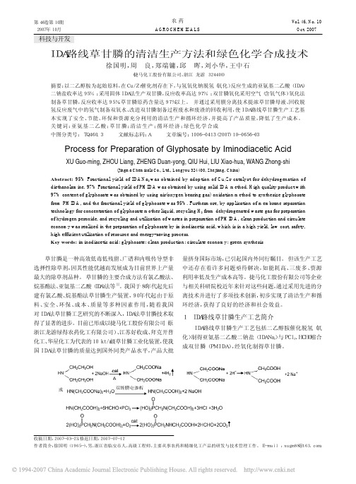
第46卷第10期2007年10月农 药AGROCHEMICALSVol. 46, No. 10Oct. 2007IDA路线草甘膦的清洁生产方法和绿色化学合成技术徐国明,周 良,郑端镛,邱 晖,刘小华,王中石(捷马化工股份有限公司,浙江 龙游 324400)摘要:以二乙醇胺为起始原料,在Cu/Zr催化剂存在下,与氢氧化钠脱氢(氧化)反应生成的亚氨基二乙酸(IDA)二钠盐收率达95% ;采用固体IDA法生产双甘膦,反应收率高达97% ;双甘膦氧化采用空气(含氧气体)氧化法制备草甘膦,反应收率达95%,草甘膦原药含量达97%以上。
并通过采用膜分离技术提浓草甘膦母液、回收脱氢反应废气中的氢气制备双氧水、改进双甘膦制备过程废水和废渣的回收利用,使IDA路线草甘膦生产工艺基本实现了安全、节能、环保和资源充分利用的清洁生产和循环经济,并提高了产品质量,降低了生产成本。
关键词:亚氨基二乙酸;草甘膦;清洁生产;循环经济;绿色化学合成中图分类号: TQ460.3 文献标志码:A 文章编号:1006-0413(2007)10-0656-03Process for Preparation of Glyphosate by Iminodiacetic AcidXU Guo-ming, ZHOU Liang, ZHENG Duan-yong, QIU Hui, LIU Xiao-hua, WANG Zhong-shi(Jingma Chemicals Co., Ltd., Longyou 324400, Zhejiang, China)Abstract: 95% Fractional yield of IDANa 2 was obtained by adoption of Cu/Zr catalyst for dehydrogenation of diethanolamine. 97% Fractional yield of PMIDA was obtained by using solid IDA method. High quality product with 97% content of glyphosate was obtained by using air(oxygen bearing gas) oxidation method to synthesize glyphosate from PMIDA, and the fractional yield of glyphosate was 95%. Furthermore, by application of membrane separationtechnology for concentration of glyphosate mother liquid, recycling H 2 from dehydrogenated waste gas for preparation of hydrogen peroxide, and recycling and utilization of wastes in preparation of PMIDA, clean production and circulate economy was realized in the preparation of glyphosate by iminodiacetic acid, which is in a high yield, low cost, safety,high efficient utilization of resource and energy-saving process.Key words: iminodiacetic acid; glyphosate; clean production; circulate economy; green synthesis草甘膦是一种高效低毒低残留、广谱和内吸传导型非选择性除草剂,因其性能优越而发展成为目前世界上产量最大的除草剂品种。
法国生物梅里埃介绍

A world leader in in vitro diagnosticsContribute to the improvement of public health worldwide through in vitro diagnosticsAnalyzing a food, drug or air sample to monitor and confirm the quality of the production processThe Company and its Market先生Mérieux)开创了科学和工业应用领域,创建梅里埃基金会创立了生物梅里埃——所致力发展和生产用于细菌学、血清学、临床生化学和凝血方面的标准化试剂的化验所,进而又把它扩展为国际性公司¾1897: Marcel Mérieux creates the “Institut Biologique Mérieux”,The Start of the Mérieux Venture¾1917: Installation in Marcy l’Etoile9Focus on 4 pathologies:tuberculosis,diphtheria, tetanus and puerperal streptococcus9Production of tuberculin(Koch’s bacillus)9Production of sera (foot-and-mouth disease)9Microbiological analyses1911: Marcel Mérieux inBrings Industrialization to Biology¾1937: Upon his fatherMarcel’s death, CharlesMérieux took over ashead of theInstitut Mérieux¾1947: introduction of invitro culture techniquesdeveloped in well-knownuniversities around theworld (Reiks University,Prof. Frenkel, JonasSalk…)1942: Dr. Charles Mérieux at a laboratory in Marcy l’EtoileBanner for vaccination campaign in Rio(in the middle) and of which he was掌控了公司的绝大多数股份,至此BD Mérieux成为了收购麦道公司的1994增加对生物梅里埃的控股并收购TransgeneApplications & ProductsBecome the undisputed leader with full microbiology lab automationVITEK ®2 CompactDiversiLab ®VIDAS ®API ®袋装肉汤致病菌检测TEMPO 卫生指标菌群定量VITEK ®2 Compact菌株分型检测环境监测BioBall BacT/ALERT Count-Tact ®Air/DEAL ®API ®Global Customer Service& Training¾Product trainingTraining by University professorsEditions (more than 6 subjectsManufacturing & QualityFlorence (Italy)Basingstoke(UK)Durham (USA)Rio de Janeiro(Brazil)Brisbane (Australia)Saint Louis (USA)GrenobleSidney (Australia)Portland (USA)Lombard (USA)2010Tres Cantos (Spain)Shanghai (China)SitesbioMérieux La BalmebioMérieux Marcy l’EtoileIDC –St Vulbas bioMérieux CraponneMarcy l’EtoileFranceHead Office¾Site since1971¾Staff9~ 1,150 people*¾Activities9bioMérieux administration9Research9Production9Quality Control laboratory forclinical chemistry, immunoassaysand microbiology¾Products9Clinical chemistry9Immunoserology9VIDAS®reagents31。
三聚氰胺
Analytica Chimica Acta 665 (2010) 84–90Contents lists available at ScienceDirectAnalytica ChimicaActaj o u r n a l h o m e p a g e :w w w.e l s e v i e r.c o m /l o c a t e /a caHapten synthesis and antibody production for the development of a melamine immunoassayHongtao Lei a ,1,Yudong Shen a ,1,Lijun Song a ,Jinyi Yang a ,Olivier P.Chevallier b ,Simon A.Haughey b ,Hong Wang a ,Yuanming Sun a ,∗,Christopher T.Elliott b ,∗∗aThe Key Laboratory of Food Safety of Guangdong Province/Institute of Food Quality and Safety,South China Agricultural University,Wushan Road,Tianhe District,Guangzhou 510642,Guangdong,PR China bInstitute of Agri-Food and Land Use,Queen’s University Belfast,Belfast BT95AG,Northern Ireland,United Kingdoma r t i c l e i n f o Article history:Received 27November 2009Received in revised form 28February 2010Accepted 7March 2010Available online 15 March 2010Keywords:Melamine Hapten AntibodyEnzyme-linked immunosorbent assaya b s t r a c tThe incorporation of melamine into food products is banned but its misuse has been widely reported in both animal feeds and food.The development of a rapid screening immunoassay for monitoring of the substance is an urgent requirement.Two haptens of melamine were synthesized by introducing spacer arms of different lengths and structures on the triazine ring of the analyte molecular structure.6-Aminocaproic acid and 3-mercaptopropionic acid were reacted with 2-chloro-4,6-diamino-1,3,5-triazine (CAAT)to produce hapten 1[3-(4,6-diamino-1,6-dihydro-1,3,5-triazin-2-ylamino)hexanoic acid]and hapten 2[3-(4,6-diamino-1,6-dihydro-1,3,5-triazin-2-ylthio)propanoic acid],respectively.The molecu-lar structures of the two haptens were identified by 1H nuclear magnetic resonance spectrometry,mass spectrometry and infrared spectrometry.An immunogen was prepared by coupling hapten 1to bovine serum albumin (BSA).Two plate coating antigens were prepared by coupling both haptens to egg oval-bumin (OVA).A competitive indirect enzyme-linked immunosorbent assay (ciELISA)was developed to evaluate homogeneous and heterogeneous assay formats.The results showed that polyclonal antibod-ies with high titers were obtained,and the heterogeneous immunoassay format demonstrated a better performance with an IC 50of 70.6ng mL −1,a LOD of 2.6ng mL −1and a LOQ of 7.6ng mL −1.Except for cyro-mazine,no obvious cross-reactivity to common compounds was found.The data showed that the hapten synthesis was successful and the resultant antisera could be used in an immunoassay for the rapid and sensitive detection of this banned chemical.© 2010 Elsevier B.V. All rights reserved.1.IntroductionMelamine,with an IUPAC name of 1,3,5-triazine-2,4,6-triamine,is an organic base.In its chainlike “polymerized”form,melamine is an industrial chemical and has been used for decades in theAbbreviations:CAAT,2-chloro-4,6-diamino-1,3,5-triazine;BSA,bovine serum albumin;DCC,dicyclohexylcarbodiimide;NHS,N-hydroxysuccinimide;TMB,3,3 ,5,5 -tetramethylbenzidine;DMF,dimethylformamide;TLC,thin-layer chro-matography;PBS,phosphate-buffered saline;PBST,phosphate-buffered saline with TW-20;HRMS,high-resolution mass spectrometry;hapten 1,3-(4,6-diamino-1,6-dihydro-1,3,5-triazin-2-ylamino)propanoic acid;hapten 2,3-(4,6-diamino-1,6-dihydro-1,3,5-triazin-2-ylthio)propanoic acid;ELISA,enzyme-linked immunosorbent assay;iELISA,indirect ELISA;ciELISA,competitive indirect ELISA;CR,cross-reactivity;UV,ultraviolet spectrometry;IR,infrared spectrometry;NMR,nuclear magnetic resonance.∗Corresponding author.Tel.:+862085280270;fax:+862085288282.∗∗Corresponding author.Tel.:+442890976549;fax:+442890976513.E-mail addresses:ymsun@ (Y.Sun),chris.elliott@ (C.T.Elliott).1Equal contributors.manufacturing of dishes,plastic resins,flame-retardant fibers,components of paper and paperboard and industrial coatings [1,2].Food has had limited exposure to melamine through a number of these contact materials.The Food and Drug Administration (FDA)states that the yield of chloroform soluble extracts should not exceed 0.5mg in.−2(77.5g cm −2)of food contact surface (21CFR 1777.1460).The maximum permitted concentration for melamine in food has been set at 2.5mg kg −1by the European Commission [3,4].Melamine is not approved to be added to foods or feeds,nor is it permitted to be used as a fertilizer anywhere in the world.Any future approval is highly unlikely as ingestion of melamine may lead to reproductive damage,bladder or kidney stones,which in turn may lead to bladder carcinogenesis [5].However,in 2007residues of melamine were detected in pet food,animal feed,wheat gluten,and other protein-based food commodities,and subsequently pet illness and death were widely reported [1].In the following year,several companies and individ-uals were implicated in a scandal involving milk and infant formula which had been adulterated with melamine,leading to kidney stones and renal failure [6–8].Thus,the discovery of illegally added0003-2670/$–see front matter © 2010 Elsevier B.V. All rights reserved.doi:10.1016/j.aca.2010.03.007H.Lei et al./Analytica Chimica Acta665 (2010) 84–9085Fig.1.Synthesis route of melamine haptens.melamine to food products resulted in the urgent requirement to detect melamine in food and feed.Liquid chromatographic determination of melamine was developed for beverage samples in1987[9].Ion-pair liquid chro-matography coupled to electrospray tandem mass spectrometry was used to determine residue of melamine in chard samples using C-18chromatography,the limit of detection(LOD)and limit of quantification(LOQ)were0.01and0.1mg kg−1,respectively[10]. Gas chromatography–mass spectrometry and ultra-performance liquid chromatography–tandem mass spectrometry were also used to detect melamine,the claimed sensitivity of this procedure was10 and5g kg−1,respectively[11].However,quantitative detection of melamine is currently limited to instrumental methods,most of which share a number of important drawbacks:they usually require costly apparatus,their sample throughput is limited and they are not suitable for on-site analysis.Immunoassay technology has been increasingly used for screen-ing food contaminants due to the sensitivity,selectivity,time efficiency,cost-effective and portability of the procedures.Over the past20years the development of immunochemical meth-ods and their potential applications,especially the enzyme-linked immunosorbent assay(ELISA),has grown significantly[12,13]. Although several commercial ELISA kits to melamine have become available in recent times[2],no studies on the hapten synthesis, antibody production,and development of immunoassays to this compound have been published to our knowledge.The aim of the present research is to prepare polyclonal anti-body against melamine for immunoassay development.To achieve this aim,haptens containing two different linkers were synthesized and one was used as an immunogen and both were used as coat-ing antigen.The recognition performance of antibody obtained was evaluated with some compounds based on their structural differ-ence and cross-reactivity data.Also,the specificity and sensitivity of the homogeneous and heterogeneous ELISA were compared. 2.Experiments2.1.Reagents and chemicalsGeneral reagents and organic solvents were of analytical grade unless specified otherwise.2-Chloro-4,6-diamino-1,3,5-triazine (CAAT)was obtained from Shanghai Chaoyan Biotechnology Co.,Ltd.(Shanghai,China).6-Aminocaproic acid was bought from Sinopharm Chemical Reagent Co.,Ltd.(Shanghai,China). Melamine,bovine serum albumin(BSA),dicyclohexylcarbodiimide (DCC),N-hydroxysuccinimide(NHS),ovalbumin(OVA),3,3 ,5,5 -tetramethylbenzidine(TMB),complete and incomplete Freund’s adjuvants were purchased from Sigma(St.Louis,MO,USA).3-Mercaptopropionic acid was bought from Alfa Aesar(Tianjin, China).Cyanuric chloride and cyanuric acid were obtained from Accela Chembio Co.,Ltd.(Shanghai,China).Atrazine was a gift from Shandong Zhongke Qiaochang Chemical Co.,Ltd.(Shandong,China).Cyromazine was bought from Changzhou Zhineng Ani-mal Pharmaceutical Co.,Ltd.(Changzhou,China).HRP-conjugated goat anti-rabbit IgG was obtained from Boster Biotech Co.,Ltd. (Wuhan,China).Thin-layer chromatography(TLC)was performed on200mesh,2.5mm precoated silica gel GF254on glass sheets obtained from Qingdao Haiyang Chemical Co.,Ltd.(Qingdao, China).Polystyrene ELISA plates were obtained from Jincanhua Co., Ltd.(Shenzhen,China).Buffers used in this study were prepared as follows:50mM carbonate buffer(pH9.6)for coating plates,10mM PBST solution phosphate buffer saline(PBS,pH7.4,containing0.1%Tween-20) was used for washing plates,0.1M citrate and sodium phosphate buffer(pH5.4)for substrate buffer,and2M H2SO4as the stopping reagent.TMB solution was prepared by addition of10mL substrate buffer and150L of15mg mL−1TMB in dimethylformamide(DMF) and2.5L of6%(w/v)H2O2.2.2.InstrumentationUltraviolet spectrometry(UV)were recorded on a UV-3010 spectrophotometer(Hitachi,Japan).High-resolution mass spec-trometry(HRMS)analyses were performed using a MAT95XP high-resolution mass spectrometer(Thermo,USA).Nuclear mag-netic resonance(NMR)spectra were obtained with the DRX-600 NMR spectrometers(Bruker,Germany–Switzerland).Infrared spectrometry(IR)was performed using Nicdet Avatar360(Thermo, USA).Melting point was determined on apparatus(Gallenkamp, UK).ELISA plates were washed using a Multiskan MK-2microplate washer(Thermo Labsystems,USA).Absorbance was measured at a wavelength of450nm using a Multiskan MK3microplate reader (Thermo Labsystems,USA).2.3.Hapten synthesis and characterization2.3.1.Synthesis of6-(4,6-diamino-1,3,5-triazin-2-ylamino) hexanoic acid(hapten1,Fig.1)CAAT(1.21g,8.1mmol)was dissolved in150mL of absolute ethanol,and6-aminocaproic acid(1.26g,9.4mmol)and KOH (1.64g,24.9mmol)in10mL of absolute ethanol were added drop-wise,refluxed at70◦C for24h,monitored by TLC.The reaction mixture wasfiltered,thefiltrate was cooled to obtain a white solid.The solid was dissolved in75mL of5%sodium carbonate and extracted with60mL of methylene chloride(3×20mL).The aqueous phase was acidified to pH3with6N HCl,concentrated and cooled,0.128g of hapten1was obtained as a white solid in 6.6%yield.m.p.185.0–186.0◦C,TLC R f=0.5(CHCl3:MeOH=60:20). HRMS(EI)m/z calculated for C9H16N2O2[M]240.1335,found 240.1329.1H NMR(DMSO-d6,600MHz)ı:1.32–1.25(m,2H), 1.56–1.45(m,4H),2.21(t,J=7.35,7.35Hz,2H),3.25(dd,J=12.97, 6.65Hz,2H);7.85–8.18(br,4H).13C NMR(MeOH-d4,150MHz)ı: 177.4(C-1),41.6(C-6),34.8(C-2),30.09(C-5),27.2(C-4),25.6(C-3). IR(KBr) max(cm−1):3492(s),3409(s),3331(s),3159(s),2937(s),86H.Lei et al./Analytica Chimica Acta665 (2010) 84–90Fig.2.Synthesis route and structures of melamine conjugates.2860(w),1642(vs),1553(s),1473(s),1442(s),1371(s),1309(w),1165(w),811(w).The maximum wavelength of UV absorption was 241nm.2.3.2.Synthesis of 3-(4,6-diamino-1,3,5-triazin-2-ylthio)propanoic acid (hapten 2,Fig.1)CAAT (1.21g,8.1mmol)was suspended in 150mL of absolute ethanol,and 3-mercaptopropionic acid (1.0g,9.4mmol)and KOH (1.64g,24.9mmol)in 10mL of absolute ethanol were added drop-wise,refluxed at 78◦C for 28h,monitored by TLC.The reaction mixture was filtered,the filtrate cake was washed with cool abso-lute ethanol.The white solid was dissolved in 10mL of cooled deionized water (0◦C)and acidified to pH 6with 6N HCl.0.74g hap-ten 2was obtained as a white solid in 42.4%yield.m.p.218–221◦C;1H NMR (600MHz,DMSO-d 6)ı:2.64(t,J =6.98Hz,2H),3.14(t,J =7.02Hz,2H),6.9(br,4H).13C NMR (DMSO-d 6,150MHz)ı:178.3(C-1);173.0(C-4);165.3(C-5,6);34.3(C-2);24.2(C-3).MS (APCI)m /z :216[M];MS 2(APCI)m /z :198[M −OH],144[M −CH 2COOH];IR (KBr) max (cm −1):3422(vs),3157(s),2924(s),2854(w),1711(s),1644(vs),1531(s),1403(s),1308(w),1252(w),1189(w),921(w),803(w).The maximum wavelength in UV absorption is 275nm.2.4.Preparation of hapten–protein conjugates (Fig.2)Hapten 1was coupled to BSA to be used as an immunogen (con-jugate 1),and both haptens 1and 2were coupled with OVA using the active ester method to obtain two plate coating antigens (con-jugates 2and 3).Briefly,20.0mg (100mol)of hapten 1or 21.0mg (100mol)of hapten 2,NHS 17.0mg (150mmol)were dissolved in 1mL of DMF,followed by addition of DCC 31.0mg (150mmol).The activation reaction was carried out at 4◦C overnight with stir-ring.The white dicyclohexylurea precipitate was removed from solution by centrifugation.The supernatant (900L)was added dropwise to BSA (113mg)or OVA (75mg)in 8mL PBS (10mM,pH 7.4).The conjugate mixture was stirred at 4◦C for 12h and then dialyzed against 10mM PBS (pH 7.4).2.5.Production of polyclonal antibodiesThree New Zealand rabbits weighing 1.5–2.0kg were immu-nized 5times using conjugate 1at intervals of 14days by theGuangdong Medical Laboratory Animal Center.The rabbits were blood sampled to detect the presence of antibodies to melamine using an indirect ELISA on the eighth day after each immunization,starting 40days after the first injection.Serum was divided into aliquots (1mL)and stored at −20◦C until use.2.6.ELISA developmentIn the indirect ELISA (iELISA),all incubations were performed at 37◦C except for the coating antigen.Flat-bottom polystyrene ELISA plates were coated with hapten–OVA (1g mL −1,100L well −1)in carbonate buffer (pH 9.6)over night at 4◦C.The wells were washed 5times with PBST solution,and then blocked with 5%skim milk in PBS buffer (200L well −1)for 1h.After washing 5times with PBST solution,the wells were incubated with 100L of diluted antibody in PBST for 1h and washed 5times with PBST solu-tion.HRP-conjugated goat anti-rabbit IgG diluted 1:9000in PBST was added (100L well −1).After incubation for 1h and washing 5times with PBST solution,TMB solution was added to the wells (100L well −1)and incubated for 15min.The reaction was stopped by addition of 50L well −1of 2M H 2SO 4,and the absorbance was recorded at 450nm.For the competitive indirect ELISA (ciELISA),the procedure was identical except for the addition of a competition step after the blocking,which was involved with adding 50L well −1of melamine standards dissolved in PBS containing 0.8%methanol,followed by 50L well −1of appropriate concentrations of antisera diluted with PBST.The concentrations of antibodies and coating antigens had been optimized by checkerboard titration and com-petitive curves were then obtained by plotting the normalized signal B /B 0against the logarithm of analyte concentration,where B 0is the signal without analyte and B is the signal of each concen-tration of analyte.A four-parameter logistic equation (OriginPro 7.5for Windows)was used to fit the sigmoidal curve according to the following for-mula (1):Y =A −D [1+(x/C )B]+D(1)where A is the asymptotic maximum (maximum absorbance in absence of analyte,A max ),B is the curve slope at the inflexion point,H.Lei et al./Analytica Chimica Acta665 (2010) 84–9087Table1Parameters comparison of homogeneous and heterogeneous ciELISA.Coating antigen Rabbit1serum Rabbit2serumTiter IC50(ng mL−1)LOD(ng mL−1)LOQ(ng mL−1)Titer IC50(ng mL−1)LOD(ng mL−1)LOQ(ng mL−1)Conjugate24×1051346.637.6153.08×104708.842.895.8Conjugate32×105758.832.290.02×10470.6 2.67.6C is the x value at the inflexion point(corresponding to analyteconcentration giving50%inhibition of A max,IC50),and D is theasymptotic minimum(background signal).10%inhibition valuewas defined as LOD;20%inhibition value was defined as LOQ.2.7.Specificity of antibodyThe specificity of the antibody was evaluated by performingcompetitive assays using several compounds structurally related tomelamine as competitors,and the obtained IC50values were usedto calculate cross-reactivity using the formula:%CR=IC50(melamine)IC50(cross reactant)×100(2)3.Results3.1.Hapten synthesisHapten design is a critical factor for the successful prepara-tion of highly specific antibodies against low molecular weight antigens[13–15].A suitable hapten and its subsequent protein conjugate for immunization should be designed as a mimic to the target molecule.If melamine itself was utilized as reactant to derive a hapten,due to its three amino groups on the triazine ring having the same reactivity,the derivation product would in all likelihood be complicated in that one,two or all three amino would be substituted when using glutaraldehyde or halogenated carboxylic acid such as sodium chloro-or bromo-acetic acid or their esters.For this reason CAAT was selected as the derivation reactant due to the active chloride group being capable of reacting relatively easily with the aliphatic primary amino or the sulphydryl group of a spacer arm reagent such as6-aminocaproic acid or 3-mercaptopropionic acid,respectively.Catalyzed with KOH,the resultant product possessed the antigenic epitopes most likely to mimic that of melamine(Fig.1).In some cases,immunogens prepared without a spacer arm are easier to produce but result in an assay with poor sensitivity and/or weak recognition of the segment of the target molecule near the attachment site on the carrier protein[12–15].Spacer groups which contain4–6carbons have been shown to be optimal in obtain-ing high quality antibodies[15,16].In the present study,a spacer arm containing6carbons was designed for immunogen prepara-tion.This was selected to expose a specific region of the hapten to the animals’immune system.Furthermore,a three carbon arm with a heterogeneous sulphur atom instead of a nitrogen atom was designed for hapten2.The strategy employed was selected to pre-vent any antibody binding to the spacer arm,which may have a detrimental effect in the assay.From the data generated by1H NMR and MS and IR,it was determined that the desired haptens,1and2,were successfully synthesized.3.2.Serum titer determinationThree rabbits were immunized with conjugate1,however one died during the immunization period(cause of death unknown). The other two rabbits produced sera that resulted in antibody titers>80,000(Table1)as determined by indirect ELISA,with titer being defined as the time of the serum dilution that results in an absorbance value that is about1.0OD units.parison of homogeneous and heterogeneous ciELISAA heterogeneous approach in the current context refers to the hapten used for immunogen production being different from that used for the coating antigen.Heterogeneous formats can often result in antibodies having a higher affinity towards the analyte in comparison to the coating antigen or tracer hapten[14–17]. Thus the sensitivity achievable using this format as opposed to a homologous one can improve sensitivity substantially.In the present study,conjugates2and3were used as homoge-neous and heterogeneous coating antigens in order to compare the sensitivity achievable.The results have been presented in Fig.3and Table1.For rabbit1serum,the IC50with heterogeneous format was found to be758.8ng mL−1(6.02nM)compared to1346.6ng mL−1 (10.68nM)with the homogeneous format.The IC50based on rabbit 2serum was found to be substantially better than that of rabbit1,parison of homogeneous and heterogeneous ciELISA.Data represented in mean±SD(standard deviation)and n=3.(a)Standard curve of rabbit1serum;(b)standard curve of rabbit2serum.88H.Lei et al./Analytica Chimica Acta 665 (2010) 84–90Table 2Cross-reactivity of antiserum to related compounds.CompoundsStructureConjugate 2Conjugate 3IC 50(nmol mL −1)Cross-reactivity (%)IC 50(nmol mL −1)Cross-reactivity (%)Melamine 5.621000.56100.0Cyromazine 2.1267.60.30186.7Hapten 10.341652.90.26215.4Hapten 213.3642.414.08 4.0CAAT 218.44 2.6181.340.3Cyanuric acid ND 0.01ND 0.01Atrazine ND 0.01ND 0.01Cyanuric chloride ND 0.01ND 0.01ND presented infinite IC 50values and could not be fitted with the four-parameter logistic equation.i.e.in the region of 10times more sensitive.Similar improvements with both LOD and LOQ measurements were also found (Table 1).The working range between melamine concentrations giving 20–80%inhibition were 0.76–41.54nM (95.8–6744.2ng mL −1)for serum 1and 0.06–5.8nM (7.6–731.4ng mL −1)for serum 2in the heterogeneous assay format.The US FDA have published several liquid chromatography methods coupled with mass spectrometer for melamine analysis,the claimed LOQ was 10g kg −1[18–20].Therefore,the LOQ determined in the presented heterogeneous immunoassay could meet the requirements of both the US FDA [18–20]and European Commission [2,3].Due to its better sensitivity,rabbit 2serum was selected for further specificity investigation.3.4.Specificity of antibodyTo determine which structural features of the molecules are important to antibody recognition,the cross-reactivity (CR)against a range of compounds structurally related to melamine was tested.Both conjugates 2and 3were employed as homogeneous and het-erogeneous coating antigens to compare the specificity differences observed (Table 2).Amongst all the related compound tested in the present study,the cross-reactivity of hapten 1was highest in both homogeneous and heterogeneous format assays,followed by cyromazine,melamine,hapten 2,CAAT,atrazine,cyanuric acid and cyanuric chloride which showed less than 0.01%cross-reactivity to the developed antibody.H.Lei et al./Analytica Chimica Acta665 (2010) 84–9089Since the immunogen was prepared using hapten1,it was rea-sonable to expect that the developed antibody would recognize hapten1with a high affinity.Although the arm of cyromazine with a large ring structure was different from that of hapten1, cyromazine displayed more%CR than melamine in both assay for-mats.However,removing the spacer arm to change hapten1into melamine resulted in the%CR decreasing from1652.9to100in the homogeneous assay,and from215.4to100in the heterogeneous assay(Table2).This data strongly indicated that the spacer arm played an important role in hapten–antigen binding.Similar phe-nomenon has been reported in other immunoassay development studies[21–24].In the homogeneous format,the267.6%CR of cyromazine was higher than the186.7%CR in the heterogeneous format.Similarly, the%CRs of hapten2and CAAT in the heterogeneous assay were lower than those in homogeneous assay.Thesefindings indicated that the assay format employed can affect not only sensitivity but also specificity.3.5.DiscussionIf the cyclopropyl group of cyromazine and amidocaproic acid group of hapten1being regarded as two different spacer arms of haptens,and comparing the structures and cross-reactivity data of melamine,cyromazine and haptens,it was interesting tofind that the compound without the spacer arm,melamine,had lower%CR than that with spacer arms,hapten1and cyromazine.Although cyromazine contains one hydrocarbon ring structure which can cause steric hindrance to hapten–antibody binding[25,26],the%CR of cyromazine was still higher than melamine but lower than hap-ten1with a straight hydrocarbon spacer arm.Thus,it is possible that the steric hindrance reduces antibody recognition of cyro-mazine compared to hapten1;and that even though a spacer arm was just a hydrocarbon straight chain,it could take part in the hapten–antibody interaction.Also,when the hapten in the immunogen contains a spacer arm,even if the arm of recognized analyte contains a ring structure with the steric hindrance effect, the analyte with ring could possibly be recognized by antibod-ies easier than that without a spacer arm.Consequently,the arm structure of analyte could be used to adjust the assay perfor-mance.However,hapten2contained one spacer arm,mercaptopropi-onic acid group,which had3carbons less than the arm of hapten1, but the%CR of hapten2was lower than not only cyromazine with a ring arm,but also melamine without a spacer arm.It seemed that there was another factor affecting the hapten–antibody recogni-tion stronger than the spacer arm effect.We think that electronic effects may contributed to the lower%CR of hapten2.In some cases,electronic features were also responsible for governing hapten–antibody recognition[26].In the present study,the sul-phur atom in hapten2contained two lone-pair electrons,while the nitrogen atom in the spacer arm of hapten1and cyromazine con-tained only one single lone-pair electrons.The additional lone-pair of electrons on the sulphur atom resulted in a stronger conjuga-tive effect with the triazine ring,which had been verified by the UV data.The UV information of the molecule can indicate the con-jugative structures,and extending conjugative effects will result in bathochromic and hyperchromic shifts in absorption[27].The max-imum absorbance wavelength of hapten1was found to be241nm, as opposed to275nm for hapten2,which are typical bathochromic and hyperchromic shifts.This data indicated that hapten2con-tained a stronger conjugative structure.Therefore,the electronic effect observed is likely to have played a critical role in the recog-nition of the melamine antibody to different compounds.Moreover,the chlorine atom on CAAT had strong electron with-drawing effects on the triazine ring,which is another obviously effect different to the conjugative effects of the sulphur atom on hapten2and the nitrogen atom of hapten1or cyromazine.When only one amino group of melamine was substituted by a chlo-rine atom,the cross-reactivity decreased dramatically from100% to2.6%in homogeneous assay or0.31%in heterogeneous assay, respectively(Table2).It is possible that the electron withdrawing effect influenced significantly the binding of the hapten and anti-body.Also,comparison of the%CR of hapten2and CAAT showed that the%CR of the latter was much lower than the former,which suggested that the electronic withdrawing effect of the substituent atom in the arm might have inversely affected the hapten–antibody binding more than the conjugative effect of the sulphur atom in hapten2.Similarly,comparing the spacer arm steric effect(based on melamine,cyromazine,hapten1)and electronic effect(based on melamine,hapten2,CAAT),it seemed that the latter resulted in more influence on the recognition of hapten–antibody than the former.Furthermore,atrazine can be regarded as a derived CAAT whose two free amino groups are blocked with one ethyl and one iso-propyl group,respectively.Then,removing the two alkyl blocking groups of atrazine to expose both naked amino groups,the cross-reactivity was observed to increase from≤0.01%of atrazine to2.57% or0.3%of CAAT in homogeneous and heterogeneous assay,respec-tively(Table2).Similarly,both cyanuric acid and cyanuric chloride –which contain a triazine ring but no naked amino groups–could not be recognized by the antibody either.The result suggested that both free amino groups were necessary epitopes for antibody recognition.Cyromazine is afly-killing pesticide frequently used for prevent-ing housefly and Fannia canicularis in livestock and Liriomyza sativae and leafminer on vegetable andflowers[1,10].It is known that melamine is the main metabolite of cyromazine[1,10].To lower the unexpected cross-reactivity from cyromazine,one hapten with a non-hydrocarbon spacer arm,e.g.6-hydrazinyl-1,3,5-triazine-2,4-diamine,could possibly be used to prepare an immunogen which could hopefully raise antibodies recognizing cyromazine weaklier because the hydrazine arm of hapten and cyclopropyl group of cyromazine are quite distinct.4.ConclusionTwo haptens with different spacer arms were synthesized and were used successfully for antibody production.Two conjugates were used to compare and evaluate the antibody performance, the result showed that sensitivity and specificity of antibody were much better in the heterogeneous format assay than in homoge-neous one.Based on the comparison of structure and biological activity data,spacer arm effects and electronic effects appear to be two important factors with regard to the antibody binding to the haptens.With further development work the antibody produced in the current study could be applied to a range of immunochemical platforms to deliver a highly rapid and sensitive screening proce-dure for melamine in foods and feeds.AcknowledgementsThis work was supported by National Key Technologies R &D Program of China during the11th Five-Year Plan Period (No.2006BAD27B02-05)and China Guangdong Provincial Science and Technology Department(2007A020100006-10,zgzhzd0808, 2009B011300005).We are grateful to Dr Yingju Liu,College of Science,South China Agricultural University,for conducting infrared spectrometry and melting point test.。
CLSI国家临床实验室标准委员会标准2010年最新标准目录(含中文翻译)
NCCLS国家临床实验室标准委员会标准2010年最新标准目录NCCLS C3-A4 2006.06.16 Preparation and Testing of Reagent Water in the Clinical Laboratory; Approved Guideline - Fourth Edition制备和检测试剂在临床实验室;批准指南-第四版NCCLS C5-P2 1987.01.01 Methodological Principles for Selected Analyses: Enzymes; Proposed Guideline方法论原则,选择分析:酶;建议指南NCCLS C12-T2 1991.01.01 Definitions of Quantities and Conventions Related to Blood pH and Gas Analysis-Second Edition Standard定义的量和公约有关的血液PH值和气体analysis-second版标准NCCLS C21-A 1992.01.01 Performance Characteristics for Devices Measuring PO2 and PCO2 in Blood Samples性能特点的设备测量氧及二氧化碳分压的血液样本NCCLS C24-A 1991.01.01 Internal Quality Control Testing: Principles and Definitions, Approved Guideline内部质量控制测试:原理和定义,批准指南NCCLS C25-T 1992.01.01 Fractional Oxyhemoglobin, Oxygen Content and Saturation, and Related Quantities in Blood: Terminology, Measurement and Reporting; Tentative Guideline分数血氧饱和度,氧含量,和相关的数量在血液:术语,测量和报告的暂定准则NCCLS C27-A 1993.01.01 Blood Gas Pre-Analytical Considerations: Specimen Collection, Calibration, and Controls, Approved Guideline血液气体分析前考虑:标本采集,校准,并控制,批准指南NCCLS C28-P 1992.01.01 How to Define, Determine, and Utilize Reference Intervals in the Clinical Laboratory; Proposed Guideline如何定义,确定,并利用参考间隔在临床实验室提出的指导方针NCCLS C29-T 1992.01.01 Standardization of Sodium and Potassium Ion Selective Electrode Systems to the Flame Photometric Reference Method; Tentative Standard标准化的钠和钾离子选择性电极系统的火焰光度参照法;试验标准NCCLS C30-T 1991.01.01 Ancillary (Bedside) Blood Glucose Testing in Acute and Chronic Care Facilities; Tentative Guideline辅助(床边)测试血糖在急性和慢性保健设施;暂定准则NCCLS C34-P 1993.01.01 Determination of Chloride and Sodium Concentrations in Sweat: Sample Collection and Quantitative Analysis; Proposed Guideline测定在汗水中氯和钠的浓度:样品采集和定量分析;提出的指导方针NCCLS DI1-A2 1992.01.01 Glossary and Guidelines for Immunodiagnostic Procedures, Reagents, and Reference Materials, Approved Guideline词汇和准则免疫诊断程序,试剂,和参考材料,批准指南NCCLS DI2-A 1986.01.01 Immunoprecipitin Assays: Procedures for Evaluating the Performance of Materials; Approved Guideline验证试验:评价程序的材料性能;批准指南NCCLS DI3-T 1986.01.01 Agglutination Analyses: Characteristics of Antibody, Methodology, Limitations, and Clinical Validation, Tentative Guideline凝集抗体分析:特点,方法,限制,和临床验证,初步的指南NCCLS DI4-T 1986.01.01 Enzyme and Fluorescence Immunoassays, Tentative Guideline酶和荧光免疫测定,初步的指南NCCLS EP 5 2004.01.01 Evaluation Precision Performance of Quantitative Measurement Methods; Approved Guideline评价精度性能的定量测量方法;批准指南NCCLS EP 7 2002.01.01 Interference Testing in Clinical Chemistry, Approved Guideline临床化学干扰试验,批准指南NCCLS EP6-P 1986.01.01 Evaluation of the Linearity of Quantitative Analytical Methods, Proposed评价定量分析方法的线性,提出NCCLS EP9-T 1993.01.01 Method Comparison and Biasa Estimation Using Patient Samples; Tentative Guideline方法比较不错的估计使用病人样本;暂定准则NCCLS EP10-T 1989.01.01 Preliminary Evaluation of Clinical Chemistry Methods; Tentative Guideline初步评价临床化学方法;初步的指南NCCLS GP2-A 2002.01.01 Clinical Laboratory Technical Procedure Manuals - Fourth Edition临床实验室技术程序手册-第四版NCCLS GP5-A 2002.01.01 Clinical Laboratory Waste Management临床实验室废弃物管理NCCLS GP6-A 1994.01.01 Inventory Control Systems for Laboratory Supplies - First Edition实验室用品库存控制系统-第一版NCCLS GP9-A 1998.01.01 Selecting and Evaluating a Referral Laboratory - First Edition选择和评价参考实验室-第一版NCCLS GP10-A 1995.01.01 Assessment of the Clinical Accuracy of Laboratory Tests Using Receiver Operating Characteristic (ROC) Plots - First Edition临床评估的准确性化验使用接收机操作特性(02)图-第一版NCCLS GP11-A 1998.01.01 Basic Cost Accounting for Clinical Services - First Edition成本会计的基本临床服务-第一版NCCLS GP14-A 1996.01.01 Labeling of Home-Use In Vitro Testing Products - First Edition家用体外测试产品的标签——第一版NCCLS GP15-A 2001.01.01 Papanicolaou Technique - Second Edition NCCLS GP15-T 1991.01.01 Papanicolaou Technique; Tentative Guideline NCCLS GP16-A 2001.01.01 Routine Urinalysis and Collection, Transportation, and Preservation of Urine Specimens - Second Edition 尿常规和收集,运输,和保存的尿液标本-第二版NCCLS GP17-A 2004.01.01 Clinical Laboratory Safety; Approved Guideline临床实验室安全;批准指南NCCLS GP18-A 1998.01.01 Laboratory Design - First EditionNCCLS GP20-A 2003.01.01 Fine-Needle Aspiration Biopsy (FNAB) Techniques - Second Edition细针穿刺活检(细针穿刺细胞学检查)技术-第二版NCCLS GP21-A 2004.01.01 Training and Competence Assessment培训和能力评价NCCLS GP22-A 1999.01.01 Continuous Quality Improvement: Essential Management Approaches - First Edition持续质量改进:管理的基本方法-第一版NCCLS GP23-A 1999.01.01 Nongynecologic Cytologic Specimens: Collection and Cytopreparatory Techniques - First EditionNCCLS GP26-A 2003.01.01 Application of a Quality System Model for Laboratory Services - Second Edition实验室服务的高质量的应用系统模型-第二版NCCLS GP27-A 1999.01.01 Using Proficiency Testing (PT) to Improve the Clinical Laboratory - First Edition使用水平测试(铂)改善临床实验室-第一版NCCLS GP28-P 2004.01.01 Microwave Device Use in the Clinical Laboratory; Proposed Guideline - First Edition微波器件在临床实验室的使用;建议指南-第一版NCCLS GP29-A 2002.01.01 Assessment of Laboratory Tests When Proficiency Testing is Not Available - First Edition评估实验室测试时,测试不可用-第一版NCCLS H1-A3 1991.01.01 Evacuated Tubes for Blood Specimen Collection, Approved Standard; Third Edition真空管采血,经批准的标准;第三版NCCLS H2-A2 2001.01.01 Refernece and Selected Procedure for the Erythrocyte Sedimentation Rate (ESR) Test参考和选择程序的红细胞沉降率(血沉)试验NCCLS H3-A3 1991.01.01 Procedures for the Collection of Diagnostic Blood Specimens by Venipuncture - Third Edition; Approved Standard静脉穿刺收集诊断血液标本的程序-第三版;批准标准NCCLS H4-A3 1991.01.01 Procedures for the Collection of Diagnostic Blood Specimens by Skin Puncture - Third Edition; Approved Standard皮肤穿刺收集诊断血液标本的程序-第三版;批准标准NCCLS H5-A2 1985.01.01 Procedures for the Handling and Transport of Domestic Diagnostic Specimens and Etiological Agents, Approved Standard; Second Edition处理和运输国内诊断标本和病因剂的程序,经批准的标准;第二版NCCLS H7-A 1985.01.01 Procedure for Determining Packed Cell Volume by the Microhematocrit Method; Approved Standard血球容量计法确定便携细胞体积的程序;批准标准NCCLS H8-A 1986.01.01 Detection of Abnormal Hemoglobin Using Cellulose Acetate Electrophoresis; Approved Standard用乙酸纤维素电泳检测异常血红蛋白;批准标准NCCLS H9-A 1989.01.01 Chromatographic (Microcolumn) Determination of Hemoglobin A2; Approved Standard色谱(微)测定血红蛋白;批准标准NCCLS H10-A 1986.01.01 Solubility Test for Confirming the Presence of Sickling Hemoglobins, Approved Standard确认镰状血红蛋白存在的溶解度试验,批准的标准NCCLS H11-A2 1992.01.01 Percutaneous Collection of Arterial Blood for Laboratory Analysis, Approved Standard Second Edition实验室认可的经皮动脉收集血液,第二版NCCLS H13-A 1989.01.01 Quantitative Measurement of Fetal Hemoglobin by the Alkali Denaturation Method, Approved Guideline定量测量胎儿血红蛋白的碱变性方法,批准指南NCCLS H14-A2 1990.01.01 Devices for Collection of Skin Puncture Blood Specimens - Second Edition, Approved Guidelines皮肤穿刺收集血标本的设备-第二版,批准指南NCCLS H15-A 2001.01.01 Reference Procedure for the Quantitative Determination of Hemoglobin in Blood, Approved Standard定量测定血液中血红蛋白的参考程序,批准标准NCCLS H16-P 1985.01.01 Method for Reticulocyte Counting, Proposed Standard网织红细胞计数方法,提出的标准NCCLS H17-P 1990.01.01 The Determination of Serum Iron and Total Iron-Binding Capacity; Proposed Standard测定血清铁和总铁结合能力;提出的标准NCCLS H18-A3 2004.01.01 Procedures for the Handling and Processing of Blood Specimens; Approved Guidelines处理和加工的血液标本的程序;批准指南NCCLS H20-A 1992.01.01 Reference Leukocyte Differential Count (Proportional) and Evaluation of Instrumrntal Methods; Approved Standard 参考白细胞计数(比例)和设备评价方法;批准标准NCCLS H21-A2 1991.01.01 Collection, Transport, and Processing of Blood Specimens for Coagulation Testing and Performance of Coagulation Assays, Approved Guideline Second Edition收集,运输,处理血液的凝血性能检测和凝血化验,批准指南第二版NCCLS H22-P 1984.01.01 Histochemical Method for Leukocyte Alkaline Phosphatase, Proposed Standard白细胞碱性磷酸酶组织的化学方法,提出的标准NCCLS H23-T 1988.01.01 Citrate Agar Electrophoresis for Confirming Identification of Variant Hemoglobins, Tentative Guideline柠檬酸琼脂电泳确认鉴定变异血红蛋白,暂定准则NCCLS H24-T 1988.01.01 Additives for Blood Collection Devices:Heparin, Tentative Standard血液采集装置的添加剂:肝素,试行标准NCCLS H26-P 1989.01.01 Performance Goals for the Internal Quality Control of Multichannel Hematology Analyzers; Proposed Standard血液分析仪内部质量控制的绩效目标,批准标准;NCCLS H28-T 1992.01.01 One-Stage Prothrombin Time Test (PT), Tentative Guideline一期凝血酶原时间测试(铂),暂定准则NCCLS H29-T 1992.01.01 Activated Partial Thromboplastin Time Test (APTT), Tentative Guideline活化部分凝血活酶时间(部分凝血活酶时间测试),暂定准则NCCLS H30-T 1991.01.01 Procedure for the Determination of Fibrinogen in Plasma; Tentative Guideline测定血浆中纤维蛋白原的程序,暂定准则;NCCLS H31-P 1986.01.01 Collection Containers for Specimens for Toxicological Analysis, Proposed Guideline收集容器的标本进行分析,提出的指导方针NCCLS H34-P 1986.01.01 Determination of Factor VIII Coagulant Activity (VIII:C), Proposed Guideline测定凝血因子Ⅷ活性(Ⅷ:丙),提议指南NCCLS H35-T 1992.01.01 Additives to Blood Collection Devices: EDTA, Tentative Standard血液采集装置的添加剂:ED TA,试行标准NCCLS H40-P 1986.01.01 Determination of Factor IX Coagulant Activity, Proposed Guideline凝血活性因子Ⅸ测定,提议指南NCCLS H42-T 1992.01.01 Clinical Applications of Flow Cytometry: Quality Assurance and Immunophenotyping of Peripheral Blood Lymphocytes; Tentative Guideline临床应用流式细胞术:质量保证和免疫外周血淋巴细胞;暂定准则NCCLS I/LA2-T 1993.01.01 Quality Assurance for the Indirect Immunofluorescence Test for Autoantibodies to Nuclear Antigen (IF-ANA) Tentative Guideline质量保证的间接免疫荧光试验自身核抗原(if-ana)暂定准则NCCLS I/LA6-T 1992.01.01 Evaluation and Performance Criteria for Multiple Component Test Products Intended for the Detection and Quantitation of Rubella Antibody, Tentative Guideline评价和业绩标准,多个组件测试产品的检测和定量检测风疹抗体,初步的指南NCCLS I/LA7-P 1984.01.01 Specimen Handling and Use of Rubella Serology Tests in the Clinical Laboratory, Proposed Guideline标本处理和使用风疹血清学试验在临床实验室,提议指南NCCLS I/LA9-P 1985.01.01 Reference Method for Digoxin by Radioimmunoassay; Proposed Standard放射免疫法测定地高辛用参考方法,批准标准;NCCLS I/LA13-A 1991.01.01 Human Immunodeficiency Virus Type 1, Reference Material Specifications; Approved Guideline人类免疫缺陷病毒1型,参考材料规范;批准指南NCCLS I/LA15-P 1991.01.01 Apolipoprotein Immunoassays: Development and Recommended Performance Characteristics; Proposed Guideline载脂蛋白免疫:发展和性能特点的指导方针提出建议;NCCLS I/LA18-P 1991.01.01 Specifications for Immunological Testing for Infectious Diseases; Proposed Guideline规格免疫学检测传染病提出的指导方针;NCCLS I2-A2 1992.01.01 Temperature Calibration of Water Baths, Instruments, and Temperature Sensors水浴温度的校准,温度传感器NCCLS I16-T 1987.01.01 Temperature Monitoring and Recording in Blood Banks; Tentative Guideline血库的温度监测记录;暂定准则NCCLS I17-P 1991.01.01 Protection of Laboratory Workers from Instrument Biohazards; Proposed Guideline对于实验室仪器生物危害对操作者的保护;提出的指导方针NCCLS LA1-A 1985.01.01 Assessing the Quality of Radioimmunoassay Systems, Approved Guideline,放射免疫分析系统质量评估,批准指南NCCLS LA4-A2 1992.01.01 Blood Collection on Filter Paper for Neonatal Screening Programs, Approved Standard Second Edition采血滤纸新生儿筛查项目,批准的标准第二版NCCLS LIS01-A 2003.04.20 STANDARD SPECIFICATION FOR LOW-LEVEL PROTOCOL TO TRANSFER MESSAGES BETWEEN CLINICAL LABORATORY INSTRUMENTS AND COMPUTER SYSTEMS - First Edition在临床实验室仪器与计算机系统间的信息传递的低层协议的规范-第一版NCCLS LIS03-A 2003.04.20 STANDARD GUIDE FOR SELECTION OF A CLINICAL LABORATORY INFORMATION MANAGEMENT SYSTEM - First Edition临床实验室信息管理系统选择的标准指南-第一版NCCLS LIS04-A 2003.04.20 STANDARD GUIDE FOR DOCUMENTATION OF CLINICAL LABORATORY COMPUTER SYSTEMS - First Edition临床实验室计算机系统文件的标准指南-第一版NCCLS LIS06-A 2003.04.20 STANDARD PRACTICE FOR REPORTING RELIABILITY OF CLINICAL LABORATORY INFORMATION SYSTEMS - First Edition临床实验室信息系统可靠性报告的标准规程-第一版NCCLS LIS08-A 2003.04.20 STANDARD GUIDE FOR FUNCTIONAL REQUIREMENTS OF CLINICAL LABORATORY INFORMATION SYSTEMS - First Edition临床实验室信息系统功能要求的标准指南-第一版NCCLS LIS09-A 2003.04.20 STANDARD GUIDE FOR COORDINATION OF CLINICAL LABORATORY INFORMATION SERVICES WITHIN THE ELECTRONIC HEALTH RECORD ENVIRONMENT AND NETWORKED ARCHITECTURES - First Edition协调标准指南临床实验室信息服务的电子健康记录环境和网络结构-第一版NCCLS M2 2009.01.01 Performance Standards for Antimicrobial Disk Susceptibility Tests - Tenth Edition抗菌盘易感性试验的性能标准-第十版; Includes NCCLS M100-S19; To Purchase Call 1-800-854-7179 USA/Canada or 303-397-7956 WorldwideNCCLS M6-P 1986.01.01 Evaluating Production Lots of Dehydrated Mueller-Hinton Agar, Proposed Standard评估生产大量脱水mueller-hinton 琼脂,提议标准NCCLS M7 2009.01.01 Methods for Dilution Antimicrobial Susceptibility Tests for Bacteria That Grow Aerobically - Eighth Edition 细菌需氧增长的稀释抗菌易感性试验方法-第八版; Includes NCCLS M100-S19; To Purchase Call 1-800-854-7179 USA/Canada or 303-397-7956 Worldwide NCCLS M11-A2 1990.01.01 Methods for Antimicrobial Susceptibility Testing of Anaerobic Bacteria厌氧菌的抗菌易感性的试验方法NCCLS M15-T 1992.01.01 Slide Preparation and Staining of Blood Films for the Laboratory Diagnosis of Parasitic Diseases; Tentative Guideline 制片和染色血片的寄生虫病实验室诊断;暂定准则NCCLS M20-CR 1985.01.01 Antifungal Susceptibility Testing, Committee Report药敏试验,委员会的报告NCCLS M21-T 1992.01.01 Methodology for the Serum Bactericidal Test; Tentative Guideline血清杀菌试验方法;暂定准则NCCLS M22-A 1990.01.01 Quality Assurance for Commercially Prepared Microbiological Culture Media, Approved Standard商业编写微生物媒体的质量保证,认可标准NCCLS M23-T2 1992.01.01 Development of In Vitro Susceptibility Testing Criteria and Quality Control Parameters; Tentative Guideline Second Edition体外药敏试验的标准和质量控制参数的发展;初步的指南第二版NCCLS M24-P 1990.01.01 Antimycobacterial Susceptibility Testing; Proposed Standard抗结核药敏试验;标准NCCLS M25-P 1990.01.01 Fetal Bovine (Calf) Serum胎牛血清(牛)NCCLS M26-T 1992.01.01 Methods for Determining Bactericidal Activity of Antimicrobial Agents; Tentative Guideline方法确定杀菌活性抗菌剂;初步的指南NCCLS M27-P 1992.01.01 Reference Method for Broth Dilution Antifungal Susceptability Testing of Yeast; Proposed Standard抗真菌药敏检测酵母菌肉汤稀释法的参考方法;标准NCCLS M29-T2 1991.01.01 Protection of Laboratory Workers From Infectious Disease Transmitted by Blood, Body Fluids, and Tissue; Tentative Guideline; Second Edition血液、体液、组织对传染病传播方面实验室工作人员的保护;暂定准则;第二版NCCLS M29-T2-SR N/A M29-T2, Summary of Recommendation摘要,推荐NCCLS M100-S4 1992.01.01 Performance Standard for Antimicrobial Susceptability Testing; Fourth Informational Supplement药敏试验的性能标准;第四信息的补充NCCLS NRSCL1-A 1991.01.01 Development of Definitive Methods for the National Reference System for the Clinical Laboratory临床实验室国家参考系统定义方法的开发NCCLS NRSCL2-A 1991.01.01 Development of Reference Methods to the National Reference System for the Clinical Laboratory临床实验室国家参考系统参考方法的开发NCCLS NRSCL3-A 1991.01.01 Development of Certified Reference Materials for the National Reference System for the Clinical Laboratory 临床实验室国家参考系统的认证参考材料的开发NCCLS NRSCL6-T 1989.01.01 Development of Methodological Principles Documents for Analytes in the Clinical Laboratory; Tentative Guideline 临床实验室分析物方法学文件中的开发;暂定准则NCCLS NRSCL8-P 1985.01.01 Nomenclature and Definitions for Use in the National Reference System for the Clinical Laboratory, Proposed Guideline 临床试验室参考系统用术语和定义NCCLS POL 1/2-T2 1992.01.01 Physician's Office Laboratory Guidelines Procedure Manual, and CLIA/NCCLS POL Index- Second Edition; Tentative Guideline医生办公室实验室程序手册和技术指南,第二版/实验室波尔指数;暂定准则NCCLS RS1-A 1988.01.01 Glucose; Approved Summary of Methods and Materials Credentialed by the NRSCL Council葡萄糖;提出简要的方法和材料证书NCCLS RS2-A 1988.01.01 Aspartate Aminotransferase (AST); Approved Summary of Methods and Materials Credentialed by the NRSCL Council谷草转氨酶;提出简要的方法和材料证书NCCLS RS3-A 1988.01.01 Cholesterol; Approved Summary of Methods and Materials Credentialed by the NRSCL Council胆固醇;提出简要的方法和材料证书NCCLS RS4-A 1988.01.01 Alanine Aminotransferase (ALT); Approved Summary of Methods and Materials Credentialed by the NRSCL Council丙氨酸氨基转移酶(谷丙);提出简要的方法和材料证书NCCLS RS5-A 1988.01.01 Total Protein; Approved Summary of Methods and Materials Credentialed by the NRSCL Council总蛋白;提出简要的方法和材料证书NCCLS RS6-A 1988.01.01 Bilirubin; Approved Summary of Methods and Materials Credentialed by the NRSCL Council胆红素;批准总结方法和材料NCCLS RS7-P 1988.01.01 Sodium; Proposed Summary of Methods and Materials Credentialed by the NRSCL Council钠;提出简要的方法和材料证书NCCLS RS8-P 1988.01.01 Potassium; Proposed Summary of Methods and Materials Credentialed by the NRSCL Council钾;提出简要的方法和材料证书NCCLS RS9-P 1989.01.01 Calcium; Proposed Summary of Methods and Materials Credentialed by the NRSCL Council钙;提出简要的方法和材料证书NCCLS RS10-P 1988.01.01 Chloride; Proposed Summary of Methods and Materials Credentialed by the NRSCL Council氯化物;提出简要的方法和材料证书NCCLS RS11-P 1988.01.01 Urea Nitrogen; Proposed Summary of Methods and Materials Credentialed by the NRSCL Council尿素氮;提出简要的方法和材料的信NCCLS RS13-P 1989.01.01 Rubella Antibody; Proposed Summary of Methods and Materials Credentialed by the NRSCL Council风疹抗体;提出简要的方法和材料的威望NCCLS SC1 N/A Evaluation ProtocolsNCCLS SC1-L 1996.01.01 Evaluation Protocols评价协议: Speciality Collections专业收藏 Includes EP5, EP6, EP7, EP9, Ep10, GP10NCCLS SC2 N/A Specimen Collection标本收集NCCLS SC3 N/A Antimicrobial Susceptibility抗菌敏感性NCCLS SC4 N/A General Laboratory Practices and SafetyNCCLS SC5 N/A pH and Blood GasNCCLS SC6 N/A Immunoassay免疫测定NCCLS SC7 N/A General Hematology血液学NCCLS SC8 N/A General ChemistryNCCLS SC9 N/A General Laboratory PracticesNCCLS SC10 N/A Laboratory SafetyNCCLS SC11 CLIA N/A SC11 CLIA CollectionNCCLS SC12 N/A Coagulation CollectionNCCLS SC14-L N/A A COLLECTION OF FORMER ASTM STANDARDS RELATING TO CLINICAL LABORATORY COMPUTER SYSTEMS. (THE COLLECTION INCLUDES LIS1-A, LIS2-A, LIS3-A, LIS4-A, LIS5-A, LIS6-A, LIS8-A, AND LIS9-A)NCCLS T/DM1-A 1991.01.01 Development of Requisition Forms for Therapeutic Drug Monitoring and/or Overdose Toxicology: Approved Guideline开发征用形式的治疗药物监测和/或过量毒理学:批准指南NCCLS T/DM6-P 1988.01.01 Blood Alcohol Testing in the Clinical Laboratory; Proposed Guideline血液酒精测试在临床实验室提出的指导;。
Cell-BasedAssay(细胞活性、毒性和凋亡)

•定量计算某种药物或某 荧光法: 刃天青
种因素对细胞的毒性或 促增殖的作用
生物发光法:ATP法
•作为其他实验的对照
同位素标记:H3Thymidine标记
高灵敏度,高通量,操 作简单
高灵敏度,高通量,操 作时间短
灵敏度高,但是操作复 杂,放射性方法,成本 高
MTT操作步骤
前期培养
加加DDyyeeSSooluluttioionn
Vijaya Ramachandran, Thiruvengadam Arumugam, Huamin Wang, and Craig D. Logsdon Cancer Res., Oct 2008; 68: 7811 - 7818.
5.NADPH Oxidase-dependent Signaling Involved In Endothelial Cell Survival And Proliferation: Role Of Nox2 And Nox4
出? • 需要叠加其他实验?
如何选择适合的检测方法
• 现有的仪器 • 通量和灵敏度 • 操作简便度和操作时间的要求
对实验效率的要求-高通量药物筛选
实验目的与常用方法
实验目的
方法
•初步观察细胞的生存状 态,镜下计算活细胞和死 台盼蓝染料排斥法 细胞比例
特点
镜下观察计数细胞
比色法:MTT,MTS 中低通量,操作简单
Lan H. Ly, Xiao-Yan Zhao, Leah Holloway, and David Feldman.Endocrinology, May 1999; 140: 2071.
4.Anterior Gradient 2 Is Expressed and Secreted during the Development of Pancreatic Cancer and Promotes Cancer Cell Survival
非转基因产品的检测报告
非转基因产品的检测报告【标题】非转基因产品的检测报告【引言】尽管转基因技术在农业和食品领域具有一定的应用,但是关于转基因食品的安全性和潜在风险引发了广泛的关注。
为了满足消费者对非转基因产品的需求,食品安全监管机构和检测机构对市场上的食品进行了严格的检测和监管。
本篇文章将深入探讨非转基因产品的检测方法、标准和相关问题,并分享对非转基因产品的理解和观点。
【正文】1. 非转基因产品的意义和定义1.1 非转基因产品对消费者的重要性在当今社会,消费者越来越注重食品的安全性和质量。
非转基因产品作为一种安全可靠的选择,能够满足这一需求。
1.2 非转基因产品的定义非转基因产品是指不含有任何转基因成分的食品或农产品。
通过识别和验证产品的原材料和生产过程,可以确保其不含转基因成分。
2. 非转基因产品的检测方法2.1 基于PCR的转基因成分检测方法PCR技术是目前最常用的转基因检测方法之一。
通过扩增目标基因序列,可以检测转基因成分的存在。
2.2 基于质谱和DNA测序的转基因成分检测方法质谱和DNA测序技术可以通过检测食品中特定的转基因DNA序列,来确定食品中是否含有转基因成分。
2.3 组织学和表型学等传统方法的应用组织学和表型学方法通过观察和分析植物或动物的形态和组织结构,可以初步判断食品中是否存在转基因成分。
3. 非转基因产品的检测标准3.1 国际公认的转基因食品标签法规国际上,许多国家和地区已经实施了转基因食品标签法规,并制定了相应的阈值和标识要求。
3.2 转基因成分检测的严格要求针对非转基因产品的检测,标准要求转基因成分的检测结果应该满足相应的法规和标准,并确保检测结果的准确性和可靠性。
4. 非转基因产品检测的挑战和措施4.1 转基因产品的溯源和混合问题转基因产品的溯源和混合问题是非转基因产品检测的重要挑战之一。
针对这一问题,可以通过建立完善的供应链管理和追溯体系来解决。
4.2 转基因成分的检测方法和技术的发展随着转基因技术和检测技术的不断发展,非转基因产品的检测方法和技术将更加完善和精确。
化学对医学的贡献英语作文

化学对医学的贡献英语作文英文回答:Chemistry has played a pivotal role in the advancementof medicine, revolutionizing our ability to diagnose, treat, and prevent diseases.Diagnostics:Chemical techniques, such as immunoassays and spectroscopy, have enabled the development of highly sensitive and specific tests for detecting diseases. These tests allow for early detection and accurate diagnosis, leading to better patient outcomes.Treatment:Chemistry has led to the creation of numerous life-saving drugs. Antibiotics, for instance, have transformed our ability to combat bacterial infections. Chemotherapydrugs target and destroy cancer cells, while antiviral medications help manage viral infections.Disease Prevention:Vaccines, developed through chemistry, provide immunity against infectious diseases. By stimulating the body's immune response, vaccines prevent the onset of diseaseslike polio, measles, and influenza.Personalized Medicine:Advances in genomics and proteomics have facilitated the development of personalized medicine. Chemical techniques allow for precise analysis of individual genetic profiles and protein expression, enabling tailored treatments based on a patient's unique characteristics.Medical Imaging:Chemical advancements have made medical imaging techniques such as X-rays, CT scans, and MRI possible.These technologies provide detailed anatomical images, aiding in the diagnosis and monitoring of diseases like cancer, heart conditions, and neurological disorders.Materials Science:Chemistry has contributed to the development of novel biomaterials for use in medical devices and implants. These materials mimic the properties of natural tissues, improving compatibility and reducing the risk of rejection.Environmental Health:Chemical research has identified and mitigated environmental hazards that impact human health. Understanding the chemical composition of pollutants and their effects on the body has led to regulations and technologies to protect public health.中文回答:化学对医学的贡献。
immunecell免疫细胞

26
Helper T cell (辅助性T细胞), cytotoxic T cell(细胞毒性T 细胞), regulatory T cell(调节性T细胞)
T cell ----CD4+Th cell
(Th1,Th2, Th17,Th9, Th22,Tfh(Follicular helper T cells) )
10
11
Development of TCR
TCR 基因重排
12
DN cells
DP cells
阳性选择:MHC限制性
13
SP cells SP cells
阴性选择:自身耐受
14
2).表面标志( receptors & antigens)
TCR-CD3 复合物 CD4 and CD8 分子 Costimulatory receptor
9
2). T 细胞的发育
• 3个阶段: Double negative stage双阴性阶段
Double positive stage双阳性阶段 Single positive stage单阳性阶段
• 3个事件: TCR的发育
Positive selection 阳性选择 Negative selection 阴性选择
Structure
• TCR-CD3 • TCR-CD3
Function
-传递活化信号
T细胞活化第一信号
CD4 and CD8 分子
辅助受体--- CD4 ( gp120受体(HIV)) CD8
增强细胞间结合与信号转导
TCR
TCR
CD 8
3
Cells of innate immune system
- 1、下载文档前请自行甄别文档内容的完整性,平台不提供额外的编辑、内容补充、找答案等附加服务。
- 2、"仅部分预览"的文档,不可在线预览部分如存在完整性等问题,可反馈申请退款(可完整预览的文档不适用该条件!)。
- 3、如文档侵犯您的权益,请联系客服反馈,我们会尽快为您处理(人工客服工作时间:9:00-18:30)。
The International Journal of Biochemistry &Cell Biology 42 (2010) 1808–1815Contents lists available at ScienceDirectThe International Journal of Biochemistry&CellBiologyj o u r n a l h o m e p a g e :w w w.e l s e v i e r.c o m /l o c a t e /b i o c elDevelopment of cell-based immunoassays to measure type I collagen in cultured fibroblastsBrian Jones,Christine Bucks,Patrick Wilkinson,Michael Pratta,Francis Farrell,Pitchumani Sivakumar ∗Immunology Research,Centocor Research &Development Inc.,145,King of Prussia Road,Radnor,PA 19087,USAa r t i c l e i n f o Article history:Received 10February 2010Received in revised form 4June 2010Accepted 16July 2010Available online 23 July 2010Keywords:CollagenExtracellular matrix TGF Fibroblasts Fibrosisa b s t r a c tExcessive deposition of type I collagen by activated fibroblasts is a hallmark of scarring and fibrotic pathologies.Quantitation of collagen I at the protein level is paramount to measure functionally relevant changes during pathological remodeling of the extracellular matrix.We describe two new cell-based assays to directly quantify the amount of collagen I incorporated into the extracellular matrix of primary human lung fibroblasts.Utilizing a monoclonal antibody specific to native human collagen I,we optimized conditions and parameters including incubation time,specificity and cell density to demonstrate dose-dependent induction of collagen I by transforming growth factor beta,as measured by in-cell enzyme linked immunosorbent assay.The results obtained by this assay were mimicked by an “In situ Quantitative Western Blot”on cultured cells using the same antibody.Results from these assays were comparable to those obtained with a commercial assay for collagen I N-propeptide,which is an index of collagen formation.These assays have been optimized for a 96-well format and provide a novel and useful approach for screening of anti-fibrotic agents in vitro.The assays described here also offer a significant improvement in throughput and specificity over conventional methods that primarily measure soluble collagen.© 2010 Elsevier Ltd. All rights reserved.1.IntroductionType I collagen (COL1)is the major fibrillar protein of the extracellular matrix (ECM)of connective tissues (Ricard-Blum and Ruggiero,2005).Although the primary function of COL1is to pro-vide a structural scaffold to maintain tissue integrity (Nimni,1983;Ottani et al.,2001),recent studies identified that the collagenous ECM could be dynamic rather than static (Kadler,2004),and regu-late multiple aspects of cell behavior including adhesion,migration and signal transduction (Heino,2007;Vogel,2001).Mutations in COL1genes,and misregulated assembly and deposition of COL1lead to several debilitating disorders of ECM metabolism,such as fibrosis (Biagini and Ballardini,1989;Raghow,1994),scar-ring (Shoshan,1979;Zhang et al.,1995),osteogenesis imperfecta (Prockop et al.,1993),and Ehlers Danlos Syndrome (de Paepe,1996).To date,most collagen assay methods have measured changes in gene expression of collagen and/or transcriptional regulation of collagen through COL1promoter-reporter assays (Rishikof etAbbreviations:ELISA,enzyme linked immunosorbent assay;NHLF,normal human lung fibroblasts;NHDF,normal human dermal fibroblasts;COL1,collagen type I;TGF ,transforming growth factor beta;ECM,extracellular matrix;ICW,in-cell-Western.∗Corresponding author.Tel.:+16102405735;fax:+16108894525.E-mail address:psivakum@ (P.Sivakumar).al.,2005;Yata et al.,2003).The biosynthetic pathway of COL1is highly complex and involves several post-and co-translational modifications such as hydroxylation of proline and lysine residues,glycosylation of hydroxylysine residues and crosslinking of colla-gen monomers resulting in the deposition of collagen fiber bundles in the ECM (Davidson and Berg,1981;Last et al.,1990;Myllyharju,2003).Although steady state mRNA levels are a useful predictor of COL1alterations,there is a significant lag between the tran-scription of COL1and the production of functional COL1protein,suggesting that measurement of gene expression may not corre-late with protein production.Quantitation of COL1at the protein level is paramount to measure functionally relevant changes during pathological remodeling of the ECM.Conventionally,the colorimetric determination of hydroxypro-line has been used as an index to measure COL1protein (McAnulty et al.,1991).Additionally,in vitro biosynthesis of collagen can be quantitated by measuring the incorporation of tritiated pro-line into cells (Nacher et al.,1999).Yet,other studies have used a Sircol dye binding assay to quantitate soluble collagens in culture supernatants.However,all of these methods measure only solu-ble collagen.Moreover,these assays are time-consuming,offer less sensitivity and specificity and may require the use of radioactivity.Therefore,there is a need to develop alternate methods to quantify COL1changes at the protein level.Here,we describe two new cell-based assays to measure rel-ative changes in COL1in cultured fibroblasts.These assays were centered on the hypothesis that an antibody specific to native COL11357-2725/$–see front matter © 2010 Elsevier Ltd. All rights reserved.doi:10.1016/j.biocel.2010.07.011B.Jones et al./The International Journal of Biochemistry&Cell Biology42 (2010) 1808–18151809will bind to COL1in the ECM of cultured cells,and such binding can be quantitated by an immunohistochemical method.We identified a commercially available antibody that bound to ECM-associated COL1and specifically detected increased COL1production induced by TGF,through an in-cell ELISA method.Specificity and selec-tivity of this antibody were confirmed through solid phase binding assays and immunohistochemistry.Secondly,a quantitative“in-cell-Western”detection method was utilized to confirm the ability of this antibody to measure the induction of COL1protein with TGF.These cell-based quantitative assays,validated and opti-mized for a semi high throughput96-well plate format,provide a novel method to screen and identify agents that modulate COL1 assembly in vitro.2.Materials and methods2.1.Proteins and antibodiesMonoclonal(mab6308)and polyclonal(pab292and pab34710) anti-collagen I antibodies and mouse IgG1isotype control were obtained from Abcam(Cambridge,MA).Rabbit IgG isotype con-trol was obtained from Jackson Immunoresearch(West Grove,PA). Fibronectin and rat tail type I collagen were obtained from BD Biosciences(Bedford,MA).Human collagen types I,III and IV and porcine gelatin were purchased from Sigma Aldrich(St.Louis,MO). Collagen I was denatured by heating native collagen for3min,at 65◦C.TGFand anti-TGFantibody were purchased from R&D Sys-tems(Minneapolis,MN).All cytokines were frozen at−20◦C at a working stock concentration of10g/ml;fresh aliquots were used for each experiment.2.2.Fibroblast culturesNormal human lungfibroblasts(NHLFs)and normal human dermalfibroblasts(NHDFs)were obtained from Lonza(CC-2512-NHLF,CC-2511-NHDFs)(Walkersville,MD).Cells were maintained in Fibroblast Growth Media(FGM-2Bullet kit)(Lonza)per man-ufacturer’s instructions,and were used between passages P3and P6.2.3.Cell-based ELISA protocolNHLF and NHDF cells were cultured in inner wells of a 96-well plate,in either FGM-2media or Dulbecco’s Modified Eagle’s Medium containing5%FBS and100units/ml penicillin and 100g/ml streptomycin(5%DMEM).Cells were stimulated with TGFfor24-h,and then re-stimulated with TGFin fresh media containing20g/ml ascorbic acid for an additional24h to promote collagen synthesis(Booth and Uitto,1981).After stimulation,cells were washed three times in phosphate buffered saline(PBS),fixed in95%ethanol for10min at room temperature,washed twice in PBS,and blocked in PBS containing1%bovine serum albumin(BSA) for2h at room temperature.To keep the cell monolayer intact, plates were inverted and gently tapped dry,followed by manual washing with a multi-channel pipette.Primary antibodies were added in the range1:500to1:2000(in blocking buffer)and incu-bated for2h at room temperature.Cells were then washed four times with PBS/0.05%Tween20.Secondary antibody(peroxidase conjugated anti-mouse IgG,1:5000in blocking buffer)(Jackson ImmunoResearch)was added and incubated for1h at room tem-perature.The plate was washed as before,developed for20min, in the dark,with TMB one solution(BD Biosciences).The assay was stopped by addition of1N sulfuric acid,and the optical density(OD) was read at450nm using a Spectramax Plus plate reader.2.4.In-cell-Western assayNHLF cells were cultured and stimulated as described for the ELISA protocol.After stimulation,cells were washed three times in PBS andfixed in95%ethanol for20min at room temperature. All washing and staining steps were performed using gentle agi-tation on plate rotator.Cells were permeabilized by washing5 times(5min each time)with PBS/0.1%Triton X-100.To keep the cell monolayer intact,plates were inverted and gently tapped dry, followed by manual washing with multi-channel pipette.Plates were blocked with100l per well Odyssey Blocking Buffer(LI-COR biosciences,Lincoln,Nebraska),for1.5h at room temperature or overnight at4◦C.Block buffer was removed,and primary antibody (mab6308)or IgG1isotype control(used in the range25–2.5g/ml, in blocking buffer)was added for2.5h at room temperature.Plates were washed5-times(5-min per wash)with PBS-0.1%Tween20. Secondary antibody(goat anti-mouse-IR800,LI-COR biosciences) was added for1h at room temperature.To discriminate between live and dead cells,DRAQ5(LI-COR)was added during the sec-ondary incubation period.The plate was washed as before,and then 100l PBS was added to each well prior to scanning the plate.Sig-nal intensity of stained cells was acquired using the Odyssey Imager and Odyssey2.1software.2.5.Immunofluorescence microscopyNHLFs were seeded at20,000cells per well on4-well chamber slides and allowed to attach overnight.Cells were stimulated for24-h with TGFand then re-stimulated with TGFin the presence of 20g/ml ascorbic acid for an additional24h.After stimulation,cells were washed three times in PBS,fixed in95%ethanol,and blocked for2h in PBS containing1%donkey serum and0.05%sodium azide. Cells were incubated with mab6308(primary antibody,1:1000 in blocking buffer)for2h at room temperature,Cells were then washed three times in PBS,and incubated for1h at room tempera-ture with Cy3conjugated donkey anti-mouse IgG–antibody(1:200 in blocking buffer)(Jackson ImmunoResearch,West Grove,PA). Washing was repeated as before,the chamber wells were removed, and cells were mounted in anti-fade media(9:1glycerol:PBS,with 1%n-propyl gallate).Images were acquired with the Nikon Eclipse E800Upright Microscope at20×magnification.2.6.Solid phase binding assays96-Well plates were coated with antigens(collagens,gelatin orfibronectin)serially diluted in PBS,for2h at37◦C.Plates were washed twice in PBS and blocked in PBS/0.5%BSA for2h at room temperature with gentle shaking.Blocking buffer was removed and plates were incubated with primary antibody(mab6308,diluted 1:2000in blocking buffer)for2h at room temperature.Plates were washed four times with PBS/0.05%tween-20and incubated with secondary antibody(peroxidase conjugated goat anti-mouse IgG, 1:5000in blocking buffer)for1h at room temperature.Plates were washed four times as before,and TMB one solution was added and incubated for15min in the dark.The reaction was stopped with1N sulfuric acid and plates were read at450nm in a Spectramax plus plate reader.2.7.Radioimmunoassay for procollagen I N-propeptideP1NP levels in NHLF supernatants were measured using a com-mercial Radio Immuno Assay Kit(Immunodiagnostic Systems, Scottsdale,AZ),per the manufacturer’s protocol.1810 B.Jones et al./The International Journal of Biochemistry&Cell Biology42 (2010) 1808–18152.8.Data analysisFor the cell-based ELISA,data was exported from SoftMax Pro 4.2,and imported into EXCEL for calculation of mean OD and normalization.Optical Densities(OD)from secondary only or iso-type controls were used to normalize the data.Data was graphed in GraphPad Prism4and expressed as mean OD±SEM,or fold change over un-stimulated controls.For the in-cell-western,data was exported from Odyssey2.1and imported into EXCEL.Data is reported as integrated intensity(ii800),or normalized for viability as integrated intensity800/integrated intensity700(ii800/ii700). Statistical significance was assessed by one-way ANOVA followed by Student’s–Newman Keuls or Tukey post-test.Differences were considered statistically significant at p≤0.05.3.Results3.1.Identification of a suitable antibody for cell-based ELISAA cell-based ELISA approach has been previously described for the detection and quantification of cytoskeletal proteins such as alpha smooth muscle actin(Cushing et al.,2008).However,to our knowledge a robust cell-based ELISA for COL1does not exist.Since COL1is an ECM protein and the epitopes on native ECM-assembled COL1could be readily available for antibody binding,we hypothe-sized that an antibody to native COL1would bind and quantitatively detect COL1incorporated in the ECM of cultured cells.To address this hypothesis,we cultured normal human lungfibroblasts in the presence of increasing doses of TGF,a known pro-fibroticstim-Fig.1.Identification of a suitable antibody for cell-based ELISA.(A–C)The effect of TGFon COLI expression in lungfibroblasts was measured by a cell-based ELISA,using three different antibodies(mab6308,pab292and pab34710).(D)dose-dependent induction of COL1with TGF,measured by in-cell ELISA with mab6308under optimized conditions(relative quantification).(E)COL1standard curve was generated using plate bound native COL1and mAb6308antibody for detection.(F)Dose response of TGFon total amount of COLI measured through cell-based ELISA with mab6308(absolute quantification)(*p<0.05,***p<0.001versus no treatment.One-way ANOVA and Student’s–Neuman–Keul’s post-test).B.Jones et al./The International Journal of Biochemistry&Cell Biology42 (2010) 1808–18151811ulus(McAnulty et al.,1991;Sheppard,2006).Two stimuli were administered24h apart,with the second one in the presence of ascorbic acid,to promote collagen synthesis and deposition(Booth and Uitto,1981).Cells were then processed via ELISA as described in Section2.Three different antibodies were tested in the ELISA–two rabbit polyclonals(pab292and pab34710)and a monoclonal(mab6308) to type I collagen,together with appropriate isotype controls. pab34710and mab6308were raised against full length native COL1 whereas pab292was raised against pepsin soluble COL1.As seen in Fig.1A–C,although all three antibodies detected COL1,only mab6308detected increased COL1as a function of TGFtreat-ment.We then optimized the dose range of TGFfor detection with mab6308,and found a linear dose response in the range of 0.05–1ng/ml of TGF(Fig.1D).The effect of TGFwas saturated at 1ng/ml.In all experiments,a no primary(secondary only)control was included for each treatment group and a background sub-traction was performed.Experiments were performed with lung fibroblasts derived from three different donors yielding identical results with TGFtreatment(data not shown).To determine if the assay could be extrapolated to measure abso-lute increases in COL1,we modified the protocol to include a COL1 standard curve.In this experiment,cells were cultured in the inner wells of a96-well plate and stimulated with a dose range of TGFfor48-h.Prior tofixation,the outer empty wells were coated with standard rat tail COL1in the range of0–2g,and the plate was incubated at37◦C for a further2h.Subsequently,the ELISA was performed on thefixed cells as well as the collagen standard.After adjusting for the blank(no collagen),a linear standard curve within the range of2–125ng of total collagen was observed(Fig.1E).Using the standard curve as a reference,we observed a robust6-fold induction of collagen at the highest dose of TGFtested(Fig.1F).3.2.Mab6308is specific for native COL1We next determined if mab6308could specifically detect native ECM-associated COL1,using solid phase binding assays.96-well plates were coated with increasing concentrations of type I,III and IV collagens orfibronectin,and an ELISA was performed with mab6308.As shown in Fig.2A,mab6308bound strongly to COL1 and very weakly to type III,but not to other ECM molecules such as COLIV orfibronectin.To determine specificity to native COL1, similar solid phase binding ELISAs were performed with gelatin or heat denatured COL1.As expected,mab6308specifically bound to native COL1but not to denatured COL1or gelatin(Fig.2B).In addi-tion,immunofluorescent staining was performed with mab6308 on NHLF cells.Consistent with the ELISAfindings,1.0ng/ml TGFtreatment showed an increased staining for COL1compared to untreated controls.Importantly,most of the staining appeared to be fibrillar suggesting that mab6308detects ECM-associatedfibrillar collagen(Fig.2C).3.3.Optimization of media conditions,time course and cellseeding densitiesAfter validating the specificity and reproducibility of COL1 detection with mab6308,we next performed experiments to opti-mize media and cell seeding conditions for the use of this assay in a96-well semi high throughput format.To determine whether mab6308detection of COL1could be influenced by media conditions,NHLF cells were cultured in one of two commonfibroblast media–DMEM/5%FBS or FGM-2media. Although increase in COL1with TGFwas detected in both media conditions,the signal and the fold increase was greater in DMEM/5% FBS media as compared to FGM-2(Fig.3A and B).Sincefibrob-lasts accumulate more collagen with increased time in culture,we Fig.2.mab6308specifically detects native COL1.(A)Solid phase binding assays for mab6308against collagens I,III,IV andfibronectin.Plates were coated with indicated proteins,blocked and incubated with mab6308followed by detection with HRP-conjugated anti-mouse IgG.mab6308specifically binds to native COLI and shows either no or negligible binding to other matrix proteins.(B)Binding of mab6308 to native and denatured collagens measured through solid phase assay(note strong binding to only native collagen).(C)Immunofluorescent staining for COL1in human lungfibroblasts using mab6308shows the ability to detectfibrillar ECM-associated collagen(magnification20×,bar=100m).determined if lengthening the incubation times with TGFcould improve the detection window of COL1.However,there was no discernable difference in total COL1in cells stimulated for72-h as compared to48-h(Fig.3C and D).Finally,we determined if the seeding density of thefibroblasts could have an effect on COL1pro-duction and detection.As shown in Fig.4A,the relative amount of COL1produced increased with increasing cell densities.How-ever,the greatest window of detection of TGFinduced COL1was obtained at a seeding density of5–10×103cells per well(Fig.4B).From these experiments,NHLFs seeded at a density of 5–10×103cells per well in a96-well plate,and stimulated for 48h post-attachment in DMEM/5%FBS media provided the opti-mal conditions to detect an increase in COL1production induced by TGF.1812 B.Jones et al./The International Journal of Biochemistry &Cell Biology42 (2010) 1808–1815Fig.3.Effect of culture time and media conditions on the measurement of TGF induced COLI production in human lung fibroblasts using mab6308.NHLF cells were cultured in either DMEM/5%FBS (A)or FGM-2(B)media to determine the effect of growth media on collagen production.Cells were stimulated with TGF for 48h (C)or 72h (D)to determine the impact of culture time on collagen production.(*p <0.05,**p <0.01,***p <0.001versus no treatment.One-way ANOVA and Student’s–Neuman–Keul’s post-test).3.4.TGF ˇinduced COL1production in fibroblasts can be reversed using a neutralizing anti-TGF ˇantibodyIn our experiments,a dose-dependent increase in COL1pro-duction by stimulation with TGF was consistently detected.To confirm the specificity of COL1induction by TGF ,ELISA was per-formed after stimulating cells with TGF ,or TGF pre-incubated with a neutralizing antibody or an isotype control antibody.As shown in Fig.5,TGF induced increase in COL1could be reversed by the neutralizing TGF antibody dose-dependently.The isotype control antibody did not have an effect on the COL1response.3.5.In-cell-Western (ICW)–an alternate method for COL1detection in fibroblastsTo confirm the utility of mab6308to detect changes in COL1production and validate the cell-based ELISA,we utilized an in-cell-Western (ICW)detection assay.The ICW method is similar to an ELISA,but is potentially more sensitive due to the use of near-infrared conjugated secondary dyes for detection.Additionally,by using two detection dyes with non-overlapping spectral emissions,we can simultaneously determine cell viability and COL1produc-tion and/or the relative quantity of two ECM components.This represents a significant advantage over a traditional ELISA read-out,since the COL1production can be normalized by cell number or other house-keeper of choice.To determine whether mab6308could be used to detect COL1in the ICW assay,NHLFs were cultured for 48h in the pres-ence of an increasing concentration of TGF .After stimulation,the cells were processed as described in Section 2.Analogous to our findings with the cell-based ELISA,mab6308detected TGF induced increase in COL1in NHLFs (Fig.6A and B).Impor-tantly,isotype control antibody did not yield a significant signal above the background stain of secondary IR800dye alone,indi-cating that mab6308specifically detected COL1in the ICW assay.Moreover,the TGF stimulation did not affect cell proliferation,as the dose-dependent induction of COL1was still maintained after normalization to the live cell stain (Fig.6C).The three-fold increase in COL1after TGF stimulation,detected by the ICW assay,was consistent with that observed in the cell-based ELISA,suggesting that either of these methods can be used to detect native COL1,increased as a function of a pro-fibrotic stimu-lus.3.6.Detection of COL1in dermal fibroblasts with the cell-based ELISA approachTo extend the utility of our method to measure COL1in other cells,we performed the cell-based ELISA on dermal fibroblasts using a similar TGF stimulation as with lung fibroblasts.As shown in Fig.7,TGF stimulation caused a dose-dependent increase in COL1production in dermal fibroblasts from the two donors tested.These data suggest that this method can be effectively applied to detect COL1protein changes in fibroblast cells from different organs.B.Jones et al./The International Journal of Biochemistry &Cell Biology 42 (2010) 1808–18151813Fig.4.Effect of initial cell seeding density on the measurement of TGF induced COLI production in NHLFs using mab6308.(A)Dose response to TGF at different seeding densities and (B)fold change in collagen production with TGF at dif-ferent seeding densities (***p <0.001versus no treatment.One-way ANOVA and Student’s–Neuman–Keul’spost-test).Fig.5.Effect of anti-TGF 1neutralizing antibody on TGF induced COLI production in NHLFs measured using cell-based ELISA with mab6308.NHLFs were stimulated with TGF or TGF and neutralizing antibody for 48h (***p <0.001versus no treat-ment.One-way ANOVA and Student’s–Neuman–Keul’spost-test).Fig.6.mab6308detects collagen production in stimulated NHLFs in an ICW assay.NHLFs were seeded and stimulated for 48-h as previously described.Mab6308or IgG1isotype control were used for detection of cell associated,native COL1in the ICW assay.(A)Scanned image of the 96-well plate after staining with mab6308or isotype control,shows specificity of COL1detection.(B)COL1production increases after stimulation with TGF (shown as signal intensity).(C)Normalization by cell viability indicates a true increase in COL1on a per-cell basis (*p <0.001,One-way ANOVA and Tukey post-test).parison of collagen ELISA with measurement of procollagen N-propeptideTo determine how our current method compares to other exist-ing methods of collagen measurement,we performed our assay in parallel with a commercial assay that measures soluble propep-tide of COL1(P1NP)in fibroblast culture supernatants.Collagen I propeptides generated upon cleavage of procollagen molecules are a reliable marker of collagen metabolism under normal and patho-1814 B.Jones et al./The International Journal of Biochemistry &Cell Biology42 (2010) 1808–1815Fig.7.mab6308detects collagen production and modulation in dermal fibroblasts.NHDF cells were cultured and stimulated with increasing amounts of TGF for 48h,as described for lung fibroblasts.Changes in COLI production were detected using mab6308(**p <0.01,***p <0.001versus no treatment for each donor.One-way ANOVA and Student’s–Neuman–Keul’s post-test).logical conditions (Orum et al.,1996;Risteli et al.,1995).NHLF cells were seeded and stimulated with a dose range of TGF as described.The cells were fixed and processed for ELISA and the supernatants were collected and assayed for levels of P1NP.As shown in Fig.8,the TGF dose response was comparable between bothmethods.parison of collagen ELISA with P1NP measurement.NHLF cells were cul-tured and stimulated with increasing amounts of TGF for 48h.Cells were processed for ELISA as described and P1NP levels were measured in the supernatants.(A)Detec-tion of ECM COLI with cell-based ELISA and (B)P1NP levels in supernatant (**p <0.01,***p <0.001versus no treatment.One-way ANOVA and Student’s–Neuman–Keul’s post-test).4.DiscussionWe have described two new cell-based immunoassays for the relative and/or absolute quantification of type I collagen in cultured fipared to existing methods,the assays described here are relatively simple,have increased specificity and through-put,and will provide a useful method for screening pro-fibrotic agents in vitro.The methods are based on the hypothesis that an antibody to native COL1molecule would bind to ECM-bound COL1in cultured cells,and that such binding can be quantitated using an immunological method.To this end,we tested several com-mercially available antibodies and identified a mouse monoclonal antibody (mab6308)that bound to COL1in human fibroblasts.Although other antibodies bound COL1,binding of mab6308was selective and sensitive to detect increases in COL1induced by TGF .Since fibroblasts produce a large amount of COL1,high basal levels can significantly hamper the ability to detect measurable dif-ferences with stimuli,more significantly in post-confluent cultures (Grinnell et al.,1989).Therefore it is important to establish optimal conditions under which the basal levels of COL1can be kept at a low but detectable level.We controlled these conditions by identifying the optimal seeding density,limiting the total time in culture,and adding ascorbic acid (a key cofactor for collagen biosynthesis)to post-confluent cells for 24h.Accordingly,we determined that a total stimulus period of 48h with cells seeded at 5–10×103/well in a 96-well plate was an optimal ing these conditions,a three-fold induction of COL1was consistently achieved at the high-est dose of TGF tested.Although initially set up to measure relative amounts of collagen,we extended this method to measure absolute amounts of collagen by including a standard collagen curve.An additional complexity in the quantitation of COL1is the high degree of homology between the different species of collagen that would affect specificity.While over 25different types of collagen have been characterized to date,many of these collagens play sup-portive roles as minor components of the ECM (Kadler et al.,2007).Of the major collagens,type II collagen is exclusively present in car-tilage.Type III collagen is mostly confined to fetal tissues (Kadler et al.,2007)but is known to be upregulated in a variety of fibrotic pathologies (Chesnutt et al.,1997;Yamada et al.,1993;Zhang et al.,1995).In vivo,lung fibroblasts reside in an ECM environment con-sisting predominantly of COL1,together with basement-membrane localized type IV collagen.Further,cultured adult fibroblasts are known to express type I and IV collagen (Lam et al.,2004;Olsen et al.,1988;Shimonishi et al.,2005).Our solid phase assays clearly demonstrate that mab6308binds strongly to type I collagen but not type III or IV –a significant improvement over conventional methods such as hydroxyproline measurement that cannot distin-guish collagen phenotypes.Another advantage of our method is the ability of the antibody to selectively bind to native COL1,but not denatured COL1,as confirmed by solid phase binding assays against heat denatured collagen or gelatin.In summary,our method pro-vides a means to specifically quantify functionally relevant changes in COL1in the ECM.The validity of our findings with the cell-based ELISA approach was confirmed by using an alternate method based on a similar ing mab6308in an ICW assay,we quantified the TGF dose-dependent induction of COL1in lung fibroblasts.The ICW assay supports the data generated with the cell-based ELISA,and also has the additional advantage of assessing cell viability.The utility of the ICW assay has previously been shown for detection of tissue factor in human mononuclear cells,another cell associated protein (Egorina et al.,2005;Egorina et al.,2006).In our studies we normalized COL1production to cell viability by staining lung fibroblasts with both mab6308and the DNA specific dye,DRAQ5.Based on this dual staining,we determined that TGF induced increased COL1production was not simply due to increased cell。
