ultra competent cell
干细胞
干细胞干细胞是一类具有自我复制能力(self-renewing)的多潜能细胞。
在一定条件下,它可以分化成多种功能细胞。
根据干细胞所处的发育阶段分为胚胎干细胞(embryonic stem cell,ES 细胞)和成体干细胞(somatic stem cell)。
根据干细胞的发育潜能分为三类:全能干细胞(totipotent stem cell,TSC)、多能干细胞(pluripotent stem cell)和单能干细胞(unipotent stem cell)(专能干细胞)。
干细胞(Stem Cell)是一种未充分分化,尚不成熟的细胞,具有再生各种组织器官和人体的潜在功能,医学界称为“万用细胞”。
2013年12月1日,美国哥伦比亚大学医学研究中心的科学家首次成功地将人体干细胞转化成了功能性的肺细胞和呼吸道细胞。
2014年4月,爱尔兰首个可用于人体的干细胞制造中心获得爱尔兰药品管理局的许可,在爱尔兰国立戈尔韦大学成立。
中文名:干细胞外文名:stem cells,SC研究开始时间:1960年代种类:全能干细胞、万能干细胞等组成结构干(gàn)细胞即为起源细胞。
干细胞干细胞具有增殖和分化潜能的细胞,具有自我更新复制的能力(Self-renewing),能够产生高度分化的功能细胞。
简单来讲,它是一类具有多向分化潜能和自我复制能力的原始的未分化细胞,是形成哺乳类动物的各组织器官的原始细胞。
干细胞在形态上具有共性,通常呈圆形或椭圆形,细胞体积小,核相对较大,细胞核多为常染色质,并具有较高的端粒酶活性。
干细胞可分为胚胎干细胞和成体干细胞。
干细胞是自我复制还是分化功能细胞,主要由于细胞本身的状态和微环境因素所决定。
包括调节细胞周期的各种周期素(Cyclin)和周期素依赖激酶(Cyclin-Dependent Kinase)、基因转录因子、影响细胞不对称分裂的细胞质因子。
微环境因素,包括干细胞与周围细胞,干细胞与外基质以及干细胞与各种可溶性因子的相互作用。
高中生物竞赛细胞生物学专业词中英文对照(1-3章)

细胞生物学专业词中英文对照第一章细胞学——Cytology细胞生物——Cell biology细胞学说——Cell theory原生质——protoplasm原生质体——protoplast有丝分裂——mitosis福尔根反应——Feulgen reaction哺乳动物雷帕霉素靶蛋白——mammalian target of rapamycin (mTOR)支原体——mycoplast真核细胞——rucaryotic cell真核生物——procaryote原核细胞——prokaryotic cell原核生物——prokaryote类群、域——domain古核细胞——archaea古核生物——archaeon古细菌——archaebacteria真细菌——eubacteria鞭毛——flagellum鞭毛蛋白——flagellin类核——nucleoid质粒——plasmid管蛋白——tubulin蓝细菌——cyanobacteria类囊体——thylakoid异形胞——heterocyst直系同源基因——orthologous gene 盐细菌——halobacteria热源体——thermoplasma硫氧化菌——sulfolobus核小体——nucleosome核纤层——nuclear lamina核纤层蛋白——lamin核基质——nuclear matrix纳米生物学——nanobiology自我装配——self-assembly协助装配——aided-assembly直接装配——direct-assembly次生代谢产物——secondary metabolite天然产物——natural product衣壳——capsid核壳体——nucleocapsid囊膜——envelope第二章光学显微镜——light microscope分辨率——resolution相差显微镜——phase-contrast microscope微分干涉显微镜——differential-interference microscope录像增差显微镜——video-enhance microscope荧光显微镜——fluorescence microscope绿色荧光蛋白——green fluorescent protein, GFP激光扫描共焦显微镜——laser scanning confocal microscope, LSCM全内反射荧光显微术——total internal reflection fluorescence microscopy 光激活定位显微术——photoactivated localization microscopy, PALM随机光学重构显微术——stochastic optical reconstruction microscopy受激发射损耗显微术——stimulated emission depletion microscopy结构照明显微术——structured-illumination microscopy, SIM电子显微镜——electron microscope, EM电荷耦合器件——charge-coupled device, CCD超薄切片——ultrathin section负染色技术——negative staining冷冻蚀刻技术——frezze etching快速冷冻深度蚀刻技术——quick freeze deep etching低温电镜技术——cryo-electron microscopy单颗粒分析技术——single particle analysis电子断层成像技术——electron tomography背散射电子成像——back scattered electron imaging扫描电镜——scanning electron microscope, SEM光-电关联技术——correlative light microscopy and electron microscopy 扫描隧道显微镜——Scanning tunnel microscope, STM原子力显微镜——atomic force microscope, AFM免疫印记——western blotting放射免疫沉淀——radioimmuno-precipitation原位杂交——in situ hybridization流式细胞术——flow cytometry原代细胞——primary culture cell传代细胞——subculture cell单层细胞——single layer cell细胞系——cell line有限细胞系——finite cell line永生细胞系——infinite cell line连续细胞系——continuous cell line细胞株——cell strain成纤维样细胞——fibroblast like cell上皮样细胞——epithelial like cell外殖体——explant愈伤组织——callus细胞融合——cell fusion电融合技术——electrofusion methodB淋巴细胞杂交瘤技术——B-lymphocyte hybridoma technique 单克隆抗体——monoclonal antibody胞质体——cytoplast核质体——karyoplast细胞松弛素B——cytochalasin B显微操作——micromanipulation微量注射——microinjection荧光漂白恢复技术——fluorescence photobleaching recovery, FPR 荧光恢复——fluorescence recovery酵母双杂交系统——yeast two-hybrid systemDNA结合域——DNA binding domain转录激活域——activation domain荧光共振能量转移——fluorescence resonance energy transfer, FRET 放射自显影技术——autoradiography第三章细胞质膜——plasma membrane细胞内膜系统——internal membrane生物膜——biomembrane单位膜模型——unit membrane model流动镶嵌模型——fluid mosaic model菌紫红质——bacteria rhodopsin脂筏模型——lipid raft model辛德毕斯病毒——sindbis virus, SbV甘油磷脂——glycerophosphatide鞘脂——sphingolipid固醇——sterol磷脂酰胆碱——phosphatidylcholine, PC(卵磷脂)磷脂酰乙醇胺——phosphatidylethanolamine, PE磷脂酰丝氨酸——phosphatidyserine, PS磷脂酰肌醇——phosphaditylinositol, PI心磷脂——cardiolipin鞘磷脂——sphingomyelin, SM磷脂——phospholipid豆固醇——stigmasterol麦角固醇——ergosterol翻转酶——flippase脂质体——liposome微团——micelle膜蛋白——membrane protein周边膜蛋白——peripheral membrane protein外在膜蛋白——extrinsic membrane protein整合膜蛋白——integral membrane protein内在膜蛋白——intrinsic membrane protein脂锚定膜蛋白——lipid-anchored membrane protein 磷脂酶——phospholipase蛋白聚糖——proteoglycan磷脂酰肌醇糖脂——glycosylphosphaditylinositol跨膜蛋白——transmembrane protein单次跨膜蛋白——single-pass transmembrane protein 多次跨膜蛋白——multipass transmembrane protein 孔蛋白——porin卷曲结构——coiled-coil水孔蛋白——aquaporin去垢剂——detergent微团临界浓度——critical micelle concentration,CMC相变温度——phase transition temperature扩散常数——diffusion constant细胞外表面——extrocytoplasmic surface, ES外小叶——outer leaflet原生质表面——protoplasmic surface, PS内小叶——inner leaflet细胞外小叶断裂面——extrocytoplasmic face,EF原生质小叶断裂面——protoplasmic face,PF脂肪细胞——adipocyte鞭毛——flagellum纤毛——cilium微绒毛——microvillus膜相关的细胞骨架——membrane associated cytoskeleton 肌动蛋白——actin基于肌动蛋白的膜骨架——actin-based membrane skeleton 细胞皮层——cortex血影——ghost血影蛋白(或红膜肽)——spectrin锚蛋白——ankyrin血型糖蛋白——glycoprotein内收蛋白——adducin阀蛋白——flotillin膜脂微区——membrane lipid microdomain 阿尔兹海默症——Alzheimer disease。
干细胞技术

人胚胎干细胞的分离及体外培养的成功,将给
人类带来医学革命。
1. 2. 3. 4.
体外研究人胚胎的发生发育 非正常发育(通过改变细胞系的靶基因) 新人类基因的发现 药物筛选和致畸实验
5.
作为组织移植、细胞治疗和基因治疗的细胞源
ES 研究面临的难题
用于干细胞研究的胚胎来源困难 如何保持胚胎干细胞的全能性,并控制向特别类型 细胞转化? 如何分离、纯化干细胞? 分化后的细胞是否有致瘤性? 干细胞在体外发育成完整的器官尚难以做到。 分化细胞移植仍有可能发生免疫排斥 伦理问题
干细胞简介
1999年12月,美国《科学》杂志公布了当年世界 科学进展的评定结果,干细胞的研究成果列在举世 瞩目耗资巨大的人类基因组工程之前,名列十大科 学进展首位。
2000年干细胞研究再次被《科学》杂志评 为该年度世界十大科学成就之一。
2000年10月发生在美国Colorado的一个故事
美国Colorado州的6岁女孩Molly Nash罹患范可 尼贫血症(Molly’s Fanconi anemia),先天性 再障,病人的端粒由于端粒DNA序列断裂而导致 缩短速度加快,致使染色体末端失去保护,从而 连接到一起。其典型特征是遗传不稳定性和染色 体脆性,易引发癌症。 2000年10月4日, Molly Nash的双亲通过遗传 学方法选择出一个胚胎,并生下小男孩Adam, 借着亚当的脐带血移植,把姐姐从鬼门关拉了回 来,成功创造出全球首例的救命宝宝。
亚洲首例救命宝宝--2008
两岁男童“辰辰”出生两周后被检出患有重度地中海贫血,医师表示, 除非造血干细胞移植,否则终身必须输血、打排铁针。家长希望生一 个小孩来做近亲移植,台大医院伦理委员会同意帮忙订做“救命宝 宝”。
一定要会的医用细胞生物学英文
一定要会的医用细胞生物学英文Cell细胞Cytology细胞学Cell biology细胞生物学Cell theory细胞学说Protoplasm原生质Nucleic acid核酸Ribonucleic acid核糖核酸Deoxylitronucleic acid脱氧核糖核酸Messenge RNA信使RNATransfer RNA转运RNARibosoaml RNA核糖体RNAAmino acid氨基酸Protein蛋白质Enzyme酶Ribozyme核酶Nucleotide核苷酸Prokaryotic cell原核细胞Eukaryotic cell真核细胞Plasma membrane质膜Biomembrane生物膜Unit membrane单位膜Fluid mosaic model流动镶嵌模型Passive transport被动运输Active reansport主动运输Exocytosis胞吐作用Endocytosis胞吞作用Pinocytosis胞饮作用Phagocytosis吞噬作用Glycoprotein糖蛋白Glycolipid糖脂Nucleos细胞膜Nuclear membrane核膜Nuclear pore complex核孔复合体Lamina核纤层Chromatin染色质Chromosome染色体Heterochromatin异染色质Euchromatin常染色质Cytoskeleton细胞骨架Microtubule微管Microtubule organizing centers微观组织中心Microfilament微丝Intermediate filament中间纤维Respiratory chain电子传递链(呼吸链)Mitochondrion线粒体Elementary particle基粒Endomembrane内膜Ribosome核糖体Signal peptide信号肽Signal hypothesis信号肽假说Endoplasmic reticulum内质网Endosome内体Golgi compplex高尔基复合体Lysosome溶酶体Primary lysosome初级溶酶体Secondary lysosome次级溶酶体Peroxisome过氧化物酶体Cytosol细胞质基质Amitosis无丝分裂Mitosis有丝分裂Spindle纺锤体Meiosis减数分裂Cell cycle细胞周期Cyclin细胞周期蛋白Cell differentiation细胞分化Cell totipotency细胞全能性Cell determination细胞决定Aging衰老Necrosis细胞坏死Apoptosis细胞凋亡Programmed cell death程序性细胞死亡Cellular aging细胞衰老。
自体脂肪间充质干细胞抗衰原理
自体脂肪间充质干细胞抗衰原理
自体脂肪间充质干细胞(ADSCs)是一种来源于脂肪组织的干细胞,具有多向分化潜能和自我更新能力。
它们被认为具有抗衰老的
潜力,主要原理如下:
1. 分泌生长因子,ADSCs可以分泌多种生长因子,如表皮生长
因子(EGF)、基本成长因子(bFGF)和血管内皮生长因子(VEGF),这些生长因子可以促进细胞增殖、修复受损组织和促进血管生成,
从而减缓皮肤老化过程。
2. 抗氧化作用,ADSCs具有显著的抗氧化能力,可以中和自由基,减少氧化应激对皮肤造成的损害,延缓皮肤衰老。
3. 促进胶原蛋白生成,ADSCs可以促进胶原蛋白的生成,胶原
蛋白是皮肤的重要成分,能够增加皮肤的弹性和紧致度,减少皱纹
的形成。
4. 免疫调节作用,ADSCs可以调节免疫反应,减少炎症反应,
改善皮肤的免疫环境,有助于减缓皮肤老化的进程。
总的来说,自体脂肪间充质干细胞通过分泌生长因子、抗氧化、促进胶原蛋白生成和免疫调节等多种途径,可以对抗皮肤衰老,改
善皮肤质地,减少皱纹和松弛,使皮肤恢复年轻、光滑和有弹性。
这些原理为其在抗衰老领域的应用提供了科学依据。
BL21 trxB(DE3)pLysS感受态细胞使用说明

BL21 trxB(DE3)pLysS Competent Cells
编号
名称
规格 单位
北京华越洋 WR4448 BL21 trxB(DE3)pLysS 感受态细胞 20×100ul 包
北京华越洋 细胞 20×100ul 包
DH5α UltraCompetent cells DMT EGY48 EHA101 EHA101,EHA101 EHA103 EHA105 ER2566 ER2738 ET12567(pUZ8002) GV3101 HB101,HB101 Competent Cell HMS174 HMS174(DE3) HMS174(DE3)pLysS JM109 JM109 UltraCompetent Cells JM110 JM83 KM71H LBA4404 M15 M15[pREP4] Mach1-T1 Phage Resistant Mach1-T1 UltraCompetent Cells MC1061 NMY51 NovaBlue NovaBlue T1 NovaBlue(DE3) NovaF-
Rosetta-gami(DE3)pLysS Rosetta-gami2(DE3)pLacI Rosseta(DE3) Stbl2 Stbl3 Stbl3 Ultracompetent Cells Sure SURE,SURE T1 TB1 TG1 TKB1 TOP10 TOP10 UltraCompetent Cells TOP10F' ,TOP10F' TOP10F` TOP10F′ UltraCompetent Cells Tuner Tuner(DE3) Tuner(DE3),Tuner(DE3) Tuner(DE3)pLacI Tuner(DE3)pLysS XL10-Gold XL10-Gold UltraCompetent Cells XL1-Blue XL1-Blue UltraCompetent Cells XL2-Blue Y187 Y1HGold Y2HGold
BJ5183-AD-1电穿孔感受态细胞说明书说明书

BJ5183-AD-1 Electroporation Competent Cells Catalog #200157*200157____xxxxxxx/*M ATERIALS P ROVIDEDMaterials Provided Quantity Transformation Efficiency (cfu/P g transformation control)aBJ5183-AD-1 electroporation competent cells (green tubes) 5 × 100 P l b t 1× 10⁷Transformation Control (0.1 ng/P l in TE buffer) 10 P l —a Stratagene guarantees this efficiency when the cells are used according to the protocol in this instruction manual.b Each 100-P l aliquot is sufficient for two transformations.Storage Store the cells immediately at the bottom of a –80°C freezer. Do not store the cells in liquid nitrogen.Store the control plasmid DNA at –20°C.A DDITIONAL M ATERIALS R EQUIREDElectroporation cuvettes, 0.2 cm gapDNase-free microcentrifuge tubesI NTRODUCTIONBJ5183-AD-1 electroporation competent cells are recombination proficient bacterial cells carrying the pAdEasy-1 plasmid that encodes the Adenovirus-5 genome (E1/E3 deleted). These cells supply the components necessary to execute a recombination event between the pAdEasy-1 vector and an AdEasy® shuttle vector containing the gene of interest, thus generating a recombinant adenovirus genome that contains the gene of interest.G ENOTYPE1end A1 sbc BC rec BC gal K met thi-1 bio T hsd R (Str r) [pAdEasy-1 (Amp r)]BJ5183-AD-1 cells are streptomycin and ampicillin resistant.T RANSFORMATION G UIDELINES FOR BJ5183-AD-1C ELLSStorage ConditionsElectroporation competent cells are sensitive to even small variations in temperature and must be stored at the bottom of a –80°C freezer.Transferring tubes from one freezer to another may result in a loss of efficiency. Electroporation competent cells should be placed at–80°C directly from the dry ice shipping container.Aliquoting CellsWhen aliquoting, keep electroporation competent cells on ice at all times. It is essential that the DNase-free microcentrifuge tubes and the electroporation cuvettes are placed on ice before the cells are thawed and that the cells are aliquoted directly into the prechilled tubes.T RANSFORMING THE BJ5183-AD-1C ELLS TO P RODUCE A DENOVIRUS R ECOMBINANTSNotes In this portion of the protocol, the BJ5183-AD-1 cells are transformed with a shuttle vector containing the gene of interest (usually in linear form after cleavage with Pme I restriction enzyme). A recombination event that takes place in the BJ5183-AD-1 cells results inthe production of supercoiled recombinant adenovirus plasmid DNA. It is important that the BJ5183 host cell strain is used for thisrecombination step; not all bacterial strains are capable of supporting homologous recombination.It is important to set up controls for the recombination event: Stratagene recommends performing the following controltransformations: 1) transformation control, 2) Pme I-linearized pShuttle-CMV-lacZ recombination control.1. Chill the required number of DNase-free microcentrifuge tubes and electroporation cuvettes (0.2 cm gap) on ice.2. Referring to the instructions provided with the electroporator, set the following parameters on the instrument: 200 :, 2.5 kV, 25 P F.3. Remove the BJ5183-AD-1 electroporation competent cells from –80°C storage and thaw on ice.4. Gently pipet 40 P l of the competent cells into each of the chilled microcentrifuge tubes.5. Into one tube of cells, pipet 1 P l (0.05 to 0.1 P g) of linearized shuttle vector. (Add no more than 6 P l of DNA into 40 P l of cells.)6. Into a second tube of cells, pipet 1 P l of the provided transformation control DNA.7. Using additional 40-P l aliquots of BJ5183-AD-1 cells, set up as many additional controls as are required.8. Transfer the shuttle vector transformation mixture (from step 5) into a chilled electroporation cuvette and tap the cuvette gently to settle themixture to the bottom.9. Slide the cuvette into the chilled electroporation chamber until the cuvette connects with the electrical contacts.10. Pulse the sample once, then quickly remove the cuvette. Immediately add 1 ml of sterile LB broth (see Preparation of Media andReagents) and pipet up and down to resuspend the cells.11. Transfer the cell suspension to a sterile 15-ml Falcon 2059 polypropylene tube.12. Repeat the electroporation for the other transformation reaction(s).13. Incubate the cell suspensions at 37°C for 1 hour with shaking at 225–250 rpm.14. For the linearized shuttle vector transformations, plate the entire volume of cell suspension onto LB agar plates containing the appropriateantibiotic. For Stratagene’s AdEasy shuttle vectors, use LB-kanamycin agar; see Preparation of Media and Reagents. Spread different volumes of cells on three plates (50 P l, 100 P l, and 850 P l respectively) such that the entire volume is plated.15. For the transformation control, plate 10 P l and 100 P l of the cells on LB-kanamycin agar. When plating less than 100 P l, first place a100-P l pool of LB broth on the plate, pipet the cells into the broth, and then spread the mixture.16. Incubate the plates overnight at 37°C.T ESTING C OLONIES FOR A DENOVIRUS R ECOMBINANTS1. Examine the transformation control plates to calculate the transformation efficiency (expect t1 × 107 cfu/P g).2. Examine the linear shuttle vector transformation plates. The linear shuttle vector transformants will appear as three populations: very largecolonies, intermediate-, and small-sized colonies. The small and intermediate colonies are the potential recombinants and the very large colonies represent background from the shuttle vector. The ratio of small plus intermediate colonies to very large colonies should beapproximately 10:1.3. Pick 10 of the smallest, best-isolated colonies from the recombinants plate into 5-ml cultures of LB broth containing the appropriateantibiotic (see Preparation of Media and Reagents).4. Incubate the cultures at 37°C overnight with shaking at 225–250 rpm.5. Prepare miniprep DNA from the overnight cultures using a method of choice. Resuspend the miniprep DNA in 50 P l of sterile dH2O.Note Do not store the BJ5183-AD-1 transformants afte r ove rnight growth as unde sire d re combinants can be ge ne rate d. Pre pare plasmid miniprep DNA first thing in the morning.6. Cut 10 P l of the miniprep DNA with restriction enzymes that are diagnostic for the recombination event and run the digest on a0.8% agarose TAE gel (see Preparation of Media and Reagents) next to 10 P g of uncut plasmid. It is also recommended that 5 P l of theminiprep DNA be cut with a restriction enzyme that will cleave somewhere within the gene of interest to confirm maintenance of the insert in the recombined adenovirus plasmid.Note Reserve a small amount of each plasmid sample for amplifying by transformation in a subsequent step.Once the construction of the recombinant adenovirus plasmid(s) has been confirmed, amplify the plasmid stock by transforming competent bacterial cells (Stratagene recommends XL10-Gold® ultracompetent cells, Catalog #200314) with an aliquot of the miniprepped DNA and preparing maxiprep DNA from these cells. Following amplification, packaged adenovirus can be produced by transfecting a human cell line such as Stratagene’s AD-293 cells (Catalog #240085) with linearized adenoviral DNA. Packaged adenovirus can then be used in geneexpression studies.2Note Do not use BJ5183-AD-1 competent cells for recombinant adenovirus plasmid amplification.P REPARATION OF M EDIA AND R EAGENTSLB Broth (per Liter)10 g of NaCl10 g of tryptone5 g of yeast extractAdd deionized H2O to a final volume of1 literAdjust to pH 7.0 with 5 N NaOHAutoclaveCool to 55°CAdd antibiotic (if required)1× TAE Buffer40 mM Tris-acetate1 mM EDTA LB Agar (per Liter)10 g of NaCl10 g of tryptone5 g of yeast extract20 g of agarAdjust pH to 7.0 with 5 N NaOHAdd deionized H2O to a final volume of 1 literAutoclaveCool to 55°CAdd antibiotic (if required)Pour into petri dishes (~25 ml/100-mm dish)LB-Kanamycin Agar (per Liter)10 g of NaCl10 g of tryptone5 g of yeast extract20 g of agarAdjust pH to 7.0 with 5 N NaOHAdd deionized H2O to a final volume of 1 literAutoclaveCool to 55°CAdd 5 ml of 10 mg/ml filter-sterilizedkanamycinPour into petri dishes (~25 ml/100-mm dish)R EFERENCES1. Hanahan, D. (1983) J Mol Biol 166(4):557-80..2. He, T. C., Zhou, S., da Costa, L. T., Yu, J., Kinzler, K. W. et al. (1998) Proc NatlAcad Sci U S A 95(5):2509-14.Q UALITY C ONTROL T ESTINGFollowing the protocol above, BJ5183-AD-1 electroporation competent cells are transformed with transformation control DNA. One microliter of the transformation control DNA is used to transform 40 P l of BJ5183-AD-1 cells. After electroporation, the cells are resuspended in 1 ml of LB broth and allowed to recover for 1 hour at 37°C with shaking. 5 P l volumes are plated in duplicate on LB agar plates containing 50 P g/ml of kanamycin. The plates are incubated overnight at 37°C. The efficiency is calculated based on the average number of colonies per plate.Following the protocol above, BJ5183-AD-1 electroporation competent cells are transformed with linearized pShuttle-CMV-lacZ control plasmid DNA. To assess recombination efficiency, DNA is prepared from 10 of the smallest colonies, digested with PmeI, and run on a 0.8% agarose gel. Greater than or equal to 90% of the prepared DNA samples contain the recombinant vector. The integrity of the pADEasy-1 plasmid is verified by restriction mapping following isolation from an overnight culture of BJ5183-AD-1 cells.L IMITED P RODUCT W ARRANTYThis warranty limits our liability to replacement of this product. No other warranties of any kind, express or implied, including without limitation, implied warranties of merchantability or fitness for a particular purpose, are provided by Stratagene. Stratagene shall have no liability for any direct, indirect,consequential, or incidental damages arising out of the use, the results of use, or the inability to use this product.E NDNOTESXL10-Gold® is a registered trademark of Stratagene in the United States.Falcon® is a registered trademark of Beckton-Dickinson and Company.AdEasy® is a registered trademark of Johns Hopkins University.PR7000-0028For Research Use Only.Not for use in diagnostic procedures.BJ5183-AD-1 Electroporation Competent Cells #200157-11 Revision #07Copyright © 2004, 2015 by Stratagene.。
安捷伦产品目录
15
Real-Time PCR
16
Mx3000P QPCR System
17
Brilliant III Ultra-Fast SYBR Green QPCR and QRT-PCR Reagents
18
Brilliant III Ultra-Fast QPCR and QRT-PCR Reagents
Agilent / STRATAGENE
Agilent website: /genomics
Welgene | Agilent Stratagene
威健股份有限公司 | Stratagene 總代理
Table of Content
Table of Contents
/ XL1-Red Competent Cells SoloPack Gold Supercompetent Cells
/ TK Competent Cells Specialty Cells
/ Classic Cells / Fine Chemicals For Competent Cells
適用於 UNG 去汙染或 bisulphite
sequencing
適用於 TA Cloning
最高敏感性
取代傳統 Taq 的好選擇
-
2
威健股份有限公司 | Stratagene 總代理
PCR Enzyme & Instrument
Agilent SureCycler 8800
市場上領先的 cycling 速度和 sample 體積 10 ~ 100 μL 簡易快速可以選擇 96 well 和 384 well 操作盤 優秀的溫控設備讓各個 well 都能保持溫度的穩定 七吋的高解析度觸控螢幕讓操作上更為簡便 可以透過網路遠端操控儀器及監控儀器 Agilent 專業的技術支援可以幫助您應對各種 PCR 的問題
超级感受态E.coli细胞制备

大肠杆菌(E.coli)超级感受态细胞制备H. Inoue, H.Nojima, and H. Okayama (1990)High efficiency transformation of Escherichia coli with plasmids. Gene 96:23-28溶液:SOB培养基 1L 500ml 1000 2% Bacto tryptone 20g 10g0.5% yeast exctract 5g 2.5g10 mM NaCl 0.6g/L 0.3g2.5mm KCl 0.18g/L 250mM 5ml 10ml10mM MgCl2 2.03g/l. .6H2O 2M 2.5ml 5ml10mM MgSO4 2.46g/l. .7H2O 1M 10ml 20ml培养细菌用。
TB buffer (transformation buffer):10mM Hepes (or Pipes) 2.5g/l Hepes15mM CaCl2 2.27g/l (.2H2O)250mM KCl 18.6g/l55mM MnCl2 10g/l除MnCl2外,所有组分以固体加水中溶解和搅拌混匀,并以KOH调pH6.7,再将MnCl2溶解上述溶液中,用0.45μm滤膜抽滤除菌,存放于4℃备用。
4℃预冷所需的器皿:离心机转子、移液管、枪尖和eppendorf管。
步骤:1,取5ml SOB 加到100ml 的小烧瓶中,从新培养的平板上接种一个单克隆。
过夜培养。
2,在一个2L的烧瓶中加200ml SOB,接种预培养的菌液1ml。
在18℃,置于摇床剧烈振荡(200-250rpm)培养至OD660=0.6,大约需要1.5-2天。
(注:下面所有处理应该在低温下操作)3,将培养物和低温离心管放置冰中,10min,将培养物转到预冷的离心管中。
4,离心收集:4℃、3000rpm、10min。
去上清,倒置于面纸上。
安捷伦SURE 2 Supercompetent Cells产品说明书
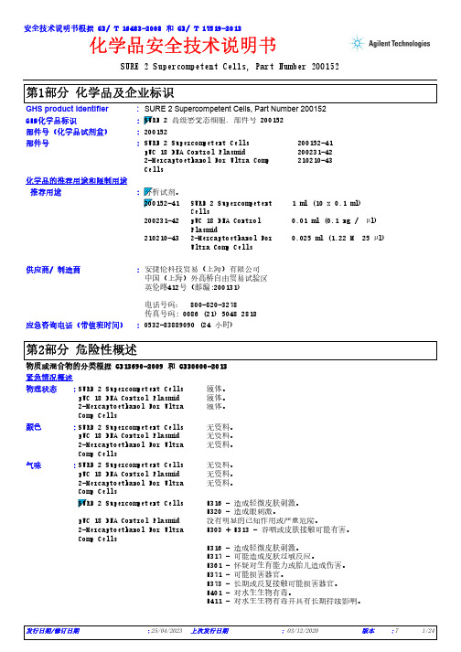
SURE 2 Supercompetent Cells, Part Number 200152*************(24小时)化学品安全技术说明书GHS product identifier 应急咨询电话(带值班时间)::供应商/ 制造商:安捷伦科技贸易(上海)有限公司中国(上海)外高桥自由贸易试验区英伦路412号(邮编:200131)电话号码: 800-820-3278传真号码: 0086 (21) 5048 2818SURE 2 Supercompetent Cells, Part Number 200152化学品的推荐用途和限制用途SURE 2 Supercompetent Cells 200152-41pUC 18 DNA Control Plasmid 200231-422-Mercaptoethanol For Ultra Comp Cells210210-43部件号:部件号(化学品试剂盒):200152安全技术说明书根据 GB/ T 16483-2008 和 GB/ T 17519-2013GHS化学品标识:推荐用途SURE 2 Supercompetent Cells1 ml (10 x 0.1 ml)200231-42pUC 18 DNA Control Plasmid0.01 ml (0.1 ng / µl)210210-432-Mercaptoethanol For Ultra Comp Cells0.025 ml (1.22 M 25 µl):物质或混合物的分类根据 GB13690-2009 和 GB30000-2013紧急情况概述SURE 2 Supercompetent Cells 液体。
pUC 18 DNA Control Plasmid 液体。
2-Mercaptoethanol For Ultra Comp Cells 液体。
SURE 2 Supercompetent Cells 无资料。
epsc英文扩展多能干细胞

epsc英文扩展多能干细胞
EPSC是英文扩展多能干细胞(Extended Pluripotent Stem Cells)的缩写,是一种新型的多能干细胞类型。
EPSC具有与传统多能干细胞(ES细胞和iPS细胞)相似的特性,但也有一些独特的特征。
EPSC可以通过特定的培养条件和信号通路来维持其多能性和自我更新能力。
EPSC的发现对干细胞研究具有重要意义。
首先,EPSC的存在拓展了我们对干细胞多能性的理解,为研究人员提供了更多的选择。
其次,EPSC具有更广泛的分化潜能,可以分化为各种不同类型的细胞,这对于再生医学和组织工程等领域具有巨大的潜力。
此外,EPSC的研究也有助于揭示干细胞的分化和自我更新机制,有助于深入了解细胞生物学和发育生物学。
EPSC的研究也面临着一些挑战和未知。
例如,如何稳定地维持EPSC的多能性和自我更新能力,以及如何准确控制其分化方向等问题都需要进一步研究。
此外,由于EPSC是相对较新的研究领域,对其在临床应用中的安全性和效果也需要进行深入的评估和研究。
总的来说,EPSC作为一种新型的多能干细胞类型,具有巨大的
潜力和重要的科学意义,但同时也需要进一步的研究和探索。
希望未来能够有更多的突破,使EPSC在干细胞研究和临床应用中发挥重要作用。
诱导型多能干细胞鉴定标准
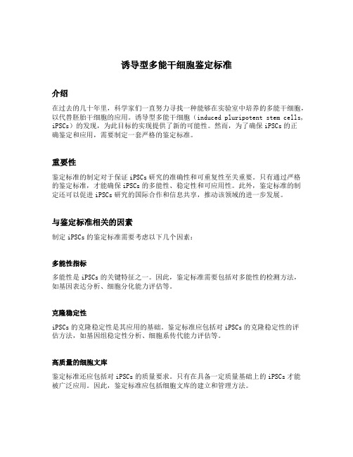
诱导型多能干细胞鉴定标准介绍在过去的几十年里,科学家们一直努力寻找一种能够在实验室中培养的多能干细胞,以代替胚胎干细胞的应用。
诱导型多能干细胞(induced pluripotent stem cells, iPSCs)的发现,为此目标的实现提供了新的可能性。
然而,为了确保iPSCs的正确鉴定和应用,需要制定一套严格的鉴定标准。
重要性鉴定标准的制定对于保证iPSCs研究的准确性和可重复性至关重要。
只有通过严格的鉴定标准,才能确保iPSCs的多能性、稳定性和可应用性。
此外,鉴定标准的制定还可以促进iPSCs研究的国际合作和信息共享,推动该领域的进一步发展。
与鉴定标准相关的因素制定iPSCs的鉴定标准需要考虑以下几个因素:多能性指标多能性是iPSCs的关键特征之一。
因此,鉴定标准需要包括对多能性的检测方法,如基因表达分析、细胞分化能力评估等。
克隆稳定性iPSCs的克隆稳定性是其应用的基础。
鉴定标准应包括对iPSCs的克隆稳定性的评估方法,如基因组稳定性分析、细胞系传代能力评估等。
高质量的细胞文库鉴定标准还应包括对iPSCs的质量要求。
只有在具备一定质量基础上的iPSCs才能被广泛应用。
因此,鉴定标准应包括细胞文库的建立和管理方法。
鉴定标准的可追溯性为了确保鉴定标准的有效性和可追溯性,需要制定明确的实验流程和数据分析方法。
此外,鉴定标准的制定还应包括数据的共享和存储规范。
诱导型多能干细胞鉴定标准的制定为了确保诱导型多能干细胞鉴定标准的全面性和有效性,应采取以下步骤:成立专家组应成立一个包括多个相关领域专家的工作组,以确保鉴定标准的全面性和权威性。
该工作组应包括基础研究、临床应用和伦理等各个方面的专家。
文献综述和经验总结工作组应对已有的文献进行综述和总结,了解当前iPSCs鉴定方法的优缺点,为鉴定标准的制定提供参考。
此外,工作组还应汇总各个实验室的经验和实践,了解当前实验室中iPSCs鉴定的常见问题和挑战。
德美研发快速自修复生物材料
德美研发快速自修复生物材料
作者:
来源:《科学大观园》2020年第17期
近日,德國和美国科学家联合研发一种生物合成蛋白材料,通过强化串联重复多肽的愈合性能,成功解决了自修复软材料目前的局限性。
该研究有望在软机器人领域获得重要应用,相关成果发表在近日的《自然·材料》杂志上。
自修复材料是一类拥有结构上具有自愈合能力的智能材料。
其灵感来自在受伤后能自我修复的生物系统。
目前,科学家合作研发一种高强度合成蛋白,可以在很短的时间内自我修复微观和宏观的机械损伤,完全恢复其结构和性能,并且具有可编程的愈合特性。
这种愈合性能为生物启发性材料设计提供了新的机会,并解决了目前用于软机器人和个人防护设备的自修复材料的局限性。
宾夕法尼亚州立大学的德米雷尔教授说,我们改变了章鱼触手蛋白质的分子结构,以便将材料的自我修复能力发挥到极致。
在自然界中,自我修复需要很长时间,例如24小时。
现在,我们将修复过程缩短到1秒钟。
马克斯·普朗克智能系统研究所的阿布顿·佩纳-弗朗切斯博士解释道,章鱼需要更长的时间才能愈合,因为其触手中的蛋白质分子只是简单地交织在一起。
而在实验室开发的材料中,改变了分子的纳米结构,使它们相互连接。
这些材料经过系统优化,具有可编程的愈合特性(愈合1秒后强度为2MPa至23MPa),愈合速度和强度均超过其他天然与合成软材料几个数量级。
◎来源|科技日报。
感受态细胞制备
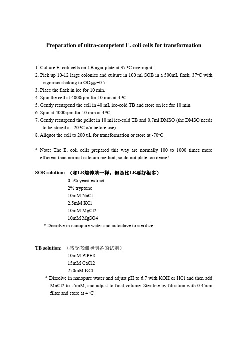
Preparation of ultra-competent E. coli cells for transformation1. Culture E. coli cells on LB agar plate at 37 o C overnight.2. Pick up 10-12 large colonies and culture in 100 ml SOB in a 500mL flask, 37o C withvigorous shaking to OD600 =0.5.3. Place the flask in ice for 10 min.4. Spin the cell at 4000rpm for 10 min at 4 o C.5. Gently resuspend the cell in 40 mL ice-cold TB and store on ice for 10 min.6. Spin at 4000rpm for 10 min at 4 o C.7. Gently resuspend the pellet in 10 ml ice-cold TB and 0.7ml DMSO (the DMSO needsto be stored at -20 o C o/n before use).8. Aliquot the cell to 200 uL for transformation or store at -70o C.* Note: The E. coli cells prepared this way are normally 100 to 1000 times more efficient than normal calcium method, so do not plate too dense!SOB solution: (和LB培养基一样,但是比LB要好很多)0.5% yeast extract2% tryptone10mM NaCl2.5mM KCl10mM MgCl210mM MgSO4* Dissolve in nanopure water and autoclave to sterilize.TB solution:(感受态细胞制备的试剂)10mM PIPES15mM CaCl2250mM KCl* Dissolve in nanopure water and adjust pH to 6.7 with KOH or HCl and then add MnCl2 to 55mM, and adjust to final volume. Sterilize by filtration with 0.45um filter and store at 4 o C。
干细胞和肿瘤干细胞
1.转化生长因子β
TGFβ(transforming growth factorβ)是TGFβ超 家族的成员之一,具有调节细胞生长和分化的 作用。 因为TGFβ能使正常成纤维细胞的表型发生转 化,即在EGF同时存在时,改变成纤维细胞贴 壁生长特性而获得在琼脂中生长的能力,并失 去密度依赖的抑制作用,故命名。
这些物质可以介导干细胞-干细胞的相互 作用,以及细胞与细胞外基质的作用,
影响干细胞的增殖和分化。
34
(一)分泌因子
分泌因子是由细胞自分泌或旁分泌的生长 因子,有的分泌因子对维持干细胞的增殖, 分化和存活具有调节作用。 如转化生长因子-β和Wnt家族的成员,在 不同组织,甚至不同种属中都发挥重要作用。
catenin与Lef/Tcf结合并入核,与DNA结合蛋白Tcf3结
合,激活c-myc、cyclinD1等基因转录,促进细胞增殖
分化。
43
如Tcf/Lef转录因子家族对上皮干细胞的 分化: Tcf/Lef与β-Catenin形成转录复合物后, 促使角质细胞转化为多能状态并分化为 毛囊。
44
五、研究干细胞的科学意义
40
(4)Wnt信号途径的作用: 在生物的正常发育中起重要作用,是组织发育、分化所必需 的关键信号通路。 (5)Wnt信号途径的作用机理: 通过TCF/LEF和β-cat对C-myc的表达进行调控,即Wnt通路的 靶基因为c-myc。
41
TCF/LEF是Wnt信号通路的中间介质,可与β-连 环 蛋 白 (β-catenin , β-cat) 结 合 成 复 合 物 , 将 βcatenin由胞浆→核内。
21
三、干细胞的分化特征
22
(一)干细胞的分化潜能
根据其分化潜能大小,干细胞可分为三类。
动物病理学概要

4. 分子病理学的发展方向
自从1953年美国学者沃森(Watson J.D.)和克 里克(Crick F.H.C.)发现了DNA分子的双螺 旋结构以后,细胞超微结构的发现吸引病理学家 将目光集中到DNA,努力探求每种疾病是否与细 胞中特定的基因表达有关,基因水平的疾病定位 诊断标准指日可待。
1.在生物医学科学研究中的“基石作用” ➢ 病理学揭示疾病规律和本质 ➢ 为防治疾病的策略、药物和疫苗的开发、新的治疗手
段的发展提供科学的理论支撑。 “如果想消灭一种疾病,透彻的理解其病理学规律是
前提和基础” 2.新动态 毒理和药理病理学 环境毒理病理学 食品、药品的安全评价 人类疾病的动物模型
病理医生称之为“doctor’s doctor”。
西方医学界流行:“最后一句话要由病理学家 来说”。
从临床兽医诊断和疾病防治的角度。
3.动物病理学是动物医学的哲学
哲学:是理论化、系统化的世界观,是自然知识、 社会知识、思维知识的概括和总结,是世界观和方 法论的统一。
病理学是理论化、系统化的疾病观。
病理学的前期课程
解剖学 组织胚胎学 生理学 生物化学 免疫学 药理学 细胞生物学、分子生物学、微生物学、寄生虫
学、传染病
病理学的技术和方法
病/尸体剖验(autopsy) 活体组织检查(biopsy) 细胞学检查(cytology) 组织、细胞、分子病理学观察 组织化学 免疫组织化学 电子显微镜技术
四体液平衡则身体健康,四体液不 调和则疾病产生。四体液之间受环 境的影响,冷热干湿条件的改变会 影响四体液平衡,使人生病。
Hippocrates
umuc3细胞形态

umuc3细胞形态
UMUC3细胞形态是一种特殊的细胞形态,它在细胞学中具有重要的意义。
UMUC3细胞是一种人类膀胱癌细胞系,可以用来研究膀胱癌的发生机制、治疗方法等。
UMUC3细胞通常呈现出长条形或椭圆形的形态,细胞体积较大,细胞质呈现出浅蓝色。
细胞核大而圆形,核质比较丰富,呈现出深蓝色。
UMUC3细胞的细胞膜完整,没有明显的突起或伸展。
UMUC3细胞在培养基中呈现出聚集生长的特点,细胞之间紧密相连,形成细胞群。
细胞群之间可以形成不规则的空隙,这些空隙可能是由于细胞的增殖和迁移造成的。
UMUC3细胞的增殖速度较快,可以快速形成细胞片和层。
UMUC3细胞在显微镜下观察,可以看到细胞内有丰富的细胞器,如线粒体、内质网和高尔基体等。
线粒体呈长条状或圆柱状,内质网呈网状分布,高尔基体则呈现为扁平或管状结构。
这些细胞器在细胞代谢、分泌和运输等方面起着重要的作用。
UMUC3细胞的形态特点与其在膀胱癌中的功能有关。
膀胱癌是一种常见的恶性肿瘤,UMUC3细胞的研究可以帮助我们更好地了解膀胱癌的发生和发展过程。
通过研究UMUC3细胞的形态变化,我们可以揭示膀胱癌细胞的特殊生理和病理特点,为膀胱癌的诊断和治疗提供新的思路和方法。
UMUC3细胞具有独特的形态特点,它是研究膀胱癌的重要模型细胞。
通过对UMUC3细胞形态的观察和分析,我们可以更好地理解膀胱癌的发生机制和治疗方法。
希望未来的研究能够深入挖掘UMUC3细胞的潜力,为膀胱癌的防治做出更大的贡献。
ucp1基因作用

ucp1基因作用
UCP1基因是一种编码蛋白质的基因,该蛋白质称为线粒体不
耦联蛋白1(uncoupling protein 1,UCP1)。
UCP1在棕色脂
肪组织中高度表达,并在体内发挥多种重要功能。
UCP1的主要作用是调节能量代谢。
它介导了非霍尔脂肪组织
的热产生(thermogenesis),即通过将食物中的化学能转化为
热量来产生热量。
这种热产生是通过线粒体不耦联呼吸的机制实现的,该机制将线粒体膜的质子梯度耦联通道分开,从而导致线粒体内质子泄漏,产生热量而不产生ATP。
UCP1的另一个作用是调节体温。
通过调节线粒体不耦联呼吸,UCP1可以增加棕色脂肪组织中的热量产生,从而提高体温。
这在动物和人类中都起着重要作用,特别是在寒冷环境下。
此外,UCP1还与体重调节和能量平衡有关。
研究表明,
UCP1参与了食物摄取和能量消耗之间的平衡。
其调节能量代
谢的能力使其成为治疗肥胖和相关代谢性疾病的潜在靶点。
总的来说,UCP1基因的作用涉及到调节能量代谢、热产生、
体温调节以及体重调节和能量平衡等方面。
- 1、下载文档前请自行甄别文档内容的完整性,平台不提供额外的编辑、内容补充、找答案等附加服务。
- 2、"仅部分预览"的文档,不可在线预览部分如存在完整性等问题,可反馈申请退款(可完整预览的文档不适用该条件!)。
- 3、如文档侵犯您的权益,请联系客服反馈,我们会尽快为您处理(人工客服工作时间:9:00-18:30)。
Inoue法制备超级感受态细胞
越干净越好,越冷越好
1.将洗干净的瓶子(500ml三角烧瓶)用Milli-Q水浸泡2-3小时,倒
去烧瓶中的水并将烧瓶倒扣在吸水纸上2-3分钟。
用Milli-Q水配置250mlLB液体培养基。
高压灭菌。
(为什么用水浸泡?)
2.准备两盒1.5ml EP管,每盒中放置两张吸水纸,共约两百个。
高压
灭菌。
3.于早晨7-8点钟小摇菌种,挑取单菌落于5ml Milli-Q水配置且灭
菌的LB液体培养基(无抗性)中,37℃培养12小时以上(OD600大于1.5)。
可以用一个新的50ml进口离心管(costa,BD等品牌都可以)来摇细菌。
4.晚上10点钟左右,按1:100大摇细菌。
超静台内吸取2.5ml小摇
细菌于高压灭菌的250ml培养基(无抗性)中,18-22℃,200rpm摇过夜。
5.第二天上午测量细菌OD600值。
Top10,Jm109等生长速度快的细菌约
在上午9点左右OD600达到0.55左右,DH5α则要到下午4-6点钟(新手可以多摇几瓶细菌,梯度接菌,如1:50,1:100,1:200等,哪瓶到了用哪瓶,其它的可以狠心倒掉)。
6.将OD600达到0.55的细菌置于冰上10min(OD600在0.4-0.8之间
都可以,对结果影响不大)。
(为什么置于冰上10min?)
7.4℃,4000rpm,10min集菌(用新的50ml 进口离心管)。
8.超静台内弃上清,将管倒扣在事先灭好的吸水纸上,巨大力向下击
打,尽可能去掉剩余LB。
9.每管倒入10ml 预冷的Inoue转化缓冲液,拧紧盖子。
在冰上来回滑
动重悬细菌(重悬的时间与向下击打的力度和来回滑动的速度相关,这两者于感受态效率没有直接关系)。
10.超静台内向每管倒入约30ml预冷的Inoue转化缓冲液,来回颠倒
混匀。
11.4℃,4000rpm,10min集菌。
12.超静台内弃上清,将管倒扣在事先灭好的吸水纸上(换一张吧,别
用刚才那张),巨大力向下击打,尽可能去掉剩余缓冲液。
13.每管倒入10ml预冷的Inoue转化缓冲液,拧紧盖子。
在冰上来回
滑动重悬细菌。
14.超静台内向每管加入750μlDMSO。
轻轻来回混匀(记住要轻轻)。
置于冰上10min。
15.将冰浴的感受态细胞分装到事先灭菌后并放到-20或4℃的EP管
中,一个一个丢到液氮罐里,然后冻到-80(分装的体积是爱分多少分多少,建议100μl每管,传说液氮速冻可以提高5倍的感受态效率)。
16.按人品的好坏,感受态效率在108-107不等。
溶液配置:
0.5M PIPES 100ml
PIPES 15.1g
用5M KOH调pH到6.7,然后用0.45μm的滤膜过滤除菌。
(PIPES似乎有PIPES酸和PIPES盐之分,如果你配好溶液后,测量的pH大于6.7,千万不要惊慌,可以用HCl调回到6.7,不影响结果的)
Inoue转化缓冲液1000ml
MnCl2.4H2O 10.88g
CaCl2.2H2O 2.2g
KCl 18.65g
PIPES,0.5M pH6.7 10ml
H2O 补至1000ml
用0.45μm的滤膜过滤除菌。
