ESO Imaging Survey Optical follow-up of 12 selected XMM-Newton fields
ZEISS Axio Imager Light Manager手册说明书
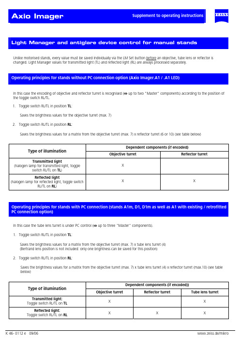
Unlike motorised stands, every value must be saved individually via the LM Set button before an objective, tube lens or reflector ischanged. Light Manager values for transmitted light (TL) and reflected light (RL) are always processed separately.In this case the encoding of objective and reflector turret is recognised (⇒ up to two "Master" components) according to the position ofthe toggle switch RL/TL.1. Toggle switch RL/TL in position TL:Saves the brightness values for the objective turret (max. 7)2. Toggle switch RL/TL in position RL:Saves the brightness values for a matrix from the objective turret (max. 7) x reflector turret (6 or 10) (see table below)Dependent components (if encoded)Type of illuminationObjective turret Reflector turretTransmitted lightX (halogen lamp for transmitted light, toggleswitch RL/TL on TL)Reflected lightX X (halogen lamp for reflected light, toggle switchRL/TL on RL)In this case the tube lens turret is under PC control (⇒ up to three "Master" components).1. Toggle switch RL/TL in position TL:Saves the brightness values for a matrix from the objective turret (max. 7) x tube lens turret (4)(Bertrand lens position is not included: only one brightness can be saved for this position)2. Toggle switch RL/TL in position RL:Saves the brightness values for a matrix from the objective turret (max. 7) x tube lens turret (4) x reflector turret (max.10) (see tablebelow)Dependent components (if encoded))Type of illuminationObjective turret Reflector turret Tube lens turretTransmitted light:X X Toggle switch RL/TL on TLReflected light:X X X Toggle switch RL/TL on RLDefault setting of the stand after switching on:Transmitted light:• Toggle switch RL/TL on TLButton TL on (shutter open or lamp on)Button RL offReflected light:• Toggle switch RL/TL on RLButton TL offButton RL on (shutter open or lamp on)Saving LM value:• To save the current lamp voltage for the current objective turret position press the LM Set button brieflySaving 3200K:This function determines whether the stand is set at 3200K when it is switched on.• To set 3200K to be active on switching on: activate 3200K and press LM Set button.• To set 3200K to be inactive on switching on: deactivate 3200K and press LM Set button.The 3200K setting is saved globally and is independent of other LM values that have already been saved. The normal LM values are available at any time as soon as 3200K is deactivated.Overwriting the LM values:• To save the new value at the relevant position press the LM Set buttonDeleting of the LM values:This is not possible.Activating an LM value:This is done by switching on and changing the position of a "Master" component.To permanently deactivate/activate Light Manager (LM) & antiglare device (AG)• Keep the "RL" button pressed down when you switch on:• One beep signifies deactivation.• Two beeps signify activation.To permanently deactivate/ activate Light Manager only• Keep "3200"button pressed down when you switch on:One beep signifies deactivation. Two beeps signify activation.To permanently deactivate/ activate antiglare device only• Keep “TL” button pressed down when you switch on:One beep signifies deactivation. Two beeps signify activation• If button "RL" is pressed when you switch on and only one of the two functions is activated, that function will be deactivated:Starting condition OutcomeLM AG LM AG⇒0 01 1⇒0 01 0⇒0 00 1⇒ 1 10 0These parameters can also be set via MTB 2004 for motorised stands.Antiglare device:If there is a shutter in the TL optical path, the lamp voltage remains constant when the objective is changed and the shutter takes over thefunction of the antiglare device.If no shutter is present, the lamp is switched off.Safety function:If the reflector turret flap is opened or the reflector turret is completely removed, the safety switch-off device automatically closes thereflected light shutter. In addition, the shutter can no longer be opened by pressing a button as long as the reflected light path is "open".The shutter also closes automatically when the stand is switched off.The brightness of the Light Control LEDs can be adjusted by the user.Manual stands:• Keep SET button pressed down for about 3 seconds until a long beep is heard.All LEDs go on.The brightness of the LEDs can now be adjusted by the brightness control (control knob).However, the brightness cannot be completely extinguished!Activating the control knob in this mode has no effect on the lamp voltage!This mode is exited automatically by releasing the LM Set buttonThe setting is saved permanently!Motorised stands:The adjustment of the LED brightness is linked with the brightness control of the TFT Display.All motorised reflector turrets can be mounted on the stands D1 and D1m. The reflector turrets for the D1 stands have been incorporated into the MTB 2004 in the same way as those for the motorised stands.The motorised reflector turrets can be operated either by the AxioVision Software or by the keyring in Z drive. If a motorised reflector turret is recognised when the microscope is switched, keys are assigned automatically in the following order:Refl.turret to the right (Pos. +), Refl.turret to the left (Pos. -), RL shutter, lamp voltage +, lamp voltage -.Otherwise the default assignment of keys applies:TL shutter, RL shutter, unassigned, lamp voltage +, lamp voltage -.The position indication is shown by the LED Bargraph. As soon as a motorised reflector turret is recognised, the LED Bargraph indicates the reflector turret position, if ”RL on” was set (by pressing button on the Light Control or on the keyring or via Software). If TL is switched on as well (only possible if the toggle switch HAL is on TL), the reflector position will continue to be displayed, overriding the display of the lamp brightness.。
TG-320 数码相机规格说明书
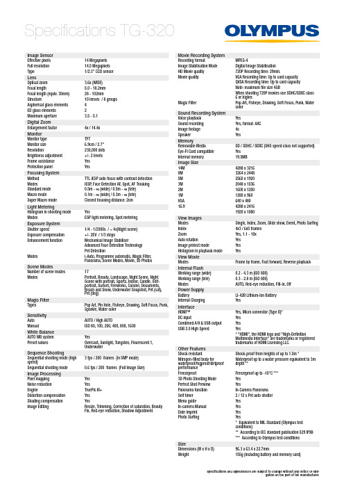
Specifications TG-320Specifications and appearances are subject to change without any notice or obli-gation on the part of the manufacturer.Image Sensor Effective pixels 14 Megapixels Full resolution 14.5 Megapixels Type1/2.3'' CCD sensor LensOptical zoom 3.6x (WIDE)Focal length5.0 - 18.2mm Focal length (equiv. 35mm)28 - 102mmStructure10 lenses / 8 groups Aspherical glass elements 4ED glass elements 2Maximum aperture 3.5 - 5.1Digital Zoom Enlargement factor 4x / 14.4x Monitor Monitor type TFTMonitor size 6.9cm / 2.7''Resolution230,000 dots Brightness adjustment +/- 2 levels Frame assistance Yes Protection panel YesFocusing System Method TTL iESP auto focus with contrast detection ModesiESP , Face Detection AF, Spot, AF Tracking Standard mode 0.5m - ∞ (wide) / 0.5m - ∞ (tele)Macro mode0.1m - ∞ (wide) / 0.3m - ∞ (tele)Super Macro mode Closest focusing distance: 2cm Light MeteringHistogram in shooting mode YesModesESP light metering, Spot metering Exposure System Shutter speed1/4 - 1/2000s / < 4s(Night scene)Exposure compensation +/- 2EV / 1/3 stepsEnhancement functionMechanical Image StabiliserAdvanced Face Detection Technology Pet DetectionModesi-Auto, Programme automatic, Magic Filter, Panorama, Scene Modes, Movie, 3D Photos Scene ModesNumber of scene modes 17ModesPortrait, Beauty, Landscape, Night Scene, Night Scene with portrait, Sports, Indoor, Candle, Self-portrait, Sunset, Fireworks, Cuisine, Documents, Beach and Snow, Underwater Snapshot, Pet (cat), Pet (dog)Magic Filter Types Pop Art, Pin Hole, Fisheye, Drawing, Soft Focus, Punk, Sparkle, Water colorSensitivity Auto AUTO / High AUTOManual ISO 80, 100, 200, 400, 800, 1600White Balance AUTO WB system YesPreset valuesOvercast, Sunlight, Tungsten, Flourescent 1, UnderwaterSequence ShootingSequential shooting mode (high speed)3 fps / 200 frames (in 5MP mode)Sequential shooting mode 0.6 fps / 200 frames (Full Image Size)Image Processing Pixel mapping Yes Noise reduction YesEngineTruePic III+ Distortion compensation Yes Shading compensation YesImage EditingResize, Trimming, Correction of saturation, Beauty Fix, Red-eye reduction, Shadow AdjustmentMovie Recording System Recording formatMPEG-4Image Stabilisation Mode Digital Image Stabilisation HD Movie quality 720P Recording time: 29min.Movie qualityVGA Recording time: Up to card capacity QVGA Recording time: Up to card capacity Note: maximum file size 4GBWhen shooting 720P movies use SDHC/SDXC class 6 or higher.Magic FilterPop Art, Fisheye, Drawing, Soft Focus, Punk, Water colorSound Recording System Voice playback YesSound recording Yes, format: AAC Image footage 4s SpeakerYesMemoryRemovable MediaSD / SDHC / SDXC (UHS speed class not supported)Eye-Fi Card compatible Yes Internal memory 19.5MB Image Size 14M 4288 x 32168M 3264 x 24485M 2560 x 19203M 2048 x 15362M 1600 x 12001M 1280 x 960VGA 640 x 48016:94288 x 24161920 x 1080View Images Modes Single, Index, Zoom, Slide show, Event, Photo Surfing Index 4x3 / 6x5 frames ZoomYes, 1.1 - 10x Auto rotationYes Image protect modeYes Histogram in playback mode YesView Movie ModesFrame by frame, Fast forward, Reverse playback Internal Flash Working range (wide)0.2 - 4.3 m (ISO 800) Working range (tele)0.3 - 2.8 m (ISO 800)ModesAUTO, Red-eye reduction, Fill-in, Off Power Supply BatteryLI-42B Lithium-Ion Battery Internal ChargingYesInterface HDMI™Yes, Micro connector (Type D)*DC inputYes Combined A/V & USB output Yes USB 2.0 High SpeedYes* "HDMI", the HDMI logo and "High-DefinitionMultimedia Interface" are trademarks or registered trademarks of HDMI Licensing LLC.Other Features Shock resistantShock-proof from heights of up to 1.5m *Nitrogen-filled body forwaterproof/fogproof/dirtproof performance Waterproof up to a water pressure equivalent to 3m depth **FreezeproofFreezeproof up to -10°C ***3D Photo Shooting Mode Yes Perfect Shot Preview YesPanorama function In-Camera Panorama Self timer 2 / 12 s Pet auto shutter Menu guideYes In-camera Manual Yes Date imprint Yes Photo SurfingYes* Equivalent to MIL Standard (Olympus test conditions)** According to IEC standard publication 529 IPX8*** According to Olympus test conditions SizeDimensions (W x H x D)96.3 x 63.4 x 22.7mmWeight155g (including battery and memory card)。
自动光学检查(AOI)
技术进步:更高精度、更快速 度、更智能的OI设备
应用领域:拓展到更多行业, 如半导体、电子、汽车等
市场竞争:国内外企业竞争加 剧,推动技术升级和创新
环保要求:满足绿色制造和可 持续发展的要求,降低能耗和 污染
感谢您的观看
汇报人:XX
OI可以大大提高生产效率,减少人 工检查的错误率。
添加标题
添加标题
添加标题
添加标题
OI主要用于检测电子产品的表面缺 陷、焊点质量、元件放置等。
OI广泛应用于电子制造行业,如 PCB、SMT等领域。
自动光学检查 (OI)是一种 通过光学手段 对电子元件进 行自动检查的
技术。
OI系统通常包 括一个摄像头, 一个光源和一 个图像处理系
统。
工作原理:摄像 头拍摄电子元件 的图像,光源提 供均匀的照明, 图像处理系统分 析图像,检测出
缺陷和异常。
OI的优点:速 度快,准确度 高,可以检测 出人工检查难 以发现的缺陷。
电子行业:用于检测电路板、半导 体等电子元件的缺陷和故障
航空航天行业:用于检测机、卫 星等航空航天设备的缺陷和故障
准备阶段:确定检测目标和标准, 准备检测设备和工具
分析阶段:对检测数据进行分析和 处理,找出存在的问题和缺陷
添加标题
添加标题
添加标题
添加标题
检测阶段:按照预定的流程和标准 进行检测,获取检测数据
报告阶段:将检测结果和分析结果 整理成报告,提供给相关人员和部 门
检测结果分为合格和不合格两种
不合格结果表示产品不符合标准要 求,需要返修或报废
添加标题
添加标题
添加标题
添加标题
合格结果表示产品符合标准要求, 可以继续生产
相机术语中英文对照表
86 87 88 89 90 91 92 93 94 95 96 97 98 99 100 101 102 103 104 105 106 107
108 109 110 111 112 113 114 115 116 117 118 119 120 121 122 123 124 125 126 127 128
照相机专用术语
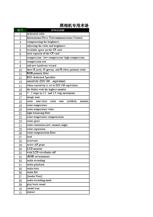
编号 1 2 3 4 5 6 7 8 9 10 11 12 13 14 15 16 17 18 19 20 21 22 23 24 25 26 27 28 29 30 31 32 33 34 35 36 37 38 39 40 41
ENGLISH dedicated cable International Press Telecommunications Council compensating the brightness adjusting the color and brightness available space on the CF card total capacity of the CF card compression / low compression/ high compression compression rate add new hardware wizard three R (red), G (green), and B (blue) primary color RGB primary filter EOS-dedicated Speedlite sensitivity (ISO 100…equivalent) when sensitivity is set to ISO 100 equivalent the folder with the highest number +/- 2 stops in 1/2- and 1/3-stop increments image zone color tone/clear color tone (reddish, normal, yellowish) color temperature color temperature value light balancing filter color temperature compensation color space color saturation (low, normal, high) color separation color compensation filter load overwrite active AF point LCD monitor with LCD viewfinder off sRGB environments audio recording audio playback audio data audio file [Audio Note] audio recording mark play back sound sound icon extract
超声造影剂Sonazoid(示卓安)用于肝脏疾病进展

Application progresses of ultrasound contrast agentSonazoid in liver diseasesZHANG Zheyuan, ZHANG Huabin, BAI Zhiyong*(Department of Ultrasound, Beijing Tsinghua Changgung Hospital, School ofClinical Medicine, Tsinghua University, Beijing 102218, China)[Abstract]With the rapid development of contrast-enhanced ultrasound (CEUS),Sonazoid,a new generation of ultrasound microbubbles contrast agent came into being.The unique Kupffer phase of Sonazoid could greatly prolong the intrahepatic developing time,hence providing more valuable information for diagnosis,treatment and follow-up of liver diseases. The progresses of Sonazoid applicated in liver diseases were reviewed in this article.[Keywords]liver; contrast media; ultrasonographyDOI:10.13929/j.issn.1672-8475.2024.02.010超声造影剂Sonazoid(示卓安)用于肝脏疾病进展张哲元,张华斌,白志勇*(清华大学附属北京清华长庚医院超声科清华大学临床医学院,北京 102218)[摘要]随着超声造影技术迅速发展,新一代超声微泡造影剂——Sonazoid(示卓安)应运而生,其特有的Kupffer相可极大地延长肝内显影时间,为诊断、治疗及随访肝脏疾病提供更多有价值的信息。
Optical Imaging
ACS, F814W - well sampled (0.025” pixels)
WFPC2, F300W - highly undersampled (0.1” pixels)
27th August 2008 NEON Archive School, Garching 8
From Optics to the Point Spread Function
s(r) = C / (1+r2/R2)b+ B
Where there are two free parameters (apart from intensity, background and position) R, the width of the PSF and b, the Moffat parameter. Software is available to fit PSFs of this form.
I is the result of sampling the continuous distribution resulting from the convolutions at the centre of a pixel and digitising the result into DN.
27th August 2008 NEON Archive School, Garching 5
http:/instruments/TinyTim/
27th August 2008 NEON Archive School, Garching 10
Simple Measures of Optical Image Quality
• Full Width at Half Maximum (FWHM) of point-spread function (PSF) - measured by simple profile fitting (eg, imexam in IRAF) • Strehl ratio (ratio of PSF peak to theoretical perfect value). • Encircled energy - fraction of total flux in PSF which falls within a given radius.
光散射学报第32卷,第1
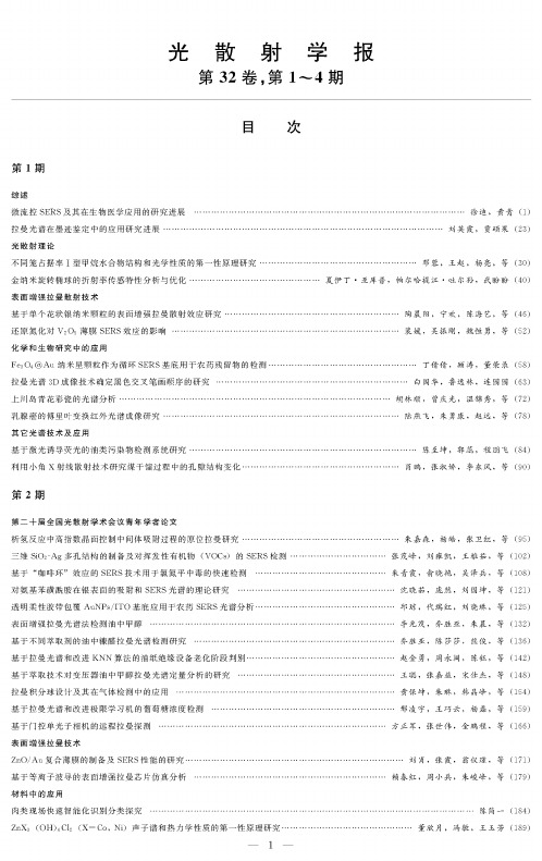
光散射学报第32卷,第#〜4期目次第1期综述微流控SERS及其在生物医学应用的研究进展..................拉曼光谱在墨迹鉴定中的应用研究进展..........................光散射理论不同笼占据率I型甲烷水合物结构和光学性质的第一性原理研究…金纳米旋转椭球的折射率传感特性分析与优化...................表面增强拉曼散射技术基于单个花状银纳米颗粒的表面增强拉曼散射效应研究..........还原氮化对V2O5薄膜SERS效应的影响........................化学和生物研究中的应用Fe3O4@Au纳米星颗粒作为循环SERS基底用于农药残留物的检测拉曼光谱&D成像技术确定黑色交叉笔画顺序的研究............上川岛青花彩瓷的光谱分析.....................................乳腺癌的傅里叶变换红外光谱成像研究..........................其它光谱技术及应用基于激光诱导荧光的油类污染物检测系统研究...................利用小角X射线散射技术研究煤干幅过程中的孔隙结构变化.........................................徐迪,黄青⑴..............................刘英霞!贾硕果(23).......................邓蓉!王赵!杨亮!等(30)夏伊丁-亚库普,帕尔哈提江-吐尔孙,武盼盼(40)...................陶晨阳,宁欢,陈海艺,等(46)...................裴媛!吴振刚,魏恒勇!等(52).......................丁倩倩!顾涛!董荣录(51).....................白国华,鲁逸林,连园园(63)................胡林顺,曾庆光,温锦秀,等(72)...................陆燕飞!朱勇康!赵远!等(71).......................陈至坤,郭蕊,程朋飞(14)...................肖鹏!张淑娇,李东风!等(50)第2期第二十届全国光散射学术会议青年学者论文析应中面控中的原拉曼研究……………………………………………三维SiO2-Ag多孔结构的制备及对挥发性有机物(VOC:)的SERS检测........................基于“咖啡环$效应的SERS技术用于氯氮平中毒的快速检测.................................对氨基苯磺酰胺在银表面的吸附和SERS光谱的理论研究...................................... 透明柔性胶带包覆AuNPs/ITO基底应用于农药SERS光谱分析.................................表面增强拉曼光谱法检测油中甲醇............................................................基于不同萃取剂的油中糠醛拉曼光谱检测研究.................................................基于拉曼光谱和改进KNN算法的油纸绝缘设备老化阶段判别...................................基于萃取技术对变压器油中甲醇拉曼光谱定量分析的研究...................................... 拉曼分球及其在中的应用………………………………………………………………基于拉曼光谱和改进极限学习机的葡萄糖浓度检测............................................基于门控单光子相机的远程拉曼探测..........................................................表面增强拉曼技术ZnO/Au复合薄膜的制备及SERS性能的研究...................................................基于等离子波导的表面增强拉曼芯片仿真分析.................................................…朱嘉森,杨皓,张卫红,等(95)张茂峰,刘雍凯,王雅茹,等(102)朱青霞,,吴,等101,,刘国坤,等121 -■邱琼,代瑞红,刘晓琳,等125 -■李光茂,乔胜亚,朱晨,等132 -■乔胜亚,陈莎莎,熊俊,等136 -■赵金勇,周永阔,陈,等142王,张嘉,,等141 -■黄保坤,朱琳,,等154,王,杨,等159,张,金鹏程,等166••…刘肖,张霞,,等171,,朱,等179材料中的应用肉类现场快速智能化识别分类探究陈简一(114)ZnX&(OB))C12(X=Co,Ni)声子谱和热力学性质的第一性原理研究.................................董欣月,冯敏,王玉芳(119)第3期综述光声成像技术表面增强拉曼散射技术基于拉曼光谱相似度比对的油纸绝缘老化阶段诊断花粉的表面增强拉曼光谱鉴别研究Au@PS阵列SERS基底的特性研究表面增强拉曼光谱对抗肿瘤药物5-氟尿的检测研究蒋文萍,吴其鑫,,等(195%张腾翼,古亮,陈等(202…符致秋,刘刚,,等(210裴君妍,徐宗伟,王,等(217周国良,黄光耀,李盼,等(224化学和生物研究中的应用取代基对观es o-四苯基CD几何结构,红外拉曼振动频率及电子光谱的影响的理论研究张坚,李秀(230) PX装置吸附塔进料非芳含量的拉曼定量分析蒋飘逸,戴连奎(237)光散射理论基于光散射理论的玻璃晶圆表面缺陷检测方法研究涂政乾,董立超,赵东峰,等(245)对称性破缺纳米核壳二聚体的近场特性研究姜继玉,姜莎莎,吕靖薇,等(251)氮化_低温高压光谱研究范春梅,刘静仪,刘珊,等(259)材料中的应用嵌入金属纳米颗粒提高晶硅薄膜太阳能电池吸收率肖亮,朱群志(266)其它光谱技术及应用一种新的布里渊光时域定位技术蒋超(274)光谱法在唐卡蓝、绿色颜料分析中的应用张蕊,方小济,巨建伟(280)基于激光拉曼光谱快速无损检测牛油果油的研究张凤娟,黄敏,刘振方(288)第4期综述气溶胶单颗粒的拉曼测量方法,张(295)表面增强拉曼散射技术基于等离子波导的表面增强拉曼芯片仿真分析赖春红,周小兵,朱峻峰,等(301)单链DNA表面增强拉曼散射检测条件的优化蒋承顺,李旺,柳艳,等(306)基于SERS阵列的铜绿假单胞菌代谢产物铜绿菌素即时检测陈燕,陈欢,陈周恬,等(312)三氧化鸭超薄纳米片SERS活性基底的制备与性能研究孙宗杰,林东岳,何遥,等(320)材料中的应用小角X射线散射法研究枣树的微孔结构武海娟,翟红生,杨春明,等(328)化学和生物研究中的应用基于等离子体四极共振的金纳米长方体颗粒的折射率传感性能研究刘静雅,张现周(335)胆酸钠自组装行为的小角X射线散射研究李博楠,李天富,刘荣灯,等(343)金纳米颗粒在全血环境中近场增强特性李俊平,陈娜,刘书朋,等(348)仪器和方法学消光光谱颗粒粒径测量方法影响因素实验研究杨斌,赵蓉,王继,等(355)自组装纳米球热压印制备金属纳米盘阵列的研究曹燕,陈溢杭(361)其它光谱技术及应用江苏仪征刘集联营西汉墓岀土彩绘陶俑颜料分析范陶峰(369)陕西绥德县博物馆馆藏青铜甑锈蚀物的拉曼光谱分析付倩丽,康卫东,张尚欣,等(375)原位ATR-IR光谱研究钳酸钠光催化全分解水机理丁倩,陈涛,冯兆池,等(381) L-半胱氨酸功能化的碳量子点为探针快速检测花旗松素程家维,张宇辉,杨季冬(386)THE JOURNAL OF LIGHT SCATTERINGVol.32No.#〜4CONTENTSNo.1OverviewMicrofluidic SERS form and its Biomedical Applications..................................................................................................XU Di&HUANG Qing(1) Research Progress and Application of Raman Spectroscopy on the Identification of Ink Marks................LIU Yingxia,51A Shuoguo(23) Theories of Light ScatteringStudies on the Structure and Optical Properties of Different Cage Occupancy on si Methane Hydrate by First-principle ............................................................................................................................................DENGRong,WANG Zhao&YANG Liang&etal(30) Analysis and Optimization of Refractive Index Sensing of Gold Nanospheroid.YAKUPU Xiayiding,TUERSUN Paerhatijiang,WU Panpan(40)Surface-Enhanced Raman Scattering(SERS)Surface-enhanced Raman Scattering Study of Individual Flower-like Silver Nanoparticles....................................................................................................................................TAO Chenyang,NING Huan,CHEN Haiyi,et al(46) The Impact of Reduction Ntridation on SERS Effect of V2O5Film....................PEI Yuan,WU Zhengang,WEI Hengyong,et al(52) Application in Chemistry and Biology ResearchesFe3O4@Au nanostar as reproducible SERS substrates for the detection of pesticide residues.....................................................................................................................................................DING Qianqian&GU Tao&DONG Ronglu(58) Study of Raman Spectroscopy3D Profilometry to Determine the Sequence of Black Pen Ink Crossings.....................................................................................................................................................BAI Guohua,LU Yilin,LIAN Yuanyuan(63) Spectral Analysis of Blue-and-Whte porcelain on Shangchuan Island............HU Linshun&ZENG Qingguang&WEN Jiniu&et al(72) Study on Breast Cancer by Fourier Transform Infrared Spectroscopy Imaging••-LU Yanfei,ZHU Yongkang,ZHAO Yuan,et al(78) Other Optical Spectroscopic Techniques and ApplicationsResearch on Oil Contaminant Detection System Based on Laser Induced Fluorescence.................................................................................................................................................CHEN Zhikun&GUO Rui&CHENG Pengfei(84) Study on Pore Structural Changes of Coal Carbonization by Small Angle X-ray Scattering Technology...................................XIAO Peng,ZHANG Shujiao&LI Dongfeng,et al(90)No.2Papers by young scholars at the20th National Conference on light scattering=nsituRamanEvidenceof=ntermediatesAdsorptionEngineeringby High-=ndexFacetsControlduring HydrogenEvolutionReaction ....................................................................................................................................ZHU Jia s en,YANG Hao,ZHANG Weihong,et al(95) PreparationofThree-Dimensional(3D)SiO2-AgPorousStructureandSERSDetectionofVolatileOrganicCompounds(VOCs) ...............................................................................................................................ZHANG Mao f e ng&LIU Yongkai&WANG Yaru&et al(102) Antipsychoticdrugpoisoning monitoringofclozapineinurinebyusingco f eeringe f ectbasedsurface-enhancedRamanspectroscopy ............................................................................................................................................ZHU Qingxia&YU Xiaoyan&WU Ze'bing&et al(108) TheoreticalStudyofAdsorptionandSERSSpectraofSulfanilamideonSilverSurfaces........................................................................................................................................SHEN Xiaoru&PANG Ran&LIU Guokun&et al(121) Transparent flexible tape coated AuNPs/ITO substrate for pesticide SERS spectral analysisQIUQiong,DAIRuihong,LIU Xiaolin,tal(125) Detection of Methanol in Oil by Surface-enhanced Raman Spectroscopy...........LI Guangmao,QIAO Shengya,ZHU Chen,et al(132) RamanSpectroscopicDetectionofFurfuralinOilBasedonDi f erentExtractants....................................................................................................................................QIAO Shengya,CHEN Shasha,XIONG Jun,et al(136) Aging Stage Discrimination of Oil-Paper Insulation Equipment Based on Raman Spectrum and Improved KNN AlgorthmsZHAO Jinyong&ZHOU Yongkuo&CHEN Yu&tal(142) Study on Quanttative Analysis Method of Methanol Raman Spectra in OilBy Extraction TechnologyWANG Cong&ZHANGJiayi&SONG Shiji&tal(148)TheDesignofRamanIntegratingSphereandtheApplicationofDetectingGasesHUANGBaokun&ZHULin&HANJingf ng&tal(154)GlucoseconcentrationdetectionbasedonRamanspectroscopyandimprovedExtremeLearning MachineXING Lingyu&WANG Qiaoyun&YANG L i&tal(159) Remote Raman Detection Based on Gated-Single-Photon Camera......FANG Zhengjun&ZHANG ShPei&Jin Pengcheng&et al(166) Surface-EnhancedRamanSca t ering(SERS)Fabricationand SERS Properties Studies of ZnO/Au Composite film..........................LIU Xiao&ZHANG Xia&WENG Yijin&et al(171) Simulation AnalysisoJSurJace-enhancedRamanSca t eringChipBasedonthePlasma WaveguideI Chunhong,ZHOU Xiaobin,ZHU Jun f e ng,et al(179)FastInte l igentIdentiicationandExplorationoJMeatOnSite..........................................................................................................CHENJianyi(184)OtherOpticalSpectroscopicTechniquesandApplicationsFirst-principles Study of Phonon Spectra and Thermodynamic Properties of Z11X3(OB))C12(X=Co,Ni).................................................................................................................................................DONG Xinyue,FENG Min&WANG Yufang(119) No.3OverviewPhotoacoustic imaging................................................................................................................J1ANG Wenping&WU Qixin&MIN Jun&e=al(195) Surface-Enhanced Raman Scattering(SERS)Aging Phase Diagnosis of Oil Paper Insulation Based on Raman Spectral Similarty Ratio...............................................................................................................................ZHANG Tengyi,GU Liang,CHEN Xingang,et al(202) Discrimination of Pollen by Surface Enhanced Raman Spectroscopy......................................FUZhiqiu&LIU Gang&AN Ran&et al(210) Study on SERS substrate properties ofAu@PS arrays................................................................................PEI Junyan&XUZongwei,et al(217) Detection of5-Fluorouracil by Surface Enhanced Raman Spectroscopy ZHOU Guoliang,HUANG Guangyao&LI Pan,et al(224) Application in Chemistry and Biology ResearchesInfluences by substtuents on the geometrical structure&infrared and raman vibration frequency and electronic spectrum forme s o-tetraphenylporphyrin:A theoretical study................................................................................................................ZHANG Jian,Li Xiu(230) Quantitative Analysis for Non-aromatic Hydrocarbon in the Feedstock of an Adsorption Tower in a P-Xylene Unit Based on Raman Spectroscopy....................................................................................................................................................................................JIANG Piaoyi,DAI Liankui(237) Theories of Light ScatteringResearch on Surface Defect Detection Methodof Glass Wafer Based on Light Scattering Theory...............................................................................................................................Tu Zhengqian&Dong Lichao,Zhao Dongfeng&et al(245) ResearchonNearFieldCharacteristicsofSymmetryBreaking multilayerednanoshe l sdimer....................................................................................................................................JIANG Jiyu,JIANG Shasha,LV Jingwei,et al(251) Low-temperature and high-pressure Spectroscopy study of Gallium N t ride............FAN Chunme i,LIU Jingyi,LIU Shan,e t al(259) Other Optical Spectroscopic Techniques and ApplicationsEmbedding Metal NanEparticles tEIncrease the AbsErptiEn RateEf Crysta l ine SilicEn Thin Film SElar Ce l s...............................................................................................................................................................................XIAO Liang,ZHU Qunzhi(266) OtherOpticalSpectroscopicTechniquesandApplicationsA new Brillouin optical time domain localization technique................................................................................................................JIANG Chao(274) ApplicaNionofSpecNromeNryon AnalysisofBlueandGreenPigmenNsinNheThangka.....................................................................................................................................................ZHANG Rui,FANG Xiaoji,JU Jianwei(210) SNudyonRapid Non-desNrucNiveTesNingofAvocadoOilBasedonLaserRamanSpecNroscopy....................................................................................................................................ZHANG Fengjuan&HUANG Min&LIUZhenfang(211) No.4OverviewRaman Spectroscopy Measurement for Aerosol Single Particle.....................................................CHANG Pianpian&ZHANG Yunhong(295) Surface-EnhancedRamanSca t ering(SERS"Simulation AnalysisoJSurJace-enhancedRamanSca t eringChipBasedonthePlasma WaveguideI Chunhong&ZHOU Xiaobin&ZHU Jun f e ng&et al(301) OptimizationoJsurJaceenhancedRamansca t eringdetectionconditionsJorsingle-strandedDNAJIANGCh ngshun,LIWang,LIUYan,tal(306" SERS array toward point-of-care detection of Pseudomonas aeruginosa metabolteCHEN Yan,CHEN Huan,CHEN Zhoutian,tal(312" Preparation of Tungsten Trioxide Ultrathin Nanosheets for SERS Active Substrate and Its Performance............................................................................................................................................SUN Zongjie,LIN Dongyue,HE Yao,et al(320) OtherOpticalSpectroscopicTechniquesandApplicationsSAXS Study on the Micropore Structure of Jujube...................................WU Haijuan,ZHAI Hongsheng,YANG Chunming,et al(321) ApplicationinChemistryandBiologyResearchesStudyEftheRefractiveIndexSensingPerfErmancesEfAu NanEcubEidParticlesBasedEnthePlasmEnicQuadrupEleResEnance ......................................................................................................................................................................LIU Jingya&ZHANG Xianzhou(335) StudyEnself-assemblybehaviErEfsEdiu34)Near-fieldEnhancementCharacteristicsEfGEld NanEparticlesin WhEleBlEEdEnvirEnmentLI Junping&CHEN Na&LIUShup ng&tal(341) Instruments and MethodsExperimentalResearchEnInfluenceFactErsEfParticleSize MeasurementMethEdbasedEnExtinctiEnSpectrEscEpy .................................................................................................................................................YANG Bin&ZHAORong,WANG Ji,e t al(355) StudyEnfabricatiEnEfMeta l icNanEdisk ArraysbyhEtpressingself-assemblednanEspheres CAOyan&CHENyihang(361) OtherOpticalSpectroscopicTechniquesandApplicationsPigment analysis of painted terracotta unearthed from the Western Han Dynasty Tomb at Lianying&Liuji in Yizheng&Jiangsu Province ........................................................................................................................................................................................................FAN Taof e ng(369) Raman Spectrum Analysis of Corrosion Products on the Bronze Bu in Suide Museum in Shaanxi Province...........................................................................................................................FU Qianii,KANG Weidong,ZHANG Shangxin,et al(375) Mechanistic Study of Photocatalytic Overall Water Splitting on NaTaOa-based Photocatalysts by In S iu ATR-IR SpectroscopyDingQian,Ch nTao,F ngZhaochi,tal(311) Rap6dDetect6onofTaxfolnbyL-cyste6neFunct6onalzedCarbon QuantumDotsCHENG Jiawei&ZHANG Yuhui&YANG Jidong(316)。
眼科英文单词
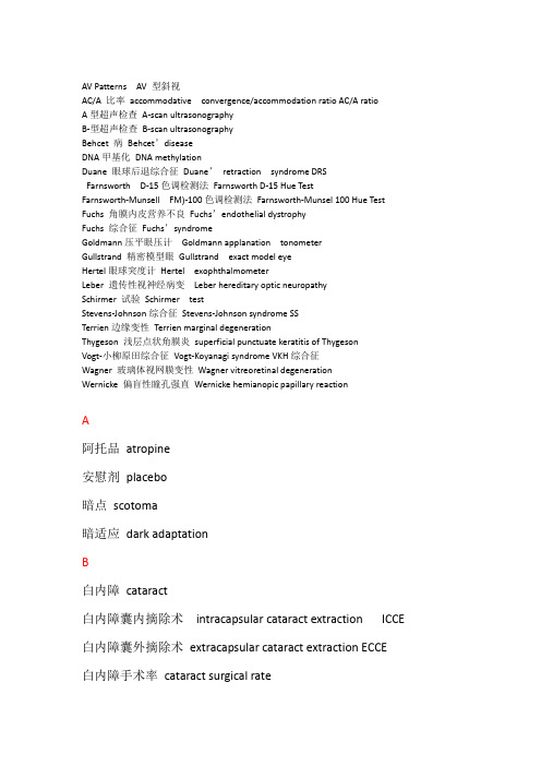
AV Patterns AV 型斜视AC/A 比率accommodative convergence/accommodation ratio AC/A ratioA型超声检查A-scan ultrasonographyB-型超声检查B-scan ultrasonographyBehcet 病Behcet’diseaseDNA甲基化DNA methylationDuane 眼球后退综合征Duane’retraction syndrome DRSFarnsworth D-15色调检测法Farnsworth D-15 Hue TestFarnsworth-Munsell FM)-100色调检测法Farnsworth-Munsel 100 Hue Test Fuchs 角膜内皮营养不良Fuchs’endothelial dystrophyFuchs 综合征Fuchs’syndromeGoldmann压平眼压计Goldmann applanation tonometerGullstrand 精密模型眼Gullstrand exact model eyeHertel眼球突度计Hertel exophthalmometerLeber 遗传性视神经病变Leber hereditary optic neuropathySchirmer 试验Schirmer testStevens-Johnson综合征Stevens-Johnson syndrome SSTerrien边缘变性Terrien marginal degenerationThygeson 浅层点状角膜炎superficial punctuate keratitis of ThygesonVogt-小柳原田综合征Vogt-Koyanagi syndrome VKH综合征Wagner 玻璃体视网膜变性Wagner vitreoretinal degenerationWernicke 偏盲性瞳孔强直Wernicke hemianopic papillary reactionA阿托品atropine安慰剂placebo暗点scotoma暗适应dark adaptationB白内障cataract白内障囊内摘除术intracapsular cataract extraction ICCE 白内障囊外摘除术extracapsular cataract extraction ECCE白内障手术率cataract surgical rate白内障针拔术couching of lens白瞳症leukocoria瘢痕性睑内翻cicatricial entropion瘢痕性睑外翻cicatricial ectropion瘢痕性类天疱疮cicatricial pemphigoid半乳糖性白内障galactose cataract包涵体性结膜炎inclusion conjunctivitis暴露性角膜炎exposure keratitis杯凹optic cup被动牵拉试验forced duction test鼻睫状神经nasociliary nerve鼻泪管nasolacrimal duct闭合小带zonula occludens边缘性角膜变性marginal degeneration扁平部pars plana扁平角膜applanation表层巩膜炎episcleritis表皮外胚叶surface ectoderm表型模拟pheoncopy并发性白内障complicated cataract病毒性结膜炎virus conjunctivitis病毒性眼睑皮炎virus palpebral dermatitis病理性近视pathologic myopia玻璃膜Bruch membrane玻璃膜疣drusen玻璃体vitreous body玻璃体后脱离posterior vitreous detachment PVD玻璃体积血vitreous hemorrhages玻璃体基底部vitreous base玻璃体劈裂vitreoschisis玻璃体脱出vitreous loss玻璃体纸样黄斑病变cellophane maculophthy不等像aniseikonia不规则散光irregular astigmatism部分调节性内斜视partially accommodative esotropia 彩色超声多普勒成像color Doppler imaging蚕蚀性角膜溃疡mooren ulcer常年性过敏性结膜炎perennial allergic conjunctivitis超级性细菌性结膜炎hyperacute bacterial conjunctivitis 穿通伤penetrating injury超声ulrtasoud超声生物显微镜ultrasound biomicroscopy UBM超声乳化白内障吸除术phacoemulsification垂直分离性斜视dissociated vertica deviation DVD垂直性斜视hypertropia春季角结膜炎vernal keratoconjunctivitis VKC春季结膜炎vernal conjunctivitis磁共振成像magnetic resonance imaging ,MRID大角膜megalocornea大泡性角膜病变bullous keratopathy带状光检影镜streak retinoscopes带状角膜病变band-shaped keratopathy单纯近视散光simple myopic astigmatism单纯疱疹病毒herpes simplex virus,HSV单纯疱疹病毒性角膜炎herpes simplex keratitis HSK单纯性表层巩膜炎simple episcleritis单纯远视散光simple hyperopic astigmatism单眼运动monocular rotation ,dunction倒睫trichiasis滴眼液eyedrops地图-点状-指纹状营养不良map-dot-finger print dystrophy 第二玻璃体secondary vitreous第二斜视角secondary deviation第二眼位secondary positions第三玻璃体tertiary vitreous第一斜视角primary deviation第一眼位primary position点状光检影镜spot retinoscopes电光性眼炎electric ophthalmia动脉硬化性视网膜病变arteriosclerotic retinopathy 动态视野检查kinetic perimetry动眼神经麻痹third crania nerve/oculomotor palsy 对比敏感度contrast sensitivity钝挫伤blunt trauma多焦ERG multifocal ERG多形性腺癌pleomorphic adenomasE恶性黑色素瘤malignant melanoma儿童盲children blindness恶性青光眼malignant glaucomaF发病率incidence房角后退性青光眼angle-recession glaucoma房角切开术goniotomy房角粘连goniosynechia房水aqueous humor房水引流装置植入术implantation drainage device放射状角膜切开术Radial keratotomy ,RK非编码RNA noneoding RNA非穿透性小梁手术nonpenetrating trabecular surgery非调节性内斜视nonaccommodative esotropia非共同性内斜视incomitant esodeviation非接触眼压计non-contact tonometer非正视ametropia分开divergence分析性研究analytic study负相对调节negative relative accommodation,NRA复合近视散光compound myopic astigmatism复合远视散光compound hyperopic astigmatism复视diplopiaG干眼dry eye感觉剥夺性内斜视sensory deprivation esodeviation感觉融合sensory fusion感觉性外斜视sensory exotropia高AC/A型调节性内斜视high AC/A ratio accommodative esotropia 高血压性视神经视网膜病变hypertensive neuroretinopathy高血压性视网膜病变hypertensive retinopathy HRP高眼压症ocular hypertension巩膜sclera巩膜葡萄肿sclera staphyloma巩膜炎scleritis骨性眼眶bony orbit贯通伤perforating injury光动力疗法photodynamic therapy,PDT光损伤photic damage光学相干断层扫描optical coherence tomography光晕halo规则散光regular astigmatism过敏性结膜炎allergic conjunctivitisH海绵窦血栓cavernous sinus thrombosis海绵状血管瘤cavernous hemangioma核性白内障nuclear cataract恒定性外斜视constant exotropia红色盲protanopia虹膜iris虹膜后粘连posterior synechia of the iris虹膜夹型iris-claw虹膜角膜内皮综合征iridocorneal endothelial syndrome ,ICE 虹膜囊肿iris cyst虹膜膨隆iris bombe虹膜前粘连anterior synechia of the iris虹膜缺损coloboma of the iris后弹力层膨出descementocele后房posterior chamber后发性白内障after cataract后巩膜加固术posterior sclera reinforcement,PSR 后巩膜炎posterior scleritis后囊膜混浊posterior capsular opacification后囊下白内障posterior subcapsular cataract 后葡萄膜炎posterior uveitis坏死性前巩膜炎necrotizing anterior scleritis患病率prevalence黄斑macula lutea黄斑部视网膜前膜macular epiretinal membrane 黄斑分裂macular splitting黄斑格栅样光凝grid pattern photocoagulation 黄斑回避macular sparing黄斑裂孔macular hole黄斑囊样水肿cystoid macular edema,CME黄斑中心凹fovea centralis黄色瘤xanthelasma混合散光mixed astigmatism混合型调节性内斜视mixed accommodative esotropia混淆视confusion活性氧reactive oxygen species ,ROS获得性上斜肌麻痹acquired superior oblique muscle palsy,ASOP J肌炎myositis基本型内斜视basic esotropia基底细胞癌basal cell carcinoma激光虹膜造瘘术laser sclerostomy激光虹膜切开术laser eridotomy激光扫描拓扑仪scanning laser topography急性闭角型青光眼acute angle-closure glaucoma急性共同性内斜视acute comitant esotropia急性泪囊炎acute dacryocystitis急性泪腺炎acute dacryoadenitis急性视网膜坏死综合征acute retinal necrosis syndrom ,ARN棘阿米巴角膜炎acanthamoeba keratitis集合convergence集合近点检查near point of convergence计算机体层成像computerized tomography,CT季节性过敏性结膜炎seasonal allergic conjunctivitis继发性青光眼secondary glaucoma继发性视神经萎缩secondary optic atrophy继发性外斜视consecutive exotropia家族性渗出性玻璃体视网膜病变familial exudative vitreoretinopathy,FEV甲状腺相关免疫眼眶病变thyroid related immune orbitopathy,TRIO 甲状腺相关眼病thyroid associated ophthalmopathy TAO假同色图pseudoisochromatic plate假性视盘水肿pseudo-papilledema假性视盘炎pseudo-papollitis间隙性外斜视intermittent exotropia睑板腺功能障碍Meibomian gland dynfunction,MGD睑板腺囊肿chalazion睑结膜palpebral conjunctiva睑结膜瘢痕tarsal conjunctival scarring睑裂palpebral fissure睑裂斑pinguecula睑内翻entropion睑外翻ectropion睑腺炎hordeolum睑缘palpebral margin睑缘炎blepharitis简略眼reduced eye渐变多焦点镜片progressive addition lens交叉柱镜Jackson cross cylinder,JCC交感性眼炎sympathetic ophthalmia交替遮盖法alternate cover test胶原盾collagen cornea shield椒盐状眼底salt and pepper fundus角结膜干燥症keratoconjunctivitis sicca角膜cornea角膜白斑corneal leucoma角膜斑翳corneal macula角膜变性corneal degeneration角膜云翳corneal nebula角膜穿孔corneal perforation角膜地形图检查corneal topography角膜共焦显微镜corneal confocal microscopy角膜后沉着物keratic precipitate ,KP角膜混浊corneal opacification角膜基质环植入术Intrastromal corneal ring segments,ICRS 角膜基质炎interstitial keratitis角膜胶原交联术Corneal collagen cross-linking ,CXL角膜浸润corneal infiltration角膜溃疡corneal ulcer角膜老年环cornea arcus senilis角膜鳞状细胞癌corneal squamous cell carcinoma角膜瘘corneal fistula角膜内皮镜corneal specular microscopy角膜皮样癌corneal dermoid tumor角膜葡萄肿corneal staphyloma角膜曲率计keratometer角膜屈光手术keratorefractive surgery角膜软化症keratomalacia角膜塑形镜orthokeratology ,OK角膜炎keratitis角膜营养不良corneal dystrophy角膜映光法Hischberg test角膜缘limbus角膜缘干细胞功能障碍limbal stem cell deficiency,LSCD 角膜脂质变性lipid degeneration接触镜contact lens接触性睑皮炎contact dermatitis of lids拮抗肌antagonist结节性表层巩膜炎nodular episcleritis结节性前巩膜炎nodular anterior scleritis结膜conjunctiva结膜结石conjunctival concretion结膜滤泡follicular conjunctival inflammation结膜囊conjunctival sac结膜囊肿conjunctival inclusion cyst结膜皮样瘤dermoid tumor结膜乳头状瘤conjunctival papilloma结膜色素痣conjunctival nevi结膜血管瘤conjunctival angioma结膜炎conjunctivitis睫状长神经long ciliary nerve睫状短神经short ciliary nerve睫状冠pars plicata睫状后长动脉long posterior ciliary artery睫状后短动脉short posterior ciliary artery睫状环阻塞性青光眼ciliary-bolck glaucoma睫状肌麻痹验光cycloplegic refraction睫状前动脉anterior ciliary artery睫状前静脉anterior ciliary vein睫状神经节ciliary ganglion睫状视网膜动脉阻塞cilioretinal artery occlusion 睫状体ciliary body睫状体光凝术cyclophotocoagulation睫状体冷凝术cyclocryotherapy睫状体透热术cyclodiathermy睫状突ciliary processes近点near point近视myopia近视性黄斑变性myopic macular degeneration经瞳孔温热疗法transpupillary therapy ,TTT晶状体lens晶状体板lens placode晶状体泡lens vesicle痉挛性睑内翻spastic entropion静态视野检查static perimetry巨乳头性结膜炎giant papillary conjunctivitis,GPC巨细胞动脉炎giant cell arteritis,GCA锯齿缘ora serrataK颗粒状角膜基质营养不良granular dystrophy空蝶鞍综合征empty sella syndrome孔源性视网膜脱离rhegmatogenous retinal detachment 枯草热性结膜炎hay fever conjunctivitis框架眼镜spectacles眶隔orbital septum眶隔前蜂窝织炎preseptal cellulitis 眶上裂superior orbital fissure眶深部蜂窝织炎deep orbital cellulite 眶下裂inferior orbita fissure溃疡性睑缘炎ulcerative blepharitis蓝色盲tritanopia老年性白内障senile cataract老年性睑外翻senile ectropion老视presbyopia泪道lacrimal passages泪点lacrimal puncta泪膜破裂时间breaking up time ,BUT 泪囊lacrimal sac泪器lacrimal apparatus泪腺Lacrimal gland泪腺脱垂lacrimal glands prolapsed泪腺炎dacryosdenitis泪小管lacrimal canaliculi泪液分泌过多lacrimal hypersecretion 泪液分泌过少lacrimal huposecretion 泪液分泌器secretory apparatus泪液排出器excretory apparatus泪溢epiphora棱镜度prismatic diopter立体视检查stereopsis testing立体视觉stereoscopic vision裂伤laceration裂隙灯活体显微镜slit-lamp biomecroscope临床试验clinical trial鳞屑性睑缘炎squamous blepharitis鳞状细胞癌squamous cell carcinoma流泪lacrimation流行性出血性结膜炎epidemic hemorrhagic conjunctivitis 流行性角结膜炎epidemic keratoconjunctivitis绿色盲deuteranopia乱睫aberrant lashesM麻痹性睑外翻paralytic ectropion马凡综合征Marfan syndrome马切山尼综合征Marchesani syndrome埋藏性玻璃膜疣buried drusen脉络膜choroid脉络膜恶性黑色素瘤malignant melanoma of the choroid脉络膜骨瘤choroidal osteoma脉络膜缺损coloboma of the choroid脉络膜新生血管膜choroidal neovascularization CNV脉络膜血管瘤choroidal hemangioma脉络膜转移癌metastatic carcinoma of the choroid慢性闭角型青光眼chronic angle-closure glaucoma慢性泪腺炎chronic dacryoadenitis慢性滤泡性结膜炎chronic follicular conjunctivitis慢性细菌性结膜炎chronic conjunctivitis盲法blind trial毛细血管瘤capillary hemangioma弥漫性层间角膜炎diffuse lamellar keratitis,DLK弥漫性结膜感染diffuse conjunctival inflammation弥漫性前巩膜炎diffuse anterior scleritis弥漫性眼眶炎症diffuse orbital inflammation棉绒斑cotton-wool spots免疫性结膜炎immunologic conjunctivitis描述性研究descriptive studyN内镜下泪囊鼻腔吻合术endoscopic dacryocystorhinostomy EDCR 内斜视esotropis,ET内转adduction内眦赘皮epicanthus难治性青光眼refractory glaucoma脑膜脑膨出meningoencephalocele逆规散光astigmatism against the rule年龄相关性白内障age-related cataract年龄相关性黄斑变性age-relate macular degeneration,ARMD 颞侧偏盲temporal hemianopsia脓毒性视网膜炎septic retinitisP旁中心注视eccentric fixation泡性角结膜炎phlyctenular keratoconjunctivitis胚裂embryonic fissure胚眼embryonic eye配偶肌yoke muscles皮痒脂肪瘤dermolipoma皮脂腺癌sebaceous gland carcinoma皮质盲cortical blindness皮质性白内障cortical cataract葡萄膜uvea葡萄膜炎uveitisQ牵拉性视网膜脱离tractional retinal detachment TRD牵牛花综合征morning-glory gyndrome前部缺血性视神经病变anterior ischemic optic neuropathy,AION 前房anterior chamber前房积血hyphema前房角anterior chamber angle前房角镜gonioscope前房闪辉anterior chamber flare前房细胞anterior chamber cell前巩膜炎anterior scleritis前葡萄膜炎anterior uveitis浅层点状角膜炎superficial punctuate keratitis,SPK强制性脊椎炎ankylosing spondylitis青光眼glaucoma青光眼睫状体炎综合征glacuomatocyclitic crisis青年性视网膜劈裂症juvenile retionschisis青少年型青光眼juvenile glaucoma穹窿结膜fornical conjunctiva球后视神经炎retrobulbar optic neuritis球结膜bulbar conjunctiva球结膜下出血subconjunctival hemorrhage球镜度数diopter of spherical power曲安奈德triamcinolone acetonide,TA屈光refraction屈光不正refractive error屈光参差anisometropia屈光度diopter屈光力refractive power屈光性调节性内斜视refractive accommodative esotropia 屈光状态refractive status全葡萄膜炎generalized uveitis全色盲monochromasia全视网膜光凝panretinal photocoagulation,PRPR染色质重塑chromosome remodeling人工晶状体植入术intraocular lens implantation日常生活视力presenting vision溶血性青光眼hemolytic glaucoma融合fusion融合储备力检查fusion potential融合交叉柱镜fused cross cylinder,FCC乳头状瘤papilloma软镜soft contact lens弱视amblyopiaS三棱镜度prism diopter,PD三棱镜加角膜映光法Krimsky test三棱镜加遮盖试验prism plus cover testing散光astigmatism散光性角膜切开术Astigmatic keratotomy ,AK扫描激光偏振仪scanning laser polarimetry色盲镜anomaloscope色素性青光眼pigmentary glaucoma色素痣nevus沙眼trachoma沙眼衣原体Chlamydia trachomatis闪光ERG Flash ERG上睑下垂ptosis上皮基底膜营养不良epithelial basement membrane dystrophy 上皮内上皮癌intraepithelial epithelioma上斜肌肌鞘综合征Brown syndrome上斜肌麻痹superior oblique muscle palsy上转supraduction ,elevation神经等量支配定律Hering’s law神经交互支配定律Sherrington‘s law神经麻痹性角膜炎neuroparalytic keratitis神经外胚叶neuroectodem神经褶neural fold渗出性视网膜脱离exudative retinal detachment ERD 实验研究experimental study世界卫生组织World Heslth Organization,WHO视杯optic cup视放射optic radiation视光学optometry视沟optic sulcus视觉科学vision science视交叉optic chiasm视交叉综合征chiasmatic syndrome视茎optic stalk视觉诱发电位visual evoked potential ,视力表vision chart视力损伤visual impairment视路visual pathway视能矫正训练orthoptics视盘optic disc视盘玻璃膜疣optic disc drusen视盘黑色素细胞瘤melanocytoma of the optic disc视盘静脉炎papilla phlebitis视盘损伤coloboma of optic disc视盘水肿optic disc edema,papilloedema 视盘小凹optic pit视盘血管瘤hemangioma of the optic disc 视盘血管炎optic disc vasculitis视盘炎papillitis视泡optic vesicle视皮质visual cortex视锐度visual acuity视神经optic nerve视神经发育不全optic nerve hypoplasia视神经管optic canal视神经脊髓炎neuromyelitis optica视神经胶质瘤glioma of optic nerve视神经孔optic foramen视神经脑膜瘤meningioma of optic nerve 视神经乳头optic papilla视神经视网膜炎neuroretinitis视神经撕脱avulsion of the optic nerve视神经头部optic nerve head视神经萎缩optic atrophy视神经炎optic neuritis视神经周围炎optic perineuritis视束optic tract视网膜retina视网膜电图electroretingogram,ERG视网膜对应retinal correspondence视网膜分支静脉阻塞branch retinal artery occlusion,BRAO 视网膜静脉周围炎retinal periphlebitis视网膜静脉阻塞retinal vein occlusion,RVO视网膜毛细血管扩张症retinal telangiectasia视网膜母细胞瘤retinoblastoma,RB视网膜前膜epiretinal membrane视网膜色素上皮retinal pigment epithelium,RPE视网膜神经感觉层neurosensory retina视网膜脱离retinal detachment,RD视网膜血管瘤retinal angiomatosis视网膜血管炎retinal vasculitis视网膜震荡commotio retinae视网膜中央动脉central retinal artery,CRA视网膜中央动脉阻塞central retinal artery occlusion,,CRAO 视网膜中央静脉central retinal vein,CRV视网膜中央静脉阻塞central retinal vein occlusion,CRVO视窝optic pit视野visual field视野计perimeter视紫蓝质iodopsin视紫红质rhodopsin手足抽搐性白内障tetany cataract双目间接检眼镜binocular indirect ophthalmoscope双上转肌麻痹double elevator palsy双行睫distichiasis双眼颞侧偏盲binocular temporal hemianopsia双眼视觉binocular vision双眼同向运动conjugate movement,version双眼异向运动disjunctive movement,vergence水平视差horizontal visual disparity水平斜视horizontal strabismus水液缺乏性干眼aqueous tear deficiency,ATD顺规散光astigmatism with the rule丝状角膜炎filamentary keratitis随机点立体图random-dot stereogramT糖尿病diabetic mellitus糖尿病性白内障diabetic cataract糖尿病性视网膜病变diabetic retinopathy,DR糖皮质激素性青光眼corticosteroid-induced glaucoma调节accommodation调节幅度amplitude ,AMP调节性内斜视accommodative esotropia 调整缝线adjustable sutures铁质沉着症siderosis同侧偏盲homonymous hemianopsia 同视机法synoptophore同型胱氨酸尿症homocystinuria铜质沉着症chalcosis瞳孔pupil瞳孔闭锁seclusion of pupil瞳孔残膜persistent pupillary membrane 瞳孔光反射light reflex瞳孔近反射pupil near reflexW歪头试验Bielschowsky head tilt test 外侧膝状体lateral geniculate body外伤性白内障traumatic cataract外斜视exotropia,XT伪盲malingering blindness伪装综合征masquerade syndrome涡静脉vortex vein无虹膜aniridiaX细菌性角膜溃疡bacterial corneal ulcer 细菌性角膜炎bacterial keratitis细菌性结膜炎bacterial conjunctivitis 下颌瞬目综合征jaw-winking syndrome 354。
雷卡D-Lux7摄影机说明书

Camera Leica D-Lux 7Order no.Leica D-Lux 7 silver: 19115 (E-Version), 19116 (U-Version), 19117 (TK-Version), 19118 (IN-Version) Leica D-Lux 7 black: 19140 (E-Version), 19141 (U-Version), 19142 (TK-Version)Lens Leica DC Vario-Summilux 10.9-34 f/1.7-2.8 ASPH., 35mm camera equivalent: 24 - 75mm,aperture range: 1.7 – 16 / 2.8 - 16 (at 10.9 / 34mm)Optical Image stabilizationOptical compensation systemDigital zoom Max. 4xFocusing rangeAF 0.5m / 1´6“ to ∞AF Macro / MF / Snapshot Modes /Motion Pictures Maximum wideangle setting: 3cm / 13/16“ to ∞Maximum telephoto setting: 30cm / 117/8“ to ∞Image sensor 4/3“ MOS sensor, total pixel number: 21,770,000,effective pixels: 17,000,000, primary color filterMinimum Illuminance approx. 5lx (when i-Low light is used, the shutter speed is 1/30 s)Shutter system Electronically and mechanically controlledShutter speedsStill pictures T (max. approx. 30min),60 - 1/4000 s (with the mechanical shutter)1 - 1/16000 s (with the electronic shutter function)Motion pictures 1/25 - 1/16000 s (When [4K/100M/24p] is set in [Rec Quality])1/2 - 1/16000 s (When Manual Exposure Mode is set and [MF] is selected)1/30 - 1/16000 s (Other than the above)Continuous recordable time:– When the resolution for [Rec Quality] is set to [FHD]: 29 minutes– When the resolution for [Rec Quality] is set to [4K]: 15 minutesSeries exposureContinuous series exposurefrequencyElectronic / mechanical shutter: 2fps (L) / 7fps (M) / 11fps (H)Number of serially recordable pictures With RAW files: 32 or more*Without RAW files: 100 or more** Based on CIPA standards and a card with a fast read/write speedExposureExposure control modes Program (P), Aperture-priority (A),Shutter-priority (S),Manual setting (M)Exposure compensation±5EV in 1/3 EV steps (±3EV dial setting range)Exposure metering modesMulti-zone, center-weighted, spotRecording file formatsStill pictures RAW/JPEG (based on “Design rule for Camera File system” and on the “Exif 2.31” standard)L EICAD-LUX 7Technical data.Motion pictures (with audio)[MP4]3840a2160/30p (100 Mbit/s) 3840a2160/24p (100 Mbit/s) 1920a1080/60p (28 Mbit/s) 1920a1080/30p (20 Mbit/s) 1280a720/30p (10 Mbit/s)Audio recording format AAC (stereo)Monitor 3.0“ TFT LCD, resolution: approx. 1,240,000 dots,field of view: approx. 100%, aspect ratio: 3:2,touch screen functionalityViewfinder0.38“ LCD viewfinder,resolution: approx. 2,760,000 dots,field of view: approx. 100%, aspect ratio: 16:9,with diopter adjustment -4 to +3 diopters,Magnification: approx. 0.7x (35mm camera equivalent),eye sensorFlash CF DExternal flash unit (included in scope of delivery) Attachment In the camera’s hot shoeGuide number10 / 7 (with ISO 200 / 100)Flash range (with ISO AUTO and no ISO limit set)Approx. 0.6 - 14.1m/2 - 46´ / 0.3 - 8.5m/1 - 27´(at shortest / longest focal length)Illumination angle Matched to cover the lens’ shortest focal length of 10.9mmFlash modes (set on camera)AUTO, AUTO/Red-Eye Reduction, ON, ON/Red-Eye Reduction, Slow Sync., Slow Sync./Red-Eye Reduction, OFFDimensions (W x H x D)Approx. 31 x 41.5 x 30mm / 17/32 x 15/16 x 111/64“Weight Approx. 25g / 0.05lbMicrophones StereoSpeaker MonauralRecording media SD / SDHC* /SDXC* memory cards,(*UHS-I/UHS Speed Class 3)Wi-FiCompliance standard IEEE 802.11b/g/n (standard wireless LAN protocol)Frequency range used (central frequency)2412- 2462MHz (1 to 11ch), maximum output power: 13dBm (EIRP)Encryption method Wi-Fi compliant WPA™ / WPA2™Access method Infrastructure modeBluetooth functionCompliance standard Bluetooth Ver. 4.2 (Bluetooth low energy (BLE))Frequency range used (central frequency)2402 to 2480MHz,maximum output power: 10dBm (EIRP)Operatingtemperature/humidity0 - 40°C (32 - 104°F) / 10 - 80% RHPower Consumption 2.1W/2.8W (When recording with monitor/viewfinder)1.7W/1.9W (When playing back with monitor/viewfinder)Terminals / Interfaces [HDMI]: Micro HDMI Type D[USB/CHARGE]: USB 2.0 (High Speed) Micro-BDimensions(W x H x D)approx. 118 x 66 x 64mm / 421/32 x 241/64 x 29/16“Weight approx. 403g/14,2 oz / 361g/12,7 oz。
摄影专业英语词汇

photo, photograph 照片,像片snapshot, snap 快照photographer, cameraman 摄影师backlighting 逆光backlighting photography 逆光照luminosity 亮度to load 装胶卷focus 焦点to focus, focusing 调焦focal length 焦距depth of field, depth of focus 景深exposure 曝光time of exposure 曝光时间automatic exposure 自动曝光to frame 取景framing 取景slide, transparency 幻灯片,透明片microfilm 微型胶卷photocopy 影印photocopier 影印机duplicate, copy 拷贝,副本reproduction 复制photogenic 易上镜头的overexposure 曝光过度underexposure 曝光不足projector 放映机still camera 照相机cinecamera 电影摄影机(美作:movie camera)television camera 电视摄像机box camera 箱式照相机folding camera 风箱式照相机lens 镜头aperture 光圈wide-angle lens 广角镜头diaphragm 光圈telephoto lens 远摄镜头,长焦镜头zoom lens 变焦头,可变焦距的镜头eyepiece 目镜filter 滤光镜shutter 快门shutter release 快门线viewfinder 取景器telemeter, range finder 测距器photometer, exposure meter 曝光表photoelectric cell 光电管mask 遮光黑纸sunshade 遮光罩tripod 三角架flash, flashlight 闪光灯guide number 闪光指数magazine (相机中的)软片盒cartridge 一卷胶卷spool 片轴film 胶片,胶卷plate 感光片spotlight, floodlight 聚光灯darkroom 暗室to develop 显影developer 显影剂bath 水洗to fix 定影emulsion 感光剂drying 烘干to enlarge, enlargement 放大enlarger 放大机image, picture 像,相oblong photography 横式照片blurred image 模糊的照片negative 负片positive 正片print 印制format 尺寸grain 颗粒foreground 近景Scale尺寸Colse-up特写High-key shot高调摄影Low-key lighting低调采光Black and white黑白摄影Camera 相机Faces脸Contrasts对比Paper相纸Exposure曝光Autofocus自动对焦Manual手调TTL镜头测光Flash闪光灯Daylight自然光Soft柔和Basic基本High key高调Low key低调Location外景Make-up化粧Modles模特儿Picture照片Auto自动Soft image柔和影像Under exposure曝光不足Depth of field景深Location work外景作业Exposure latitude曝光宽容度Image system影像系统Film speed感光度Photo studio摄影棚Flash umbrella 闪光伞Zoom lens变焦镜头High-speed film高感度软片Abstract抽象Lights光线Lighting采光Overexposure曝光过度IS(Japanese Industrial Standards) 日本工业标准LLandscape 风景Latitude 宽容度LCD data panel LCD数据面板LCD(Liquid Crystal Display) 液晶显示LED(Light Emitting Diode) 发光二极管Lens 镜头、透镜Lens cap 镜头盖Lens hood 镜头遮光罩Lens release 镜头释放钮Lithium battery 锂电池Lock 闭锁、锁定Low key 低调Low light 低亮度、低光LSI(Large Scale Integrated) 大规模集成MMacro ?微距、巨像Magnification ?放大倍率Main switch ?主开关Manual ?手动Manual exposure ?手动曝光Manual focusing ?手动聚焦Matrix metering ?矩阵式测光Metering Coupling,测光耦合Metered manual ?测光手动Metering ?测光Micro prism ?微棱Mirage ?倒影镜Mirror ?反光镜Mirror box ?反光镜箱Mirror lens ?折反射镜头Module ?模块Monitor ?监视、监视器Monopod ?独脚架Motor ?电动机、马达Mount ?卡口MTF (Modulation Transfer Function ?调制传递函数Multi beam ?多束Multi-layer Caoting ?Multi-coated,多层镀膜Multi control ?多重控制Multi-dimensional ?多维Multi-exposure ?多重曝光Multi-image ?多重影Multi-mode ?多模式Multi-pattern ?多区、多分区、多模式Multi-program ?多程序Multi sensor ?多传感器、多感光元件Multi spot metering ?多点测光Multi task ?多任务Neutral 中性Neutral density filter 中灰密度滤光镜Ni-Cd battery 镍铬(可充电)电池Noctilux,Leica消彗差镜头OOff camera 离机Off center 偏离中心OTF(Off The Film) 偏离胶卷平面One ring zoom 单环式变焦镜头One touch 单环式Orange filter 橙色滤光镜Over exposure 曝光过度Panning 摇拍Panorama 全景Parallel 平行Parallax 平行视差Partial metering 局部测光Passive 被动的、无源的Pastels filter 水粉滤光镜PC(Perspective Control) 透视控制Pearl,珠面相纸Pentaprism 五棱镜Perspective 透视的Phase detection 相位检测Photography 摄影Pincushion distortion 枕形畸变Plane of focus 焦点平面Point of view 视点polarisation 偏振polariser偏振镜Polarizing 偏振、偏光Polarizer 偏振镜Portrait 人像、肖像Power 电源、功率、电动Power focus 电动聚焦Power zoom 电动变焦Predictive 预测Predictive focus control 预测焦点控制Preflash 预闪Professional 专业的Program 程序Program back 程序机背Program flash 程序闪光Program reset 程序复位Program shift 程序偏移Programmed Image Control (PIC) 程序化影像控制QQuartz data back 石英数据机背RRainbows filter 彩虹滤光镜Range finder 测距取景器Release priority 释放优先Resin Coated,涂塑相纸Rear curtain 后帘Reciprocity failure 倒易律失效Reciprocity Law 倒易律Recompose 重新构图Red eye 红眼Red eye reduction 红眼减少Reflector 反射器、反光板Reflex 反光Remote control terminal 快门线插孔Remote cord 遥控线、快门线Resolution 分辨率Reversal films 反转胶片Rewind 退卷Ring flash 环形闪光灯ROM(Read Only Memory) 只读存储器Rotating zoom 旋转式变焦镜头RTF(Retractable TTL Flash) 可收缩TTL闪光灯image, picture 像,相oblong photography 横式照片blurred image 模糊的照片negative 负片positive 正片print 印制format 尺寸grain 颗粒foreground 近景abaxial 【光】离中心光轴ABBE number 雅比数值,即相对色散倒数aberration change 析光差变化﹝因设计及应用光圈产生之光差变化﹞aberrations 【光】析光差abrasion marks ﹝底片﹞花痕abrasive reducer 局部减薄剂absolute temperature 绝对温度absorption 吸收性能absorption curve 吸收曲线absorption filter = frequency filter色谱滤片AC = alternating current交流电AC coupler 交流电耦合器accelerator 促进剂accessories 配件accessory shoe 配件插座accumulator 储电器acetate base 醋酸片基acetate film 醋酸质胶片或菲林acetate filter 醋酸质滤光片acetic acid 【化】醋酸﹝用于停影、定影、漂白及过调药﹞,亦乙酸acetic acid, glacial 【化】冰醋酸﹝即结晶如冰状的醋酸,用于急制及定影药﹞acetone 【化】丙酮﹝有机溶剂,配用于不溶于水的化学物﹞achromat = achromatic lens消色差镜头achromatic 【光】消色差的achromatic lens 消色差镜头acid 【化】酸acid fixer 酸性定影药acid rinse 酸漂acoustic 音响学,音响学的actinic 光化的,由光产生的化学变化action grip 快速手柄Action Photography 动态摄影acutance 明锐度,常指底片结像adapter 转接器adapter cable 转接导线adapter ring 转接环additive color printing method 加色法彩色放相技巧﹝参阅附表﹞additive synthesis 【光】原色混合﹝原色包括红、绿、蓝色,三色相加产生白色,红绿产生黄色,红蓝产生洋红,绿蓝产生青靛色﹞adhesive tape 胶纸advance lever advance leveraerial camera 空中摄影机,或称遥感摄影机aerial film 空中摄影菲林,或称遥感摄影菲林aerial image 空间凝象﹝指凝聚在焦点平面位置的影像﹞aerial oxidation 氧化﹝指与空气接触的氧化﹞aerial perspective 透视感﹝由气层产生远物模糊的透视现像﹞Aerial Photography 空中摄影,或称遥感摄影aerial survey lens 空中测量镜头,应用于在空中测量地面,取景角度达120度,光圈多数固定于afocal lens 改焦镜头ageing 成熟过程 1. 使感光物体成熟的过程 2. 光学玻璃性能变为稳定所需的过程agitate 搅动agitation 搅动过程air brush 喷笔,执底或执相之用air lens 空气镜片﹝指镜片与镜片之空间,其作用如镜片﹞aircraft camera 航空摄影机album 相簿albumen 蛋白albumen pager 蛋白相纸,以蛋白作为乳化剂的相纸albumen print 蛋白相片,以蛋白相纸放成的作品albumin 蛋白质alcohol 酒精alcohol thermometer 酒精温度计alkali 【化】碱alkali earth 【化】碱土﹝例如钡barium,钙calcium﹞alkali metal 【化】碱金属﹝例如锂lithium,钠sodium﹞Alpine Photography 山景摄影alternating current 交流电amateur 业余amateur photographer 业余摄影师amber 琥珀色Ambrotype 火棉胶正摄影法﹝参阅附表﹞American National Standard Institute 美国国家标准学会,ANSI是感光度单位之一American Standards Association 1. 美国标准协会 2. ASA是感光度单位之一amidol 【化】二氨基酚,苯系化合物,俗称克美力,显影剂之一ammonium bichromate 【化】重铬酸铵,感光剂之一ammonium bifluoride 【化】氟化氢铵,用于使感光膜脱离玻璃片基ammonium carbonate 【化】碳酸铵,用于暖调显影药ammonium choloride 【化】氯化铵,用于漂白,过调药及感光剂ammonium persulphate 【化】过硫酸铵,显影剂之一ammonium sulphocyanate 【化】= ammonium thiocyanate硫氰酸铵,用于过金﹝色﹞药ammonium thiocyanate 【化】= ammonium sulphocyanate硫氰酸铵,用于过金﹝色﹞药Amphitype 正负双性相片amplifier 扩大器anamorphic process 变形拍摄方法anamorphotic lens 变形镜头,可将影像高度或阔度压缩或扩展anastigmat 消像散的anastigmat lens 消像散镜头angle coverage ﹝镜头﹞取景角度angle finder 量角器angle of gaze 凝视角﹝人类视角通常是120度,当集中注意力时约为五分之一,即25度﹞angle of incidence 【光】入射角angle of lens 镜头涵角angle of reflection 【光】反射角angle of refraction 【光】折射角angle of shooting 拍摄角度angle of view 观景角度Angstrom 〈埃〉长度单位=10-10公尺anhydrous 无水的animation 动画Animation Photography 动画摄影animation stand 动画台annealing 【光】热炼﹝制玻璃﹞法﹝这个方法是把玻璃在350至600度的电焗炉焗很长的时间,可减低制镜是时产生的扭曲﹞ANSI 1. American National Standard Institute ﹝美国国家标准学会﹞ 2. 美国国家标准学会订出的感光度单位之一anti-fogging agent 防雾化剂anti-halation backing 防晕光底层anti-reflection coating 防反光膜anti-static wetting agent 消静电湿润剂anti-vignetting filter 消除黑角滤片aperture 光圈aperture display 光圈显示aperture needle 圈指针aperture ring 光圈环aperture scale 光圈刻度apochromatic 【光】复消色差Applied Photography 应用摄影arabic gum 阿拉伯树胶arc lamp 弧光灯Architectural Photography 建筑摄影area masking 局部加网area metering 区域测光artificial light 人造光源ASA 1. American Standards Association﹝美国标准协会﹞ 2. 感光度单位之一ASA setting device 感光度调校器asphalt 沥青aspherical lens 非球面镜头astigmatism 【光】像散,结像松散现像Astrophotography 天文摄影attachment 附加器audio 听觉性audio visual 视听auto = automatic自动的简称automatic 自动化automatic loading loading> 自动上片automatic bellows 自动近摄皮腔,自动回校光圈的近摄皮腔automatic camera 自动化相机automatic extension tube 自动延长管,自动回校光圈的延长管automatic flash 自动闪灯automatic focusing 自动对焦automatic rewinding 自动回卷automatic shooting range 自动拍摄范围automatic tray siphon 自动虹吸器,用于冲盆automatic winding 自动卷片auxiliary lens 附加镜头available light 现场光average gradient 平均倾斜率,平均梯度average metering 平均测光axial 【光】光轴back focal distance 【光】后焦距﹝指镜头与菲林间的距离﹞back projection 后方投影background 背景backlighting 背光bag bellow 袋型皮腔bar chart 棒形测试图bar static 线形静电纹﹝因拉开过度卷紧菲林时产生的现象﹞barn doors 遮光掩门barrel distortion 【光】桶形变形﹝影像四边线条呈外弯线变形﹞bas-relief 浮雕,黑房特技之一base 片基batch number 分批编号battery 电池battery charger 电池充电器battery charger 电池充电器battery pack 电池箱bayonet mount 刀环,镜头接环之一BCPS =beam candlepower second光束烛光秒bead static 珠形静电纹,亦称pearl static,在冲洗未完成前,用手拉擦过而产生的现象beam splitter 分光器bellows 皮腔bellows extension 皮腔延长度,多指近摄benzene 【化】苯benzotriazole 【化】苯并三唑﹝用于防雾化剂﹞between-the-lens-shutter 镜间快门bi-convex 【光】双凸镜片bi-prism 双棱镜bi-prism focusing 双棱镜对焦bichromated albumen process 重铬酸盐蛋白蚀刻法﹝参阅附录﹞binocular vision 视觉三维效果birefringence =double refraction双重折射,因镜片结构缺点产生重复折射现象bitumen 沥青bitumen grain process 沥青微粒蚀刻法﹝参阅附录﹞Black & White Photography 黑白摄影black filter 透紫外光滤片,只让紫外光透过的滤片black light 紫外光灯的俗称black opaqueopague 黑丹,修饰底片颜料bladed shutter 片闸式快门blank 【光】粗模,制镜过程中,经rough shaping 粗铸而成的镜片=dummy filter空白滤光片,作为对焦等操作的预备,使应用滤镜拍摄时不会产生误差bleach 漂白药bleach-fix 漂定bleach-out process 漂移方法﹝参阅附录﹞bleaching 漂白bleeding 无边﹝相片﹞blimp 1. 闪烁 2.保温隔音机套blocking 【光】粗磨,制造镜头过程之一,使blank 粗模﹝镜片﹞磨成Blocking out 遮挡blotch static 雀斑形静电纹,亦称moisture static,因在湿度高的环境下回卷菲林而产生的现象。
光学元器件的英语
光学元器件的英语
【原创版】
目录
1.光学元器件的定义与重要性
2.光学元器件的英文表达
3.常见光学元器件的英文名称
正文
光学元器件的英语是 "optical components",它是指用于构建光学系统的基本元件,包括透镜、反射镜、光栅、光纤等。
光学元器件在现代科技领域中具有重要的地位,被广泛应用于通讯、医疗、科研、军事等多个领域。
光学元器件的英文表达主要有以下几个方面:
1.Lens:透镜,是光学元器件中最常见的一种,它可以对光线进行折射和聚焦。
2.Mirror:反射镜,也是一种常见的光学元器件,它可以反射光线并改变其传播方向。
3.Prism:光栅,它可以将光线分解成不同颜色的光谱。
4.Fiber:光纤,是一种用光来传输信息的高速、远距通信工具。
5.Waveplate:波板,一种可以使光线发生偏振的元器件。
6.Polarizer:偏振片,可以使光线的振动方向单一化,从而减少光的反射和提高光的透射率。
以上就是光学元器件的英语以及一些常见光学元器件的英文名称。
第1页共1页。
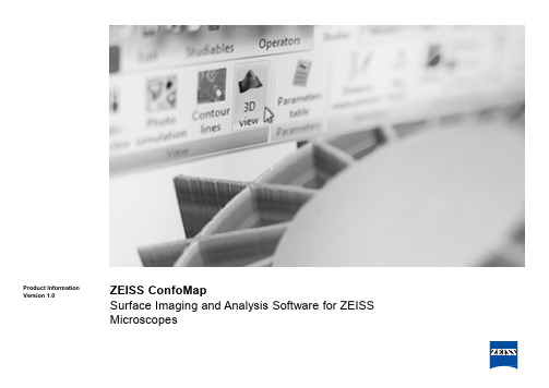
ZEISS ConfoMap Surface Imaging和分析软件:ZEISS微显微镜产品信息(
ZEISS ConfoMapSurface Imaging and Analysis Software for ZEISS MicroscopesProduct Information Version1.0› In Brief› The Advantages › The Features› TheModules The standard ConfoMap® ST package, based on MountiansMap® softwarefrom Digital Surf, includes numerous analytical studies. Geometric studiescalculate distances, angles, areas, volumes and step heights on 2D profilesand 3D surfaces. Functional studies, including the bearing ratio curve andheight distribution, facilitate the assessment of friction and wear onengineering surfaces. The roughness and waviness components of a surfaceare separated using the latest ISO advanced filtering techniques and 3Dsurface texture parameters are calculated in accordance with ISO 25178standard (see modules for selected parameters). Additionally a layer orregion of interest on a micro-mechanical or electronic component can beextracted and analyzed in exactly the same way as a full surface.ConfoMap® ST software can be extended by adding modules for advancedsurface texture analysis, dimensional analysis, grain and particle analysis,3D Fourier analysis, the analysis of surface evolution, and statistics.Working in ConfoMap®, a visual surface analysis report is built straightforward frame by frame in accordance with latest international and nationalmetrology standards and methods. Every analysis step is recorded in ananalysis workflow to assure full metrological traceability.This powerful software package works in conjunction with both ZEISSconfocal and widefield microscope systems providing ease of use forcomplexanalyses.ZEISS ConfoMap: Surface Imaging and Analysis Software for ZEISS Light Microscopes› In Brief› The Advantages › The Features › The ModulesExpand Your PossibilitiesFor use with ZEISS microscope systems:•Smartproof 5 confocal microscope •LSM 800 MAT laser scanning confocal microscope•Smartzoom 5 digital microscope•Many other ZEISS microscopes using ZEN 2 core imagingsoftwareConfoMapConfocalWidefieldZEN 2 core softwareZEISS Smartproof 5ZEISS LSM 800MATA variety of Stereo, Zoom, Inverted, and Upright Light Microscopes ZEISS Smartzoom 52Dimensional3Dimensional› In Brief› The Advantages › The Features › TheModulesVisualize. Analyze.Report.See your sample in a new wayConfoMap® ST provides the highest quality surface imaging. You can visualize a surface in 3D, zoom in and rotate in real time, apply different renderings, select the height amplification and control the lighting type. A standard oruser-defined palette can be selected for the vertical scale and the palette can be fine tuned automatically or interactively to highlight surface features. In addition true color overly is available for a real life view.Provide fast reportingFast, automated, traceable 2D, 3D and surface analysis report creation is simple and quick.Documents are built visually frame by frame. Every step (e.g. 3D surface view, application of filter, geometric study and parameter table) is shown in a graphical analysis workflow that assures full metrological traceability. Once a document has been created it can be applied as a template to automate the analysis of all similar data sets. The results of analyses can be exported in a format compatible with third party software.All the tools at your finger tipsModern look, easy to use with total GUI (Graphical User Interface) flexibility to configure your workspace.Simplify your GUI.Panels can be moved, stacked, docked, and combined for optimal use of screen space. All functions are organized in groups and sub-groups that are clearly labeled and in logical order.The standard version software contains a wide array of imaging and analytical tools. Depending on the application requirements modules can be added to expand the tool set.› In Brief› The Advantages › The Features› The Modules Work Made EasyOrganized to fit your needs.Easy and simple navigation. Page View provides an overview of your document Report Pages. Shortcuts allow quick access to often used tools. Thevaluable Analysis Workflow records step by step processes preformed during analysis which can be reused on subsequent images with the click of the mouse. Results Manager tracks the critical data generated as a result of analysis. All analysis results can be exported in Excel format for interfacing withthird party software, for example quality management systems.1.Configurable tool bars.Context Sensitive Text2.Analysis Workflow3.Page View4.Shortcuts5.Report Pages6.ResultsManager23654ConfoMap at WorkTrue Color Overlay with surface topography making surface features more 3D Surface Texture Measurement. User selectable standards and parameters.analysis of tribological studiesISO 4287Amplitude parameters - Roughness profile Rp 5.37µm Gaussian filter, 0.25 mm Rv 5.03µm Gaussian filter, 0.25 mm Rz 10.4µm Gaussian filter, 0.25 mmRc 10.3µm Gaussian filter, 0.25 mm, ISO 4287 w/o amendme …Rt 10.4µm Gaussian filter, 0.25 mm Ra 3.00µm Gaussian filter, 0.25 mm Rq 3.41µm Gaussian filter, 0.25 mmVolumetric measurement of a hole / valley or peak. Parameters such as volume, surface, Threshold slicing of 2D and 3D images for % area (porosity) and volumetric measurement.Motifs Analysis is a segmentation method (new for ISO 25178) fallowing detection of the hills and dales on a surface with morphological parameters.› In Brief› The Advantages › The Features › The ModulesConfoMap at Work12345501001502002503003504004501.01.5Mean values on 5 steps Value Unit› In Brief› The Advantages › The Features › TheModulesConfoMap at WorkContour Extraction extracts a horizontal profile following the edges of a segmented imageresulting in a parametric profile.Advanced Contour Analysis provides metrology on 2D images and surface profiles with automatic contourdetection.Tolerance limits can be set on measurement parameters and surface roughness measurements. Comparison to CAD files as well.Statistics module is used to monitor numerical results and present in control charts , tables and charts with user defined control limits.› In Brief› The Advantages › The Features › The ModulesConfoMap at WorkAdvanced surface leveling by plane fit, subtraction or defined multipoint while ignoring surface structures.Form removal function -mathematically removing the general form such as of cylinders,spheres and more complex shapes.Outlier removal function. Under certain optical conditions “spikes” or non-measured pointsmay be created in the image. The outlier removal function handles this issue in a elegantand efficient way.Standard filter (Cut-off) used to separate the roughness and waviness phenomena of the profile. Selectable filter types (e.g.ISO).Spectral Analysis -Interactive representation and filtering of frequency spectrum(FFT). In addition avg. power spectrum density (PSD) and wavelet transform are available.Texture Direction and Texture Isotropy for analysis of surfaces having main directions and / or periodic structures in two directions.› In Brief› The Advantages› The Features › The ModulesConfoMap at Work› In Brief› The Advantages› The Features› TheModules ConfoMap Applications Milled aluminum surface with height profile...Smartproof 5with C Epiplan Apochromat 20x/0.7 objective lens.Metalized finger on solar cell surface 3D view with color overlay... Smartzoom 5 with Plan Apochromat D 5x/0.3 objective lens.Wear mark on polymer surface of a medical device…LSM 800 with C Epiplan Apochromat 50x/0.95 objective lens.Typical Areas of Use Machining and Micromechanics Automobile& Aerospace MedicalDevicesElectronicsMicro-optics / MicroReplication Materials ScienceForensicsTasks Measure surface texture/roughness (2D/3D) Measure 3D geometric features Measure wear (tribology) Measure geometical features Measure porosity Measure volume Measure form And much more!› In Brief› The Advantages › The Features› TheModulesFunctions and› In Brief› The Advantages › The Features› TheModules Functions and Modules› In Brief› The Advantages › The Features› The Modules Functions and* Distance between the highest profile peak and the intersection line of the surface ratio Mr1 or distance between the intersection line of the surface ratio Mr2 and the deepest valley respectively.› In Brief› The Advantages › The Features› The Modules Functions and› In Brief› The Advantages › The Features› The Modules Functions andModules。
ZEISS VISUCAM 524 224 24-兆像素传感器筛查眼科设备说明书
VISUCAM Fundus Imaging Brilliance in every detailN E WV I S U CA M 524/224 24-m eg a p i x els e n s orVISUCAM Fundus ImagingExcellent clarity, ultra-high resolution, legendary ZEISS optics.The new ZEISS VISUCAM fundus camera with a 24-megapixel sensor produces brilliant, detail-rich images to effectively aid in diagnosing and monitoring a broad range of eye diseases – from glaucoma and diabetic retinopathy to AMD.Greater diagnostic insight – High-resolution fundus imagingVersatility –Fully-featured camera with a full spectrum of imaging modes* Enhanced practice performance –Simple design, user friendly, full integration with clinic workflowSetting a new standard for resolutionDetails define your decisionsUltra-high resolution and excellent clarity promote efficient navigation from full-image overview to magnification of the smallest detail, allowing precise visualization within a particular area of interest.Fundus autofluorescence (FAF)FAF, included on both VISUCAM models, is an important non-invasive tool for the diagnosis and monitoring of dry AMD, including geographic atrophy.More than a pretty pictureVISUCAM is a complete system with numerous on-board image capture modes – fundus autofluorescence, non-mydriatic Color, Red-free, Red, Blue – and visualization functionality that provide powerful diagnostic insights for optimal patient care.Advanced features such as fluorescein angiography and indocyanine green angiography* further extend its diagnostic applications. * Available only on VISUCAM 524Color VISUCAM Fundus Camera with24-megapixel SensorStereo image pairRed-free// U LTRA-HIGH RESOLUTIONMADE BY ZEISSBest-in-class images from a 24-megapixel sensorAvailable in two modelsVISUCAM 224 with FAF is a fully featured non-mydriatic and mydriatic color camera.VISUCAM 524 adds fluorescein angiography with an optional ICGA mode for doctorswho perform their own dye-based angiography.Red FAF FA ICGAAnterior segmentFundus camera systemField angle 45° and 30°Capture modesColor, red-free, blue, red and fundus autofluorescence images, stereo pairs and images of the anterior segmentVISUCAM 524 only fluorescein angiography VISUCAM 524 only optional: ICG angiographyFiltersOptical filters for capture modes: Filters for green and blue pictures, filters for fundus autofluorescence images, UV/IR barrier filters Compensation for ametropia +35 D … -35 D, continuousCapture sequence from 1.5 seconds (depends on flash energy)Pupil diameter≥ 4.0 mm≥ 3.3 mm (30° small pupil mode)Working distance 40 mm (patient’s eye – front lens)Capture sensor CCD 24-megapixelsMonitor 23” TFT (1920 x 1080), connected via medical power supplyFixation targetsExternal and internal; four sizes of internal fixation target including a circle (for AMD patients). Attention mode for internal fixation target; various programmed sequences or freely positionable as combination with stereo mode too Flash energy Xenon flash lamp, 24 flash levels (max 80 Ws)DatabasePatient information and images with field angle, FA time, R/L recognition and date of visit are storedComputer / AccessoriesOperating system Windows Embedded Standard 7Hard drive Storage of approx. 80,000 images possible (present size of HDD: 420 GB) Interfaces USB ports and network connectors, DVI portExport/import Supported image formats: DICOM-OP and VL, BMP , TIFF, JPEG Patient list, DICOM MWL, DICOM storage Instrument table Asymmetric, suitable for wheelchairAccessoriesNetwork printer, USB memory stick, monitor bracket, sliding keyboard shelf for instrument table, VISUPAC archiving and image analysis system, Network isolatorDimensionsBasic device 410 mm x 480 mm x 735 mm (W 16.14 x D 18.90 x H 28.94 inches)Monitor544 mm x 45 mm x 329 mm (W 21.4 x D 1.8 x H 12.9 inches) (depends on model)Weight (basic device)27.5 kg (60.7 lbs)Rated voltage 100 … 240 V ±10% (self-adjusting)Frequency50 / 60 HzPower consumption340 VA maximum (basic device); 60 VA maximum (monitor)Technical dataE N _31_022_0024I / U S _31_022_0024I P r i n t e d i n G e r m a n y C Z -07/2016T h e c o n t e n t s o f t h i s b r o c h u r e m a y d i f f e r f r o m t h e c u r r e n t s t a t u s o f a p p r o v a l o f t h e p r o d u c t o r s e r v i c e o f f e r i n g i n y o u r c o u n t r y . P l e a s e c o n t a c t o u r r e g i o n a l r e p r e s e n t a t i v e s f o r m o r e i n f o r m a t i o n . S u b j e c t t o c h a n g e i n d e s i g n a n d s c o p e o f d e l i v e r y a n d d u e t o o n g o i n g t e c h n i c a l d e v e l o p m e n t . V I S U C A M i s e i t h e r a t r a d e m a r k o r r e g i s t e r e d t r a d e m a r k o f C a r l Z e i s s M e d i t e c , I n c . i n t h e U n i t e d S t a t e s a n d /o r o t h e r c o u n t r i e s . © C a r l Z e i s s M e d i t e c , I n c . 2016 A l l c o p y r i g h t s r e s e r v e d .Carl Zeiss Meditec AG Goeschwitzer Str. 51-5207745 Jena Germany/med0297VISUCAM NM/FAVISUCAM 524/224。
摄像机术语
摄像机术语&特征1.OLPF(光低通滤波器)俗称滤光片。
滤除干扰图像采集IC的信号,控制图像采集的质量。
用于有Day/night转换功能的机型。
光学低通滤波器大都是由两块或多块石英晶体薄板构成的,放在CCD传感器的前面。
目标图象信息的光束经过OLPF后产生双折射(分为寻常光o光束和异常光e光束)。
根据CCD像素尺寸的大小和总感光面积计算出抽样截止频率,同时也可计算出o光和e光分开的距离。
改变入射光束将会形成差频的目标频率,达到减弱或消除低频干扰条纹的目的,特别是彩色CCD出现的伪彩色干扰条纹的目的。
D(电荷耦合作)电荷耦合器件。
这是一个固态半导体成像装置经常被称为集成电路、芯片或者“影像器”固定型摄像机有时被称为CCD摄像机。
中文全称:电荷耦合元件。
可以称为CCD图像传感器。
CCD是一种半导体器件,能够把光学影像转化为数字信号。
CCD上植入的微小光敏物质称作像素(Pixel)。
一块CCD上包含的像素数越多,其提供的画面分辨率也就越高。
CCD的作用就像胶片一样,但它是把图像像素转换成数字信号。
CCD 上有许多排列整齐的电容,能感应光线,并将影像转变成数字信号。
经由外部电路的控制,每个小电容能将其所带的电荷转给它相邻的电容。
3.IRIS(光圈)是CCD CAMERA成像的基础,控制进光量,可以控制图像明暗。
当处在黑暗环境下时,IRIS开口扩大,增大进光量,物体可以被观察到;相反,明亮环境下。
IRIS将缩小开口防止曝光过大造成图像不良。
如果IRIS没有打开,将无法采集到图像。
4.ZOOM(变焦)是调焦部分,也就是控制图像的放大缩小。
是在IRIS打开之。
后动作的部分,由控制芯片发出信号将图像调整到我们需要的大小。
变焦分为光学变焦和数字变焦。
光学变焦是指在镜头部分的焦距可以变化,焦距就是透镜中心到焦点的距离。
数字变焦是画面放大到整个画面,数码变焦后景物放大了,但是清晰度会下降。
5.FOCUS(聚焦)这个部分是在将图像大小调整到目标值之后进行调整图像变得清晰的。
固态成像器件和成像装置[发明专利]
![固态成像器件和成像装置[发明专利]](https://img.taocdn.com/s3/m/63882d4cf524ccbff021847f.png)
专利名称:固态成像器件和成像装置专利类型:发明专利
发明人:野林和哉
申请号:CN201280062328.5
申请日:20121129
公开号:CN103999221A
公开日:
20140820
专利内容由知识产权出版社提供
摘要:一种固态成像器件包括多个像素,其中,多个像素中的一个或更多个具有光瞳分割部分和受光部分,受光部分包含多个光电转换区域,元件隔离区域被设置在多个光电转换区域之中的相邻的光电转换区域之间,并且其中,散射体被设置在光瞳分割部分内和元件隔离区域之上,并且,散射体由折射率比处于散射体周边的光瞳分割部分的材料的折射率小的材料形成。
申请人:佳能株式会社
地址:日本东京
国籍:JP
代理机构:中国国际贸易促进委员会专利商标事务所
代理人:杨小明
更多信息请下载全文后查看。
Panasonic LUMIX GX85 4K Mirrorless Camera 产品说明书

LUMIX GX85 4K Mirrorless Camera, with 12-32mm and 45-150mm Lenses - Black - DMC-GX85WK∙TYPEo Type Digital Single Lens Mirrorless camerao Recording media SD Memory Card, SDHC Memory Card, SDXC Memory Cardo Recording media (Compatible with UHS-I UHS Speed Class 3 standard SDHC / SDXC Memory Cards)o Image sensor size 17.3 x 13.0 mm (in 4:3 aspect ratio)o Lens mount Micro Four Thirds mount∙IMAGE SENSORo Type Live MOS Sensoro Total pixels 16.84 Megapixelso Camera effective pixels 16.00 Megapixelso Color filter Primary color filtero Dust reduction system Supersonic wave filter∙RECORDING SYSTEMo Aspect ratio 4:3, 3:2, 16:9, 1:1o Image quality RAW, RAW+Fine, RAW+Standard, Fine, Standardo Image quality MPO+Fine / MPO+Standard (with 3D lens in Micro Four Thirds system standard)o Color Space sRGB, AdobeRGBo Continuous recordable time (Motion picture) AVCHD [FHD/60i]: Approx. 100 min (rear monitor), 90 min (LVF) with H-FS12032o Continuous recordable time (Motion picture) MP4 [4K/30p]: Approx. 80 min (rear monitor), 70 min (LVF) with H-FS12032o Actual recordable time (Motion picture) AVCHD [FHD/60i]: Approx. 50 min (rear monitor), 45 min (LVF) with H-FS12032o Actual recordable time (Motion picture) MP4 [4K/30p]: Approx. 40 min (rear monitor), 35 min (LVF) with H-FS12032o Recording file formato Still image JPEG (DCF, Exif 2.3), RAW, MPO (When attaching 3D lens in Micro Four Thirds system standard)o Motion picture AVCHD (Audio format: Dolby Digital 2ch), MP4 (Audio format: AAC 2ch)o File size(Pixels)o Still Image [4:3] 4592x3448(L) / 3232x2424(M) / 2272x1704(S) / 1824x1368 (When attaching 3D lens in Micro Four Third system standard)o Still Image [3:2] 4592x3064(L) / 3232x2160(M) / 2272x1520(S) / 1824x1216 (When attaching 3D lens in Micro Four Third system standard)o Still Image [16:9] 4592x2584(L) / 3840x2160(M) / 1920x1080(S) / 1824x1024 (When attaching 3D lens in Micro Four Third system standard)o Still Image [1:1] 3424x3424(L) / 2416x2416(M) / 1712x1712(S) / 1712x1712 (When attaching 3D lens in Micro Four Third system standard)o Motion picture*o MP4 N/Ao AVCHD N/Ao MP4* [4K] 3840x2160: 4K/30p 100Mbps, 4K/24p 100Mbpso MP4* [Full HD] 1920x1080: FHD/60p 28Mbps, FHD/60p 28Mbpso MP4* [HD] 1280x720: HD/30p 10Mbpso MP4* [VGA] 640x480: VGA/30p 4Mbpso AVCHD* [Full HD] 1920x1080 FHD/60p: 28Mbps, 60p recordingo AVCHD* [Full HD] 1920x1080 FHD/60i: 17Mbps, 60i recordingo AVCHD* [Full HD] 1920x1080 FHD/30p: 24Mbps, 60i recording (sensor output is 30fps)o AVCHD* [Full HD] 1920x1080 FHD/24p: 24Mbps, 24p recording∙WiFi FUNCTIONo WiFi IEEE 802.11b/g/n, 2412 MHz - 2462 MHz (1-11 ch), Wi-Fi / WPA / WPA2, Infrastructure modeo NFC Noo QR Code Connection Yeso Password-less connection Yes (ON / OFF selectable)∙VIEWFINDERo Type LCD Live View Finder (2,764,800 dots equivalent)o Field of view Approx. 100%o Magnification Approx. 1.39x / 0.7x (35mm camera equivalent) with 50 mm lens at infinity; -1.0 m-1o Eye point Approx. 17.5 mm from eyepiece lenso Diopter adjustment -4.0 - +3.0 (dpt)o Eye sensor Yeso Eye sensor adjustment High / Low∙FOCUSo Type Contrast AF systemo DFD technology Yeso Post Focus Yeso Focus Stacking N/Ao Focus mode AFS (Single) / AFF (Flexible) / AFC (Continuous) / MFo AF mode Face/Eye Detection / Tracking / 49-Area / Custom Multi / 1-Area / Pinpointo AF mode (Full area touch is available)o AF detective range EV -4 - 18 (ISO100 equivalent)o Starlight AF Yeso AF assist lamp Yeso AF lock Yes (AF/AE LOCK button)o Others One Shot AF, Shutter AF, Half Press Release, Quick AF, Continuous AF (during motion picture recording), Eye Sensor AF, AF+MF, MF Assist, TouchMF Assist, Focus Peaking, Touch AF/AE Function, Touch Pad AF, TouchShutter∙EXPOSURE CONTROLo Light metering system 1728-zone multi-pattern sensing systemo Light metering mode Multiple / Center Weighted / Spoto Metering range EV 0 - 18 (F2.0 lens, ISO100 equivalent)o Exposure mode Program AE, Aperture Priority AE, Shutter Priority AE, Manual o ISO sensitivity (Standard Output Sensitivity) Auto / Intelligent ISO / 100 (Extended) / 200 / 400 / 800 / 1600 / 3200 / 6400 / 12800 / 25600 (Changeable to1/3 EV step)o ISO sensitivity (Standard Output Sensitivity) (Up to ISO6400 in motion picture recording) (ISO Auto in M mode)o Exposure compensation 1/3 EV step ±5EV (±3EV for motion picture)o AE lock Yes (AF/AE LOCK button)o AE bracket N/A∙WHITE BALANCEo White balance Auto / Daylight / Cloudy / Shade / incandescent / Flash / White Set 1, 2, 3, 4 / Color temperature settingo White balance adjustment Blue/Amber bias, Magenta/Green biaso Color temperature setting 2500-10000K in 100Ko White balance bracket N/A∙SHUTTERo Type Focal-plane shuttero Shutter speed Still image: Still image: Time (Max. 2 minutes), 1/4,000 - 60o Shutter speed Motion picture: 1/16,000 - 1/25o Shutter speed Electronic shutter: 1/16,000 - 1o Self timer 10sec, 3 images / 2sec / 10seco Remote control N/A∙SCENE GUIDEo Still image N/Ao Motion picture N/A∙BRACKETo AE bracket 3, 5, 7 frames in 1/3, 2/3 or 1 EV Step, Max. ±3 EV, single/bursto Aperture Bracket 3, 5 or all positions in 1 EV stepo Focus Bracket 1 to 999 frames, focus steps can be set in 5 levelso White balance bracket 3 exposures in blue/amber axis or in magenta/green axis ∙PANORAMA SHOTo Panorama shot Yes (Standard / Wide)∙BURST SHOOTINGo Burst speed [Mechanical shutter] AFS: H: 8 frames/sec, M: 6 frames/sec (with Live View), L: 2 frames/sec (with Live View)o Burst speed [Mechanical shutter] AFC: H: 6 frames/sec, M: 6 frames/sec (with Live View), L: 2 frames/sec (with Live View)o Burst speed [Electronic shutter] SH: 40 frames/seco Burst speed [Electronic shutter] AFS: H: 10 frames/sec, M: 6 frames/sec (with Live View), L: 2 frames/sec (with Live View)o Burst speed [Electronic shutter] AFC: H: 6 frames/sec, M: 6 frames/sec (with Live View), L: 2 frames/sec (with Live View)o Number of recordable images More than 13 images (when there are RAW files with the particular speed)o Number of recordable images More than 100 images (when there are no RAW files)o Number of recordable images (depending on memory card type, aspect, picture size and compression)∙4K PHOTO MODEo4K Photo mode* 4K Burst: 30 frames/seco4K Photo mode* 4K Burst (S/S): 30 frames/seco4K Photo mode* 4K Pre-Burst: 30 frames/sec, approx. 2 secondso4K Photo mode* (depending on memory card size and battery power)o Exif information Yeso Selectable aspect ratio Yes (4:3 / 3:2 / 16:9 / 1:1 are selectable)o Exposure mode Program AE/ Aperture-Priority / Sutter-Priority / Manual Exposureo Marking function Yes (in 4K Burst (S/S) mode)o Loop rec function Yes (in 4K Burst (S/S) mode)∙FLASHo Flash type TTL Built-in-Flash, GN6.0 equivalent (ISO200 ・m) / GN4.2 equivalent (ISO100 ・m), Built-in Pop-up (Reference)o Flash Mode Auto*, Auto/Red-eye Reduction*, Forced On, Forced On/Red-eye Reduction, Slow Sync., Slow Sync./Red-eye Reduction, Forced Off * For iA, iA+only.o Synchronization speed Less than 1/160 secondo Flash output adjustment 1/3EV step ±3EVo Flash synchronization 1st. Curtain Sync, 2nd Curtain Sync.o Synchronization for flash dimming and exposure compensation Yeso Wireless control N/A∙REAR MONITORo Type TFT LCD monitor with static touch controlo Monitor size Tilt 3.0-inch / 3:2 aspect / Wide viewing angleo Pixels Approx. 1,040k dotso Field of view Approx. 100%o Monitor adjustment Brightness, Contrast, Saturation, Red-Green, Blue-Yellow ∙LIVE VIEWo Digital zoom 2x, 4xo Extra Tele Conversion Still image: Max. 2xo Extra Tele Conversion Motion picture: 2.4x (FHD), 3.6x (HD), 4.8x (VGA)o Other functions Level Gauge, Real-time Histogram, Guide Lines (3 patterns), Highlight display (Still image / motion picture), Zebra pattern (Still image /motion picture)∙DIRECTION DETECTION FUNCTIONo Direction Detection Function Yes∙SELF SHOTo Self Shot Mode N/Ao Shutter N/Ao Effect N/A∙FUNCTION BUTTONo Fn1, Fn2, Fn3, Fn4, Fn5, Fn6 N/Ao Fn1, Fn2, Fn3, Fn4, Fn5, Fn6, Fn7, Fn8, Fn9 4K Photo Mode / Wi-Fi / Q.MENU / LVF/Monitor Switch / AF/AE LOCK / AF-ON / Preview / One Push AE /Touch AE / Level Gauge / Focus Area Set / Zoom Control / Cursor Button Lock /Dial Operation Switch / Photo Style / Filter Select / Aspect Ratio / Picture Size /Quality / Metering Mode / Bracket / Focus Mode / Highlight Shadow / i. Dynamic/ i. Resolution / Post Focus / HDR / Shutter Type / Flash Mode / Flash Adjust. /Ex. Tele Conv. / Digital Zoom / Stabilizer / Snap Movie / Motion Pic. Set /Picture Mode / Silent Mode / Peaking / Histogram / Guide Line / Zebra Pattern /Monochrome Live View / Rec Area / Step Zoom / Zoom Speed / Touch Screen /Sensitivity / White Balance / AF Mode/MF / Drive Mode / Restore to Default o Fn1, Fn2, Fn3, Fn4, Fn5, Fn6, Fn7, Fn8, Fn9, Fn10 N/A∙PHOTO STYLEo Still image and motion picture Standard / Vivid / Natural / Monochrome / L.Monochrome / Scenery / Portrait / Custom∙CREATIVE CONTROLo Still image Expressive / Retro / Old Days / High Key / Low Key / Sepia / Monochrome / Dynamic Monochrome / Rough Monochrome / SilkyMonochrome / Impressive Art / High Dynamic / Cross Process / Toy Effect / ToyPop / Bleach Bypass / Miniature Effect / Soft Focus / Fantasy / Star Filter / OnePoint Color / Sunshineo Motion picture Expressive / Retro / Old Days / High Key / Low Key / Sepia / Monochrome / Dynamic Monochrome / Impressive Art / High Dynamic / CrossProcess / Toy Effect / Toy Pop / Bleach Bypass / Miniature Effect / Fantasy / OnePoint Color∙CREATIVE VIDEO MODEo Exposure mode Program AE / Aperture-Priority / Sutter-Priority / Manual Exposure∙MOTION PICTURE FUNCTIONo Cinelike gamma N/Ao Flicker reduction [1/50] / [1/60] / [1/100] / [1/120] / OFF∙PLAYBACKo Playback function 30-thumbnail display, 12-thumbnail display, Calendar display, Zoomed playback (Max. 16x), Slideshow (All / Picture Only / Video Only / 4KPHOTO / Post Focus / 3D / Category Selection / Favorite, duration & effect isselectable), Playback Mode (Normal / Picture Only / Video Only / 4K PHOTO /Post Focus / 3D Play / Category / Favorite), Location Logging, RAW Processing,Light Composition, Clear Retouch, Title Edit, Text Stamp, Video Divide, TimeLapse Video, Stop Motion Video, Resize, Cropping, Rotate, Rotation Display,Favorite, DPOF Print Set, Protect, Face Recognition Edit, Picture Sort, CreatingStill Pictures from a Motion Picture∙IMAGE PROTECTION / ERASEo Protection Single / Multio Erase Single / Multi / All / Except Favorite∙PRINTo Direct Print PictBridge compatible∙INTERFACEo USB USB 2.0 High Speed Multio HDMI N/Ao HDMI** microHDMI TypeD / VIERA Linko HDMI** Video: Auto / 4K / 1080p / 1080i / 720p / 480po HDMI** Audio: Stereoo Audio video output Noo Remote input N/Ao External microphone input N/Ao Microphone Stereo, Wind-cut: OFF / Standard / Higho Speaker Monaural∙LANGUAGEo OSD language Japanese, English, German, French, Italian, Spanish, Portuguese, Chinese (Traditional)∙GENERALo POWERo Battery Li-ion Battery Pack (7.2V, 1025mAh, 7.4Wh) (Included)o Battery AC Adaptor (Input: 110 - 240V AC) (Included, connect with USB cable) o Battery life (CIPA standard) Approx. 290 images (rear monitor), 270 images (LVF) with H-FS12032o Battery life (CIPA standard) Approx. 290 images (rear monitor), 270 images (LVF) with H-FS12032 / H-FS35100o Battery grip N/Ao DIMENSIONS / WEIGHTo Dimensions (W x H x D) 122 x 70.6 x 43.9 mm / 4.80 x 2.78 x 1.73 inch (excluding protrusions)o Weight Approx. 426g / 0.94 lb (SD card, Battery, Body)o Weight Approx. 383g / 0.84 lb (Body only)o Weight Approx. 493g / 1.09 lb (SD card, Battery, H-FS12032 lens included)o Weight Approx. 628g / 1.39 lb (SD card, Battery, H-FS12032 + H-FS35100 lenses included)o OPERATING ENVIRONMENTo Operating temperature 0℃ to 40℃ (32°F to 104°F)o Operating humidity 10%RH to 80%RHo STANDARD ACCESSORIESo Software ・ The software to edit and playback images on computer is not bundled with DMC-GX85. To do this, PHOTOfunSTUDIO is available for download atPanasonic website using computer connected to the Internet.o http://panasonic.jp/support/global/cs/soft/download/d_pfs99pe.html (For Windows)oo・ The software to process RAW file on computer is not bundled with DMC-GX85. To do this, SILKYPIX Developer Studio is available for download atIchikawa Soft Laboratory's website using computer connected to the Internet.o http://www.isl.co.jp/SILKYPIX/english/p/ (For Windows / Mac)o Standard accessories DMC-GX85W Kito Standard accessories Hot Shoe Cover, Battery Pack, AC Adaptor, USB Connection Cable, Shoulder Strap, Lens Cap x 2, Lens Hood, Lens Rear Cap o Standard accessories ・ The DMC-GX85 Operating Instructions for advanced features is available for downloaded at Panasonic LUMIX Customer Support Siteusing PC, smartphone or tablet connected to the Internet.∙INTERCHANGEABLE LENS-1o Lens Name LUMIX G VARIO 12-32mm / F3.5-5.6 ASPH. / MEGA O.I.S.o Lens Construction 8 elements in 7 groups (3 aspherical lenses, 1 ED lens)o Nano Surface Coating -o Mount Micro Four Thirds mounto Optical Image Stabilizer Yes (MEGA O.I.S.)o Focal Length f=12-32mm (35mm camera equivalent 24-64mm)o Aperture Type 7 diaphragm blades / Circular aperture diaphragmo Aperture range N/Ao Aperture N/Ao Maximum Aperture F3.5(Wide) - F5.6(Tele)o Minimum Aperture F22o Closest Focusing Distance 0.20m/0.66ft (at focal lenghts 12-20mm) /0.30m/0.98ft (at focal lenghts 21-32mm)o Maximum magnification Approx. 0.13x / 0.26x (35mm camera equivalent)o Diagonal Angle of View 84°(Wide) to 37°(TELE)o Weatherproof N/Ao Generalo Filter Size 37mm / 1.5ino Max. Diameter φ55.5mm / 2.2ino Overall Length Approx. 24mm / 0.94in (from the tip of the lens to the base side of the lens mount)o Weight [g] Approx. 70g (excluding lens cap, lens rear cap )o Weight [oz] Approx. 2.47oz (excluding lens cap, lens rear cap )o Others N/A∙INTERCHANGEABLE LENS-2o Lens Name LUMIX G VARIO 35-100mm / F4.0-5.6 ASPH. / MEGA O.I.S.o Lens Construction 12 elements in 9 groups (1 aspherical lens, 2 ED lenses)o Nano Surface Coating -o Mount Micro Four Thirds mounto Optical Image Stabilizer Yes (MEGA O.I.S.)o Focal Length f=35-100mm (35mm camera equivalent 70-200mm)o Aperture Type 7 diaphragm blades / Circular aperture diaphragmo Aperture range N/Ao Aperture N/Ao Maximum Aperture F4.0 (Wide) - F5.6 (Tele)o Minimum Aperture F22o Closest Focusing Distance 0.90m / 3.00fto Maximum magnification Approx. 0.11x / 0.22x (35mm camera equivalent)o Diagonal Angle of View 34°(Wide) to 12°(TELE)o Weatherproof N/Ao Generalo Filter Size 46mm / 1.8ino Max. Diameter φ55.5mm / 2.2ino Overall Length Approx. 50mm / 1.97in (from the tip of the lens to the base side of the lens mount)o Weight [g] Approx. 135g (excluding lens cap, lens rear cap and lens hood)o Weight [oz] Approx. 4.76oz (excluding lens cap, lens rear cap and lens hood)o Others N/A∙NOTESo Image Stabilization System Image Sensor Shift Type (5-axis)o TIME LAPSE SHOT Yeso STOP MOTION ANIMATION Yeso SILENT MODE Yeso NOTE * About motion picture recording / 4K Photo recordingo- Use a card with SD Speed Class with "Class 4" or higher when recording motion pictures.o- Use a card with SD Speed Class with "UHS-I UHS Speed Class 3 (U3)" when recording motion pictures with [MP4] in [4K] or [4K PHOTO].o(SD speed class is the speed standard regarding continuous writing.)o- Recording stops when the continuous recording time exceeds 29 minutes and 59 seconds or the file size exceeds 4GB with [MP4] in [FHD] [HD] [VGA].o- MP4 motion pictures with [MP4] in [4K]:o- When using an SDHC memory card: You can continue recording without interruption even if the file size exceeds 4 GB, but the motion picture file will bedivided and recorded/played back separately.o- When using an SDXC memory card: You can record a motion picture in a single file.o- When the ambient temperature is high or continuous recording is performed, the camera may stop the recording to protect itself. Wait until the camera cools down.oo** For [4K] video output, use an HDMI cable that has the HDMI logo on it, and that is described as"4K compatible".∙o UPC 885170338524。
光学检测算法 -回复
光学检测算法-回复光学检测算法在现代科技领域中起着至关重要的作用。
随着计算机视觉技术的发展,越来越多的应用场景需要对图像和视频进行精确的光学检测。
光学检测算法可以帮助我们从图像和视频中提取出关键信息,如物体的位置、形状、颜色等。
本文将从光学检测算法的基本原理、常见应用领域和算法优化等方面,对光学检测算法进行详细介绍。
光学检测算法的基本原理是基于计算机视觉的图像处理和模式识别技术。
首先,在图像处理阶段,需要对图像进行预处理,包括去噪、灰度化、边缘检测等。
然后,在模式识别阶段,通过比对已有的模型和特征描述,将图像中的关键信息提取出来。
最后,根据提取到的信息,进行判断、分类或识别。
光学检测算法的应用十分广泛。
首先,在工业领域中,光学检测算法可以用于产品质量检测。
例如,在生产线上,通过对产品照片进行光学检测,可以自动识别出缺陷产品,并实现实时的质量控制。
其次,在医学领域中,光学检测算法可以用于医学影像分析。
例如,在CT扫描中,光学检测算法可以帮助医生定位肿瘤的位置和大小,从而辅助医生进行诊断和治疗。
此外,光学检测算法还可以应用于交通管理、安防监控、自动驾驶等领域。
然而,光学检测算法在实际应用中面临一些挑战。
首先是复杂背景环境的影响。
图像中的复杂背景可能干扰光学检测算法对目标物体的检测效果。
为了解决这个问题,可以采用背景建模和背景差法等方法来进行背景分离和目标提取。
其次是光照条件的影响。
光照不均衡、阴影等问题可能导致光学检测算法的准确性下降。
为了解决这个问题,可以采用直方图均衡化、光照补偿等方法来进行图像增强。
此外,算法的效率和稳定性也是需要考虑的因素,可以使用并行计算、硬件加速等技术进行算法优化。
为了提高光学检测算法的准确性和可靠性,研究人员也在不断进行算法改进和优化。
例如,研究人员提出了基于深度学习的光学检测算法,利用深度神经网络来提取图像中的特征,并进行目标识别和分类。
此外,还有一些基于传统图像处理技术的光学检测算法,如边缘检测、模板匹配等。
光谱 epo算法 -回复
光谱epo算法-回复什么是光谱?为什么需要光谱算法?光谱算法的基本原理是什么?以及光谱算法在实际应用中的重要性。
【光谱】指的是将电磁辐射按照波长进行分类和排序的过程。
光谱算法是一种数学方法,用于从光谱数据中提取有用的信息和特征。
在各个领域中,包括物理、化学、天文学和工程学等等,光谱数据经常用来研究、分析和识别物质。
光谱算法之所以如此重要,是因为它可以帮助我们理解物质的组成、结构和性质。
通过分析光谱数据,我们可以得到关于物质的各种信息,例如其化学元素组成、分子结构、浓度、温度,以及性质等等。
因此,光谱算法在许多实际应用中发挥着重要作用。
光谱算法的基本原理是基于不同物质对光的吸收、散射或发射的不同特性。
当光通过物质时,根据该物质的分子结构和组成,它会吸收或发射特定波长的光。
通过测量物质与光的相互作用,我们可以得到该物质的光谱数据。
这些数据通常以光的强度随波长的变化来表示,称为光谱图。
在实际应用中,光谱算法可以用于许多不同的目的。
其中一个重要的应用领域是光谱分析,用于确定物质的组成和浓度。
例如,在环境科学中,我们可以使用光谱算法来检测并监测水体中的污染物,根据光谱数据判断是否存在特定的化学物质,同时还可以确定其浓度。
在农业领域,光谱算法可以用来检测和识别作物的营养和水分状况,为农民提供决策基础。
此外,光谱算法还被广泛应用于材料科学中的表征和质检、医学诊断中的病理分析和药物研发,以及天文学中的星际物质分析等等。
光谱算法的实施需要一系列的步骤和技术。
首先,我们需要准备样品并获取其光谱数据。
通常情况下,我们会使用光谱仪或其他光学设备来收集样品的光谱信息。
然后,我们会对所获得的光谱数据进行预处理,以去除噪声、校正光谱和进行数据清洗。
接下来,根据特定的分析目的,我们可以应用不同的光谱算法进行数据处理和分析。
这些算法可以包括传统的数学方法,如光谱峰识别、光谱拟合和数据提取等,也可以是一些先进的机器学习和人工智能技术,如光谱聚类、光谱分类和光谱回归等。
- 1、下载文档前请自行甄别文档内容的完整性,平台不提供额外的编辑、内容补充、找答案等附加服务。
- 2、"仅部分预览"的文档,不可在线预览部分如存在完整性等问题,可反馈申请退款(可完整预览的文档不适用该条件!)。
- 3、如文档侵犯您的权益,请联系客服反馈,我们会尽快为您处理(人工客服工作时间:9:00-18:30)。
a r X i v :a s t r o -p h /0510223v 1 7 O c t 2005Astronomy &Astrophysics manuscript no.3785cESO 2008February 5,2008ESO Imaging Survey:Optical follow-up of 12selectedXMM-Newton fields ⋆,⋆⋆J.P.Dietrich 1,2,J.-M.Miralles 2,L.F.Olsen 2,3,4,L.da Costa 2,A.Schwope 5,C.Benoist 2,4,V .Hambaryan 5,A.Mignano 6,2,7,C.Motch 8,C.Rit´e 2,R.Slijkhuis 2,J.Tedds 9,B.Vandame 2,M.G.Watson 9,and S.Zaggia 2,101Institut f¨u r Astrophysik und extraterrestrische Forschung,University of Bonn,Auf dem H¨u gel 71,53121Bonn,Germany e-mail:dietrich@astro.uni-bonn.de2European Southern Observatory,Karl-Schwarzschild-Str.2,85748Garching b.M¨u nchen,Germany 3Copenhagen University Observatory,Juliane Maries Vej 30,DK-2100Copenhagen,Denmark 4Observatoire de la Cˆo te d’Azur,Laboratoire Cassiop´e e,BP4229,06304Nice Cedex 4,France 5Astrophysikalisches Institut Potsdam,An der Sternwarte 16,14482Potsdam,Germany 6Dipartimento di Astronomia,Universit`a di Bologna,via Ranzani 1,I-40126Bologna,Italy 7Istituto di Radioastronomia,INAF,Via Gobetti 101,I-40129Bologna,Italy 8Observatoire Astronomique,CNRS UMR 7550,11rue de l’Universit´e ,67000Strasbourg,France 9Department of Physics and Astronomy,University of Leicester,Leicester LE17RH,UK 10Osservatorio Astronomico di Trieste,Via G.B.Tiepolo,11,I-34131Trieste,ItalyReceived 7July 2005;Accepted 7October 2005ABSTRACTThis paper presents the data recently released for the XMM-Newton /WFI survey carried out as part of the ESO Imaging Survey (EIS)project.The aim of this survey is to provide optical imaging follow-up data in BVRI for identification of serendipitously detected X-ray sources in selected XMM-Newton fields.In this paper,fully calibrated individual and stacked images of 12fields as well as science-grade catalogs for the 8fields located at high-galactic latitude are presented.These products were created,calibrated and released using the infrastructure provided by the EIS Data Reduction system and its associated EIS /MVM image processing engine,both of which are briefly described here.The data covers an area of ∼3square degrees for each of the four passbands.The median seeing as measured in the final stacked images is 0.′′94,rangingfrom 0.′′60and 1.′′51.The median limiting magnitudes (AB system,2′′aperture,5σdetection limit)are 25.20,24.92,24.66,and 24.39mag for B -,V -,R -,and I -band,respectively.When only the 8high-galactic latitude fields are included these become 25.33,25.05,25.36,and 24.58mag,in good agreement with the planned depth of the survey.Visual inspection of images and catalogs,comparison of statistics derived from the present data with those obtained by other authors and model predictions,as well as direct comparison of the results obtained from independent reductions of the same data,demonstrate the science-grade quality of the automatically produced final images and catalogs.These survey products,together with their logs,are available to the community for science exploitation in conjunction with their X-ray counterparts.Preliminary results from the X-ray /optical cross-correlation analysis show that about 61%of the detected X-ray point sources in deep XMM-Newton exposures have at least one optical counterpart within 2′′radius down to R ≃25mag,50%of which are so faint as to require VLT observations thereby meeting one of the top requirements of the survey,namely to produce large samples for spectroscopic follow-up with the VLT,whereas only 15%of the objects have counterparts down to the DSS limiting magnitude.Key words.Catalogs –Surveys –Stars:general –Galaxies:general –X-rays:general1.IntroductionThe new generation of highly sensitive X-ray observatories such as Chandra and XMM-Newton is generating large vol-umes of X-ray data,which through public archives are made2J.P.Dietrich et al.:ESO Imaging Survey:Optical follow-up of12selected XMM-NewtonfieldsTable1.Central positions in right ascension,declination and galactic longitude and latitude of the12XMM-Newtonfields observed as part of the XMM-Newton follow-up survey.Field Targetα(J2000.0)δ(J2000.0)l bproposed optical follow-up observations of XMM-Newtonfields for its X-ray Identification(XID)program(Watson et al.2001;Barcons et al.2002;Della Ceca et al.2004).This pro-posal was evaluated and accepted by ESO’s Survey WorkingGroup(SWG)and turned into a proposal for an ESO large pro-gram submitted to the ESO OPC.1The XMM-Newton optical follow-up survey aims at ob-taining optical observations of XMM-Newton SerendipitousSky Surveyfields,publicly available in the XMM-Newtonarchive,using the wide-field imager(WFI)at the ESO/MPG2.2m telescope at the La Silla Observatory.WFI has a FOVwhich is an excellent match to that of the X-ray detectors on-board the XMM-Newton satellite,making this instrument anobvious choice for this survey in the South.A complemen-tary multiband optical imaging program(to median5σlimitingmagnitudes reaching i′=23.1)for over150XMM-Newtonfields is nearing completion in the North using the similarlywell matched Wide Field Camera on the2.5m Isaac NewtonTelescope(Yuan et al.2003;Watson et al.2003).In order toprovide data for minimum spectral discrimination and photo-metric redshift estimates of the optical counterparts of previ-ously detected X-ray sources,the survey has been carried outin the B-,V-,R-,and I-passbands.The survey has been admin-istered and carried out by the ESO Imaging Survey(EIS)team.This paper describes observations,reduction,and scienceverification of data publicly released as part of this follow-up survey.Section2briefly describes the X-ray observationswhile Sect.3focus on the optical imaging.In Sect.4the re-duction and calibration of optical data are presented and theresults discussed.Final survey products such as stacked im-ages and science-grade catalogs extracted from them are pre-sented in Sect.5.The quality of these products is evaluated inSect.6by comparing statistical measures obtained from thesedata to those of other authors as well as from a direct compar-ison with the results of an independent reduction of the samedataset.In this section the results of a preliminary assessmentof X-ray/optical cross-correlation are also discussed.Finally,inSect.7a brief summary of the paper is presented.J.P.Dietrich et al.:ESO Imaging Survey:Optical follow-up of12selected XMM-Newtonfields3Fig.1.Color composite X-ray images for the12fields considered in this paper(XMM-01to XMM-12from top left to bottom right).The color images are composites within the so-called XID-band(0.5–4.5keV).Red,green and blue channels comprise the energy ranges0.5–1.0keV, 1.0–2.0keV,and2.0–4.5keV,respectively.Weighting of the sub-images was done in a manner that a typical extragalactic source with a power law spectrum with photon index1.5and absorption column density N H=1×1020cm−2would have equal photon numbers in all three bands. North is up and East to the left.The size of the images is typically30′×30′but varies slightly with camera orientation.4J.P.Dietrich et al.:ESO Imaging Survey:Optical follow-up of12selected XMM-Newtonfields rmation about X-ray imaging used to create composite X-ray images.Field Obs.ID T exp(s)Camera settingsXMM-02016456050150059EPN FF MOS1FF MOS2FF015696020130243EPN FF MOS1FF MOS2FF015696040132039EPN FF MOS1FF MOS2FFXMM-04009480010141021EPN FF MOS1FF MOS2FFXMM-06011116020149616EPN EFF MOS1FF MOS2FFXMM-08011098020158237EPN EFF MOS1FF MOS2FFXMM-10001244030135366EPN FF MOS1FF MOS2FFXMM-12010911010176625EPN EFF MOS1FF MOS2FF2http://xmm.vilspa.esa.es/external/xmm acc/ xsa/index.shtml WFI is a focal reducer-type mosaic camera mounted at the Cassegrain focus of the telescope.The mosaic consists of4×2 CCD chips with2048×4096pixels with a projected pixel size of0.′′238,giving a FOV of8.′12×16.′25for each individual chip.The chips are separated by gaps of23.′′8and14.′′3along the right ascension and declination direction,respectively.The full FOV of WFI is thus34′×33′with afilling factor of95.9%.The WFI data described in this paper are from the following two sources:1.the ESO Large Programme170.A-0789(A)(Principal In-vestigator:J.Krautter,as chair of the SWG)which has ac-cumulated data from January27,2003to March24,2004 at the time of writing.2.the contributing programs70.A-0529(A);71.A-0110(A);71.A-0110(B)with P.Schneider as the Principal Invest-igator,which have contributed data from October14,2002 to September29,2003Observations were performed in the B-,V-,R-,and I-passbands.These were split into OBs consisting of a sequence offive(ten in the I-band)dithered sub-exposures with the typi-cal exposure time given in Table3.The table gives:in Col.1the passband;in Col.2thefilter id adopting the unique naming convention of the La Silla Science Operations Team;in Col.3 the total exposure time in seconds;in Col.4the number of ob-serving blocks(OBs)perfield;and in Col.5the integration time of the individual sub-exposures in the OB.The dither pat-J.P.Dietrich et al.:ESO Imaging Survey:Optical follow-up of12selected XMM-Newtonfields5 Table3.Planned observing strategy for the XMM-Newton follow-upsurvey.Passband Filter T tot(s)N OB T exp(s)ESO87918001360V V/89ESO84435001700I I/203tern with a radius of80′′was optimized for the bestfilling ofthe gaps.Filter curves can be found in Arnouts et al.(2001)andon the web page of the La Silla Science Operations Team.3Even though the nominal total survey exposure time for theR-band is3500s,the data contributed by the Bonn group pro-vided additional exposures totaling11500s each,spread over4OBs.For the same reason the B-band data for thefield XMM-07has a significantly larger exposure time than that given inTable3(see Table9).Service mode observing provides the option for constraintson e.g.,seeing,transparency,and airmass to be specified in or-der to meet the requirements of the survey.The adopted con-straints were:(1)dark sky with a fractional lunar illuminationof less than0.4;(2)clear sky with no cirrus though not neces-sarily photometric;and(3)seeing≤1.′′2.The R-band imagesof the contributing program were taken with a seeing constraintof 1.′′0so that the data can be used for weak lensing studies.The total integration time in somefields may be higher thanthe nominal one listed in Table3because unexpected variationsin ambient conditions during the execution of an OB can cause,for instance,the seeing and transparency to exceed the origi-nally imposed constraints.If this happens,the OB is normallyexecuted again at a later time.In these cases the decision of us-ing or not all the available data must be taken during the datareduction process.In the case of the present survey all avail-able data were included in the reduction,which explains whyin some cases the total integration time exceeds that originallyplanned.This paper describes data accumulated prior to October16,2003,amounting to about80h on-target integration.The sci-ence data comprises720exposures split into130OBs.About15%of the B-band and85%of the R-band data are from thecontributing programs.4.Data reductionThe accumulated optical exposures were reduced and cali-brated using the EIS Data Reduction System(da Costa etal.,in preparation)and its associated image processing enginebased on the C++EIS/MVM library routines(Vandame2004,Vandame et al.,in preparation).4This library incorporates rou-tines from the multi-resolution visual model package(MVM)described in Bijaoui&Ru´e(1995)and Ru´e&Bijaoui(1997).It was developed by the EIS project to enable handling and5/science/eis/6J.P.Dietrich et al.:ESO Imaging Survey:Optical follow-up of12selected XMM-NewtonfieldsTable4.Summary of the number of nights with standard star obser-vations and type of solution.Passband default1-par2-par3-par totalparison between the EIS3-parameterfit solutions and the Telescope Team’s best solution.Passband∆ZP∆k∆colorAugust6,2003)the solutions obtained in the passbands V,I, R,respectively(either2-or3-parameterfits)deviate from the median by−0.26,−0.5,−0.25mag.Of those,only the I-band zeropoint obtained for April2,2003deviates by more than3σfrom the solutions obtained for other nights.Note that the type of solution obtained depends on the available airmass and color coverage,which in the case of the XMM-Newton survey de-pends on the calibration plan adopted by the La Silla Science Operations Team.Because the EIS Survey System automatically carries out the photometric calibrations it is interesting to compare the so-lutions to those obtained by other means.Therefore,the auto-matically computed3-parameter solutions of the EIS Survey System are compared with the best solution recently obtained by the La Silla Science Operations Team.The results of this comparison are presented in Table5which lists:in Col.1the passband;in Cols.2–4the mean offsets in zeropoint(ZP),ex-tinction(k)and color term(color),respectively.The agreement of the solutions is excellent for all passbands.However,it is worth emphasizing that the periods of observations of standard stars available to the two teams do not coincide.Not surprisingly,larger offsets are found when2-and1-parameterfits are included,depending on the passband and es-timator used to derive the estimates for extinction and color term.Finally,taking into consideration only3-parameterfit so-lutions and after rejecting3σoutliers onefinds that the scatter of the zeropoints is 0.08mag.This number is still uncertain given the small number of3-parameterfits currently available, especially in the R-band.The obtained scatter is a reasonable estimate for the current accuracy of the absolute photometric calibration of the XMM-Newton survey data.There are two more points that should be considered in evaluating the accuracy of the photometric calibration of the present data.First,for detectors consisting of a mosaic of individual CCDs it is important to estimate and correct for possible chip-to-chip variations of the gain.For the present data these variations were estimated by comparing the median background values of sub-regions bordering adjacent CCDs. The determined variations were used to bring the gain to a common value for all CCDs in the mosaic.This was applied to both science and standard exposures.Second,it is also known that large-scale variations due to non-uniform illumi-nation over thefield of view of a wide-field camera exist.The significance of this effect is passband-dependent and becomes more pronounced with increasing distance from the optical axis (Manfroid&Selman2001;Koch et al.2004;Vandame et al.in preparation).Automated software to correct for this effect has been developed but due to time constraints it has not yet been applied to these data.Thefinal step of the data reduction process involves the assessment of the quality of the reduced images.Following vi-sual inspection,each reduced image is graded,with the grades ranging from A(best)to D(worst).This grade refers only to the visual aspect of the data(e.g.background,cosmetics).Out of 150reduced images covering(see Sect.3)the selected XMM-Newtonfields,104were graded A,35B,7C and4D.The images with grades C and D are listed in Table6.The table, ordered byfield and date,lists:in Col.1thefield name;in Col.2the passband;in Col.3the civil date when the night started(YYYY-MM-DD);in Col.4the grade given by the vi-sual inspection;and in Col.5the primary motive for the grade. It is important to emphasize that the reduced images must be graded,as grades are used in the preparation of thefinal image stacks.In particular,reduced images with grade D have no sci-entific value and were not released and were discarded in the stacking process discussed in the next section.The success rate of the automatic reduction process is bet-ter than95%and most of the lower grades are associated with observational problems rather than inadequate performance of the software operating in an un-supervised mode.An interest-ing point is that occasionally R-band images are also affected by fringing(see Table6)–for instance,in the nights of August 6and September23and29,2003,all from the contributing program.The night of August6is one of the nights for which the computed R-band zeropoint deviates from the median.This points out the need to consider applying fringing correction also in the R-band,at least in some cases.The R-band fring-ing problem accounts forfive out of seven grade C images.The remaining cases are due to stray-light and strong shape distor-tions.It should also be pointed out that the reduced images show a number of cosmic ray hits.This is because the construction of RBs was optimized for removing cosmic ray features in thefi-nal stacks using a thresholding technique.To this end the num-ber of images in an RB was minimized for somefield andfilter combinations to have at least three reduced images entering the SB.5.Final products5.1.ImagesThe146reduced images with grades better than D were con-verted into44stacked(co-added)images using the EIS Data Reduction System.The system creates both afinal stack,by co-adding different reduced images taken of the samefield with the samefilter(see Appendix C),and an associated product log with additional information about the stacking process and theJ.P.Dietrich et al.:ESO Imaging Survey:Optical follow-up of12selected XMM-Newtonfields7 Table6.Grades representing the visual assessment of the reduced images.Field Passband Date Grade Commentfinal image.Note that all stacks(and catalogs)and their asso-ciated product logs are publicly available from the EIS surveyrelease and ESO Science Archive Facility pages.6Thefinal stacks are illustrated in Fig.2which showscutouts from color composite images of the12fields.From thisfigure,one can easily see the broad variety offields observed bythis survey–dense stellarfields(XMM-01,XMM-02,XMM-12),sometimes with diffuse emission(XMM-11),extended ob-jects(e.g.XMM-08),and emptyfields at high galactic latitude(e.g.XMM-07).While the constraints imposed by the systemnormally lead to good results,visual inspection of the imagesafter stacking revealed that at least in one case thefinal stackedimage was significantly degraded by the inclusion of a reducedimage(graded B)with high-amplitude noise.Therefore,thisimage was not included in the production of the correspond-ing stack.The reason for this problem is being investigated andmay lead to the definition of additional constraints for the au-tomatic rejection algorithm being currently used.Before being released the stacks were again examined byeye and graded.Out of44stacks,33were graded A,10B,and1C,with no grade D being assigned.In addition to the grade acomment may be associated and a list of all images with somecomment can be found in the READMEfile associated to thisrelease in the EIS web-pages.The comments refer mostly toimages with poor background subtraction either due to verybright stars(XMM-12)or extended,bright galaxies(XMM-08,XMM-09)in thefield.It is important to emphasize that thereduction mode for these data was optimized for extragalac-tic,non-crowdedfields,which is not optimal for some of thesefields.Residual fringing is also observed in some stacks suchas that of XMM-10in the R-band and XMM-04,XMM-06inthe I-band.As mentioned in the previous section,to improve the rejec-tion of cosmic rays,the RBs were constructed so that in mostcases the stack blocks(SB)consist of at least3reduced imagesas input.This allows for the use of a thresholding procedure,with the threshold set to2.5σ,to remove cosmic ray hits fromthefinal stacked image.Even with this thresholding the stacksconsisting of only three RBs(totaling5exposures),mostly B-XMM.html for catalogs and http://archive.eso.org/archive/public8J.P.Dietrich et al.:ESO Imaging Survey:Optical follow-up of 12selected XMM-NewtonfieldsFig.2.Above are cut-outs from color images of XMM-01to XMM-12(from top left to bottom right)to illustrate the wide variety of fields the pipeline can successfully handle.The color images are BVR composite were R -band data is available,BVI otherwise.The side length ofthe images displayed here is 7.′9×5.′6.In these images North is up and East is to the left.These composite color images also demonstrate the accuracy of the astrometric calibration independently achieved in each passband.for a 2′′aperture,5σdetection limit in the AB system;in Col.9the grade assigned to the final image during visual inspection (ranging from A to D);in Col.10the fraction (in percentage)of observing time relative to that originally planned.This table shows that for most stacks the desired limiting magnitude was met in V (24.92mag)or even slightly exceeded in B (25.20mag).The R -and I -band images are slightly shal-lower than originally proposed with median limiting magni-tudes of 24.66mag and 24.39mag.Still,when only the high-galactic latitude fields are included the median limiting mag-nitudes are fainter –25.33(B ),25.05(V ),25.36(R )and 24.58(I )mag.All magnitudes are given in the AB system.The me-dian seeing of all stacked images is 0.′′94with the best andworst values being 0.′′60and 1.′′51,respectively.This is signif-icantly better than the seeing requirement of 1.′′2specified forthis survey.Finally,the following remarks can be made concerning the image stacks and their calibration:–XMM-01(R )–The background subtraction near bright stars is poor.This field was observed as a single OB on February 3,2003for which no standard stars were ob-served.Since this is a galactic field there are no comple-mentary observations from the contributing program,and therefore these observations cannot be calibrated.–XMM-01(I )–This field at low galactic latitude is very crowded and no acceptable fringing map could be producedJ.P.Dietrich et al.:ESO Imaging Survey:Optical follow-up of12selected XMM-Newtonfields9 Table9.Overview of the properties of the produced image stacks.Field Passband T int#RBs#Exp.Seeing PSF rms m lim Grade Completeness(s)(arcsec)(mag)(%) XMM-02B180035 1.170.05124.51A100XMM-02V43993100.960.07624.63A100XMM-02R3500350.640.08724.69A100XMM-02I59983200.940.07923.84A67XMM-04B180035 1.170.04125.22A100XMM-04V4399310 1.070.05025.05A100XMM-04R117484200.760.06925.57A336XMM-04I89983300.870.06624.83A100XMM-06B1800350.870.05225.57A100XMM-06V43993100.730.03925.43A100XMM-06R149986250.850.06024.54A429XMM-06I89983300.740.04424.40A100XMM-08B180035 1.280.06225.62A100XMM-08V4399310 1.030.08224.93A100XMM-08I89983300.790.05224.76B100XMM-10B150035 1.120.04224.26B83XMM-10R117485200.880.04924.62C336XMM-12B180035 1.090.08723.41B100XMM-12V3519280.790.08523.48B80XMM-12R4899370.640.11123.16B140XMM-12I3599312 1.210.09322.01B4010J.P.Dietrich et al.:ESO Imaging Survey:Optical follow-up of12selected XMM-NewtonfieldsTable7.Summary of available data–number of reduced images and in parentheses number of independent nights–for eachfield and pass-band.Field B V R ITable8.Type of best photometric solution available for eachfield.Field Default1-par2-par3-par–XMM-07(B)–From the three reduced images available only two were used for stacking because of the high ampli-tude of noise in one of them which greatly affected thefinal product.–XMM-07(R)–Thisfield was observed in four nights (August6,and September23,27,and28,2003)as part of the contributing program.For the night of August6a3-parameterfit solution was obtained.However,this solution deviates by roughly0.25mag relative to the median of all R-band solutions.–XMM-07(I)–There is a visible stray light reflection at the lower right corner of the image.–XMM-08(V)–The bright central galaxy is larger than the dithering pattern,thus making it difficult to estimate the background in its neighborhood.As a consequence the background subtraction procedure does not work properly.–XMM-08(I)–The comments about the background sub-traction for the V-band image also apply to the I-band.This field was observed using3OBs(which in this case also correspond to3RBs)on two nights(March30,2003,one OB and April2,2003,two OBs).On the night of April 2a3-parameter solution was obtained for which the ZP determined deviates significantly(more than3σ)from the median of all solutions,even though the conditions of the night seem to have been adequate.The reason for this poorsolution is at present unknown.Poor fringing correction is a possibility but needs to be confirmed.The zeropoint for the two reduced images taken in this night has been replaced by a default value.–XMM-09(BVI)–The preceding comment about back-ground subtraction(see XMM-08)can be repeated here for the large galaxy in the North-West corner of the image.The background subtraction procedure fails,creating a strong variation around the galaxy.–XMM-10(B):This stack has a shorter exposure time than the others released,leading to higher background noise.–XMM-10(R)–Thisfield was observed in the nights of August6,and September23and29,2003as part of con-tributing program.As in case of XMM-07the solution for August6deviates somewhat from the median.–XMM-11(V)–The same comments as for the photometric calibration of XMM-03(V)apply to this image.–XMM-11(I)–Like for XMM-01(I)an external fringing map was used.–XMM-12(BR)–The background subtraction near bright stars is poor.–XMM-12(V)–The preceding comment about background subtraction also applies to this image.In addition the com-ment about the photometric calibration of XMM-03(V-band)also applies to this image.–XMM-12(I)–The comment about background subtraction also applies to the I-band image.Like for XMM-01(I)an external fringing map was used.Some improvements in the image quality may be possible in the future by adopting a different observing strategy such as larger dithering patterns to deal with more extended objects or shorter exposure times to minimize the impact of fringing. 5.2.CatalogsFor the8fields located at high-galactic latitudes with|b|>30◦, a total of28catalogs were produced(not allfields were ob-served in allfilters,see Table7).Catalogs for the remaining low-galactic latitudefields were not produced since these are crowded stellarfields for which SExtractor alone is not well suited.As in the case of the Pre-FLAMES survey(Zaggia et al., in preparation),it is preferable to use a PSFfitting algorithm such as DAOPHOT(Stetson1987).Details about the catalog production pipeline available in the EIS data reduction system are presented in Appendix D.As mentioned earlier,thefields considered here cover a range of galactic latitudes of varying density of objects,in some cases with bright point and extended sources in thefield.In this sense this survey is a useful benchmark to evaluate the performance of the procedures adopted for the un-supervised extraction of sources and the production of science-grade cat-alogs.This also required carrying out tests tofine-tune the choice of input parameters to provide the best possible compro-mise.Still,it should be emphasized that the catalogs produced are in some sense general-purpose catalogs.Specific science goals may require other choices of software(e.g.DAOPHOT, IMCAT)and/or input parameters.J.P.Dietrich et al.:ESO Imaging Survey:Optical follow-up of12selected XMM-Newtonfields11A key issue in the creation of catalogs is to minimize the number of spurious detections and in general,the adopted ex-traction parameters work well.However,there are unavoidable situations where this is not the case.Among these are:(1)the presence of ghost images near bright stars.Their location and size vary with position and magnitude making it difficult to deal with them in an automatic way;(2)the presence of bright galaxies because the algorithm for automatic masking does not work well in this case;(3)residual fringing in the image;(4) the presence of stray light,in particular,associated with bright objects just outside the observedfield;(5)when the image is slightly rotated,the trimming procedure does not trim the cor-ners of the image correctly,leading to the inclusion of regions with a low S/N.In these corners many spuriously detected ob-jects are notflagged as such.The XMM-Newtonfields are a good showcase for these various situations.Another important issue to consider is the choice of the parameter that controls the deblending of sources.Experience shows that the effects of deblending depend on the type offield being considered(e.g.empty or crowdedfields,extended ob-ject,etc.)and vary across the image.Some tests were carried out but further analysis of this topic may be required.A number of tests have also been carried out tofind an ade-quate compromise for the scaling factor used in the calculation of the size of the automatic masks(see Appendix D)which de-pends on the passband and the magnitude of the object.While the current masking procedure generally works well,the op-timal scaling will require further investigation.It is also clear that for precision work,such as e.g.lensing studies,additional masking by hand is unavoidable.It should also be mentioned that occasionally the masking of saturated stars fails.This oc-curs infive out of the28catalogs released and only for∼10%of the saturated stars in them.These cases are likely to be of stars just barely saturated,at the limit of the settings for automatic masking.Bearing these points in mind,the following comments can be made regarding some of the released catalogs:–XMM-03(B)–The automatic masking misses a few satu-rated stars.–XMM-06(B)–Due to a small rotation of the image of a few degrees the trimming frame does not mask the borders completely.–XMM-06(V)–The deblending near bright galaxies is in-sufficient.Deblending near bright stars is too strong.–XMM-06(R)–As in the V-band image the deblending near bright galaxies is insufficient.–XMM-06(I)–As in the V-band image the deblending near bright galaxies is insufficient.Spurious object detections are caused by reflection features of bright stars and stray light reflections.–XMM-07(B)–Spurious objects in the corners are caused by insufficient trimming.–XMM-08(B)–Masks are missing for a number of satu-rated stars.XMM-08contains an extended,bright galaxy (NGC4666)at the center of the image,plus a compan-ion galaxy located South-East of it.The presence of thesegalaxies leads to a large number of spurious object detec-tions in their surroundings in all bands.–XMM-08(VRI)–See the comments about spurious object detections for XMM-08B-band.–XMM-09(B)–Cosmic rays are misidentified as real ob-jects.The very bright galaxy located at the North-West of the image leads to the detection of a large number of spurious objects extending over a large area(10′×10′)in all bands.Even though the galaxy has been automatically masked,the affected area is much larger than that predicted by the algorithm,which is optimized for stars.Thus,addi-tional masking by hand would be required.–XMM-09(VI)–See the comments about spurious object detections for XMM-09B-band.–XMM-10(R)–The stacked image was graded C because of fringing.The fringing pattern causes a high number of spurious object detections along the fringing pattern,lead-ing to a catalog with no scientific value.This catalog is released exclusively as an illustration.6.Discussionparison of counts and colorsA key element in public surveys is to provide potential users with information regarding the quality of the products released. To this end a number of checks of the data are carried out and several diagnostic plots summarizing the results are automati-cally produced by the EIS Survey System.They are an integral part of the product logs available from the survey release page. Due to the large number of plots produced in the verification process these are not reproduced here.Instead a small set illus-trating the results are presented.A relatively simple statistics that can be used to check the catalogs and the star/galaxy separation criteria is to compare the star and galaxy number counts derived from the data to that of other authors and/or to model predictions.As an example, Fig.3shows the galaxy counts in different observed passbands for thefield XMM-07.Here objects with CLASSSTAR versus magnitude show a less well defined stellar locus for these bands.It is thus reasonable to assume that the observed differences between catalogs and model predictions are due to misclassification of stars as galax-ies.Alternatively,these may also reflect short-comings in the。
