Mesenchymal Stem Cells Isolation
人骨髓间充质干细胞体外成骨分化过程中成骨相关基因的动态表达

人骨髓间充质干细胞体外成骨分化过程中成骨相关基因的动态表达目的:系统研究人骨髓间充质干细胞(hBMSCs)体外成骨诱导分化过程中成骨相关基因的表达变化。
方法:应用密度梯度离心法分离hBMSCs,取第2代细胞通过流式检测及多向诱导分化方法进行干细胞鉴定;应用RT-PCR法对hBMSCs在体外成骨诱导不同时间点的成骨相关基因表达进行检测。
结果:第2代hBMSCs表达间充质干细胞表面标志CD44、CD90,具有成脂和成骨分化潜能。
成骨相关基因在诱导早期部分表达,中期均有表达,基因表达大部分在14天达高峰,与矿化相关的基因表达在21天达高峰。
结论:hBMSCs体外成骨诱导过程中成骨相关基因呈动态表达,其表达时序与成骨细胞生理发育基本相似。
Abstracts:ObjectiveTo investigate the osteogenic related genes expression during in vitro osteogenic differentiation of human bone mesenchymal stem cells (hBMSCs).MethodshBMSCs were isolated by density gradient centrifugation. Surface markers and cell cycle were analyzed by flow cytometry. The multiple differentiation potential of hBMSCs were identified using in vitro osteogenic and adipogenic induction. The osteogenic related genes expression of hBMSCs at different induction time point were evaluated by semiquantitative RT-PCR analysis.ResultsThe hBMSCs were derived from bone marrow and expressed CD44 and CD90, markers of mesenchymal stem cell. The second passage of hBMSCs showed adipogenic and osteogenic differentiation potential. During the osteogenic induction process,hBMSCs expressed part of osteogenic related genes in early stage, and all of them in middle stage.The peak expression of most of genes were on the 14th day, and the mineralization associated genes on the 21st day. ConclusionsThe osteogenic related gene expression of hBMSCs shows a dynamic progress during in vitro osteogenic induction, which is consistent with in vivo development of osteoblast.Key words:hBMSCs;in vitro differentiation;osteogenic genes;expression pattern骨缺损的修补和重建是修复重建外科临床面临的常见问题。
骨髓间充质干细胞的主要表面标志
骨髓间充质干细胞的主要表面标志1 骨髓间充质干细胞的发现和来源骨髓组织中有多种细胞成分,除基质细胞等已经分化的细胞外,还含有两类多潜能干细胞:造血干细胞和间充质干细胞。
1987 年Friedenstein 等发现在塑料培养皿中培养的贴壁的骨髓单个细胞在一定条件下可分化为多种类型的细胞,而且经过20-30个培养周期仍能保持其多向分化潜能。
由于骨髓中的这种多能细胞能够分化为多种中胚层来源的间质细胞, 故称之为间充质干细胞(Mesenchymal stem cells,MSCs),或间质祖细胞(MPCs),是成人多能干细胞的一类。
早期分离培养时,发现其形状呈成纤维细胞样而称其为成纤维细胞集落形成单位(Colony-forming unit-fibroblast,CFU-F),或骨髓基质成纤维细胞(Marrow stromal fibroblast,MSF)。
Friedenstein AJ , Chailakhyan RK, Gerasimov UV. Bone marrow o steogenic stem cells: in vit ro cult ivat ion and t ransp lantat ion in diffusion chambers. Cell T issue Kinet, 1987, 20 (3) : 263-267]2 鉴于其强大的增殖能力及多向分化潜能,可在体外长期培养和遗传背景较稳定,而且用自体干细胞诱导构建的组织不涉及伦理问题,也不存在MHC限制,所以骨髓间充质干细胞日益受到重视。
但是与造血干细胞等其他细胞相比,骨髓中MSCs的数量非常少,约占整个骨髓有核细胞的十万分之一,并随年龄的增加,细胞数量逐渐减少。
因此,如何简便有效地从骨髓中获取高纯度的MSCs显得尤为重要,寻找高度特异性的MSCs的表面抗原也就成为MSCs研究中的一项重要任务和目标。
不仅如此,一种同样来源于骨髓、贴壁生长、被认为更原始(可以分化为MSCs)也具有更强增殖能力的干细胞也被鉴定,它就是多能成体祖细胞(multipotent adult progenitor cell (MAPC) or mesodermal progenitor cell(MPC))[Reyes, M., Lund, T., Leuvik, T., Aguiar, D., Koodie, L., Verfaillie,C.M. (2001) Purification and in vivo expansion of postnatal human marrow mesodermal progenitor cells. Blood 98, 2615-2625],因能和MSCs一起被纯化而统称BM stromal stem cell。
小鼠骨髓间充质干细胞的分离、培养、纯化及鉴定

小鼠骨髓间充质干细胞的分离、培养、纯化及鉴定赵继学;王广义;张海玉;伏鑫【摘要】目的探讨分离、培养、纯化和鉴定小鼠骨髓间充质干细胞(mesenchymal stem cells,MSCs)方法,观察MSCs体外生长特征.方法取小鼠胫骨和股骨,取出骨髓细胞,用1.073 g/ml的Percoll分离液梯度离心,培养皿培养、换液除去非贴壁细胞,获得MSCs,通过传代对MSCs进行纯化和扩增培养.进行形态学观察,测定生长曲线,用流式细胞仪检测P3代MSCs细胞周期及表面抗原.结果新分离的MSCs呈小圆形,形态规整.培养传代后,细胞大小均匀,形态较一致,多为梭形.P1、P2、P3代MSCs生长曲线均呈S型.P3代MSCs 75.27%的细胞处于G0-G1期.P3代MSCs CD44表达阳性,表达率为88.71%,CD34表达阴性.结论利用密度梯度离心法联合贴壁培养法分离纯化骨髓MSCs,在含15%FBS的DMEM-LG培养基中培养MSCs,可获得稳定生长的昆明小鼠MSCs.培养的MSCs细胞系性状稳定,表型稳定均一,适于做进一步研究.%Objective To explore a new method for the isolation, cultivaton, purification and identification of MSCs and observe the biological features of mice MSCs in vitro. Methods Bone marrow was extracted from the tibia and thighbone of mice. The marrow liquid were isolated with 1. 073 g/ml ocrcoll. MSCs were obtained by removing the non-adherent cells. Then the MSCs were purified and expanded through passage in time. The growth curve was drawn and the morphology was observed. Cell cycle and the antigen expression of P3 MSCs were measured with FACS. Results The MSCs exhibited a small round shape after fresh separation. After cultivated and passaged,the MSCs were homogcnenously fusiform shaped. The growth curves of P1 ,P2 and P3MSCs were "S" shape. The cells of G0-G1 stage account for 75. 27%. The expression of CD 44 was positive, while the expression of CD34 was negative. Conclusion The method of density gradient centrifugation combined with adherent culture could isolate MSCs from bone marrow simplcly. DMEM-LG medium supplemented with 15% fetal bovine scrum is suitable for the culture of MSCs. The cultured MSCs lineage is stable and can be used for further research.【期刊名称】《中国实验诊断学》【年(卷),期】2012(016)001【总页数】3页(P11-13)【关键词】骨髓间充质干细胞;细胞培养;鉴定;小鼠【作者】赵继学;王广义;张海玉;伏鑫【作者单位】吉林大学第一医院儿外科,吉林长春130021;吉林大学第一医院普外科,吉林长春130021;吉林大学第一医院儿外科,吉林长春130021;中日联谊医院【正文语种】中文【中图分类】Q78骨髓间充质干细胞(MSCs)是骨髓中存在的除造血干细胞(HSCs)外的另一类干细胞。
间充质干细胞-是在中胚层的早期发展中形成的多能干细胞,具有不断的自我更新、多向分化潜能
间充质干细胞-是在中胚层的早期发展中形成的多能干细胞,具有不断的自我更新、多向分化潜能间充质干细胞-是在中胚层的早期发展中形成的多能干细胞,具有不断的自我更新、多向分化潜能、低免疫原性、免疫抑制作用和修复作用。
学术术语来源---无动物源性成分培养基体外扩增的间充质干细胞文章亮点:1 此问题的已知信息:传统培养基中含有胎牛血清,可能携带动物细菌、病毒、蛋白传染性疾病或朊病毒,用其培养的间充质干细胞输入人体后,具有潜在传播疾病的风险。
2 文章增加的新信息:在间充质干细胞的体外扩增过程中,为了避免培养基中的动物源性成分,本文着重讨论了替代胎牛血清的其他添加剂的优缺点以及应用前景。
3 临床应用的意义:间充质干细胞体外大规模扩增培养是实验研究和临床应用的基础,还需在此基础上构建一个立体全面、有效、安全的扩增间充质干细胞的评价体系,保证间充质干细胞临床治疗安全化和规范化。
关键词:干细胞;间充质干细胞;人血清;脐血清;富血小板血浆;血小板裂解液;化学成分明确无血清培养基主题词:间质干细胞;培养基;血清;富血小板血浆摘要背景:间充质干细胞是组织工程、再生医学和免疫抑制治疗领域重要的种子细胞来源,在体外大量扩增成为其进入临床应用的关键环节。
目的:对无动物源性成分培养基体外扩增间充质干细胞的研究进展做一综述。
方法:由第一作者应用计算机检索2004年1月至2014年4月中国期刊全文数据库(CNKI)及PubMed数据库,英文检索词为“mesenchymal (stromal) stem cells”,“animal serum-free media”,“humanized media”,“human serum”,“umbilical cord blood serum”,“platelet rich plasma”,“platelet lysate”,“defined medium”,中文检索词为“间充质干细胞、无动物血清培养基、人源性培养基、人血清、脐血清、富血小板血浆、血小板裂解液、化学成分明确无血清培养基”,最终保留41篇文献。
间充质细胞

间充质干细胞研究进展【摘要】间充质干细胞是一种源于中胚层的早期干细胞,具有多向分化潜能,特定的条件下可分化为骨细胞、软骨细胞和神经细胞等,支持造血,具备低免疫原性和免疫调节活性,具有广泛的科研和临床应用价值。
本文针对间充质干细胞的研究进展和在临床医学应用进行综述。
【关键词】间充质干细胞、分化、免疫调节、应用1 引言间充质干细胞(mesenchymal stem cells,MSC)就是指在胚胎发育过程中形成的成体间叶组织(如骨髓基质、脂肪、胎盘和脐带等)中留存下来未分化的原始细胞。
MSCs主要存在于结缔组织和器官间质中,以骨髓中含量最为丰富,少量存在于血液及其他组织中。
MSCs承担着支持造血系统细胞的使命,为造血干细胞的生长、分化及自我更新提供重要的微环境,还能分化为肌细胞、肝细胞、成骨细胞、软骨细胞等多种细胞。
此外,MSCs还具有免疫调节功能,通过细胞间的相互作用及产生细胞因子抑制T细胞的增殖及其免疫反应,发挥免疫重建的功能。
MSCs来源方便,易于分离、扩增和纯化,多次传代扩增后仍具有干细胞特性。
MSCs的这些特性,使其在自身免疫性疾病治疗和细胞治疗等方面具有广阔的临床应用前景。
2 MSCs的来源最常见的MSCs来源是骨髓。
外周血、脂肪和胎盘等组织也可进行MSCs提取。
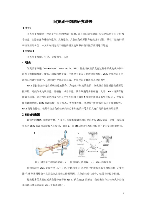
此外,越来越多新的MSCs来源也逐渐被人们发现,如图1,为MSCs的研究与应用提供了更丰富多样的供体。
a b图1.间充质干细胞的来源。
a :骨髓MSCs的提取;b :MSCs的新来源骨髓来源的MSCs来源方便,易于分离、扩增和纯化,多次传代扩增后仍具干细胞特性,无免疫排斥,体外基因转染率高并稳定高效表达外源基因,且能最终分化成骨、软骨和神经等组织。
越来越多的实验证明脐血能分离得到MSCs。
脐血MSCs的形态、免疫表型和生长方式等生物学特征与其他来源的MSCs大致类似[1]。
Cheng等从十字交叉韧带中发现了MSCs,可诱导分化为软骨细胞、脂肪细胞、骨细胞等。
人脐血间充质干细胞来源的外泌体_分离鉴定及生物学特性_张娟
中国组织工程研究 第18卷 第37期 2014–09–03出版Chinese Journal of Tissue Engineering Research September 3, 2014 Vol.18, No.37ISSN 2095-4344 CN 21-1581/R CODEN: ZLKHAH5955www.CRTER.org张娟,女,1988年生,河北省玉田县人,汉族,内蒙古医科大学免疫学在读硕士,医师,主要从事干细胞的基础与临床应用研究。
通讯作者:王彩生,硕士,主任医师,内蒙古医科大学研究生学院,内蒙古自治区呼和浩特市 010110;呼和浩特市第二医院,内蒙古自治区呼和浩特市 010031并列通讯作者:魏来,博士,教授,北京大学人民医院,北京大学肝病研究所,丙型肝炎和肝病免疫治疗北京市重点实验室,北京市 100044doi:10.3969/j.issn.2095-4344. 2014.37.009 []中图分类号:R394.2 文献标识码:A 文章编号:2095-4344 (2014)37-05955-06 稿件接受:2014-07-31Zhang Juan, Studying for master’s degree, Physician, Faculty of Graduate Studies, Inner Mongolia Medical University, Hohhot 010110, Inner Mongolia Autonomous Region, ChinaCorresponding author: Wang Cai-sheng, Master, Chief physician, Faculty of Graduate Studies, Inner Mongolia Medical University, Hohhot 010110, Inner Mongolia Autonomous Region, China; the Second Hospital of Hohhot City, Hohhot 010031, Inner Mongolia Autonomous Region, ChinaCorresponding author: Wei Lai, M.D., Professor, Peking University People’s Hospital, Peking University Hepatology Institute, Beijing Key Laboratory of Hepatitis C and Immunotherapy for Liver Diseases, Beijing 100044, ChinaAccepted: 2014-07-31人脐血间充质干细胞来源的外泌体:分离鉴定及生物学特性张 娟1,刘 峰2,张 薇2,丛 旭2,王彩生1,3,魏 来2(1内蒙古医科大学研究生学院,内蒙古自治区呼和浩特市 010110;2北京大学人民医院,北京大学肝病研究所,丙型肝炎和肝病免疫治疗北京市重点实验室,北京市 100044;3呼和浩特市第二医院,内蒙古自治区呼和浩特市 010031)文章亮点:1 研究发现间充质干细胞是通过旁分泌有效生物学活性物质外泌体在细胞间进行信息传递,进而通过某种机制发挥组织损伤修复作用。
人羊膜间充质干细胞(hAMSCs)的分离、体外培养及诱导分化
万方数据
万方数据
人羊膜间充质干细胞(hAMSCs)的分离、体外培养及诱导分化
作者:丛姗, 宋瑾, 张惠娟, 李岩, 白立恒, 曹贵方, CONG Shan, SONG Jin, ZHANG Hui-Juan, LI Yan, BAI Li-Heng, CAO Gui-Fang
作者单位:丛姗,宋瑾,张惠娟,李岩,曹贵方,CONG Shan,SONG Jin,ZHANG Hui-Juan,LI Yan,CAO Gui-Fang(内蒙古农业大学兽医学院/动物组织胚胎学与发育生物学实验室,呼和浩特,010018), 白立恒,BAI Li-Heng(内蒙古
妇幼保健医院妇产科,呼和浩特,010020)
刊名:
农业生物技术学报
英文刊名:Journal of Agricultural Biotechnology
年,卷(期):2015,23(1)
引用本文格式:丛姗.宋瑾.张惠娟.李岩.白立恒.曹贵方.CONG Shan.SONG Jin.ZHANG Hui-Juan.LI Yan.BAI Li-Heng.CAO Gui-Fang 人羊膜间充质干细胞(hAMSCs)的分离、体外培养及诱导分化[期刊论文]-农业生物技术学报 2015(1)。
人羊膜间充质干细胞分离培养:胰蛋白酶及胶原酶消化时间及浓度的选择

人羊膜间充质干细胞分离培养:胰蛋白酶及胶原酶消化时间及浓度的选择张惠娟;丛姗;梁美萍;刘俊平;黄利刚;宋瑾;曹贵方【摘要】背景:文献报道人羊膜间充质干细胞提取方法各不一致,得到的细胞数量各不相同。
目的:探索人羊膜间充质干细胞在体外分离培养的最适方法。
<br> 方法:无菌条件下取正常足月剖腹产胎儿的羊膜剪成碎片,分别通过4个实验7种方法体外培养人羊膜间充质干细胞。
①实验一:分别采用3种方法:0.05 g/L胰蛋白酶消化10 min后,再加0.75 g/L胶原酶Ⅰ消化60 min;0.75 g/L胶原酶Ⅰ直接消化120 min;0.05 g/L胰蛋白酶与0.75 g/L胶原酶Ⅰ同时消化60 min。
②实验二:采用0.05 g/L的胰蛋白酶消化30 min后,再加0.75 g/L胶原酶Ⅰ消化30 min。
③实验三:分别采用2种方法:0.05 g/L的胰蛋白酶连续2次消化30 min后,再加入0.75 g/L的Ⅰ型胶原酶消化60 min;0.05 g/L的胰蛋白酶连续2次消化40 min后,再加0.75 g/L胶原酶Ⅰ消化60 min。
④实验四:采用0.05 g/L的胰蛋白酶连续2次消化30 min后,再加1 g/L的Ⅰ型胶原酶消化60 min。
显微镜下观察其形态,研究人羊膜间充质干细胞在体外分离培养的最适方法。
<br> 结果与结论:用0.05 g/L的胰蛋白酶连续消化2次每次消化30 min,然后用1 g/L的胶原酶消化60 min是最合适的体外分离培养条件。
细胞成细长梭形或星形,胞质丰富,细胞核呈圆形,1-3个核仁。
说明实验四采用的胰酶和胶原酶消化的时间合适,而且胶原酶的浓度合适,得到的细胞数量最多。
%BACKGROUND:Extraction methods of human amniotic mesenchymal stem cells are inconsistent in the number of cells. <br> OBJECTIVE:To explore the optimal method to in vitro isolate and culture human amniotic mesenchymal stem cells. <br> METHODS:Under sterile conditions, ful-term cesarean fetal amniotic membrane was cut into pieces, then to isolate human amniotic mesenchymal stem cells by seven methods in four experiments. In experiment 1, human amniotic mesenchymal stem cells were isolated by the fol owing three methods:(1) 0.05 g/L trypsin digestion for 10 minutes fol owed by 0.75 g/L col agenase digestion for 60 minutes;(2) 0.75 g/L col agenase I for 120 minutes;(3) co-digestion with 0.05 g/L trypsin and 0.75 g/L col agenase for 60 minutes. In experiment 2, the samples were digested with 0.05 g/L trypsin digestion for 30 minutes fol owed by 0.75 g/L col agenase digestion for 30 minutes. In experiment 3, the samples were digested by two methods:(1) 0.05 g/L trypsin digestion for 30 minutes×2, fol owed by 0.75 g/L col agenase digestion for 60 minutes;(2) 0.05 g/L trypsin digestion for 40 minutes×2, fol owed by 0.75g/L col agenase digestion for 60 minutes. In experiment 4, the samples were digested with 0.05 g/L trypsin digestion for 30 minutes×2, fol owed by 1 g/L col agenase digestion for 60 minutes. Folowing morphology observation under a microscope, we studied the most suitable method for isolating human amniotic mesenchymal stem cells. <br> RESULTS AND CONCLUSION:Digestion with 0.05 g/L trypsin for 30 minutes twice fol owed by 1 g/L of col agenase digestion of 60 minutes was the most suitable isolation and culture condition in vitro. cells became elongated fusiform or star-shaped with rich cytoplasm, and nuclei were round with 1-3 nuts. We can harvest the most number of human amniotic mesenchymal stem cells using the method described in experiment 4.【期刊名称】《中国组织工程研究》【年(卷),期】2014(000)006【总页数】6页(P944-949)【关键词】干细胞;培养;间充质干细胞;羊膜间充质干细胞;细胞培养;胰蛋白酶;胶原酶;863项目【作者】张惠娟;丛姗;梁美萍;刘俊平;黄利刚;宋瑾;曹贵方【作者单位】内蒙古农业大学动物组织胚胎与发育生物学实验室,内蒙古自治区呼和浩特市 010018;内蒙古农业大学动物组织胚胎与发育生物学实验室,内蒙古自治区呼和浩特市 010018;内蒙古农业大学动物组织胚胎与发育生物学实验室,内蒙古自治区呼和浩特市 010018;内蒙古农业大学动物组织胚胎与发育生物学实验室,内蒙古自治区呼和浩特市 010018;内蒙古农业大学动物组织胚胎与发育生物学实验室,内蒙古自治区呼和浩特市 010018;内蒙古农业大学动物组织胚胎与发育生物学实验室,内蒙古自治区呼和浩特市 010018;内蒙古农业大学动物组织胚胎与发育生物学实验室,内蒙古自治区呼和浩特市 010018【正文语种】中文【中图分类】R394.20 引言 Introduction干细胞是一种能够自我更新且未分化的细胞,在一定的条件下,干细胞能够分化成为多种成熟细胞类型。
普利莱基因技术线粒体提取试剂盒说明书

升级版线粒体提取试剂盒C0010描述:线粒体制备试剂盒(Mitochondria Isolation Kit)用于从组织或培养细胞中分离线粒体和细胞胞浆成分。
加入分离溶液,匀浆破碎组织细胞,经过数次800g和12000g离心,在60分钟内即可分离出完整的线粒体和胞浆成分。
制备的线粒体具有很高的生物学活性,可进行各种功能研究如酶学测定,更可用于Western Blot、2D-胶、线粒体蛋白或DNA提取、蛋白质组学等研究。
严格按照说明操作,总是能制备获得高纯度线粒体。
一篇方法学研究论文发现,用普利莱试剂盒制备线粒体的得率、活性、纯度优于蔗糖密度梯度离心法和Invotrogen/Pierce线粒体提取试剂盒方法。
适用:从组织、培养细胞制备高纯度线粒体,同时分离细胞胞浆成分。
组成:Mito Solution100ml for50次制备200ml for100次制备储存:−20ºC12个月有效操作步骤:以下所有操作均在4ºC进行1.组织匀浆:100~200mg新鲜组织如肝、脑、肾、心肌等,剪为0.5cm2碎块放入小容量玻璃匀浆器内。
估计组织块总体积。
加入1.5ml冰预冷的Mito Solution。
用间隙严紧的研杵上下研磨组织20次。
培养细胞匀浆:800×g5min离心收集细胞。
单次提取需2-5×10⁷个细胞。
加入1.5ml冰预冷Mito Solution 重悬细胞,将细胞悬液转移到小容量玻璃匀浆器内,用间隙严密的研杵研磨细胞30次。
2.将匀浆液转移到离心管中,800×g,4ºC离心5min。
(胞核、膜碎片、未裂解细胞等在管底,弃去)3.收集上清液并转移到新的离心管。
再次800×g离心5min at4ºC,弃沉淀。
4.将上清液转移到新的离心管。
10,000×g离心10min4ºC。
线粒体沉淀在管底。
离心后的上清含胞浆成分,可收集用于对照实验。
间充质细胞汇总
间充质干细胞研究进展【摘要】间充质干细胞是一种源于中胚层的早期干细胞,具有多向分化潜能,特定的条件下可分化为骨细胞、软骨细胞和神经细胞等,支持造血,具备低免疫原性和免疫调节活性,具有广泛的科研和临床应用价值。
本文针对间充质干细胞的研究进展和在临床医学应用进行综述。
【关键词】间充质干细胞、分化、免疫调节、应用1 引言间充质干细胞(mesenchymal stem cells,MSC)就是指在胚胎发育过程中形成的成体间叶组织(如骨髓基质、脂肪、胎盘和脐带等)中留存下来未分化的原始细胞。
MSCs主要存在于结缔组织和器官间质中,以骨髓中含量最为丰富,少量存在于血液及其他组织中。
MSCs承担着支持造血系统细胞的使命,为造血干细胞的生长、分化及自我更新提供重要的微环境,还能分化为肌细胞、肝细胞、成骨细胞、软骨细胞等多种细胞。
此外,MSCs还具有免疫调节功能,通过细胞间的相互作用及产生细胞因子抑制T细胞的增殖及其免疫反应,发挥免疫重建的功能。
MSCs来源方便,易于分离、扩增和纯化,多次传代扩增后仍具有干细胞特性。
MSCs的这些特性,使其在自身免疫性疾病治疗和细胞治疗等方面具有广阔的临床应用前景。
2 MSCs的来源最常见的MSCs来源是骨髓。
外周血、脂肪和胎盘等组织也可进行MSCs提取。
此外,越来越多新的MSCs来源也逐渐被人们发现,如图1,为MSCs的研究与应用提供了更丰富多样的供体。
a b图1.间充质干细胞的来源。
a :骨髓MSCs的提取;b :MSCs的新来源骨髓来源的MSCs来源方便,易于分离、扩增和纯化,多次传代扩增后仍具干细胞特性,无免疫排斥,体外基因转染率高并稳定高效表达外源基因,且能最终分化成骨、软骨和神经等组织。
越来越多的实验证明脐血能分离得到MSCs。
脐血MSCs的形态、免疫表型和生长方式等生物学特征与其他来源的MSCs大致类似[1]。
Cheng等从十字交叉韧带中发现了MSCs,可诱导分化为软骨细胞、脂肪细胞、骨细胞等。
BMSCs的分离培育纯化与鉴定
BMSCs的分离培育、纯化与鉴定马民吕志刚杜以宽张桂娟马义【摘要】目的成立一种持续、稳固的可多向分化的骨髓间充质干细胞(BMSCs)体外分离培育体系。
方式运用密度梯度离心法从1月龄新西兰兔骨髓中分离培育BMSCs,利用相差显微镜观看其形态及生长情形,扫描电镜观看其细胞结构,运用流式细胞术分析分离细胞群所处细胞周期和细胞活力,用MTT比色法绘制细胞生长曲线,并用特定诱导液将分离的BMSCs向成骨细胞和成脂肪细胞定向诱导分化,利用ALP和油红O进行染色鉴定。
结果所分离的BMSCs细胞在形态学观看与生长动力学上均符合BMSCs特点,分离培育的BMSCs细胞在第3天进入对数生长期,第10天进入平台期;在成骨、成脂肪的诱导培育条件下,别离显现成骨、成脂肪表型特点,可进一步定向分化,结论所收成的细胞具有BMSCs的特异性。
【关键词】骨髓间充质干细胞;分离;成骨分化;成脂分化【Abstract】 Objective To establish a sustained and stable bone marrow derived mesenchymal stem cells (BMSCs) isolation and culture system which could be pluripotential in vitro. Methods BMSCs were isolated and cultured from the 1month old New Zealand white rabbits through density gradientcentrifugation. Growth and ultromicrostructure of the BMSCs were observed by light microscope and scan electron microscope. The cell activities and cell cycle were observed by flow cytometry. The grow curve of the BMSCs was described through MTT assay. BMSCs were induced and differentiate into osteoblasts and adipocytes by specific induction culture medium. Then the cells were stained with alkaline phosphatase (ALP) and Oil red O , and analyzed the result of all staining. Results The isolated BMSCs cells were in line with the characteristics of BMSCs in morphology and growth kinetics, and the isolated BMSCs cells would be into the logarithmic growth phase in the third day and into the platform phase in the tenth day. Conclusions In the osteogenic and adipogenic induced culture conditions, BMSCs have appeared the phenotypic characteristics of osteogenic and adipogenic respectively, and could be further directed differentiation. These indicated the harvested cells possessed BMSCs specificity.【Key words】 Bone marrow derived mesenchymal stem cells(BMSCs); Isolation; Osteogenic differentiation; Adipogenic differentiation软骨组织工程技术〔自体软骨细胞的移植、生长因子对软骨细胞的作用、骨髓间充质干细胞(又称骨髓基质干细胞,BMSCs)在软骨组织工程中的作用、转基因技术〕是现今的研究热点〔1,2〕。
间充质干细胞

MSC在自身免疫系统疾病中的应用
治疗基础:自身免疫系统疾病(系统性 红斑狼疮、类风湿性关节炎、甲状腺机能 亢进、I型糖尿病等)目前方法是使用免疫 抑制药物抑制免疫系统表达。MSC具有良好 的免疫调节作用。
MSC在心血管疾病治疗中的应用
*机制:分化为心肌细胞;促进血管新 生;旁分泌机制;抑制心室重构;激 活内源性修复机制,引起干细胞龛的 再生 。
++
来源易,细胞
数量较多,但
+
++
++
++ 体外优化培养
后是否能适应
体内的环境还
有待研究
常 间充见质干干细细胞
间充质干细胞克服了其 他一些干细胞的缺陷
不容易获得 分离困难
伦理学问题
单向分化
All kill!
MSC主要功能
免疫
调节
支持
促血管
造血
新生
基因 组织 治疗 修复
组织修复:(骨骼肌系统再生)
Bad News
一位患有狼疮性肾炎的患者,接受了干细胞治 疗。患者在接受治疗后就出现了肾衰竭,检查 后发现,在接受干细胞注射的肾脏部位出现了 奇怪的肿块与损伤,其后证明它们是骨髓和血 管组织的混合物。此外,她的肝脏和肾上腺也 出现了相同的肿块。两年后她因肾坏死死亡。 对于这些肿块的一个解释是:这些肿块来自注 射的干细胞。
*临床应用:急性心肌梗死;缺血性心 肌病;慢性心功能不全。
MSC在神经系统疾病中的应用
*临床应用:新生儿缺氧-缺血性脑病(HIE小儿脑 瘫);神经变性疾病,如肌萎缩侧索化症(ALS)、帕 金森病、阿尔茨海默病等。
*干细胞可释放神经营养因子及重建运动神经元微环境 等机制来保护濒死的运动神经元。
小鼠骨髓间充质干细胞的分离培养与鉴定
小鼠骨髓间充质干细胞的分离培养与鉴定发表时间:2012-05-24T09:50:06.677Z 来源:《医药前沿》2012年第1期供稿作者:林芸1 蔡鹏威2 陈为民1 孟春3[导读] 分离、培养符合实验要求的小鼠骨髓间充质干细胞并进行鉴定,为进一步的研究打基础。
林芸1 蔡鹏威2 陈为民1 孟春3( 1 福建医科大学省立医院临床学院血液科福建福州 3 5 0 0 0 1 )( 2 福建省立医院检验科福建福州 3 5 0 0 0 1 )( 3 福州大学生物工程学院福建福州 3 5 0 0 0 1 )【摘要】目的分离、培养符合实验要求的小鼠骨髓间充质干细胞并进行鉴定,为进一步的研究打基础。
方法采用贴壁培养法培养小鼠骨髓间充质干细胞,观察细胞的形态及生长特性,并应用流式细胞仪对细胞表面抗原CD34、CD45、CD29、CD44进行表型鉴定。
结果原代分离的间充质干细胞在接种后培养24小时,细胞开始贴壁,胞体呈圆形或多边形,培养第3-5天,细胞开始较紧密贴附壁上并开始有细胞呈梭形,培养第7天开始观察到细胞分裂,随着细胞数的增加,细胞的生长速度变快,逐渐成漩涡状排列,培养第12天贴壁细胞长满瓶底的80%,并融合成片。
传代后细胞生长迅速,培养7天左右即可长满瓶底的80%。
传至10代仍具有良好的增殖活性。
流式细胞仪检测第4代及第8代MSCs细胞均不表达CD34、CD45,但表达CD29、CD44,纯度分别为73.8% 、91.65%。
结论采用贴壁培养法可获得生长状态良好、增殖能力强的间充质干细胞,随传代数增加,其纯度增加,且该方法简单、实用。
【关键词】骨髓间充质干细胞细胞培养流式细胞术表型鉴定【中图分类号】R392.2 【文献标识码】A 【文章编号】2095-1752(2012)01-0082-02间充质干细胞(mesenchymal stem cells,MSCs)起源于中胚层,具有高度增殖和自我更新的能力,有向骨、软骨、脂肪、血管内皮细胞、神经星型胶质细胞等分化的潜能[1],可分化成骨髓基质支持造血,并可分泌多种细胞因子促进造血干细胞增殖分化,同时它能抑制同种异体反应性T淋巴细胞,在同种异基因造血干细胞移植后的造血重建及免疫调节,预防移植物抗宿主病等方面有广阔的应用前景[2],但骨髓间充质干细胞含量极低,仅占骨髓单个核细胞的0.001%-0.010%[3],因此,培养出生长状态良好,足够数量的骨髓间充质干细胞是应用的前提。
人脐带华通氏胶间充质干细胞的分离_培养_鉴定及冻存_复苏
MSCs 消化后以 1×10⁶/ml 密度冻存, 冻存液 DMSO∶ FBS∶DMEM 为 1∶1∶8,将冻存细胞放入 4 ℃预冷的程 序冷冻盒内,直接将程序冷冻盒放入-80 ℃冰箱,第 2 天放入-196 ℃液氮保存。 冻存 6 个月后将程序冷 冻盒从-80 ℃冰箱取出,细胞直接放入 37 ℃预热的 水浴锅内,用 4 ℃预冷的 PBS 洗涤细胞 2 遍,将细胞 接种于含 10%胎 牛血清的 DMEM 培 养 瓶 内 , 放 入 37 ℃、5%CO2 培养箱中培 养。 待细胞传 代至 P3 代 时, 用流式细胞 仪检测 CD14、CD45、CD79a、CD90、 CD34、CD73、CD105、HLA-DR,并 进 行 成 骨 细 胞 、成 脂细胞诱导分化,方法同前。
YANG Xiaoqing1**, ZHANG Mu2, YANG Bing3, ZGANG Hu3, ZHANG Yuquan1***
(1Department of Gynaecology and Ob-
sterics, Affiliated Hospital of Nantong University; 2Department of Clinical Medical class 071, Medical College of Nantong U-
来源 MSCs 具有来 源广泛 、取 材 方 便 、相 对 纯 净 、含 量丰富及免疫原性低等优点, 正逐渐成为 MSCs 研 究领域的热点之一。 人脐带 MSCs 的来源有 4 种,其 中华通氏胶(WJ)来源的 MSCs 含量丰富但对其 分离 培养的研究相对较少, 本文就探讨从人脐带华通氏 胶 中 分 离 出 脐 带 间 充 质 干 细 胞 (umbilical cord mesenchymal stem cells, UC-MSCs)并进行体外培养、鉴 定及定向分化,为 MSCs 的应用提供更广泛的来源。
Mesenchymal stem cell
Mesenchymal stem cellFrom Wikipedia, the free encyclopediaJump to: navigation, search Mesenchymal stem cellMesenchymal stem cell showing typical ultrastructural morphology.Latin cellula mesenchymatica precursoriaCode TH H2.00.01.0.00008Mesenchymal stem cells, or MSCs, are multipotent stromal cells that can differentiate into a variety of cell types,[1] including: osteoblasts (bone cells),[2] chondrocytes (cartilage cells),[3] and adipocytes (fat cells). This phenomenon has been documented in specific cells and tissues in living animals and their counterparts growing in tissue culture.DefinitionWhile the terms Mesenchymal Stem Cell and Marrow Stromal Cell have been used interchangeably, neither term is sufficiently descriptive as discussed below:Mesenchyme is embryonic connective tissue that is derived from the mesoderm and that differentiates into hematopoietic and connective tissue, whereas MSCs do not differentiate into hematopoietic cells.[4]Stromal cells are connective tissue cells that form the supportive structure in which the functional cells of the tissue reside. While this is an accurate description for one function of MSCs, the term fails to convey the relatively recently-discovered roles of MSCs in the repair of tissue.[5] Because the cells, called MSCs by many labs today, can encompass multipotent cells derived from other non-marrow tissues, such as umbilical cord blood, adipose tissue, adult muscle, corneal stroma[6] or the dental pulp of deciduous baby teeth, yet do not have the capacity to reconstitute an entire organ, the term Multipotent Stromal Cell has been proposed as a better replacement.4.^ Porcellini Adolfo. Regenerative medicine: a review. Rev. Bras. Hematol. Hemoter. [serial on the Internet]. [cited 2011 Dec 11]. Available from: http://www.scielo.br/scielo.php?script=sci_arttext&pid=S1516-84842009000800017&lng=en. /10.1590/S1516-848420090008000175.^ "PLoS ONE: Eccentric Exercise Facilitates Mesenchymal Stem Cell Appearance in Skeletal Muscle." PLoS ONE : Accelerating the Publication of Peer-reviewed Science. Available from: </article/info:doi/10.1371/journal.pone.0029760>.6.^ Branch, M. J. et al. 2012 Mesenchymal Stem Cells in the Human Corneal Limbal Stroma /content/early/2012/06/25/iovs.11-8673.abstractThe youngest, most primitive MSCs can be obtained from the umbilical cord tissue, namely Wharton's jelly and the umbilical cord blood. However the MSCs are found in much higher concentration in the Wharton’s jelly compared to the umbilical cord blood, which is a rich source of hematopoietic stem cells. The umbilical cord is easily obtained after the birth of the newborn, is normally thrown away and poses no risk for collection. The umbilical cord MSCs have more primitive properties than other adult MSCs obtained later in life, which might make them a usefulsource of MSCs for clinical applications.An extremely rich source for mesenchymal stem cells is the developing tooth bud of the mandibular third molar. While considered multipotent, they may prove to be pluripotent. The stem cells eventually form enamel, dentin, blood vessels, dental pulp, nervous tissues, including a minimum of 29 different unique end organs. Because of extreme ease in collection at 8–10 years of age before calcification and minimal to no morbidity they will probably constitute a major source for personal banking, research and multiple therapies. These stem cells have been shown capable of producing hepatocytes. Additionally, amniotic fluid has been shown to be a very rich source of stem cells. As many as 1 in 100 cells collected from and genetic amniocentesis has been shown to be a pluripotent mesenchymal stem cell.[citation needed]Adipose tissue is one of the richest sources of MSCs. When compared to bone marrow, there is more than 500 times more stem cells in 1 gram of fat when compared to 1 gram of aspirated bone marrow. Adipose stem cells are currently actively being researched in clinical trials for treatment in a variety of diseases.HistoryIn 1924, Russian-born morphologist Alexander A. Maximow used extensive histological findings to identify a singular type of precursor cell within mesenchyme that develops into different types of blood cells.[7]Scientists Ernest A. McCulloch and James E. Till first revealed the clonal nature of marrow cells in the 1960s.[8][9] An ex vivo assay for examining the clonogenic potential of multipotent marrow cells was later reported in the 1970s by Friedenstein and colleagues.[10][11] In this assay system, stromal cells were referred to as colony-forming unit-fibroblasts (CFU-f).Subsequent experimentation revealed the plasticity of marrow cells and how their fate could be determined by environmental cues. Culturing marrow stromal cells in the presence of osteogenic stimuli such as ascorbic acid, inorganic phosphate, and dexamethasone could promote their differentiation into osteoblasts. In contrast, the addition of transforming growth factor-beta (TGF-b) could induce chondrogenic markers.[citation needed]7.^ Sell, Stewart (Stem cell handbook). Humana Press. p. 143.8.^ Becker, A. J.; McCulloch, E. A.; Till, J. E. (1963). "Cytological Demonstration of the Clonal Nature of Spleen Colonies Derived from Transplanted Mouse Marrow Cells". Nature 197 (4866): 452–4. doi:10.1038/197452a0. PMID 13970094.9.^ Siminovitch, L.; McCulloch, E. A.; Till, J. E. (1963). "The distribution of colony-forming cells among spleen colonies". Journal of Cellular and Comparative Physiology 62 (3): 327–36. doi:10.1002/jcp.1030620313. PMID 14086156.CulturingThe majority of modern culture techniques still take a colony-forming unit-fibroblasts (CFU-F) approach, where raw unpurified bone marrow or ficoll-purified bone marrow Mononuclear cell are plated directly into cell culture plates or flasks. Mesenchymal stem cells, but not red blood cells or haematopoetic progenitors, are adherent to tissue culture plastic within 24 to48 hours. However, at least one publication has identified a population of non-adherent MSCs that are not obtained by the direct-plating technique.[22]Other flow cytometry-based methods allow the sorting of bone marrow cells for specific surface markers, such as STRO-1.[23] STRO-1+ cells are generally more homogenous, and have higher rates of adherence and higher rates of proliferation, but the exact differences between STRO-1+ cells and MSCs are not clear.[24]Methods of immunodepletion using such techniques as MACS have also been used in the negative selection of MSCs.[25]22.^ Wan, Chao; He, Qiling; McCaigue, Mervyn; Marsh, David; Li, Gang (2006). "Nonadherent cell population of human marrow culture is a complementary source of mesenchymal stem cells (MSCs)". Journal of Orthopaedic Research 24 (1): 21–8. doi:10.1002/jor.20023. PMID 16419965.23.^ Gronthos, S; Graves, SE; Ohta, S; Simmons, PJ (1994). "The STRO-1+ fraction of adult human bone marrow contains the osteogenic precursors". Blood 84 (12): 4164–73. PMID 7994030. /cgi/pmidlookup?view=long&pmid=7994030.24.^ Oyajobi, Babatunde O.; Lomri, Abderrahim; Hott, Monique; Marie, Pierre J. (1999). "Isolation and Characterization of Human Clonogenic Osteoblast Progenitors Immunoselected from Fetal Bone Marrow Stroma Using STRO-1 Monoclonal Antibody". Journal of Bone and Mineral Research 14 (3): 351–61. doi:10.1359/jbmr.1999.14.3.351. PMID 10027900.25.^ Tondreau, T; Lagneaux, L; Dejeneffe, M; Delforge, A; Massy, M; Mortier, C; Bron, D (1 January 2004). "Isolation of BM mesenchymal stem cells by plastic adhesion or negative selection: phenotype, proliferation kinetics and differentiation potential". Cytotherapy 6 (4): 372–379. doi:10.1080/14653240410004943.。
间充质干细胞能量代谢 力学
间充质干细胞能量代谢力学间充质干细胞(mesenchymal stem cells,MSCs)是一类来源于胚胎或成体组织的多潜能干细胞,具有自我更新和多向分化能力。
它们广泛存在于人体各个组织中,包括骨髓、脂肪、骨骼、肌肉、心脏和脑组织等。
能量代谢是维持细胞生存和生物学功能正常运转的重要过程。
间充质干细胞的能量代谢与其生物学特性密切相关。
在干细胞的能量代谢中,主要有两个重要的通路:糖酵解和线粒体三磷酸腺苷(ATP)生成。
糖酵解是一种不需要氧气的代谢途径,能够将葡萄糖分解为丙酮酸和乳酸,并生成少量的ATP。
这一过程被称为厌氧糖酵解,因为它在缺氧条件下进行。
糖酵解对于维持间充质干细胞的自我更新和增殖非常重要,因为干细胞生长需要大量的能量供应。
此外,糖酵解还可以产生丙酮酸,可以进一步参与线粒体三羧酸循环。
线粒体是细胞内的一个重要细胞器,负责产生ATP,维持细胞的能量供应。
线粒体内的线粒体呼吸链(mitochondrial respiratory chain,MRC)能够通过氧化还原反应从葡萄糖、脂肪酸和氨基酸等物质中释放大量的ATP。
线粒体的功能受到细胞内和细胞外环境的影响。
在正常细胞中,线粒体的形状、结构和数量可根据能量需要进行调节。
间充质干细胞的线粒体功能在其能量代谢和多向分化中起着重要作用。
线粒体功能障碍或线粒体损伤会导致线粒体功能减弱,ATP生成减少,细胞能量供应不足,从而影响干细胞的生物学活性。
因此,维持线粒体功能的稳定对于间充质干细胞的功能维持和自我更新非常重要。
除了糖酵解和线粒体呼吸链之外,脂肪酸氧化也是间充质干细胞的一个重要能量代谢途径。
脂肪酸氧化是指将脂肪酸分解为丙酮酸,然后进入线粒体三羧酸循环,最终产生ATP。
脂肪酸氧化是一种有氧代谢过程,需要氧气的参与。
脂肪酸氧化在间充质干细胞的增殖和分化中起着重要作用。
总的来说,间充质干细胞能量代谢是一个复杂的过程,包括糖酵解、线粒体呼吸链和脂肪酸氧化等途径。
间充质干细胞是什么?
间充质干细胞是什么?间充质干细胞(mesenchymal stem cells,MSCs)应具有如下基本特征:⑴贴壁生长;⑵具有特定的表面标志,如不表达CD14、CD34、CD45、HLA-Ⅱ,但表达CD29、CD73和CD105;⑶能进行自我更新,也能在体外分化为骨、软骨和脂肪等多种细胞系。
虽然不同研究组对人脐带中分离出的细胞给出基质细胞、基质干细胞和MSCs等多种命名,但通常均具有上述基本特征,本文拟对这一来源的MSCs的生物学特征进行综述。
1、基因分析对人脐带MSCs进行基因分析表明,该细胞与造血干细胞(hematopoietic stem cells,HSCs)和胚胎干细胞(embroynic stem cells,ESCs)类似,其高表达的常见基因包括未分化的ESCs表达的基因、形态发生相关蛋白、细胞外黏附分子、神经营养因子以及3个胚层衍生的子代细胞标志物[1]。
此外,RT-PCR分析显示人脐带基质干细胞还表达多种未分化细胞标志、3个胚层和滋养外胚层相关的基因和一系列多能干细胞标志,如Nanog、Oct-4、Sox-2、Rex-1、SSEA-3、SSEA-4、Tra-1-60和Tra-1-81[2]。
2、细胞标志物的表达以流式细胞学技术、PCR技术、微点阵方法和免疫组织化学方法对脐带基质细胞的表面标志表达情况进行分析的诸多研究表明,这些细胞与其他来源的MSCs类似,表达CD10、CD13、CD29、CD44、CD49 b、CD49 c、CD49 d、CD49e、CD51、CD73、CD90、CD105、CD146、CD166、HLA-1和HLA-A,B,C等;但不表达CD14、CD31、CD33、CD34、CD38、CD45、CD56、CD123、CD133、CD235a、HLA-G、HLA-DP、HLA-DQ、HLA-DR和Strol-1[21。
3、端粒酶活性端粒酶活性在干细胞的增殖能力方面发挥重要作用。
- 1、下载文档前请自行甄别文档内容的完整性,平台不提供额外的编辑、内容补充、找答案等附加服务。
- 2、"仅部分预览"的文档,不可在线预览部分如存在完整性等问题,可反馈申请退款(可完整预览的文档不适用该条件!)。
- 3、如文档侵犯您的权益,请联系客服反馈,我们会尽快为您处理(人工客服工作时间:9:00-18:30)。
O R I G I N A L A R T I C L EOriginal ArticleMesenchymal Stem Cells:Isolation,Characterisation and In Vivo Fluorescent Dye TrackingChristopher Weir,PhD a ,b ,d ,Marie-Christine Morel-Kopp,PhD b ,d ,Anthony Gill,FRCPA c ,d ,Kellie Tinworth,MSc a ,d ,Leigh Ladd,PhD a ,d ,Stephen N.Hunyor,MD a ,d ,∗and Christopher Ward,PhD b ,daCardiac Technology Centre &Kolling Institute,Sydney,AustraliabNorthern Blood Research Centre and Department of Haematology,Sydney,Australiac Anatomical Pathology Department,Sydney,Australiad University of Sydney at Royal North Shore Hospital,Sydney,AustraliaCell therapies have been used to regenerate the heart by direct myocardial delivery,by coronary infusion and by surface attached scaffolds.Multipotent mesenchymal stem cells (MSC)with capacity to differentiate into cardiomyocytes and other cell lines have been predominantly trialled in rodents.However,large animal models are increasingly needed to translate basic research into new,safe regenerative therapies.Understanding the mode of action of cell therapies in the mammalian heart has been limited by cell tracking capability.This study examined the ability to track the fate of allogeneic MSC in sheep using various fluorescent dyes.MSC isolated from sheep bone marrow were grown in culture following extraction and flow cytometric characterisation.After labelling with fluorescent tracking dyes (e.g.CFSE and DiI)cells were tested for in vitro and in vivo signal up to six belling effect on cell division and differentiation was studied.Several dyes lost fluorescence and slowed cell division.However,the thiol reactive dye CM-DiI showed detectable in vivo fluorescence in labelled MSC six weeks after injection into sheep skeletal muscle and two weeks after implantation of an MSC coated biomaterial scaffold.CM-DiI labelled MSC differentiated in vitro showed label retention over four weeks.The fluorescent membrane dye CM-DiI tracks implanted sheep MSC and provides an alternative to traditional cell markers such as gene modified GFP .(Heart,Lung and Circulation 2008;17:395–403)©2008Australasian Society of Cardiac and Thoracic Surgeons and the Cardiac Society of Australia andNew Zealand.Published by Elsevier Inc.All rights reserved.Keywords.Mesenchymal stem cells (MSC);CM-DiI;Cell division and differentiation;Sheep;Biomaterials;ScaffoldIntroductionCoronary artery disease leading to myocardial ischaemia and infarction is characterised by loss of cardiomyocytes,with frequent progression to heart failure whose incidence has increased markedly due to coronary angioplasty/stenting,bypass surgery and pharmacotherapy.Stem cell therapy has been proposed as a repair mechanism,preferably before hearts enter a phase of rapid decline in pump function.Received 13December 2007;received in revised form 15January 2008;accepted 20January 2008;available online 8April 2008∗Corresponding author at:Cardiac Technology Centre,Depart-ment of Cardiology,Royal North Shore Hospital,St Leonards,Sydney,NSW 2065,Australia.Tel.:+61299268679;fax:+61299014097.E-mail address:stephenh@.au (S.N.Hunyor).The potential for mesenchymal stem cells (MSC)to dif-ferentiate into specialised cell lineages,such as osteocytes,adipocytes and chondrocytes has been documented,1as has differentiation into cardiomyocytes in vitro .2However,there is evidence that MSC exert their beneficial effect pre-dominantly via paracrine mechanisms resulting from cell death.3Short-term in vivo rat studies show that MSC injected into damaged myocardium adopt characteristics of car-diomyocytes and occupy a major part of the damaged area.4In another study Orlic et al.5mobilised bone marrow (BM)cells in mice with myocardial infarction by inject-ing granulocyte colony stimulating factor (G-CSF),with cells localised in the infarcted zone generating myocardial cells.Despite such promise,doubts persist concerning long-term cell survival,and reliable MSC markers and evidence of differentiation into cardiomyocytes is lacking,especially©2008Australasian Society of Cardiac and Thoracic Surgeons and the Cardiac Society of Australia and New Zealand.Published by Elsevier Inc.All rights reserved.1443-9506/04/$30.00doi:10.1016/j.hlc.2008.01.006ORIGINAL ARTICLE 396Weir et al.Heart,Lung and Circulation Fluorescent stem cell tracking2008;17:395–403in large animal models.6Sheep appear particularly suit-able for refining stem cell techniques for heart repair.Current cellular therapies involve taking either uns-elected autologous BM cells7or homogenous antibodyselected‘specific’sub-populations,8which aim pre-dominantly for vasculogenesis in ischaemic or necroticmyocardium.In our sheep we used MSC adherenceselection and growth under standardised conditions toprovide a cell population capable of differentiation intocardiomyocytes.7,9Work on human MSC has utilised spe-cific antibody labelling(e.g.STRO-1,V-CAM)to isolateand define particular cells10but use of specific selectedcells for cardiac repair in animals is limited.3Because avail-able specific ovine monoclonal antibodies do not reactwith sheep MSC(except anti-ovine CD44),we examinedlabelling and tracking of MSC withfluorescent ingdyes such as CFSE or CM-DiI(Molecular Probes,USA)we optimised ovine MSC labelling in vitro,noted its effecton cell division and studied dye retention in differenti-ating MSC.Having defined optimal in vitro staining,weassessed in vivo dye stability following MSC implantationdirectly into muscle,or subcutaneously when grown on abiomaterial scaffold.While recent work suggests that adult skin cells can begenetically modified to recreate pluripotent cells resem-bling hESCs,11their clinical application appears remote.Exploring the full potential of non-immunogenic,read-ily available MSC for heart repair is therefore warranted.This study focused on the potential of CM-DiI as an MSCtracking dye for use in heart regeneration.Materials and MethodsMerino-cross sheep(42.5–44.5kg)received humane carein compliance with the Guide for the Care and Use of Labora-tory Animals(US National Institutes of Health,publication85–23,revised1996)in a protocol approved by the Institu-tional Animal Care and Ethics Committee.Surgical PreparationSterile surgery was performed under alfaxalone(1–1.5mg/kg)induced general anaesthesia maintainedwith 1.5–2%isoflurane in40%O2.Following intuba-tion expired CO2was maintained at30–35mmHg.Nopost-operative drug therapy was given.Bone Marrow(BM)ExtractionBM was collected aseptically into Na2EDTA from thesheep’s iliac crest under general anaesthetic.The buffycoat was isolated by centrifugation(2500×g,10min)andresuspended into1.5ml PBS.A500l sample served forflow cytometric analysis and residual for culture(A).Thebuffy coat(1ml)wasfiltered through a meshfilter(Bec-ton Dickinson,USA)and residualfiltrate washed off withsterile PBS;half that cell suspension was cultured(B).Thefiltered buffy coat left(500l)was layered onto10mlFicoll(Amersham Bioscience,Uppsala,Sweden)and cen-trifuged(1200×g,20min).Cells at the interface wereremoved,washed twice in sterile PBS and split into twofractions(500l).One fraction was cultured directly(C)and the other selected for CD106(V-CAM)(D)usingindirect immuno-magnetic beads(MACs TM microbeads,Miltenyti Biotech,Bergish Gladbach,Germany)accordingto manufacturer’s protocol.All cell groups were culturedin DMEM with10%foetal calf serum(FCS),1×antibiotics(penicillin,streptomycin)and5mM L-glutamine.Bone Marrow AnalysisA small BM sample was analysed for anti-ovine markersCD44,CD45,CD34,CD11b,CD31(Serotec,Oxford,UK)and the anti-human marker CD106(BD Pharmingen TM,San Diego,USA).Briefly,30l of complete marrow wasadded to the antibodies and incubated at room tempera-ture for15min in the dark.Cells werefixed(0.5ml10%formaldehyde,0.6ml10×HBSS and0.9ml dH2O)for10min,400l dH2O added and red blood cells allowed tolyse for10min at4◦C,followed byflow cytometric analysis.Cultured MSC AnalysisAfter culturing the cells in DMEM with10%FCS and peni-cillin and streptomycin as antibiotics(Trace Biosciences)for1–2weeks until each fraction had reached confluencein a75cm2flask,MSC from each sample were analysedbyflow cytometry using anti-ovine markers CD44,CD45and CD11b and anti-human CD106.Carboxyfluorescein Diacetate,Succinimidyl Ester(CFSE)LabellingCFSE dye stock was made up to a concentration of100M/ml in DMSO as recommended by manufacturer(Molecular Probes,Oregon,USA).MSC were detachedusing trypsin and resuspended at a concentration of1×107cells/ml in PBS and then labelled at1M,2Mand4M concentrations(6×10⁶cells)for10min at37◦Cbefore washing in PBS and putting back into culture formonitoring offluorescent staining retention over a periodof two weeks using unlabelled control MSC.Chloromethylbenzamido-1,1 -Dioctadecyl-3,3,3 3 -Tetramethylindocarbocyanine Perchlorate(CM-DiI;Lipophilic Carbocyanine Dye)LabellingCM-DiI stock was made up at a1mg/ml concentrationin ethanol as recommended by manufacturer(MolecularProbes,USA)and MSC staining at4M,6M and8Mconcentrations was performed for15or30min at37◦Cor room temperature,andfinally for15min at4◦C to opti-mise staining levels.All staining was performed on75cm2flasks with a confluent layer of3–4×belledcells were cultured,maintained at sub-confluence andmonitored forfluorescence levels overfive weeks usingflow belled and unlabelled control MSCwere monitored for cell growth with cell numbers countedusing a haemocytometer.CM-DiI Labelling Effect on Cell Growth and ViabilityT o assess labelling effect on cell growth aflask of MSCs waslabelled with4M CM-DiI for15min at37◦C and4◦C.A plate of unlabelled cells served as control.Cells fromO R I G I N A L A R T I C L EHeart,Lung and Circulation Weir et al.3972008;17:395–403Fluorescent stem cell trackingTable 1.Staining Characteristics and Follow-up of MSCs ±Biomaterial Scaffold Implanted Directly into Muscle or Subcutaneously in SheepImplantation SiteT wo WeeksSix Weeks(i)MSC directly into muscle2×106MSC in 0.2ml serum-free DMEMLateral gastrocnemiusPeak staining group (stained 2days pre-injection)7.5×10⁷CM-DiI labelled MSCs in serum-free DMEM (high dose).Cranial tibial CM-DiI stained group (stained on the day) 2.5×10⁶unlabelled MSC in 0.4%trypan blue (low dose)Dorsal gastrocnemius Control unlabelled MSC1×107CM-DiI labelled MSC in 0.4%trypan blue (low dose)Central cranial tibial 7.5×10⁶MSC in serum-free DMEM (high dose)(ii)MSC on biomaterialCranial dorsal region Control unlabelled MSC2×106unlabelled MSCMiddle dorsal regionCM-DiI labelled MSC (stained 2days pre-injection)0.5×10⁶CM-DiI labelled MSC spiked on biomaterial (LD)Caudal dorsal regionCM-DiI labelled MSC +additional 2×10⁶cells1.5×107CM-DiI labelled MSC (HD)both flasks were washed in PBS and detached using 0.25%trypsin-EDTA.Exactly 100cells were sorted by flow cytom-etry based on FL-2v SSC both for CM-DiI labelled and unlabelled cells in triplicate groups for each day (i.e.days 4,7,14)and placed on six-well plates.On sampling days media was removed and cells washed in sterile PBS before treatment,spun down (6000rpm/1min)and counted using a haemocytometer.In Vivo Two-and Six-Week CM-Dil Label Retention Trials (T able 1)Flasks of MSC were grown to confluence and stained with 4M CM-DiI.A sample of cells showed pre-injection staining level.MSC were also cultured for two weeks in DMEM and 10%FCS with Penicillin-Streptomycin on a prosthetic silicone biomaterial scaffold that has random sized and random shaped cavities in its matrix structure (Seare Matrix ®,Seare Biomatrix Systems,Inc,Salt Lake City,USA).The biomaterial had been sterilised and then cut into circular sections to cover the whole well of a six-well plate.The cells were stained on the biomaterial two days prior to its implantation.This served to establish proof of principle for MSC growth in this matrix material and also to provide a substrate for in vivo MSC tracking studies.Extra labelled cells were placed on one bioma-terial sample prior to its insertion back into the sheep.A negative control consisted of biomaterial plus unla-belled MSC.The biomaterial was inserted subcutaneously in sheep that also had direct MSC injection.Six-week in vivo CM-DiI labelling persistence served to mimic the timeframe of ovine myocardial infarct repair.Differentiation of MSCMultipotent capacity of ovine MSC was proven after in vitro culturing with specific supplements by induc-ing differentiation into chondrogenic and cardiomyocyte phenotypes.Samples were compared with control cells from the same cell lineage cultured without differentiating supplement.Chondrogenic supplement comprised 1%ITS-premix (BD Biosciences,Australia),100M ascorbate-2phosphate,10−7M dexamethasone and 10ng/ml TGF-1.12Cardiomyocyte supplement was 3M/l 5-azacytidine (Sigma,Australia).2Differentiation of CM-DiI MSCAfter CM-DiI dye labelled cells were induced to differ-entiate,chondrogenic and cardiomyocyte differentiating groups were monitored for four weeks by flow cytome-try (FL-2)and fluorescence microscopy (Olympus BX51epi-fluorescent microscope with a 520–550nm filter block).CM-DiI labelled undifferentiated MSC and unlabelled MSC served as controls.Chondrogenic induced cells were filtered (mesh:BD,USA)to produce a single cell popula-tion.ImmunohistochemistryMorphological analysis and immunohistochemistry was performed on cell block preparations.Briefly,cells grow-ing in culture medium were suspended in Hanks solution and centrifuged to form a pellet of cellular material which was fixed in 10%buffered formalin,processed routinely and paraffin embedded.This tissue was sec-tioned onto positively charged glass slides (Superfrost plus,Menzel-Glaser,Germany).Sequential slides were stained with haematoxylin and eosin (H&E)and used for desmin immunohistochemistry and correlation with mor-phology.Desmin immunohistochemistry used a BondmaX TM autostainer (Vision Biosystems,Mt Waverley,Victoria,Australia)with manufacturer’s settings.Antigen retrieval was performed in a pH 6buffered medium (Bond TM Epitope Retrieval solution 1,AR9961,Vision Biosys-tems)for 30min at 97◦C.Slides were incubated with mouse monoclonal anti-human desmin antibody (clone D33,M0760,Dako Carpinteria CA,USA)for 30min at 25◦C.Washing and antigen detection were performed using Bond Define biotin-free detection system (DS9713,Vision Biosystems)according to manufacturer’s instruc-tion.ORIGINAL ARTICLE 398Weir et al.Heart,Lung and Circulation Fluorescent stem cell tracking2008;17:395–403Figure1.Flow cytometry marrow screening using anti-ovine CD44/45/34/11b and anti-human CD106.( )Isotype control.H&E stained and desmin labelled slides were exam-ined together by a single pathologist not blinded to otherdata.T wo blinded pathologists independently assessedslides for morphological and histochemical evidence ofcartilaginous differentiation(H&E and alcian blue stainedsections).ResultsMSC Flow CharacterisationInitial screening of sheep BM demonstrated cell pop-ulations with hemopoetic(CD34+,CD45+)andmesenchymal(CD44+and CD106+)stem cellpopu-Figure2.CD106selected and adherent MSC populations using anti-ovine CD11b/44/45and anti-human CD106after3weeks culture.(—)CD 106,(....)adherent.O R I G I N A L A R T I C L EHeart,Lung and Circulation Weir et al.3992008;17:395–403Fluorescent stem celltrackingFigure bel retention (A and B)and cell division (C and D).(A)CFSE,(B)CM-DiI;cell division (C)DiI,(D)CM-DiI.lations (Fig.1),indicating similarity to human mark-ers.Adherence or anti-human CD 106was used to select ovine MSC characterised by anti-ovine CD 11b,CD 44,CD 45and anti-human CD 1063weeks post-culture (Fig.2).Similarly,residual marrow and filtrate showed no significant difference in population characteristics.All MSC populations extracted were CD 44+,CD 106+,CD 11b −,CD 34−and CD 45−,indicating they were non-haemopoeitic.All MSC groups lost CD 106sig-nal after two more passages.Because of equivalence of isolation methods for cell markers,adherent selec-tion was chosen for further experiments because of rapid cell production rate and reproducible cell num-bers.CFSE and CM-DiI Cell LabellingInitial CFSE labelling of ovine MSC showed rapid signal loss over eight days at all staining concentrations (FL-1;flow cytometry—Fig.3A;and fluorescent microscopy).This rendered CFSE unsuitable for cell tracking.Initial studies of cells stained with 4M,6M or 8M CM-DiI concentrations optimised dye labelling at 37◦C and showed a fluorescent spike at day 2,with highest signal at 8M (Fig.3B).By day 7,cell fluores-cence dropped significantly and levelled out but remained readily detectable by flow cytometry and fluorescence microscopy,and significantly above control for five weeks at all concentrations (Fig.3C).While stain retention did not differ between 4M and 8M CM-DiI incubation of MSC,concentrations of ≥6Mimpaired cell division (Fig.3D).Therefore,4M staining was chosen.Staining at 37◦C compared to room temperature or 4◦C produced a stronger signal at 4M and temperature sensitivity was also evident with 6–8M.Staining time (15–30min)at 37◦C (4M)showed no effect.Optimised labelling was therefore standardised at 4M CM-DiI con-centration for 15min at 37◦C.In Vivo CM-DiI Labelling ExperimentsInitial injection of CM-DiI labelled MSC into skeletal mus-cle showed dye retention for six weeks (Fig.4B and D).Similarly CM-DiI stained cells on biomaterial show dis-tinct staining two weeks after implantation (Fig.4F and H).Tissue sections typically show moderate red auto-fluorescence (Fig.4E and G).This pattern of stain retention supports CM-DiI as a suitable labelling system for in vivo cell tracking.MSC DifferentiationLineage-specific differentiation of ovine MSC was carried out after a period of four weeks.T wo blinded surgical pathologists both identified cartilaginous differentiation by morphology and by presence of alcian blue positive extra-cellular matrix in treated MSC and not in other cell lines or controls (Fig.5C,F,G,H).Immunohistochemistry for desmin in the chondrogenic cell lines was focally positive.No alcian blue positive extra-cellular matrix was present in the control or cardiomyocyte cells.MSC induced with 5-azacytidine to cardiomy-ocyte formation did not show characteristic cell ‘beating’ORIGINAL ARTICLE 400Weir et al.Heart,Lung and Circulation Fluorescent stem cell tracking2008;17:395–403Figure4.MSC±biomaterial CM-DiI retention:6weeks post MSCmuscle implantation.Unlabelled:(A)×20;(C)×100.DiI labelled:(B)×20;(D)×100.Two weeks post biomaterial+MSC implantation:(E,F)control and CM-DiI labelled,×40;(G,H)×100.Cells cultured onglass slides(I)control;(J)CM-DiI label,×40.reported in approximately30%of murine MSC,2,13rais-ing the possibility of species differences.Genetic andimmuno-histochemical markers of myocyte differentia-tion have however,been reported in porcine studies.14Theovine MSC when induced with5-azacytidine did demon-strate different growth and adherence patterns and alsostained desmin positive indicating primitive differentia-tion towards muscle cells.CM-DiI Labelled Cell DifferentiationMSC labelled with CM-DiI were split into four groups toassess whether CM-DiI staining affected differentiationand whether staining was retained.All CM-DiI labelledMSC differentiation groups showed identicalhistologicalFigure5.Differentiated MSC morphology:(A)control,(B and C)cardiomyocyte,chrondrogenic induced.No morphological features ofcardiomyocyte differentiation.Desmin immunohistochemistry:(D)negative control,(E)cardiomyocytes,(F)chondrogenic cells.Chondrogenic differentiation:(G)morphologic,(H)alcian bluestaining.(All H&E stain except(H).)and immunohistochemical features to unlabelled differ-entiated MSC.Monitoring of CM-DiI label retention by differentiatedMSC usingflow cytometry demonstrated that all groupsexcept chondrocyte maintainedfluorescence levels higherthan control(Fig.6E).The cardiomyocyte induced groupshowed highestfluorescence signal after four weeks.When examined byfluorescence microscopy however allgroups showedfluorescence similar to control.DiscussionThe lineage-specific differentiation potential of MSC hassuggested a therapeutic role in cardiac repair,15but stud-ies have mostly been in small animals with limited abilityto refine strategies and techniques for human use.Theovine model currently lacks‘tools’such as monoclonalantibodies and GFP to track,identify and analyse MSC.Therefore the key objective of this study was to optimiseisolation of ovine MSC and to test cell tracking dyes foreffectiveness,stability and cell damage.O R I G I N A L A R T I C L EHeart,Lung and Circulation Weir et al.4012008;17:395–403Fluorescent stem celltrackingFigure 6.CM-DiI fluorescence of differentiated MSC groups after 3weeks in culture (×40).(A and B)Negative/positivecontrol—unlabelled/labelled undifferentiated MSC,(C and D)cardiomyocyte/chondrogenic induced MSC,(E)flow cytometric mean fluorescence intensity of CM-DiI labelled MSC after 4weeks in vitro differentiation (n =3,mean ±S.D.).Initial work focused on isolation and characterisation of ovine MSC which,unlike murine MSC,have not been well characterised or isolated according to surface mark-ers such as c-kit and SCA-1.8Initial selection involved flask adherence,well documented for MSC isolation from hemopoietic stem cells.7Subsequently,anti-human CD 106antibody with magnetic bead separation isolated a small population of cells which formed adherent colonies after several days,indicating that V-CAM antigen is species conserved.However,several weeks in culture changed surface markers with loss of CD 106.Adherent and CD 106selected populations showed no significant difference in surface markers after several weeks culture.Carboxyfluorescein diacetate,succinimidyl ester (CFSE)labelling,initially trialled as tracker for ovine MSC had been used to track cell division,e.g.lymphocytes,16but MSC so stained and grown in culture rapidly lost staining.In contrast,MSC staining intensity with chloromethylbenzamido-DiI (CM-DiI)labelling peaked at day 2and levelled out by day 7.It was maintained for four to five weeks in vitro in sub-confluence maintained cultures.In experiments labelling MSC with CM-DiI before 100cells were sorted by flow cytometry and then cultured,CM-DiI signal was lost after five divisions.While expected in rapidly dividing cultures,MSC were expected not to divide rapidly in vivo .Therefore CM-DiI (4M concentration)labelling which did not affect cell division was chosen.The use of a non-specific DiI version for in vivo track-ing over 24hours of injected endothelial progenitor cells into rat heart has been documented.17Our initial two-week sheep study showed retention of CM-DiI staining in MSC injected into skeletal muscle or placed subcuta-neously on biomaterial.Thicker sections served to clearly visualise cells by fluorescence microscopy at six weeks.Additionally,biomaterial proved effective in culturing and applying MSC in different applications.The reservoir of cells in such scaffolds could replace cells,compensating for loss during direct injection,shown to approach 80–90%in beating pig and rat heart.18DiI,without the CM derivative,has been used to stain rat MSC in vivo to six months,19and similar stain retention after eight weeks with nerve growth 20indicates that cells with low division rates retain DiI stain for long periods.CM-DiI has improved solubility giving better dye reten-tion and stability through fixation and permeabilisation procedures 21without requiring cytotoxic DMSO.Our ovine MSC were induced to chondrogenic differentiation 12and after four weeks of culture were examined by two surgical pathologists blinded to con-ditions.The cells showed early morphological and histochemical features of cartilaginous differentiation not present in other cell lines.These cells also showed pos-itive labelling for the muscle marker desmin.Desmin expression (“myochondroblastic phenotype”)is likely to be a feature of regenerating,dividing and differentiat-ing cartilage cells 22akin to myofibroblastic cells present in regenerating fibrous tissue.Differentiation of ovine MSC into cardiomyocytes did not demonstrate the characteristic ‘beating’and myotube formation described by Makino et al.2whether using large numbers of 5-azacytidine induced cells or cloned MSCs.However,the cardiomyocyte induced MSC did show desmin staining although morphological identifica-tion provided no evidence of myocyte differentiation.Only chondrogenic and cardiomyocyte lines showed desmin positivity as opposed to desmin negative controls.Unlabelled and CM-DiI labelled MSC when induced to differentiate,produced the same histological pat-terns,demonstrating that 4M CM-DiI does not affect cell division/differentiation.Flow cytometry showed cardiomyocyte induced MSC retained the highest flu-orescence for four weeks.Chondrogenic induced cells showed a rapid seven-day drop in fluorescence,per-haps due to CM-DiI dye incorporation into different cell areas or excretion during ing fluores-cent microscopy each differentiated group had a differentORIGINAL ARTICLE 402Weir et al.Heart,Lung and Circulation Fluorescent stem cell tracking2008;17:395–403staining pattern indicating CM-DiI’s incorporation intodifferent cell components.Chondrogenic CM-DiI stainingis quite distinctive compared to cardiomyocyte inducedcells,possibly relative to level of differentiation.Others have shown that DiI dye is retained in MSC wheninjected into an infarcted heart region in rats,where theyalso begin to show differential changes.19Left longer in theinfarct zone(up to six months)cells showed more musclemarkers(␣-actinin,myosin heavy chain)but no decreasein DiI signal.19This corresponds with our results in sheepshowing CM-DiI retained even with MSC differentiatingand expressing different cell markers.Similar to our invitro differentiation studies Dai et al.reported that whileMSC expressed cardiac cell markers,differentiation wasincomplete.19The key advantage of CM-DiI and derivatives is pro-vision of a stable marker should MSC surface markers(CD44)change during differentiation.It also obviatesneed for genetic manipulation of cells used to induceGFP.ConclusionIn conclusion,our study demonstrated that characterisa-tion of ovine MSC reveals similar markers to other species(i.e.CD44+,and CD45−and CD11b−).However,lack ofdistinct ovine specific MSC markers(CD105,CD106,CD271,SCA-1and c-kit)make in vitro characterisation andstudy difficult.In our studies in vitro differentiation andidentification by blinded pathologists confirmed them asMSC.CM-DiI dye studies demonstrated it to be a simple alter-native for tracking cells that had been prepared in a fewhours,both in vitro and in vivo.In contrast to GFP there isno genetic manipulation.We demonstrated that CM-DiIlabelling provides a stablefluorescent tracking system notinfluenced by changes in surface markers or cell differen-tiation,yet does not affect cell division.AcknowledgementThis project was supported by North Shore Heart ResearchFoundation Grant#13-04/05.References1.Li WJ,T uli R,Huang X,Laquerriere P,T uan RS,Li W-J,T uliR,Huang X,Laquerriere P,T uan RS.Multilineage differentia-tion of human mesenchymal stem cells in a three-dimensionalnanofibrous scaffold.Biomaterials2005;26(25):5158–66.2.Makino S,Fukuda K,Miyoshi S,Konishi F,Kodama H,PanJ,Sano M,T akahashi T,Hori S,Abe H,Hata J,UmezawaA,Ogawa S.Cardiomyocytes can be generated from marrowstromal cells in vitro.J Clin Invest1999;103(5):697–705.3.Zeng L,Hu Q,Wang X,Mansoor A,Lee J,Feygin J,Zhang G,Suntharalingam P,Boozer S,Mhashilkar A,Panetta CJ,Swin-gen C,Deans R,From AH,Bache RJ,Verfaillie CM,Zhang J.Bioenergetic and functional consequences of bone marrow-derived multipotent progenitor cell transplantation in heartswith postinfarction left ventricular remodeling.Circulation2007;115(14):1866–75.4.Piao H,Y oun TJ,Kwon JS,Kim YH,Bae JW,Bora S,Kim DW,Cho MC,Lee MM,Park YB.Effects of bone marrow derivedmesenchymal stem cells transplantation in acutely infarctingmyocardium.Eur J Heart Fail2005;7(5):730–8.5.Orlic D,Kajstura J,Chimenti S,Jakoniuk I,Anderson SM,Li B,Pickel J,McKay R,Nadal-Ginard B,Bodine DM,Leri A,Anversa P.Bone marrow cells regenerate infarctedmyocardium.Nature2001;410(6829):701–5.6.Hassink RJ,Brutel de la Riviere A,Mummery CL,DoevendansPA.Transplantation of cells for cardiac repair.J Am Coll Cardiol2003;41(5):711–7.7.Bosnakovski D,Mizuno M,Kim G,T akagi S,OkumuraM,Fujinaga T.Isolation and multilineage differentiation ofbovine bone marrow mesenchymal stem cells.Cell Tissue Res2005;319(2):243–53.8.Kawada H,Fujita J,Kinjo K,Matsuzaki Y,Tsuma M,Miy-atake H,Muguruma Y,Tsuboi K,Itabashi Y,Ikeda Y,OgawaS,Okano H,Hotta T,Ando K,Fukuda K.Nonhematopoieticmesenchymal stem cells can be mobilized and differenti-ate into cardiomyocytes after myocardial infarction.Blood2004;104(12):3581–7.9.Pittenger MF,Mackay AM,Beck SC,Jaiswal RK,DouglasR,Mosca JD,Moorman MA,Simonetti DW,Craig S,Mar-shak DR.Multilineage potential of adult human mesenchymalstem cells.Science1999;284(5411):143–7.10.Gronthos S,Zannettino AC,Hay SJ,Shi S,Graves SE,Korte-sidis A,Simmons PJ.Molecular and cellular characterisationof highly purified stromal stem cells derived from humanbone marrow.J Cell Sci2003;116(Pt9):1827–35.11.T akahashi K,T anabe K,Ohnuki M,Narita M,IchisakaT,T omoda K,Y amanaka S.Induction of pluripotent stemcells from adult humanfibroblasts by defined factors.Cell2007;131(5):861–72.12.Rhodes NP,Srivastava JK,Smith RF,Longinotti C.Metabolicand histological analysis of mesenchymal stem cells grownin3-D hyaluronan-based scaffolds.J Mater Sci Mater Med2004;15(4):391–5.13.Fukuda K.Development of regenerative cardiomyocytes frommesenchymal stem cells for cardiovascular tissue engineer-ing.Artif Organs2001;25(3):187–93.14.Liu J,Hu Q,Wang Z,Xu C,Wang X,Gong G,Mansoor A,LeeJ,Hou M,Zeng L,Zhang JR,Jerosch-Herold M,Guo T,BacheRJ,Zhang J.Autologous stem cell transplantation for myocar-dial repair.Am J Physiol Heart Circ Physiol2004;287(2):H501–11.15.Kovacic JC,Muller DW,Harvey R,Graham RM.Updateon the use of stem cells for cardiac disease.Intern Med J2005;35(6):348–56.16.Lyons AB,Parish CR.Determination of lymphocyte divisionbyflow cytometry.J Immunol Methods1994;171(1):131–7.17.Weber A,Pedrosa I,Kawamoto A,Himes N,Munasinghe J,Asahara T,Rofsky NM,Losordo DW.Magnetic resonancemapping of transplanted endothelial progenitor cells for ther-apeutic neovascularization in ischemic heart disease.Eur JCardiothorac Surg2004;26(1):137–43.18.Teng CJ,Luo J,Chiu RC,Shum-Tim D.Massive mechanicalloss of microspheres with direct intramyocardial injection inthe beating heart:implications for cellular cardiomyoplasty.JThorac Cardiovasc Surg2006;132(3):628–32.19.Dai W,Hale SL,Martin BJ,Kuang JQ,Dow JS,Wold LE,Kloner RA.Allogeneic mesenchymal stem cell transplanta-tion in postinfarcted rat myocardium:short-and long-termeffects.Circulation2005;112(2):214–23.20.Choi D,Li D,Raisman G.Fluorescent retrograde neuronaltracers that label the rat facial nucleus:a comparison of Fast。
