古壁画_陶彩颜料的拉曼光谱分析
拉曼光谱分析技术ppt课件精选全文完整版

拉曼发明的拉曼光谱仪
为了规范事业单位聘用关系,建立和 完善适 应社会 主义市 场经济 体制的 事业单 位工作 人员聘 用制度 ,保障 用人单 位和职 工的合 法权益
1928~1940年,受到广泛的重视,曾是研究分子结 构的主要手段。这是因为可见光分光技术和照相感 光技术已经发展起来的缘故;
1940~1960年,拉曼光谱的地位一落千丈。主要是 因为拉曼效应太弱(约为入射光强的10-6),并要求 被测样品的体积必须足够大、无色、无尘埃、无荧 光等等。所以到40年代中期,红外技术的进步和商 品化更使拉曼光谱的应用一度衰落;
b. 在以波数为变量的拉曼光谱图上,斯托克斯线和反斯 托克斯线对称地分布在瑞利散射线两侧, 这是由于在上 述两种情况下分别相应的得到或失去了一个振动量子的 能量。
为了规范事业单位聘用关系,建立和 完善适 应社会 主义市 场经济 体制的 事业单 位工作 人员聘 用制度 ,保障 用人单 位和职 工的合 法权益
(1)拉曼光谱是一个散射过程,因而任何尺寸、形状、 透明度的样品,只要能被激光照射到,就可直接用来 测量。由于激光束的直径较小,且可进一步聚焦,因 而极微量样品都可测量。
(2)水是极性很强的分子,因而其红外吸收非常强烈。 但水的拉曼散射却极微弱,因而水溶液样品可直接进 行测量,这对生物大分子的研究非常有利。
1.3 几种重要的拉曼光谱分析技术
1、单道检测的拉曼光谱分析技术 2、以CCD为代表的多通道探测器用于拉
曼光谱的检测仪的分析技术 3、采用傅立叶变换技术的FT-Raman光
谱分析技术 4、共振拉曼光谱分析技术 5、表面增强拉曼效应分析技术
为了规范事业单位聘用关系,建立和 完善适 应社会 主义市 场经济 体制的 事业单 位工作 人员聘 用制度 ,保障 用人单 位和职 工的合 法权益
壁画颜料和彩陶颜料XRF分析
X-RAY SPECTROMETRYX-Ray Spectrom.29,18–24(2000)Pigment Analysis of Wall Paintings and Ceramics from Greece and Cyprus.The Optimum Use of X-Ray Spectrometry onSpecific Archaeological IssuesE.Aloupi,1A.G.Karydas2and T.Paradellis2*1THETIS–Science and Techniques for Art History Conservation Ltd,41M.Moussourou Street,11636Athens Greece2Laboratory for Material Analysis,Institute of Nuclear Physics,NCSR Demokritos,15310Aghia Paraskevi Attiki,GreeceThis paper deals with archaeological issues which lend themselves to a simple but very effective treatment by means of x-ray spectroscopy.The common feature of all the samples presented here is that they can be reduced to a simple spectroscopic question concerning the presence or absence of certain chemical elements in the ancient pigments.Copyright©2000John Wiley&Sons,Ltd.INTRODUCTIONThe identification and quantification of the elements that compose objects with an archaeological interest provides afirst strong indication for the origin of the raw materials and the technology applied in their production.In some cases(in metals or pottery materials)the concentration of the minor and trace elements might also address the question of age and provenance of the objects.The strong requirement by archaeologists and curators for the exclu-sive use of non-destructive analytical techniques during the examination of archaeological material makes x-ray analytical techniques a unique tool in the understanding of the cultural heritage.1,2The variety of the analyti-cal techniques that have been developed over the last three decades,employing different excitation probes(ion or x-ray beams)and instrumentation,have successfully addressed the above questions in a very effective way. For example,the external proton induced x-ray emission (PIXE)technique with variable incident proton energy allows the determination of the elemental depth concen-tration profile of pigments and thus the arrangement of paint materials in definite layers.3In addition,when PIXE is combined with the Rutherford backscattering(RBS) technique,the characterization of the surface composi-tion relative to the bulk,e.g.slip versus fabric in pottery materials,can be achieved.However,x-ray techniques based on energy dispersion (PIXE,x-rayfluorescence)are suitable for determining only the elemental composition and not the chemical or geochemical form of the materials analysed.This cer-tainly is a disadvantage,especially in pigment analysis. If archaeologists can provide some additional material so that x-ray diffraction(XRD)techniques may also be applied,the combination of the two methods is a powerful*Correspondence to:T.Paradellis,Laboratory for Material Analysis, Institute of Nuclear Physics,NCSR Demokritos,15310Aghia Paraskevi Attiki,Greece.tool for the understanding of ancient pigment technol-ogy.If not,x-rayfluorescence alone may provide answers in cases where only well-defined questions have been posed,whose answer relies on the resolution of a given dichotomy.In the case of Bronze Age wall paintings from Knossos,Thera,Pylos,Tiryns and Mycenae,for instance,4 the blue pigments identified were either the natural glauco-phane,Na2 Mg,Fe 3Al2Si8O22 OH 2,from the amphibole group,or the synthetic Egyptian blue,CaCuSi4O10.The diagnostic elements in this case are Fe and Cu,respec-tively,and the question is whether glaucophane or Egyp-tian blue is spectroscopically translated to Fe or Cu.A number of case studies dealing with similar questions which were undertaken by the authors during the last quar-ter of a century are presented chronologically,admitting simple qualitative but definite answers,and are discussed in terms of their archaeological relevance.1976;BRONZE AGE WALL PAINTINGS: GLAUCOPHANE OR EGYPTIAN BLUE?In the course of a broad research project dealing with systematic analyses of the inorganic pigments from the wall paintings discovered at Knossos,Thera,Pylos,Tiryns and Mycenae,4–6the blue pigments presented an intrigu-ing chronological variation scheme in the case of Thera and Knossos(Fig.1).The colour palette in the Bronze Age was mainly based on mineral pigments(i.e.goethite, limonite)and carbon for the red,yellow and black.The blue was either the well known Egyptian blue,the syn-thetic pigment produced in Egypt and imported to Greece as described in detail by Tite et al.,7or glaucophane,a hydrous sodium magnesium aluminium silicate mineral rich in iron from the amphibole group.Different shades and colour variations were obtained by mixing or over-painting the above basic colours.The50samples of blue pigments selected were anal-ysed by the x-rayfluorescence technique.A few mil-ligrams of the pigment were placed on a thin Mylar sheetPIGMENT ANALYSIS OF WALL PAINTINGS AND CERAMICS19Figure1.Map of Greece and Cyprus showing the places referred to in this paper.and analysed.The elemental concentrations were esti-mated on a semiquantitative basis by normalizing all peak intensities to the strongest one.According to the XRF data,4the samples were divided into three types.type A[Fig.2(a)]:the dominant element is copper with significant Ca content.Pb,As,Sr,Sn as trace elements and Fe of the order of a few percent normalized to Cu were also detected.Type B[Fig.2(b)]: the dominant element is iron with traces of Ti,Mn Rb, Cr,Ni,Y.The total absence of Cu is characteristic of this type.Type C:iron is the prominent element with varying amounts of copper.The above analyses were complemented by x-ray diffraction and petrographic examination4,8that correlated type A with the presence of pure Egyptian blue,type B with glucophane and type C with a mixture of glauco-phane with Egyptian blue.As shown in Table1,Egyp-tian blue was used at allfive sites.The samples from the palaces of Mycenae,Tiryns and Pylos(Mainland in Table1)dated after1400BC and the samples from Knos-sos after1500BC show exclusive use of pure Egyptian blue(type A).For dates earlier than1500BC the Knossos pigments indicate use of the Egyptian blue as early as ca 3000BC.The glaucophane was identified only in Middle and Late Bronze Age samples dated earlier than1700BC and not later than1500BC and in most Theran samples from the destruction level of Akrotiri due to the volcano eruption are dated before1500BC.In both Knossos and Thera,type C blue,i.e.mixtures of the two pigments,is associated with Late Minoan I period and the late part of Middle Bronze Age.Although glaucophane occurs as a mineral in both Thera and Crete,its presence has not been established in the geographic area from Knossos. The introduction of glaucophane in the colour palette of Minoan wall paintings was attributed to Theran artisans and in view of the extended relationship between the two settlements its presence in the wall paintings from Knos-sos was explained as an import from Thera.This claim is further supported by its abandonment after¾1500BC or rather after the end of Late Minoan IA defined by the eruption of the volcano in Thera which destroyed the island.The discussion above is not affected by the abso-lute chronology of the Thera eruption.A recent analysis of well documented specimens from the wall paintings of the Xesti3(basically rooms3and15,but also from the staircase of room5),the House of the Benches and some other parts of the excavation area at Akrotiri(Sectors A, B and C),9verified the scheme of the parallel use of both pigments in Thera,included in Table1.This last publica-tion allowed a more precise identification of the mineral in the form of riebeckite Na2 Fe2C,Mg,Fe3C 3Si8O22 OH 2, which like glaucophane belongs to the group of amphi-boles.Also the presence of exactly the same mineralogical phase in the‘glaucophane’blue pigments from Knossos pointed decisively to the Theran origin of the pigment.8 Another interesting result of the work by Filippakis et al.4was the identification of Egyptian blue inKnossos Figure2.Typical XRF spectra of blue pigments based on(a)Egyptian and(b)glaucophane blue.20 E.ALOUPI,A.G.KARYDAS AND T.PARADELLISTable 1.Overview of the blue pigment distribution as a function of provenanceand chronology (ž=Egyptian blue (presence of Cu); =amphiboles (glaucophane,presence of Fe); =mixture of both (presence of both Cu andFe))as early as the 3rd millenium BC that coincides with the first appearance of the pigment in Egypt during the 4th Dynasty.This observation which contradicts earlier assertions by Sir Arthur Evans 10led the authors to raise interesting archaeological questions introducing the idea of a simultaneous local production of the synthetic blue pigment in early Minon Crete,which still remain to be addressed.1989;LATE BRONZE AGE WALL PAINTINGS FROM THERA:EARTHEN OR MARINE PURPLE?Contrary to the previous study,the question to be answered here concerns a single sample of a purple material found in 1969in Akrotiri,Thera.The material was sampled for comparison with the pigments of Theran wall paintings within the framework of a larger pigment analysis study as a follow-up of the work described above.The visual examination of the material was compatible with a ferruginous nature (i.e.ochre).However,thenon-destructive XRF analysis of about 50mg of this very light and powdered material (Fig.3)revealed a calcitic matrix (Ca 34%)with a low Fe content (1.5%)combined with very high Br concentration (5300ppm).Traces of Mn (2300ppm),Cu (600ppm)and Zn (600ppm)were also detected.In general,bromine offers a very powerful discriminating criterion between marine and terrestrial environments.Br occurs in the hydrosphere as soluble bromide salts.Its concentration in seawater is 65–70ppm whereas in the earth’s crust and streams are only 4.0and 0.02ppm,respectively.This is further accentuated between the marine and terrestrial biosphere (seaweed,sponges,shells,plants,etc.)owing to the formation of organic bromine compounds.The use of bromine and its compounds as a tracer of the contact between seawater and sea-salt with ceramic and lithic artifacts is the subject of an on-going research project.11In the case of the purple Theran material,the high Br concentration strongly indicates a Br-enrichment mechanism which naturally led to the possible presence of an organic dye.More specifically it pointed to the precious ‘royal’or ‘Tyrian’purple,based on 6,6-dibromoindigotin,C 16H 8N 2O 2Br 2,derived fromPIGMENT ANALYSIS OF WALL PAINTINGS AND CERAMICS21Figure3.XRF spectrum of the purple material from Akrotiri,Thera(inset photograph),showing high Br concentration.murex shells(Murex brandaris and trunculus)and related species(Purpura haemastoma),which was identified by Friendl¨a nder in the beginning of the century.12The organic nature of the dye was confirmed by dis-solving a small quantity in HCI and treating the solution with CHCl3and observing the purple colouring in the phase of the solvent.A stoichiometric calculation of Br content in the molecule of6,6-dibromoindigotin leads to1.5%(w/w)for the dye compound contained in the sample.XRD analysis of the bulk material indicated the abun-dant presence of aragonite and calcite.The presence of aragonite,which is the characteristic phase of CaCO3,in sea-shells combined with the high Br concentration led to the conclusion that the material in question originated from crushed and pulverized live molluscs possibly fol-lowed by sieving,thus leading to a concentrated dye.The original study13suggests a cosmetic use of the material although its use as a wall painting pigment cannot be excluded,especially in view of a recent identification of the material in Minoan wall-paintings from the Minoan Palace at Malia,14Crete(Fig.1).Subsequent analysis of pigments from Theran wall paintings9based on the use of analytical scanning electron microscopy–electron probe microanalysis(SEM–EPMA)would not have been able to detect Br.We therefore believe that all future analyses of wall paintings,Theran or Minoan,should include XRF-based Br detection.This becomes partic-ularly relevant following the advent of portable XRF systems.Tyrian purple in a calcitic matrix,referred to as ‘purpurissum,’15had been also identified in most of the purple pigments contained in the ceramic bowls found in Pompeii.16It is widely known that Tyrian purple was amongst the most expensive of antiquity’s goods,reserved for kings,emperors and the upper classes of society.It is also known that Phoenicians dominated the trade of the precious dye in the Mediterranean basin during the his-toric period.It therefore becomes clear that the evidence of its use in Crete and the Aegean,prior to its introduction by Phoenicians,is obviously important for the prehistory of the Aegean.17,181994–96;CYPRIOT TERRACOTTA FIGURINES: CINNABAR OR OCHRE-BASED RED?The study of six Cypriot–Archaic polychrome terracotta figurines(750–475BC)of the Louvre Collection19by using the proton induced x-ray emission(PIXE)non-destructive technique of the AGLAE accelerator facility20 of the Laboratoire de Recherches des Mus´e es de France revealed the presence of cinnabar(mercury sulphide,HgS) in the red pigment of onefigurine representing a horse-rider(Fig.4,left).Interestingly,the blue-green pigment on the samefigurine was identified as a zinc-based material, which was initially related to the natural zinc carbonate (smisthonite).These observations provided a contrast with the most frequent use of ochre for the red and green earth(celadonite)for the green,detected in the rest of thefigurines which have been analysed.The latter two minerals(i.e.iron hydroxides and green earth)are abun-dant in Cyprus,whereas cinnabar and smisthonite are not known to be present in the island.A plausible explana-tion was that the use of such pigments,probably imported from Anatolia or even Spain,characterizes the produc-tion of a distinct ceramic workshop.The consolidation and interpretation of this suggestive evidence could only be achieved through a large-scale systematic study,which was undertaken during a wider project referring to the diachronic investigation of ceramic decoration techniques in ancient Cyprus.As a follow-up of the above study,allfigurines of the Nicosia Museum collection on which the red paint was still preserved(43pieces in total)were analysed in situ using a portable XRF system.The system was built at the Institute of Nuclear Physics,NCSR Demokritos and con-sisted of a109Cd source,a Peltier-cooled Si(PIN)detector and portable data acquisition and analysis systems.A typical example of these terracottafigurines that rep-resent singers and musicians,riders and horses,chariots, animals and birds is given in Fig.5.As shown in a typ-ical x-ray spectrum in Fig.6(a),the red pigment is an iron-rich material obviously derived by the use of ochre without signs of mercury in their XRF spectra.In view of these results it was then safe to conclude that the pres-ence of mercury sulphide(i.e.cinnabar)in the single22 E.ALOUPI,A.G.KARYDAS AND T.PARADELLISFigure 4.Cypriot terracotta figurines representing a complex of a horse and a rider (Cypriot-Archaic I,ca 750–600BC ),Mus ´ee du Louvre.The one on the left (No.AM 235,height 12.3cm)revealed the unusual presence of cinnabar for the red and a zinc-based material for the green (photograph provided by D.Bagualt).Figure 5.Cyproarchaic terracotta figurines (750–475BC )from the collection of the Nicosia Museum analysed in situ with a portableXRF system.figurine in the Louvre Museum must be attributed to post-excavation retouching having taken place in the period 1870–80when the above terracotta collection was bought by the Louvre Museum.The date coincides with the first introduction of synthetic cinnabar,commonly known as vermilion.As for the blue–green Zn-based pigment,given the restriction of performing exclusively non-destructive analyses,PIXE results alone could not allow the drawingof any reliable conclusion on the nature of this pig-ment.We note,however,the introduction of a synthetic blue–green pigment known as Rinmann’s green which was based on ZnO with varying CoO content 21by the end of 19th century.The collection of 43figurines was examined in less than 2h.This illustrates the power of new technology in a case where the archaeological question is very specific.PIGMENT ANALYSIS OF WALL PAINTINGS AND CERAMICS23Figure6.Typical XRF spectra of(a)Fe-rich red pigments and (b)Fe-rich and Mn-rich black pigments on Cypriot ceramics from the Nicosia Museum.The spectra were obtained in situ at the Nicosia Museum with a portable XRF system consisting of a Peltier-cooled x-ray detector(XR-100T,240eV resolution at Mn K˛and a109Cd radioactive x-ray source(20mCi).A recent analysis of similar terracottafigurines from the Cesnola Collection of the Metropolitan Museum of Fine Arts in New York by the SEM–EPMA technique verified the presence of iron-based red pigment in all figurines examined.The analysis was undertaken in the course of the conservation procedure,22as part of the reinstallation of the Metropolitan Museum of the Cypriot galleries scheduled for the spring of2000.The230 Cypriot–Archaicfigurines of the Cesnola collection con-stitute one of the most significant collections of these objects and this will be theirfirst exhibition since1873, when they were brought to New York from Cyprus.1996;CERAMIC DECORATION TECHNIQUES IN CYPRUS:Fe-OR Mn-BASED BLACK?Ceramic artifacts provide excellent material against which cultural interactions can be studied since they contain multi-dimensional information with respect to the shape, the style of decoration(incised,painted,plastic),the fab-ric,the raw materials used,the manufacturing techniques, etc.It is now widely recognized that the investigation of ancient ceramic technology,which was usually based on the analysis of the ceramic body in the past,can be com-plemented through the analysis of the pigments used for the surface decoration.The ceramics in the Cyprus Museum in Nicosia provide a complete and comprehensive archaeological collection for the study,which spans more than40centuries from Neolithic to Hellenistic times(5000–325BC).Owing to the nature and wealth of the material,thefirst step of the project consisted of an in situ survey using non-destructive XRF analysis and examination under a stereoscope,in conjuction with digital recording of visual information (digital camera,3-D image recording system).23The XRF analysis of75ceramic artifacts revealed a very clear chronological pattern in the nature of the ubiq-uitous black or dark colour[Fig.6(b)].Essentially all dark decorations in Cypriot pottery from the Neolithic to the Middle Bronze Age(5000–1625BC)are based on the use of iron-rich materials.As is well known,iron-rich clays (Fe2O3¾9–18%)with low CaO(<3%)and relatively high K2O content(¾3.5–6%)produce dark-coloured pig-ments whenfired in a reducing atmosphere and for this reason the technique is mostly known as‘the iron reduc-tion technique.’From the end of the Late Bronze Age onwards(1050–325BC),the dark colours were achieved through the use of Mn ores(umbrae).These materials with varying Mn3O4(2.5–15%)and Fe2O3(20–65%)contents produce black or brown easily withfiring without any special requirements in kiln atmosphere.Figure7summa-rize the XRF results and shows clearly that the transition between the two dark-colour techniques occurs during the Late Bronze Age(1625–1050BC)on the so-called White Slip Pottery(WSI and WSII shreds in Fig.7).The alternative use of Mn-rich and Fe-based black indi-cates the use of both different raw materials andfiring processes,which consequently point to different tech-nological traditions.24–26The latter,seen in the context of the different ethnic origins of the various potters in Cyprus(native Cypriots,Cretans,Mycenaeans,Syro-Palestinians,Phoenicians)during several periods was initially attributed27either to the introduction of new production techniques or the resistance of local tradi-tion to external influence.Recent detailed analyses on a well documented sequence of this characteristic Cypriot pottery28revealed that the change from Fe-black,in WSI, to Mn-black,in WSII monochrome ware was introduced through the bichrome WSI wares in order to facilitate the simultaneous production of red and black on the same object.Whereas the ancient craftsmen were able to pro-duce black and red,separately,using iron-based pigments, when called upon to produce a bichrome effect they found it more convenient to use Mn for the black.This is under-standable if we consider the difficulties of thefine tuning betweenfiring atmosphere and temperature required to produce a bichrome effect based on Fe only.29It can then be argued that given the availability of all required raw materials in Cyprus,the subsequent adoption of a more convenient technique for the production of dark monochrome wares[see proto White Painted I(pWPI) and White Painted I(WPI)samples in Fig.7]and its sub-sequent spread over the whole island was not surprising.CONCLUSIONSIt is clear that the use of x-ray-based analytical techniques provide archaeologists with extremely important clues and information about our ancestors’technology,commercial and cultural contacts.In return,physicists who employ24 E.ALOUPI,A.G.KARYDAS AND T.PARADELLISFigure 7.Chronological distribution of Fe-and Mn-based pigments produced by non-destructive in situ XRF analysis of 75Cypriot ceramic artifacts from the collection of the Nicosia Museum.The time-scale focuses on Late Bronze Age objects bearing monochrome dark decoration.these techniques do share with them the excitement of these discoveries and the joy of a significant participation in the process of understanding our history.Today,all European Union research-funding agencies give significant priority to the understanding and conservation of cultural heritage.We are confident that in this framework,x-ray-based analytical techniques developed so far and the significant expertise accumulated will prove relevant to these projects.REFERENCES1.C.P.Swann,Nucl.Instrum.Methods B ,130,289(1997).2.M.F.Guerra,X-Ray Spectrom.27,73(1998).3.C.Neelmeijer,W.Wagner and H.P.Schramm,Nucl.Instrum.Methods B ,118,338(1996).4.S. E.Filippakis, B.Perdikatsis and T.Paradellis,Stud.Conserv.21,143(1976).5.S.Profi,L.Weier and S.E.Filippakis,Stud.Conserv.19,105(1974).6.S.Profi,L.Weier and S.E.Filippakis,Stud.Conserv.21,34(1976).7.M.S.Tite,M.Bimson and M.R.Cowell,in Archaeological Chemistry III ,edited by mbert,Advances in Chem-istry Series,Vol.205,p.215.American Chemical Society,Washington,DC (1984).8.V.Perdikatsis,in La Couleur dans la Peinture et l’Emaillage de l’Egypte Ancienne ,edited by S.Colinart and M.Menu,Vol.4,p.103.Centro Universitario Europeo per i Beni Culturali,Ravello (1998).9.V.Perdikatsis,V.Kilikoglou,S.Sotiropoulou and E.Chrys-sikopoulou,in The Wall Paintings of Thera ,Vol.I,edited by S.Sherratt,Thera Foundation Petros M.Nomikos and Thera Foundation,Athens,in press.10.A.Evans,The Palace of Minos at Knossos,I .Macmillan,London (1921).11.E.Aloupi,A.Karydas,T.Paradellis and I.Siotis,paper pre-sented at the 31st International Symposium of Archaeome-try,Budapest,April–May 1998.12.P.Friendl ¨ander,Ber.Dtsch.Chem.Ges.42,765(1909).13.E.Aloupi,Y.Maniatis,T.Paradellis and L.Karali-Yanna-copoulou,in Thera and the Aegean World III ,edited by D. A.Hardy, C.G.Doumas,J. A.Sakellarakis and P.M.Warren,Vol.I,p.488.Thera Foundation,London (1990).14.Ch.Boulotis,Glaas III:the Frescoes .Archaeological Society,Athens,to be published.15.S.Augusti,I Colori Pompeiani ,pp.73–76De Luca,Rome(1967).16.A.Donati,Romana Pictura ,pp.95,203.Electa Milan.(1998).17.D.S.Reese,Annu.Br.Sch.Athens 82,201(1987).18.R.R.Stieglitz,Bibl.Archaeol.57,46(1987).19.E.Aloupi and D.Mc Arthur,in The Coroplastic Art ofAncient Cyprus ,IV ,edited by V.Karageorghis,Vol.IV,p.145.A.G.Leventis Foundation,Nicosia (1995).20.M.Menu,Nucl.Instrum.Methods B ,45,597(1990).21.R.J.Gettens and G.L.Stout,Painting Materials .Dover,NewYork (1966).22.L.Barnes and E.Salzman,in Glass,Ceramics and RelatedMaterials ,edited by A.B.Paterakis,p.71.EVTEK Institute of Arts and Design,Department of Conservation Studies,Vantaa,Finland (1998).23.E.Aloupi,A.Karydas,P.Kokkinias,D.Loukas,T.Paradellis,A.Lekka and V.Karageorghis,in Proceedings of the 3rd Symposium on Archaeometry of the Greek Society for Archaeometry,Athens,1999,edited by Y.Bassiakos,E.Aloupi and G.Fakorellis,in press.24.W.Noll,Ber.Dtsch.Keram.Ges.59,3(1982).25.R.E.Jones,in Greek and Cypriot Pottery ,The British School atAthens,Athens,Fitch Laboratory Occasional Papers 1(1986).26.E.Aloupi and Y.Maniatis,in Thera and the Aegean WorldIII ,edited by D.A.Hardy,C.G.Doumas,J.A.Sakellarakis and P.M.Warren,Vol.I,p.459.Thera Foundation,London,(1990).27.V.Karageorghis,N.Kourou and E.Aloupi,in OpticalTechnologies in the Humanities,OWLS IV ,edited by D.Dirksen and G.von Bally,p.3.Springer Berlin (1997).28.E.Aloupi,V.Perdikatsis and A.Lekka,in White Slip Ware ,edited by V.Karageorghis. A.G.Leventis Foundation,Nicosia,not yet published,in press.29.M.Tite,M.Bimson and I.C.Freestone,Archaeometry 25,17(1983).。
古陶片的拉曼光谱研究1

拉曼光谱在古陶片测量中的应用李婧 李倩 王晓波 屈晓田(山西大学物理实验中心030006)摘要 近年来激光技术的快速发展,使得拉曼光谱技术成为激光分析研究领域中的热门之一。
本文利用LRS-Ⅱ型激光拉曼光谱仪对不同年代的古陶片进行了测试研究,用X 射线衍射图谱分析确定了古陶片的主要成分。
测试结果表明:不同年代的古陶片的主要成分相同,但从所获得的拉曼光谱图还可以看出,随着地质年代的不同拉曼光谱图向高波数方向移动,这对确定它们的历史年代提供了很重要的依据。
通过该实验有助于学生对激光拉曼技术的进一步了解。
关键词 拉曼光谱;X 射线衍射;PDF 标准卡片;古陶片1引言1928年印度物理学家C. V. Raman 发现拉曼效应,1960年以后激光技术的发展使拉曼光谱法获得了新生快速发展。
拉曼光谱学是基于光与物质相互作用的特性,是一种基于非弹性光散射(即入射激光的能量/频率发生改变)的无损伤探测方法。
每个分子的化学结构或物理状态不同,因此各分子都有特定的拉曼光谱,称为拉曼指纹图谱。
利用拉曼光谱对古文物进行分析研究是目前发展较迅速的领域之一。
到目前为止,依靠人体感官来鉴定古陶器,仍占主导地位,但要准确的确定陶器的历史年代,传统的方法也难免有失误之处。
最近,我们利用拉曼光谱对不同时期的古陶片进行了测试研究,取得了一些结果。
2拉曼光谱原理拉曼散射是光散射现象的一种,单色光束的入射光光子与分子相互作用时可发生弹性碰撞和非弹性碰撞(见图1),在弹性碰撞过程中,光子与分子间没有能量交换,光子只改变运动方向而不改变频率,这种散射过程称为瑞利散射。
而在非弹性碰撞过程中,光子与分子之间发生能量交换,光子不仅仅改变运动方向,同时光子的一部分能量传递给分子,或者分子的振动和转动能量传递给光子,从而改变了光子的频率,这种散射过程称为拉曼散射。
拉曼散射分为斯托克斯散射和反斯托克斯散射(见图2)。
图1 拉曼散射模型 图2 拉曼散射能级图Incident light okes Rama n sc atteringRayle igh scatteri ngn scatteri ng3样品及实验方法激光拉曼光谱仪、半导体激光器波长532nm ,输出功率≥40mw 。
拉曼光谱仪原理

拉曼光谱仪原理
拉曼光谱仪是一种通过拉曼散射现象对样品进行光谱分析的仪器。
其工作原理基于拉曼散射现象,即当激发光通过样品时,一部分光子与样品中的分子相互作用,而发生频率发生轻微改变的散射。
拉曼光谱仪通过测量散射光的频率偏移,即拉曼位移,来分析样品的分子结构和化学成分。
拉曼光谱仪主要由光源、样品装置、光学系统、光谱探测器和数据处理部分组成。
光源发出单色或紧凑的激发光,通常使用激光器产生的单色光源。
样品装置将样品放置在光路中,保持样品与光线的高度对准,并可实现样品的旋转、移动等操作。
光学系统包括光路的调节装置,如光栅、滤光片等,用于调节光的光谱范围和分辨率。
当激发光通过样品时,部分光子与样品中的分子发生相互作用,发生拉曼散射。
散射光经过光学系统后,进入光谱探测器进行检测。
光谱探测器可以是单通道探测器或多通道探测器,用于测量不同频率的散射光强度。
数据处理部分接收探测器输出的信号,并进行信号处理和数据分析,得到样品的拉曼光谱图。
拉曼光谱仪广泛应用于材料科学、化学、生物学等领域的研究和分析。
它可以提供样品的分子结构和成分信息,具有非破坏性、无需样品处理、高灵敏度等优点。
通过对样品的拉曼光谱分析,可以实现物质的快速鉴定、质量控制、研究反应动力学等应用。
第5章拉曼光谱分析法
第5章拉曼光谱分析法拉曼光谱分析法是一种基于拉曼散射原理的光谱分析技术。
该技术利用物质分子产生的拉曼散射光谱,通过测定光谱的频移来分析样品的成分和结构信息。
相比于传统的红外光谱分析法,拉曼光谱分析法具有高分辨率、非破坏性等优点,因此在各个领域得到了广泛应用。
拉曼光谱的基本原理是:当物质受到入射光的作用后,部分光子的能量被物质分子吸收,并在分子的振动和转动过程中增加或减少了能量,此时吸收光谱已经发生了位移。
通过分析这种能量的位移,可以获取样品的结构和成分信息。
通过拉曼光谱分析法,可以对各种物质进行非破坏性的分析。
例如,在化学领域,可以利用拉曼光谱分析法来确定化学反应中的中间产物和催化剂,以及分析有机化合物的结构。
在生物领域,可以用来研究生物分子之间的相互作用和结构变化。
在材料科学领域,可以分析材料的晶格结构和缺陷情况。
在环境领域,可以用来分析水和空气中的污染物。
拉曼光谱分析法的实施一般需要一个拉曼光谱仪。
这种仪器由激光系统、照射样品的光学系统、通过光学系统收集和分析拉曼散射光的系统以及数据处理系统组成。
首先,激光器产生一个单色激光束,照射到样品上。
样品散射的光经过光学系统聚焦到检测器上,并通过光电倍增管转化为电信号。
最后,数据处理系统会对电信号进行处理,得到拉曼光谱图。
在拉曼光谱分析法中,有两种常用的技术:常规拉曼光谱和表面增强拉曼光谱(SERS)。
常规拉曼光谱的灵敏度较低,需要较高的浓度才能获得良好的信噪比。
而SERS可以通过将样品与金属表面接触来放大拉曼信号,因此可以在极低浓度下进行分析。
总之,拉曼光谱分析法是一种高分辨率且非破坏性的光谱分析技术。
它在不同领域中有着广泛的应用,能够为我们提供样品的结构和成分信息。
随着技术的不断进步,相信拉曼光谱分析法将会在更多的领域得到应用。
Raman(拉曼)光谱原理和图解

excitation excit.-vib.
拉曼光谱的优点和特点 Ÿ对样品无接触,无损伤; Ÿ样品无需制备; Ÿ快速分析,鉴别各种材料的特性与结构; Ÿ能适合黑色和含水样品; Ÿ高、低温及高压条件下测量; Ÿ光谱成像快速、简便,分辨率高; Ÿ仪器稳固,体积适中, Ÿ维护成本低,使用简单。
数字化显微共焦系统专利技术 共焦应用 - 石英内的气、液包裹体
1390
2500
N2
4000
quartz
3000 2000
2000
H2O
1287
1500
1086 3648
1087
1000
1 164 2914 1627 2333
1000 1164 1280 1387 1640 2331
500
1500
2000
光散射 - 瑞利散射
• 散射光中,弹性 (瑞利) 散射占主导 • 前… 后…
入射光 分子 分子
散射光
• 散射光与入射光有相同的频率
emission
excitation
光散射 - 拉曼
• 散射光中的1010光子之一是非弹性散射(拉曼) • 前… 后…
入射光 分子 分子振动
散射光
• 光损失能量,使分子振动
14220 cm-1 14430 cm-1
Frequency cm-1
14885 cm-1 14971 cm-1
This error plot show that during normal working day all the errors track and the typical errors are less than 0.05 cm-1
数字化显微共焦系统专利技术
古代壁画中常用颜料的拉曼光谱
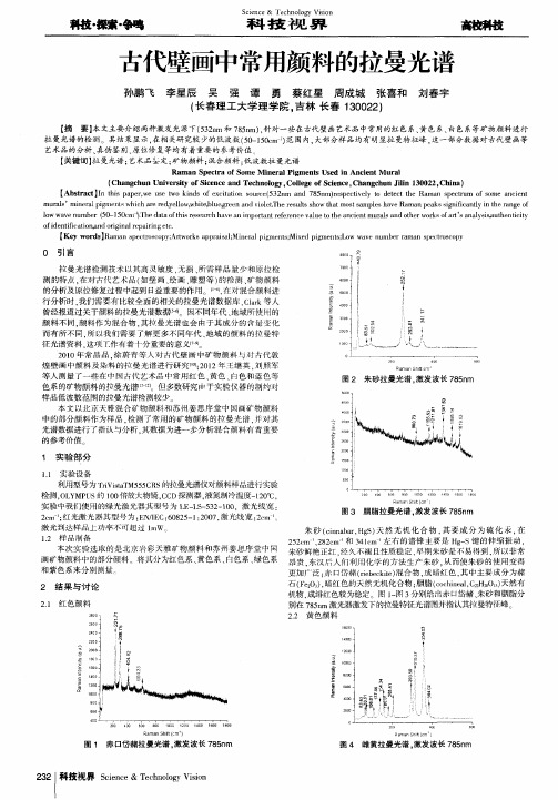
( Ch a n g c h u n U n i v e r s i t y o f S i c e n c e a n d T e c h n o l o g y 。 C o l l e g e o f S c i e n c e , C h a n g c h u n J i l i n 1 3 0 0 2 2 , C l l i n a) 【 Ab s t r a c t 】 I n t h i s p a p e r , w e u s e t w o k i n d s o t e x c i t a t i o n s o u r c e ( 5 3 2 m n a n d 7 8 5 n m) r e s p e c t i v e l y t o d e t e c t t h e R a m a n s p e c t r u m o f s o m e a n c i e n t
【 摘 要】 本文主要介 绍两种激发 光源下( 5 3 2 n r n 和7 8 5 ) 一) , 针对一 些在古代壁画 艺术品 中常用的红 色系、 黄 色系、 白色系等矿物颜料进行
拉 曼光谱 的检测 。其 结果显 示, 在相 关研 究较 少的低波数( 5 0 — 1 5 0 e m ) 范围 内, 大部分样品均有 明显拉 曼特征峰 . 这一部分数据对古代壁 画等 艺术品 的分析 、 真伪鉴别 、 原位修复等均有着重要的参考价值
科技・ 探索・ 争鸣
S c 科 i e n c e & 技 T e c h 视 n o l o g y 界 V i s i o n
古代壁画中常用颜料的拉曼光谱
孙鹏 飞 李星辰 吴 强 谭 勇 蔡红 星 院 , 吉 林 长春 1 3 0 0 2 2 ) 刘春 字
彩绘文物表面颜料光谱分段识别与填图方法研究

摘要受到环境变化和人类活动的影响,彩绘文物表面会出现褪色或颜色缺失的情况,需要尽快对其进行数字化留存和色彩修复。
对修复区域使用的颜料种类进行确定是色彩还原的关键步骤。
因此,亟需利用现代科技手段判断彩绘文物表面颜料类型,留存颜料信息,为修复工作者提供科学依据,提高修复效率。
为了避免对文物造成二次损害,本文选择无损的高光谱技术对彩绘文物全表面颜料信息进行获取和分析,建立了针对中国绘画的典型颜料光谱库,有利于文化遗产的数字化存档和永续留存。
根据绘画技法,画家在创作时经常会使用多种颜料混合,使作品呈现出绚丽的色彩。
因此,通过肉眼或高光谱等仪器观察到的字画、壁画表面的颜色往往是多种颜料的复合色。
大多数光谱识别方法一次只能识别一种颜料,或是通过光谱解混后再进行匹配,较为复杂且需要先验知识。
基于此,本文提出一种顾及离子吸收特征的光谱分段识别方法,可以实现绘画表面混合区域多种颜料的同时识别。
本文基于国画和壁画的高光谱影像和反射光谱,通过端元估计、图像聚类、光谱识别、以及颜料填图“四步走”的流程,实现绘画表面颜料类别的确定和空间分布的可视化。
主要研究成果如下:(1)构建了一个典型绘画颜料光谱库。
目前国内外针对中国绘画表面典型颜料建立的光谱库还很少。
本文选择了中国绘画使用的典型颜料制作样本,利用电子显微镜、拉曼光谱仪、扫描电镜以及便携式X荧光仪测试颜料的化学成份,利用光谱辐射仪采集350-2500 nm的反射光谱,建立典型颜料光谱库,作为光谱识别的基础。
并收集真实文物表面颜料光谱数据,留存于典型绘画颜料光谱库中,为其建立数字化档案,助力文物的永续留存。
(2)提出了一种顾及离子吸收特征的光谱分段识别方法。
分析典型颜料中主要离子和官能团的光谱吸收位置,以此作为划分特征子区间的依据。
然后从待测的整条光谱中提取出特定的特征子区间,逐区间进行光谱吸收系数匹配,以保证准确定位且不遗漏光谱的关键吸收特征。
接着对各区间结果进行权重分配,计算每种颜料的加权指数。
古代壁画中常用颜料的拉曼光谱
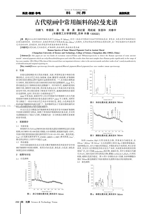
红色颜料图1赤口岱赭拉曼光谱,激发波长785nm图2朱砂拉曼光谱,激发波长785nm图3胭脂拉曼光谱,激发波长785nm(cinnabar,HgS)天然无机化合物,其要成分为硫化汞、282cm-1和341cm-1左右的谱锋主要是Hg-S键的伸缩振动朱砂鲜艳正红,经久不褪且性质稳定,早期朱砂是不易得到东汉后人们利用化学的方法生产朱砂,从而使朱砂的使用变得赤口岱赭(riebeckite)混合物,成暗红色,其中主要成分为赭暗红色的天然无机化合物;胭脂(cochineal,C22成绯红色较为稳定。
图1~图3分别给出赤口岱赭、激光器激发下的拉曼特征光谱图并指认其拉曼特征峰黄色颜料图4雌黄拉曼光谱,激发波长785nm图5黄拉曼光谱,激发波长785nm黄色颜料我们选取雌黄(orpiment,As2S3)成金黄色,天然无机金属其特征峰在180cm-1处为As-S-As键的弯曲振动S-As-S键的伸缩振动,而在293cm-1~384cm-1处是As-S(yellow)成淡黄色,混合矿物颜料,通过其拉曼光谱我们可以300~400cm-1附近看出其中含有雄黄(realgar,As4S4)成分的特征峰分别给出雌黄和黄在785激光器激发下的拉曼特征光谱图并指认了其拉曼特征峰。
白色颜料在艺术品中能够作为白色的颜料很多,我们主要对蛤白3)成白色,天然无机化合物,蛤粉属于矿物颜料有显著特点是不易退色、色彩鲜艳;进口钛白(titanium dioxide,TiO2)成白色白色固体或粉末状的两性氧化物,是使用最为广泛的白色多样结晶型态化合物。
图6给出了蛤白分别在532nm激发下的拉曼光谱,图7给出了进口钛白在532nm激发下的拉曼光并指出了蛤白与进口钛白拉曼特征峰。
6蛤白拉曼光谱,激发波长532nm(a)和785nm(b)图7进口钛白拉曼光谱,激发波长532nm绿色颜料图8孔雀绿拉曼光谱,激发波长532nm图9青紫拉曼光谱,激发波长785nm图10桃色拉曼光谱,激发波长532nm本文讨论了古代壁画、绘画等艺术品的真伪鉴定及原位修复是考古工作中的重要研究内容,利用拉曼光谱技术,以532nm为激发光源,检测了古代艺术品中常用红色系、黄色系、绿色系和紫色系的矿物颜料拉曼光谱,尤其针对国内外研究较少(50-200cm-1)范围的拉曼光谱进行检测分析。
大同地区寺观壁画常用颜料的拉曼光谱分析
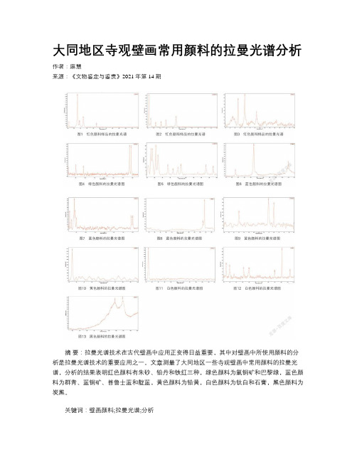
大同地区寺观壁画常用颜料的拉曼光谱分析作者:麻慧来源:《文物鉴定与鉴赏》2021年第14期摘要:拉曼光谱技术在古代壁画中应用正变得日益重要,其中对壁画中所使用颜料的分析是拉曼光谱技术的重要应用之一。
文章测量了大同地区一些寺观壁画中常用颜料的拉曼光谱,分析的结果表明红色颜料有朱砂、铅丹和铁红三种,绿色颜料为氯铜矿和巴黎绿,蓝色颜料为群青、蓝铜矿、普鲁士蓝和靛蓝,黄色颜料为铅黄,白色颜料为钛白和石膏,黑色颜料为炭黑。
关键词:壁画颜料;拉曼光谱;分析0 引言利用拉曼光谱技术分析古代壁画所使用的颜料,是一种非常有效便捷的方法。
拉曼光谱技术作为一种分析手段,可以确定样品的成分,此分析手段具有非接触无损探测的优点,样品无须特殊制备,测量需要的样品量很少,并且具有在位测量等突出的优势,这些突出的优势正是珍贵的艺术文物所需要的,这也是拉曼光谱技术在珍贵文物中得到大量应用的原因。
本文测量了大同地区一些寺观壁画中常用颜料的拉曼光谱,并对这些光谱进行分析,对研究大同地区寺观壁画颜料的应用有重要的参考价值。
1 实验大同地区寺观壁画颜料运用比较丰富,通过大量调查明确可以辨识的颜色有红色、绿色、蓝色、黄色、白色和黑色,对大量寺观壁画不同颜色进行取样,采用拉曼分析对成分进行确定。
对颜料样品的拉曼光谱采用法国JYBIN YVON公司生产的XploRA拉曼光谱仪进行分析。
实验中根据样品的颜色,选用了适当的激发波长,对红色、白色、黑色和黄色颜料使用近红外785nm激发是最佳的选择,对绿色和蓝色颜料采用绿光532nm激发。
为避免因激光功率过大导致颜料分解、碳化,在样品上使用的激光功率都不能超过1MW。
①2 结果与讨论中国古代壁画颜料主要来源于无机矿物、植物、金属以及化学合成,其中天然的无机矿物颜料占绝大多数。
以下根据颜色分类对大同地区部分寺观壁画颜料的拉曼光谱图进行分析。
2.1 红色颜料图1~图3分别给出了大同地区三种不同红色颜料样品的拉曼光谱,并标出了其特征峰。
基于光谱分析技术的文物材料鉴定研究

基于光谱分析技术的文物材料鉴定研究基于光谱分析技术的文物材料鉴定研究文物是一种珍贵的文化遗产,它们不仅记录了历史,也反映了人类文明的发展。
然而,由于年代久远、保存环境恶劣等原因,文物材料的鉴定工作变得尤为重要。
传统的文物材料鉴定方法通常需要对文物进行破坏性取样,这不仅会破坏文物本身,而且还会影响到文物的价值和完整性。
因此,基于光谱分析技术的文物材料鉴定方法成为了一种非常重要的文物保护手段。
光谱分析技术是一种非破坏性的分析方法,它可以通过对光谱信号的分析,来确定样品的组成和结构。
在文物材料鉴定中,光谱分析技术可以用来确定文物的材料类型、制作工艺、年代等信息。
目前,应用比较广泛的光谱分析技术包括红外光谱、紫外-可见光谱、拉曼光谱、荧光光谱等。
红外光谱是一种常用的光谱分析技术,它可以用来确定样品中的化学键和官能团。
在文物材料鉴定中,红外光谱可以用来确定文物中有机物和无机物的含量以及结构。
例如,在陶瓷材料鉴定中,红外光谱可以用来确定陶瓷中的氧化物、硅酸盐等成分;在纸张材料鉴定中,红外光谱可以用来确定纸张中的纤维素、淀粉质等成分。
紫外-可见光谱是一种可以测量样品吸收或反射光线的光谱分析技术。
在文物材料鉴定中,紫外-可见光谱可以用来确定文物中的染料和颜料成分。
例如,在壁画颜料鉴定中,紫外-可见光谱可以用来确定颜料中的铁、铜、锰等元素;在古代书画鉴定中,紫外-可见光谱可以用来确定墨汁中的碳黑和树脂等成分。
拉曼光谱是一种可以测量样品散射光线的光谱分析技术。
在文物材料鉴定中,拉曼光谱可以用来确定文物中的晶体结构和分子振动信息。
例如,在宝石材料鉴定中,拉曼光谱可以用来确定宝石中的晶体结构和杂质元素;在古代玻璃鉴定中,拉曼光谱可以用来确定玻璃中的硅氧四面体结构和金属离子等成分。
荧光光谱是一种可以测量样品荧光强度和荧光寿命的光谱分析技术。
在文物材料鉴定中,荧光光谱可以用来确定文物中的染料和颜料成分以及其年代。
例如,在古代壁画鉴定中,荧光光谱可以用来确定颜料中的铜绿和铜蓝等成分以及其年代。
基于光谱吸收特征分析的彩绘文物颜料识别研究

基于光谱吸收特征分析的彩绘文物颜料识别研究张陈峰;胡云岗;侯妙乐;吕书强;张学东【摘要】高光谱成像技术作为一种无损高效的检测方法,对于彩绘文物颜料的鉴别具有重要意义.由于全波段参与光谱相似度计算会造成数据的冗余,没有充分利用光谱的细微特征.为此,本文首先对文物颜料光谱吸收特征进行参量化分析,并通过改进的光谱吸收特征拟合算法与标准光谱进行匹配识别,从而得到识别结果.实验以获取的一幅波长为400~1000nm的古代壁画高光谱影像为例,通过光谱吸收特征分析识别出壁画的颜料主要成分有朱砂、赭粉、石绿和石青,4种颜料的光谱吸收特征拟合度分别是:0.95、0.77、0.92、0.81.实验结果表明:对光谱吸收特征分析可以帮助识别彩绘文物的颜料信息,该方法可为以后文物修复提供参考.%As a non-destructive and efficient detection method,hyper-spectral imaging technology has a great significance to the identification of pigment on colored relics.The results show that the spectral similarity calculation of the full band will cause the data redundancy,and do not make full use of the subtle features of the spectrum.In this paper,the spectral absorption characteristics of the pigment were analyzed,and the spectral feature fitting algorithm was used to match the standard spectrum,and the results were obtained.The experimental study took a hyper-spectral image of an ancient mural as an example which wavelength range is 400-1000 nm,and distinguished the pigments by spectral absorption analysis.The results show that the major pigments are cinnabar,ochre powder,malachite and azurite,the spectral fitting characteristics are 0.95,0.77,0.92,0.81 respectively.The analysis of spectral absorption features can help toidentify the color information of colored relics.The method can provide reference for cultural relic restoration.【期刊名称】《地理信息世界》【年(卷),期】2017(024)003【总页数】5页(P119-123)【关键词】高光谱成像;颜料识别;光谱特征分析【作者】张陈峰;胡云岗;侯妙乐;吕书强;张学东【作者单位】北京建筑大学测绘与城市空间信息学院,北京102500;北京建筑大学测绘与城市空间信息学院,北京102500;北京建筑大学测绘与城市空间信息学院,北京102500;北京建筑大学测绘与城市空间信息学院,北京102500;北京建筑大学测绘与城市空间信息学院,北京102500【正文语种】中文【中图分类】P2370 引言对于彩绘文物保护工作,需要尽量保持文物的原貌,然而由于岁月久远,文物表面丰富的色彩会因为自然侵蚀、人为破坏等因素遭到损害,这就要求在尊重历史的基础上对彩绘文物进行修复。
定边郝滩东汉壁画墓绿色底层颜料分析研究_付倩丽

第24卷第1期2012年2月文物保护与考古科学SCIENCES OF CONSERVATION AND ARCHAEOLOGYVol.24,No.1Feb ,2012收稿日期:2011-06-03;修回日期:2011-07-11基金项目:国家文物局文物保护科学和技术研究课题资助(20080216),国家科技支撑计划资助(2010BAK67B12)作者简介:付倩丽(1979—),女,毕业于西北大学文物保护技术专业,馆员,E -mail :fuqianli2008@163.com 文章编号:1005-1538(2012)01-0038-06定边郝滩东汉壁画墓绿色底层颜料分析研究付倩丽1,夏寅1,王伟锋1,杨军昌2,吕智荣2,惠娜1,张尚欣1(1.陶质彩绘文物保护国家文物局重点科研基地,秦始皇兵马俑博物馆,陕西临潼710600;2.陕西省考古研究院,陕西西安710043))摘要:本研究以定边郝滩东汉壁画墓中绿色底层为研究对象,采用偏光显微镜(PLM )、带能谱的扫描电子显微镜(SEM /EDS )、X 射线衍射仪(XRD )和拉曼光谱(RS )分析了绿色底层的成分与物相,同时和四种已知国外绿土相比较,得出绿色底色为绿土,对其进行深入分析研究以期为考古学和后期文物保护工作提供科学信息。
关键词:壁画底层;绿土;颜料;分析中图分类号:G262;K854.2文献标识码:A0引言定边郝滩东汉壁画墓,位于陕西靖边定边县郝滩乡四十里铺村西北,于2003年4月发现并开始发掘。
该壁画墓保存之完整,颜色之艳丽,场面之热烈宏大,绘画技艺之娴熟,在迄今为止所发现的东汉壁画墓中尚不多见。
庭院图、农作图、放牧图,在已发现的两汉壁画墓中保存得如此完整还属首例,其内容不仅表现了墓主人的家庭生产活动和经济状况,还为研究陕北地区经济和文化的发展及毛乌素沙漠地区的环境变迁提供了具有重要价值的资料。
墓室内除左侧额耳室形龛内没有绘制壁画外,其它部位均绘有壁画,面积有25m 2。
光谱技术在文物鉴定中的应用探索

摘要光谱技术是科技物体研究中的高技术之一,属于无损分析研究。
文章首先谈论了文物鉴定的传统方法和现代科技鉴定现状以及利弊,然后分别介绍了激光拉曼光谱技术在古玉器、古陶瓷、古颜料、古代青铜器等无损鉴定的应用和X-射线荧光能谱仪在古代服饰文物的无损鉴定的应用,从而阐明光谱技术在文物鉴定中是一个很好的无损分析方法。
最后,讨论了光谱技术在文物鉴定应用研究中出现的局限性并对发展前景进行了展望。
关键词:光谱技术,文物鉴定,无损分析。
ABSTRACTSpectroscopy is one of the high-tech in the area of the science and technology research, belonging to non-destructive analysis. Firstly, the thesis discussed the traditional methods for identification of Cultural Relics and present study for identification of modern technology as well as the pros and cons. Then, the application of laser Roman spectroscopy in ancient jade, ancient ceramics, ancient pigments, ancient bronze and other non-destructive identification were introduced. Besides, the application of energy dispersive X-ray fluorescence spectrometry in the non-destructive identification of ancient costumes were also discussed, which clarifies spectroscopy in the identification of Cultural Relics is a good method for non-destructive analysis.Finally, the limitations of the technology and the development prospects of the spectral artifacts appear in the application of identification were discussed.Key words: spectroscopy identification of cultural relics non-destructive analysis目录摘要 (I)ABSTRACT ........................................................................................................ I I1、引言 (1)2、文物鉴定的现状和方法 (1)2.1文物鉴定的现状 (1)2.2文物鉴定的方法 (1)2.2.1传统方法 (1)2.2.2现代科技鉴定方法 (2)2.3光谱技术鉴定文物的优越性 (3)3、常用光谱技术简介 (3)3.1拉曼光谱 (3)3.1.1拉曼散射简介 (3)3.1.2拉曼散射的原理 (4)3.1.3拉曼光谱的特点 (5)3.2红外光谱 (6)3.2.1红外光谱简介和原理 (6)3.2.2红外光谱特点 (6)3.3荧光光谱 (7)3.3.1荧光光谱简介及原理 (7)4、拉曼光谱技术在文物鉴定中的应用 (8)4.1中国古玉、古玉器鉴定和研究的分析 (8)4.1.1五种常见玉石的激光拉曼研究结果 (8)4.1.2激光拉曼光谱技术在中国古代玉器无损鉴定中的应用 (9)4.2古颜料的分析 (11)4.3古陶器的分析 (13)4.4古代青铜器的分析 (14)4.5结论 ......................................................................... 错误!未定义书签。
龙门石窟彩绘颜料的拉曼光谱、X射线荧光和扫描电镜分析
龙门石窟彩绘颜料的拉曼光谱、X射线荧光和扫描电镜分析马朝龙;刘照军;程琳【摘要】作为一种无损分析技术,拉曼光谱近几十年来被越来越广泛地应用于文物艺术品分析和考古学领域.色彩是艺术品的灵魂,颜料分析是研究文物艺术品的一个重要课题.本文利用显微拉曼光谱技术,测量了龙门石窟洞窟中彩绘使用的颜料的拉曼光谱,通过对这些光谱的解析,对颜料的化学成分做出了识别.利用X射线荧光光谱进行的元素分析确定金色装饰为高纯度黄金,扫描电镜分析进一步测出了贴金的厚度.这些分析结果对于进一步开展龙门石窟彩绘的保护和修复有着重要的参考价值.【期刊名称】《光散射学报》【年(卷),期】2018(030)004【总页数】5页(P357-361)【关键词】拉曼光谱;X射线荧光;扫描电镜;龙门石窟;颜料识别【作者】马朝龙;刘照军;程琳【作者单位】龙门石窟研究院,河南洛阳471023;洛阳师范学院物理与电子信息学院,河南洛阳471934;洛阳师范学院物理与电子信息学院,河南洛阳471934【正文语种】中文【中图分类】O657.371 引言龙门石窟位于洛阳南郊伊河两岸的东西两山上,开始建造于北魏年间,历经隋、唐等多个朝代的不断营造,留下了2345个窟龛、10万余尊造像、2860余块碑刻题记的石窟遗存。
龙门石窟是全国第一批重点文物保护单位,2000年11月联合国教科文组织列入《世界遗产名录》。
龙门石窟的建造者在完成石窟雕刻后,大量采用彩绘和贴敷金方式对洞窟和雕刻进行装饰,以衬托雕刻的精美,表现佛像的华丽庄严。
彩绘在露天环境中历经了千余年岁月洗礼,受风吹日晒、雨水和岩体渗水冲蚀、温湿度变化以及其它人为因素的影响,目前石窟内大部分彩绘已经剥落消失,残存的也大多污损、褪色、变色,失去了往日的华彩。
仅有少数岩体整体性好的洞窟中仍存有一些较为完整的彩绘装饰,在奉先寺卢舍那大佛身体表面、擂鼓台南洞等洞窟的造像和雕刻表面还保留有贴金装饰[1]。
图1a所示为龙门石窟宾阳中洞彩绘现状照片,该洞窟开凿于北魏时期(~520A.D.),洞内主佛为释迦牟尼,穹顶中央雕刻莲花宝盖,周围是衣带飘扬、姿态优美的伎乐天和供养天人,其间彩绘保存现状较好。
基于光谱吸收特征分析的彩绘文物颜料识别研究
基于光谱吸收特征分析的彩绘文物颜料识别研究张陈峰;胡云岗;侯妙乐;吕书强;张学东【期刊名称】《地理信息世界》【年(卷),期】2017(24)3【摘要】高光谱成像技术作为一种无损高效的检测方法,对于彩绘文物颜料的鉴别具有重要意义.由于全波段参与光谱相似度计算会造成数据的冗余,没有充分利用光谱的细微特征.为此,本文首先对文物颜料光谱吸收特征进行参量化分析,并通过改进的光谱吸收特征拟合算法与标准光谱进行匹配识别,从而得到识别结果.实验以获取的一幅波长为400~1000nm的古代壁画高光谱影像为例,通过光谱吸收特征分析识别出壁画的颜料主要成分有朱砂、赭粉、石绿和石青,4种颜料的光谱吸收特征拟合度分别是:0.95、0.77、0.92、0.81.实验结果表明:对光谱吸收特征分析可以帮助识别彩绘文物的颜料信息,该方法可为以后文物修复提供参考.%As a non-destructive and efficient detection method,hyper-spectral imaging technology has a great significance to the identification of pigment on colored relics.The results show that the spectral similarity calculation of the full band will cause the data redundancy,and do not make full use of the subtle features of the spectrum.In this paper,the spectral absorption characteristics of the pigment were analyzed,and the spectral feature fitting algorithm was used to match the standard spectrum,and the results were obtained.The experimental study took a hyper-spectral image of an ancient mural as an example which wavelength range is 400-1000 nm,and distinguished the pigments by spectral absorption analysis.The resultsshow that the major pigments are cinnabar,ochre powder,malachite and azurite,the spectral fitting characteristics are 0.95,0.77,0.92,0.81 respectively.The analysis of spectral absorption features can help to identify the color information of colored relics.The method can provide reference for cultural relic restoration.【总页数】5页(P119-123)【作者】张陈峰;胡云岗;侯妙乐;吕书强;张学东【作者单位】北京建筑大学测绘与城市空间信息学院,北京102500;北京建筑大学测绘与城市空间信息学院,北京102500;北京建筑大学测绘与城市空间信息学院,北京102500;北京建筑大学测绘与城市空间信息学院,北京102500;北京建筑大学测绘与城市空间信息学院,北京102500【正文语种】中文【中图分类】P237【相关文献】1.光导纤维光谱技术无损鉴定彩绘文物颜料的研究 [J], 王丽琴;周文晖;赵静2.可见光谱法无损识别壁画文物矿物质颜料的研究 [J], 李俊锋;万晓霞3.光纤反射光谱在彩绘文物颜料鉴别中的应用研究 [J], 李广华;陈垚;马越;雷勇4.彩绘文物中蓝色颜料群青的鉴定技术研究 [J], 刘璐瑶;张秉坚5.彩绘文物颜料胶结材料分析与表征研究进展 [J], 闫宏涛;安晶晶;周铁;容波;夏寅因版权原因,仅展示原文概要,查看原文内容请购买。
光谱分析技术在彩绘文物颜料分析中的应用

文物,尤其是其上的颜料保存较差。如何对颜料进行准确、
无损或微损分析,一直是文物保护领域研究的热点与难点。
近年来,激光拉曼光谱法、光导纤维反射光谱法、激光诱导 击穿光谱法等多种光谱分析技术逐渐被应用于彩绘文物颜料 信息的提取、分析,使人们掌握了更为有利的颜料分析工
中的主要元素,而且发现红色、橘黄色、灰色颜料等都含有 铁的氧化物[7]。本课题组结合XRF和X射线衍射分析结果,
500
en'l-1的散射光,检测光
斑直径只有几肛m,颜料用量<1 Pg,无需前期制样即可进行 快速鉴定[22。2“。拉曼光谱的主要发展有:(1)光导纤维、显微 技术等的应用,使可移动拉曼技术被广泛的应用于文物的分 析中。(2)共焦型设备的应用,使拉曼的分辨率提高。(3)主 成分分析(PCA)等化学统计学方法被应用到拉曼数据的处理
干扰等因素会对结果产生较大影响[291。利用Ⅺm对埃及
Wadi
El
Natrun遗址建筑遗迹的绿色颜料进行检测,结果表
明颜料为含碱式碳酸铜(石绿)与碱式碳酸铅(铅白)的混合
万方数据
3396
光谱学与光谱分析
第32卷
物[驯。伦敦大学学院Petrie博物馆用XRD对馆藏埃及不同 时期木乃伊棺进行分析,发现了大量在中世纪才使用的绿色 颜料“green earth”。证实了希腊罗马统治时期该颜料的使用 量远远超过了人们的估计,并将其已知应用年代从希腊罗马 统治时期提前到公元前9世纪的第三中间期,为考古学研究 提供了实物证据。同时,还发现了以黄色、蓝色颜料混合成 绿色颜料的工艺【31 J。
Pascual利用SEM研究了Maya文化墓葬壁画的制作工艺, 发现部分绿色颜料是由蓝色与黄色颜料混合而成的,且蓝色 颜料表面存在一层含有2~5 nlTl大小细孔的凹痕状的坡缕石 晶体(图4)[4引。
拉曼光谱分析测试技术及其在陶瓷结构测试中的应用

拉曼光谱分析测试技术及其在陶瓷结构测试中的应用应用部分,首先说明可做哪些结构测试,然后详细说明从制样到出结果的各测试步骤,最后就各条结构测试各举2-3具体例子予以说明,包括图、表、分析方法、结果、外文参考文献等)19周特冶楼230摘要:拉曼光谱分析技术由于具有无损、信息丰富、灵敏度高、所需测试样品量小等优点,可进行现场快速筛查、检测及鉴别,在食品、材料、环境监测等众多领域得到了越来越广泛的应用。
随着全国经济的不断发展,陶瓷材料在工业中应用逐渐增多,而陶瓷材料的结构对性能影响非常大。
本文阐述了拉曼光谱产生的原理,介绍相关的拉曼光谱测试技术及其在纳米BaTiO3陶瓷结构测试中的应用,并对实验结果进行了讨论。
1 拉曼光谱1.1简介1923年,Smekal从理论上描述了拉曼散射效应。
1928年,印度物理学家Raman 发现了光的非弹性碰撞现象,记录了散射光谱,并以他的名字将这一现象命名为拉曼效应/拉曼光谱。
拉曼光谱(Raman spectrosopy)技术是基于拉曼散射效应而发展起来的光谱分析技术,研究的是分子振动、转动信息。
与常规化学分析技术相比,具有检测时间短、操作简单、样品所需量少等特点,故随着激光光源的不断发展,拉曼光谱在食品、生物监测、医药、刑事司法、地质考古、宝石鉴定等领域都已得到广泛的应用[1]。
因此拉曼光谱技术成为了人们研究分子结构的新手段之一。
拉曼光谱最初是用聚焦的日光作为光源,之后改用汞弧灯,但是光源强度仍然不够,限制了拉曼光谱的发展。
直到20世纪60年代,高功率,单色性和相干性好,准直性好,偏振特性好的激光出现,为拉曼散射提供了空前优异的光源,拉曼光谱学也因此被冠以激光二字称为激光拉曼光谱学[2]。
拉曼光谱采用激光作为单色光源,使激光拉曼光谱在分析化学等领域中得到了广泛的应用。
拉曼光谱技术的基本原理:单色光束照射会产生两种类型的光散射,弹性散射和非弹性散射。
在弹性散射过程中,光子的频率不发生改变,其波长和能量上没有任何改变,这种散射也称为瑞利散射。
- 1、下载文档前请自行甄别文档内容的完整性,平台不提供额外的编辑、内容补充、找答案等附加服务。
- 2、"仅部分预览"的文档,不可在线预览部分如存在完整性等问题,可反馈申请退款(可完整预览的文档不适用该条件!)。
- 3、如文档侵犯您的权益,请联系客服反馈,我们会尽快为您处理(人工客服工作时间:9:00-18:30)。
1999年10月CHINESE JOURNAL OF LIGHT S CATT ERING Oct.1999文章编号:1004 5929(1999)03-0215-05古壁画、陶彩颜料的拉曼光谱分析左 健1,2,许存义1(1 中国科学技术大学结构分析开放研究实验室;合肥 2300262 中国科学技术大学科技史与科技考古系,合肥 230026)摘 要 本文利用拉曼光谱对河南班村遗址出土的仰韶彩陶陶彩以及河北磁县湾漳东魏北齐大型壁画墓中的壁画颜料进行了分析,成功的测定出陶彩及壁画颜料的成分。
这一研究工作表明,拉曼光谱作为现代技术非常适合于易损和不允许取样的珍贵艺术品颜料的无损分析。
关键词 激光拉曼光谱;古壁画;陶彩;颜料中图分类号:O657 37 文献标识符:AThe Study of Ancient Coating Pottery andWall Painting by Raman SpectraZU O Jian1,2,XU Cun yi1(1 Structure Resear ch L aboratory,University of Science and Technologyo f China,H eif ei A nhui230026,China)(2 Dep artment o f Scientif ic H istory and Ar chaeom etry,University o fScience and Technology o f China,H ef ei,Anhui230026,China)Abstract:The ancient coating pottery and w all painting w ere analyzed by RamanSpectra.The w ork described here confirms that Raman scattering is a very effectiveanalytical method w hen applied to the pig ments used in ancient coating pottery andw all painting.Key Words: Raman scattering;pigment;ancient coating pottery;w all painting1 引 言利用拉曼光谱对古代材料进行分析研究是目前发展较迅速的领域之一。
近年来,国外已有许多这方面的研究报导,特别是关于古颜料的研究[1,2]。
我国是一个文物大国,祖先给我们留下了丰富的文化遗产,但这一方面的工作却开展的很少。
最近,我们利用拉曼光谱对河南班村遗址出土的仰韶彩陶陶彩以及河北磁县湾漳东魏北齐大型壁画墓中的壁画颜料进行了分析,取得了一些较为满意的结果。
收稿日期:1999 02 29基金项目:国家自然科学基金资助项目(29775023)2 陶彩颜料的分析2 1 考古学背景实验所选的彩陶片取自河南班村遗址,该遗址与著名的仰韶遗址相邻,为典型的仰韶文化遗址,距今约6000~7000年。
仰韶文化闻名于世,其彩陶艺术和工艺尤其令人叹服。
多少年来人们十分重视彩陶的研究,但主要是器型和艺术风格的研究。
仰韶彩陶工艺的发明、传播及相互影响是一个极其复杂而又十分重要的问题。
因此,利用现代分析手段探索陶彩的矿物来源,有着明显的考古学意义。
2 2 实验及结果分析拉曼光谱分析是在美国SPEX-1403型激光拉曼光谱仪上进行的,激发光为氩离子激光器514 5nm线,背散射配置。
被测样品为红彩3块、白彩2块、黑彩3块,实验结果表明红彩为赤铁矿,这和其他遗址出土的红彩是相同的;而白彩和黑彩比较特殊,具有一定的地方特色。
图1给出了白彩和天然铝土矿的拉曼光谱,从图中可看出的拉曼光谱和天然铝土矿非常相似,说明白彩的矿物源为铝土矿,这和X射线衍射(XRD)的结果是一致的,但白彩位于446cm 1的峰明显向高波数移动,原因尚不清楚。
据了解,河南北部有丰富的铝土矿,看来班村遗址出土的白彩为就地选材的结果。
用铝土矿作为白彩的颜料,国内尚不多见。
Fig.1 The Raman spectra of white coat ing on painted pottery(sample A)and a naturebauxite Fig.2 The Raman spectra of the black coating on painted pottery(sample C、D、E)and the nature m agnetite图2给出了黑彩和天然磁铁矿的拉曼光谱,从图中可看出黑彩的拉曼光谱和天然磁铁1999年10月CHINESE JOURNAL OF LIGHT S CATT ERING Oct.1999矿很相似,但黑彩的拉曼谱峰明显宽化并向低波数移动。
和天然磁铁矿相比,样品E的峰位红移了19cm 1、半高宽增加了24cm 1。
拉曼谱峰的宽化和移动可能是由于晶粒尺寸效应所产生,随着晶粒尺寸的减小,不少纳米材料的拉曼峰均发生明显的红移和宽化,这一现象通常可用声子限制效应来解释[3~6]。
为了证实这一推测,取下了部分黑彩粉末进行了XRD 分析和透射电镜(TEM)观察,XRD和TEM的结果表明黑彩确实为磁铁矿,其尺寸为纳米范围,并且样品E的尺寸比样品C小。
从外观上看,样品E的颜色比样品C明显要暗一些,看来陶彩的颜色不仅决定于颜料的矿物成分,而且与颜料的颗粒尺寸有关。
原则上说拉曼光谱不仅可以判断颜料的成分,还可以估计颜料的晶粒尺寸。
3 壁画颜料的分析3 1 考古学背景河北省出土的墓葬壁画十分精彩、丰富,并具有地方特色。
本文所分析的壁画样品取自河北磁县湾漳大型壁画墓,该墓位于磁县南部,据推测为东魏北齐文宣帝高洋的陵墓[7]。
该墓由墓道、甬道和墓室三部分组成。
墓道、甬道和墓室的壁顶全部抹白灰(碳酸钙),上面满绘壁画,所绘壁画色彩丰富、内容生动、技艺高超。
准确地测定这些壁画颜料的成分,无疑对壁画的有效保护与修复具有重要的意义。
3 2 实验及结果分析选择了几块壁画残片进行了原位拉曼光谱分析,实验条件同上。
图3给出了红色颜料和天然辰砂的拉曼光谱,从图中可看出红色颜料的拉曼光谱和天然辰砂很相似,确认红色颜料为辰砂。
R.J.H.Clark教授曾对10世纪前期的8张有红色颜料痕迹的纸片和一块纺织品进行了拉曼光谱分析,并从其中四块纸片和那块纺织品中发现了辰砂的存在。
R.J.H.Clark教授认为辰砂为10世纪初期中国红墨水的主要矿物源[8]。
上述实验结果进一步支持了这一推论。
图4给出了黄色颜料和天然针铁矿的拉曼光谱,从图4可以确认黄色颜料为针铁矿。
图5给出了黑色颜料的拉曼光谱。
从拉曼谱的峰型和峰位看,这种颜料为石墨类材料,即碳黑。
用碳黑作黑颜料已有很长的历史。
图6给出了浅蓝色颜料和地仗(衬底)的拉曼光谱。
从图中可见有4个明显的拉曼峰分别位于155、281、711和1085cm 1,这和方解石结构的碳酸钙是完全一致的[9]。
但和地仗(衬底)的拉曼光谱相比,浅蓝色颜料在低频段的两个谱峰(144和281cm 1)的宽度明显增加;而在高频段的两个谱峰(711和1085cm 1)的宽度与峰位均无明显变化。
位于155、281cm 1的低频振动属于与Ca2+离子有关的外振动模,而位于711、1085cm 1的高频振动属于与CO2 3离子有关的内振动模。
H.N.Rutt[9]等注意到晶格畸变对外振动模的频率有较大的影响。
如果部分Ca2+离子被其他金属离子所取代,那么将引起一定的晶格畸变,从而导致外振动模的展宽。
为了证实这一推论,取下部分浅蓝色颜料进行了X射线荧光光谱(XRF)分析。
XRF的结果表明,浅蓝色颜料中除含有Ca外,确实还含有少量的M g、Fe、Mn等金属原子。
因而可以认为这种浅蓝色颜料为有杂质替代的方解石,其化学式为Ca x(M g、Fe、Mn)1 xFig.3 The Raman spectra of the redpigment on ancient wall painting and the naturebauxite Fig.4 The Raman spectra of the yellow pigment on ancient wall paintingand the naturegoethite.Fig.5 The Raman spectra of the black pigment on ancient wallpainting.Fig.6 The Raman spectra of the pale bluepigment on ancient wall painting andthe ground coating.1999年10月CHINESE JOURNAL OF LIGHT S CATT ERING Oct.1999CO3。
在此之前,曾利用X射线衍射(XRD)对上述壁画颜料进行了分析,除了红色颜料被确认为辰砂外,其他颜料均未得到明确的结论。
主要困难是衬底(地仗)的衍射信号太强,将颜料的信息淹没。
4 小 结以上工作表明,拉曼光谱这一现代技术非常适合于易损和不允许取样的珍贵艺术品颜料的无损分析。
准确地测定这些珍贵文物所用的颜料,不仅可以为这些文物的有效保护和修复以及真伪鉴别提供依据,还可以帮助人们了解当时的工艺水平、文化和贸易交流、社会经济状况等方面的信息。
我国是世界上唯一历史连绵不断,文明常盛不衰的大国,祖先给我们留下了无数的珍贵文物,其古代绘画和彩陶等艺术更是闻名世界。
可以预计,拉曼光谱分析技术必将在我国珍贵文物的研究中发挥重要的作用。
参考文献:[1] L.A.Lyon, C.D.Krating, A.P.Fox,et.al.,Anal.Chem.1998,70:341R[2] R.J.H.Clark,Chemical Society Review,1995,24:187[3] M.Yosikaw a,Y.M ori,H.Obata,et.al.,J.Appl.Phys.Lett.,1995,67:694[4] Z.Jian,H.Bnsoher, C.Falter,et.Al.,J.Appl.Phys.Lett.,1996,69:200[5] C.W.Grahan,W.H.W fber, C.R.Peters,et.al.,J.Catal.,1991,130:310[6] H.Ri chter,Z.P.Wang and L.Ley,Solid State Commun.,1981,58:739[7] 徐光冀,文物,1996,9:69[8] R.J.H.Clark,P.J.Gibbs,K.R.S edden.J.Raman S pectrose.1997,28:91[9] H.N.Rutt,J.H.Nicola,J.Phys.C:Solid State Phys.,1974,7:4522。
