Single-chain antibody-mediated intracellular retention of ErbB-2 impairs
《新型冠状病毒感染合并急性肾损伤诊治专家共识》解读
[2] TANG Y, XIN Y, DENG F. Prevention and management of COVID-19 in hemodialysis centers[J]. Am J Manag Care, 2020, 26(8): e237-e238.
[3] KLIGER A, COZZOLINO M, JHA V, et al. Managing the COVID-19 pandemic: international comparisons in dialysis patients[J]. Kidney Int, 2020, 98(1): 12-16.
[6] CHAWKI S, BUCHARD A, SAKHI H, et al. Treatment impact on COVID-19 evolution in hemodialysis patients[J]. Kidney Int, 2020, 98(4): 1053-1054.
[7] MURT A, ALTIPARMAK M R. Convalescent COVID-19 patients on hemodialysis: when should we end isolation?[J]. Nephron, 2020, 144(7): 343-344. 收稿日期:2021-03-25;修回日期:2021-04-22 (本文编辑:王丽)
抗氯霉素单链抗体的高效生产
1.8
抗体亲和力常数测定 参 照 Batty 等 非 竞 争 ELISA 法 [9] 进 行 。 scFvCAP 样 品 和 母 本
Abstract : [ Objective ] To construct a single chain variable fragment ( scFv ) directed against chloramphenicol ( scFvCAP ). [Methods ] The scFvCAP gene was assembled by overlap PCR and optimized by synonymous mutation , then cloned into pET21a and pET32a which is a fusion expression vector with Trx gene at the terminal 5' of foreign gene for expression in Es cherichia coli. The inclusion protein of scFvCAP was refolded by β -cyclodextrin companion system. [ Results ] The scFvCAP gene (Genebank ID : GU258048 ) was assembled. Under the control of strong promoter T7 , the scFvCAP gene was failed to be individually expressed , but over-expressed when fused to Trx gene. An optimized scFvCAP mut ( Genebank ID : GU258048 ) was cloned by reducing the content of G and C from 60.0% to 36.7% in the terminal 5' of 30 base pairs , and over-expressed was as inclusions. The inclusion protein of scFvCAP was successfully refolded by β -cyclodextrin companion system with an antibody titer similar to its parent monoclonal antibody (MAb ). [Conclusion ] We successfully construct a over-production system of scFvCAP. Key words : Chloramphenicol ; Single chain variable fragment ; Over-expression ; Renaturation
Antibody structure, instability, and formulation

MINIREVIEWAntibody Structure,Instability,and FormulationWEI WANG,SATISH SINGH,DAVID L.ZENG,KEVIN KING,SANDEEP NEMAPfizer,Inc.,Global Biologics,700Chesterfield Parkway West,Chesterfield,Missouri63017Received14March2006;revised17May2006;accepted4June2006Published online in Wiley InterScience().DOI10.1002/jps.20727 ABSTRACT:The number of therapeutic monoclonal antibody in development hasincreased tremendously over the last several years and this trend continues.At presentthere are more than23approved antibodies on the US market and an estimated200ormore are in development.Although antibodies share certain structural similarities,development of commercially viable antibody pharmaceuticals has not been straightfor-ward because of their unique and somewhat unpredictable solution behavior.This articlereviews the structure and function of antibodies and the mechanisms of physical andchemical instabilities.Various aspects of formulation development have been examinedto identify the critical attributes for the stabilization of antibodies.ß2006Wiley-Liss,Inc.and the American Pharmacists Association J Pharm Sci96:1–26,2007Keywords:biotechnology;stabilization;protein formulation;protein aggregation;freeze drying/lyophilizationINTRODUCTIONProtein therapies are entering a new era with the influx of a significant number of antibody pharmaceuticals.Generally,protein drugs are effective at low concentrations with less side effects relative to small molecule drugs,even though,in rare cases,protein-induced antibody formation could be serious.1Therefore,this category of therapeutics is gaining tremendous momentum and widespread recognition both in small and large drugfirms.Among protein drug therapies,antibodies play a major role in control-ling many types of diseases such as cancer, infectious diseases,allergy,autoimmune dis-eases,and inflammation.Since the approval of thefirst monoclonal antibody(MAb)product -OKT-3in1986,more than23MAb drug products have entered the market(Tab.1).The estimated number of antibodies and antibody derivatives constitute20%of biopharmaceutical products currently in development(about200).2The global therapeutic antibody market was predicted to reach$16.7billion in2008.3There are several reasons for the increasing popularity of antibodies for commercial develop-ment.First,their action is specific,generally leading to fewer side effects.Second,antibodies may be conjugated to another therapeutic entity for efficient delivery of this entity to a target site, thus reducing potential side effects.For instance, Mylotarg is an approved chemotherapy agent composed of calicheamicin conjugated to huma-nized IgG4,which binds specifically to CD33for the treatment of CD33-positive acute myeloid leukemia.Another example is the conjugation of immunotoxic barnase with the light chain of the anti-human ferritin monoclonal antibody F11as potential targeting agents for cancer immuno-therapy.4Third,antibodies may be conjugated to radioisotopes for specific diagnostic purposes. Examples include CEA-Scan for detection of color-ectal cancer and ProstaScint for detection of prostate stly,technology advancement has made complete human MAb available,which are lessimmunogenic.JOURNAL OF PHARMACEUTICAL SCIENCES,VOL.96,NO.1,JANUARY20071 Correspondence to:Wei Wang(Telephone:(636)-247-2111;Fax:(636)-247-5030;E-mail:wei.2.wang@pfi)Journal of Pharmaceutical Sciences,Vol.96,1–26(2007)Pharmacists AssociationT a b l e 1.C o m m e r c i a l M o n o c l o n a l A n t i b o d y P r o d u c t s#B r a n d n a m e M o l e c u l eM A bY e a r C o m p a n y R o u t e I n d i c a t i o n M A b C o n c B u f f e r E x c i p i e n t s S u r f a c t a n t p H1A v a s t i n B e v a c i z u m a bH u m a n i z e d I g G 1,149k D a2004G e n e t e c h a n d B i o O n c o l o g y I V i n f u s i o nM e t a s t a t i c c a r c i n o m a o f c o l o n o r r e c t u m ,b i n d s V E G F 100m g a n d 400m g /v i a l (25m g /m L )s o l u t i o n 5.8m g /m L m o n o b a s i c N a P h o s H 2O ;1.2m g /m L d i b a s i c N a P h o s a n h y d r o u s (4m L ,16m L fil l i n v i a l )60m g /m L a -T r e h a l o s e d i h y d r a t e (4m L ,16m L fil l i n v i a l )0.4m g /m L P S 20(4m L ,16m L fil l i n v i a l )6.22B e x x a rT o s i t u m o m a b a n d I -131T o s i t u m a b M u r i n e I g G 2l2003C o r i x a a n d G S KI V I n f u s i o nC D 20p o s i t i v e f o l l i c u l a r n o n H o d g k i n s l y m p h o m aK i t :14m g /m L M A b s o l u t i o n i n 35m g a n d 225m g v i a l s ;1.1m g /m L I 131-M A b s o l u t i o n10m M p h o s p h a t e (M A b v i a l )145m M N a C l ,10%w /v M a l t o s e ;I 131-M A b :5–6%P o v i d o n e ,1–2,9–15m g /m L M a l t o s e ,0.9m g /m L N a C l ,0.9–1.3m g /m L A s c o r b i c a c i d 7.23C a m p a t h A l e m t u z u m a bH u m a n i z e d ,I g G 1k ,150k D a2001I l e x O n c o l o g y ;M i l l e n i u m a n d B e r l e xI V i n f u s i o nB -c e l l c h r o n i c l y m p h o c y t i c l e u k e m i a ,CD 52-a n t i g e n 30m g /3m L s o l u t i o n3.5m g /3m L d i b a s i c N a P h o s ,0.6m g /3m L m o n o b a s i c K P h o s 24m g /3m L N a C l ,0.6m g /3m L K C l ,0.056m g /3m L N a 2E D T A 0.3m g /3m L P S 806.8–7.44C E A -S c a n (l y o )A c r i t u o m a b ;T c -99M u r i n e F a b ,50k D a1996I m m u n o m e d i c s I V i n j e c t i o n o r i n f u s i o nI m a g i n g a g e n t f o r c o l o r e c t a l c a n c e r1.25m g /v i a l L y o p h i l i z e d M A b .R e c o n s t i t u t e w 1m L S a l i n e w T c 99m 0.29m g /v i a l S t a n n o u s c h l o r i d e ,p o t a s s i u m s o d i u m t a r t r a t e t e t r a h y d r a t e ,N a A c e t a t e .3H 2O ,N a C l ,g l a c i a l a c e t i c a c i d ,H C l S u c r o s e5.75E r b i t u x C e t u x i m a bC h i m e r i c h u m a n /m o u s e I g G 1k ,152kD a 2004I m C l o n e a n d B M S I V i n f u s i o n T r e a t m e n t o fE GF R -e x p r e s s i n g c o l o r e c t a l c a r c i n o m a 100m g M A b i n 50m L ;2m g /m L s o l u t i o n1.88m g /m L D i b a s i c N a P h o s Á7H 2O ;0.42m g /m L M o n o b a s i c N a P h o s ÁH 2O8.48m g /m L N a C l 7.0–7.46H e r c e p t i n (l y o )T r a s t u z u m a bH u m a n i z e d I g G 1k1998G e n e t e c h I V i n f u s i o n M e t a s t a t i c b r e a s t c a n c e r w h o s e t u m o r o v e r e x p r e s s H E R 2p r o t e i n 440m g /v i a l ,21m g /m L a f t e r r e c o n s t i t u t i o n 9.9m g /20m L L -H i s t i d i n e H C l ,6.4m g /20m L L -H i s t i d i n e400m g /20m L a -T r e h a l o s e D i h y d r a t e 1.8m g /20m L P S 2067H u m i r a A d a l i m u m a bH u m a n I g G 1k ,148k D a2002C A T a n d A b b o t t S CR A p a t i e n t s n o t r e s p o n d i n g t o D M A R D s .B l o c k s T N F -a l p h a40m g /0.8m L s o l u t i o n (50m g /m L )0.69m g /0.8m L M o n o b a s i c N a P h o s Á2H 2O ;1.22m g /0.8m L D i b a s i c N a P h o s Á2H 2O ;0.24m g /0.8m L N a C i t r a t e ,1.04m g /0.8m L C i t r i c a c i d ÁH 2O 4.93m g /0.8m L N a C l ;9.6m g /0.8m L M a n n n i t o l 0.8m g /0.8m L P S 805.28L u c e n t i s R a n i b i z u m a bH u m a n i z e d I g G 1k f r a g m e n t2006G e n e n t e c h I n t r a v i t r e a l i n j e c t i o n A g e -r e l a t e d m a c u l a r d e g e n e r a t i o n (w e t )10m g /m L s o l u t i o n10m M H i s t i d i n e H C l10%a -T r e h a l o s e -D i h y d r a t e 0.01%P S 205.52WANG ET AL.JOURNAL OF PHARMACEUTICAL SCIENCES,VOL.96,NO.1,JANUARY 2007DOI 10.1002/jps9M y l o t a r g (l y o )G e m t u z u m a b o z o g a m i c i nH u m a n i z e d I g G 4k c o n j u g a t e d w i t h c a l i c h e a m i c i n2000C e l l t e c h a n d W y e t h I V i n f u s i o nH u m a n i z e d A b l i n k e d t o c a l i c h e a m i c i n f o r t r e a t m e n t o f C D 33p o s i t i v e a c u t e m y e l o i d l e u k e m i a 5m g p r o t e i n -e q u i v a l e n t l y o p h i l i z e d p o w d e r /20-m L v i a l M o n o b a s i c a n d d i b a s i c N a P h o s p h a t e D e x t r a n 40,S u c r o s e ,N a C l 10O n c o S c i n tS a t u m o m a b p e n d e t i d eM u r i n e I g G 1k c o n j u g a t e d t o G Y K -D T P A1992C y t o g e n I V i n j e c t i o nI m a g i n g a g e n t f o r c o l o r e c t a l a n d o v a r i a n c a n c e r0.5m g c o n j u g a t e /m L s o l u t i o n (2m L p e r v i a l )P h o s p h a t e b u f f e r s a l i n e 6.011O r t h o c l o n e O K TM u r o m o m a b -C D 3M u r i n e ,I g G 2a ,170k D a1986O r t h o B i o t e c h I V i n j e c t i o nR e v e r s a l o f a c u t e k i d n e y t r a n s p l a n t r e j e c t i o n (a n t i C D 3-a n t i g e n )1m g /m L s o l u t i o n2.25m g /5m L m o n o b a s i c N a P h o s ,9.0m g /5m L d i b a s i c N a P h o s 43m g /5m L N a C l 1m g /m L P S 807Æ0.512P r o s t a S c i n tI n d i u m -111c a p r o m a b p e n d e t i d e M u r i n e I g G 1k -c o n j u g a t e d t o G Y K -D T P A1996C y t o g e n I V i n j e c t i o nI m a g i n g a g e n t f o r p r o s t a t e c a n c e r0.5m g c o n j u g a t e /m L s o l u t i o n (1m L p e r v i a l )P h o s p h a t e b u f f e r s a l i n e 5–713R a p t i v a (l y o )E f a l i z u m a bH u m a n i z e d I g G 1k2003X o m a a n d G e n e n t e c h S C C h r o n i c m o d e r a t e t o s e v e r e p l a q u e p s o r i a s i s ,b i n d s t o C D 11a s u b u n i t o f L F A -1150m g M A b /v i a l ;125m g /1.25m L (100m g /m L )a f t e r r e c o n s t i t u t i o n w i t h 1.3m L S W F I 6.8m g /v i a l L -H i s t i d i n e H C l ÁH 2O ;4.3m g /v i a l L -H i s t i d i n e123.2m g /v i a l S u c r o s e 3m g /v i a l P S 206.214R e m i c a d e (l y o )I n fli x i m a bC h i m e r i c h u m a n /m u r i n e M A b a g a i n s t T N F a l p h a (a p p .30%m u r i n e ,70%c o r r e s p o n d s t o h u m a n I g G 1h e a v y c h a i n a n d h u m a n k a p p a l i g h t c h a i n c o n s t a n t r e g i o n s )1998C e n t o c o r I V i n f u s i o nR A a n d C r o h n ’s d i s e a s e (a n t i T N F a l p h a )100m g /20-m L V i a l ,10m g /m L o n r e c o n s t i t u t i o n2.2m g /10m L M o n o b a s i c N a P h o s H 2O ,6.1m g /10m L D i b a s i c N a P h o s Á2H 2O 500m g /10m L S u c r o s e 0.5m g /10m L P S 807.215R e o P r o A b c i x i m a bF a b .C h i m e r i c h u m a n -m u r i n e ,48k D a 1994C e n t o c o r /L i l l y I V i n j e c t i o n a n d i n f u s i o n R e d u c t i o n o f a c u t e b l o o d c l o t r e l a t e d c o m p l i c a t i o n s 2m g /m L s o l u t i o n 0.01M N a P h o s p h a t e 0.15M N a C l 0.001%(0.01m g /m L )P S 807.216R i t u x a n R i t u x i m a bC h i m e r i c m o u s e /h u m a n I g G 1k w i t h m u r i n e l i g h t a n d h e a v y c h a i n v a r i a b l e r e g i o n (F a b d o m a i n ),145kD a1997I D E C a n d G e n e n t e c h I V i n f u s i o nN o n H o d g k i n ’s l y m p h o m a .(a n t i C D 20-a n t i g e n )10m g /m L s o l u t i o n7.35m g /m L N a C i t r a t e Á2H 2O9m g /m L N a C l 0.7m g /m L P S 806.5(C o n t i n u e d )ANTIBODY FORMULATION3DOI 10.1002/jpsJOURNAL OF PHARMACEUTICAL SCIENCES,VOL.96,NO.1,JANUARY 200717S i m u l e c t (l y o )B a s i l i x i m a bC h i m a r i c I g G 1k ,144kD a1998N o v a r t i s I V i n j e c t i o n a n d i n f u s i o nP r e v e n t i o n o f a c u t e k i d n e y t r a n s p l a n t r e j e c t i o n ,I L -2r e c e p t o r a n t a g o n i s t10m g a n d 20m g /v i a l ,4m g /m L o n r e c o n s t i t u t i o n 3.61m g ,7.21m g M o n o b a s i c K P h o s ;0.50m g ,0.99m g N a 2H P O 40.8m g ,1.61m g N a C l ;10m g ,20m g S u c r o s e ;40m g ,80m g M a n n i t o l ;20m g 40m g G l y c i n e 18S y n a g i s (l y o )P a l i v i z u m a bH u m a n i z e d I g G 1k ,C D R o f m u r i n e M A b 1129,148k D a 1998M e d I m m u n e I M i n j e c t i o nP r e v e n t r e p l i c a t i o n o f t h e R e s p i r a t o r y s y n c y t i a l v i r u s (R S V )50m g a n d 100m g /v i a l ,100m g /m L o n r e c o n s t i t u t i o n47m M H i s t i d i n e ,3.0m M G l y c i n e 5.6%M a n n i t o l19T y s a b r i N a t a l i z u m a bH u m a i n z e d I g G 4k2004B i o g e n I D E C I V I n f u s i o nM S r e l a p s e 300m g /15m L s o l u t i o n 17.0m g M o n o b a s i c N a P h o s ÁH 2O ,7.24m g d i B a s i c N a P h o s Á7H 2O f o r 15m L 123m g /15m L N a C l3.0m g /15m L P S 806.120V e r l u m a N o f e t u m o m a b M u r i n e F a b 1996B o e h r i n g e r I n g e l h e i m a n d D u P o n t M e r c k I V i n j e c t i o n I m a g i n g a g e n t f o r l u n g c a n c e r10m g /m L s o l u t i o nP h o s p h a t e b u f f e r s a l i n e?21X o l a i r (l y o )O m a l i z u m a bH u m a n i z e d I g G 1k ,149k D aG e n e n t e c h w N o v a r t i s a n d T a n o xS CA s t h m a ,i n h i b i t s b i n d i n g o f I g E t o I g E r e c e p t o r F C e R I202.5m g /v i a l ,D e l i v e r 150m g /1.2m L o n r e c o n s t i t u t i o n w i t h 1.4m L S W F I 2.8m g L H i s t i d i n e H C l ÁH 2O ;1.8m g L H i s t i d i n e145.5m g S u c r o s e 0.5m g P S 2022Z e n a p a x D a c l i z u m a bH u m a n i z e d I g G 1,144k D a1997R o c h e I V i n f u s i o nP r o p h y l a x i s o f a c u t e o r g a n r e j e c t i o n i n p a t i e n t s r e c e i v i n g r e n a l t r a n s p l a n t s .I n h i b i t s I L -2b i n d i n g t o t h e T a c s u b u n i t o f I L -2r e c e p t o r c o m p l e x 25m g /5m L M A b S o l u t i o n3.6m g /m L M o n o b a s i c N a P h o s ÁH 2O ;11m g /m L D i b a s i c N a P h o s Á7H 2O4.6m g /m L N a C l 0.2m g /m L P S 806.923Z e v a l i nI b r i t u m o m a b -T i u x e t a nM u r i n e I g G 1k -t h i o u r e a c o v a l e n t l i n k a g e t o T i u x e t a nI D E C I V i n f u s i o nC D 20a n t i g e n .(K i t w i t h Y t t e r i u m -90i n d u c e s c e l l u l a r d a m a g e b y b e t a e m i s s i o n )3.2m g /2m L s o l u t i o n 09%N a C l 7.1T a b l e 1.(C o n t i n u e d )#B r a n d n a m e M o l e c u l eM A bY e a r C o m p a n y R o u t e I n d i c a t i o n M A b C o n c B u f f e r E x c i p i e n t s S u r f a c t a n t p H4WANG ET AL.JOURNAL OF PHARMACEUTICAL SCIENCES,VOL.96,NO.1,JANUARY 2007DOI 10.1002/jpsDevelopment of commercially viable antibody pharmaceuticals has,however,not been straight-forward.This is because the behavior of antibodies seems to vary,even though they have similar structures.In attempting to address some of the challenges in developing antibody therapeutics, Harris et al.5reviewed the commercial-scale formulation and characterization of therapeutic recombinant antibodies.In a different review, antibody production and purification have been discussed.2Nevertheless,the overall instability and stabilization of antibody drug candidates have not been carefully examined in the litera-ture.This article,not meant to be exhaustive, intends to review the structure and functions of antibodies,discuss their instabilities,and sum-marize the methods for stabilizing/formulating antibodies.ANTIBODY STRUCTUREAntibodies(immunoglobulins)are roughly Y-shaped molecules or combination of such molecules(Fig.1). Their structures are divided into two regions—the variable(V)region(top of the Y)defining antigen-binding properties and the constant(C)region (stem of the Y),interacting with effector cells and molecules.Immunoglobulins can be divided into five different classesÀIgA,IgD,IgE,IgM,and IgG based on their C regions,respectively desig-nated as a,d,e,m,and g(five main heavy-chain classes).6Most IgGs are monomers,but IgA and IgM are respectively,dimmers and pentamers linked by J chains.IgGs are the most abundant,widely used for therapeutic purposes,and their structures will be discussed as antibody examples in detail.Primary StructureThe structure of IgGs have been thoroughly reviewed.6The features of the primary structure of antibodies include heavy and light chains, glycosylation,disulfide bond,and heterogeneity. Heavy and Light ChainsIgGs contain two identical heavy(H,50kDa)and two identical light(L,25kDa)chains(Fig.1). Therefore,the total molecular weight is approxi-mately150kDa.There are several disulfide bonds linking the two heavy chains,linking the heavy and light chains,and residing inside the chains (also see next section).IgGs are further divided into several subclasses—IgG1,IgG2,IgG3,and IgG4(in order of relative abundance in human plasma),with different heavy chains,named g1, g2,g3,and g4,respectively.The structural differences among these subtypes are the number and location of interchain disulfide bonds and the length of the hinge region.The light chains consist of two types—lambda(l)and kappa(k). In mice,the average of k to l ratio is20:1,whereas it is2:1in humans.6The variable(V)regions of both chains cover approximately thefirst 110amino acids,forming the antigen-binding (Fab)regions,whereas the remaining sequences are constant(C)regions,forming Fc(fragment crystallizable)regions for effector recognition and binding.6The N-terminal sequences of both the heavy and light chains vary greatly between different antibodies.It was suggested that the conserved sequences in human IgG1antibodies Figure1.Linear(upper panel)and steric(lower panel)structures of immunoglobulins(IgG).ANTIBODY FORMULATION5DOI10.1002/jps JOURNAL OF PHARMACEUTICAL SCIENCES,VOL.96,NO.1,JANUARY2007are approximately95%and the remaining5% is variable and creates their antigen-binding specificity.5The V regions are further divided into three hypervariable sequences(HV1,HV2,and HV3)on both H and L chains.In the light chains,these are roughly from residues28to35,from49to59,and from92to103,respectively.6Other regions are the framework regions(FR1,FR2,FR3,and FR4).The HV regions are also called the complementarity determining regions(CDR1,CDR2,and CDR3). While the framework regions form the b-sheets, the HV sequences form three loops at the outer edge of the b barrel(also see Section2.2).Disulfide BondsMost IgGs have four interchain disulfide bonds—two connecting the two H chains at the hinge region and the other two connecting the two L chains to the H chains.6Exceptions do exist.Two disulfide bonds were found in IgG1and IgG4 linking the two heavy chain in the hinge region but four in IgG2.7In IgG1MAb,HC is linked to the LC between thefifth Cys(C217)of HC and C213on the LC.In IgG2and IgG4MAbs,it is the third Cys of HC(C123)linking to the LC.7A disulfide bond between HC C128and LC C214 was found for mouse catalytic monoclonal anti-bodies(IgG2a).8IgGs have four intrachain disulfide bonds, residing in each domain of the H and L chains, stabilizing these domains.The intrachain disul-fide bonds in V H and V L are required in functional antigen binding.9Native IgG MAbs should not have any free sulfhydryl groups.7However, detailed examination of the free sulfhydryl groups in recombinant MAbs(one IgG1,two IgG2,and one IgG4)suggests presence of a small portion of free sulfhydryl group(approximately0.02mol per mole of IgG2or IgG4MAb and0.03for IgG1.7In rare cases,a free cysteine is found.A nondisulfide-bonded Cys at residue105was found on the heavy chain of a mouse monoclonal antibody,OKT3 (IgG2a).10OligosaccharidesThere is one oligosaccharide chain in IgGs.6This N-linked biantennary sugar chain resides mostly on the conserved Asn297,which is buried between the C H2domains.5,11For example,the oligosaccharide resides on Asn-297of the C H2 domain of chimeric IgG1and IgG3molecules12but on Asn299in a monoclonal antibody,OKT3 (IgG2a).10The oligosaccharide,often microheter-ogeneous,is typically fucosylated in antibodies produced in CHO or myeloma cell lines5and may differ in other cell lines.2,11There are many factors that dictate the nature of the glycan microheterogenity on IgGs.These include cell line,the bioreactor conditions and the nature of the downstream processing.An additional oligo-saccharide can be found in rare cases.A human IgG produced by a human-human-mouse hetero-hybridoma contains an additional oligosaccharide on Asn75in the variable region of its heavy chain.13In addition,O-linked carbohydrates could also exist in this antibody.Proper glycosylation is critical for correct functioning of antibodies.11It was demonstrated that removal of the oligosaccharide in IgGs(IgG1 and IgG3)made them ineffective in binding to C1q, in binding to the human Fc g RI and activating C; and generally more sensitive to most proteases than their corresponding wild-type IgGs(one exception).12This is because the binding site on IgG for C1q,thefirst component of the complement cascade,is localized in the C H2domains.11 Furthermore,the glycosylation can affect the antibody conformation.12Oligosaccharides in other regions can also play a critical role.Removal of an oligosaccharide in a Fv region of the CBGA1antibody resulted in a decreased antigen-binding activity in several ELISA systems.13In addition,this oligosaccharide might play critical role in reducing the antigenicity of the protein.14The sugar composition of the oligosaccharide is also critical in antibody functions.It has been shown that a low fucose(Fuc)content in the complex-type oligosaccharide in a humanized chimeric IgG1is responsible for a50-fold higher antibody-dependent cellular cytotoxicity(ADCC) compared with a high Fuc counterpart.15 HeterogeneityPurified antibodies are heterogeneous in struc-ture.This is true for all monoclonal antibodies (MAbs)due to differences in glycosylation pat-terns,instability during production,and terminal processing.5For example,five charged isoforms were found in recombinant humanized monoclo-nal antibody HER2as found by capillary iso-electric focusing(cIEF)and sodium dodecyl sulfate–capillary gel electrophoresis(SDS–CGE).16Six separate bands were focused under6WANG ET AL.JOURNAL OF PHARMACEUTICAL SCIENCES,VOL.96,NO.1,JANUARY2007DOI10.1002/jpsIEF for two mouse monoclonal antibodies IgG2a (k)and IgG1(k).17A mature monoclonal antibody, OKT3(IgG2a),contain cyclized N-terminus (pyroglutamic acid,À17D)in both H and L chains, processed C-terminus(no Lys,À128D)of the H chains,and a small amount of deamidated form.10 Similar observation was also reported for a huma-nized IgG1(k).18In rare cases,gene cross-over may lead to formation of abnormal heavy chains.For example,a purified monoclonal anti-IgE antibody contains a small amount of a variant H chain, which had16fewer amino acid residues than the normal H chain(position is between Arg108of the L chain and Ala124of the H chain).19 Secondary and Higher-Order StructureThe basic secondary and higher-order structural features of IgGs have been reviewed.6Only a small portion of the three-dimensional structures of IgGs has been solved.20The antibody’s secon-day structure is formed as the polypeptide chains form anti-parallel b-sheets.The major type of secondary structure in IgGs is these b-sheets and its content is roughly70%as measured by FTIR.21The light chain consists of two and the heavy chain contains four domains,each about 110amino acid long.6,20All these domains have similar folded structures—b barrel,also called immunoglobulin fold,which is stabilized by a disulfide bond and hydrophobic interaction(pri-mary).These individual domains($12kDa in size)interact with one another(V H and V L;C H1 and C L;and between two C H3domains except the carbohydrate-containing C H2domain)and fold into three equal-sized spherical shape linked by a flexible hinge region.These three spheres form a Y shape(mostly)and/or a T shape.22The less globular shape of IgGs is maintained both by disulfide bonds and by strong noncovalent interactions between the two heavy chains and between each of the heavy-chain/light-chain pairs.23Through noncovalent interactions,a less stable domain becomes more stable,and thus,the whole molecule can be stabilized.24A detailed study indicates that the interaction between two CH3domains are dominated by six contact residues,five of these residues(T366,L368, F405,Y407,and K409)forming a patch at the center of the interface.25These noncovalent interactions are spatially oriented such that variable domain exchange(switching V H and V L; inside-out IgG;ioIgG)induces noncovalent multimerization.26The six hypervariable regions in CDR(L1,L2, L3,H1,H2,and H3)form loops of a few predictable main-chain conformations(or canonical forms), except H3loop,which has too many variations in conformation to be predicted accurately.27,28 There is a slight difference in the loop composition and shape between the two types of light chains.20 However,no functional difference was found in antibodies having l or k chain.6Basic Functions of AntibodiesThe basic functions of antibodies have been reviewed.6There are two functional areas in IgGs—the V and C regions.The V regions of the two heavy and light chains offer two identical antigen-binding sites.The binding of the two sites (bivalent)can be independent of each other and does not seem to depend on the C region.29The exact antigen-binding sites are the CDR regions with participation of the frame work regions.30 Binding of antigens seems through the induced-fit mechanism.31,32The induced-fit mechanism allows multispecificity and polyreactivity.It has been suggested that about5–10residues usually contribute significantly to the binding energy.32 The C regions of antibodies have three main effector functions(1)being recognized by receptors on immune effector cells,initiating antibody-dependent cell cytotoxicities(ADCC),(2)binding to complement,helping to recruit activated pha-gocytes,and(3)being transported to a variety of places,such as tears and milk.6In addition,C domains also modulate in vivo stability.23,29,33The function of Fc is affected by the structure of Fab. Variable domain exchange(switching V H and V L; inside-out IgG;ioIgG)affected Fc-associated func-tions such as serum half-life and binding to protein G and Fc g RI.26The hinge region providesflexibility in bivalent antigen binding and activation of Fc effector functions.26Two chimeric IgG3antibodies lacking a genetic hinge but with Cys residues in CH2 regions was found to be deficient in their inter-molecular assembly,and both IgG3D HþCys and IgG3D Hþ2Cys lost greatly their ability to bind Fc g RI and failed to bind C1q and activate the complement cascade.34Alternative Forms of AntibodiesIn addition to species-specific antibodies,other antibody forms are generated to meet various needs.In the early development of antibody therapies,antibodies were made from murineANTIBODY FORMULATION7DOI10.1002/jps JOURNAL OF PHARMACEUTICAL SCIENCES,VOL.96,NO.1,JANUARY2007sources.However,these antibodies easily elicit formation of human anti-mouse antibody (HAMA).Therefore,humanized chimeric antibo-dies were generated.Chimeric monoclonal anti-bodies(60–70%human)are made of mouse variable regions and human constant regions.2 Such antibodies can still induce formation of human anti-chimeric antibody(HACA).Highly humanized antibodies,CDR-grafted antibodies, are made by replacing only the human CDR with mouse CDR regions(90–95%human).2These antibodies are almost the same in immunogeni-city potential as completely human antibodies, which may illicit formation of human anti-human antibody(HAHA).Other alternative forms of antibodies have also been generated and these different forms have been reviewed.35Treatment with papain would cleave the N-terminal side of the disulfide bonds and generate two identical Fab fragments and one Fc fragment.Fab0s are50kDa(V HþC H1)/ (V LþC L)heterodimers linked by a single disul-fide bond.Treatment with pepsin cleaves the C-terminal side of the disulfide bonds and pro-duces a F(ab)02fragment.The remaining H chains were cut into several small fragments.6Cleavage by papain occurs at the C-terminal side of His-H22836or His-H227.37Reduction of F(ab0)2will produce two Fab0.23Fv fragments are noncovalent heterodimers of V H and V L.Stabilization of the fragment by a hydrophilicflexible peptide linker generates single-chain Fv(scFvs).2Fragments without constant domains can also be made into domain antibodies (dAbs).These scFvs are25–30kDa variable domain (V HþV L)dimers joined by polypeptide linkers of at least12residues.Shorter linkers(5–10residues)do not allow pairing of the variable domains but allow association with another scFv form a bivalent dimer (diabody)(about60kDa,or trimer:triabody about 90kDa).38Two diabodies can be further linked together to generate bispecific tandem diabody (tandab).39Disulfide-free scFv molecules are rela-tively stable and useful for intracellular applica-tions of antibodies—‘‘intrabodies.’’38The smallest of the antibody fragments is the minimal recognition unit(MRU)that can be derived from the peptide sequences of a single CDR.2ANTIBODY INSTABILITYAntibodies,like other proteins,are prone to a variety of physical and chemical degradation path-ways,although antibodies,on the average,seem to be more stable than other proteins.Antibody instabilities can be observed in liquid,frozen,and lyophilized states.The glycosylation state of an antibody can significantly affect its degradation rate.40In many cases,multiple degradation path-ways can occur at the same time and the degrada-tion mechanism may change depending on the stress conditions.41These degradation pathways are divided into two major categories—physical and chemical instabilities.This section will explore the possible degradation pathways of antibodies and their influencing factors.Physical InstabilityAntibodies can show physical instability via two major pathways—denaturation and aggregation. DenaturationAntibodies can denature under a variety of conditions.These conditions include temperature change,shear,and various processing steps. Compared with other proteins,antibodies seem to be more resistant to thermal stress.They may not melt completely until temperature is raised above708C,21,42,43while most other mesophilic proteins seem to melt below708C.44Shear may cause antibody denaturation.For example,the antigen-binding activity of a recombinant scFv antibody fragment was reduced with afirst-order rate constant of0.83/h in a buffer solution at a shear of approximately20,000/s.45Lyophilization can denature a protein to var-ious extents.An anti-idiotypic antibody(MMA 383)in a formulation containing mannitol,sac-charose,NaCl,and phosphate was found to loose its in vivo immunogenic properties(only10–20% of normal response rate)upon lyophilization.46 Since the protein showed no evidence of degrada-tion after lyophilization,no change in secondary structure by CD(29%b-sheet,14%a-helix,and 57%‘‘other’’),the loss of activity was attributed to the conformational change.Indeed,tryptophan fluorescence properties were different between the lyophilized and unlyophilized antibodies.46 AggregationAntibody aggregation is a more common manifes-tation of physical instability.The concentration-dependent antibody aggregation was considered the greatest challenge to developing protein formulations at higher concentrations.47This is8WANG ET AL.JOURNAL OF PHARMACEUTICAL SCIENCES,VOL.96,NO.1,JANUARY2007DOI10.1002/jps。
单抗体夹心ELISA定量检测人神经生长因子试剂盒的研制_徐春娥_王威_丁锐_纪宏

神 经 生 长 因 子 (nerve growth factor,NGF)是 由 其 效应神经元支配的靶组织合成和分泌的一种对神经 细胞生长发育、分化、新陈代谢等方面具有重要调控 作 用 的 活 性 多 肽[1-5],影 响 外 周 和 中 枢 神 经 系 统 某 些 神 经元的存活和分化,在神经系统的发育以及损伤和修 复中具有重要作用 。 [6-10] 多项研究[11-14]成 果表明,神经 生长因子具有广泛的临床应用前景。
细胞株。 建立 1 种检测 hNGF 含量的 ELISA 试剂盒,该检测方法的回收率为 85%~105%,灵敏度为 0.63 ng/ml,变
异系数<10%,且特异性良好。 结论 成功制备了抗 hNGF 的单克隆抗体,并建立了一种可快速检测 hNGF 含量的
ELISA 试剂盒。
[关键词] 人神经生长因子;单克隆抗体;ELISA 定量检测
实用中医药杂志投稿邮箱syzyaozz@
·论 著·
1 材料与方法 1.1 材料
重组人 NGF 制品和 rhNGF 蛋白标准品由北京华 安科创生物技术有限公司提供,为精确定量的 rhNGF 纯品;mNGF 标准品 (苏肽生,舒泰 神 药 业 有 限 公 司 ) 为市 售产品;BALB/c 小鼠 由北京大 学 医 学 部 实 验 动 物中心提供;可拆卸式 96 孔板、浓缩洗涤液(20×)、终 止液、封板膜、自封袋均为常规实验室耗材,试剂级别 为分析纯即可;微量移液器、漩涡混匀器、电热恒温培 养箱、超微量微孔板分光光度计均为市售实验室常规 检测仪器。 1.2 抗 hNGF 单克隆抗体的制备 1.2.1 BALB/c 小 鼠 免 疫 选 取 5 只 雌 性 BALB/c 小 鼠, 将重组人神经生长因子抗原与弗氏完全佐剂混 合,乳化完全后皮下注射,每只小鼠的免疫剂量为 80 μg。 首次免疫后,间隔 2 周及 3 周,将 rhNGF 抗原 与弗式不完全佐剂进行乳化后重复免疫 2 次,小鼠尾 部取血,测血清效价,选取效价高的小鼠进行融合试 验。 融合前 3 d,腹腔加强免疫 1 次。 1.2.2 细胞融合及杂交瘤细胞 筛选 超净台 内无菌环 境 下 取 免 疫 BALB/c 小 鼠 的 脾 脏 研 磨 制 成 脾 细 胞 悬 液,用 RPMI 1640 培养基洗涤 2 次,收取 1×10⁸ 脾 细 胞与 2×10⁷~5×10⁷ 骨髓瘤细胞 SP2/0 混合于一支 50 ml 融 合 管 中 ,补 加 RPMI 1640 培 养 基 至 30 ml,充 分 混 匀。 1000 r/min 离心 10 min,将上清尽量吸净。 在手掌 上轻击融合管底,使沉淀细胞松散均匀。 用 1 ml 吸管 在 1min 左 右 加 预 热 至 37℃的 50% PEG 2000 (pH= 8.0)1 ml,边加边轻轻转动,肉眼可见有颗粒出现,缓 慢加入 RPMI 1640 培养基至 20 ml。 1000 r/min 离心 6 min,弃去上清。 前 10 d 在 HAT 选择性培养基中进 行培养,之后直至第 1 次克隆化完成前选用 HT 培养 基培养。 2 周后用间接酶联免疫吸附法(ELISA 法)检 测融合细胞的阳性率,选出阳性值较高的孔,经有限 稀释对检测出的阳性杂交瘤细胞进行克隆,连续克隆 化 3 次使阳性克隆孔的阳性率达 2 次 100%后, 选出 阳性值高的孔转孔并扩大培养并冻存。 1.2.3 抗 hNGF 单 克 隆 抗 体 腹 水 的 制 备 及 抗 体 纯 化 采用体内诱生法大量制备单克隆抗体。 取 6~8 周龄的 健康 BALB/c 雌性小鼠,腹腔注射石蜡(0.5 ml/只)。 1 周 后 ,杂 交 瘤 细 胞 离 心 洗 涤 ,用 PBS 调 整 细 胞 数 至 1× 10⁶ 个/ml,每只小鼠注射 0.5 ml。 5~7 d 后,待小鼠腹 部增大,采集腹水。 腹水离心后收集上清,用 HITRAP Protein A HP 预装柱纯化抗体,分装后于-70℃保存。 1.3 抗 hNGF 单克隆抗体性质鉴定 1.3.1 抗 hNGF 单 克 隆 抗 体 亚 类 鉴 定 采 用 Rapid
【doc】抗人CD19单链抗体基因的构建、表达及功能测定
抗人CD19单链抗体基因的构建、表达及功能测定21卷5期2005年9月生物工程ChineseJournalofBiotechnologyV01.2lNo.5September2005抗人CD19单链抗体基因的构建,表达及功能测定ConstructionandExpressionofSingleChainVariableFragments(ScFv)AgainstHumanCD19Antigen陈森,饶青,王建祥,王敏CHENSen,RAOQing,WANGJian—XiangandWANGMin中国医学科学院中国协和医科大学血液学研究所血液病医院,天津300020 InstituteofHematologyandBloodDiseasesHospital,ChineseAcademyofM edicalSciencesandPekingUnionMedicalCollege,Tianjin300020,China摘要采用RT.PCR方法从分泌抗人类白细胞表面分化抗原CD19单克隆抗体的杂交瘤细胞中克隆出v和v.可变区基因,再通过重叠延伸拼接(splice—overlapextension)PCR方法在vH和V.可变区基因之间引入连接肽(Gly4Ser)3,体外构建抗人CD19单链抗体(抗CD19一ScFv)基因.将其克隆至表达载体PET28a 并在大肠杆菌中表达.SDS—PAGE和Western—blot分析结果表明,抗CDI9一SeFv在BL21(DE3)菌中获得表达,重组蛋白的相对分子量为27kD,表达产物以不溶性包涵体形式存在,经过溶解包涵体,镍柱亲和层析纯化和体外复性过程,获得了高纯度的单链抗体片段.流式细胞分析结果证实抗CD19一ScFv可与人类白细胞表面的分化抗原CD19结合,保留了鼠源性单抗与CD19结合活性.抗人CD19一ScFv的构建与表达,为下一步针对B淋巴系统恶性肿瘤的靶向治疗奠定了基础.关键词CD19,单链抗体(ScFv),原核表达中图分类号Q786文献标识码A文章编号1000—3061(2005)05—0686—06 AbstractThegenesencodingforthelightandheavychainvariableregionswer eclonedbyRT—PCRfromamurinemonoclonalhybridomacellline,whichcouldproducemonoclonalantibodytorecognize CD19antigenonhumanBlymphocyte.Thenfused thelightandheavychainvariableregionstogetherbyashortpeptidelinkercon taining15aminoacid(G1y4Set)3usingsplice—overlapextensivePCR.Therecombinantanti—CD19一ScFvwassubclonedintotheexpressionvectorpET28aandinducedtobe expressedbyIPTGinE.coliB121.SDS—PAGEandWesternblotanalysisshowedthattherecombinantanti—CD19一ScFvgene wasexpressedinE.coliBI21.ScFvexpressionwasintheforlnofaninclusionb odiesandthepurifiedfusionproteinwas obtainedafteraseriesofpurificationstepsincludingcellbreak,inclusionbody solubilization,Nimetalaffinity chromatographyandproteinrefolding.Flowcytometryanalysisshowedthat theScFvcanreactwithhumanCD19antigen.Inconclusion,recombinantanti—CD19一ScFvgenehasbeenSuccessfulconstructedandexpressedinE.c0如B121.whichcould provideabasicstudyforthefuturetargettherapytotheBlymphoidleukemiaan dBlymphoma.KeywordsCD19,ScFv,prokaryoticexpression应用基因工程抗体进行肿瘤免疫治疗是当前生物治疗的研究热点之一.CD19分子是B淋巴细胞表面的一种分化抗原…,在B淋巴系统恶性肿瘤如急性淋巴细胞白血病,非何杰金氏淋巴瘤中广泛表Received:April25,2005;Accepted:July2,2005. ThisworkwassupportedbythegrantsfromTianjin(No.033801311)andTian jinNaturalScienceFundKeyTackleProject(No.05YFSZSF02400)*Correspondingauthor.Tel:86-22-27307938~3169;E-mail:*******************天津市应用基础研究重点项目(No033801311),天津市科技发展计划项目(No.05YFSZSF02400).陈森等:抗人CD19单链抗体基因的构建,表达及功能测定687 达].而在造血干细胞,浆细胞,T细胞及其他组织中则没有表达.上述特点使其成为一种理想的靶向治疗的靶点’’.但由于鼠源性单抗具有较高的免疫原性,多次用药易产生人抗鼠抗体反应,限制了其在I临床的应用.而单链抗体(ScFv)因其具有分子量小,穿透力强,能较好的保持抗原亲和性及特异性同时降低免疫原性等特点,已成为当前肿瘤免疫治疗中的一种重要手段.为此我们将鼠源性单克隆抗体经基因工程方法改造成单链抗体,并成功地在大肠杆菌中进行了功能性表达.抗CD19单链抗体的构建为下一步针对B淋巴系统恶性肿瘤的靶向治疗奠定了基础.1材料和方法1.1材料1.1.1细胞与菌株:抗人CD19单克隆抗体的杂交瘤细胞株HI19a,由中国医学科学院血液学研究所自行研制.原核表达载体pET28a(+)和大肠杆菌BL21(DE3)购自Novagen公司,pMD一18T载体,大肠杆菌DH5a购自大连TaKaRa公司.1.1.2试剂:限制酶,MML V逆转录酶及高保真pymbest聚合酶购自大连TaKaRa公司;Trizol试剂盒购自Invitrogen公司;胶回收试剂盒为上海华舜生物工程公司产品;纯化镍柱购自Novagen公司;尿素,盐酸胍,精氨酸盐酸购自上海生工生物工程有限公司.鼠抗His—tag抗体为Novagen公司产品;PE标记的兔抗鼠IgG为协和干细胞公司产品.1.1.3PCR引物:为扩增抗体轻重链可变区基因通用兼并引物,引物均由上海生工公司合成,画线部分为酶切位点.表1PCR扩增所用的引物及其序列Table1PrimersequencesusedforthePCRampUcation 5.GACAITrGTGCTCACCCAGWCTSMH.3GrrAGA TCTCCARBITrKGTSCS一35.CAGGTSMARCTGCAGSAGTCWGG.35’GAGGAGAC(GTGACCGTGGTCCCITrGGCC CC.35CGGAA TTCGACAITrGTGCTCACCCAGTCT CCA.35-GGAGCCGCCGCCGCCAGAACCACCACCACC CCG1fIfrTA TTTCCACCITrGGTCCC.35—GGCGGCGGCGGCTCCGGTGGTGGTGGTTCT CAGGTCCAGCTGCAGCAGTC.38)5’-CCCGTGAGGAGACTGTGAGAGTGG TGCC.31.2方法1.2.1抗人CD19单抗轻,重链可变区基因的克隆:取生长良好,能持续分泌抗CD19单克隆抗体的杂交瘤细胞,用Trizol试剂盒提取总RNA,应用oligodT 为引物逆转录为cDNA.用设计的通用兼并引物1和2,3和4分别扩增抗CD19抗体的轻,重链可变区基因.将轻,重链可变区基因PCR产物回收,与pMD一18T载体连接,连接反应按试剂盒要求进行. 取连接产物5FL,转化大肠杆菌DH5a感受态,以蓝/ 白斑方法挑选阳性克隆.1.2.2序列及分析:应用菌落PCR方法进一步鉴定阳性克隆,随机各挑取10个阳性克隆采用Sanger双脱氧链末端终止法测序,并参照基因序列分析库中抗体特征对氨基酸序列进行分析,以区分抗体轻链, 重链框架区(FR)和抗体互补决定区(CDR).1.2.3抗CD19单链抗体表达载体的构建:根据轻,重链可变区基因的酶切图谱及表达载体pET28a的酶切位点,设计并合成了用于v和v基因拼接的引物.首先用引物5和6,7和8分别扩增轻,重链可变区基因,再用引物5和8经重叠延伸拼接PCR方法将抗体重链,轻链可变区基因拼接成5vL_Linker-VH3片段,最后使用扩增全长ScFv基因并在5端和3端分别引入EcoRI和HindⅢ限制性酶切位点,通过酶切后将全长的抗CD19一ScFv基因组装到pET28a表达载体,构建抗CD19一ScFv表达载体.1.2.4目的基因的诱导表达:将表达载体pET28a-抗CD19一ScFv转化Bff21菌,挑取单菌落接种于含氨苄青霉素(100mg/mL)的LB培养基中,37℃,220r/min剧烈振荡至OD=0.6~0.8,加入IPTG至终浓度为100mmol/L,37℃继续振荡诱导培养5h后,4℃离心收集菌体.1.2.5包涵体蛋白的变性,纯化和复性:将表达的菌体沉淀重悬于预冷的50mmol/L Tris—HC1,100 mmol/LNaC1,1mmol/LEDTA(pH7.0)中溶解,超声破碎细菌,4℃,30000g离心30min.沉淀用3mol/L尿素和50mmol/L Tris—HC1(pH7.0)洗涤,30000g,4℃离心30min收集包涵体.6mol/L盐酸胍和0.1mo1/LTris-HC1(pH7.0),于4~C摇动过夜溶解包涵体.4℃,30000g离心30min去除沉淀,用0.45Fm的滤膜过滤,上清用于纯化.上样于镍金属亲和层析柱,用6mol/L尿素,50mmol/L Tris—HC1和50mmo1/L咪唑(pH7.0)洗柱,用含有250mmo酸纤维素睦上,5%脱瞻册4:i2:封I过夜,基于表达物末端晌6xHis一【短赋.州鼠抗His—tag抗件室温孵育2h,PBS充分洗涤3次昕,人稀释的辣根过氧化物酶杯【的羊抗鼠;多抗.室滥孵育Ih,PBS一一1一n洗涤3攻每扶10rnin.冉川二氯基苯胺¨l】1AJ显色..1.2.7机CDI9-一1-”抗原结台活rl删定:取CD19表达刚性的人类臼血病IMdin6fill腱系,5×细胞,L旧性对j{}l纽nUII』,AB血清,4孵育Ih,PBS洗细胞2欢,加鼠豫r,2[)/1fImgfm[,):阳性对照纽于』,AB清封削IhJJⅡ人机CDI9克降抗体HII9ar作}收2(1rI:斌验鲲『l【l几AB血清封闭先打uI(】¨l抗)I9-So-F,,,.4.阵育Ih,PBS洗细胞2次,Pf抗C1)I9甲克降抗体Hl19a2[)IJ{II11ril1).4孵育1h.PBS冼细胞2献三组细胞分别蘑悬干t’BS巾,枷人0一J『_抗鼠IgG—FITC抗, 4C孵育45rriiri,PBs洗d-:末结呐荧光抗体后以1:17f5洗涤3次,加人批hisI抗37’孵育2h.甩去第一抗体溶液.PBS洗3扶’i加人觫攫过氧化物酶偶联羊抗限,37.孵育21--甩点反应淑.PBS洗3状.加包淑【OPD.¨,O.?厦心约I{hnin,用酶f,ji仪(vⅢnedEl,ghteering,.lsl『ia1圳定各孔OD嗽兜(}【2结果2.L抗CD19.ScFv轻,重链可变区基因的克隆从交辔细胞巾提取总RNA.经RT—PCtt扩增后j_}J1的琼脂惭毹胶I泳榆删R扩增产物,清晰的v..和v.噌祭r廿.t段k小舒别约力360hpf¨32(}bp扩增v.Iv闻分Il插人到pUMD—lgT敏体.得到岔抗体重链,轻链可变区基因的重组质村一PCR扩增结见闻1.分别转化DH5~x 感受奄细胞.挑取克隆谢序..2扩j曾的扎(nI9-甚片段琼脂特磺胶电泳Fig21ililhall--li,flanli—CI)19(F#I2.2序列测定及分析对构建抗体轻,重链口f变区困免隆载倬进行洲.坫果显示挑取fl’,J】0个克隆的轻重链入&R1和tlinc】In 限制H:酶切位点.通过酶切.将长St-n毡凼组装到pET28a表达载体.构建抗CDI9--n表达载体pET28a一抗CD19—SeF【阁3),质粒转化E”lj121感受惫细胞,姚取克降删序.测序结粜示所掬建的pET28a一抗CD]9I质牲与抗Ci)19单抗轻,链可变区基凼一数.两扦问连控(Glyr)f¨连2.4抗CDI9-ScFv的诱导表达,鉴定和纯化禽有表达戴体[d71”28抗【I)I9-¨的E』IlL2I.经IPTGr371:诱导5h后萁表达产物包汹体形式住sns—PA(,E凝睦电冰示在分子约为3DkD处有一叫尼的污导絷:..滚单谜抗体『=I勺理论计算{=}1符.w_卜blot试啦结删进步lJE实厂其特井性.包涵体经盐酸胍辫解变J:镍离子亲f『『层析纯化和l体外复过,破终获得高纯度的I健抗体片段(45)复『斫得活’H-赁自产量为6Ii,旅缩后J到c,185;nil2.5抗CDI9.ScFv抗原结合活性测定为进步榆测经复的抗c.I’19-cFvrrl—blolanalysisofthPilurilledanti—CDl9一台活胜进行榆测,结粜表咧复肚后的表选产物能够与抗CD19单电隆抗体竞争怍结合细胞表面的CDI9机原..结果见6.6a为’对心组(AB血清+鼠辉lg(.)61)为阳性刘照蛆f卜Ill9a十PBS),6为竞争性抑制实暄组(抗(:D19一十HI19a),6d为阳埘照组61-Ij实验组6重吾图,峰I为竞争性抑制实验蛆,峰2为阳肚对照组.医蹰I-’i6¨-Iivir{1fnNo.5差,为加入的重组的抗CD19ScFv的浓度.回归分析,计算解离常数K值.经计算解离常数值,Kd=1.7×10mol/L(R=0.99),结果见图7.图7抗CD19一ScFv解离常数的测定及Scatchard曲线Fig.7anti—CD19?ScFvdissociationconstant andScatchardCUFV e3讨论急性淋巴细胞白血病(acutelymphoidleukemia,ALL)是一种以原始淋巴细胞积聚为特征的血液系统恶性疾患,占所有白血病的25%.而在ALL中,B细胞ALL(B.ALL)占70%~80%.由于成人ALL患者的临床治疗效果较差,大多不能达到长期无病生存.因此开展以化疗,干细胞移植,免疫治疗及生物治疗等综合手段以提高患者远期疗效的研究方兴未艾.应用基因工程抗体进行肿瘤免疫治疗是当前的研究热点之一,生物工程类抗体美罗华的临床应用为肿瘤的生物免疫治疗开创了新的局面,研究表明联合应用美罗华及化疗可以明显提高B—ALL和B 细胞淋巴瘤(B—NHL)患者的临床预后.CD19是广泛分布在B淋巴细胞表面的一种功能受体分子,在干细胞以外的B淋巴细胞所有发育阶段均有表达,在B淋巴系统恶性肿瘤如急性淋巴细胞白血病,非何杰金氏淋巴瘤中广泛表达.而在造血干细胞,浆细胞,T细胞及其他组织中则没有表达.因此可以作为B—ALL和B—NHL的一个理想治疗靶点.与毒素相联结的CD19单克隆抗体的白血病和淋巴瘤治疗现已应用于临床试验.由于鼠源性抗体因具有较强的免疫原性,应用于人体时会产生强烈的人抗鼠抗体反应,因此严重限制了其在临床的应用;单链抗体是由接头(1inker)将轻链可变区和重链可变区基因以两种取向(5VrLinker—VH3和5VH—Linker—VI3)连接而成的具有特异性抗原结合功能的小分子片段.因其具有分子量小,穿透力强,能较好的保持抗原亲和性及特异性同时降低免疫原性,体内半衰期短,易与效应分子相连构成多种新功能的抗体分子,在细菌中易于大量生产等特点,已成为当前应用生物工程方法进行肿瘤免疫治疗的一种重要手段.为此我们进行了抗CD19单链抗体的研制.目前构建单链抗体基因片段应用最为广泛的Linker序列是由重复的4个甘氨酸和一个丝氨酸构成的15个氨基酸序列的短肽,其中甘氨酸是分子量最小,侧链最短的氨基酸,可增加侧链的柔性;丝氨酸是亲水性最强的氨基酸,可增加其亲水性.本研究采用了(GlySer)作为柔性连接短肽,经复性及透析后所得单链抗体表现出良好的稳定性.本文采用了pET28a表达载体进行诱导表达,此载体所表达的外源性蛋白以包涵体形式存在,表达产量高,同时其目的蛋白比较稳定,不会被蛋白酶所降解.其多克隆位点的下游有一个能编码6×His—Tag序列,可与上游的表达序列形成融合蛋白,它通常不影响表达产物的生物学活性,因此不必通过酶水解来获得目的蛋白.同时该序列可作为蛋白标签用于目的蛋白的纯化和检测.经过镍柱亲和层析,变性及透析复性,我们得到了具有良好生物学活性的单链抗体,复性后所得活性蛋白产量为6ttg/mL,浓缩后达到85p.g/mL.体外研究证明此ScFv可与抗CD19单克隆抗体竞争性结合CD19细胞表面抗原,解离常数检测显示K=1.7×10(R=0.99).结合活性较强.CD19单链抗体的成功构建为下一步在此基础上进行B淋巴细胞白血病和淋巴瘤的靶向免疫诊断,治疗奠定了基础.REFERENCES(参考文献)BarclayAN.BeyersAD.BirkelandMLeta1.TheLeukocyte AntigenFactsBook.AcademicPress,London,1994,PP.142 AndersonKC,BatesMP,SlaughenhouptBL.Expressionofhuman Bcell—associatedantigensonleukemiasandlymphomas:amodelof humanBcel1differentiation.Blood.1984.63:1424—1433 MayRD,VitettaES,MoldenhauerGeta1.Selectivekillingof normalandneoplastichumanBcellswithanti-CD19-andanti??CD22-ricinAchainimmunotoxins.CancerDrugDeliv,1986,3: 261—272l~fflerA,KuferP,LutterbtiseReta1.Arecombinantbispecific single-chainantibody,CD19XCD3,inducesrapidandhighlymphoma-directedcytotoxicitybyunstimulatedTlymphocytes.口f00d,2O00.95:2O98—2103SapraP,AllenTM.ImpmvedoutcomewhenB-celllymphomais treatedwithcombinationsofinununoliposomalanticancerdrugs targetedtoboththeCD19andCD20epltopes.ClinCancerRes,2004.10:2530—2537陈森等:抗人CD19单链抗体基因的构建,表达及功能测定691生物技术应用于改造沙漠沙漠化的危害是人所共知的,沙尘暴的威力更是让越来越多的人领教了.人类是沙漠的导致者,也是沙漠的受害者.改造,治理和利用沙漠是人类面临的一项永恒的任务,科技工作者为此作出了不懈的努力.目前,改造,治理沙漠地带有众多途径,除了新型固沙剂,木质素固沙新材料用于固沙,治理沙尘暴之外,如何发挥生物技术的特定功能,有效改造和治理沙漠,已引起广大生命科学工作者的高度关注.从1998年开始,有关生物技术治沙大致有6种思路和措施.1.寻找稀有沙生植物,并加强其繁殖和生态研究,为改造沙漠地带提供先锋植物;2.引进适应性强的沙生植物,使其在沙漠地带”安家落户”,并加强对该类植物抗旱性及生态学方面的研究;3.用某些特定微生物改造沙漠性质.如加强对硅酸盐细菌的研究,通过该菌剂的利用以达到变沙子为土壤的目的;4.发展高吸水性生物聚合物(某些微生物可生产),用于改造沙漠;5.基因工程技术建构,培育抗旱植物用于改造沙漠;6.将各类有机废弃物与有效微生物(好氧者与厌氧者)相结合的使用,有利于改造沙漠.近年来,随着生物技术研究的深入和快速发展,科学家们对于如何有效治理沙漠,又有了进一步的更新的想法:1.引入两种抗旱能力强又具有共生固氮能力的植物——沙棘和甘草.这两种植物都与固氮微生物息息相关,不仅可增强植物氮素营养,而且还能使沙漠化基地逐渐增加有机质和肥沃化;这样,既有利于沙漠化土地的改造,反过来又有利于这两种植物的生长繁殖,还可以同时获得可观的高附加值产品,如具有保健功能的医药产品,实现一举多得.2.引入一种耐贫瘠,又与菌根菌建立共生关系的能源植物——麻疯树.麻疯树是一种能在干旱环境中生长的野生灌木,其果实能提供优质油料(生物柴油),是一种不含硫,无污染,无毒害,纯天然的,能被生物降解的生物燃料,可作为柴油替代品,广泛用于交通,电器设备及其他靠矿石燃料提供动力的机器.为了提高麻疯树种子繁育能力和产油率,可有效利用其共生真菌——菌根菌作为接种剂,一般可使其产量提高20%~30%. 3.发展微型蓝藻,绿藻用于荒漠化土地改造.有一种蓝藻,即普通念珠藻(Nostoccommune),系地木耳(注:一种黑木耳,是开发抗癌物质和降血脂产品的重要原料),将其制成标本,干燥保存80年仍没有丧失生命,只要给它一点水分,即可恢复生机“死而复生”.在自然界干旱的岩壁表层常布满带色的”结皮”(erust),其中含有微型生物的不同成分,包括蓝藻与真菌共生的地衣等,一旦有了水分,它们照样恢复生命活动.结皮生物的这种特性可否用来进行荒漠化土地的改造呢?如果能攫取其中的耐旱基因,通过转基因技术培育出高度抗旱的,有极强生命力的转基因植物,那将为沙漠化的有效治理带来福音.4.我国”生物地毯式治沙工程”正在启动.中国科学院拟联合国内有关研究单位,探索综合利用包括微生物,孢子植物的微型生物结皮治理荒漠化的新途径.就是以干旱,半干旱区,荒漠地区自然形成的微生物结皮为”模板”,通过现代生物技术途径予以复制,为活化的沙漠表层铺上微型生物结皮式色”地毯”,以达到控制流沙和治理沙漠化的目的.这是我国首次拟将“生物地毯式治沙工程”引向实用化,产业化,其优越性通过实战必将显现出来.用生物技术治理荒漠的好处是不言而喻的,应大力提倡和发展这项”绿色”生物技术的应用,这对维持地球的生态平衡将起到关键性的作用.(柯为)。
自噬双标腺病毒使用指南

自噬双标腺病毒(HBAD-mRFP-GFP-LC3)使用指南汉恒生物科技(上海)有限公司目录背景: (1).病毒实验操作注意事项 (2).收到病毒后的处理 (2)HBAD-mRFP-GFP-LC3 腺病毒的操作 (3).腺病毒感染细胞预实验(MOI的摸索) (3).感染目的细胞 (5)(一)细胞准备 (5)(二)病毒感染 (5)I、贴壁细胞 (5)II、特殊细胞 (6)(三)观察感染情况 (6)(四)结果分析与统计 (6).参考文献: (9)附录1 (10)附录2 实验室病毒操作应急预案 (11)背景:自噬是细胞内的一种“自食(Self-eating)”的现象,凋亡是“自杀(Self-killing)”的现象,二者共用相同的刺激因素和调节蛋白,但是诱发阈值和门槛不同,如何转换和协调目前还不清楚。
自噬是指膜(目前来源还有争议,大部分表现为双层膜,有时多层或双层)包裹部分胞质和细胞内需降解的细胞器、蛋白质等形成自噬体,最后与溶酶体融合形成自噬溶酶体,降解其所包裹的内容物,以实现细胞稳态和细胞器的更新。
目前文献对自噬过程进行观察和检测常用的策略和手段有:通过western blot检测LC3的剪切;通过电镜观测自噬体的形成;在荧光显微镜下采用GFP/RFP-LC3等融合蛋白来示踪自噬体形成以及降解。
近几年对自噬流的研究日趋增多,针对于此我们汉恒生物科技(上海)有限公司自主研发了用于实时监测自噬(流)的mRFP-GFP-LC3腺病毒,mRFP 用于标记及追踪LC3,GFP的减弱可指示溶酶体与自噬小体的融合形成自噬溶酶体,即由于GFP 荧光蛋白对酸性敏感,当自噬体与溶酶体融合后GFP 荧光发生淬灭,此时只能检测到红色荧光。
这种串联的荧光蛋白表达载体系统直观清晰的指示了细胞自噬流的水平,是我们研究自噬尤其是自噬流发生的不可或缺的利器。
病毒实验操作注意事项1)病毒相关实验请在生物安全柜(BL-2级别)内操作。
从噬菌体抗体库中筛选抗HIV-Tat蛋白单链抗体

序列 测定 ,并检测其抗原结合活性 。结果 经过筛选 ,获得了 1株能 与 Tal蛋白特 异性结合 的 阳性 克隆 ,经检索 kabat 数据 库 ,发现其轻 、重链 可变 区分别属于 v I型 、Ⅶ Ⅲ型。结论 利用 噬菌体抗 体库技术可 以不经 过免 疫制备 出高
特异性 的人源化抗 Tat蛋白单链抗体。
brary.M ethods Panning of phage antibody library against HIV—Tat was conducted to select specif ic anti—HIV—Tat scFv.The ant igen
binding activity and the variable genes of the selected antibodies were analyzed and identif ied,Results After the panning,1 Positive
scFv also ca/1 be prepared by using phage antibody library,
K ey words,Phage antibody library;HIV-Tat;Single-chain antibodies
(Chin.『lab脚 ,20O8,12:0592)
,ZHANG
Cuo-li,el a1.(The Military Veterinary Institute,Academy of删 如 Sciences ofPIA,Changch.n 130062,China) Abstract:Objective To eloile human ant i—HIV—Tat single—chain antibodies(scFv)from Tomlinson(I+J)phage at ̄tibody li—
scfv名词解释(二)

scfv名词解释(二)SCFV名词解释1. SCFV 介绍•SCFV,全称为单链可变片段 (Single-chain variable fragment),是转基因技术的产物,是源自抗体的小分子蛋白。
•抗体是一种由免疫系统产生的蛋白质,用于识别并结合体内外的特定抗原。
SCFV是抗体分子的重要组成部分,具有保留了抗体结合特异性的功能,同时拥有较小的体积和更好的组织渗透能力。
2. SCFV的结构•SCFV是由可变区域 (Variable region) 的轻链和重链连接而成的单一线性链,是一种单链抗体分子。
•可变区域包括了抗体结合抗原所关键的亚区域,负责与特定抗原结合并实现特异性识别。
•SCFV通常由经过基因工程技术合并的单链抗体模块生成,结构更简单,组装更方便,且具有较小的分子量。
3. SCFV应用领域•SCFV在生物医药领域具有广泛的应用前景,尤其在新药研发、诊断和治疗等方面。
•新药研发:SCFV可以通过对抗原进行高效的定向筛选,用于发现和优化具有高亲和力和特异性的新药分子。
•诊断:SCFV可以用于开发高灵敏度和高特异性的诊断试剂盒,用于检测疾病标记物、病原体和药物残留等。
•治疗:通过将SCFV结合到适当的药物或免疫治疗药物上,可以增强药物的靶向性和疗效,减少副作用。
4. SCFV应用案例Case 1: 新药研发•某生物制药公司正在开发一种治疗乳腺癌的新药。
他们使用基因工程技术构建了一种靶向乳腺癌细胞表面特定抗原的SCFV。
通过体外细胞实验和动物试验,确定这种SCFV具有较高的亲和力和特异性,能够有效地识别并杀死乳腺癌细胞。
这为进一步的临床研究奠定了基础。
Case 2: 诊断试剂开发•一家诊断试剂公司正在研发一款用于早期检测艾滋病的检测试剂盒。
他们利用基因工程技术制备了一种特异性与艾滋病毒抗原结合的SCFV。
实验证实,该SCFV能够高灵敏度地结合艾滋病毒抗原,并产生可观察的荧光信号。
这一发现为艾滋病的早期诊断提供了新的工具。
一种新型结缔组织生长因子人源单链抗体的可溶性表达以及分离纯化
范小波 苟丽霞 刘昭 , , , 沈子龙 刘乃丰 吴国球 奚涛 , , ,
( .中国药科 大学 生命科 学与技术 学院 , 1 江苏 南京 20 0 2 109; .东南大学附属 中大医 院 临床检验 中心 , 江苏 南京 2 00 ) 10 9
[ 要 ]目的 : 究单链抗 体 (cv 在 不 同载体 和 菌株 中的表 达 以及 表 达 产物 的 免 疫 学活性 评 价 。方 法 : 摘 研 sF ) 将 人 源化单链 抗体 的基 因通过 酶 切 、 连接 等 生 物技 术 的 方 法克 隆到 p T 8 、E 3 a和 p T E : 不 同的 E 2 ap T 2 E — IX 3种
a antc n et et seg o h fco ( T / N2 g iபைடு நூலகம் o n ci i u r wt tr C GF CC ) v s a
F a 。 o , GOU i i L U Zh o AN Xio b L。 a , l a 。 SHEN Z 。 n L U x i o g , l Na。e g , U l i n W Gu 。 i f o qu ,
载体 中, 并选用不同的表达菌株进行表达 , 观察单链抗体的表达情况并对抗体 的可溶性表达进行优化 , 用 采
亲和层 析和 分子 筛层析 对 目的蛋 白进 行 纯化 , 并在体 外研 究 目的蛋 白的生物 学活性 。结果 : 结缔 组织 生长 抗 因子 单链抗 体在 p T 8/ L 1 D 3 中不能表 达 , p T 2 / L 1 D 3 和 p T E : / L D 3 中均成 功 E 2 aB 2 ( E ) 在 E 3 a B 2 ( E ) E 。 IX B R( E )
d i1 . 9 9 ji n 17 -2 4 2 1 .4 0 2 o:0 3 6 /.s .6 16 6 . 0 0 0 .0 s
常用免疫学名词解释
附录:常用免疫学名词解释Aabsorption吸收应用特异性抗原与溶液中的抗体结合,形成不溶性复合物而除去抗体,例如用此法处理血清,即称为吸收血清。
用作吸收的抗原称为吸收剂。
accessory cell辅佐细胞特异性免疫应答需要的细胞,但不是实际介导的,通常用于描述抗原呈递细胞(APC)。
acquired immunity获得性免疫机体在生活过程中所获得的免疫力,称为获得性免疫,它与先天性免疫或天然免疫相反。
获得性免疫可分为:自动免疫,被动免疫,体液免疫与细胞免疫。
参看适应性免疫(adaptive immunity),过继性免疫(adoptive immunity),免疫耐受(immune tolerance)。
acquired immunodeficiency Sydrom(AIDS)获得性免疫缺陷综合征(艾滋病)由人类免疫缺陷病毒(HIV)所致的免疫缺陷病。
HIV感染主要引起T淋巴细胞CD4亚群的极度减少。
患者表现为迟发型超敏反应降低或消失,对机会感染菌极其易感,易发生某些少见的,如Kaposi氏肉瘤或Burkitt氏淋巴瘤。
HIV也可引起B 淋巴细胞多克隆性扩增,导致高丙种球蛋白血症。
尽管血清中免疫球蛋白量明显增加,但对抗原不能发生免疫反应。
这种综合征发生于“危险”人群,包括同性恋的男子,滥用静脉药物者,血液或血液制品的接受者,以及某些来自中非或加勒比海的人群。
在“危险”人群的异性伙伴中和AIDS母亲的婴儿中也已发现了这种综合症。
active immunization主动免疫(作用)抗原进入机体起免疫应答(自动免疫)adaptation tolerance适应性耐受生物在长期进化过程,宿主与寄生物在相互反应中,宿主的防御能力选择性地被减弱。
adaptative immunity适应免疫机体与抗原接触而发生的免疫力(包括主动体液免疫与主动细胞免疫)。
adherent cell粘附细胞在体外能粘附于表面的细胞。
核盘菌通过类似整联蛋白SSITL...
核盘菌通过类似整联蛋白(SSITL)抑制寄主的抗病反应目 录摘 要 (I)ABSTRACT (IV)缩略词表 (VIII)1. 前言综述 (1)1.1 核盘菌的危害及其防治 (1)1.1.1 核盘菌的危害及其生物学特性 (1)1.1.2 作物菌核病的防治研究 (1)1.2 植物病原菌与寄主植物的互作 (5)1.2.1 植物天然的的物理及生理生化防卫屏障 (5)1.2.2 植物的先天免疫系统 (6)1.2.3 植物的后天免疫系统 (10)1.2.4 植物的非寄主抗性 (13)1.2.5 不同类型植物病原菌的侵染策略以及互作方式 (14)1.2.6 核盘菌的侵染策略 (16)1.3基因功能研究的策略 (19)1.3.1丝状真菌的遗传转化的研究进展 (19)1.3.2基因的超标达、敲除和沉默 (20)1.3.3 蛋白质的定位 (24)1.4 Integrin以及Integrin–like基因的研究进展 (26)1.4.1 整联蛋白的结构 (27)1.4.2 整联蛋白的信号传导 (29)1.4.3整联蛋白在微生物中的生物学功能 (30)1.5 本项研究的目的和意义 (32)2. 材料与方法 (33)2.1 菌株及植物材料 (33)2.2 基因的生物信息学分析 (33)2.3 核酸的实验操作 (34)2.3.1 DNA的提取 (34)2.3.2 质粒的提取 (34)2.3.3 总RNA的提取 (35)2.3.4 RT和Real–Time PCR (35)2.3.5 Northern blot (36)2.4 蛋白质的实验操作 (37)2.4.1 SSITL的原核表达 (37)2.4.2 抗体血清的制备、效价(ELISA)以及特异性(Western blot)的检测 (37)2.4.3 SSITL的免疫胶体金亚细胞定位 (39)2.4.4 核盘菌侵染洋葱表皮过程中SSITL的免疫荧光定位 (40)2.5 相关载体的构建 (40)2.6 ATMT介导的真菌和植物转化 (41)2.7 生物学特性的实验研究 (43)2.7.1 生长速度、致病力、菌丝顶端分支以及菌落形态的观察 (43)华中农业大学2012届博士研究生学位论文2.7.2 菌核的培养、大小及重量的测定和菌核萌发的研究 (43)2.7.3 核盘菌产草酸能力的测定 (44)2.7.4 核盘菌侵染拟南芥叶片过程的观察 (45)2.8 SSITL与植物诱导抗性的关系 (45)2.8.1 核盘菌侵染拟南芥过程中SSITL基因的表达情况 (45)2.8.2 核盘菌侵侵染拟南芥过程中拟南芥局部抗性的动态变化 (45)2.8.3 核盘菌侵侵染拟南芥过程中拟南芥系统抗性的动态变化 (46)2.8.4 SSITL在植物中表达对植物的抗病性的影响 (46)3. 结果与分析 (47)3.1 SSITL的生物信息学分析 (47)3.1.1 SSITL的序列分析 (47)3.1.2 SSITL蛋白的同源比对分析及高级结构预测 (49)3.2 SSITL对核盘菌生物学特性的影响 (53)3.2.1 SSITL基因在核盘菌不同生长时期的表达 (53)3.2.2 SSITL基因沉默对核盘菌生物学特性的影响 (53)3.3 SSITL抗体的制备以及免疫胶体金亚细胞定位 (61)3.3.1 SSITL的原核诱导表达 (61)3.3.2 抗血清效价以及特异性测定 (63)3.3.3 SSITL蛋白的亚细胞定位 (63)3.4 SSITL基因在核盘菌与植物互作过程中的作用 (67)3.4.1 核盘菌侵染拟南芥时,SSITL基因的表达情况 (67)3.4.2 核盘菌SSITL对拟南芥局部防卫反应的影响 (68)3.4.3 核盘菌SSITL对拟南芥系统防卫反应的影响 (70)3.4.4 SSITL在寄主植物中瞬时表达对植物抗病性的影响 (74)3.4.5 SSITL在寄主植物中组成型表达对植物抗病性的影响 (79)3.4.6 SSITL的表达对烟草的影响 (81)4. 讨论 (83)4.1 SSITL基因生物学功能的深入探讨 (83)4.1.1 SSITL基因的序列分析 (83)4.1.2 SSITL基因的功能分析 (85)4.2 SSITL参与抑制植物诱导抗性 (87)4.2.1 SSITL基因在核盘菌侵染过程中被诱导表达 (88)4.2.2 SSITL参与抑制植物的局部抗性 (88)4.2.3 SSITL参与抑制植物的系统抗性 (89)4.2.4 SSITL基因在植物中表达后,植物的抗性受到抑制 (90)4.3 研究SSITL的互作蛋白以及作用机理 (90)4.4 结论与展望 (92)5. 参考文献 (94)附录: (116)博士期间发表的论文 (121)致 谢 (122)核盘菌通过类似整联蛋白(SSITL)抑制寄主的抗病反应摘 要核盘菌(Sclerotinia sclerotiorum)属于子囊菌门,是一种世界性分布的典型的死体营养型病原真菌。
沉默信息调节因子过表达或谷氨酰胺剥夺对肾透明细胞癌细胞凋亡、增殖的影响
《中国癌症杂志》2020年第30卷第6期 CHINA ONCOLOGY 2020 Vol.30 No.6435欢迎关注本刊公众号·论 著·通信作者:卢仁泉 E-mail: lurenquan_fudan15@沉默信息调节因子过表达或谷氨酰胺剥夺对肾透明细胞癌细胞凋亡、增殖的影响童 颖,余怡雯,谢素红,王砚春,卢仁泉,郭 林复旦大学附属肿瘤医院检验科,复旦大学上海医学院肿瘤学系,上海 200032[摘要] 背景与目的:肾透明细胞癌(clear cell renal cell carcinoma ,ccRCC )是最常见的肾癌类型,它与代谢密切相关。
探讨沉默信息调节因子4(silent information regulator 4,SIRT4)过表达或谷氨酰胺(glutamine ,Gln )剥夺对ccRCC 细胞增殖、凋亡的影响。
方法:慢病毒构建SIRT4和突变体H161Y 过表达的Caki-2细胞株,利用无Gln 的培养基来构建Gln 剥夺模型,并通过体外增殖活力实验[细胞计数试剂盒-8(cell counting kit-8,CCK-8)]和克隆形成实验来分析两者对Caki-2细胞增殖和生长能力的影响;利用DCFH-DA 荧光探针检测细胞内活性氧自由基(reactive oxygen species ,ROS )水平进而评估Gln 代谢对细胞ROS 含量的影响;进一步通过线粒体膜电位检测、凋亡检测和蛋白质印迹法(Western blot )检测凋亡相关分子,分析SIRT4过表达以及Gln 剥夺对Caki-2细胞凋亡的影响。
结果:过表达SIRT4可抑制Gln 代谢从而抑制Caki-2细胞增殖,另外还原性物质还原型烟酰胺腺嘌呤二核苷酸磷酸(reduced nicotinamide adenine dinucleotide phostate ,NADPH )的生成减少能够增加细胞内ROS 含量,促进细胞凋亡。
single chain antibody fragment(scfv)名词解释

single chain antibody fragment(scfv)名词解释单链抗体(single chain antibody fragment,scFv),是一种重组蛋白,由免疫球蛋白的重链可变区(VH)和轻链可变区(VL)通过一个短的连接肽(通常为15-23个氨基酸残基)连接而成。
scFv具有完整的抗原结合活性,但由于其分子量小、结构简单、稳定性好等特点,因此在生物技术领域具有广泛的应用前景。
scFv的结构特点使其具有以下优势:1. 分子量小:scFv的分子量仅为传统抗体的1/10左右,这使得其在生物体内更容易穿透细胞膜,进入细胞内发挥作用。
此外,小分子量的scFv还有利于提高药物的溶解度和生物利用度。
2. 结构简单:scFv仅由VH和VL两个结构域组成,避免了传统抗体中其他结构域可能带来的不良影响。
同时,scFv的结构相对简单,有利于降低生产成本。
3. 稳定性好:scFv的稳定性主要取决于VH和VL之间的相互作用。
由于VH和VL之间的接触面积较大,且连接肽的长度较短,因此scFv的稳定性通常优于传统抗体。
此外,scFv还可以通过突变技术对其结构进行优化,进一步提高其稳定性。
4. 易于表达和纯化:scFv可以通过原核或真核表达系统进行表达,且表达水平较高。
此外,scFv具有较高的溶解性,可以通过亲和层析等方法进行高效纯化。
5. 灵活性高:scFv可以与其他蛋白质或多肽序列进行融合,形成具有特定功能的融合蛋白。
例如,将scFv与毒素、酶、荧光蛋白等融合,可以实现靶向治疗、生物成像等功能。
scFv在生物技术领域的应用主要包括以下几个方面:1. 诊断试剂:scFv可以作为抗原检测的特异性识别元件,用于制备各种诊断试剂,如ELISA试剂盒、免疫组化染色试剂等。
2. 靶向治疗:scFv可以与药物、毒素、放射性同位素等结合,形成具有靶向杀伤作用的融合蛋白,用于肿瘤、病毒感染等疾病的治疗。
自噬免疫荧光共定位
自噬免疫荧光共定位自噬是细胞在应对压力、维持稳态和调节代谢等方面的一种重要机制。
免疫荧光共定位则是一种用于研究蛋白质亚细胞定位的技术。
自噬免疫荧光共定位技术结合了这两种机制,可以用于研究自噬相关蛋白质在不同亚细胞结构中的定位和相互作用。
自噬机制自噬是一种通过溶酶体降解细胞内部分或全部成分的过程。
它包括三个主要步骤:形成自噬体、运输到溶酶体和降解。
形成自噬体的过程包括以下步骤:1. 起始阶段:该阶段由ATG1/ULK1复合物、ATG9和PI3K复合物组成,主要功能是启动自噬过程。
2. 膜扩张阶段:该阶段由VPS34-PI3K复合物、Beclin-1和ATG14L等蛋白质组成,主要功能是形成双层隔膜并扩张形成孤立的双层隔膜囊泡(autophagosome)。
3. 运输阶段:该阶段由微管相关蛋白LC3和ATG12等蛋白质组成,主要功能是将孤立的双层隔膜囊泡运输到溶酶体。
4. 降解阶段:该阶段由酸性水解酶和其他水解酶组成,主要功能是将自噬体内的物质降解为小分子物质并释放到细胞质中。
自噬免疫荧光共定位技术自噬免疫荧光共定位技术可以用于研究自噬相关蛋白质在不同亚细胞结构中的定位和相互作用。
该技术主要包括以下步骤:1. 细胞处理:将需要研究的细胞处理成需要的状态(例如处理成诱导自噬状态)。
2. 免疫染色:使用特异性抗体标记需要研究的蛋白质。
3. 荧光染色:使用荧光探针标记不同亚细胞结构(例如核、线粒体、高尔基体等)。
4. 显微镜观察:使用显微镜观察标记的荧光信号,确定需要研究的蛋白质在不同亚细胞结构中的定位和相互作用。
自噬免疫荧光共定位技术的优点是可以直观地观察蛋白质在不同亚细胞结构中的定位和相互作用,同时可以定量分析这些蛋白质在不同亚细胞结构中的表达水平。
该技术已经广泛应用于自噬相关疾病、肿瘤等领域的研究。
总结自噬是一种重要的细胞代谢机制,可以通过溶酶体降解细胞内部分或全部成分。
自噬免疫荧光共定位技术则是一种用于研究自噬相关蛋白质在不同亚细胞结构中的定位和相互作用的技术。
细胞自噬相关通路关键分子的检测方法

细胞自噬是一种重要的细胞生物学过程,涉及多个关键分子和通路。
以下是一些常用的检测方法,用于研究细胞自噬相关通路的关键分子:
1. 免疫荧光染色:通过使用特异性抗体标记关键分子,然后观察其在细胞中的分布和表达水平变化。
例如,可以使用LC3B抗体来标记自噬囊泡膜结构,并观察自噬囊泡的形成和降解。
2. 免疫印迹(Western blot):通过提取细胞蛋白质并使用特异性抗体进行免疫印迹分析,以检测关键分子的表达水平变化。
例如,可以检测LC3B-II(转化后的形式)和p62等自噬相关蛋白的表达水平。
3. 转基因小鼠模型:利用基因工程技术构建具有特定基因缺陷或突变的小鼠模型,从而研究关键分子对细胞自噬的影响。
通过对这些小鼠进行组织或细胞水平的分析,可以评估关键分子在自噬调控中的作用。
4. 实时荧光显微镜:利用荧光标记的自噬囊泡膜结构或关键分子,观察细胞内自噬过程的实时动态变化。
例如,可以使用mCherry-GFP-LC3B融合蛋白来监测自噬囊泡的形成和降解过程。
5. 基因组学和转录组学方法:通过测定关键分子的基因表达水平变化和调控机制,以了解自噬相关通路的调控网络。
这包括RNA测序(RNA-seq)和质谱法等高通量技术。
这些方法的选择取决于具体研究问题和实验要求。
综合应用多种技术可以更全面地揭示细胞自噬相关通路的关键分子的功能和调控机制。
1。
单链抗体 分子量

单链抗体分子量单链抗体(single-chain antibody,scFv)是一种通过基因工程技术将Fab片段与链间肽链连接而成的单一多肽链分子,其分子量约为30 kDa。
单链抗体具有与传统抗体相似的结构和功能,但其特殊的构造使其在抗体治疗、生物传感和分子诊断等领域具有广泛的应用前景。
单链抗体由两个重链变量区域(VH)和两个轻链变量区域(VL)组成,通过一个柔性的多肽链桥(linker)连接在一起。
这个桥区通常由15-25个氨基酸组成,可以提供足够的长度和灵活性,使得VH 和VL之间可以相对自由地结合。
由于其单链结构,单链抗体相较于传统抗体更容易合成和表达,也更便于在细胞内外进行定位和传递。
单链抗体的分子量较小,有利于其在体内的扩散和渗透能力。
与传统抗体相比,单链抗体可以更好地穿透组织间隙,更快速地与靶点结合,从而提高治疗效果。
此外,单链抗体还可以被用于构建具有多种功能的融合蛋白,如光学成像探针、药物载体和基因传递载体等。
单链抗体的制备主要通过基因工程技术实现。
首先,从免疫动物中获得特定抗原的B细胞,提取其mRNA并进行逆转录,得到cDNA。
然后,使用PCR扩增得到VH和VL基因片段,并通过SOE-PCR等方法将其连接在一起,形成scFv基因。
最后,将scFv基因插入表达载体中,经过转染和蛋白表达、纯化等步骤,最终得到单链抗体。
单链抗体具有多种应用。
在抗体治疗方面,单链抗体可以通过与靶点结合,抑制异常细胞生长、调节免疫应答和促进细胞凋亡等方式发挥治疗作用。
在生物传感方面,单链抗体可以作为分子识别元件,与特定的分子结合并转导信号,实现对目标物的高灵敏检测。
在分子诊断方面,单链抗体可以通过与特定抗原结合,标记荧光染料或放射性同位素等,用于肿瘤标记和疾病诊断。
尽管单链抗体具有许多优势和潜在应用,但其也存在一些限制。
由于其单链结构,单链抗体的亲和力和稳定性较传统抗体较低,因此需要进行合理的设计和优化。
scfv法
SCFV法介绍SCFV(Single Chain Fragment Variable)法是一种用于抗体工程和抗体选择的技术。
SCFV是一种由抗原结合位点的抗体片段,可以通过基因重组技术得到。
它具有许多优点,如较小的分子量、较高的稳定性和较低的免疫原性,因此在生物医学研究和药物开发中得到广泛应用。
原理SCFV法基于抗体的可变区域,通过将抗体的重链可变区(VH)和轻链可变区(VL)连接起来构建出单链抗体片段。
这种单链抗体片段保留了抗体的抗原结合能力,但相对于完整的抗体,它的分子量更小,结构更简单。
SCFV的构建通常通过基因工程技术实现。
首先,从免疫动物中获得特异性抗体细胞,提取其mRNA,然后通过逆转录和PCR扩增得到抗体的可变区序列。
接下来,将这些可变区序列与适当的框架序列连接起来,形成单链抗体片段的基因。
最后,将这些基因导入表达系统,如细菌或哺乳动物细胞,使其产生大量的单链抗体片段。
应用抗体工程SCFV法在抗体工程中得到广泛应用。
通过基因重组技术,可以对单链抗体片段进行改造,如引入特定的突变或连接不同的功能模块,从而增强其抗原结合能力或改变其生物活性。
此外,由于SCFV具有较小的分子量和较高的稳定性,可以更容易地渗透到组织和细胞内,因此被广泛用于肿瘤治疗等领域。
抗体选择SCFV法也可以用于抗体选择。
通过将SCFV片段与抗原进行结合,可以筛选出具有高亲和力和特异性的SCFV片段。
这种筛选过程通常通过蛋白质相互作用的方法实现,如免疫沉淀、酵母双杂交和表面展示技术等。
选出的高亲和力SCFV片段可以用于研究抗原的功能和机制,也可以作为药物开发的候选物。
优势和挑战优势•较小的分子量:SCFV片段相对较小,可以更容易穿透组织和细胞内,提高药物的传递效率。
•较高的稳定性:SCFV片段相对较简单,结构较稳定,可以更好地抵抗酶解和热变性等不利条件。
•较低的免疫原性:由于SCFV片段缺少Fc区域,其免疫原性较低,可以降低患者的免疫反应。
抗黄曲霉毒素单链抗体在毕赤酵母X-33中的表达
抗黄曲霉毒素单链抗体在毕赤酵母X-33中的表达王婷;雷佳文;李培武;张奇;张文;何婷【期刊名称】《中国油料作物学报》【年(卷),期】2017(039)001【摘要】To reduce the cost of single-chain antibody for aflatoxin and increase the expression activity of sin-gle-chain antibody,the recombinant plasmid containing the aflatoxin scFv gene pCANTAB 5E-scFv1A7 was cloned to obtain scFv gene fragment and construct a yeast expression vector pPICZαA-scFv1A7.Then Sac Ⅰ en-zyme was linearized into Pichia pastoris X-33 chromosome genome to construct pPICZαA-scFv1A7 X-33 re-combinant yeast.Single-chain antibody was successfully expressed with 0.8% methanol induced secretion.Indi-rect competitive ELISA method was used to detect aflatoxin B1 revealed the detection inhibition rate (IC50) of 4.5 ng/mL,which indicated that the recombinant expression product scFv had good antigen binding activity and could be applied in diagnosis of aflatoxin contamination.%为降低黄曲霉毒素单链抗体制备成本,提高单链抗体表达活性,利用获得的含抗黄曲霉毒素scFv基因的重组质粒pCANTAB 5E-scFv1A7,克隆出了scFv基因片段,并构建了酵母表达载体pPICZαA-scFv1A7,利用SacⅠ酶将重组载体线性化后插入到毕赤酵母X-33染色体基因组中,构建了pPICZαA-scFv1A7 X-33重组酵母,用0.8%甲醇诱导其分泌表达成功.间接竞争ELISA方法检测到该scFv对黄曲霉毒素B1的抑制率(IC50)值为4.5ng/mL,表明重组酵母表达产物scFv具有很好的抗原结合活性,可用于黄曲霉毒素的检测.【总页数】4页(P113-116)【作者】王婷;雷佳文;李培武;张奇;张文;何婷【作者单位】中国农业科学院油料作物研究所,湖北武汉,430062;农业部油料作物生物学与遗传育种重点实验室,湖北武汉,430062;农业部生物毒素检测重点实验室,湖北武汉,430062;农业部油料及制品质量监督检验测试中心,湖北武汉,430062;中国农业科学院油料作物研究所,湖北武汉,430062;农业部油料作物生物学与遗传育种重点实验室,湖北武汉,430062;农业部生物毒素检测重点实验室,湖北武汉,430062;农业部油料及制品质量监督检验测试中心,湖北武汉,430062;中国农业科学院油料作物研究所,湖北武汉,430062;农业部油料作物生物学与遗传育种重点实验室,湖北武汉,430062;农业部生物毒素检测重点实验室,湖北武汉,430062;农业部油料产品质量安全风险评估实验室,湖北武汉,430062;农业部油料及制品质量监督检验测试中心,湖北武汉,430062;中国农业科学院油料作物研究所,湖北武汉,430062;农业部油料作物生物学与遗传育种重点实验室,湖北武汉,430062;中国农业科学院油料作物研究所,湖北武汉,430062;农业部油料及制品质量监督检验测试中心,湖北武汉,430062;中国农业科学院油料作物研究所,湖北武汉,430062;农业部油料作物生物学与遗传育种重点实验室,湖北武汉,430062;农业部生物毒素检测重点实验室,湖北武汉,430062;农业部油料及制品质量监督检验测试中心,湖北武汉,430062【正文语种】中文【中图分类】S379.7【相关文献】1.人源抗HBsAg单链抗体与白细胞介素2融合蛋白在巴氏毕赤酵母中的表达 [J], 陈文吟;粟宽源;饶桂荣;余宙耀2.抗对虾白斑综合症病毒单链抗体P1D3基因在毕赤酵母中的分泌表达 [J], 杨毅;张敏;袁丽;张晓华;戴和平3.抗黄曲霉毒素B1单链抗体在大肠杆菌和毕赤酵母中的表达和活性研究 [J], 胡莉;王小红4.特异性抗肝癌单链抗体二聚体在毕赤酵母中的表达、纯化及生物学活性鉴定 [J], 别彩群;杨冬华;汤绍辉;丁世华5.抗速灭威单链抗体基因在巴斯德毕赤酵母中的表达及性质研究 [J], 蔡凤;李铁军;解彦刚;李德全因版权原因,仅展示原文概要,查看原文内容请购买。
大麻素1型受体拮抗剂对激活态小胶质细胞的免疫调节功能研究
大麻素1型受体拮抗剂对激活态小胶质细胞的免疫调节功能研究李琳;程洁;楼之茵;赵忠新【期刊名称】《海军医学杂志》【年(卷),期】2017(038)002【摘要】Objective To observe the immunomodulation of activated BV2 microglia by cannabinoid 1 receptor (CBIR) antagonist.Methods IFN-γ was used to stimulate BV2 microglia,and the cell model for the simulation of EAE inflammatory environment was developed.The expression levels of the CB1R mRNA and the protein in the rest BV2 group,the activated BV2 group and the SR1 intervention group were detected respectively.The concentrations of cytokines and chemokines were monitored by ELISA method,the concentration of nitric oxide was detected by Griess reagent,and proliferation rate of cells was detected by MTT.Results The expression levels of CB1RmRNA and protein of the activated BV2 were significantly higher than those of the rest BV2 group (P <0.05).Cannabinoid 1 receptor antagonist SR1 could decrease the expression levels of CB1RmRNA and protein of the activated BV2 (P <0.05).SR1 could significantly up-regulate the expression levels of IFN-γ,IL-6 and TNF-α,increase the releasing level of NO of BV2 (P < 0.05) and significantly down-regulate the expression levels of IL-4,IL-17 and MCP-1.However,it had no effect on the regulation of IL-10,IL-1β and CX3CL1,andalso it had no effect on the proliferation of the IFN-γ-activated BV2 (P >0.05).Conclusion CB1R was involved in the immunoregulation mediated by microglia,CB1R played a certain role in the regulation of cytokine network balance and NO secretion.%目的观察大麻素1型受体拮抗剂SR141716A(SR1)对BV2小胶质细胞免疫调节功能的影响.方法使用致炎因子干扰素-γ(IFN-γ)刺激激活BV2小胶质细胞,建立模拟EAE炎性环境的细胞模型,比较静息态BV2细胞组、激活态BV2细胞组和SR1干预组CB1R mRNA和蛋白的表达情况,用ELISA方法检测细胞因子和趋化因子的浓度,Griess试剂法检测一氧化氮(NO)浓度,MTT法检测细胞增殖率.结果激活态的BV2小胶质细胞CB1R mRNA和蛋白的表达均高于静息态BV2细胞组(P<0.05);大麻素1型受体拮抗剂SR1可降低激活态的BV2细胞CB1R mRNA和蛋白的表达(P <0.05);SR1可显著上调IFN-γ、白细胞介素-6(IL-6)和肿瘤坏死因子-α(TNF-α)的水平,增加BV2小胶质细胞NO的释放(P<0.05),而显著下调IL-4、IL-17和MCP-1的浓度,对IL-10、IL-1β和CX3CL1无调节作用.SR1对IFN-γ活化的BV2小胶质细胞增殖无影响(P>0.05).结论 CB1R参与了小胶质细胞介导的炎症反应,CB1R在调节细胞因子网络平衡和NO分泌中发挥了一定的作用.【总页数】5页(P122-125,163)【作者】李琳;程洁;楼之茵;赵忠新【作者单位】200092上海,上海交通大学医学院附属新华医院;200092上海,上海交通大学医学院附属新华医院;200092上海,上海交通大学医学院附属新华医院;第二军医大学附属长征医院神经内科【正文语种】中文【中图分类】R392.11【相关文献】1.鞘氨醇1-磷酸受体拮抗剂的免疫调节功能及在眼科的应用 [J], 刘勇2.大麻素2型受体拮抗剂对激活态BV2小胶质细胞的免疫调节作用 [J], 李琳;楼之茵;程洁;赵忠新3.大麻素受体1型基因多态性对利拉鲁肽治疗早期2型糖尿病患者临床疗效的影响研究 [J], 任丽君; 王军杰; 马豪莉; 侯会娟; 司马盼盼; 宋瑞捧4.大麻素cb1受体拮抗剂利莫那班用于糖尿病眼病的研究 [J], 莫慧;袁嘉丽;蔡丹;林晓斌;邓子恒5.Ⅰ型大麻素受体对脊髓损伤小鼠和小胶质细胞活化的影响 [J], 安康;马正良因版权原因,仅展示原文概要,查看原文内容请购买。
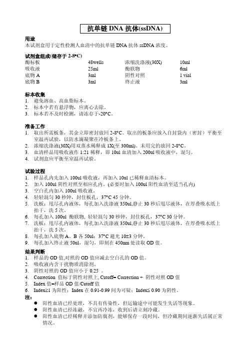
抗单链DNA抗体(ssDNA)
准备工作 1. 取出所需板条,其余立即密封放回 2-8°C。取出的板条应放入自封袋内(密封)平衡至
室温再试验,以防水滴凝聚在冷板条上。 2. 浓缩洗涤液(30X)用双蒸水稀释成 1X(至 600ml)。未用完的放回 2-8°C。 3. 血清样品用吸收液作 1:21 稀释,即 10ul 血清加入 200ul 吸收液中,混匀。 4. 试剂盒应平衡至室温再试验。
拍干,洗 5 次。 6. 每孔加入 100ul 酶联物, 轻轻混匀 30 秒钟,封住板孔,37°C 30 分钟。 7. 洗板:甩尽孔内液体,每孔加入洗涤液 350ul,静止 30 秒后甩尽液体,在厚叠吸水纸上
拍干,洗 5 次。 8. 每孔加入底物 A、B 各 50ul;37°C 避光 10±3 分钟。 9. 每孔加入终止液 50ul,混匀,即刻在 450nm 处读取 OD 值。
标本收集 1. 避免溶血、高血脂标本。 2. 标本中若有悬浮物,应离心去除。 3. 标本若不及时检测,请冻存于-20°C。
准备工作 1. 取出所需板条,其余立即密封放回 2-8°C。取出的板条应放入自封袋内(密封)平衡至
室温再试验,以防水滴凝聚在冷板条上。 2. 浓缩洗涤液(30X)用双蒸水稀释成 1X(至 300ml)。未用完的放回 2-8°C。 3. 血清样品用吸收液作 1:21 稀释,即 10ul 血清加入 200ul 吸收液中,混匀。 4. 试剂盒应平衡至室温再试验。
抗单链 DNA 抗体(ssDNA)
用途 本试剂盒用于定性检测人血清中的抗单链 DNA 抗体 ssDNA 浓度。
试剂盒组成(储存于 2-8°C) 酶标板 吸收液 底物 A 底物 B
48wells 25ml 3ml 3ml
浓缩洗涤液(30X) 酶联物 阴性对照 终止液
- 1、下载文档前请自行甄别文档内容的完整性,平台不提供额外的编辑、内容补充、找答案等附加服务。
- 2、"仅部分预览"的文档,不可在线预览部分如存在完整性等问题,可反馈申请退款(可完整预览的文档不适用该条件!)。
- 3、如文档侵犯您的权益,请联系客服反馈,我们会尽快为您处理(人工客服工作时间:9:00-18:30)。
M OLECULAR AND C ELLULAR B IOLOGY,Mar.1995,p.1182–1191Vol.15,No.3 0270-7306/95/$04.00ϩ0Copyright᭧1995,American Society for MicrobiologySingle-Chain Antibody-Mediated Intracellular Retention ofErbB-2Impairs Neu Differentiation Factor andEpidermal Growth Factor SignalingDIANA GRAUS-PORTA,ROGER R.BEERLI,AND NANCY E.HYNES*Friedrich Miescher-Institut,CH-4002Basel,SwitzerlandReceived28September1994/Returned for modification15November1994/Accepted7December1994 ErbB-2becomes rapidly phosphorylated and activated following treatment of many cell lines with epidermalgrowth factor(EGF)or Neu differentiation factor(NDF).However,these factors do not directly bind ErbB-2,and its activation is likely to be mediated via transmodulation by other members of the type I/EGF receptor(EGFR)-related family of receptor tyrosine kinases.The precise role of ErbB-2in the transduction of thesignals elicited by EGF and NDF is unclear.We have used a novel approach to study the role of ErbB-2insignaling through this family of receptors.An ErbB-2-specific single-chain antibody,designed to preventtransit through the endoplasmic reticulum and cell surface localization of ErbB-2,has been expressed in T47Dmammary carcinoma cells,which express all four known members of the EGFR family.We show that cellsurface expression of ErbB-2was selectively suppressed in these cells and that the activation of the mitogen-activated protein kinase pathway and p70/p85S6K,induction of c-fos expression,and stimulation of growth byNDF were dramatically impaired.Activation of mitogen-activated protein kinase and p70/p85S6K and inductionof c-fos expression by EGF were also significantly reduced.We conclude that in T47D cells,ErbB-2is a majorNDF signal transducer and a potentiator of the EGF signal.Thus,our observations demonstrate that ErbB-2plays a central role in the type I/EGFR-related family of receptors and that receptor transmodulationrepresents a crucial step in growth factor signaling.Receptor tyrosine kinases(RTKs)are involved in the regu-lation of cell growth and differentiation.Ligand binding to the extracellular domain of these receptors induces dimerization, stimulation of the intrinsic kinase activity,and autophosphor-ylation.RTKs elicit their functions by binding and/or phospho-rylating intracellular substrates(9,48,60).Tyrosine-phospho-rylated sites in the activated receptors function as high-affinity binding sites for SH2domain-containing proteins including Grb2,Shc,phospholipase C␥,and the p85subunit of phospha-tidylinositol(PtdIns)3-kinase,which couple RTKs to signal transduction pathways(19,41,49,51,66).The Ras-MAPK (mitogen-activated protein kinase)/ERK(extracellular signal-regulated kinase)cascade involves sequential kinase reactions which lead to the stimulation of MAPK and the induction of expression of transcription factors such as c-Jun and c-Fos(18, 35,63).RTKs couple to the Ras-MAPK signaling network via the SH2-SH3domain-containing adaptor protein Grb2and/or the SH2domain-containing protein Shc,which complexes with Grb2following its phosphorylation on tyrosine(41,51,58).By binding Grb2,the Ras guanine nucleotide exchange factor Sos is brought within the vicinity of p21ras,which allows modula-tion of Ras(12,40)and subsequently MAPK activity.Distinct intracellular pathways,such as the one leading to the activation of p70/p85S6K(4),whose link to RTKs appears to be mediated by PtdIns3-kinase(13,15),have been reported to exist. Four members of the type I/epidermal growth factor recep-tor(EGFR)-related family of RTKs have been identified: ErbB/EGFR(69),ErbB-2(76),ErbB-3(37,54),and ErbB-4 (52).The four proteins are normally coexpressed in various combinations in diverse tissues excluding the hematopoietic system.Aberrant expression of EGFR has been observed in a variety of human tumors(24).ErbB-2gene amplification and/or overexpression is frequently found in tumors arising at many sites,including breast and ovary(29).High levels of ErbB-3have been found in a number of mammary tumor cell lines(36)as well as in primary breast cancer(39).Ligands binding to different members of the type I RTKs have been identified.Several growth factors,including EGF (59),transforming growth factor␣(43),and amphiregulin (62),bind and activate the EGFR.Recently,a number of peptide factors which were simultaneously identified by differ-ent groups were shown to induce tyrosine phosphorylation of ErbB-2,and thus they were described as putative ligands for this receptor.The names reflect the source of isolation and include Neu differentiation factor(NDF),heregulin,glial growth factor,and acetylcholine receptor-inducing activity(20, 26,42,50,73).However,it has recently been shown that ErbB-2does not directly bind NDF.Instead,ErbB-3and ErbB-4function as direct receptors for NDF(11,53),and NDF-induced tyrosine phosphorylation of ErbB-2seems to occur only in the presence of either ErbB-3or ErbB-4,pre-sumably by heterodimerization and transphosphorylation(10, 53,64).Similarly,activation of EGFR by EGF and other ago-nists stimulates tyrosine phosphorylation of ErbB-2by het-erodimer formation(22,31,32).Although ErbB-2alone can-not bind any of these ligands,it has been shown to modulate ligand affinities.ErbB-2confers high-affinity binding sites for EGF by heterodimerizing with EGFR(70)as well as for NDF by heterodimerizing with ErbB-3(64).It is likely that ErbB-2 also modulates intracellular signals elicited by EGF and NDF, since both factors cause its phosphorylation and activation. We have used a novel approach to investigate the involve-ment of ErbB-2in EGF-and NDF-induced signaling.We have recently described experimental strategies for the targeted in-activation of RTKs by using single-chain antibodies(scFv), recombinant proteins consisting of immunoglobulin heavy-*Corresponding author.Mailing address:Friedrich Miescher-Insti-tut,P.O.Box2543,CH-4002Basel,Switzerland.Phone:4161696 6869.Fax:41616963835.1182 on July 4, 2017 by guest / Downloaded fromand light-chain variable domains linked over a short flexible linker peptide.A secreted form of an scFv competing with EGF for binding to the EGFR has been expressed in EGFR-transformed cells and was shown to inhibit receptor activity in an autocrine fashion (6).In an alternative approach,scFvs directed against the extracellular domain of ErbB-2have been expressed intracellularly and targeted to the lumen of the en-doplasmic reticulum (ER).By binding to the extracellular do-main of newly synthesized ErbB-2,the scFvs prevented its transit through the ER to the cell surface,thereby causing its functional inactivation (7).In the present study,we used the same approach to inactivate ErbB-2in T47D human mammary carcinoma cells.These cells express moderate levels of all known members of the EGFR family (30)and proliferate in response to NDF.We show that EGF-and NDF-induced ac-tivation of MAPK and p70/p85S6K as well as stimulation of growth by NDF is impaired in cells devoid of cell surface ErbB-2.MATERIALS AND METHODSMaterials.Recombinant human EGF and bovine pancreatic insulin were from Sigma,recombinant human basic fibroblast growth factor was from Collaborative Biomedical Products,and recombinant human NDF isoforms were a generous gift from Amgen,Thousand Oaks,Calif.For all of the experiments described in this report,the NDF-3isoform (amino acids 14to 241)was used.Numbering is according to Wen et al.(74).Antibodies used were EGFR-specific monoclonal antibody (MAb)EGFR1(Amersham),ErbB-2-specific antiserum 21N (28)and MAb FSP77(25),ErbB-3-specific affinity-purified rabbit polyclonal antibodies C17(Santa Cruz Biotechnology)and E38530(Transduction Laboratories),Shc-specific rabbit immunoglobulin G (Upstate Biotechnology,Inc.),Grb2-specific antiserum (Upstate Biotechnology,Inc.),ERK1-and ERK2-specific antisera (44),ERK2-specific MAb (Gibco BRL),phosphotyrosine-specific MAb (17),and scFv-specific antiserum (7).Expression of the ErbB-2-directed scFv in T47D cells.The heavy-and light-chain variable domains of MAb FRP5were cloned from hybridoma cells and used to construct a chimeric gene encoding the scFv FRP5as described previ-ously (72).For expression in mammalian cells and localization to the lumen of the ER,the scFv-5R cDNA was created as described elsewhere (7).Briefly,the cDNA was modified to encode an N-terminal immunoglobulin heavy-chain sig-nal peptide and a C-terminal ER retention signal (KDEL)and was cloned into the retroviral vector pBabePuro (46).Ecotropic virus encoding scFv-5R as well as an empty vector control virus were used to infect the amphotropic packaging cell line PA317(45)in the presence of 8g of Polybrene (Sigma)per ml.Two days after infection,the cells were subjected to selection in 5g of puromycin (Fluka)per ml for 10days.Virus-containing conditioned medium was collected from pools of puromycin-resistant PA317and used to infect the human mam-mary carcinoma cell line T47D.Four days after infection,the cells were sub-jected to selection in 2g of puromycin per ml for 14days.Pools of puromycin-resistant cells were analyzed in all experiments.Cell culture.T47D/puro and T47D/5R cells were maintained in Dulbecco modified Eagle medium (DMEM;Gibco BRL)supplemented with 10%fetal calf serum (Gibco BRL),5g of insulin (Sigma)per ml,and 1g of puromycin per ml.Prior to growth factor stimulation,cells were starved for 24h in serum-free medium (DMEM containing 1mg of fetuin [Sigma]per ml and 10g of transferrin [Sigma]per ml).Flow cytometric analysis.Cells were trypsinized and washed with 2ml of fluorescence-activated cell sorting (FACS)buffer (phosphate-buffered saline containing 0.1%sodium azide and 1%bovine serum albumin)prior to staining;106cells were suspended in 200l of FACS buffer containing MAb EGFR1,MAb FSP77(25),or affinity-purified rabbit polyclonal antibody E38530(20g of each per ml),specific for,respectively,the extracellular domain of human EGFR,ErbB-2,or ErbB-3.Following incubation on ice for 30min,the cells were washed in 2ml of FACS buffer.Bound antibodies were stained with either fluorescein-linked sheep anti-mouse antibody (Amersham)or goat anti-rabbit F(ab)2(Jackson ImmunoResearch)for 30min on ice.Finally,the cells were washed twice in 2ml of FACS buffer,resuspended in 500l of FACS buffer,and analyzed for fluorescence in a Becton Dickinson FACScan.Immunoprecipitation and Western blot (immunoblot)analysis.Cells were solubilized in Triton extraction buffer (50mM Tris [pH 7.5],5mM EGTA [pH 8.5],150mM NaCl,1%Triton X-100,2mM sodium orthovanadate,50mM sodium fluoride,10mM sodium molybdate,20M phenylarsine oxide,1mM phenylmethylsulfonyl fluoride,10g of leupeptin per ml,10g of aprotinin per ml)except for MAPK and S6K assays,for which ERK lysis buffer (ELB;see below)was used.The lysates were clarified by centrifugation at 16,000ϫg for 15min.For immunoprecipitations,equal amounts of protein were incubated with specific antibodies for 2h at 4ЊC.Immune complexes were collected with proteinA-Sepharose (Sigma)and washed three times with lysis buffer and once with TNE buffer (50mM Tris-HCl [pH 7.5],140mM NaCl,5mM EDTA).For immunoprecipitation of EGFR,protein A-Sepharose was precoated with poly-clonal rabbit anti-mouse immunoglobulin G (ICN Immunobiologicals).Bound proteins were released by boiling in sample buffer.Total cell lysates or immu-noprecipitates were subjected to sodium dodecyl sulfate (SDS)-polyacrylamide gel electrophoresis (PAGE),and proteins were blotted to polyvinylidene difluo-ride membranes.After blocking with 20%horse serum (Gibco BRL)in TTBS (50mM Tris-HCl [pH 7.5],150mM NaCl,0.05%Tween 20),filters were probed with specific antibodies and proteins were visualized with peroxidase-coupled secondary antibody,using the ECL detection system (Amersham).In some experiments,filters were stripped in SDS buffer (100mM -mercaptoethanol,2%SDS,62.5mM Tris-HCl [pH 6.8])for 30min at 65ЊC,washed three times in TTBS,blocked,and reprobed with the indicated antibodies.Exposed films were quantitated with a Molecular Dynamics densitometer.MAPK (ERK)assays.Cells were stimulated at 37ЊC with different factors for the indicated times and doses,lysed in ELB (50mM -sodium glycerophosphate,1.5mM EGTA,2mM sodium orthovanadate,1mM dithiothreitol,2g of leupeptin per ml,2g of aprotinin per ml,1mM benzamidine,1%[vol/vol]Nonidet P-40).ERK2was immunoprecipitated from 200g of total lysate with 2l of specific antiserum (44)and protein A-Sepharose.The immunoprecipi-tates were washed three times with ELB and once with kinase buffer (30mM Tris-HCl [pH 8.0],20mM MnCl 2,and 2mM MgCl 2).The activity of MAPK was then determined by incubating the immune complexes at 37ЊC for 30min with 30l of kinase buffer containing 15g of myeloid basic protein (MBP),10M unlabeled ATP,and 0.1M [␥-32P]ATP (1,200Ci/mmol).The reaction was stopped with sample buffer,proteins were subjected to SDS-PAGE (15%gel)and blotted,and the phosphorylation of MBP was quantitated with a Phosphor-Imager (Molecular Dynamics).To verify that equal amounts of ERK2were immunoprecipitated,a Western blot analysis with a specific MAb was carried out.S6kinase assays.Cells were stimulated as described above and lysed in ELB,and p70/p85S6K activity was determined as previously described (38).Briefly,0.4g of crude lysate was incubated for 30min at 37ЊC with 10g of 40S ribosomal subunit in a reaction buffer containing 100mM morpholine propane sulfonic acid (MOPS),2mM dithiothreitol,20mM MgCl 2,20mM p -nitrophenylphosphate,60M unlabeled ATP,0.05g of cyclic AMP-dependent protein kinase inhib-itor (Sigma),and 1Ci of [␥-32P]ATP (1,200Ci/mmol).The reaction was stopped with sample buffer,proteins were subjected to SDS-PAGE (15%gel)and blotted,and phosphorylation of S6ribosomal protein was quantitated with a PhosphorImager (Molecular Dynamics).Northern (RNA)blotting.Total RNA was extracted from growth factor-treated cells by using Trizol (Gibco BRL).Ten micrograms of RNA was elec-trophoresed on a 0.66M formaldehyde–1%agarose gel,transferred to a nylon membrane,and hybridized to a digoxigenin-labeled riboprobe encompassing the 1.3-kb Bgl II-Pvu II fragment of the v-fos cDNA (8).The hybridized probe was detected by using alkaline phosphatase-conjugated antidigoxigenin F(ab)2frag-ments and the chemiluminescent substrate Lumigen PPD (Boehringer Mann-heim).Cell growth assays.Factor responsiveness for growth was examined under anchorage-independent conditions.Cells (3ϫ103)were plated in duplicate in 6-cm-diameter dishes in 6ml of culture medium supplemented with 0.35%Noble agar overlaying a 0.7%agar layer.For growth induction,1nM growth factor was added to the medium.The cells were incubated for 4weeks at 37ЊC,after which colonies were stained by adding 2ml of phosphate-buffered saline containing 0.5mg of nitroblue tetrazolium per ml for 24h.Colonies larger than 200m were counted with an Artek 880colony counter (Dynatech Laboratories,Inc.).RESULTSIntracellular retention of ErbB-2in T47D cells.To investi-gate the involvement of ErbB-2in EGF-and NDF-induced signaling,we have specifically inactivated ErbB-2in T47D hu-man mammary carcinoma cells.We have previously shown that an ER-retained variant of the ErbB-2-specific scFv FRP5,scFv-5R,specifically inhibits ER transit of ErbB-2and leads to a loss of cell surface ErbB-2(7).We therefore established a cell line,T47D/5R,which intracellularly expresses scFv-5R at high levels as judged by Western blotting (Fig.1A).scFv-5R was detected as a double band,the upper of which corresponds to a N-glycosylated form (7).When analyzed by Western blot-ting,the ErbB-2protein in the T47D/5R cells showed a faster electrophoretic mobility than the T47D/puro vector control cells (Fig.1B).ErbB-2is a glycoprotein,and its carbohydrate side chains undergo modification and extension in the Golgi complex (1).It is likely that ER retention of ErbB-2causes hypoglycosylation,which might explain the reduced apparentV OL .15,1995TARGETED INACTIVATION OF ErbB-2IN MAMMARY CELLS 1183on July 4, 2017 by guest/Downloaded frommolecular weight.Specific intracellular retention of ErbB-2was demonstrated by flow cytometric analysis.Intact cells were stained for the presence of cell surface EGFR,ErbB-2,or ErbB-3(Fig.2).ErbB-2was absent from the cell surface in T47D/5R cells but present on T47D/puro control cells.In con-trast,the related EGFR and ErbB-3proteins were expressed atequal levels on the cell surface of both cell lines,indicating that the scFv-5R-mediated abolishment of ErbB-2was highly spe-cific.EGF-and NDF-induced tyrosine phosphorylation of type I RTKs.Following activation of EGFR by EGF,heterodimer-ization with ErbB-2and cross-phosphorylation occur (22).It is likely that heterodimerization and receptor cross-phosphory-lation of ErbB-2with ErbB-3and/or ErbB-4follow NDF stim-ulation (53,64).Thus,we investigated how the presence or absence of ErbB-2influences the phosphorylation of other members of the EGFR family in response to EGF and NDF.T47D/puro and T47D/5R cells were treated with 50ng of each factor per ml for 5min,and the phosphotyrosine content of EGFR,ErbB-2,or ErbB-3was analyzed (Fig.3).The tyrosine phosphorylation of ErbB-2dramatically increased in response to EGF and NDF in the T47D/puro control cells.In contrast,there was no increase in ErbB-2tyrosine phosphorylation in the T47D/5R cells,likely because of the lack of cell surface ErbB-2(Fig.3B).In response to EGF,the EGFR was phos-phorylated in both cell lines.However,in the T47D/5R cells,the EGFR phosphotyrosine content was reduced by 46%com-pared with the control cells.No tyrosine phosphorylation could be detected in response to NDF (Fig.3A).NDF treatment of T47D/puro cells resulted in a strong tyrosine phosphorylation of ErbB-3.Remarkably,the phosphotyrosine content of ErbB-3was reduced by 89%in the T47D/5R compared with the T47D/puro cells,suggesting that ErbB-2contributes to a large extent to the stimulation of tyrosine phosphorylation of ErbB-3triggered by NDF.No ErbB-3phosphorylation was detected upon EGF treatment (Fig.3C),which may be due to the low levels of EGFR in T47D cells (approximately 7ϫ103[27a]).It has been shown that in EGFR-overexpressing A431cells,EGF induces tyrosine phosphorylation of ErbB-3(65).FIG.1.Expression of scFv-5R and ErbB-2in T47D/puro and T47D/5R cells.(A)scFv-5R Western blot.Fifty micrograms of total protein was subjected to SDS-PAGE (15%gel),blotted,and analyzed for expression of scFv-5R with an scFv-specific antiserum (7).(B)ErbB-2Western blot.Fifty micrograms of total protein was subjected to SDS-PAGE (7.5%gel),blotted,and analyzed for ErbB-2expression with antiserum 21N(28).FIG.2.Loss of cell surface ErbB-2in T47D/5R cells.T47D/puro and T47D/5R cells were incubated with MAb EGFR1,MAb FSP77(25),and rabbit polyclonal antibody E38530,which react with EGFR,ErbB-2,and ErbB-3,re-spectively.Specifically bound antibody was detected with fluorescein-linked an-tibody,and cells were analyzed for fluorescence in a FACScan.Right-hand curves,specific staining;left-hand curves,nonspecific staining by omitting the primary antibody;ordinates,relative cell number;abscissas,logfluorescence.FIG.3.EGF-and NDF-induced tyrosine phosphorylation of EGFR,ErbB-2,and ErbB-3.After 24h of starvation,cells were either treated for 5min with 50ng of EGF or NDF per ml or left untreated.EGFR (A),ErbB-2(B),and ErbB-3(C)were immunoprecipitated (IP)from 1.5mg of total protein with,respec-tively,MAb EGFR1,antiserum 21N (28),and rabbit polyclonal antibody C17.Proteins were subjected to SDS-PAGE (7.5%gel),Western blotted (WB),and probed with a phosphotyrosine-specific MAb (␣-PY)(17).Filters were stripped and reprobed with specific antibodies (21N [B]and C17[C])to verify that equal amounts of proteins were immunoprecipitated.The amounts of EGFR immu-noprecipitated were too low to be detected.1184GRAUS-PORTA ET AL.M OL .C ELL .B IOL .on July 4, 2017 by guest/Downloaded fromEGF-and NDF-induced tyrosine phosphorylation of cellu-lar proteins.To assess the effect that loss of cell surface ErbB-2has on EGF-and NDF-induced signaling through the type I RTKs,total cell lysates were initially analyzed by West-ern blotting with a phosphotyrosine-specific MAb.Following EGF treatment (Fig.4A),two proteins in the high-molecular-weight region of the gel (indicated with an arrow)rapidly became tyrosine phosphorylated in the T47D/puro control cells.In the T47D/5R cells,only the lower protein was phos-phorylated.It is likely that the upper protein corresponds to ErbB-2and that the lower protein corresponds to EGFR.Phosphorylation of several other proteins (indicated by arrow-heads)with molecular masses of approximately 60,48,44,and 42kDa was also induced by EGF treatment.While the 60-and the 48-kDa proteins have not yet been identified,the last two proteins were found to comigrate with ERK1and ERK2,re-spectively.Phosphorylation of both ERKs remained high for 2h in the T47D/puro cells,whereas it decreased to the basal level in the T47D/5R cells after 2h.More striking differences between the two cell lines were observed when cells were stimulated with NDF (Fig.4B).Rapid and strong phosphory-lation of a protein(s)in the high-molecular-weight region of the gel (indicated with an arrow)was observed in T47D/puro control cells.This phosphorylation was dramatically reduced in the T47D/5R cells,presumably because of the lack of cell surface ErbB-2and/or reduced ErbB-3/4phosphorylation.As seen for EGF stimulation,phosphorylation of several other proteins with molecular masses of approximately 60,48,44,and 42kDa was also induced.While phosphorylation of the 60-kDa protein was observed in both cell lines,phosphoryla-tion of the 48-kDa protein was visible only in the T47D/puro cells.Strikingly,phosphorylation of the 44-and 42-kDa pro-teins,which comigrated with ERK1and ERK2,respectively,remained high after 2h of NDF treatment in the T47D/puro cells,whereas it started decreasing after 10min in the T47D/5R cells.Absence of cell surface ErbB-2impairs EGF-and NDF-dependent activation of MAPK.The distinct time-dependent tyrosine phosphorylation of the two proteins comigrating with ERK1and ERK2prompted us to examine the activities of these two MAPK isoforms.In vitro kinase assays using MBP as a substrate were performed.Following EGF stimulation (Fig.5A),ERK2was rapidly activated with similar kinetics,and its activity declined slowly in both cell lines.However,throughout the time course,activation of ERK2was always more pro-nounced in the T47D/puro cells.When cells were treated with NDF (Fig.5B),ERK2activity rapidly increased in both T47D/puro and T47D/5R cells,although again to a higher level in the former.Strikingly,in the T47D/puro cells,NDF-induced acti-vation of ERK2was sustained for 2h,whereas in the cells lacking ErbB-2on the cell surface,it was only transient and rapidly returned to the basal level.Activity of ERK1in re-sponse to both EGF and NDF was also analyzed and found to follow the same pattern as the ERK2activity (not shown).To show that the effects observed were specific for EGF and NDF,basic fibroblast growth factor,which interacts with a nonre-lated receptor tyrosine kinase,was tested for its ability to stimulate MAPK.In both cell lines,the fold induction and kinetics of ERK2activity (Fig.5C)and ERK1activity (not shown)followed the same pattern.The EGF-and NDF-induced ERK1and ERK2stimulation in T47D/5R cells was always lower than that in T47D/puro cells.Therefore,different doses of the two factors were applied to the cells for 10min.Similar results for EGF (Fig.6A)and for NDF (Fig.6B)were found:picomolar concentrations of both factors stimulated ERK2activity in both cell lines,and even with saturating amounts,the activity in the T47D/5R cells was lower.For both growth factors,the same results were obtained with ERK1(not shown).Therefore,we conclude that both EGF-and NDF-induced MAPK activation is impaired by the absence of cell surface ErbB-2.EGF-and NDF-induced tyrosine phosphorylation of Shc and association with type I RTKs and Grb2.Activated type I RTKs are coupled to the Ras-MAPK cascade by binding to the SH2-SH3domain-containing adaptor protein Grb2and/or the SH2domain-containing protein Shc,which is phosphorylated on tyrosine and then complexes with Grb2(58).Given the evidence that EGF-and NDF-induced MAPK activation was diminished by the absence of cell surface ErbB-2(Fig.5),we reasoned that the stimulation of Shc tyrosine phosphorylation and Grb2association might be affected.To test this hypothesis,cells were stimulated with 50ng of EGF or NDF per ml for 5min,proteins were extracted,and Shc was immunoprecipi-tated.A Western blot analysis showed that equal amounts of Shc proteins were immunoprecipitated from all samples (not shown).An analysis using a phosphotyrosine-specific antibody revealed that in T47D/puro cells,two of the three Shc iso-forms,p46shc and p52shc ,became highly phosphorylated in response to EGF and NDF.In the T47D/5R cells,the NDF-induced tyrosine phosphorylation of both proteins was reduced by 90%(Fig.7A).EGF-induced Shc phosphorylation was also reduced in these cells,albeit to a lesser extent (63and 41%for p46shc and p52shc ,respectively).High-molecular-weight ty-rosine-phosphorylated proteins (indicated with an arrow)were coimmunoprecipitated with the Shc proteins in growth factor-treated cells.A reprobing of the filter with different antibodies revealed that in T47D/puro but not T47D/5R cells,ErbB-2was coimmunoprecipitated with Shc,both in response to EGFandFIG.4.Total phosphotyrosine blot of EGF-and NDF-induced T47D/puro and T47D/5R cells.Cells were stimulated with 0.5nM EGF (A)or NDF (B)for the indicated times prior to lysis.One hundred micrograms of total protein was subjected to SDS-PAGE (9%gel),immunoblotted,and probed with a phospho-tyrosine-specific MAb.Arrows indicate the position of proteins showing elevated phosphotyrosine content upon factor treatment.V OL .15,1995TARGETED INACTIVATION OF ErbB-2IN MAMMARY CELLS 1185on July 4, 2017 by guest/Downloaded fromin response to NDF (Fig.7B).ErbB-3was found to be com-plexed with Shc in both T47D/puro and T47D/5R cells in response to NDF.However,81%less Shc–ErbB-3complexes were found in T47D/5R cells,a finding that can be explained by the strongly reduced NDF-induced phosphorylation of ErbB-3in these cells (Fig.3C).No EGFR was detected in the Shc precipitates (not shown).However,this might be due to the low EGFR level in these cells.In fact,the high-molecular-weight tyrosine-phosphorylated protein that is coimmunopre-cipitated with Shc in both EGF-treated T47D/puro and T47D/5R cells is likely to be EGFR (Fig.7A).Grb2was foundto be associated with Shc in both EGF-and NDF-treated T47D/puro cells.This association was reduced by 24%in EGF-treated and by 76%in NDF-treated T47D/5R cells.These observations provide a molecular basis for the reduced activa-tion of MAPK.EGF-and NDF-induced c-fos expression is impaired in cells lacking cell surface ErbB-2.c-fos expression can be induced by a variety of growth factors through the Ras-MAPK pathway (23,34).Moreover it was recently shown that ERK2is directly involved in the activation of c-fos expression (34).Our results show that EGF-and NDF-induced activation of ERK2is im-paired in the T47D/5R cells;therefore,we evaluated whether c-fos expression was also affected.Cells were treated with 0.5nM EGF and NDF for 30min,and c-fos expression was ex-amined by Northern blot analysis (Fig.8).EGF and NDF induced the expression of c-fos in both cell lines.However,there were 23and 81%reductions in,respectively,EGF-and NDF-treated T47D/5R cells.Our results suggest that the ulti-mate step in the activation of the Ras-MAPK pathway by ligands of type I RTKs,i.e.,the phosphorylation of nuclear targets and induction of gene expression,is impaired by the lack of ErbB-2.Absence of cell surface ErbB-2impairs EGF-and NDF-dependent activation of p70/p85S6K .We have previously shown that NDF and EGF induce p70/p85S6K activity in T47D cells (44).Therefore,we investigated whether the activation oftheFIG.5.Time course of ERK2activation.T47D/puro (closed squares)and T47D/5R (open squares)cells were treated with 0.5nM EGF (A),NDF (B),or basic fibroblast growth factor (C)for the indicated times.ERK2was immuno-precipitated from 200g of cell lysate with a specific antibody (44),and kinase activity was determined by using MBP as a substrate.Proteins were subjected to SDS-PAGE (15%gel)and blotted,and phosphorylation of MBP was quantitated with a PhosphorImager (MolecularDynamics).FIG.6.Dose response of ERK2activation.T47D/puro (closed squares)and T47D/5R (open squares)cells were treated for 10min with the indicated con-centrations of EGF (A)or NDF (B).ERK2was immunoprecipitated from 200g of cell lysate with a specific antibody (44),and kinase activity was determined by using MBP as a substrate.Proteins were subjected to SDS-PAGE (15%gel)and blotted,and phosphorylation of MBP was quantitated with a PhosphorIm-ager (Molecular Dynamics).1186GRAUS-PORTA ET AL.M OL .C ELL .B IOL .on July 4, 2017 by guest/Downloaded from。
