AI-2 Key Enzyme S-Ribosylhomocysteinase from Strain Klebsiella pneumoniae CICC 10011 Producing 2
PPARγ基因shRNA真核表达载体的构建及其干扰效果鉴定
P P A M基因shRNA真核表达载体的构建及其干扰效果鉴定辛婧、沈亚非、邓飞\谢亚争、张日华2,刘云21.漯河市中心医院内分泌科,河南漯河462000;2•南京医科大学第一附属医院老年医学内分泌科摘要:目的构建过氧化物酶体增殖物激活受体y(PPARy)基因RNA干扰的真核表达载体,转染小鼠3T3-L1前体脂肪 细胞,并鉴定其干扰效果。
方法选择设计2条针对小鼠PPA R y基因的干扰耙序列,构建真核表达载体PLKO. 1-PPARy-GFP-shRNAl/2,以PCR鉴定并进行序列分析。
证实质粒构建成功后,转染小鼠3T3-U前体脂肪细胞,荧光显微镜下观察 绿色荧光蛋白(eGFP)的表达,计算转染效率;采用实时定量PC R法和W estern印迹检测载体对PPA R y基因的表达干扰效 果;采用油红染色观察PPA R y在脂肪细胞分化过程中对脂滴形成的作用。
结果成功构建PPA R y干扰质粒PLKO.1- PPARy-GFP-shRNAl/2,PC R和DNA测序证实序列与设计完全一致;荧光显微镜下观察到3T3-L1细胞绿色荧光蛋白的表 达,证实重组质粒成功转人细胞,转染效率(92. 67士1.53)%;实时荧光定量PC R和Western B lot印记试验均显示PLKO. 1-PPARy-GFP-shRNAl/2转染的3T3-L1细胞PPARy基因分别被特异性抑制;耙点1干扰效率[(78. 2士2.1)%]高于耙点2 [(55.8±4.3)%],差异有统计学意义(P C0.05),靶点1初步鉴定为有效靶点。
油红染色显示PLKO. 1-PPARy-GFP-shR-NA1/2转染均可抑制3T3-L1脂肪细胞分化和脂滴聚集。
结论成功构建了耙向干扰PPA R y基因的shRNA真核表达质粒 PLKO.l-PPARy-GFP-shRNAl/2,并筛选出有效抑制靶基因的表达质粒,初步验证PPA R y基因对脂肪细胞分化的作用,为 进一步研究提供了有效的分子生物学工具。
小鼠Ⅰ型胶原α2 (COL1a2)-ELISA试剂盒说明书
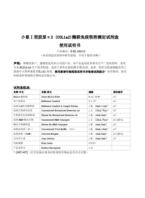
小鼠Ⅰ型胶原α2 (COL1a2)酶联免疫吸附测定试剂盒使用说明书产品编号:E-EL-M0318(本试剂盒仅供体外研究使用、不用于临床诊断!)声明:尊敬的客户,感谢您选用本公司的产品。
本产品选用世界著名生产厂家的原料,采用专业ELISA kit生产技术制造。
适用于体外定量检测小鼠血清、血浆、组织匀浆或细胞培养上清液中天然和重组COL1a2浓度。
使用前请仔细阅读说明书并检查试剂组分!如有疑问,请及时联系伊莱瑞特生物科技有限公司。
*: [96T/48T](打开包装后请及时检查所有物品是否齐全完整)检测原理:本试剂盒采用双抗体夹心ELISA法。
用抗小鼠COL1a2抗体包被于酶标板上,实验时标本或标准品中的COL1a2会与包被抗体结合,游离的成分被洗去。
依次加入生物素化的抗小鼠COL1a2抗体和辣根过氧化物酶标记的亲和素。
抗小鼠COL1a2抗体与结合在包被抗体上的小鼠COL1a2结合、生物素与亲和素特异性结合而形成免疫复合物,游离的成分被洗去。
加入显色底物(TMB),TMB 在辣根过氧化物酶的催化下现蓝色,加终止液后变黄。
用酶标仪在450nm波长处测OD值,COL1a2浓度与OD450值之间呈正比,通过绘制标准曲线求出标本中COL1a2的浓度。
标本收集:1.血清:全血标本于室温放置2小时或4℃过夜后于1000×g离心20分钟,取上清即可检测,收集血液的试管应为一次性的无热原,无内毒素试管。
2.血浆:抗凝剂推荐使用EDTA.Na2,标本采集后30分钟内于1000×g离心15分钟,取上清即可检测。
避免使用溶血,高血脂标本。
3.组织匀浆:用预冷的PBS (0.01M, pH=7.4)冲洗组织,以去除残留血液(匀浆中裂解的红细胞会影响测量结果),称重后将组织剪碎。
将剪碎的组织与对应体积的PBS(一般按1:9的重量体积比,比如1g的组织样本对应9mL的PBS,具体体积可根据实验需要适当调整,并做好记录。
marked manuscript

Quality evaluation of Flos Lonicerae through a simultaneous determination of seven saponins by HPLC with ELSDXing-Yun Chai1, Song-Lin Li2, Ping Li1*1Key Laboratory of Modern Chinese Medicines and Department of Pharmacognosy, China Pharmaceutical University, Nanjing, 210009, People’s Republic of China2Institute of Nanjing Military Command for Drug Control, Nanjing, 210002, People’s Republic of China*Corresponding author: Ping LiKey Laboratory of Modern Chinese Medicines and Department of Pharmacognosy, China Pharmaceutical University, Nanjing 210009, People’s Republic of China.E-mail address: lipingli@Tel.: +86-25-8324-2299; 8539-1244; 135********Fax: +86-25-8532-2747AbstractA new HPLC coupled with evaporative light scattering detection (ELSD) method has been developed for the simultaneous quantitative determination of seven major saponins, namely macranthoidinB (1), macranthoidin A (2), dipsacoside B (3), hederagenin-28-O-β-D-glucopyranosyl(6→1)-O-β-D- glucopyranosyl ester (4), macranthoside B (5), macranthoside A (6), and hederagenin-3-O-α-L-arabinopyranosyl(2→1)-O-α-L-rhamnopyranoside (7)in Flos Lonicerae, a commonly used traditional Chinese medicine (TCM) herb.Simultaneous separation of these seven saponins was achieved on a C18 analytical column with a mixed mobile phase consisting of acetonitrile(A)-water(B)(29:71 v/v) acidified with 0.5% acetic acid. The elution was operated from keeping 29%A for 10min, then gradually to 54%B from 10 to 25 min on linear gradient, and then keep isocratic elution with 54%B from 25 to 30min.The drift tube temperature of ELSD was set at 106℃, and with the nitrogen flow-rate of 2.6 l/min. All calibration curves showed good linear regression (r2 0.9922) within test ranges. This method showed good reproducibility for the quantification of these seven saponins in Flos Lonicerae with intra- and inter-day variations of less than 3.0% and 6.0% respectively. The validated method was successfully applied to quantify seven saponins in five sources of Flos Lonicerae, which provides a new basis of overall assessment on quality of Flos Lonicerae.Keywords: HPLC-ELSD; Flos Lonicerae; Saponins; Quantification1. IntroductionFlos Lonicerae (Jinyinhua in Chinese), the dried buds of several species of the genus Lonicera (Caprifoliaceae), is a commonly used traditional Chinese medicine (TCM) herb. It has been used for centuries in TCM practice for the treatment of sores, carbuncles, furuncles, swelling and affections caused by exopathogenic wind-heat or epidemic febrile diseases at the early stage [1]. Though four species of Lonicera are documented as the sources of Flos Lonicerae in China Pharmacopeia (2000 edition), i.e. L. japonica, L. hypoglauca,L. daystyla and L. confusa, other species such as L. similes and L. macranthoides have also been used on the same purpose in some local areas in China [2]. So it is an important issue to comprehensively evaluate the different sources of Flos Lonicerae, so as to ensure the clinical efficacy of this Chinese herbal drug.Chemical and pharmacological investigations on Flos Lonicerae resulted in discovering several kinds of bioactive components, i.e. chlorogenic acid and its analogues, flavonoids, iridoid glucosides and triterpenoid saponins [3]. Previously, chlorogenic acid has been used as the chemical marker for the quality evaluation of Flos Lonicerae,owing to its antipyretic and antibiotic property as well as its high content in the herb. But this compound is not a characteristic component of Flos Lonicerae, as it has also been used as the chemical marker for other Chinese herbal drugs such as Flos Chrysanthemi and so on[4-5]. Moreover, chlorogenic acid alone could not be responsible for the overall pharmacological activities of Flos Lonicerae[6].On the other hand, many studies revealed that triterpenoidal saponins of Flos Lonicerae possess protection effects on hepatic injury caused by Acetaminophen, Cd, and CCl4, and conspicuous depressant effects on swelling of ear croton oil [7-11]. Therefore, saponins should also be considered as one of the markers for quality control of Flos Lonicerae. Consequently, determinations of all types of components such as chlorogenic acid, flavonoids, iridoid glucosides and triterpenoidal saponins in Flos Lonicerae could be a better strategy for the comprehensive quality evaluation of Flos Lonicerae.Recently an HPLC-ELSD method has been established in our laboratory for qualitative and quantitative determination of iridoid glucosides in Flos Lonicerae [12]. But no method was reported for the determination of triterpenoidal saponins in Flos Lonicera. As a series studies on the comprehensive evaluation of Flos Lonicera, we report here, for the first time, the development of an HPLC-ELSD method for simultaneous determination of seven triterpenoidal saponins in the Chinese herbal drug Flos Lonicerae, i.e.macranthoidin B (1), macranthoidin A (2), dipsacoside B (3), hederagenin-28-O-β-D-glucopyranosyl(6→1)-O-β-D- glucopyranosyl ester (4), macranthoside B (5), macranthoside A (6), and hederagenin-3-O-α-L-arabinopyranosyl(2→1)-O-α-L-rhamnopyranoside (7) (Fig. 1).2. Experimental2.1. Samples, chemicals and reagentsFive samples of Lonicera species,L. japonica from Mi county, HeNan province (LJ1999-07), L. hypoglauca from Jiujang county, JiangXi province (LH2001-06), L. similes from Fei county, ShanDong province (LS2001-07), L. confuse from Xupu county, HuNan province (LC2001-07), and L. macranthoides from Longhu county, HuNan province (LM2000-06) respectively, were collected in China. All samples were authenticated by Dr. Ping Li, professor of department of Pharmacognosy, China Pharmaceutical University, Nanjing, China. The voucher specimens were deposited in the department of Pharmacognosy, China Pharmaceutical University, Nanjing, China. Seven saponin reference compounds: macranthoidin B (1), macranthoidin A (2), dipsacoside B (3), hederagenin-28-O-β-D-glucopyranosyl(6→1)-O-β-D- glucopyranosyl ester (4), macranthoside B (5), macranthoside A (6), and hederagenin-3-O-α-L-arabinopyranosyl(2→1)-O-α-L-rhamnopyranoside (7) were isolated previously from the dried buds of L. confusa by repeated silica gel, sephadex LH-20 and Rp-18 silica gel column chromatography, their structures were elucidated by comparison of their spectral data (UV, IR, MS, 1H- NMR and 13C-NMR) with references [13-15]. The purity of these saponins were determined to be more than 98% by normalization of the peak areas detected by HPLC with ELSD, and showed very stable in methanol solution.HPLC-grade acetonitrile from Merck (Darmstadt, Germany), the deionized water from Robust (Guangzhou, China), were purchased. The other solvents, purchased from Nanjing Chemical Factory (Nanjing, China) were of analytical grade.2.2. Apparatus and chromatographic conditionsAglient1100 series HPLC apparatus was used. Chromatography was carried out on an Aglient Zorbax SB-C18 column(250 4.6mm, 5.0µm)at a column temperature of 25℃.A Rheodyne 7125i sampling valve (Cotati, USA) equipped with a sample loop of 20µl was used for sample injection. The analog signal from Alltech ELSD 2000 (Alltech, Deerfield, IL, USA)was transmitted to a HP Chemstation for processing through an Agilent 35900E (Agilent Technologies, USA).The optimum resolution was obtained by using a linear gradient elution. The mobile phase was composed of acetonitrile(A) and water(B) which acidified with 0.5% acetic acid. The elution was operated from keeping 29%A for 10min, then gradually to 54%B from 10 to 25 min in linear gradient, and back to the isocratic elution of 54%B from 25 to 30 min.The drift tube temperature for ELSD was set at 106℃and the nitrogen flow-rate was of 2.6 l/min. The chromatographic peaks were identified by comparing their retention time with that of each reference compound tried under the same chromatographic conditions with a series of mobile phases. In addition, spiking samples with the reference compounds further confirmed the identities of the peaks.2.3. Calibration curvesMethanol stock solutions containing seven analytes were prepared and diluted to appropriate concentration for the construction of calibration curves. Six concentrationof the seven analytes’ solution were injected in triplicate, and then the calibration curves were constructed by plotting the peak areas versus the concentration of each analyte. The results were demonstrated in Table1.2.4. Limits of detection and quantificationMethanol stock solution containing seven reference compounds were diluted to a series of appropriate concentrations with methanol, and an aliquot of the diluted solutions were injected into HPLC for analysis.The limits of detection (LOD) and quantification (LOQ) under the present chromatographic conditions were determined at a signal-to-noise ratio (S/N) of 3 and 10, respectively. LOD and LOQ for each compound were shown in Table1.2.5. Precision and accuracyIntra- and inter-day variations were chosen to determine the precision of the developed assay. Approximate 2.0g of the pulverized samples of L. macranthoides were weighted, extracted and analyzed as described in 2.6 Sample preparation section. For intra-day variability test, the samples were analyzed in triplicate for three times within one day, while for inter-day variability test, the samples were examined in triplicate for consecutive three days. Variations were expressed by the relative standard deviations. The results were given in Table 2.Recovery test was used to evaluate the accuracy of this method. Accurate amounts of seven saponins were added to approximate 1.0g of L. macranthoides,and then extracted and analyzed as described in 2.6 Sample preparation section. The average recoveries were counted by the formula: recovery (%) = (amount found –original amount)/ amount spiked ×100%, and RSD (%) = (SD/mean) ×100%. The results were given in Table 3.2.6. Sample preparationSamples of Flos Lonicerae were dried at 50℃until constant weight. Approximate 2.0g of the pulverized samples, accurately weighed, was extracted with 60% ethanol in a flask for 4h. The ethanol was evaporated to dryness with a rotary evaporator. Residue was dissolved in water, followed by defatting with 60ml of petroleum ether for 2 times, and then the water solution was evaporated, residue was dissolved with methanol into a 25ml flask. One ml of the methanol solution was drawn and transferred to a 5ml flask, diluted to the mark with methanol. The resultant solution was at last filtrated through a 0.45µm syringe filter (Type Millex-HA, Millipore, USA) and 20µl of the filtrate was injected to HPLC system. The contents of the analytes were determined from the corresponding calibration curves.3. Results and discussionsThe temperature of drift tube and the gas flow-rate are two most important adjustable parameters for ELSD, they play a prominent role to an analyte response. In ourprevious work [12], the temperature of drift tube was optimized at 90°C for the determination of iridoids. As the polarity of saponins are higher than that of iridoids, more water was used in the mobile phase for the separation of saponins, therefore the temperature for saponins determination was optimized systematically from 95°C to 110°C, the flow-rate from 2.2 to 3.0 l/min. Dipsacoside B was selected as the testing saponin for optimizing ELSD conditions, as it was contained in all samples. Eventually, the drift tube temperature of 106℃and a gas flow of 2.6 l/min were optimized to detect the analytes. And these two exact experimental parameters should be strictly controlled in the analytical procedure [16].All calibration curves showed good linear regression (r2 0.9922) within test ranges. Validation studies of this method proved that this assay has good reproducibility. As shown in Table 2, the overall intra- and inter-day variations are less than 6% for all seven analytes. As demonstrated in Table 3, the developed analytical method has good accuracy with the overall recovery of high than 96% for the analytes concerned. The limit of detection (S/N=3) and the limit of quantification (S/N=10) are less than 0.26μg and 0.88μg respectively (Table1), indicating that this HPLC-ELSD method is precise, accurate and se nsitive enough for the quantitative evaluation of major non- chromaphoric saponins in Flos Lonicerae.It has been reported that there are two major types of saponins in Flos Lonicerae, i.e. saponins with hederagenin as aglycone and saponins with oleanolic acid as the aglycone [17]. But hederagenin type saponins of the herb were reported to have distinct activities of liver protection and anti-inflammatory [7-11]. So we adoptedseven hederagenin type saponins as representative markers to establish a quality control method.The newly established HPLC-ELSD method was applied to analyze seven analytes in five plant sources of Flos Lonicerae, i.e. L. japonica,L. hypoglauca,L. confusa,L. similes and L. macranthoides(Table 4). It was found that there were remarkable differences of seven saponins contents between different plant sources of Flos Lonicerae. All seven saponins analyzed could be detected in L. confusa and L. hypoglauca, while only dipsacoside B was detected in L. japonica. Among all seven saponins interested, only dipsacoside B was found in all five plant species of Flos Lonicerae analyzed, and this compound was determined as the major saponin with content of 53.7 mg/g in L. hypoglauca. On the other hand, macranthoidin B was found to be the major saponin with the content higher than 41.0mg/g in L. macranthoides,L. confusa, and L. similis, while the contents of other analytes were much lower.In our previous study [12], overall HPLC profiles of iridoid glucosides was used to qualitatively and quantitatively distinguish different origins of Flos Lonicerae. As shown in Fig.2, the chromatogram profiles of L. confusa, L. japonica and L. similes seem to be similar, resulting in the difficulty of clarifying the origins of Flos Lonicerae solely by HPLC profiles of saponins, in addition to the clear difference of the HPLC profiles of saponins from L. macranthoides and L. hypoglauca.Therefore, in addition to the conventional morphological and histological identification methods, the contents and the HPLC profiles of saponins and iridoids could also be used as accessory chemical evidence toclarify the botanical origin and comprehensive quality evaluation of Flos Lonicerae.4. ConclusionsThis is the first report on validation of an analytical method for qualification and quantification of saponins in Flos Lonicerae. This newly established HPLC-ELSD method can be used to simultaneously quantify seven saponins, i.e. macranthoidin B, macranthoidin A, dipsacoside B, hederagenin-28-O-β-D-glucopyranosyl(6→1)-O-β-D- glucopyranosyl ester, macranthoside B, macranthoside A, and hederagenin-3-O-α-L-arabinopyranosyl(2→1)-O-α-L-rhamnopyranoside in Flos Lonicerae. Together with the HPLC profiles of iridoids, the HPLC-ELSD profiles of saponins could also be used as an accessory chemical evidence to clarify the botanical origin and comprehensive quality evaluation of Flos Lonicerae.AcknowledgementsThis project is financially supported by Fund for Distinguished Chinese Young Scholars of the National Science Foundation of China (30325046) and the National High Tech Program(2003AA2Z2010).[1]Ministry of Public Health of the People’s Republic of China, Pharmacopoeia ofthe People’s Republic of China, V ol.1, 2000, p. 177.[2]W. Shi, R.B. Shi, Y.R. Lu, Chin. Pharm. J., 34(1999) 724.[3]J.B. Xing, P. Li, D.L. Wen, Chin. Med. Mater., 26(2001) 457.[4]Y.Q. Zhang, L.C. Xu, L.P. Wang, J. Chin. Med. Mater., 21(1996) 204.[5] D. Zhang, Z.W. Li, Y. Jiang, J. Pharm. Anal., 16(1996) 83.[6]T.Z. Wang, Y.M. Li, Huaxiyaoxue Zazhi, 15(2000) 292.[7]J.ZH. Shi, G.T. Liu. Acta Pharm. Sin., 30(1995) 311.[8]Y. P. Liu, J. Liu, X.SH. Jia, et al. Acta Pharmacol. Sin., 13 (1992) 209.[9]Y. P. Liu, J. Liu, X.SH. Jia, et al. Acta Pharmacol. Sin., 13 (1992) 213.[10]J.ZH. Shi, L. Wan, X.F. Chen.ZhongYao YaoLi Yu LinChuang, 6 (1990) 33.[11]J. Liu, L. Xia, X.F. Chen. Acta Pharmacol. Sin., 9 (1988) 395[12]H.J. Li, P. Li, W.C. Ye, J. Chromatogr. A 1008(2003) 167-72.[13]Q. Mao, D. Cao, X.SH. Jia. Acta Pharm. Sin., 28(1993) 273.[14]H. Kizu, S. Hirabayashi, M. Suzuki, et al. Chem. Pharm. Bull., 33(1985) 3473.[15]S. Saito, S. Sumita, N. Tamura, et al. Chem Pharm Bull., 38(1990) 411.[16]Alltech ELSD 2000 Operating Manual, Alltech, 2001, p. 16. In Chinese.[17]J.B. Xing, P. Li, Chin. Med. Mater., 22(1999) 366.Fig. 1 Chemical structures of seven saponins from Lonicera confusa macranthoidin B (1), macranthoidin A (2), dipsacoside B (3), hederagenin-28-O-β-D-glucopyranosyl(6→1)-O-β-D- glucopyranosyl ester (4), macranthoside B (5), macranthoside A (6), and hederagenin-3-O-α-L-arabinopyranosyl(2→1)-O-α-L-rhamnopyranoside (7)Fig. 2Representative HPLC chromatograms of mixed standards and methanol extracts of Flos Lonicerae.Column: Agilent Zorbax SB-C18 column(250 4.6mm, 5.0µm), temperature of 25℃; Detector: ELSD, drift tube temperature 106℃, nitrogen flow-rate 2.6 l/min.A: Mixed standards, B: L. confusa, C: L. japonica, D: L. macranthoides, E: L. hypoglauca, F: L. similes.Table 1 Calibration curves for seven saponinsAnalytes Calibration curve ar2Test range(μg)LOD(μg)LOQ(μg)1 y=6711.9x-377.6 0.9940 0.56–22.01 0.26 0.882 y=7812.6x-411.9 0.9922 0.54–21.63 0.26 0.843 y=6798.5x-299.0 0.9958 0.46–18.42 0.22 0.724 y=12805x-487.9 0.9961 0.38–15.66 0.10 0.345 y=4143.8x-88.62 0.9989 0.42–16.82 0.18 0.246 y=3946.8x-94.4 0.9977 0.40–16.02 0.16 0.207 y=4287.8x-95.2 0.9982 0.42–16.46 0.12 0.22a y: Peak area; x: concentration (mg/ml)Table 2 Reproducibility of the assayAnalyteIntra-day variability Inter-day variability Content (mg/g) Mean RSD (%) Content (mg/g) Mean RSD (%)1 46.1646.2846.2246.22 0.1346.2245.3647.4226.33 2.232 5.385.385.165.31 2.405.285.345.045.22 3.043 4.374.304.184.28 2.244.284.464.024.255.204 nd1)-- -- nd -- --5 1.761.801.821.79 1.701.801.681.841.77 4.706 1.281.241.221.252.451.241.341.201.26 5.727 tr2)-- -- tr -- -- 1): not detected; 2): trace. RSD (%) = (SD/Mean) ×100%Table 3 Recovery of the seven analytesAnalyteOriginal(mg) Spiked(mg)Found(mg)Recovery(%)Mean(%)RSD(%)1 23.0823.1423.1119.7122.8628.1042.7346.1351.0199.7100.699.399.8 0.722.692.672.582.082.913.164.735.515.7698.197.6100.698.8 1.632.172.152.091.732.182.623.884.404.6598.8103.297.799.9 2.94nd1)1.011.050.980.981.101.0297.0104.8104.1102.0 4.250.880.900.910.700.871.081.561.752.0197.197.7101.898.9 2.660.640.620.610.450.610.751.081.211.3397.796.796.096.8 0.97tr2)1.021.101.081.031.111.07100.9102.799.1100.9 1.81): not detected; 2): trace.a Recovery (%) = (Amount found –Original amount)/ Amount spiked ×100%, RSD (%) = (SD/Mean) ×100%Table 4 Contents of seven saponins in Lonicera spp.Content (mg/g)1 2 3 4 5 6 7 L. confusa45.65±0.32 5.13±0.08 4.45±0.11tr1) 2.04±0.04tr 1.81±0.03 L. japonica nd2)nd 3.44±0.09nd nd nd nd L. macranthoides46.22±0.06 5.31±0.13 4.28±0.10 tr 1.79±0.03 1.25±0.03 tr L. hypoglauca11.17±0.07 nq3)53.78±1.18nd 1.72±0.02 2.23±0.06 2.52±0.04 L. similes41.22±0.25 4.57±0.07 3.79±0.09nd 1.75±0.02tr nd 1): trace; 2): not detected.. 3) not quantified owing to the suspicious purity of the peak.。
艾玛昔替尼化学结构

艾玛昔替尼化学结构艾玛昔替尼(Imatinib Mesylate),是一种口服的小分子靶向抗癌药物,属于酪氨酸激酶抑制剂。
它被广泛应用于慢性骨髓性白血病(CML)和胃肠道间质瘤(GIST)等肿瘤的治疗。
艾玛昔替尼的化学结构是一种咪唑基取代的苯甲酰胺类化合物。
它的分子式为C29H31N7O,相对分子质量为493.61。
艾玛昔替尼的结构中含有苯环、咪唑环和胺基等功能基团。
艾玛昔替尼通过抑制酪氨酸激酶(tyrosine kinase)的活性,从而干扰了癌细胞的信号传导通路,抑制了癌细胞的增殖和存活。
具体来说,艾玛昔替尼通过与BCR-ABL融合蛋白结合,阻断了BCR-ABL激酶的活性,从而抑制了CML细胞的增殖和存活。
此外,艾玛昔替尼还可以抑制肿瘤细胞中的KIT受体激酶,从而发挥对GIST 的治疗作用。
艾玛昔替尼的药物代谢途径主要是通过肝脏的细胞色素P450酶系统进行。
它主要经过CYP3A4酶代谢,形成活性代谢产物N-甲基艾玛昔替尼。
这些代谢产物具有较强的抗肿瘤活性,对于药物的治疗效果起到了重要的作用。
在临床应用中,艾玛昔替尼一般口服给药,每日一次。
由于其强烈的作用靶点选择性,艾玛昔替尼在治疗CML和GIST方面取得了显著的疗效。
然而,艾玛昔替尼也存在一定的副作用,如恶心、呕吐、腹泻等消化系统反应,以及水肿、皮疹、疲乏等全身反应。
此外,由于艾玛昔替尼的药物代谢途径主要经过肝脏,因此在用药过程中需要注意肝功能的监测。
尽管艾玛昔替尼在治疗白血病和肿瘤方面取得了巨大的成功,但也有部分患者出现耐药现象。
这主要是由于药物靶点的突变导致药物失效。
为了克服耐药问题,科学家们进行了大量的研究工作,开发了多种新型的靶向抗癌药物,如二代和三代BCR-ABL抑制剂。
这些药物在治疗耐药患者方面显示出了很大的潜力。
艾玛昔替尼作为一种重要的靶向抗癌药物,通过抑制酪氨酸激酶的活性,抑制了癌细胞的增殖和存活,取得了显著的疗效。
然而,其副作用和耐药问题仍然需要我们进一步研究和解决。
恩美曲妥单抗化学式

恩美曲妥单抗化学式
恩美曲妥单抗化学式:C43H58N10O13
恩美曲妥单抗(Trastuzumab)的化学式为C43H58N10O13,它是一种人源化的IgG1κ单克隆抗体,用于治疗HER2阳性的乳腺癌和胃癌等恶性肿瘤。
恩美曲妥单抗是一种单克隆抗体药物,由人类IgG1κ Fc 区域和人源化的单克隆抗体结合片段组成。
它的化学式为C43H58N10O13,分子量为约1100 kDa。
恩美曲妥单抗的结构中包含了两个重链和两个轻链,每个链都由多个氨基酸组成。
恩美曲妥单抗是一种人源化的单克隆抗体药物,它可以与HER2受体结合,抑制HER2受体的信号转导,从而减少癌细胞的增殖和转移。
恩美曲妥单抗主要用于治疗HER2阳性的乳腺癌和胃癌等恶性肿瘤。
盐酸苯达莫司汀的合成优化
盐酸苯达莫司汀的合成优化关键词:盐酸苯达莫司汀合成优化盐酸苯达莫司汀(bendamustine hydrochloride,如图化合物iii)是一种双功能基烷化剂,具有抗肿瘤和杀细胞作用。
19世纪60年代初期由ozegowski和其同事在德国耶拿的微生物试验协会研制。
美国fda于2008年3月21日批准cephalon公司的盐酸苯达莫司汀(bendamustine hydrochloride,treanda)用于慢性淋巴细胞性白血病(cll)。
盐酸苯达莫司汀的化学名为4-[5-(双(2-氯乙基)氨基-1-甲基-2-苯并咪唑]丁酸盐酸盐,可用于治疗多种恶性肿瘤,具有良好的市场前景,有开发的潜力。
目前,有关其合成方法主要如下:通过2,4-二硝基氯苯与甲胺反应,得到化合物1;化合物1经硝基还原得到化合物2;化合物2经戊二酸酐处理得到化合物3,;化合物3经硫酸乙醇处理,得到化合物4;化合物4经再次硝基还原得到化合物5;化合物5经环氧乙烷反应,得到化合物6;化合物6在氯化亚砜的作用下氯代后用浓盐酸处理,得到化合物7,即盐酸苯达莫司汀。
本方法是合成盐酸苯达莫司汀的主要方法。
但是,在合成过程中,我们发现该合成路线有如下几个缺点:1)起始原料2,4-二硝基氯苯是剧毒化学品,按照我国的法律法规,不容易得到(或则需要较大的代价才可以得到);2)在化合物2的制备过程中,需用到具有难闻气味的硫化物,给反应处理带来的难度,增加了工作人员的劳动保护成本;3)在最后一步的氯代过程中,我们发现有大量的氯化氢气体产生,对工作人员和反应设备的要求都比较高。
因此我们计划对此工艺进行了部分改进。
通过对工艺的分析并参考文献,我们计划直接采购中间体化合物4,即5-硝基-1-甲基-2-苯并咪唑丁酸乙酯为原料,经催化氢化、酰化、还原、成盐四步完成产品的合成。
其主要路线如下:一、仪器与试剂1h nmr核磁共振仪brucker 400mhz;elementar vario micro元素分析仪;安捷伦质谱仪;yrt-3熔点仪(温度计未校正)。
四种烷基咪唑磷酸酯离子液体的热力学性质

化工进展Chemical Industry and Engineering Progress2024 年第 43 卷第 3 期四种烷基咪唑磷酸酯离子液体的热力学性质刘泽鹏,曾纪珺,唐晓博,赵波,韩升,廖袁淏,张伟(西安近代化学研究所氟氮化工资源高效开发与利用国家重点实验室,陕西 西安 710065)摘要:针对烷基咪唑磷酸酯离子液体的热物性数据较少的问题,本文在常压下测定了1-乙基-3-甲基咪唑磷酸二氢盐([EMIM][DHP])、1-乙基-3-甲基咪唑磷酸二甲酯盐([EMIM][DMP])、1-乙基-3-甲基咪唑磷酸二乙酯盐([EMIM][DEP])、1-丁基-3-甲基咪唑磷酸二丁酯盐([BMIM][DBP])四种烷基咪唑磷酸酯离子液体的密度、黏度(293.15~353.15K )和电导率(293.15~343.15K ),并且测定了四种离子液体的热稳定性。
结果表明,离子液体的密度、黏度随温度的升高而减小,而电导率随温度的升高而增大。
采用自然对数方程关联四种离子液体的密度,根据实验值计算到了离子液体体积性质;采用VFT 方程关联离子液体黏度和电导率,其中密度与电导率的实验值与模型相关系数R 2达到0.9999,黏度相关系数R 2达到0.99999,实验测定的数据与模型一致;四种离子液体的热稳定性相近,分解温度均在271.9~278.6℃范围内;瓦尔登规则分析表明,四种烷基咪唑磷酸酯离子液体符合Walden 规则,而[EMIM][DMP]和[EMIM][DEP]被归类为“good ionic liquids ”。
关键词:烷基咪唑磷酸酯离子液体;密度;黏度;电导率;热稳定性中图分类号:TQ013.1 文献标志码:A 文章编号:1000-6613(2024)03-1484-08Thermodynamic properties of four alkyl imidazolium phosphate ionicliquidsLIU Zepeng ,ZENG Jijun ,TANG Xiaobo ,ZHAO Bo ,HAN Sheng ,LIAO Yuanhao ,ZHANG Wei(State Key Laboratory of Fluorine & Nitrogen Chemicals, Xi ’an Modern Chemistry Research Institute, Xi ’an 710065,Shaanxi, China)Abstract: The density, viscosity, and conductivity of 1-ethyl-3-methylimidazolium dihydrogen-phosphate ([EMIM][DHP]), 1-ethyl-3-methylimidazolium dimethylphosphate ([EMIM][DMP]), 1-ethyl-3-methylimidazolium diethylphosphate ([EMIM][DEP]) and 1-butyl-3-methylimidazolium dibutyl-phosphate ([BMIM][DBP]) ionic liquids were measured in the temperature range of 293.15K to 353.15K under ambient conditions. Some important volumetric properties, including the isobaric thermal expansion coefficients, molecular volume, standard entropy and lattice potential energy were calculated from the experimental density values. The thermal gravimetric analysis was performed in the temperature range of 35℃ to 700℃, resulting in thermal decomposition temperatures up to 271.9—278.6℃. The Walden rule analysis demonstrated that four phosphate ionic liquids complied with the Walden rule well, while [EMIM][DMP] and [EMIM][DEP] were classified as “good ionic liquids ”.Keywords: alkyl imidazolium phosphate ionic liquids; density; viscosity; conductivity; thermal stability研究开发DOI :10.16085/j.issn.1000-6613.2023-1722收稿日期:2023-09-28;修改稿日期:2023-12-05。
伊布替尼 平面结构 溶解度

伊布替尼平面结构溶解度
【原创实用版】
目录
1.伊布替尼的简介
2.伊布替尼的平面结构
3.伊布替尼的溶解度
4.伊布替尼的应用领域
正文
伊布替尼是一种用于治疗慢性髓系白血病和费城染色体阳性急性淋巴细胞性白血病的药物,其化学名为“4-[(4-甲基 -1-哌嗪) 甲
基]-N-[4-[(3-甲基 -2-氧代 -5-咪唑啉 -4-基) 甲基]-2-甲基苯胺]-3-甲基 -2-氧代 -5-咪唑啉 -4-羧酸”。
它的平面结构由一个哌嗪环、一个咪唑啉环和一个苯胺环组成,这三个环通过共价键连接在一起,构成了伊布替尼的基本骨架。
伊布替尼的溶解度是指其在水中的溶解程度。
一般来说,伊布替尼在水中的溶解度较低,但在某些有机溶剂中的溶解度较高。
这一特性决定了伊布替尼在制备和应用过程中需要特别注意溶解度的问题,以保证药物的有效成分能够被充分吸收和利用。
伊布替尼的应用领域非常广泛,除了上述提到的治疗慢性髓系白血病和费城染色体阳性急性淋巴细胞性白血病外,还用于治疗多种实体瘤,如肺癌、胃癌等。
第1页共1页。
希美替尼结构式-概述说明以及解释

希美替尼结构式-概述说明以及解释1.引言1.1 概述希美替尼(Imatinib)是一种广泛应用于白血病和其他恶性肿瘤治疗的靶向治疗药物。
它是第一代酪氨酸激酶抑制剂,通过抑制异常的酪氨酸激酶活性,阻止了癌细胞的生长和扩散。
希美替尼已被证明在治疗慢性髓性白血病(CML)、急性淋巴性白血病(ALL)和一些消化道肿瘤等疾病中具有显著的疗效。
随着对希美替尼的深入研究,人们对其治疗机制和潜在的临床应用也有了更深入的了解。
本文将着重介绍希美替尼的化学结构、药理作用以及临床应用,以期为读者提供更全面的了解和认识。
1.2 文章结构文章结构部分主要是说明整篇文章的结构安排,帮助读者更好地理解文章的内容组织。
本文的结构分为引言、正文和结论三个部分。
引言部分主要包括概述、文章结构和目的三个方面。
在概述部分,会简要介绍希美替尼这种药物的背景和重要性。
文章结构部分即是本段,将会介绍整篇文章的结构安排。
目的部分则明确了本文撰写的目的和意义。
正文部分包括了希美替尼的化学结构、药理作用以及临床应用三个方面,分别介绍了这种药物的化学成分、作用机制以及临床使用的情况。
结论部分则对全文的内容进行总结,强调希美替尼的重要性和未来发展的展望。
同时,通过结论部分可以让读者更清晰地理解全文的核心意义和价值。
最后,结尾的结束语也是文章的收尾部分,可以对全文进行一个简短的总结或思考。
整个文章结构分明,逻辑清晰,能够很好地引导读者了解希美替尼这一药物的相关知识和重要性。
1.3 目的:本文的主要目的是详细介绍希美替尼这一药物的化学结构、药理作用以及临床应用,以便读者能够更全面地了解这一重要药物。
通过对希美替尼的研究和分析,可以帮助读者更好地认识并理解希美替尼在临床上的作用机制和应用领域,为医学研究和医疗实践提供参考和指导。
希美替尼作为一种重要的药物,对于肿瘤治疗以及其他相关疾病的治疗具有重要的意义,因此深入了解和研究希美替尼,可以为临床医生和研究人员提供更多选择和参考,有助于促进医疗领域的发展和进步。
阿布昔替尼化学结构-概述说明以及解释

阿布昔替尼化学结构-概述说明以及解释1.引言1.1 概述阿布昔替尼(Abemaciclib)是一种新型的抗癌药物,属于一类被称为“CDK4/6抑制剂”的药物。
CDK4和CDK6是细胞周期调节蛋白激酶,它们在细胞生长和增殖中起着重要的调控作用。
阿布昔替尼的化学结构与其他CDK4/6抑制剂有所不同,这使得它具有独特的药理特性和潜在的药效。
阿布昔替尼的化学结构中包含苯并三唑环和吡嗪环,并与CDK4/6结合,从而阻止其与细胞周期相关的底物结合,进而抑制肿瘤细胞的增殖。
与传统的化疗药物相比,阿布昔替尼能够更准确地靶向肿瘤细胞,并且对正常细胞的毒副作用较小。
这使得阿布昔替尼成为治疗某些癌症类型的有前景的药物。
阿布昔替尼的合成方法是通过有机合成化学反应来合成的。
具体来说,它是通过苯并三唑和吡嗪为原料,经过多步反应合成而成。
这种合成方法具有高效、高选择性和可控性的特点,能够在合成过程中控制反应的条件和操作,从而得到高纯度和高产率的阿布昔替尼。
目前,阿布昔替尼已经在临床试验中显示出良好的治疗效果,特别适用于乳腺癌、胰腺癌和肾透明细胞癌等恶性肿瘤的治疗。
它可以作为单药使用,也可与其他抗癌药物联合使用,以增强疗效。
在医药领域的前景中,阿布昔替尼被认为是一种具有重要且广泛应用的药物。
随着对其作用机制的深入研究和临床试验的不断推进,阿布昔替尼有望成为治疗多种癌症的首选药物之一。
未来,可以预见,阿布昔替尼的化学结构还有很大的发展潜力。
通过对其结构进行改造和修饰,可以进一步提高其抗肿瘤活性和药物代谢特性,使其更适合临床应用。
因此,对阿布昔替尼化学结构的研究和展望具有重要意义,并且值得进一步深入探索。
1.2文章结构文章结构本文分为引言、正文和结论三个部分。
引言部分主要是对阿布昔替尼的背景和重要性进行概述。
通过引入相关背景知识和对阿布昔替尼的简要介绍,让读者对接下来的内容有一个整体的认识。
正文部分是整篇文章的核心部分,主要包括阿布昔替尼的化学结构及特点、阿布昔替尼的合成方法和阿布昔替尼的应用领域三个方面的内容。
重组贻贝粘蛋白的表征及功效评价

生物技术进展 2023 年 第 13 卷 第 4 期 596 ~ 603Current Biotechnology ISSN 2095‑2341研究论文Articles重组贻贝粘蛋白的表征及功效评价李敏 , 魏文培 , 乔莎 , 郝东 , 周浩 , 赵硕文 , 张立峰 , 侯增淼 *西安德诺海思医疗科技有限公司,西安 710000摘要:为了推进重组贻贝粘蛋白在医疗、化妆品领域的应用,对大肠杆菌规模化发酵及纯化生产获得的重组贻贝粘蛋白进行了表征及功效评价。
经Edman 降解法、基质辅助激光解吸电离飞行时间质谱、PITC 法、非还原型SDS -聚丙烯酰胺凝胶电泳法、凝胶法、改良的Arnow 法对重组贻贝粘蛋白进行氨基酸N 端测序、相对分子量分析、氨基酸组成分析、蛋白纯度分析、内毒素含量测定、多巴含量测定;通过细胞迁移、斑马鱼尾鳍修复效果对重组贻贝粘蛋白进行功效评价。
结果显示,获得的重组贻贝粘蛋白与理论的一级结构一致,蛋白纯度达95%以上,内毒素<10 EU ·mg -1,多巴含量大于5%;重组贻贝粘蛋白浓度为60 μg ·mL -1时能够显著促进细胞增殖的活性(P <0.01);斑马鱼尾鳍面积样品组与模型对照组相比极显著增加(P <0.001)。
研究结果表明,重组贻贝粘蛋白具有显著的促细胞迁移和修复愈合的功效,具备作为生物医学材料的潜质。
关键词:贻贝粘蛋白;基因重组;生物材料;表征;功效评价DOI :10.19586/j.20952341.2023.0021 中图分类号:S985.3+1 文献标志码:ACharacterization and Efficacy Evaluation of Recombinant Mussel Adhesive ProteinLI Min , WEI Wenpei , QIAO Sha , HAO Dong , ZHOU Hao , ZHAO Shuowen , ZHANG Lifeng ,HOU Zengmiao *Xi'an DeNovo Hith Medical Technology Co., Ltd , Xi'an 710000, ChinaAbstract :In order to promote the application of recombinant mussel adhesive protein in the medical and cosmetics field , the recombi⁃nant mussel adhesive protein obtained from scale fermentation and purification of Escherichia coli was characterized and its efficacy was evaluated. Amino acid N -terminal sequencing , relative molecular weight analysis , amino acid composition analysis , protein purityanalysis , endotoxin content , dihydroxyphenylalanine (DOPA ) content of recombinant mussel adhesive protein were determined by the following methods : Edman degradation , matrix -assisted laser desorption ionization time -of -flight mass spectrometry (MALDI -TOF -MS ), phenyl -isothiocyanate (PITC ), nonreductive SDS -polyacrylamide gel electrophoresis (SDS -PAGE ), gel method , modified Ar⁃now. The efficacy of recombinant mussel adhesive protein was evaluated by cell migration and repairing effect of zebrafish tail fin. Re⁃sults showed that the obtained recombinant mussel adhesive protein was confirmed to be consistent with the theoretical primary structure , protein purity of more than 95%, endotoxin <10 EU ·mg -1, DOPA content above 5%. When the recombinant mussel adhesive protein concentration was 60 μg ·mL -1, the effect of promoting cell proliferation was the most obvious , and it had very significant activity (P <0.01). The caudal fin area of zebrafish in sample group was significantly increased compared with model control group (P <0.001). The results indicated that recombinant mussel adhesive protein can promote cell migration and repair healing and has the potential to be used as biomedical materials.Key words :mussel adhesive protein ; gene recombination ; biological materials ; representation ; efficacy evaluation贻贝粘蛋白(mussel adhesive protein , MAP )也称作贻贝足丝蛋白(mussel foot protein ,Mfps ),收稿日期:2023⁃02⁃24; 接受日期:2023⁃03⁃31联系方式:李敏 E -mail:*******************;*通信作者 侯增淼 E -mail:***********************.cn李敏,等:重组贻贝粘蛋白的表征及功效评价是海洋贝类——紫贻贝(Mytilus galloprovincalis)、厚壳贻贝(Mytilus coruscus)、翡翠贻贝(Perna viri⁃dis)等分泌的一种特殊的蛋白质,贻贝中含有多种贻贝粘蛋白,包括贻贝粘蛋白(Mfp 1~6)、前胶原蛋白(precollagens)和基质蛋白(matrix proteins)等[1]。
乙双吗啉

介绍
目录
01 基本信息
03
急性髓性白血病的相 关分析
02 药物说明
乙双吗啉对恶性淋巴瘤有一定疗效,与放射合用对软组织肉瘤的疗效明显增强。本品对葡萄膜炎也有较好疗 效。对扁平疣有一定疗效。乙双吗啉对牛皮癣疗效明显,但近年来文献报道服用本品可诱发急性白血病,因此本 品是否用于牛皮癣治疗,必须慎之又慎。
存在争论的问题是银屑病本身是否增加白血病发生的危险。上海市银屑病协作组的报告提示银屑病患者的白 血病发生率和正常人群相似,即银屑病本身不增加白血病的发生率。但上述报告为回顾性分析,而银屑病患者可以 在罹患银屑病数10年后发生白血病,如本组患者中发生白血病的中位时间为14年,最长者竟达50年。
高源香等报道同患银屑病的姐弟两人在相同的年龄出现ΑML,其中弟弟无乙双吗琳类用药史,提示银屑病和白 血病可能有遗传学上的相关性。邱镜谨等的病例研究提示银屑病本身可能导致白血病的发生,他们进-步对无白血 病的银屑病患者进行研究发现银屑病患者存在染色体不稳定性,此不稳定性在银屑病患者和正常人之间具有显著差 异,而与是否使用乙双吗琳无明确的相关性,进-步支持银屑病本身有致白血病作用。
乙双吗啉治疗银屑病可能诱发白血病
乙双吗啉系乙亚胺的衍生物,用于治疗银屑病。20世纪80年代以来陆续有文献报道指出使用乙双吗啉治疗银 屑病可能引发白血病。国家药品不良反应监测中心收集有关乙双吗啉治疗银屑病所致可疑药品不良反应病例报告 和文献报道主要累及皮肤和血液两大系统。涉及血液系统的文献报道共64例,其中白血病达57例,确诊为急性早 幼粒细胞型(M3)白血病达28例。报道中疑为乙双吗啉治疗相关血液病死亡病例共23例,除1例因恶性淋巴瘤引 起死亡外,均由白血病导致死亡。典型病例如下:
一男性患者,42岁,1997年6月经皮肤科诊断为银屑病,给予乙双吗啉200mg,1日3次口服,半个月后皮疹消 退。此后间断服用,总剂量16g。1997年12月,出现高热、皮肤出血点、瘀斑、鼻衄。入院查体:体温38.5℃, 脉搏105次/分,呼吸20次/分,血压120/75mmHg。意识清楚,贫血貌,全身皮肤散在出血点,颈部可触及数个黄 豆大淋巴结。实验室检查:血红蛋白100g/L,白细胞2.2×109/L,幼稚粒细胞0.90,血小板5.1×109/L,织红 细胞0.008。骨髓象示急性早幼粒细胞白血病。住院后输注白细胞、血小板混悬液,并予小剂量阿糖胞苷、高三 尖杉酯碱治疗。病情渐恶化,入院第4天出现意识不清,头颅 CT示右放射冠区出血,抢救无效死亡。
儿童急性髓系白血病的遗传基因异常及其意义
中国小儿血液与肿瘤杂志 2021年 6月第 26卷第 3期 JChinaPediatrBloodCancer,June2021,Vol26,No.3
·131·
2 异常基因在 MRD监测中的作用 21 融 合 基 因 RUNX1RUNX1T1/t(8;21)和 CBFBMYH11/inv(16)/t(16;16)均可用 于 治 疗 后 的 MRD监测 。 [1,811] 诱导或巩固结束后,RTqPCR 检测其转录产物 mRNA拷贝数(ABL做内参),较初 诊时降低 >3个 log者预后良好。究竟在诱导后还 是巩固后降低 >3个 log较有预后意义,视不同方案 而定。如果诱导结束后降低不足 3个 log,在巩固期 间继续降低,直到治疗完全结束后 <初诊时的 4个 log者预后仍好,提示连续监测的重要性。但如果将 RUNX1RUNX1T1和 CBFBMYH11分开统计,可 能 有不同 的 结 果,因 此 需 后 续 更 多 的 研 究[10]。关 于 KMT2A(MLL)基 因 重 排,在 AML 只 有 MLLT3 KMT2A/t(9;11)有详细研究[4],结果显示诱导缓解 后 RTqPCR <10-3和后续监测仍 <10-3复发率低。 BCRABL融合基因在 AML发生率低,用于 MRD监 测的意义尚未有足够的临床验证 。 [11] 以上研究结 果提示在用融合基因检测 MRD方面,连续监测的 重要性,治疗结束后仍低度阳性不影响预后。 22 突变基因 不是所有与 AML相关的突变基因 都适用于 MRD监 测 。 [1,45,1011] 造 血 干 细 胞 基 因 突 变后,获得竞争优势导致克隆造血,但仍有多系分化 和成熟的造血功能不属于白血病细胞,只有在继发 其他突变后才出现分化阻滞和无限增殖,发展为白 血病。随年龄增长干细胞的突变率增加,因此克隆 造血主要见于老年人,儿童少见。较常见的克隆造 血突变基因是编码表观遗传修饰因子的基因 DNMT3A、TET2、IDH、ASXL1,以 及 RNA 剪 接 子 和 Cohesins蛋 白 的 基 因 等。胚 系 突 变 基 因 RUNX1、 GATA2、CEBPA、DDX41和 ANKRD26也 不 适 用 于 MRD检测。与治疗前比较,缓解后突变基因的表达 水平仍没有明显下降,需考虑是克隆造血突变或胚 系突变基因。胚系突变基因可通过检测胚系组织 / 细胞(例如口腔黏膜等)DNA发现。
治疗转移性乳腺癌新药——甲磺酸阿贝西尼(abemaciclib mesylate)
治疗转移性乳腺癌新药——甲磺酸阿贝西尼(abemaciclibmesylate)陈本川【摘要】治疗转移性乳腺癌新药甲磺酸阿贝西尼(abemaciclib mesylate)是周期蛋白依赖激酶(CDK) 4/6抑制药,由美国礼来公司(Eli Lilly and company)研发,曾用名为Bemaciclib,用于治疗激素受体(HR)阳性(HR+)及人表皮生长因子受体2(HER2)阴性(HER2-),经内分泌治疗后乳腺癌已进展或转移的成人晚期患者.阿贝西尼于2015年10月9日获得美国食品药品管理局(FDA)突破性疗法的认定,给予在HR+/HER2-乳腺癌患者中优先评审资格,2017年9月28日批准上市,商品名为Verzenio@.该文对甲磺酸阿贝西尼的非临床和临床药理毒理学、临床研究、不良反应、适应证、剂量与用法、用药注意事项及知识产权状态和国内外研究进展等进行介绍.【期刊名称】《医药导报》【年(卷),期】2018(037)006【总页数】9页(P783-784,后插1-后插7)【关键词】阿贝西尼,甲磺酸;Abemaciclib mesylate;癌,乳腺,转移性;人表皮生长因子受体2【作者】陈本川【作者单位】湖北丽益医药科技有限公司,武汉430205【正文语种】中文【中图分类】R979.1;R737.9乳腺癌是全球妇女最常见的癌症,2012年新确诊病例接近170万例。
2013年美国新确诊侵袭性乳腺癌病例约246 660例,约有40 450例患者死于乳腺癌。
2017年预计约63 410例女性确诊为原位乳腺癌,女性3种最常见癌症是乳腺癌、肺癌、支气管癌,总计约占一半;仅乳腺癌预计将占女性新发癌症的30%。
据估计,2017年将有600 920例美国人死于癌症,相当于每天约1 650人死亡。
在美国确诊的所有早期乳腺癌病例中,约30%会成为转移性乳腺癌,扩散至身体其他部位。
此外,在全部乳腺癌病例中,有6%~10%患者一开始便诊断为Ⅳ期或转移性疾病。
α-酮戊二酸依赖型酶的酶活检测试剂盒及其应用[发明专利]
![α-酮戊二酸依赖型酶的酶活检测试剂盒及其应用[发明专利]](https://img.taocdn.com/s3/m/54c0cc3fbb1aa8114431b90d6c85ec3a87c28b18.png)
(19)中华人民共和国国家知识产权局(12)发明专利申请(10)申请公布号 (43)申请公布日 (21)申请号 201711346017.2(22)申请日 2017.12.15(71)申请人 上海交通大学医学院地址 200025 上海市黄浦区重庆南路227号(72)发明人 张良 罗静 张会冰 孟周文理 (74)专利代理机构 上海翼胜专利商标事务所(普通合伙) 31218代理人 翟羽(51)Int.Cl.C12Q 1/32(2006.01)C12Q 1/26(2006.01)(54)发明名称α-酮戊二酸依赖型酶的酶活检测试剂盒及其应用(57)摘要本发明公开α-酮戊二酸依赖型酶的酶活检测试剂盒及其应用,所述试剂盒包括还原型辅酶Ⅱ(NADPH)或者还原型辅酶Ⅰ(NADH)中的一种,异柠檬酸脱氢酶IDH突变体蛋白和缓冲盐,所述IDH突变体蛋白是指IDH特定氨基酸突变后所获得的具有催化α-酮戊二酸(2-OG )新功能的IDH突变体蛋白。
作为一个优选方案,所述试剂盒还包括竞争亚铁离子的金属离子。
本发明提供了一种高效率高精确度检测α-酮戊二酸含量的方法,可用于检测α-酮戊二酸依赖型酶的酶催化活性,测定其酶动力学参数,及针对该类型酶展开高通量药物筛选。
该方法解决了α-酮戊二酸依赖型酶的酶活检测操作繁琐,准确度和可重复性差,通量低,难以进行高通量药物筛选等诸多问题。
权利要求书2页 说明书9页 附图5页CN 109929908 A 2019.06.25C N 109929908A1.α-酮戊二酸依赖型酶的酶活检测试剂盒,其特征在于,所述试剂盒包括还原型辅酶Ⅱ或者还原型辅酶Ⅰ中的一种,异柠檬酸脱氢酶IDH突变体蛋白和缓冲盐,所述IDH突变体蛋白是指IDH特定氨基酸突变后所获得的具有催化α-酮戊二酸新功能的IDH突变体蛋白。
2.根据权利要求1所述的α-酮戊二酸依赖型酶的酶活检测试剂盒,其特征在于,所述试剂盒还包括竞争亚铁离子的金属离子,所述竞争亚铁离子的金属离子包括锰离子、镁离子、钙离子、钴离子、镍离子、铜离子、锌离子及其同族金属中的一种或几种金属离子。
吩噻嗪类衍生物
1. Introduction Phenothiazines are important psychotropic compounds, but they also have further biological activities.1–3 For example, phenothiazines have recently been considered as potential drugs in the management of CreutzfeldtJacob disease.4 Metabolism of phenothiazine-based drugs often results in the formation of 7-hydroxylated derivatives or 5-sulfoxides.5–7 Because oxidation of asymmetrically substituted phenothiazines at the S(5) position introduces a new stereogenic center, these 5-sulfoxides are chiral. Although chiral 5-sulfoxide metabolites of the phenothiazine drug thioridazine in human plasma were separated by HPLC,8 to date optically active phenothiazine 5-oxides have not been obtained on preparative scale. Hence, stereoselective methods for the synthesis of optically active phenothiazine-5-oxides would extend the possibilities for investigation of the S -oxide metabolites of phenothiazine-based drugs.
AI-2群体感应系统抑制剂细菌生物膜感染的潜在新药
AI-2群体感应系统抑制剂: 细菌生物膜感染的潜在新药夏培元,史惠卿(400038 重庆,第三军医大学西南医院药剂科)[关键词] 生物膜;群体感应系统;luxS/AI-2;细菌耐药;毒力因子[中图法分类号] [文献标志码] AAI-2 quorum sensing inhibitors: potential drug to bacterial biofilm infections[Abstract] Bacterial biofilms possess strong drug resistance and can evade from immune attack, resulting in chronic infection, which is difficult to control and can’t be effectively treated by conventional anti-infective therapy. In the study of finding an effective drug therapeutic target for anti-biofilm, bacterial quorum sensing (QS) system has been attached great importance. QS system is an important signal regulatory system of bacterial cells and it participates in a large number of gene expressions. AI-2 QS system is widespread in gram-positive bacteria and gram-negative bacteria, which is the only currently-known intra- and interspecific QS signal transduction system. The phaenotypes of AI-2 QS system include luminescent effect, locomotory capacity, biofilm formation, release of virulence factors. QS suppression system may inhibit bacterial biofilm formation and significantly affect the expression of bacterial toxin or virulence. This provides a new research direction for the prevention and control of bacterial biofilm infection. This article reviews the progress of AI-2 QS system inhibitors and discusses its potential as a new anti-infective therapy .[Key words] biofilm; quorum sensing; luxS/AI-2; bacterial resistance; virulence factorsCorresponding author: Xia Peiyuan, Tel: 86-23-68754438, E-mail: peiyuan_xia@[通信作者] 夏培元,男,教授,博士生导师。