Reference_Scan
AQUITY UPLC Q-TOF micro液质联用操作步骤+图文版
Waters UPLC/Q-TOF操作手册目录一、开关机标准程序 (2)二、主要操作界面…………………………………………………………………3-4三、质谱方法和液相方法的编写…………………………………………………5-11四、正负离子模式切换方法……………………………………………………12-13五、质谱校正方法………………………………………………………………14-31六、单质谱以及液质联用方法…………………………………………………32-38七、图谱处理及电子版拷贝方法………………………………………………39-46八、注意事项 (47)九、清洗离子源和样品瓶方法 (47)一、开关机标准程序1、开启电脑电源2、开启Q-TOFmicro总电源开关、电子电路的电源、内建式电脑电源(Embedded PC,EPC),此时仪器内建式电脑会自动开启并与电脑主机(Host PC)连线。
3、开启Masslynx软件,此时Masslynx将自动侦测是否有仪器,即Q-TOFmicro连线上,又得话才可以控制仪器,不然,在侦测不到仪器的状态下将维持只有软件单独使用的状态。
4、检查冷却水系统是否连接妥当。
5、在质谱调谐页面(MS Tune)的选项(Options)选择抽真空(Pump),当真空够好的时候才能开始做实验。
真空的好坏可以从三个地方来看,一是从真空计的读值判断,点击真空(Vacuum)的图示:即显示真空计,如欲恢复原扫描分析峰设定,则点击其左侧分析峰(Peaks)图示。
6、从仪器面板上查看,LED灯如为固定绿灯,则真空度为正常,如为橘橙则表示真空度较差。
7、在开始操作质谱仪前,务必先平衡MCP。
平衡MCP是逐渐缓慢增加电压以赶走吸附于MCP 的水气。
平衡MCP的步骤:(1)确定飞行管的压力小于3×10⁻⁶mbar至少一小时。
(2)确定Capillary、Flight Tube、MCP电压皆为0。
(3)切换至Operate。
Quick Reference Scanner Guide

For details about C, E, F, and G, see Scan on the supplied CD-ROM.
1. {Home} key Press to display the [Home] screen. 2. Function keys No functions are registered to the function keys as a factory default. You can register often used functions, proቤተ መጻሕፍቲ ባይዱrams, and Web pages. 3. Display panel
4. {Reset} key Press to clear the current settings. 5. {Program} key Press to register frequently used
settings, or to recall registered settings.
6. Main power indicator 7. {Energy Saver} key 8. {Login/Logout} key
B Press the {Reset} key.
C Press the [Email] or [Folder] tab.
D Place originals.
E If necessary, select [Send Settings] or [Original], and specify the scan settings according to the original you want to scan.
MonoScan用户手册说明书

Catalog Number: 680002C
Date: JP . 02 P . 03 P . 04 P . 05 P . 06 P . 07 P . 08 P . 09
FLOW
RESET
HEIGHT
INTERFERING SIGNALS
4mA
20mA
LOW / HIGH WORKING
DYNAMIC
AREA
LEVEL / DISTANCE
Revision: 1.3
Software Version: 1.069 to 4.08 English
8.8.8.8 ec11 e555 5544 8818
Appears for several seconds after restarting the unit.
Noise in area.
Faulty power supply.
GAS COMP.
00.16 00.17
DEFINING 22mA ERROR SIGNAL VERSION / ADDRESS
p .10
CLEAR SETUP
PRECAUTIONS
ERRORS
Ensure that MonoScan is mounted in an area that meets the stated technical specifications. Ensure that high-voltage sources or cables are at least 1 m away from the sensor and its cable. Use round cables with a minimum diameter of 6 - 7 mm to ensure that the unit remains sealed, IP 65 / IP 67. Ensure that cables are routed correctly and tightened along walls / pipes. Ensure you are connecting the MonoScan to a proper Power Supply. When installing MonoScan, ensure that it is: • Mounted above the dead-zone area • Positioned at least 0.5 m (1.64 ft) away from the tank walls • Perpendicular to the surface of the target • Placed as far as possible from noisy areas, such as a filling inlet
Photoscan工作流程
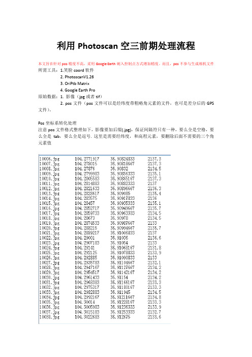
利用Photoscan空三前期处理流程本文旨在针对pos精度不高,采用Google Earth刺入控制点方式增加精度。
而且,pos不参与生成相机文件所需工具:1.笑脸coord软件2. PhotoscanV1.263. OriPtb Matrix4. Google Earth Pro原始数据:1. 影像(jpg或者tif)2. pos文件(pos文件可以是经纬度带粗略角元素的文件,也可是差分后的GPS 文件)。
Pos坐标系转化处理注意pos文件格式整理如下,影像要加后缀(.jpg),保证间隔符只有一种,要么全是空格,要么全是tab,要么全是逗号. 这里是需要经纬度,和高程元素,要删除后面不需要的三个角元素值这里我们需要将pos 大地坐标转化为平面坐标具体操作如下1.打开笑脸coord软件,设置-地图投影-2. 在投影设置里,选择高斯六度带,中央子午线设置105,其他保持不变3.点击文件转化,选择文件格式:点号经度纬度如果没有,点自定义格式设置,然后设置依次设置名称,扩展名,分隔符号,数据列表如下完成点击新建4.导入需要转换的pos文件,进行转化修改得到文件扩展名为.txt进行下一步正式处理处理流程:1. 打开photoscan,工作流-添加照片2. 在参考(reference)窗口选择导入,导入pos文件3. 选择好间隔符后,检查下X和Y值没有搞混,X为6位或8位,Y是7位,点确定4. 在主窗口界面可以浏览pos点概况5. 工作流-对齐照片,精度推荐用Low(速度快,普通和高匹配速度太慢),如果导入过pos 文件,下面可以选择参考(reference)模式,高级设置里的关键点限制和连接点限制最多不要超过默认值的2倍,也就是80000和2000。
6.检查对齐照片精度,精度小于5m说明Pos精度比较好,可以用于产生相机文件,这里由于精度不高,错误值比较大,此时采取不导入pos,产生相机文件7.当第6步中的错误有超过5的(accurac为默认的10.0情况下),则不能依据POS文件来输出自检校的相机文件。
[转载]skim和scan的区别
![[转载]skim和scan的区别](https://img.taocdn.com/s3/m/e329335e2f3f5727a5e9856a561252d380eb2060.png)
[转载]skim和scan的区别
原⽂地址:skim和scan的区别作者:Vivian
scan,是浏览,⼀般是带着问题,为寻找答案去读。
skim,是略读,是要找出主旨,看⽂章在讲什么,get the general idea
skim略读,理解掌握⽂章⼤意,与扫读(glance)意思⼀样。
属于⽅法⽅⾯。
scan审读,快速索所需特定信息,与跳读(skip)意思⼀样,属于技能⽅⾯。
skim就是 read sth quickly in order to find a particular point or the main point即快速阅读以找到⽂章的中⼼思想/主要意思。
If you skim through the play too quickly, you'll forget the plot. 如果你读剧本读得太快,就会忘记剧中主要情节。
scan就是 look at every part of sth carefully, especially because you are looking for a particular thing or person即快速阅读以找到某⼀具体的信息
His mother scanned his face to see if he was telling the truth. 他母亲察看他的⾯⾊看他是不是在讲真话。
skim和scan的区别是,skim是选择性的跳读,scan是浏览式的泛读,在国外每学期每门课程的reference book都有⼏⼗本,不像国内每门课就⼀本所谓的教材,但事实上没有⼀本教材是可以称得上完全没有纰漏的,所以在国外写essay的时候就要看很多书,但并不是每本书从头看到尾.。
NanoSpec操作说明
1.0 目的:规范NanoSpec 膜厚仪操作。
2.0 范围:所有使用NanoSpec 膜厚仪的作业人员。
3.0 定义3.1 Scan New Reference---扫描新的参考片3.2 Please Focus on Reference with Objective---请聚焦到参考片上。
3.3 Please MEASURE When Ready---准备好后请按测量键。
3.4 Enter Sample ID---输入样片代号。
3.5 Enter Refractive Index---输入参考片折射率。
4.0 内容4.1 清洗参考片4.1.1 频次:1 time/ 1 day。
4.1.2 清洗:每天Office QC 之前,将参考片放入USL-4L(HF:H2O=1:1)清洗槽中清洗30 秒,然后用水冲洗10 分钟,甩干后放回原处。
4.2 聚焦4.2.1 放好参考片,然后聚焦4.2.1.1 粗调:调整粗调旋钮,见到规则的正八边形为止。
4.2.1.2 细调:调整细调旋钮,见到清晰的正八边形为止,如图所示。
4.2.2 调视场亮度:调整遮光板,找到合适的视场亮度,使得既不刺眼,又有足够强的测量亮度。
4.2.3 调视场范围:调整视场旋钮,使得被测区域刚好落在视场内。
4.3 测量膜厚4.3.1 按CALIB 键,待菜单出现后,输入待测量的菜单号。
4.3.2 根据被测区域的面积大小,输入显微镜放大倍数,选择1。
4.3.3 提示Scan New Reference 时,按YES 键。
4.3.4 提示Please Focus on Reference with XX Objective Press MEASURE when ready 时,按MEAS 键。
4.3.5 提示Enter Sample ID 时,按ENTER。
4.3.6 提示Enter Refractive Index 时,如不输入自己定义的膜的折射率,就按ENTER,仪器会显示本身给出的折射率数值,测试者默认此数值,再按ENTER 键即可。
DS2208数字扫描器产品参考指南说明书
-05 Rev. A
6/2018
Rev. B Software Updates Added: - New Feedback email address. - Grid Matrix parameters - Febraban parameter - USB HID POS (formerly known as Microsoft UWP USB) - Product ID (PID) Type - Product ID (PID) Value - ECLevel
-06 Rev. A
10/2018 - Added Grid Matrix sample bar code. - Moved 123Scan chapter.
-07 Rev. A
11/2019
Added: - SITA and ARINC parameters. - IBM-485 Specification Version.
No part of this publication may be reproduced or used in any form, or by any electrical or mechanical means, without permission in writing from Zebra. This includes electronic or mechanical means, such as photocopying, recording, or information storage and retrieval systems. The material in this manual is subject to change without notice.
FARO激光扫描仪软件说明书

color photographs of the FARO color option, automatically detecting and equalizing differences in the distortion and alignment of the photos. Measuring and Analyzing Generation of objects, including points, spheres, planes, and cylinders directly from the scan data. Measuring between scan points and objects. Checks for flatness.Global Sales Offices: USA • Mexico • Germany • Switzerland • France • United Kingdom • Spain • Italy • Netherlands • Poland • Singapore • China • Japan • India •Brazil800.736.023404REF201-042.pdf Revised: 3/26/09Specifications Editing Scan Data■ Automatic search for reference spheres and black and white reference targets■ Object markers for the manual identification of spheres, black and white reference targets, circular reference targets, planes and slabs ■ Online correspondence search for the automatic assignment of reference points■ Automatic coloring of the scans with the high-resolution color photographs of the FARO color option■ Coloring of scan points with the aid of imported color photos ■ Deletion of scan areas■ Generation of new scan files of selected areas ■ Filters (including “dark points”, and “stray points”)Data Management of Extensive Projects ■ Hierarchical structure■ Bundling of unlimited number of scans to one project Analysis■ Distance measurement ■ Analysis of evennessImport & Export■ Control points for geo-referencing (.cor, .csv)■ Scan points (FARO Scan, FARO Cloud, .dxf, VRML, .igs, .txt, .xyz, .xyb, .pts, .ptx, .ptc, .ptz [import only], .pod [export only])■ CAD objects (.wrl [im- & export], .igs and .dxf [export only])■ Import digital photos (.jpg, .png, .bmp)■ Export panoramic images (.jpg)■ Online data transfer to FARO Cloud for AutoCAD Navigation■ Displaying of scan positions for viewpoint selection and changing to other scans by clicking■ 3D navigation in flight and inspection mode■ Predefined views (front view, side view, top view)Creating Workspaces ■ Scans & CAD objects■ Object fitting with visual quality indicators for spheres/tubes/planes (including automatic border detection)■ Meshing■ Measurements■ Intuitive user interface with structure viewViews■ 3D view■ Planar & Quick view■ Color scans are shown either in black & white or color ■ CAD object display ■ Print preview■ Color gradient depiction for displaying point distances from reference planes or the scanner locationGeneral。
中英文超声无损检测名词术语
中英文超声无损检测名词术语Acceptance limits 验收范围Acceptance level 验收水平Acceptance standard 验收标准Accumulation test 累积检测Acoustic emission transducer 声发射换能器〔声发射传感器〕Acoustic impedance 声阻抗Acoustic impedance matching 声阻抗匹配Acoustic impedance method 声阻法Acoustic wave 声波Acoustical lens 声透镜Acoustic—ultrasonic 声-超声〔AU〕Adequate shielding 安全屏蔽Amplitude 幅度Angle beam method 斜射法Angle of incidence 入射角Angle of reflection 反射角Angle of spread 指向角Angle of squint 偏向角Angle probe 斜探头Area amplitude response curve 面积幅度曲线Area of interest 评定区Artificial discontinuity 人工不连续性Artifact 假缺陷Artificial defect 人工缺陷Artificial discontinuity 标准人工缺陷A-scan A型扫描A-scope; A-scan A型显示Attenuation coefficient 衰减系数Attenuator 衰减器Automatic testing 自动检测Evaluation 评定Beam 声束Beam ratio 光束比Beam angle 束张角Beam axis 声束轴线Beam index 声束入射点Beam path location 声程定位Beam path; path length 声程Beam spread 声束扩散Bottom echo 底面回波Bottom surface 底面Boundary echo<first> 边界一次回波Broad-beam condition 宽射束B-scan presentation B型扫描显示B-scope; B-scan B型显示C- scan C型扫描Calibration,instrument 设备校准pressional wave 压缩波Continuous emission 连续发射microstructureContinuous linear array 连续线阵Continuous method 连续法Continuous spectrum 连续谱Continuous wave 连续波Contract stretch 对比度宽限Contrast 对比度Contrast sensitivity 对比灵敏度Control echo 监视回波Control echo 参考回波Couplant 耦合剂Coupling 耦合Coupling losses 耦合损失Creeping wave 爬波Critical angle 临界角Cross section 横截面Cross talk 串音Cross-drilled hole 横孔Crystal 晶片C-scope; C-scan C型显示Curie point 居里点Curie temperature 居里温度Curie<Ci> 居里Dead zone 盲区Decibel<dB> 分贝Defect 缺陷Defect resolution 缺陷分辨力Defect detection sensitivity 缺陷检出灵敏度Definition 清晰度Definition, image definition 清晰度,图像清晰度Direct contact method 直接接触法Directivity 指向性Discontinuity 不连续性Distance- gain- size-German A VG 距离- 增益- 尺寸〔DGS德文为A VG〕Distance marker; time marker 距离刻度Double crystal probe 双晶片探头Double probe technique 双探头法Double transceiver technique 双发双收法Double traverse technique 二次波法D-scope; D-scan D型显示Dual search unit 双探头Dynamic range 动态范围Echo 回波Echo frequency 回波频率Echo height 回波高度Echo indication 回波指示Echo transmittance of sound pressure 往复透过率Echo width 回波宽度Equivalent 当量Equivalent method 当量法Evaluation 评定Examination area 检测范围Examination region 检验区域Final test 复探Flat-bottomed hole 平底孔Flat-bottomed hole equivalent 平底孔当量Flaw 伤Flaw characterization 伤特性Flaw echo 缺陷回波Flexural wave 弯曲波Focal spot 焦点Focal distance 焦距Focus length 焦点长度Focus size 焦点尺寸Focus width 焦点宽度Focused beam 聚焦声束Focusing probe 聚焦探头Focus-to-film distance<f.f.d> 焦点-胶片距离〔焦距〕Frequency 频率Frequency constant 频率常数Fringe 干涉带Front distance 前沿距离Front distance of flaw 缺陷前沿距离Fundamental frequency 基频Gain 增益Gap scanning 间隙扫查Gate 闸门Gating technique 选通技术Gauss 高斯Grazing incidence 掠入射Grazing angle 掠射角Group velocity 群速度Half life 半衰期Half-value method 半波高度法Harmonic analysis 谐波分析Harmonics 谐频Head wave 头波Image definition 图像清晰度Image contrast 图像对比度Image enhancement 图像增强Image magnification 图像放大Image quality 图像质量Imaging line scanner 图像线扫描器Immersion probe 液浸探头Immersion rinse 浸没清洗Immersion testing 液浸法Impedance 阻抗Impedance plane diagram 阻抗平面图Imperfection 不完整性Indicated defect area 缺陷指示面积Indicated defect length 缺陷指示长度Indication 指示Initial pulse 始脉冲Initial pulse width 始波宽度Inspection 检查Inspection medium 检查介质Inspection frequency/ test frequency 检测频率Interface boundary 界面Interface echo 界面回波Interface trigger 界面触发Interference 干涉Interpretation 解释Lamb wave 兰姆波Lateral scan 左右扫查Lateral scan with oblique angle 斜平行扫查Limiting resolution 极限分辨率Line scanner 线扫描器Linear scan 线扫查Location 定位Location accuracy 定位精度Location puted 定位,计算Location marker 定位标记Longitudinal wave 纵波Longitudinal wave probe 纵波探头Longitudinal wave technique 纵波法Loss of back reflection 背面反射损失Loss of back reflection 底面反射损失Magnetostrictive effect 磁致伸缩效应Magnetostrictive transducer 磁致伸缩换能器Main beam 主声束Manual testing 手动检测MA-scope; MA-scan MA型显示Micrometre 微米Micron of mercury 微米汞柱Mode 波型Mode conversion 波型转换Mode transformation 波型转换Multiple back reflections 多次背面反射Multiple reflections 多次反射Multiple back reflections 多次底面反射Multiple echo method 多次反射法Multiple probe technique 多探头法Multiple triangular array 多三角形阵列Narrow beam condition 窄射束Near field 近场Near field length 近场长度Near surface defect 近表面缺陷Noise 噪声Nominal angle 标称角度Nominal frequency 标称频率Nondestructive Examination〔NDE〕无损试验Nondestructive Evaluation〔NDE〕无损评价Nondestructive Inspection〔NDI〕无损检验Nondestructive Testing〔NDT〕无损检测Normal incidence 垂直入射〔亦见直射声束〕Normal beam method; straight beam method 垂直法Normal probe 直探头Parallel scan 平行扫查Parasitic echo 干扰回波Pattern 探伤图形Penetrant flaw detection 渗透探伤Phantom echo 幻象回波Phase detection 相位检测Plane wave 平面波Plate wave 板波Plate wave technique 板波法Point source 点源Probe test 探头检测Probe index 探头入射点Probe to weld distance 探头-焊缝距离Probe/ search unit 探头Pulse 脉冲波Pulse 脉冲Pulse echo method 脉冲回波法Pulse repetition rate 脉冲重复率Pulse amplitude 脉冲幅度Pulse echo method 脉冲反射法Pulse energy 脉冲能量Pulse envelope 脉冲包络Pulse length 脉冲长度Pulse repetition frequency 脉冲重复频率Pulse tuning 脉冲调谐Quadruple traverse technique 四次波法Range 量程Rayleigh wave 瑞利波Rayleigh scattering 瑞利散射Reference block 参考试块Reference block 对比试块Reference block method 对比试块法Reference standard 参考标准Reflection 反射Reflection coefficient 反射系数Reflector 反射体Refraction 折射Refractive index 折射率Reject; suppression 抑制Rejection level 拒收水平Resolution 分辨力Sampling probe 取样探头Saturation 饱和Saturation,magnetic 磁饱和Scan on grid lines 格子线扫查Scan pitch 扫查间距Scanning 扫查Scanning index 扫查标记Scanning directly on the weld 焊缝上扫查Scanning path 扫查轨迹Scanning sensitivity 扫查灵敏度Scanning speed 扫查速度Scanning zone 扫查区域SE probe SE探头Second critical angle 第二临界角Sensitivity va1ue 灵敏度值Sensitivity 灵敏度Sensitivity of leak test 泄漏检测灵敏度Sensitivity control 灵敏度控制Shear wave 切变波Shear wave probe 横波探头Shear wave technique 横波法Signal to noise ratio 信噪比Single crystal probe 单晶片探头Single probe technique 单探头法Single traverse technique 一次波法Sizing technique 定量法Sound diffraction 声绕射Sound insulating layer 隔声层Sound intensity 声强Sound intensity level 声强级Sound pressure 声压Sound scattering 声散射Sound transparent layer 透声层Sound velocity 声速Source 源Specified sensitivity 规定灵敏度Standard 标准Standard 标准试样Standard test block 标准试块Standardization instrument 设备标准化Standing wave; stationary wave 驻波Subsurface discontinuity 近表面不连续性Suppression 抑制Surface echo 表面回波Surface wave 表面波Surface wave probe 表面波探头Surface wave technique 表面波法Surplus sensitivity 灵敏度余量Sweep 扫描Sweep range 扫描范围Sweep speed 扫描速度Swept gain 扫描增益Swivel scan 环绕扫查System exanlillatien threshold 系统检验阈值System noise 系统噪声Tandem scan 串列扫查Test block 试块Test frequency 试验频率Test range 探测范围Test surface 探测面Testing,ultrasonic 超声检测Third critical angle 第三临界角Through transmission technique 穿透技术Through penetration technique 贯穿渗透法Through transmission technique; transmission technique 穿透法Transducer 换能器/传感器Transmission 透射Transverse wave 横波Traveling echo 游动回波Travering scan; depth scan 前后扫查Triangular array 正三角形阵列Trigger/alarm condition 触发/报警状态Trigger/alarm level 触发/报警标准Triple traverse technique 三次波法True continuous technique 准确连续法技术Ultrasonic noise level 超声噪声电平Ultrasonic field 超声场Ultrasonic flaw detection 超声探伤Ultrasonic flaw detector 超声探伤仪Variable angle probe 可变角探头Vertical linearity 垂直线性Vertical location 垂直定位Visible light 可见光Wave 波Wave train 波列Wave from 波形Wave front 波前Wave length 波长Wave node 波节Wave train 波列Wedge 斜楔Wheel type probe; wheel search unit 轮式探头Working sensitivity 探伤灵敏度Zigzag scan 锯齿扫查。
生产文件半成品引物溶解、定量和稀释标准操作规程[1.2]
![生产文件半成品引物溶解、定量和稀释标准操作规程[1.2]](https://img.taocdn.com/s3/m/a77e6b1f54270722192e453610661ed9ad5155d2.png)
引物溶解、定量和稀释标准操作规程1.引物接收接收引物时,仔细核对验收,确保货单与货物一致,然后将引物管保存于-20℃冰箱,并妥善保管引物说明单。
收货单交于保管人备案。
2.准备工作2.1 打开引物分装室内超净台的紫外灯开关,照射30min。
2.2 打开Ultrospec 2100 pro分光光度仪开关,预热30min。
2。
3 准备ddH2O(引物的管数×1000μl),放在4℃冰箱内待用.2。
4 引物的溶解、稀释操作均应在超净台和冰浴中进行。
3.溶解干粉管以12,000rpm离心1min,按合成OD值加入不同量的TE溶液,具体标准如下:1—2 OD加50μl TE;2-10 O D加100μl TE;10 OD以上加200 μl TE。
充分振荡混匀,12,000rpm离心1min。
此为原液。
4。
定量4。
1 取0。
2 ml EP管,加入98 μl ddH2O,再加入2 μl引物原液,振荡混匀,离心.ddH2O作为空白对照,记下260 nm的吸光度值、260/230值、260/280值及浓度(单位为ng/μl)。
4.2 分光光度仪主菜单依次选择:Nucleic Acids→Oligo→Pa thlength: 10 mm(默认值)→Units:ng/μl (默认值)→320 nm correction:Yes (默认值)→Scan Option: On(默认值)→Dilution Factor:50→Factor: 33 (默认值)→显示Reference→Run键运行。
5。
稀释分装5。
1 浓度换算:分光光度仪测出的引物原液浓度为x ng/μl,则其摩尔浓度y=(x×1000)/MW (μM)(MW为引物的分子量)。
5。
2 如原液浓度大于100 μM(即y>100μM),则分别取出100 μl原液,加入不同的0。
5ml EP管内,然后向其中加入(y-100)μl TE溶液,振荡混匀,此为储存液。
ICP算法和NDT算法

ICP算法一、ICP算法概述二、转换矩阵T其中,R是旋转矩阵,t是转换向量。
三、滤波器(Data Filters)滤波器的目的是为了增加差异性,减少处理时间,去噪。
四、匹配度(match function)匹配度,是测量两个点集的关联度的,又称之为association solver。
比如,用欧式距离来测量两个点集的关联度,定义如下:五、离群滤波器(outlier filters)离群滤波器主要是降低匹配带来的误差。
表ICP算法模块的实现方法图ICP算法流程图NDT算法一、NDT算法概述ICP算法存在两个重要问题:一是点与点的匹配没有考虑到点与周围平面的联系;二是算法中用到的最邻近搜索计算量比较大。
二、NDT算法具体步骤将空间(reference scan)划分成各个格子cell将点云投票到各个格子计算格子的正态分布PDF参数将第二幅scan的每个点按转移矩阵T的变换第二幅scan的点落于reference的哪个格子,计算响应的概率分布函数求所有点的最优值,目标函数为PDF可以当做表面的近似表达,协方差矩阵的特征向量和特征值可以表达表面信息(朝向、平整度)格子内少于3个点,经常会协方差矩阵不存在逆矩阵,所以只计算点数大于5的cell,涉及到下采样方法。
二、2D-NDT算法2D-NDT算法是在2维平面上描述连续,可微的概率密度。
具体步骤如下:参考文献:[1]P Besl and H McKay, 1992. A method for registration of 3-D shapes. Pattern Analysis and Machine Intelligence, IEEE Transactions on, 14, pp. 239 - 256.[2]A Review of Point Cloud Registration Algorithms for Mobile Robotics.[3]Comparing ICP variants on real-world data sets.[4]Peter Biber and Wolfgang Straßer. The normal distributions transform: A new approach to laser scan matching. In Proceedings of the IEEE International Conference on Intelligent Robots and Systems (IROS), pages 2743–2748, Las Vegas, USA, October 2003.。
codesys中的reference to的意思
Codesys中的Reference To的意思什么是Codesys?Codesys是一种广泛使用的用于工业自动化系统编程的集成开发环境。
它提供了一个灵活、可扩展和易于使用的平台,用于开发和管理自动化控制系统。
Codesys支持多种编程语言,包括结构化文本、函数图表、连线图和IL(指令列表)等。
Reference To的概念在Codesys中,Reference To是一种用于引用变量或数据对象的机制。
Reference To允许在程序中传递和操作变量的引用,而不是复制它们的值。
这种引用机制可以提高代码的可读性和可维护性,同时还可以节省内存和处理器资源。
使用Reference To的好处使用Reference To有许多好处,以下是一些主要的优点:1.代码模块化:通过使用Reference To,可以将代码分成更小、更可管理的模块。
这样可以使代码更易于理解、测试和维护。
2.内存和处理器资源的节省:使用Reference To可以避免在程序中频繁复制变量的值。
这样可以节省内存和减少处理器的负载。
3.支持动态数据结构:Reference To使得可以在运行时动态创建和操作数据结构,而不需要在编程阶段就确定固定的内存分配。
4.减少错误和提高效率:通过使用Reference To,可以减少因为复制变量值导致的错误。
此外,使用Reference To还可以减少不必要的临时变量,提高代码的执行效率。
Reference To的使用方法在Codesys中,可以使用Reference To来引用变量或数据对象。
以下是一些使用Reference To的常见情况:1. 将变量作为函数参数传递可以使用Reference To将变量作为函数的参数传递。
通过这种方式,函数可以直接修改变量的值,而不需要返回值。
例如:VARvalue: INT := 0;END_VARPROCEDURE IncrementValue REF_TO INTvalue := value + 1;END_PROCEDURE// 调用函数IncrementValue REF_TO value;在上面的例子中,函数IncrementValue接受一个引用类型为INT的参数,并将value的值加1。
fMRI实验设计及数据处理

AAA_SCCAI_3DMCT_SD3DSS4.00mm_THPGLMF2c.fmr 项目名_Slice校正_头懂校正_空间平滑(高斯过滤4毫米)_
Temporal High Pass (GLM Fourier) with 2 cycles / points _. Fmr
6,可以把比较的结果存下来以后打开直接看。
第五步 标Байду номын сангаас化处理
类似spm的预处理,处理后可以再进行第四步的统计 分析,增加推论力度。
1, 点击菜单“Analysis”--》 “FMR Data Preprocessing...”。 2,对原始数据处理越多,增加的error越多。一般后 四项比较常用。
注意事项
1,删除前面不稳定的5个左右TR。包括预处理和数 据分析。
2,同一个被试不同的run,预处理要分开处理,统 计分析时要放在不同的section处理。
Brainvoyager 处理步骤
第一步,建立一个项目
1,运行brainvoyager,创建项目。 File -> Create Project Wizard -> 选择FMR project -> 选
实验设计—混合实验设计
特点及注意事项: 1 能分离持续性神经活动(block)和短暂性神经活动 (event) 2 处理和设计起来比较复杂,需要权衡的东西较多 3 要保证block和event的相关性不高
二 数据处理
打开spm 点击右下角 dicom import,转换成spm格式的 图。
Overview 1 预处理(preprocess) ➢ Slice timing ➢ Motion correction ➢ Normalization ➢ Smooth 2 数据分析
简单矢量ScanIP软件介绍说明书

Description of Simpleware ScanIPSimpleware™ ScanIP provides a core image processing interface with additional modules available for Finite Element model generation, CAD integration, NURBS export, and material property calculation. Simpleware ScanIP is the software device, with modules integrating into the same software, rather than representing separate software programs in their own right. Indications for UseScanIP is intended for use as a software interface and image segmentation system for the transfer of imaging informationfrom a medical scanner such as a CT scanner or a Magnetic Resonance Imaging scanner to an output file. It is also intendedas pre-operative software for simulating/evaluating surgical treatment options. ScanIP is not intended to be used for mammography imaging.Warnings and RecommendationsThis product is for professional use only and should be used only by trained technicians with a professional level of English. English is the language used in the Simpleware ScanIP software interface.The output must be verified by the responsible clinician.Simpleware ScanIP has the ability to process, store or discard information contained in medical image files such as DICOM files during the import process of these files. The import process can involve different data transfer methods including USB, CD/DVD, disk drive or server-based storage systems such as PACS. When such files contain personal patient information, it is the responsibilityof the end-users to follow the local laws related to appropriate handling of personal data – for example HIPAA (USA) and GDPR (EU) – and to discard any information when required. Please refer to the relevant section in the Reference Guide regarding how personal data is stored and accessible within Simpleware ScanIP, to ensure your usage is compliant.It is recommended to use Simpleware ScanIP within a hardware and/or network environment in which cyber security controls have been implemented including anti-virus and use of firewall.AccuracySimpleware ScanIP image processing and meshing algorithms are designed to use partial volume effects to improve surface accuracy. The reconstructed 3D surface typically has a maximal error of ½ of a voxel size.Note: the accuracy of a model is dependent on the image resolution and the quality of the original scan. The accuracy of a model for simulation is also dependent on user requirements and choice of simulation software.During surface reconstruction, error can be found near sharp edges, which are difficult to reconstruct when using any image-based meshing techniques. Excessive noise in scanned images can also affect surface reconstruction accuracy./simplewareOther Ways of Viewing this Information and EmergenciesEnd-users of Simpleware ScanIP can request a free paper copy of this document. To do so, please contact***********************.If information is needed in an emergency, please call +44(0)1392 428750.In the event that you experience temporary unavailability of this document through the Synopsys website or to the Internet in general, or of your institutional access, we recommend temporarily suspending use of the software until access is restored, unless you have a paper copy of this document.There are no foreseeable medical emergencies related to this device. If you believe that the device may have directly or indirectly contributed to a patient’s injury or death, then please immediately contact *********************** or call +44(0)1392 428750. Dialog SymbolsSimpleware ScanIP uses a set of standard symbols (icons) when displaying information dialogs. The table below provides information about the severity of the risk associated with each type of symbol.Instructions for UseStarting Simpleware ScanIPAfter installing the software on your PC, double click the Simpleware ScanIP icon on your desktop. Alternatively, you can click on the Windows icon in the “taskbar” and navigate to Synopsys > Simpleware ScanIP Q-2020.06.The Reference Guide can be opened by clicking on the “Help” button on the Welcome page that appears after starting Simpleware ScanIP.Simpleware ScanIP usage is controlled through a license key file which may either be node-locked or floating. Instructions for setting up both license options are described in the Reference Guide. All required software installers (product and licensing tools) and license keys can be downloaded from SolvNet (https://). Please note that only active licenses of Simpleware ScanIP will be able to get access to SolvNet.Supported Operating Systems• Windows 7*• Windows 10*†• Windows Server 2008 R2*• Windows Server 2016*• Licensing tools for Linux 32-bit and 64-bit*Only 64-bit versions of these operating systems are supported.†Simpleware ScanIP is fully tested on this operating system.2Recommended System RequirementsManufacturer Contact DetailsSimpleware ScanIP Q-2020.06Manufactured in 2020 by Synopsys Simpleware Product GroupSynopsys (Northern Europe) Ltd.Bradninch Hall, Castle Street, Exeter, EX4 3PL, United KingdomPhone: +44(0)1392 428750Email: ***********************Simpleware ScanIP is a CE-marked productUDI Number: (01)008635200003522797Current Availability as a CE-Marked ProductFor more information on the current availability and plans for Simpleware ScanIP as a CE-marked product in your country, please contact ***********************.©2020 Synopsys, Inc. All rights reserved. Synopsys is a trademark of Synopsys, Inc. in the United States and other countries. A list of Synopsys trademarks isavailable at /copyright.html. All other names mentioned herein are trademarks or registered trademarks of their respective owners.05/16/20.CS12917_SimplewareScanIP-IFU_Q-2020-06.。
Bionano Genomics技术文档说明书
Guidelines for Interpreting the Bionano MoleculeQuality ReportImportant: The guidelines described herein are based on internal experiences at Bionano Genomics and are provided as-is. The purpose of this technical note is to provide guidelines to customers who want to evaluate the quality of data generated from the Irys® System. For questions, please contact the Technical Support Team at .Molecule Quality Report (MQR)The Molecule Quality Report provides a summary report on the molecule quality. The report is generated based on results from a molecule-to-reference alignment. The RefAligner tool aligns Bionano molecules to a given reference and identifies regions of similarity between Bionano molecules and reference. The input includes the molecule BNX file and the reference CMAP file. See the Molecule Quality Report Output Files section for more details.The MQR identifies and outputs the best alignment of each molecule to the reference, provided that the alignment meets the minimum alignment quality criteria.To determine if the data quality is sufficient to continue data collection and to run de novo assembly, the best indicators* are the following:1)Map rate: What percentage of the Bionano molecules aligns to the reference (meeting minimumalignment quality criteria)?2)Noise parameters: How different are the aligned Bionano molecules when compared to the reference?* The evaluation of the MQR results is highly dependent on the accuracy and completeness of the given reference and the identity of the sample with the reference. Many sequence assemblies, even at advanced stages, could have a high degree of structural inaccuracy that may compromise the use of the MQR. See the Interpret Molecule Quality Report Results section for details.Run the Molecule Quality Report1.For instructions on performing a MQR in IrysView, see the IrysView® v2.5.1 Software Training Guide,section 6.18.2.The recommended default MQR alignment parameters for human samples in IrysView v2.5.1 are thefollowing:-nosplit 2 -BestRef 1 -biaswt 0 -Mfast 0 -FP 1.5 -FN 0.15 -sf 0.2 -sd 0.0 -A 5 -outlier 1e-3 -outlierMax 40 -endoutlier 1e-4 -S -1000 -sr 0.03 -se 0.2 -MaxSF 0.25 -MaxSE 0.5 -resbias 4 64 -maxmem 64 -M 3 3 -minlen 150 -T 1e-11 -maxthreads 32 -hashgen 5 3 2.4 1.5 0.05 5.0 1 1 3 -hash -hashdelta 10 -hashoffset 1 -hashmaxmem 64 -insertThreads 4 -maptype 0 -PVres 2 -PVendoutlier -AlignRes 2.0 -rres 0.9 -resEstimate -ScanScaling 2 -RepeatMask 5 0.01 -RepeatRec 0.7 0.6 1.4 -maxEnd 50 –usecolor 1 -stdout –stderr –randomize –subset 1 50003.Modify the following alignment parameters as necessary:M designates how many alignment iterations to perform. After each iteration of alignment, the noise parameters are estimated, and those noise parameters are used for the next iteration.The number of iterations is chosen, considering a tradeoff of computational time and potentially more accurate noise estimates. Larger genomes or datasets could require a significant computation.Using the default argument -M 3 3, the hash table is regenerated 3 times (second “3” in the argument) and perform 3 iterations for each hash table result (first “3” in the argument). This process gives more accurate error estimates than just -M 3 or -M 9.Without hashing arguments (-hashgen 5 3 2.4 1.5 0.05 5.0 1 1 3 -hash -hashdelta 10 -hashoffset 1 -hashmaxmem 64), -M 3 3 is treated the same as -M 9, in which the alignment is repeated for 9 times.T is the P-value cutoff. The cutoff should be set according to the genome complexity, which is scaled with the genome size and average label density. Therefore, the P-value can be adjusted based on the size of genome and average label density. We recommend 1e-11 for genomes larger than 1 Gbp in size with an average label density less than 15 labels per 100 kbp.Genomes (larger than 1 Gbp) with higher label densities (> 15/100 kbp) require a lower P-value (more stringent). We suggest to lower the P-value by the factor of 100, per 1 label density increase from 15/100 kbp.The table below lists the suggested P-value for example species with different genome size and average label density.Species Genome Size Average Label Density(Nicking Enzyme) Suggested P-value (-T)E. coli 5 Mbp 9/100 kbp (Nt.BspQI) 1e-7Drosophila 120 Mbp 11/100 kbp (Nt.BspQI) 1e-9Human 3.3 Gbp 9/100 kbp (Nt.BspQI) 1e-11Sample Species 2 Gbp 16/100 kbp 1e-13outlierMax limits the maximum size of outlier* in kbp. It controls how tolerant the RefAligner is of the size of the outliers. If significant structural differences are expected between the sample and the reference, this argument (-outlierMax 40) may be modified or removed. See Figure 1.*The size difference between aligned label intervals in reference and Bionano molecules (see Figure 1).Figure 1: Outlier is the size difference between aligned label intervals in reference and Bionano molecules.Molecule Quality Report Output Files1.The key output files generated in MQR are the following:File DescriptionMoleculeQualityReportInput.tar.gz* The ZIP file containing input BNX file and reference CMAP.This file is transferred from IrysView to the server whenthe MQR computation starts.MQR_files.tar.gz* The ZIP file containing multiple MQR result files. The fileis transferred back to IrysView from the server to displaythe MQR results in IrysView, when the MQR computationfinishes.MoleculeQualityReport.stdout The log file for the alignment.MoleculeQualityReport_rescaled.bnx When per-scan scaling of molecules is enabled (viadefault parameter “–ScanScaling 2”), all the scaledmolecules are included in this file, not just subset ofmolecules used in alignment. MoleculeQualityReport.maprate The summary of map rate at increasingly stringent P-values.MoleculeQualityReport.err The summary of noise parameters for each alignmentiteration (-M).MoleculeQualityReport.errbin The binary ERR file (same contents as *.err)MoleculeQualityReport.xmap The alignment result between Bionano molecules andreference CMAP. The alignment can be viewed in IrysViewby opening the XMAP file.MoleculeQualityReport_q.cmap The alignment result of the aligned Bionano molecules forviewing.MoleculeQualityReport_r.cmap The alignment result of the aligned reference maps forviewing.MoleculeQualityReport.scan The alignment result of the Bionano molecules in eachscan to the reference, using the same argument.Molecules.bnx The input BNX file for MQR alignment, which containsBionano molecule and label information.Reference.cmap The input reference CMAP file for MQR alignment, whichcontains in silico digestion result of reference (contiglength and label information) MoleculeQualityReport.errbiasThe information in these files are not used. MoleculeQualityReport_intervals.txtMoleculeQualityReport.map*These 2 files are only generated when users run MQR on a remote server via IrysView.2. The metric results of MQR in IrysView are the following: MetricsDescription Map Rate (%)The percentage of molecules (N Molecules) aligned to reference. N MoleculesThe number of molecules used in the alignment to reference FP (/100 kbp)The density of unaligned labels in molecules (relative to reference length). FP (%)The percentage of unaligned labels in molecules (relative to number of labels in molecules). FN (%)The percentage of unaligned reference labels (relative to number of reference labels). SiteSD (kbp) : sfThese parameters describe which inter-label distances in Bionano molecules match the reference. This is referred to as sizing error relative to reference. These parameters are components of the variance of the distance observed in the Bionano molecules for a given interval (distance between two labels) on the reference. variance(x) = sf^2 + x|sd|sd + x^2*sr^2The x value is the interval length (kbp) and the other noise parameters are reported in the ERR file.SMin is not reported in the ERR file; it is either the minimum value of sqrt(variance) at x > 1 kbp OR the value of sqrt(variance) at x = 1 kbp, whichever is lower.ScalingSD (kbp^1/2) : sdRelativeSD : srSMin (kbp) BppThe calculated base pairs per pixel in the alignment by comparing molecule intervals to reference intervals. Stretch (%)The stretch factor of DNA molecules in the chip. This is computed from Bpp and average chip stretch factor.Interpret Molecule Quality Report Results1.List of metrics with ranges based on Bionano internal human data (source: Irys instrument).Metrics RangeMap Rate (%) 60%-80%N Molecules User-specified; recommended to be at least 5000FP (/100kbp) < 1.7FP (%) < 15%FN (%) < 21%SiteSD (kbp): sf < 0.25ScalingSD (kbp^1/2): sd (-0.07) ~ 0.05RelativeSD: sr < 0.04SMin (kbp) < 0.25Bpp 450 ~ 510Stretch (%) 83% ~ 94%2.Interpret the MQR ResultsTo interpret MQR results, check the molecule-to-reference map rate (%) first. The map rate is also closely tied to the completeness and accurateness of the reference (i.e. how much non-sequenced part, gap and ambiguities in the sequence assembly?).Additionally, the map is tied to the degree of identity of the Bionano sample with the reference sample (i.e. is the sample from the same individual as the reference?).For example, the human reference is highly complete, so the map rate can be as high as 90% for a good molecule dataset. If the reference or sequence assembly is only 50% complete, then the expected map rate range may be half or 30-40%, even if the Bionano molecules are of good quality.If the obtained map rate is significantly lower than the minimum desired map rate (i.e., < 60% for high quality reference or 30% for half-complete reference), check the noise parameters. If the noise parameters are within the recommended range, it could mean that the part of the Bionano data that does align to the reference is of good quality. In this case, these molecules can be used for de novo assembly; however, users may need to collect extra depth of the same data to compensate for low mapping rate.When interpreting the results, it is important to consider the accurateness of the provided reference. However, evaluating the reference accuracy is often challenging. If the map rate is lower than expected based on the completeness of the reference, it is possible that the molecule quality is still good, but because of the inaccuracy of the reference, some molecules do not align. In this case, it is challenging to evaluate molecule quality using MQR.Another way to evaluate alignment between the Bionano molecules and reference CMAP is to view alignments in IrysView. The aligned molecules should cover most of the reference genome (or reference contigs) relatively uniformly and without large errors (see Figure 2 and 3).Figure 2: An example of a good alignment.Figure 3: An example of a chaotic alignment.In cases when it is difficult to evaluate the reference completeness or accurateness or when it is not sufficient to obtain reliable noise parameters from MQR, we recommend that users perform de novo assembly using default noise parameters with at least 100X coverage data. When most of the genomes (>50%) can be assembled with reasonable data quality (i.e. a good alignment of the Bionano molecules to the assembled map is visualized; see Figure 2), the data quality are more likely to be sufficient.。
紫外可见漫反射光谱
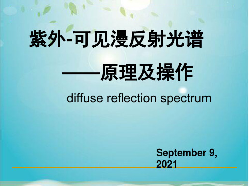
不同颜色叶子的UV-vis漫反射光谱图
比比谁 的手更 白!
你能猜出每条反射曲线对应 的是哪只手吗?
B
右上图:手背皮肤的紫外可见漫
反射曲线
A
C
左下图:上图所测曲线的各个“样
品”
视频
化学系 中心实验室
仪器展示 基本操作
1.Dark Scan—— Uncheck
2.ReSferte“nIcneteSgcraant—io—n TCihmeec”k to an 3.SaamppplreoSpcriaant—e —vaCluhee,ctkhat make the
“Intensity” of “Reference Scan” RefulepctoanacreouTnedst6: 0,000. Usually 800 •AlwisayascCcehpectakble.
1. Open the sliding cover on the top
Take off the port plug or port reducer .
material into the cuvette holder
Always Uncheck 左下图:上图所测曲线的各个“样
Acquire one spectrum 紫外-可见漫反射光谱仪器及操作
体、粉末、乳浊液和悬浊液
•漫反射光是指从光源发出的光进入样品内部,经过多 次反射、折射、散射及吸收后返回样品表面的光.
二.漫反射光与积分球 :
Diffused reflectance and integrating sphere:
Sample有吸收 反射量减少
The characteristics of typical integrating sphere coatings
蛋白质谱数据label-free quantitation

Label‐free Quantitation Discover the significantlychanging features in your dataChengpin ShenCloudscientific4/27/2013Quantitation Method基于MS1 的非标记定量•计算来自于多次实验重复中校正得到的同一肽段丰度(Area under Curve) ; (即Match between Runs 方法)•可以比较多次生物学、技术重复性或者多组样品的差异•基于内标情况结果可以辅助进行绝对定量MS1Area + Isotopic Distribution = Abundancem/zRTRTm/z为何MS1 LFQ 比Spc 更复杂?•多次重复间校正(非常重要!)•归一化•MS1谱峰识别和丰度计算Alignment in different runs Normalization AUC CalcTime windowalignment in one run AUC CalcNormalizationVisualization of TIC+m/zRT RTm/zVisualization of TICMS1Area + Isotopic Distribution = Abundancem/zRTRTm/zMS1 LFQ可视化定量结果(Progenesis LC‐MS)质控•对于谱图鉴定来说,我们只需要考虑:1.二级碎裂效率;2.母离子峰强;3.扫描速度(MS2量越多, 鉴定的越多);4.分辨率(精度越高越准确)•而对于MS1非标定量来说,我们还需要确保:1.ESI喷雾连续性;2.样品中尽可能的不含有盐、PEG等污染物;3.MS1谱峰半峰宽尽可能一致;4.MS1的分离效率尽可能的好;5.采用精确校正的高分辨质谱(峰型良好的同位素峰分布)其他定性定量方法其实一样适用,不过经常会被忽略A good MS1Sample for quantitationData from Chengpin Shen Mol Biosystems 2012Replicate 1Replicate 2Replicate 3ESI 喷雾连续;盐污染少; 没有PEG 污染;保留时间窗口<1min;谱峰分离效果好(15min ‐75min of 90min LC) ;A good MS1Sample for quantitationCV range02040608010012014016018020005001000150020002500Maximum CVAverage 11.58%RT window avg 0.9minMS1 LFQ数据的质控案例•本例中两次重复试验其中一次ESI喷雾不连续:m/ztMS1 LFQ 数据的质控案例•本例中左图数据喷雾不连续、且含有PEG 污染,右图盐污染、空气峰强度较高:•Proper separation setup;m/z tm/ztMS1 LFQ数据的质控案例•保留时间窗口过宽,分离效果差,高丰度蛋白可能没有去干净,或者色谱柱填充有问题m/zt 26.0min 35.0minRT Window = 10minutesMS1 LFQ 数据的质控案例•分离梯度设置不佳,导致分离效率较低m/zt45.0minWhy is MS1 LFQ complex?•Data alignment •Normalization •Abundance CalcAlignment in different runs Normalization AUC CalcProgenesis LC ‐MS 谱峰校正原理•自动选择最佳“reference”样本作为校正的标准. •其他样本以此为标准进行保留时间对齐•依次对齐后,所有谱峰均能进行定量“overlay”reference scanother scanProgenesis LC‐MS 谱峰校正原理reference scanpeptide signalsother scanProgenesis LC‐MS 谱峰校正原理自动找到的对齐坐标• Progenesis会自动找到一系列“对齐坐标”用以将每个样本中的相同信号进行对齐Progenesis LC‐MS 谱峰校正原理单独的各个样本• 在校正之后, 分布模式相同的谱峰将进行共识别Co‐detection of dataNo expression change Small expression changeLarge expression change数据识别可以应用在2个甚至200个样本之间识别共流出肽段蓝色区域标明了该肽段的 所有同位素分布及丰度红色区域标明了共流 出肽段的丰度分布情 况Co‐detection of datapeptide ion identified in one sample, comparable across allNo missing data!Identification can be confirmed by any other sample(s)Analyse complex samplesInclusion listsMS Peptide QuantificationLocate peaks of significant expression behaviourAlignmentPeptide separation and RT shift correction MS‐MS IdentificationPeptide fragmentation“Protein” QuantificationResultsLC‐MS data alignment algorithms corrects for the positional bias introduced by the LCCo‐detection and relative quantification of all peptide ions and statistical tools to define subsets of experimental interestIntegration with search engines to identify peptide ions. Create inclusion lists for repeat analysis of peptides unidentified. Combining of all data at the protein level and calculation of the protein quantificationAnalyse fractionated samplesAnalysis of Each FractionReduce sample complexity but quantify more runsRecombine FractionsNormalise across fractions to see global viewProtein ViewQuantification & identification at the protein level View Peptide DataDelve into underlying peptide informationInclusion lists定量结果是否进行谱图校正的效果比较Progenesis AligenmentControl 1 _DNLTLWTSDIpS 441191 EDAAEEMKDAPK _QAFDEAISELDS 1260277 LpSEESYK _GLAYDIpSDDQQ 1129633 DITR _pSFQCELVFAK 324246 Control 2 351108 1292169 896475 260030 Control 3 184023 1239171 880010 228720 CV Treat 1 0.33 550744 Treat 2 501116 Treat 3 339250 CV 0.19 0.00 0.02 0.09Maxquant No AlignementControl 1 1104600 3960500 709050 2059500 Control 2 1205400 3540200 0 0 Control 3 CV 0 0.71 Treat 1 608060 Treat 2 672900 Treat 3 823280 CV 0.13 0.070.02 1013422 1002884 1006317 0.12 759156 0.15 154668 733623 128324 718231 1268043909300 0.05 1918100 2178200 2260900 495000 0.74 1186400 0.78 0 603210 0 6436600 #DIV/0! 727040 0.08CV plotCV plot1D‐LC or 2D‐LC2DLC 能够得到更多的蛋白鉴定结果和覆盖度,但是相对来说重现性比较差 1DLC 重现性较好,但限于分离效果和MS2的数量,鉴定数目和覆盖度会低些1.非标定量方法可以大大减少样本预处理的时间及对样品本身产生的干扰,并且可以实现最大量的定量及定性结果2.非标定量方法需要较好的色谱重现性,ESI喷雾连续性和污染物均会干扰其定量重现性3.非标定量方法需要进行正确的谱图校正,否则将影响其定量准确性和重现性。
read的同义词是什么

read的同义词是什么read表阅读,朗读; 显示的意思,那么你知道read的同义词有哪些吗?接下来小编为大家整理了read的同义词,希望对你有帮助哦!read的同义词辨析:read, devour, scan, skim这些动词均有"读,阅读"之意。
read :最普通用词,含义广泛。
既指朗读又可指默读。
devor :指贪婪地读,暗含对某些作者或作品迷恋之义。
scan :指快速扫视文章等以抓住其要旨。
skim :指略读或浏览。
词组习语:read out1. 大声诵读请大声念出名单上的名字Please read out the names on the list.read up1. 研读:通过阅读进行研究或学习在旅游之前,对你准备前往的地方先作一番调查研究Read up on the places you plan to visit before you travel.read someone like a book1. 了解…的心思(或动机);对…了如指掌read someone's mind (或 thoughts)1. 猜透…的心思,知道…在想些什么read my lips1. (北美,非正式)请认真听(用来强调所说的话的重要性或表达说话人迫切的心情)take something as read1. 不加讨论地接受you wouldn't read about it1. (澳/新西兰,非正式)太离奇了(用来表示怀疑、厌恶、悲伤)read something back1. 朗读信息(或书面材料)(以检验其准确性);重念,复述read something into1. 把…加进对…的理解中去;对…做某种解释我是否对他的行为做了过于武断的解释?。
was I reading too much into his behaviour?.read someone out of1. (主美)(从组织、团体)开除,除名read up on something (或 read something up)1. 研读,攻读;系统地研究(某一科目)她投入时间系统研究产前保健问题。
医院科室英文对译

英文翻译专家办公室Expertr's Office副主任医师办公室Vice-director Physician Office专家办公室Expert's Office办公室Office护士长办公室Charge Nurse's Office医生办公室Doctor's Office主任办公室Director's Office男更衣室Gentlement`s Dressing Room女更衣室Medicate RoomLadies`Dressing Room外用药Medicine Outside陪住室(1)Accompany Room (1)医生值班室Doctor on Duty Room体外循环灌注室Extracorporeal Circulation Perfusion Room骨科资料室Date Room窥镜室Endoscope Room眼科B超室Ultrasonography B检查室Examination Room处置室Treating Room暗室Dark Room影像室Image Room储物室Store Room清洗室Cleaning Room血液检验Blood Laboratory血细胞分离室Blood Cell Room血液分离间Split Units of Blood血液储存Blood Preservation配血室Matching of Blood Room发血室Blood Discharge Room 无菌间No Bacteriological Room换药室Medicate Room心电图检查室ECG Scan Room主检室Primary Scan Room体检登记Vegistration of Physical Examination抽血室Phlebotomize Room体检咨询Physical Examination Counseling体检登记Vegistration of Physical Examination抽血室Phlebotomize Room检验服务室After Check Service RoomVIP 诊室VIP Consulting RoomVIP 休息室VIP Retiring Room市场部Marketing Department微机室/装订室PC Room/Bookbinding Room血压/身高/体重Blood Pressure/Height/Weight主检室Primary Scan Room健康教育室Health Education Room人体机能Human FunctionB 超检查室B Super Check Room内科检查室Medical Scan Room外科检查室Surgical Scan Room心电图检查室ECG Scan Room心电图检查室红外线乳腺检查Infrared Mammary Glands Check 妇科检查室Gynecology Check Room放射科Radiology Department检验科Examination Department谈话室Talking Room液体储存间Liquid Storage Room更鞋室Change-shoes Zone清洗间Cleaning Room麻醉恢复室PACU麻醉物品间Anaesthesia Goods Bank麻醉准备间Preparative Room for Anaesthesia麻醉实验室Anaesthesia Laboratory麻醉资料室Reference Room for Anaesthesia无菌熬料间Sterile Dressing Room熬料间Dressing Room药品间Drug Room一次性物品间One-off Utility Room体位垫间Postural Cushion Room精密仪器室Precision Instrument Room刷手区Brush Hands Zone标本室Specimen Room消毒室Disinfection Room手术病人等候区Wating Area for Operation Patients气体间Gas Store Room健康教育室Health Education Room人体机能Human Function主检室Primary Scan Room总住院值班室Total Hospitalization on Duty Room治疗室Therapy Room男值班室Male On Duty Room女值班室Female On Duty Room医生值班室Doctor Duty Room护士值班室Nurse Duty Room处置室Deposition Room暗室Dark Room冲击波治疗室Pound at Wave Treatment Room器械库Instrument BankExpertr's Office专家办公室D irector's Office主任办公室E xpertr's Office专家办公室Vice-director Physician Office 副主任医师办公室Expert's Office专家办公室Office办公室Charge Nurse's Office 护士长办公室Doctor's Office医生办公室Director's Office主任办公室Medicate RoomLadies` Dressing Room女更衣室Gentlement`s Dressing Room 男更衣室Medicine Outside 外用药Accompany Room(1) 陪住室(1)Doctor on Duty Room 医生值班室Extracorporeal Circulation Perfusion Room 体外循环灌注室Date Room 骨科资料室Endoscope Room 窥镜室眼科B超室Ultrasonography BExamination Room 检查室Treating Room 处置室暗室Dark RoomImage Room影像室Store Room 储物室Cleaning Room 清洗室Blood Laboratory血液检验Matching of Blood Room配血室Blood Cell Room 血细胞分离室Split Units of Blood血液分离间Blood Discharge Room 发血室No Bacteriological Room 无菌间Blood Preservation血液储存换药室Medicate RoomFemale On Duty Room 女值班室Male On Duty Room 男值班室专家办公室Expertr's Office主任办公室Director's Office副主任医师办公室Vice-director Physician Office专家办公室Expert's Office办公室Office护士长办公室Charge Nurse's Office医生办公室Doctor's Office主任办公室Director's Office女更衣室Medicate RoomLadies`Dressing Room男更衣室Gentlement`s Dressing Room外用药Medicine Outside陪住室(1)Accompany Room (1)医生值班室Doctor on Duty Room体外循环灌注室Extracorporeal Circulation Perfusion Room骨科资料室Date Room窥镜室Endoscope Room眼科B超室Ultrasonography B检查室Examination Room处置室Treating Room暗室Dark Room影像室Image Room储物室Store Room清洗室Cleaning Room血液检验Blood Laboratory配血室Matching of Blood Room血细胞分离室Blood Cell Room血液分离间Split Units of Blood发血室Blood Discharge Room无菌间No Bacteriological Room血液储存Blood Preservation换药室Medicate Room心电图检查室ECG Scan Room主检室Primary Scan Room体检登记Vegistration of Physical Examination抽血室Phlebotomize Room体检咨询Physical Examination Counseling体检登记Vegistration of Physical Examination抽血室Phlebotomize Room检验服务室After Check Service RoomVIP 诊室VIP Consulting RoomVIP 休息室VIP Retiring Room市场部Marketing Department微机室/装订室PC Room/Bookbinding Room血压/身高/体重Blood Pressure/Height/Weight主检室Primary Scan Room健康教育室Health Education Room人体机能Human FunctionB 超检查室B Super Check Room内科检查室Medical Scan Room外科检查室Surgical Scan Room心电图检查室ECG Scan Room心电图检查室红外线乳腺检查Infrared Mammary Glands Check 妇科检查室Gynecology Check Room放射科Radiology Department检验科Examination Department谈话室Talking Room液体储存间Liquid Storage Room更鞋室Change-shoes Zone清洗间Cleaning Room麻醉恢复室PACU麻醉物品间Anaesthesia Goods Bank麻醉准备间Preparative Room for Anaesthesia麻醉实验室Anaesthesia Laboratory麻醉资料室Reference Room for Anaesthesia无菌熬料间Sterile Dressing Room熬料间Dressing Room药品间Drug Room一次性物品间One-off Utility Room体位垫间Postural Cushion Room精密仪器室Precision Instrument Room刷手区Brush Hands Zone标本室Specimen Room消毒室Disinfection Room手术病人等候区Wating Area for Operation Patients 气体间Gas Store Room健康教育室Health Education Room人体机能Human Function主检室Primary Scan Room总住院值班室Total Hospitalization on Duty Room 治疗室Therapy Room男值班室Male On Duty Room女值班室Female On Duty Room医生值班室Doctor Duty Room护士值班室Nurse Duty Room处置室Deposition Room暗室Dark Room冲击波治疗室Pound at Wave Treatment Room器械库Instrument Bank科室英文医生办公室Doctor's Office护士长办公室Chief Nurse's Office主任办公室Director's Office副主任办公室Vic-director’s Office透析办公室Dialysis's Office教授办公室Professor's office透析护士更衣值班室Dialysis Nurse on Duty Room 病房护士更衣值班室Nurse on Duty Room护士值班室Nurse on Duty Room医生值班室Doctor on Duty Room透析洗消间Wash Room to Eliminate透析间Dialysis Room透析候诊间Dialysis Waiting Area治疗室Therapy Room处置室Treating Room换药室Dressing Room水箱间Water Tank配电间Distrbution Room水处理间Water Processing Room肾穿刺间Renal Biopsy Room病房护士站/透析护士站/护士站Nursing Staton乳腺检查室Mammary Glands Check Room脑功能检查室Brain Function Examination Room男医生值班室Male on Duty Room女医生值班室Female on Duty Room经颅多普勒超声TCD动态血压监测室Ambulatory Blood Pressure Monitoring Room饮水处Drinking Water洗漱间Wash Room开水间Boiled Water Room更衣室Dressing Room女更衣室Ladies`s Dressing Room男更衣室Gemtlement`s Dressing Room药剂科Medicament Department礼堂/通往干部病房Hall/Lead to VIP Wards神经内科Neurology Department病室Wards干一科International Medical Center内分泌科Endocrionology& Metabolism Departmen乳腺科Breast Department肾脏内科(含血液净化中心) Nephrology Department(Blood Purification Center) 介入中心Cardial Intervention Center行政办公区Administration Office Area步行梯On foot stairs电梯间Elevator口腔颌面外科Oral and Maxillofacial Surgery肿瘤一病区Oncologic Ward NO.one呼吸内科Respiration Depart资料室Reference Room护理Nursing Grade一级Grade One二级Grade Two三级Grade Three饮食Diet禁食Nil by Mouth普食Regular Diet特种Special Diet主管医生Doctor in Charge责任护士Nurse in Charge陪住室(1)Accompany Room (1)医生值班室Doctor on Duty Room体外循环灌注室Extracorporeal Circulation Perfusion Room 骨科资料室Date Room窥镜室Endoscope Room眼科B超室Ultrasonography B检查室Examination Room处置室Treating Room暗室Dark Room影像室Image Room储物室Store Room清洗室Cleaning Room血液检验Blood Laboratory配血室Matching of Blood Room血细胞分离室Blood Cell Room血液分离间Split Units of Blood发血室Blood Discharge Room无菌间No Bacteriological Room血液储存Blood Preservation换药室Medicate Room心电图检查室ECG Scan Room主检室Primary Scan Room体检登记Vegistration of Physical Examination抽血室Phlebotomize Room体检咨询Physical Examination Counseling体检登记Vegistration of Physical Examination抽血室Phlebotomize Room检验服务室After Check Service RoomVIP 诊室VIP Consulting RoomVIP 休息室VIP Retiring Room市场部Marketing Department微机室/装订室PC Room/Bookbinding Room血压/身高/体重Blood Pressure/Height/Weight主检室Primary Scan Room健康教育室Health Education Room人体机能Human FunctionB 超检查室B Super Check Room内科检查室Medical Scan Room外科检查室Surgical Scan Room心电图检查室ECG Scan Room心电图检查室红外线乳腺检查Infrared Mammary Glands Check 妇科检查室Gynecology Check Room放射科Radiology Department检验科Examination Department谈话室Talking Room液体储存间Liquid Storage Room更鞋室Change-shoes Zone清洗间Cleaning Room麻醉恢复室PACU麻醉物品间Anaesthesia Goods Bank麻醉准备间Preparative Room for Anaesthesia麻醉实验室Anaesthesia Laboratory麻醉资料室Reference Room for Anaesthesia无菌熬料间Sterile Dressing Room熬料间Dressing Room药品间Drug Room一次性物品间One-off Utility Room体位垫间Postural Cushion Room精密仪器室Precision Instrument Room刷手区Brush Hands Zone标本室Specimen Room消毒室Disinfection Room手术病人等候区Wating Area for Operation Patients气体间Gas Store Room健康教育室Health Education Room人体机能Human Function主检室Primary Scan Room总住院值班室Total Hospitalization on Duty Room治疗室Therapy Room男值班室Male On Duty Room护士值班室Nurse Duty Room处置室Deposition Room暗室Dark Room冲击波治疗室Pound at Wave Treatment Room器械库Instrument Bank等候区Waiting Area医生专用Doctor Only护士专用Nurse Only避免感染Avoid Infecting眼科会诊Ophthalmology Consulting处置室Treating Room标本放置处Specimens女更衣室Ladies Dressing Room您已进入24小时监控区You are in videa surreillance影像室Image Room保持安静keep quiet小心烫伤Be Careful Burns管道间Pipeline RoomExpertr's Office专家办公室D irector's Office主任办公室E xpertr's Office专家办公室Vice-director Physician Office 副主任医师办公室Office办公室Charge Nurse's Office 护士长办公室Doctor's Office医生办公室Medicate RoomLadies` Dressing Room女更衣室Gentlement`s Dressing Room 男更衣室Medicine Outside 外用药Accompany Room(1) 陪住室(1)Doctor on Duty Room 医生值班室Extracorporeal Circulation Perfusion Room 体外循环灌注室Date Room 骨科资料室Endoscope Room 窥镜室眼科B超室Ultrasonography BExamination Room 检查室Treating Room 处置室Therapeutic Room暗室Dark RoomImage Room影像室Store Room 储物室Cleaning Room 清洗室Blood Laboratory血液检验Matching of Blood Room配血室Blood Cell Room 血细胞分离室Split Units of Blood血液分离间Blood Discharge Room 发血室No Bacteriological Room 无菌间Blood Preservation血液储存换药室Medicate RoomFemale On Duty Room 女值班室Male On Duty Room 男值班室妇科采血/输液室:Hemospsia/Infusion Room内科门诊:Internal Medicine Clinic。
扫描仪校准

扫描仪校准及灯泡更换一、Bottom背景校准1.点“configuration”“user level”,选择Service 权限,输入权限密码,确认。
2、点STOP或F5,使Dye (Final)Scanner设为离线状态。
3、拿走Chuck上的碟片。
4、点Service Bottom Scanner Absolute Reference ,点击BKGND.Calib. 灯泡自动熄灭。
4、点Yes,接受对话框上的A、B、C、D值。
5、击OK离开校准。
二、Bottom Scanner的Absolute Reference1、点Service Bottom Scanner Refernce absolute ,2、确认Chuck上无碟,点Start,出现闪烁信号3、点Adapt,使光强度信号自动调整到150左右,4、点OK,完成绝对参考。
三、Bottom Scanner的Reference on disc1、点Service Bottom Scanner Reference on disc.2、在Chuck 上放一张A 级的染料片(Dye Scanner)或溅镀片(Final Scanner)。
3、点Start出现闪烁信号,点Adapt调整信号强度,平均值达150左右。
4、点OK,离开校准。
四、更换灯泡当描描仪出现报警信息“too low intensity detected,check scanner lamp-replace if necessary .”或“lacquer checker lamp defect. Exchange lamp.”时,且Scanner的Exp Tim.超过60时,需要更换灯泡。
扫描仪灯泡规格为12V,35W的卤素灯。
更换程序:1.将生产线后段停下。
2.切换至扫描仪操作界面,点Configuration user level,选择service,输入权限密码。
3.点STOP或F5,使生产线处于离线状态。
- 1、下载文档前请自行甄别文档内容的完整性,平台不提供额外的编辑、内容补充、找答案等附加服务。
- 2、"仅部分预览"的文档,不可在线预览部分如存在完整性等问题,可反馈申请退款(可完整预览的文档不适用该条件!)。
- 3、如文档侵犯您的权益,请联系客服反馈,我们会尽快为您处理(人工客服工作时间:9:00-18:30)。
SE 300 Reference Height Calculation
• Utilizes Full FOV Image
– Robust – Highly repeatable – Not affected by small changes in images
• Silk screen registration • Dust
SE 300 Reference Height Calculation
• Uses Full Image • Excludes Solder Paste
Copper Layer Substrate Layer
Field of View Height Signature
• Each FOV contains a unique height topology Substate Copper
• Compare “non pad” reference scan height information (blue) with newly acquired height image
Step 2: Measure Solder Paste
• Determine pad height from stored reference height pad data • Height of Pad (red) relative to reference height (blue) determined using stored reference scan data
7000 6000 5000 4000 3000 2000 1000 0 0 50 100 15ls
Height in microns
Solder Paste Measurements
• Step 1: Height map is generated
Histogram of Height Pixels
• No Complicated Programming Steps Require
– Reference scan automatic – Uses programmed locations of solder paste
• Accurate
– Uses Baseline Reference Scan
Step 2: Measure Solder Paste
• Solder paste is now measured relative to copper pad • Height, volume and area measurements accurately referenced to copper
Number of pixels
Height in microns
Reference Scan Operation
• Height of Each pad compared to FOV height signature
Histogram of Height Pixels
7000 6000 5000 4000 3000 2000 1000 0 0 50 100 150 200 250
– – – – Board Color Solder Mask Thickness Silkscreen registration Debris
• Not sensitive to Vibration • Works Even When Copper Layer Is Not Visible
Other Design Features
Number of pixels
4000
3000
2000
1000
0 0 50 100 150 200 250
Height in microns
Reference Plane Calculation
• Each image site has unique histogram profile
Field of View Height Signature
7000
6000
5000
Number of pixels
4000
3000
2000
1000
0 0 50 100 150 200 250
Height in microns
Solder Paste Measurements
• Step 2: Solder paste deposits are isolated
Height in microns
Field of View Height Signature
• Each Layer is characterized
– Copper
Histogram of Height Pixels
7000 6000 5000 4000 3000 2000 1000 0 0 50 100 150 200 250
Solder Paste Measurements
• Step 4: Each solder paste deposit measured relative to zero reference point
Reference Plane Calculation
• Reference Plane is Repeatable • No Programming Time Required • Not Sensitive to Board Changes
Histogram of Height Pixels
7000 6000 5000 4000 3000 2000 1000 0 0 50 100 150 200 250
Number of pixels
Height in microns
Reference Plane Calculation
• Step 3: Histogram is Compared for Every Height Image • Reference Scan Ensures Accuracy
Step 1: Obtain Reference Height Map
• Measure height of each pad (red) relative to height of all pixels not on pads (blue) • Each pad is measured separately and stored • Pad locations determined by programming
SE300 Reference Plane Operation
SE300 Reference Plane Measurement
• Goal: Measure height of Solder Paste above copper pad • Challenge: Copper Pad is covered by solder paste • Solution: Measure height of copper pads using reference scan of each FOV
• Algorithm Designed to Reject Noise
– Uses same data image-to-image to improve repeatability – Rejects outliers
SE300 Reference Plane Operation
Details
Step 1: Obtain Reference Height Map
Acquire height image of bare board (reference scan)
Step 1: Obtain Reference Height Map
For each FOV: A height
value is obtained for every pixel
Step 2: Measure Solder Paste
• Acquire height image of pasted board (production boards) • Height data is acquired at each pixel
Step 2: Measure Solder Paste
• Designed to Optimize Reproducibility
– Illuminator design – Vision algorithms – Measurement algorithms
• No Problems with Vibrations
– SE300 images acquired in a “Flash” – Scanner and line projection based solutions require vibration free environment
Number of pixels
Height in microns
Field of View Height Signature
• Each Layer is characterized
– Solder Paste
Histogram of Height Pixels
7000 6000 5000 4000 3000 2000 1000 0 0 50 100 150 200 250
Solder Paste
Field of View Height Signature
• Each FOV has unique height structure • Board are not flat planes! Histogram of Height Pixels
7000 6000
5000
Number of pixels
Height in microns
