CY3-SE_146368-16-3_DataSheet_MedChemExpress
国际化妆品原料标准中文名称、INCI名、CAS号查询表(2010年版)
序号
1681 5894 10462 12033 1493 1494 1682 1679 1685 8598 10441 5892 5893 5896 5897 5895 8599 6505 6506 8241 8602 6523 66 71 660 14686 8209 8210 1683 3847 8600 3852 3851 3854 3855 3856 3857 3848 4272 8206 14846 4080 3860 3858 3859
中文名称
1,2-丁二醇 1,2-己二醇 1,2-戊二醇 1,3-丙二醇 1,3-双-(2,4-二氨基苯氧基)丙烷 1,3-双-(2,4-二氨基苯氧基)丙烷 HCl 1,4-丁二醇 1,4-丁二醇/琥珀酸/己二酸/HDI 共聚物 1,4-丁二醇二(甲基丙烯酸)酯 1,5-萘二酚 1,5-戊二醇 1,6-己二胺 1,6-己二醇 1,6-己二醇二水杨酸酯 1,6-己二醇二硬脂酸酯 1,6-己二醇蜂蜡酸酯 1,7-萘二酚 10-羟基癸酸 10-羟基癸烯酸 1-甲基乙内酰脲-2-酰亚胺 1-萘酚 1-羟乙基-4,5-二氨基吡唑硫酸盐 1-乙酰萘 1-乙酰氧基-2-甲基萘 2-(2-氨基乙氧基)乙醇 2,2'-硫代双(4-氯苯酚) 2,2'-亚甲基双 4-氨基苯酚 2,2'-亚甲基双-4-氨基苯酚 HCl 2,3-丁二醇
国际化妆品原料标准中文名称目录(2010年版)
序号 INCI名称 中文名称
酸 (C10-18 脂酸甘油三酯类聚甘油-3酯类) 磷酸酯类 (动物)肝水解产物 (动物)睾丸水解产物 (动物)脊髓索带提取物 (动物)脊髓索带脂质 (动物)脊髓脂质提取物 (动物)脐带提取物 (动物)气管水解产物 (动物)乳房提取物 (动物)神经提取物 (动物)胎盘蛋白 (动物)胎盘酶 (动物)胎盘脂质 (动物)心脏水解产物 (动物)心脏提取物 (动物)胸腺水解产物 (动物)胸腺提取物 (镁/钾/硅)(氟化物/氢氧化物/氧化 物) (牛)肝提取物 (牛)睾丸提取物 (牛)骨髓类脂质 (牛)骨髓提取物 (牛)肌肉提取物 (牛)卵巢提取物 (牛)脾提取物 (牛)网膜类脂质 (牛/猪)脑提取物 (日用)香精 (三油酰氧甲基)甲氨基乙醇硫酸酯 (神经)鞘磷脂 (神经)鞘脂类 (四丁氧基)丙基三硅氧烷 (天冬氨酸/谷氨酸)金 (辛/癸)酸异辛酯 (椰油/葵花籽油)酰胺丙基甜菜碱 (月桂/肉豆蔻)基二醇羟丙基醚 1-(3,4-二甲氧基苯基)-4,4-二甲基1,3-戊二酮 1,10-癸二醇 1,2,4-苯三酚三乙酸酯 1,2,4-三羟基苯 1,2,6-己三醇 1
Caspase 3 活性检测试剂盒(比色法)

Caspase 3 活性检测试剂盒(比色法)产品简介:Caspase(Cysteine-requiring Aspartate Protease)家族在介导细胞凋亡过程的起着极其重要的作用,其成员包括Caspase1~11等,均属于蛋白酶家族。
Caspase 3又称CPP32、Yama、apopain、Caspase-3,属于CED-3亚家族,是细胞凋亡过程中的一个关键酶。
Caspase 3 可以剪切procaspase 2、6、7和9,可直接特异性剪切许多Caspase底物(如PARP、ICAD等),并在细胞核凋亡过程中起到重要作用。
Jimei Caspase 3活性检测试剂盒(比色法)(Caspase 3 Colorimetric A ssay Kit)的检测原理是利用Caspase 3催化底物acetyl-Asp-Glu-Val-Asp p-nitroanilide(Ac-DEVD-pNA)产生黄色的游离硝基笨胺pNA (p-nitroaniline),通过测定分光光度比色法测定pNA在400~410nm处吸光值,从而间接获得Caspase 3的活性。
自备材料:1、水浴锅或恒温箱2、96孔板3、酶标仪或分光光度操作步骤(仅供参考):1、制备标准曲线:①按照Caspase Lysis buffer:Assay buffer=1:9的比例配制适量的标准品稀释液。
②用标准品稀释液稀释pNA(10mM),使pNA分别达到200μM、100μM、50μM、25μM、12.5μM、6.25μM,另外设置一般不加pNA仅含标准品稀释液作为零管,把以上系列浓度物质作为标准品。
③取pNA浓度分别为200μM、100μM、50μM、25μM、12.5μM、6.25μM、0的标准品各100μl加入至96孔板或取适量体积加入至容量不超过100μl的比色杯,测定405nm处吸光值即A405。
④用每一个标准品的A405 减去不含pNA的空白对照的A405,计算出实际的吸光值。
CFDA SE (细胞增殖示踪荧光探针) 说明书

CFDA SE (细胞增殖示踪荧光探针) 产品编号产品名称包装C1031 CFDA SE (细胞增殖示踪荧光探针) 5mg产品简介:CFDA SE 的全称为Carboxyfluorescein diacetate, succinimidyl ester ,是一种近年来被广泛应用的细胞增殖检测用荧光探针,也可以用于细胞的荧光示踪。
基于CFDA SE 荧光标记的细胞增殖检测和[3H]-thymidine 掺入、BrdU 标记获得的检测结果完全一致,但同时可以提供更多的细胞增殖信息。
使用CFDA SE 检测可以提供整个细胞群中有多少比例的细胞分裂了1次、2次或更多次数,同时如果和其它荧光探针联用,可以获取不同分裂次数细胞的其它相关信息。
CFDA-SE 的分子式为C 29H 19NO 11,分子量为557.47,CAS number 为150347-59-4。
CFDA SE 可以通透细胞膜,进入细胞后可以被细胞内的酯酶(esterase)催化分解成CFSE ,CFSE 可以偶发性地(spontaneously)并不可逆地和细胞内蛋白的Lysine 残基或其它氨基发生结合反应,并标记这些蛋白。
在加入荧光探针CFDA SE 后大约24小时,即可充分标记细胞。
被CFDA SE 标记的非分裂细胞的荧光非常稳定,稳定标记的时间可达数个月。
CFDA SE 标记细胞的荧光非常均一,比以前使用的其它细胞示踪荧光探针例如PKH26的荧光更加均一,并且分裂后的子代细胞的荧光分配也更均匀。
由于CFDA SE 标记细胞的荧光非常均匀和稳定,每分裂一次子代细胞的荧光会减弱一半,这样通过流式细胞仪检测就可以检测出没有分裂的细胞,分裂一次的细胞(1/2的荧光强度),分离两次的细胞(1/4的荧光强度),分裂三次的细胞(1/8的荧光强度)以及类似的其它分裂次数的细胞。
采用CFDA SE 通过流式细胞仪检测获得的检测结果参考右图。
每一个峰代表一种分裂次数的细胞,从右至左的峰通常依次为分裂0次、1次、2次、3次等次数的细胞。
《中国药典》(2020年版)复方磺胺恶唑片中的甲氧苄啶含量测

《中国药典》(2020年版)复方磺胺噁唑片中的甲氧苄啶含量测甲氧苄啶(Trimethoprim, TMP),又称为甲氧苄氨嘧啶、甲氧苄嘧啶,是一种抗菌增效药,与磺胺类药物联合使用时,能使磺胺类药物抗菌谱扩大、抗菌活性大大增强。
由于其独特的作用,甲氧苄啶在养殖业病害防治中被广泛应用。
迪信泰检测平台采用高效液相色谱(HPLC)和液相色谱-三重四极杆质谱(LC-MS/MS)法,可高效、精准的检测甲氧苄啶的含量变化。
此外,我们还提供其他抗生素检测服务,以满足您的不同需求。
HPLC和LC-MS测定甲氧苄啶样本要求:1. 请确保样本量大于0.2g或者0.2mL。
周期:2~3周项目结束后迪信泰检测平台将会提供详细中英文双语技术报告,报告包括:1. 实验步骤(中英文)2. 相关质谱参数(中英文)3. 质谱图片4. 原始数据5. 甲氧苄啶含量信息应用范围:本方法采用高效液相色谱法测定复方磺胺甲恶唑片中磺胺甲恶唑和甲氧苄啶的含量。
本方法适用于复方磺胺甲恶唑片。
方法原理:供试品加甲醇稀释,最终用流动相定量稀释后,进入高效液相色谱仪进行色谱分离,用紫外吸收检测器,于波长240nm处检测磺胺甲恶唑和甲氧苄啶的吸收值,计算出其含量。
试剂: 1. 0.1mol/L盐酸2. 乙腈3. 三乙胺4. 氢氧化钠试液5. 冰醋酸仪器设备: 1. 仪器1.1 高效液相色谱仪1.2 色谱柱十八烷基硅烷键合硅胶为填充剂,理论塔板数按磺胺甲恶唑峰计算不低于4000。
磺胺甲恶唑和甲氧苄啶的分离度应符合要求。
1.3 紫外吸收检测器2. 色谱条件2.1 流动相:水乙腈三乙胺=799 200 1(用氢氧化钠试液或冰醋酸调节pH 值至5.9。
)2.2 检测波长:240nm2.3 柱温:室温试样制备:1. 称取供试品取本品10片,精密称定,研细,精密称取适量(约相当于磺胺甲恶唑44mg)置100mL量瓶中。
2. 对照品溶液的制备精密称取磺胺甲恶唑对照品和甲氧苄啶对照品适量,用0.1mol/L盐酸溶液溶解并定量稀释制成每1mL中约含有磺胺甲恶唑0.44mg的和甲氧苄啶89µg的溶液。
糖尿病患者诊断应用血清C肽及糖化血红蛋白联合检测的价值分析

DOI:10.16658/ki.1672-4062.2023.14.085糖尿病患者诊断应用血清C肽及糖化血红蛋白联合检测的价值分析倪胜南,陈少,陈一鸣泗阳康达医院检验科,江苏宿迁223700[摘要]目的探讨糖尿病患者诊断应用血清C肽联合糖化血红蛋白检测的价值。
方法将2022年1月—2023年1月泗阳康达医院收治的74例疑似糖尿病患者作为研究对象,检测入组患者糖化血红蛋白(glycosylated hemoglobin, HbA1c)以及血清C肽水平,以口服葡萄糖耐量试验(glucose tolerance test check, OGTT)为金标准,统计血清C肽联合糖化血红蛋白检测与单一项目检测的敏感性、特异度和诊断符合率。
结果74例疑似糖尿病患者根据葡萄糖耐量试验结果,确诊患者67例,确诊率为90.54%(67/74);与血清C肽、HbA1c单一检测相比,血清C肽+HbA1c联合检测敏感度更高,差异有统计学意义(P<0.05);血清C肽+HbA1c联合检测的特异度略高于血清C肽、HbA1c单一检测,但差异无统计学意义(P>0.05);联合检测诊断符合率明显高于血清C 肽、HbA1c单项检测,差异有统计学意义(P<0.05)。
结论血清C肽与糖化血红蛋白是临床诊断糖尿病的重要参考指标,二者表达水平的变化有助于检测患者胰岛素分泌功能,评估疾病严重程度,两者联合检验灵敏性与特异度良好,有助于早期明确诊断,临床参考价值较高。
[关键词] 糖尿病;血清C肽;糖化血红蛋白;诊断价值[中图分类号] R446.1 [文献标识码] A [文章编号] 1672-4062(2023)07(b)-0085-04Analysis of the Value of the Diagnostic Application of Combined Serum C-peptide and Glycosylated Hemoglobin Testing in Patients with Diabetes MellitusNI Shengnan, CHEN Shao, CHEN YimingDepartment of Laboratory Medicine, Siyang Kangda Hospital, Suqian, Jiangsu Province, 223700 China[Abstract] Objective To explore the value of applying serum C-peptide combined with glycated hemoglobin test for the diagnosis of diabetic patients. Methods A total of 74 patients with suspected diabetes admitted to Siyang Kangda Hospital from January 2022 to January 2023 were selected as the research objects. The levels of glycosylated hemoglo‐bin (HbA1c) and serum C-peptide were detected. Oral glucose tolerance test (OGTT) was used as the gold standard. The sensitivity, specificity and diagnostic coincidence rate of serum C-peptide combined with glycosylated hemoglo‐bin detection and single item detection were statistically analyzed. Results According to the results of glucose toler‐ance test, 67 patients were diagnosed in 74 patients with suspected diabetes, and the diagnosis rate was 90.54% (67/ 74). Compared with the single detection of serum C-peptide and HbA1c, the sensitivity of combined detection of se‐rum C peptide and HbA1c was higher, and the difference was statistically significant (P<0.05). The specificity of com‐bined detection of serum C-peptide and HbA1c was slightly higher than that of single detection of serum C-peptide and HbA1c, but the difference was no statistically significant (P>0.05). The diagnostic coincidence rate of combined detection was significantly higher than that of single detection of serum C-peptide and HbA1c, and the difference was statistically significant (P<0.05). Conclusion Serum C-peptide and glycosylated hemoglobin are important reference indexes for clinical diagnosis of diabetes mellitus, and changes in the expression levels of the two can help to detect the insulin secretion function of patients and assess the severity of the disease. The sensitivity and specificity of the [作者简介]倪胜南(1991-),女,本科,主管检验师,研究方向为免疫学、分子生物学检验。
恩格列净联合西格列汀治疗老年2_型糖尿病患者的临床疗效分析

·药物与临床·糖尿病新世界 2023年3月DOI:10.16658/ki.1672-4062.2023.05.059恩格列净联合西格列汀治疗老年2型糖尿病患者的临床疗效分析臧道军,龚红燕江苏省常州市德安医院老年内科,江苏常州213000[摘要]目的探讨老年2型糖尿病患者使用恩格列净+西格列汀治疗的临床效果。
方法选取2020年1月—2021年12月常州市德安医院接诊的100例老年2型糖尿病患者作为研究对象,根据不同用药方式分为对照组与研究组,各50例,对照组接受西格列汀治疗,研究组接受恩格列净+西格列汀治疗,就两组患者血糖指标、炎性指标、胱抑素C(Cys-C)、血尿素氮(BUN)、血同型半胱氨酸(Hcy)指标进行比较。
结果治疗前两组血糖指标相比,差异无统计学意义(P>0.05),治疗后,研究组HbA1c、FPG及2 hPG明显低于对照组,差异有统计学意义(P<0.05);治疗前两组炎性指标比较,差异无统计学意义(P>0.05),治疗后,研究组IL-4、IL-6及TNF-α明显低于对照组,差异有统计学意义(P<0.05);治疗前两组Cys-C、BUN及Hcy相比,差异无统计学意义(P>0.05),治疗后,研究组患者Cys-C、BUN及Hcy明显低于对照组,差异有统计学意义(P<0.05)。
结论对于老年2型糖尿病患者开展恩格列净+西格列汀治疗能有效改善血糖指标,降低Hcy,提升肾功能,治疗效果显著。
[关键词] 老年人群;恩格列净;西格列汀;2型糖尿病[中图分类号] R4 [文献标识码] A [文章编号] 1672-4062(2023)03(a)-0059-04Clinical Efficacy Analysis of Empagliflozin Combined with Sitagliptin in the Treatment of Elderly Patients with Type 2 Diabetes MellitusZANG Daojun, GONG HongyanDepartment of Geriatric Medicine, Changzhou De'an Hospital, Changzhou, Jiangsu Province, 213000 China[Abstract] Objective To investigate the clinical effect of treatment with empagliflozin + sitagliptin in elderly patients with type 2 diabetes mellitus.Methods A total of 100 elderly patients with type 2 diabetes mellitus admitted to Chang⁃zhou De'an Hospital from January 2020 to December 2021 were selected as study subjects. The cases were divided into control group and study group according to different medication administration, fifty cases in each. The control group received sitagliptin treatment and the study group received empagliflozin + sitagliptin treatment. The blood glu⁃cose index, inflammatory index, cystatin C (Cys-C), blood urea nitrogen (BUN), and blood homocysteine (Hcy) index were compared between the two groups.Results There was no statistically significant difference in blood glucose in⁃dexes between the two groups before treatment (P>0.05). After treatment, HbA1c, FPG and 2 hPG of the study group were significantly lower than those in the control group, the difference was statistically significant (P<0.05). There was no statistically significant difference in inflammatory indexes between the two groups before treatment (P>0.05). After treatment, IL-4, IL-6 and TNF-α in the study group were significantly lower than those in the control group, the dif⁃ference was statistically significant (P<0.05). There was no statistically significant difference in the Cys-C, BUN and Hcy between the two groups before treatment (P>0.05). After treatment, the Cys-C, BUN and Hcy of the study group were significantly lower than those in the control group, the difference was statistically significant (P<0.05).Conclusion For elderly patients with type 2 diabetes mellitus, treatment with empagliflozin + sitagliptin can effec⁃tively improve blood glucose index, reduce Hcy and enhance renal function, with significant therapeutic effects.[作者简介]臧道军(1974-),男,本科,副主任医师,研究方向为老年内科。
尿微量白蛋白联合血清胱抑素C在糖尿病肾病诊断中的应用
DOI:10.19368/ki.2096-1782.2023.09.018尿微量白蛋白联合血清胱抑素C在糖尿病肾病诊断中的应用孙前进江苏省徐州市中医院检验科,江苏徐州221000[摘要]目的探讨临床诊断糖尿病肾病时联合检测尿微量白蛋白(urine microalbumin, U-mAlb)、血清胱抑素C (serum cystatin C, CysC)的作用与效能。
方法随机抽取2021年7月—2022年10月江苏省徐州市中医院检验科收治的41例2型糖尿病合并肾病患者,另择取同期在本院进行体检的健康志愿者45例,两组受检者均进行U-mAlb、CysC水平检测,并比较两组检测结果,同时根据临床结果,分析U-mAlb、CysC单一检测和联合检测的诊断效能。
结果糖尿病肾病患者的U-mAlb(51.48±5.46)mg/L、CysC(1.57±0.38)mg/L与健康者的U-mAlb (9.35±1.55)mg/L、CysC(0.70±0.11)mg/L比较,差异有统计学意义(t=47.688、14.130,P<0.05)。
对糖尿病肾病患者进行U-mAlb以及CysC联合检测的诊断准确度93.02%、敏感度95.12%、特异度93.33%,阳性预测度90.48%与阴性预测度95.45%均比各指标单一检测的诊断效能高,差异有统计学意义(P<0.05)。
结论在糖尿病肾病的诊断中联合检测U-mAlb以及CysC指标可获得较高的敏感度、特异度,可为疾病的诊疗提供有效依据,因此可在临床中进行推广应用。
[关键词]尿微量白蛋白;血清胱抑素C;糖尿病肾病;临床诊断[中图分类号]R59 [文献标识码]A [文章编号]2096-1782(2023)05(a)-0018-04Urine Microalbumin Combined with Serum Cystatin C in the Diagnosis of Diabetic NephropathySUN QianjinDepartment of Laboratory, Xuzhou Hospital of Traditional Chinese Medicine, Xuzhou, Jiangsu Province, 221000 China [Abstract] Objective To investigate the role and efficacy of combined urine microalbumin (U-mAlb) and serum cys‐tatin C (CysC) in the clinical diagnosis of diabetic nephropathy. Methods From July 2021 to October 2022, 41 pa‐tients with type 2 diabetes complicated with kidney disease were randomly selected from the Department of Laboratory Medicine of Xuzhou Hospital of Traditional Chinese Medicine in Jiangsu Province, and 45 healthy volunteers who had physical examination in this hospital at the same time were selected. The levels of U-mAlb and CysC were tested in both groups, and the results of the two groups were compared. At the same time, according to the clinical results, the diagnostic efficacy of single and joint U-mAlb and CysC tests was analyzed. Results The U-mAlb (51.48±5.46) mg/L, CysC (1.57±0.38) mg/L in patients with diabetes nephropathy were compared with those in healthy subjects (9.35±1.55) mg/L, CysC (0.70±0.11) mg/L, and the difference was statistically significant (t=47.688, 14.130, P<0.05). The combined detection of U-mAlb and CysC in patients with diabetes nephropathy had a diagnostic accuracy of 93.02%,a sensitivity of 95.12%, a specificity of 93.33%, a positive predictive rate of 90.48% and a negative predictive rate of95.45%, which were higher than the diagnostic efficacy of single detection of each index, and the difference was statis‐tically significant (P<0.05). Conclusion Joint detection of U-mAlb and CysC in the diagnosis of diabetes nephropathy can obtain high sensitivity and specificity, which can provide effective basis for disease diagnosis and treatment, so it can be popularized in clinical application.[作者简介] 孙前进(1983-),男,本科,副主任检验师,研究方向为输血与凝血。
WHO International Standard 1st WHO International Standard for Human Papillomavirus (HPV) Type 16 DNA

WHO International Standard1st WHO International Standard for Human Papillomavirus (HPV)Type 16 DNA NIBSC code: 06/202 Instructions for use(Version 2.0, Dated 10/11/2010)1. INTENDED USEThe 1st International Standard for HPV Type 16 (HPV-16) DNA Nucleic Acid Amplification Techniques consists of a freeze-dried preparation of recombinant plasmid containing full-length HPV-16 DNA cloned via its unique BamH1 site (Quint et al., 2006). The standard has been formulated in a background of purified human genomic DNA, lyophilized in 0.5 ml aliquots and stored at -20 °C. The material was calibrated in an international collaborative study involving 19 laboratories (Wilkinson et al., 2008). The International Standard contains material that is proprietory to third parties and should be used for the sole purpose of calibrating in-house or working standards for the amplification and detection of HPV-16 DNA. The International Standard should not be used for any other purpose and should be discarded after use. 2. CAUTIONThis preparation is not for administration to humans .This material contains DNA derived from C33A cells. As with all materials of biological origin, this preparation should be regarded as potentially hazardous to health. It should be used and discarded according to your own laboratory's safety procedures. Such safety procedures should include the wearing of protective gloves and avoiding the generation of aerosols. Care should be exercised in opening ampoules or vials, to avoid cuts.3. UNITAGEThe 1st International Standard for HPV-16 DNA Nucleic Acid Amplification Techniques has been assigned a unitage of 5 x 10⁶ International Units (IU) per ampoule.Traceability statement:It was proposed at a WHO meeting in January 2008 (WHO Meeting Report, 2008) that the instructions for use of the International Standard for HPV-16 DNA include the calculations and assumptions used in determining the theoretical HPV-16 qenome equivalents (GEq) of the bulk material used in formulating the International Standard, thus demonstrating that 1 IU is equivalent to 1 GEq for HPV-16 DNA . The definitive unitage of the 1st WHO International Standard for HPV-16 DNA therefore remains as IU while the traceability statement would allow users to equate IU with GEq.Assays for DNA concentration of the recombinant HPV-16 plasmid stock preparation were performed in Dr Cosette Wheeler‟s laboratory, University of New Mexico (UNM). DNA concentrations were determined by absorbance at 260 nm as well as spectrofluorometrically using the Picogreen assay (Invitrogen Corporation, USA). A correlation coefficient of 0.95 or higher was obtained between the two DNA measurements. 10 ng HPV-16 plasmid DNA/μl was supplied to NIBSC for formulating the bulk material for subsequent freeze-drying. The UNM laboratory also provided NIBSC with a statement indicating that 1.0 x 10¹¹ GEq/ml for HPV-16 is equal to 1.17 ng/μl. 10 ng HPV-16 plasmid DNA/μl plasmid stock preparation is therefore equivalent to 8.547 x 10¹¹ HPV-16 GEq/ml. NIBSC used this data in formulating the 1st International Standard for HPV Type 16 DNA.Formulation of bulk material for the 1st International Standard for HPV Type 16 DNA (NIBSC code 06/202):At NIBSC, the bulk HPV-16 plasmid DNA material was prepared according to the formula:HPV GEq/ml of bulk material = (HPV GEq/ml of plasmid stock x volume plasmid stock) / volume bulk material.Therefore,HPV-16 GEq/ml of bulk material = (8.547 x 10¹¹ HPV-16 GEq/ml plasmid stock) x (0.02223 ml HPV-16 plasmid stock) / 1900 ml HPV-16 bulk material = 1.0 x 10⁷ HPV-16 GEq/ml bulk materialThe HPV-16 DNA bulk material was subsequently freeze-dried in 0.5 ml aliquots.Certain assumptions are required for equating IU to GEq for the 1st International Standard for HPV-16 DNA: 1) 1.0 x 10¹¹ GEq/ml for HPV-16 is equal to 1.17 ng/μl. 2) There is no loss in activity of the HPV-16 DNA upon lyophilization. 3) The recombinant HPV-16 plasmid DNA accurately mimics the activity of HPV-16 viral DNA in biological samples.Independent calculation of GEq/ml for recombinant HPV-16 plasmid DNA.NIBSC also independently calculated the genome equivalence of the HPV-16 plasmid stock preparation and bulk preparation in which the molecular weights of the full-length HPV-16 genome and pBR322 DNA were based on sequence content using BioEdit Sequence Alignment Editor v7.0.5.3 (Tom Hall, Isis Pharmaceuticals Inc., USA). The sequences used for determining the molecular weights are GenBank Accession number J01749.1 for pBR322 and the reference sequence for HPV16 (Accession K02718).BioEdit dataDNA molecule: HPV16 Accession K02718 Length = 7904 base pairsMW= 4786756.00 Daltons, double strandedDNA molecule: cloning vector pBR322 Length = 4361 base pairsMW= 2653867.00 Daltons, double strandedFormulaeGEq/ml of the HPV plasmid stock was calculated according to the formula: GEq/ml of the HPV plasmid stock = (DNA concentration of HPV plasmid stock) x (MW of HPV DNA + MW of pBR322)-1 x (Avogadro‟s Number) where Avogadro‟s Number = 6.022x10²³ molecules/molGEq/ml of the bulk HPV DNA materials was calculated according to the formula:HPV GEq/ml of bulk material = (HPV GEq/ml of plasmid stock x volume plasmid stock) / volume bulk material.CalculationThe recombinant HPV-16 plasmid stock preparation was supplied to NIBSC at a concentration of 10 ng/μl. Using the MW determinations shown above, the GEq/ml of the HPV-16 plasmid stock is:= (10 x 10-9 g/μl) x (mol/(7440623 g) x (6.022x10²³ molecules/mol) = 8.093 x 10⁸ molecules/μl = 8.093 x 10¹¹molecules/ml = 8.093 x 10¹¹ HPV-16 GEq/ml22.23μl of the recombinant HPV-16 plasmid stock was diluted to a final volume of 1900ml, therefore,HPV-16 GEq/ml of bulk material = (8.093 x 10¹¹ HPV-16 GEq/ml plasmid stock) x (0.02223 ml HPV-16 plasmid stock) / 1900 ml HPV-16 bulk material = 0.947 x 10⁷ HPV-16 GEq/ml bulk material4. CONTENTSCountry of origin of biological material: United Kingdom.Each ampoule contains the lyophilized equivalent of 0.5 ml HPV-16 plasmid DNA in 10mM Tris buffer pH7.4 containing 1mM EDTA, 5 mg/ml trehalose and ~1 x 10⁶ human GEq/ml derived from C33a cells.5. STORAGEThe ampoule should be stored at -20 °C or below on receipt.Please note: because of the inherent stability of lyophilized material, NIBSC may ship these materials at ambient temperature.6. DIRECTIONS FOR OPENINGDIN ampoules have an …easy -open‟ coloured stress point, where the narrow ampoule stem joins the wider ampoule body.Tap the ampoule gently to collect the material at the bottom (labeled) end. Ensure that the disposable ampoule safety breaker provided is pushed down on the stem of the ampoule and against the shoulder of the ampoule body. Hold the body of the ampoule in one hand and the disposable ampoule breaker covering the ampoule stem between the thumb and first finger of the other hand. Apply a bending force to open the ampoule at the coloured stress point, primarily using the hand holding the plastic collar.Care should be taken to avoid cuts and projectile glass fragments that might enter the eyes, for example, by the use of suitable gloves and an eye shield. Take care that no material is lost from the ampoule and no glass falls into the ampoule. Within the ampoule is dry nitrogen gas at slightly less than atmospheric pressure. A new disposable ampoule breaker is provided with each DIN ampoule.7. USE OF MATERIALNo attempt should be made to weigh out any portion of the freeze-dried material prior to reconstitution.The 1st International Standard for HPV-16 DNA contains high copy number template. There is a high risk of HPV-16 plasmid DNA contamination via aerosolization upon opening of the glass ampoule. The material must be opened and handled in a separate laboratory environment, away from other pre-amplification components such as reagents, labware and samples.The material is supplied lyophilized and, before use, should be reconstituted in 0.5 ml sterile nuclease-free water. Ensure that the inside surface of the ampoule is wetted with the added water so that any particles of freeze-dried material adhering to the glass are reconstituted. The reconstituted material has a final concentration of 1 X 10⁷ IU/ml. The reconstituted material is suitable for calibration of in-house or working standards for the amplification and detection of HPV-16 DNA.. The material is not suitable for calibrating or assessing extraction, precipitation or centrifugation procedures. The material has NOT been calibrated for human DNA nucleic acid amplification techniques.8. STABILITYReference materials are held at NIBSC within assured, temperature-controlled storage facilities. The 1st International Standard for HPV-16 DNA should be stored at -20 °C or below on receipt.Studies on the stability of reconstituted standard are underway. Users should determine the stability of the reconstituted material according to their own method of preparation, storage and use.NIBSC follows the policy of WHO with respect to its reference materials.9. REFERENCESQuint, W. G. V., Pagliusi, S. R., Lelie, N., de Villiers, E. M., Wheeler, C. M. and the World Health Organization Human Papillomavirus DNA International Collaborative Study Group. (2006). Results of the First WorldHealth Organization International Collaborative Study of Detection of Human Papillomavirus DNA. J. Clin. Microbiol. 44: 571-579.Wilkinson, D.E., Baylis, S.A., Padley, D., Heath, A.B., Ferguson, M., Pagliusi, S.R., et al. Establishment of the 1st World Health Organization international standards for human papillomavirus type 16 DNA and type 18 DNA. Int J Cancer 2010 Jun 15;126(12):2969-83.WHO meeting report, on “Standardization of HPV assays and the role of HPV LabNet in supporting vaccine introduction” Geneva, Switzerland, 23-25 January 2008, in preparation.10. ACKNOWLEDGEMENTS11. FURTHER INFORMATIONFurther information can be obtained as follows; This material: enquiries@ WHO Biological Standards:http://www.who.int/biologicals/en/JCTLM Higher order reference materials: /en/committees/jc/jctlm/ Derivation of International Units:/products/biological_reference_materials/frequently _asked_questions/how_are_international_units.aspx Ordering standards from NIBSC:/products/ordering_information/frequently_asked_q uestions.aspxNIBSC Terms & Conditions:/terms_and_conditions.aspx12. CUSTOMER FEEDBACKCustomers are encouraged to provide feedback on the suitability or use of the material provided or other aspects of our service. Please send any comments to enquiries@13. CITATIONIn all publications, including data sheets, in which this material is referenced, it is important that the preparation's title, its status, the NIBSC code number, and the name and address of NIBSC are cited and cited correctly.15. LIABILITY AND LOSSInformation provided by the Institute is given after the exercise of all reasonable care and skill in its compilation, preparation and issue, but it is provided without liability to the Recipient in its application and use. It is the responsibility of the Recipient to determine the appropriateness of the standards or reference materials supplied by the Institute to the Recip ient (“the Goods”) for the proposed application and ensure that it has the necessary technical skills to determine that they are appropriate. Results obtained from the Goods are likely to be dependant on conditions of use by the Recipient and the variability of materials beyond the control of the Institute.All warranties are excluded to the fullest extent permitted by law, including without limitation that the Goods are free from infectious agents or that the supply of Goods will not infringe any rights of any third party.The Institute shall not be liable to the Recipient for any economic loss whether direct or indirect, which arise in connection with this agreement.The total liability of the Institute in connection with this agreement, whether for negligence or breach of contract or otherwise, shall in no event exceed 120% of any price paid or payable by the Recipient for the supply of the Goods.If any of the Goods supplied by the Institute should prove not to meet their specification when stored and used correctly (and provided that the Recipient has returned the Goods to the Institute together with written notification of such alleged defect within seven days of the time when the Recipient discovers or ought to have discovered the defect), the Institute shall either replace the Goods or, at its sole option, refund the handling charge provided that performance of either one of the above options shall constitute an entire discharge of the Institute‟s liability under this Condition.。
YM-201636_DataSheet_MedChemExpress

Inhibitors, Agonists, Screening Libraries Data SheetBIOLOGICAL ACTIVITY:YM–201636 is a potent and selective PIKfyve inhibitor with an IC 50 of 33 nM.IC50 & Target: IC50: 33 nM (PIKfyve)[1]In Vitro: Acute treatment of cells with YM–201636 shows that the PIKfyve pathway is involved in the sorting of endosomaltransport, with inhibition leading to the accumulation of a late endosomal compartment and blockade of retroviral exit. The yeast orthologue of PIKfyve, Fab1, is found to be insensitive to YM–201636 (IC 50>5 μM). YM–201636 does not inhibit a type IIγ PtdInsP kinase even at 10 μM and inhibits a mouse type Iα PtdInsP kinase with an IC 50>2 μM [1]. YM–201636 almost completely inhibits basal and insulin–activated 2–deoxyglucose uptake at doses as low as 160 nM, with IC 50=54 nM for the net insulin response.YM–201636 also completely inhibits insulin–dependent activation of class IA PI 3–kinase [2]. At low doses (10–25 nM), YM–201636inhibits preferentially PtdIns5P rather than PtdIns(3,5)P2 production, whereas at higher doses, the two products are similarly inhibited. YM–201636 at 160 nM inhibits PtdIns5P synthesis twice more effectively compared with PtdIns(3,5)P2 synthesis [3]. MDCK cells treated with YM–201636 accumulate the tight junction protein claudin–1 intracellularly. YM–201636 treatment blocks the continuous recycling of claudin–1/claudin–2 and delays epithelial barrier formation [4].PROTOCOL (Extracted from published papers and Only for reference)Kinase Assay:[2]Following 3T3L1 adipocyte serum–starvation and insulin stimulation, cell lysates containing protease inhibitors are clarified and then subjected to immunoprecipitation with anti–PIKfyve antibodies. Washed beads are mixed with 100 μM PtdIns and preincubated for 15 min with YM–201636 (100 nM) or vehicle in the assay buffer (50 mM Tris–HCl, pH 7.5, 1 mMEGTA and 10 mM MgCl2). The kinase assay (50 μL final volume) is carried out for 15 min at 37 °C with 15 μM ATP and [γ–32P]ATP (30 μCi). Lipids are extracted, spotted on TLC glass plates (250 μm), resolved by a chloroform/methanol/water/ammonia solvent system and detected by autoradiography [2].Cell Assay:[4]YM–201636 is dissolved in DMSO and diluted with DMEM and added to cells at a final concentration of 800 nM. Cells are treated with YM–201636 or a DMSO control for 2 h. For TER measurements cells are plated at confluency on Transwell permeable polyester filters (0.4 μm pore size) with surface area of 0.33 cm 2. Media is changed ever 2–3 days and cells are grown for 7 days prior to TER measurements [4].References:[1]. Jefferies HB, et al. A selective PIKfyve inhibitor blocks PtdIns(3,5)P(2) production and disrupts endomembrane transport and retroviral budding. EMBO Rep, 2008, 9(2), 164–170.[2]. Ikonomov OC, et al. YM–201636, an inhibitor of retroviral budding and PIKfyve–catalyzed PtdIns(3,5)P2 synthesis, halts glucose entry by insulin in adipocytes. Biochem Biophys Res Commun. 2009 May 8;382(3):566–70.Product Name:YM–201636Cat. No.:HY-13228CAS No.:371942-69-7Molecular Formula:C 25H 21N 7O 3Molecular Weight:467.48Target:PIKfyve; Autophagy Pathway:PI3K/Akt/mTOR; Autophagy Solubility:DMSO: ≥ 47 mg/mL[3]. Sbrissa D, et al. Functional dissociation between PIKfyve–synthesized PtdIns5P and PtdIns(3,5)P2 by means of the PIKfyve inhibitor YM–201636. Am J Physiol Cell Physiol. 2012 Aug 15;303(4):C436–46.[4]. Dukes JD, et al. The PIKfyve inhibitor YM–201636 blocks the continuous recycling of the tight junction proteins claudin–1 and claudin–2 in MDCK cells. PLoS One. 2012;7(3):e28659.Caution: Product has not been fully validated for medical applications. For research use only.Tel: 609-228-6898 Fax: 609-228-5909 E-mail: tech@Address: 1 Deer Park Dr, Suite Q, Monmouth Junction, NJ 08852, USA。
guidance-technical-documentation-and-design-dossiers
Whereas the term “Technical Documentation” or “Technical File“ is used for medical devices of class I, class IIa, andclass IIb, the term “Design Dossier“ is used for class III products.术语“技术文档”或“技术文件”用于医疗设备的I类,IIA类和IIB类,术语“设计档案”用于第三类产品。
Technical Documentation is retained in the premises of the manufacturer or the Authorized Representative for potential review of Competent Authorities and Notified Bodies.技术文件保留在制造商或授权代表处,以备主管部门和公告机构审查。
Part B of the Technical File may be available at the manufacturer only, whereas Design Dossiers have to be submitted to the Notified Body for review prior to CE marking of the product技术文件的B部分可能仅在制造商提供,然而在产品标识"CE"之前,设计档案材料必须提交给认证机构(use form MDD Application for CE Conformity Assessment MED_F_03.15; http://www.tuev-sued.de/industry-and-consumer- products/download-center/applications).(使用MDD应用程序MED_F_03.15进行CE合格评估)We will assign a project coordinator who will entrust one or more further experts with the review of particular modules.我们将指派一个项目协调员,他将委托一个或多个进一步的专家对特定的模块进行评审。
Expression, Purification and Crystallization of
Expression, Purification and Crystallization of the Mycobacterium Tuberculosis HSP16.3 Molecular Chaperone Background of Mycobacterium Tuberculosis HSP16.3HSP16.3, a 16.3 kDa protein from Mycobacterium Tuberculosis, was originally identified as a prominent antigen (Kingston et al., 1987). During the stationary phase, HSP16.3 is maximally expressed and becomes a main protein of the latent phase (Yuan et al., 1996). Previous studies showed that HSP16.3 can make the cell structure stable and prevent stationary Mycobacterium Tuberculosis from autolysing (Cunningham et al., 1998). In previous studies, HSP16.3 was found as one of theα-crystallin-related small heat shock proteins (sHSP) with molecular chaperone activity. Experiments in vitro revealed that HSP16.3 can suppress the thermal aggregation of citrate synthase at 39.5˚C, without consumption of A TP (Chang et al., 1996).Now the Mycobacterium Tuberculosis HSP16.3 gene was cloned to the plasmid pSTE-HSP16.3, and transformed to E.Coli. BL21(DE3) strain.Material and MethodExpressionThings to have ready before Starting.-Plate or glycerol culture-Sterile LB 25ml in a 50mL shaker flasker, 250ml in a 500mL shaker flasker, all together autoclaved, antibiotic added afterword.- antibiotic and sterile water- TipsPrepare the LB and autoclave:Fomula of the LB medium for 1 Liter:Bacto Tryptone (BT) 10 gBacto Y east Extract (BYE) 10 gNaCl 10gThe LB medium, dd H2O and the tips all together autoclaved at 121 ˚C for 20 minutes.Method:1 Innoculate 25 ml LB Medium ( containing 100 ug) and grow culture overnight(37˚C, 200rpm).2 Next morning inoculate 250 ml prewarmed LB Medium ( containing 100 ug) with the 25 ml overnight culture and grow at 37 ˚C, 200rpm, HSP16.3 was overexpressed in soluble form intracellularly without IPTG induction.3 Incubate the Culture for 10 hours before havesting the cell at 4000 g for 20 minutes.4 Resuspend the cell pellet in 30 ml Butter A and freeze the Sample in -80˚C refigerator.PurificationDE52 Ion-Exchange columnThings to have ready before Starting.-Butter A: 50 mM Imidazole pH 6.5 (1 liter)-Butter B: 50 mM Imidazole pH 6.5 , 300mM NaClall together Fitrate with 0.2 um membrane.- DE52 medium , column ,Gradient maker, UV-monitor and Fractioner- TipsMethod:1 Thaw the cell pellet and vortex .2 Add 0.4ml 100 mM PMSF and sonicate (400kw, 4s-6s 50 cycle* 5 )3 Centrifuge 15000 rpm, 30 minutes to pellet debris4 Transfer supernatant to a 50 ml conicale tube and discard the pellet.5 The supernatant dilute to 50 ml with Buffer A and then load to DE52 ion-exchange columns (20ml), which was pre-equibrated with 100ml Buffer A. And then wash the unbound proteins with 100 ml Buffer A.6 Elute the protein with a linear gradient : 200ml buffer A plus 200ml buffer B, 2ml/min, 6ml each fraction.7 Run 15% SDS-PAGE to determine the HSP16.3 peak.Desalting by dialysis1 Preparation of the dialysis tubeCut the tube in a suitable length (20-30 cm)Boil the tube in solution containing 10 mM NaHCO3 for a few minutes.Boil the tube in solution containing 10 mM EDTA for a few minutes.Rasin the tube with de-ion water2 Pool the HSP16.3 peak and dialysis the Sample against 1000ml Buffer A for more than 6hours.Q-Separose (HP) Ion-Exchange Column1 load the sample to Q-Separose (HP) Ion-Exchange column (20ml), which was pre-equibrated with 100ml Buffer A. And then wash the unbound proteins with 100 ml Buffer A.2 Elute the protein with a linear gradient : 200ml buffer A plus 200ml buffer B, 2ml/min, 6ml each fraction.3 Run 15% SDS-PAGE to determine the purity of the HSP16.3 peak.Gel filtration ColumnThe HSP peak was a final volumn 0.3ml and then run though a Superdex75 (HR, 10/30mm) gel filtration column in 150mM NaCl and 5mM Imdazole, pH6.5. Crystallization1 The purified HSP16.3 was solvent-exchanged to water and concentrated to 20mg/ml before crystallization trails (Bradford). All the crystallization trials were carried out using the hanging-drop vapor-diffusion method at 291K: drops consisted of2 microlitres of HSP16.3 protein solution plus 2 microlitres of the precipitant. The drops were equilibrated against 0.2 ml precipitant at room temperature. The crystallization conditions were investigated with a PEG4000 Kit.Result and discussionThe purity of the final HSP16.3 was over 95% by SDS-PAGE. The crystallization trials of HSP16.3 yielded Cubic crystals with a size of 0.8*0.8*0.6mm in a few days.20040060080010001200mAUBuffer Tris-HCL pH 8.5 Precipitant PEG 4000 MethodV apor Diffusion Temperature 293 K Size0.8*0.8*0.6mmReferencesChang Z., Primm, T.P., Jakana J., Lee H. I., Serysheva I., Chiu W., Gilber H. F., Quiocho F. A., (1996) J Biol Chem 271:7218-7223Cunningham A. F., Spreadbury C. L., (1998) J. Bacteriol. 184:801-808Kingston A. E., Salgame P. R., Mitchison N.A., Colston M. J. (1987) Infect. Immun 55,3149-3154Yuan Y., Crane D. D., Barry C. E. III (1996) J Bacteriol178: 4484-4492。
Incucyte
Product Information Presentation, Storage and StabilityThe Incucyte® Fabfluor-pH Antibody Labeling Reagents for antibody internalization are supplied as lyophilized solids in sufficient quantity to label 50 μg of test antibody, when used at the suggested molar ratio (1:3 of test antibody to labeling Fab). The lyophilized solid can be stored at 2-8° C for one year. Once re-hydrated, any unused reagent should be aliquoted and stored at -80° C for up to one year. Avoid repeated freeze-thaw cycles.Incucyte® Fabfluor-pH Antibody Labeling ReagentsFor Antibody Internalization AssaysAntibody Labeling Reagent Rehydrated: -80° C *Excitation and Emission maxima were determined at a pH of 4.5.Fabfluor_quick_guideBackgroundIncucyte ® Fabfluor-pH Antibody Labeling Reagents are designed for quick, easy labeling of Fc-containing test antibodies with a Fab fragment-conjugated pH-sensitive fluorophore. The pH-sensitive dye based system exploits the acidic environment of the lysosomes to quantify in-ternalization of the labeled antibody. As Fabfluor labeled antibodies reside in the neutral extracellular solution (pH 7.4), they interact with cell surface specific antigens and are internalized. Once in the lysosomes, they enter an acidic environment (pH 4.5–5.5) and a substantial in-crease in fluorescence is observed. In the absence of ex-pression of the specific antigen, no internalization occurs and the fluorescence intensity of the labeled antibodies remains low. With the Incucyte ® integrated analysis soft-ware, background fluorescence is minimized. These reagents have been validated for use with a number of different antibodies in a range of cell types. The Incucyte ® Live-Cell Analysis System enables real-time, kinetic eval -uation of antibody internalization.Recommended UseWe recommend that the Incucyte ® Fabfluor-pH Antibody Labeling Reagents are prepared at a stock concentration of 0.5 mg/mL by the addition of 100 μL of sterile water and triturated (centrifuge if solution not clear). The reagent may then be diluted directly into the labeling mixture with test antibody. Do NOT sonicate the solution.Additional InformationThe Fab antibody was purified from antisera by a combination of papain digestion and immunoaffinity chromatography using antigens coupled to agarose beads. Fc fragments and whole IgG molecules have been removed.Human Red (Cat. No. 4722) or Human Orange (Cat. No. 4812)—Based on immunoelectrophoresis and/ or ELISA, the antibody reacts with the Fc portion of human IgG heavy chain but not the Fab portion of human IgG. No antibody was detected against human IgM, IgA or against non-immunoglobulin serum proteins. The anti-body may cross-react with other immunoglobulins from other species.Mouse IgG1 (Cat. No. 4723), IgG2a (Cat. No. 4750) or IgG2b (Cat. No. 4751)—Based on antigen-binding assay and/or ELISA, the antibody reacts with the Fc portion of mouse IgG, IgG2a or IgG2b, respectively, but not the Fab portion of mouse immunoglobulins. No antibody was detected against mouse IgM or against non–immunoglobulin serum proteins. The antibody may cross-react with other mouse IgG subclasses or with immunoglobulins from other species.Rat (Cat. No. 4737)—Based on immunoelectrophoresis and/or ELISA, the antibody reacts with the Fc portion of rat IgG heavy chain but not the Fab portion of rat IgG. No antibody was detected against rat IgM, IgA or against non-immunoglobulin serum proteins. The antibody may cross-react with other immunoglobulins from other species.A.B.C.D.R e d O b j e c t A r e a (x 105 μm 2 p e r w e l l )Time (hours)A U C x 106 (0–12 h )log [α–CD71] (g/mL)Example DataFigure 1: Concentration-dependent increase in antibody internalization of Incucyte ® Fabfluor labeled-α-CD71 in HT1080 cells. α-CD71 and mouse IgG1 isotype control were labeled with Incucyte ® Mouse IgG1 Fabfluor-pH Red Antibody Labeling Reagent. HT1080 cells were treated with either Fabfluor-α-CD71 or Fabfluor-IgG1 (4 μg/mL); HD phase and red fluorescence images were captured every 30 minutes over 12 hours using a 10X magnification. (A) Images of cells treated with Fabfluor-α-CD71 display red fluorescence in the cytoplasm (images shown at 6 h). (B) Cells treated with labeled isotype control display no cellular fluorescence. (C) Time-course of Fabfluor-α-CD71 internalization with increasing concentrations of Fabfluor-α-CD71 (progressively darker symbols). Internalization has been quantified as the red object area for each time-point. (D) Concentration response curve to Fabfluor-α-CD71. Area under the curve (AUC) values have been determined from the time-course shown in panel C (0-12 hours) and are presented as the mean ± SEM, n=3 wells.CD71-FabfluorIgG-FabfluorProtocols and ProceduresMaterialsIncucyte® Fabfluor-pH Antibody Labeling ReagentTest antibody of interest containing human, mouse, or rat IgG Fc region (at known concentration)Target cells of interestTarget cell growth mediaSterile distilled water96-well flat bottom microplate (e.g. Corning Cat. No. 3595) for imaging96-well round black round bottom ULA plate (e.g. Corning Cat. No. 45913799) or amber microtube (e.g. Cole Parmer Cat. No. MCT-150-X, autoclaved) for conjugation step0.01% Poly-L-Ornithine (PLO) solution (e.g. Sigma Cat. No. P4957), optional for non-adherent cells Recommended control antibodiesIt is strongly recommended that a positive and negative control is run alongside test antibodies and cell lines. For example, CD71, which is a mouse anti-human antibody, is recommended as a positive control for the mouse Fab.Anti-CD71, clone MEM-189, IgG1 e.g. Sigma Cat. No. SAB4700520-100UGAnti-CD71, clone CYG4, IgG2a e.g. BioLegend Cat. No. 334102Isotype controls, depending on isotype being studied—Mouse IgG1, e.g. BioLegend Cat. No. 400124, Mouse IgG2a e.g. BioLegend Cat. No. 401501Preparation of Incucyte® Antibody Internalization Assay 1. Seed target cells of interest1.1 Harvest cells of interest and determine cell concentra-tion (e.g. trypan blue + hemocytometer).1.2 Prepare cell seeding stock in target cell growth mediawith a cell density to achieve 40–50% confluence be-fore the addition of labeled antibodies. The suggested starting range is 5,000–30,000 cells/well, although the seeding density will need to be optimized for each cell type.Note: For non-adherent cell types, a well coating may be required to maintain even cell distribution in the well. For a 96-well flat bottom plate, we recommend coating with 50 μL of either 0.01% Poly-L-Or-nithine (PLO) solution or 5 μg/mL fibronectin diluted in 0.1% BSA.Coat plates for 1 hour at ambient temperature, remove solution from wells and then allow the plates to dry for 30-60 minutes prior to cell addition.1.3 Using a multi-channel pipette, seed cells (50 µL perwell) into a 96-well flat bottom microplate. Lightly tapplate side to ensure even liquid distribution in well. Toensure uniform distribution of cells in each well, allowthe covered plate sit on a level surface undisturbed at room temperature in the tissue culture hood for 30minutes. After cells are settled, place the plate insidethe Incucyte® Live-Cell Analysis System to monitor cell confluence.Note: Depending on cell type, plates can be used in assay once cells have adhered to plastic and achieved normal cell morphology e.g.2-3 hours for HT1080 or 1-2 hours for non-adherent cell types. Some cell types may require overnight incubation.2. Label Test Antibody2.1 Rehydrate the Incucyte® Fabfluor-pH Antibody Label-ing Reagent with 100 µL sterile water to result in a final concentration of 0.5 mg/mL. Triturate to mix (centrifuge if solution is not clear).Note: The reagent is light sensitive and should be protected fromlight. Rehydrated reagent can be aliquoted into amber or foilwrapped tubes and stored at -80° C for up to 1 year (avoid freezing and thawing).2.2 Mix test antibody with rehydrated Incucyte® Fabfluor–pH Antibody Labeling Reagent and target cell growth media in a black round bottom microplate or ambertube to protect from light (50 µL/well).a. Add test antibody and Incucyte® Fabfluor–pH Anti-body Labeling Reagent at 2X the final concentration.We suggest optimizing the assay by starting with afinal concentration of 4 µg/mL of test antibody or theFabfluor-pH Antibody Labeling Reagent (i.e. 2Xworking concentration = 8 µg/mL).Note: A 1:3 molar ratio of test antibody to Incucyte® Fabfluor-pHAntibody Labeling Reagent is recommended. The labeling re-agent is a third of the size of a standard antibody (50 and 150KDa, respectively). Therefore, labeling equal quantities will pro-duce a 1:3 molar ratio of test antibody to labeling Fab.b. Make sufficient volume of 2X labeling solution for50 µL/well for each sample. Triturate to mix.c. Incubate at 37° C for 15 minutes protected from light.Note: If performing a range of concentrations of test antibody,e.g. concentration response-curve, it is recommended to createthe dilution series post the conjugation step to ensure consistentmolar ratio. We strongly recommend the use of both a negativeand positive control antibody in the same plate.3. Add labeled antibody to cells3.1 Remove cell plate from incubator.3.2 Using a multi-channel pipette, add 50 µL of 2X labeledantibody and control solutions to designated wells.Remove any bubbles and immediately place plate in the Incucyte® Live-Cell Analysis System and start scanning.Note: To reduce the risk of condensation formation on the lid priorto first image acquisition, maintain all reagents at 37° C prior toplate addition.4. Acquire images and analyze4.1 In the Incucyte® Software, schedule to image every15-30 minutes, depending on the speed of the specific antibody internalization.a Scan on schedule, standard. If the Incucyte® Cell-by-Cell Analysis Software Module (Cat. No. 9600-0031)is available, adherent cell-by-cell or non-adherentcell-by-cell scan types can be selected.b Channel selection: select “phase” and “red” or“phase” and "orange” (depending on reagent used).c Objective: 10X or 20X depending on cell types used,generally 10X is recommended for adherent cells,and 20X for non-adherent or smaller cells.NOTE: The optional Incucyte® Cell-by-Cell Analysis SoftwareModule enables the classification of cells into sub-populationsbased on properties including fluorescence intensity, size andshape. For further details on this analysis module and its appli-cation, please see: /cell-by-cell.4.2 To generate the metrics, user must create an AnalysisDefinition suited to the cell type, assay conditions andmagnification selected.4.3 Select images from a well containing a positiveinternalization signal and an isotype control well(negative signal) at a time point where internalizationis visible.4.4 In the Analysis Definition:Basic Analyzer:a. Set up the mask for the phase confluence measurewith fluorescence channel turned off.b. Once the phase mask is determined, turn the fluores-cence channel on: Exclude background fluorescencefrom the mask using the background subtractionfeature. The feature “Top-Hat” will subtract localbackground from brightly fluorescent objects withina given radius; this is a useful tool for analyzing ob-jects which change in fluorescence intensity overtime.i The radius chosen should reflect the size of thefluorescent object but contain enough backgroundto reliably estimate background fluorescence inthe image; 20-30 μm is often a useful startingpoint.ii The threshold chosen will ensure that objectsbelow a fluorescence threshold will not bemasked.iii Choose a threshold in which red or orange objectsare masked in the positive response image but lownumbers in the isotype control, negative responsewell. For a very sensitive measurement, for example,if interested in early responses, we suggest athreshold of 0.2.NOTE: The Adaptive feature can be used for analysis but maynot be as sensitive and may miss early responses. If interestedin rate of response, Top-Hat may be preferable.Cell-by-Cell (if available):a. Create a Cell-by-Cell mask following the softwaremanual.b. There is no need to separate phase and fluorescencemasks. The default setting of Top-Hat No Mask forthe fluorescence channel will enable backgroundsubtraction without generation of a mask. Ensurethat the Top-Hat radius is set to a value higher thanthe radius of the larger clusters to avoid excess back-ground subtraction.c. The threshold of fluorescence can be determined inCell-by-Cell Classification.Specifications subject to change without notice.© 2020. All rights reserved. Incucyte, Essen BioScience, and all names of Essen BioScience prod -ucts are registered trademarks and the property of Essen BioScience unless otherwise specified. Essen BioScience is a Sartorius Company. Publication No.: 8000-0728-A00Version 1 | 2020 | 04Sales and Service ContactsFor further contacts, visit Essen BioScience, A Sartorius Company /incucyte Sartorius Lab Instruments GmbH & Co. KGOtto-Brenner-Strasse 20 37079 Goettingen, Germany Phone +49 551 308 0North AmericaEssen BioScience Inc. 300 West Morgan Road Ann Arbor, Michigan, 48108USATelephone +1 734 769 1600E-Mail:***************************EuropeEssen BioScience Ltd.Units 2 & 3 The Quadrant Newark CloseRoyston Hertfordshire SG8 5HLUnited KingdomTelephone +44 (0) 1763 227400E-Mail:***************************APACEssen BioScience K.K.4th floor Daiwa Shinagawa North Bldg.1-8-11 Kita-Shinagawa Shinagawa-ku, Tokyo 140-0001 JapanTelephone: +81 3 6478 5202E-Mail:*************************5. Analysis GuidelinesAs the labeled antibody is internalized into the acidic environment of the lysosome, the area of fluorescence intensity inside the cells increases.This can be reported in two ways:Ways to Report Basic AnalyzerCell-by-Cell Analysis* To correct for cell proliferation, it is advisable to normalize the fluorescence area to the total cell area using User Defined Metrics.For Research Use Only. Not For Therapeutic or Diagnostic Use.LicensesFor non-commercial research use only. Not for therapeutic or in vivo applications. Other license needs contact Essen BioS cience.Fabfluor-pH Red Antibody Labeling Reagent: This product or portions thereof is manufactured under license from Carnegie Mellon University and U.S. patent numbers 7615646 and 8044203 and related patents. This product is licensed for sale only for research. It is not licensed for any other use. There is no implied license hereunder for any commercial use.Fabfluor-pH Orange Antibody Labeling Reagent: This product or portions thereof is manufactured under a license from Tokyo University and is covered by issued patents EP2098529B1, JP5636080B2, US8258171, and US9784732 and related patent applications. This product and related products are trademarks of Goryo Chemical. Any application of above mentioned technology for commercial purpose requires a separate li -cense from: Goryo Chemical, EAREE Bldg., SF Kita 8 Nishi 18-35-100, Chuo-Ku, Sapporo, 060-0008 Japan.SupportA complete suite of cell health applications is available to fit your experimental needs. Find more information at /incucyte Foradditionalproductortechnicalinformation,************************************************************/incucyte。
瑞禧生物554566nm激发发射CY3 maleimide 脂溶性CY3 MAL 荧光探针试剂
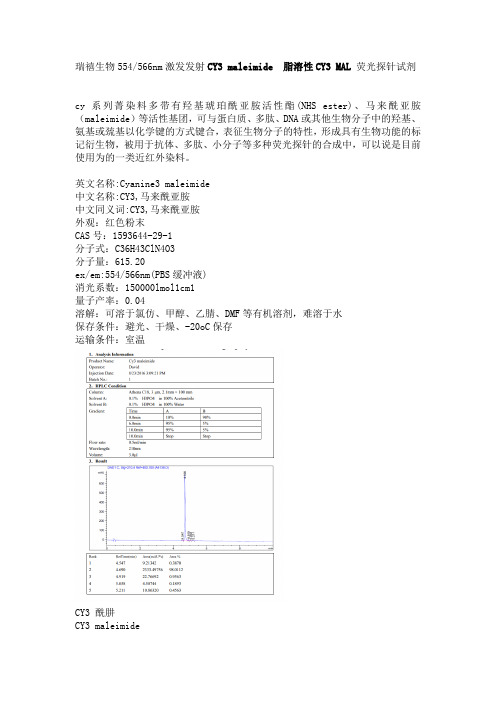
瑞禧生物554/566nm激发发射CY3 maleimide 脂溶性CY3 MAL 荧光探针试剂
cy系列菁染料多带有羟基琥珀酰亚胺活性酯(NHS ester)、马来酰亚胺(maleimide)等活性基团,可与蛋白质、多肽、DNA或其他生物分子中的羟基、氨基或巯基以化学键的方式键合,表征生物分子的特性,形成具有生物功能的标记衍生物,被用于抗体、多肽、小分子等多种荧光探针的合成中,可以说是目前使用为的一类近红外染料。
英文名称:Cyanine3 maleimide
中文名称:CY3,马来酰亚胺
中文同义词:CY3,马来酰亚胺
外观:红色粉末
CAS号:1593644-29-1
分子式:C36H43ClN4O3
分子量:615.20
ex/em:554/566nm(PBS缓冲液)
消光系数:150000lmol1cm1
量子产率:0.04
溶解:可溶于氯仿、甲醇、乙腈、DMF等有机溶剂,难溶于水
保存条件:避光、干燥、-20oC保存
运输条件:室温
CY3 酰肼
CY3 maleimide
CY5 HZ
CY3 叠氮
Cy3 活性酯
CY3 硫醇
CY3 硫基
CY3 马来酰亚胺
CY3 炔基
CY3 炔烃
CY7 carboxylic acid CY7 COOH
CY7 hydrazide
CY7 HZ
Cy3 羧酸
CY5 硫基
Sulfo-CY5 马来酰亚胺Sulfo-Cy5 羧酸
Sulfo-CY5 酰肼
Sulfo-CY7 ALK
Sulfo-CY5 NH2
Sulfo-CY5 ALK
Sulfo-CY3 硫醇。
赛沃替尼化学结构

赛沃替尼化学结构
赛沃替尼是一种化学药物,其化学名称为1-[(1S)-1-(咪唑并[1,2-a]吡啶-6-基)乙基]-6-(1-甲基-1H-吡唑-4-基)-1H-[1,2,3]三唑并[4,5-b]吡嗪,分子式为C17H15N9,分子量为345.36。
赛沃替尼的化学结构十分独特,它包含了一个咪唑并[1,2-a]吡啶-6-基团,一个1-甲基-1H-吡唑-4-基团,以及一个1H-[1,2,3]三唑并[4,5-b]吡嗪基团。
这些基团通过碳碳键和氮氮键相互连接,形成了一个复杂的分子结构。
咪唑并[1,2-a]吡啶-6-基团是赛沃替尼的一个关键部分,它具有强烈的芳香性,为分子提供了稳定性和反应活性。
1-甲基-1H-吡唑-4-基团则是一个具有生物活性的基团,它可以通过与生物体内的受体结合来发挥药效。
1H-[1,2,3]三唑并[4,5-b]吡嗪基团是赛沃替尼分子的核心部分,它具有高度的平面性和共轭性,能够稳定地存在于分子中,并且与咪唑并[1,2-a]吡啶-6-基团和1-甲基-1H-吡唑-4-基团相互作用,共同构成了赛沃替尼的分子骨架。
总的来说,赛沃替尼的化学结构具有高度的复杂性和独特性,这种结构使得它能够在生物体内发挥强大的药理作用,成为一种有效的药物分子。
转染试剂盒-萤火虫荧光素+IRES-mCherry Lentiviral Particles 数据表

Data Sheet ●Firefly Luciferase + IRES-mCherry Lentifect™ Lentiviral Particles ●Cat Nos. LP-HLUC-LV177-0200, LP- HLUC-LV177-0205Ready-to-use lentiviral particles for the transduction of a variety of mammalian cells including difficult-to-transfect, primary, stem and non-dividing cells as well as in vivo use for transgenic animals.Description•Produced with a standardized protocol using highly purified plasmids and EndoFectin-Lenti™ transfection and TiterBoost™ reagents. The protocol uses a third generation self-inactivating packaging system meeting BioSafety Level 2 requirements.•CMV promoter for the expression of HLUC (humanized firefly luciferase) in target cells•mCherry bicistronically coexpressed with FLUC•Drug selection marker: noneContents and storageProvided as 1 vial of 200 µl or 5 vials of 200 µl of lentiviral particles with titers of ~4x 10⁸copies/ml.Lentifect particles are shipped on dry ice and must be stored at –80°C immediately upon receipt. Avoid repeated freeze-thaw cycles as this will reduce titers.Quality controlThe lentiviral expression construct was validated by full-length sequencing, restriction enzyme digestion and PCR-size validation using gene-specific and vector-specific primers. Product is confirmed free of bacteria, fungi and common Mycoplasma contamination.Viral titerLentivirus products were titrated using qRT-PCR, which determines the physical copy numbers of viral genomic RNA. We suggest that the customer estimate the transduction unit (TU or IFU) for the specific host cells based on the following formula before transduction:TU=Titer (physical copy number)/100.The customer should test the transduction at MOI=0.3, 1, 3, 5, 10 using TU for the best transduction efficiency. Overview of productionThe Firefly Luciferase + IRES-mCherry OmicsLink™ ORF lentiviral expression plasmid (GeneCopoeia Cat. No. EX-HLUC-LV177) was constructed using GeneCopoeia proprietary RecJoin™ technology. This plasmid was co-transfected into 293Ta cells (GeneCopoeia Cat. No. CLv-PK-01) with the Lenti-Pac HIV Packaging Mix (GeneCopoeia Cat. No. HPK-LvTR-20). Lentivirus-containing supernatants were harvested 48 hours after transfection and stored at –80°C.User manualPlease contact GeneCopoeia for a copy or download at:/product/lentiviral/pdf/packaging_kit_manual.pdfGeneCopoeia, Inc.9620 Medical Center Drive, #101Rockville, Maryland 20850Tel: 301-762-0888 Fax: 301-762-8333Email:***********************Web: GeneCopoeia Products are for Research Use Only Copyright © 2011 GeneCopoeia Inc. Trademarks: Lentifect™, Lenti-Pac™, OmicsLink™, EndoFectin™, TiterBoost™ (GeneCopoeia Inc.) LPHLUCM061311www.g en ecopoeia.co m。
Elabscience CY3标记试剂盒说明书

(本试剂盒仅供体外研究使用,不用于临床诊断!)产品货号:E-LK-C002产品规格:1次标记/3次标记/10次标记Elabscience® CY3标记试剂盒使用说明书CY3 Labeling Kit使用前请仔细阅读说明书。
如果有任何问题,请通过以下方式联系我们:销售部电话************,************技术部电话************具体保质期请见试剂盒外包装标签。
请在保质期内使用试剂盒。
联系时请提供产品批号(见试剂盒标签),以便我们更高效地为您服务。
产品介绍Elabscience® CY3标记试剂盒提供了CY3标记所需全部试剂,能简单有效地对抗体和其他含有伯氨基(NH2-)分子的物质进行CY3标记。
试剂盒内的CY3已经活化,可直接使用,每次可标记0.1-1 mg 抗体。
试剂盒中包含用于抗体标记脱盐的Filtration tube,不用透析,操作简便,熟练操作100 min可完成整个标记过程。
基本信息Reactive CY3结构式:产品特点✓试剂全面: 本试剂盒提供了CY3标记所需全部试剂。
✓快速: 整个过程仅需100 min。
✓方便: 通过Filtration tube 即可脱盐,无需透析或者凝胶过滤。
✓使用灵活: 既可用于微量标记又可大量标记,每次可标记0.1-1 mg抗体。
✓理想的标记效果: 已经优化确定了最适的标记比例,降低标记不足或由于过度标记而失活的可能性。
试剂盒组成及保存未拆封的试剂盒可在2-8℃保存一年;溶解后的Reactive CY3 能在2-8℃保存一周。
试验所需自备物品1.高精度移液器及一次性吸头:0.5-10μL, 2-20μL, 20-200μL, 200-1000μL2.37℃恒温箱3.离心机(离心力可到12,000×g)标记原理Reactive CY3专一地与伯胺(N-末端及赖氨酸残基侧链)反应形成稳定的酰胺键:CY3标记中Reactive CY3用量的计算:每个反应中Reactive CY3试剂的使用量取决于待标记蛋白质的量和浓度。
《2024年三阴性乳腺癌关键通路筛选及丝胶抑制MDA-MB-468细胞生长的机制研究》范文
《三阴性乳腺癌关键通路筛选及丝胶抑制MDA-MB-468细胞生长的机制研究》篇一一、引言三阴性乳腺癌(TNBC)是一种恶性程度较高的乳腺癌亚型,因其缺乏雌激素受体、孕激素受体及HER2基因的扩增或过表达而具有独特的生物学特性及治疗挑战。
针对TNBC的治疗,研究其关键信号通路及其抑制机制对于新药研发和治疗方法优化具有重要意义。
丝胶作为一种天然的生物材料,其抗肿瘤效果近年来备受关注。
本文旨在研究三阴性乳腺癌关键通路的筛选以及丝胶对MDA-MB-468细胞生长的抑制机制。
二、三阴性乳腺癌关键通路筛选2.1 方法与材料利用生物信息学分析,结合基因组学及蛋白质组学数据,对三阴性乳腺癌关键信号通路进行筛选。
主要利用高通量测序技术、生物芯片技术和信号通路数据库等手段。
2.2 结果与讨论通过对多组三阴性乳腺癌患者样本的基因和蛋白质组数据进行分析,我们初步确定了PI3K/AKT、NF-kB和Wnt等信号通路为三阴性乳腺癌的关键调控通路。
这些通路与细胞的增殖、分化、凋亡等生物过程密切相关,并可被多种生物分子(如生长因子、细胞因子等)所调控。
这些通路的激活或抑制,都可能影响肿瘤细胞的生长和转移。
三、丝胶抑制MDA-MB-468细胞生长的机制研究3.1 方法与材料采用细胞培养、MTT法、流式细胞术和蛋白质印迹法(Western Blot)等实验技术,研究丝胶对MDA-MB-468细胞的生长抑制作用及其潜在机制。
3.2 结果与讨论研究发现,丝胶对MDA-MB-468细胞具有显著的生长抑制作用。
通过对细胞周期、凋亡及相关基因的表达水平进行检测,发现丝胶可显著诱导MDA-MB-468细胞的凋亡,抑制其细胞周期进程。
同时,我们发现在丝胶的作用下,NF-kB等关键信号通路的活性显著降低,提示丝胶可能通过影响这些信号通路的活性来抑制肿瘤细胞的生长。
此外,我们还发现丝胶可能通过影响肿瘤细胞的自噬、坏死等过程来进一步抑制其生长。
四、结论本研究初步确定了三阴性乳腺癌的关键信号通路,并研究了丝胶对MDA-MB-468细胞的生长抑制作用及其潜在机制。
