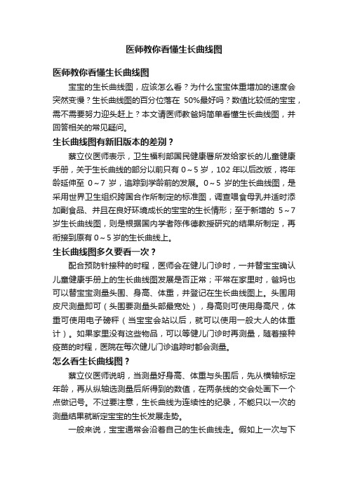
早产儿生长曲线图
医师教你看懂生长曲线图

医师教你看懂生长曲线图医师教你看懂生长曲线图宝宝的生长曲线图,应该怎么看?为什么宝宝体重增加的速度会突然变慢?生长曲线图的百分位落在50%最好吗?数值比较低的宝宝,需不需要努力迎头赶上?本文请医师教爸妈简单看懂生长曲线图,并回答相关的常见疑问。
生长曲线图有新旧版本的差别?蔡立仪医师表示,卫生福利部国民健康署所发给家长的儿童健康手册,关于生长曲线的部分以前只有0~5岁,102年以后改版,将年龄延伸至0~7岁,追踪到学龄前的发展。
0~5岁的生长曲线图,是采用世界卫生组织跨国合作所制定的标准图,调查喂食母乳并适时添加副食品、并且在良好环境成长的宝宝的生长情形;至于新增的5~7岁生长曲线图,则是根据国内学者陈伟德教授研究的结果所制定,再衔接到原有0~5岁的生长曲线上。
生长曲线图多久要看一次?配合预防针接种的时程,医师会在健儿门诊时,一并替宝宝确认儿童健康手册上的生长曲线图发展是否正常;平常在家里时,爸妈也可以替宝宝测量头围、身高、体重,并登记在生长曲线图上。
头围用皮尺测量即可(头围要测量头部最宽处),身高则可使用身高尺,体重可使用电子磅秤(当宝宝会站以后,就可以使用一般大人的体重计)。
如果家里没有这些物品,可以等健儿门诊时再测量,随着接种疫苗的时程,医院在每次健儿门诊追踪时都会测量。
怎么看生长曲线图?蔡立仪医师说明,当测量好身高、体重与头围后,先从横轴标定年龄,再从纵轴选测量后所得到的数值,在两条线的交会处画下一个点做记号。
不过要注意,生长曲线为连续性的纪录,不能只以一次的测量结果就断定宝宝的生长发展走势。
一般来说,宝宝通常会沿着自己的生长曲线走。
假如上一次与下一次的测量结果改变的幅度超过两个曲线区间(例如从85%掉到15%),可能是异常的警讯,必须特别留意与追踪。
百分位所代表的意义?生长曲线图上所显示的百分位,如果是50%,表示在100个同年龄的宝宝中,大约平均为第50名,属于中间值(这个数值是根据先前的研究结果所统计出来的)。
最新使用世界卫生组织(WHO)的生长曲线图

使用世界卫生组织(WHO)的生长曲线图成长不仅是营养的结果,还是遗传因素的结果。
种族会影响孩子的成长模式,因此有些国家有他们自己的生长曲线图。
不过,世界卫生组织(WHO)的生长曲线图用得最普遍,并被认为是全世界的标准。
了解更多关于:»怎样进行测量»怎样将测量应用到生长曲线图中怎样进行测量0-24 月龄的孩子进行的典型测量包括:•头围•身长•体重测量应定期进行,以观察可靠的趋势。
建议的测量时间间隔包括:•婴儿(0-12 月龄):每2 个月•幼儿:分别在15, 18, 24 和30 月龄• 3 岁以上:每年点击这里获取详细的测量时间表。
头围头围是在孩子头部最大部分进行的测量。
这种测量通常为0-3 岁的孩子进行。
测量时应当使用不可拉伸的卷尺。
卷尺通常是可弯曲的金属量尺。
测量时,卷尺尽可能紧贴着头部最宽围度缠绕。
通常,此部位在前额眉毛上方1-2 指宽到后脑勺最突出的部分。
测量三次,取精确到0.1 厘米的最大测量值。
在孩子生命的早期,头围是一种很重要的测量,因为它间接地反映大脑尺寸和发育。
几乎所有的大脑发育都在两岁以前,因此绘制的头部生长曲线可以作为幼儿大脑健康的通用指标。
了解关于头围-年龄生长曲线图。
身长身长是为不足24 月龄的婴儿进行的线性测量。
24 到36 月龄的孩子,如果无法独立站立,也可以进行身长测量(代替身高)。
身长是在孩子卧位(平躺)时测量的。
测量身长最准确的方法是使用校准的身长量板。
身长量板应当有一块与板表面垂直的固定头部挡板和一块可移动的足板。
测量时,将孩子平放在板上,头靠着固定挡板。
确定孩子没有穿鞋或戴帽子。
有个助手也许可以帮助保持孩子不动并在板中间。
让孩子的腿伸直,调整活动足板,使孩子的脚底靠着足板。
精确到0.1 厘米记录身长。
身长是孩子营养状况的一个重要决定因素。
如果孩子长期营养不良,可能会表现出身长增长缓慢。
了解关于身长-年龄生长曲线图。
体重体重是在整个生命期都需要进行的测量,以帮助确定当前的营养状况和趋势。
儿童身高体重百分位曲线

儿童身高体重百分位曲线
这个曲线图通常包含多条曲线,每条曲线代表不同百分位的儿童。
例如,常见的百分位包括5th、10th、25th、50th、75th、90th和95th百分位。
每个百分位表示有多少比例的同龄儿童身高或体重落在该曲线以下。
儿童身高体重百分位曲线的作用是帮助医生和家长判断儿童的生长发育是否正常。
当儿童的身高和体重曲线与某个百分位曲线相符时,说明他们的生长发育与同龄儿童相当。
如果曲线偏离某个百分位,可能意味着儿童的生长发育存在问题,需要进一步评估和干预。
此外,儿童身高体重百分位曲线还可以用来监测儿童的生长趋势。
通过定期测量和记录儿童的身高和体重,并与曲线进行比较,可以观察到他们的生长速度和趋势,及时发现潜在的生长问题。
需要注意的是,儿童身高体重百分位曲线只是一种参考工具,不能作为唯一的评估标准。
每个儿童的生长发育都有个体差异,还需要考虑其他因素,如家族遗传、营养状况、健康状况等。
总结起来,儿童身高体重百分位曲线是用来评估儿童生长发育情况的工具,通过与曲线进行比较,可以判断儿童的生长发育是否正常,并监测他们的生长趋势。
然而,它只是一个参考工具,需要综合考虑其他因素来做出准确的评估和判断。
世卫组织最新标准 - (0-5岁)体重生长曲线图 ~ 身高生长曲线图 ~ BMI生长曲线图

世卫组织最新标准 - (0-5岁)体重生长曲线图 ~ 身高生长曲线图 ~ BMI生长曲线图世界卫生组织的最新标准是完全根据母乳宝宝的生长情况制定的。
而我国的标准是综合国内不同地区和不同喂养分方式的数据统计出来的。
但这样的数据并不能说明它更正确,而恰恰会由于混合或人工喂养宝宝的因素使数据偏高。
这些新指标是基于8440名母乳喂养的孩子的生长发育状况做出的。
与吃母乳的婴幼儿比起来,吃配方奶的孩子体重比吃母乳的孩子要增长得快些。
而“世卫组织”旧的有关婴幼儿的成长发育指标却是根据吃配方奶的孩子的发育情况制定的,这就意味着这一套标准存在着重大缺陷。
从1997年到2003年间,世界卫生组织对包括巴西等6个国家的孩子进行了跟踪调查。
这些孩子来自巴西、加纳、印度、挪威、阿曼和美国等6个不同的国家,身体都很健康,且他们的母亲都不吸烟,对孩子的照顾也非常周到。
而之前的指标只是取单独一个国家的儿童为样本(也就是美国儿科学会公布的美国婴幼儿生长发育曲线)。
男宝BMI生长曲线图女宝BMI生长曲线图新的婴幼儿生长发育指标中还包含了身体质量指数(BMI=体重(公斤)÷身高(米)的平方,单位为公斤/平方米),这是WHO首次在婴幼儿生长发育指标中引入此项指标。
BMI为评估体重与身高比例提供了工具,对于监控孩子的肥胖症非常有效。
它是评估儿童健康的一个重大革新。
男宝宝体重生长曲线图专家认为,新指标的发表不会给中国孩子生长发育情况带来大影响。
我国每10年都要在北部、中部、南部各选三地,对0-18岁儿童和青少年的身高、体重等情况进行调查统计,并综合了各种喂养方式,得出我国儿童的身高、体重等指标的参照值。
用生长曲线检测孩子的身高、体重的发育,比起简单用一个数字断定孩子是高是胖要更科学。
使用方法如下:1、做顺时记录。
每个月为孩子测量一次身高、体重,把测量结果描绘在生长曲线图上(不要在孩子生病期间测量),连成一条曲线。
如果孩子的生长曲线一直在正常值范围内(3号线到-3号线之间)匀速顺时增长就是正常的。
早产儿的护理ppt课件

如何帮助早产儿建立良好的睡眠习惯等。
保持安静: 为早产儿创 造一个安静、 舒适的睡眠 环境,避免 噪音干扰。
规律作息: 帮助早产儿 建立规律的 作息时间, 包括喂食、 换尿布、玩 耍和睡眠等。
调整光线: 在早产儿睡 觉时,尽量 保持室内光 线昏暗,避 免光线刺激 影响睡眠。
早产儿肠道菌 群失调,可能 导致肠道感染 和炎症。
3
早产儿营养吸 收能力较差, 容易出现营养 不良、贫血等 问题。
4
早产儿容易出 现胃食道返流, 导致呕吐、呛 咳等问题。
神经系统问题
STEP1
STEP2
STEP3
STEP4
早产儿神经系统 发育不成熟,易 出现神经系统问 题
常见问题包括脑 损伤、脑瘫、癫 痫等
围的差异
头围变化与早 产儿发育状况
的关系
其他生长发育指标的变化
01
体重:早产儿出生时体重较 轻,但出生后体重增长迅速
02
身高:早产儿出生时身高较 短,但出生后身高增长较快
03
头围:早产儿出生时头围较 小,但出生后头围增长较快
04
胸围:早产儿出生时胸围较 小,但出生后胸围增长较快
05
皮肤:早产儿皮肤较薄,但 出生后皮肤逐渐增厚
06
肌肉:早产儿肌肉较弱,但 出生后肌肉逐渐增强
3
早产儿健康问题
呼吸系统问题
01
早产儿呼吸系统发育不成熟, 容易出现呼吸困难、呼吸暂 停等问题。
03
早产儿容易发生呼吸窘迫综 合征(RDS),需要及时进 行呼吸支持治疗。
02
早产儿肺部表面活性物质不 足,容易导致肺泡塌陷,影 响呼吸功能。
【VIP专享】世卫组织最新标准 - (0-5岁)体重生长曲线图 ~ 身高生长曲线图 ~ BMI生长曲线图

世卫组织最新标准 - (0-5岁)体重生长曲线图 ~ 身高生长曲线图 ~ BMI生长曲线图世界卫生组织的最新标准是完全根据母乳宝宝的生长情况制定的。
而我国的标准是综合国内不同地区和不同喂养分方式的数据统计出来的。
但这样的数据并不能说明它更正确,而恰恰会由于混合或人工喂养宝宝的因素使数据偏高。
这些新指标是基于8440名母乳喂养的孩子的生长发育状况做出的。
与吃母乳的婴幼儿比起来,吃配方奶的孩子体重比吃母乳的孩子要增长得快些。
而“世卫组织”旧的有关婴幼儿的成长发育指标却是根据吃配方奶的孩子的发育情况制定的,这就意味着这一套标准存在着重大缺陷。
从1997年到2003年间,世界卫生组织对包括巴西等6个国家的孩子进行了跟踪调查。
这些孩子来自巴西、加纳、印度、挪威、阿曼和美国等6个不同的国家,身体都很健康,且他们的母亲都不吸烟,对孩子的照顾也非常周到。
而之前的指标只是取单独一个国家的儿童为样本(也就是美国儿科学会公布的美国婴幼儿生长发育曲线)。
新的婴幼儿生长发育指标中还包含了身体质量指数(BMI=体重(公斤)÷身高(米)的平方,单位为公斤/平方米),这是WHO首次在婴幼儿生长发育指标中引入此项指标。
BMI为评估体重与身高比例提供了工具,对于监控孩子的肥胖症非常有效。
它是评估儿童健康的一个重大革新。
男宝宝体重生长曲线图 专家认为,新指标的发表不会给中国孩子生长发育情况带来大影响。
我国每10年都要在北部、中部、南部各选三地,对0-18岁儿童和青少年的身高、体重等情况进行调查统计,并综合了各种喂养方式,得出我国儿童的身高、体重等指标的参照值。
用生长曲线检测孩子的身高、体重的发育,比起简单用一个数字断定孩子是高是胖要更科学。
使用方法如下: 1、做顺时记录。
每个月为孩子测量一次身高、体重,把测量结果描绘在生长曲线图上(不要在孩子生病期间测量),连成一条曲线。
如果孩子的生长曲线一直在正常值范围内(3号线到-3号线之间)匀速顺时增长就是正常的。
世卫组织最新标准 - (0-5岁)体重生长曲线图 ~ 身高生长曲线图 ~ BMI生

世卫组织最新标准 - (0-5岁)体重生长曲线图 ~ 身高生长曲线图 ~ BMI生长曲线图世界卫生组织的最新标准是完全根据母乳宝宝的生长情况制定的。
而我国的标准是综合国内不同地区和不同喂养分方式的数据统计出来的。
但这样的数据并不能说明它更正确,而恰恰会由于混合或人工喂养宝宝的因素使数据偏高。
这些新指标是基于8440名母乳喂养的孩子的生长发育状况做出的。
与吃母乳的婴幼儿比起来,吃配方奶的孩子体重比吃母乳的孩子要增长得快些。
而“世卫组织”旧的有关婴幼儿的成长发育指标却是根据吃配方奶的孩子的发育情况制定的,这就意味着这一套标准存在着重大缺陷。
从1997年到2003年间,世界卫生组织对包括巴西等6个国家的孩子进行了跟踪调查。
这些孩子来自巴西、加纳、印度、挪威、阿曼和美国等6个不同的国家,身体都很健康,且他们的母亲都不吸烟,对孩子的照顾也非常周到。
而之前的指标只是取单独一个国家的儿童为样本(也就是美国儿科学会公布的美国婴幼儿生长发育曲线)。
男宝BMI生长曲线图女宝BMI生长曲线图新的婴幼儿生长发育指标中还包含了身体质量指数(BMI=体重(公斤)÷身高(米)的平方,单位为公斤/平方米),这是WHO首次在婴幼儿生长发育指标中引入此项指标。
BMI为评估体重与身高比例提供了工具,对于监控孩子的肥胖症非常有效。
它是评估儿童健康的一个重大革新。
男宝宝体重生长曲线图专家认为,新指标的发表不会给中国孩子生长发育情况带来大影响。
我国每10年都要在北部、中部、南部各选三地,对0-18岁儿童和青少年的身高、体重等情况进行调查统计,并综合了各种喂养方式,得出我国儿童的身高、体重等指标的参照值。
用生长曲线检测孩子的身高、体重的发育,比起简单用一个数字断定孩子是高是胖要更科学。
使用方法如下:1、做顺时记录。
每个月为孩子测量一次身高、体重,把测量结果描绘在生长曲线图上(不要在孩子生病期间测量),连成一条曲线。
如果孩子的生长曲线一直在正常值范围内(3号线到-3号线之间)匀速顺时增长就是正常的。
中国不同出生胎龄新生儿体重身长比、体质指数和重量指数的参照标准及生长曲线(全文)

中国不同出生胎龄新生儿体重身长比、体质指数和重量指数的参照标准及生长曲线(全文)新生儿出生时的体格生长评价是了解宫内生长发育状况、预测疾病风险以及日后生长发育甚至成年后健康状况的重要手段。
出生体重、身长和头围是常用的新生儿生长水平评价指标,但这些指标尚不能充分满足对新生儿的身体比例与营养状况的准确评价。
最近有研究显示体重身长比是反映新生儿体成分的良好指标,体质指数是反映新生儿身体比例或匀称度的有效指标。
重量指数是传统的较为常用的评价新生儿身体匀称度的指标。
我国1988年新生儿调查获得的有关数据已不适合应用于评价当今新生儿的生长和营养状况。
2020年9月九市儿童体格发育调查协作组报道了中国不同出生胎龄新生儿出生体重、身长和头围的生长参照标准,为进一步完善我国新生儿生长发育评价指标体系,本研究旨在建立我国不同出生胎龄新生儿体重身长比、体质指数和重量指数的参照标准及生长曲线,为新生儿身体比例及营养状况评价提供更加全面的参考依据。
对象和方法一、对象于2015年6月至2018年11月在中国13个城市开展不同出生胎龄新生儿体格发育横断面调查,其中北京、哈尔滨、西安、上海、南京、武汉、广州、福州、昆明是沿用1975年开展首次调查且连续实施5次调查的城市,收集出生胎龄24+0~42+6周的单胎活产新生儿;考虑到小早产儿出生人数少,调查开始后在九城市周边增加了天津、沈阳、长沙、深圳4个城市补充收集出生胎龄32周及以下早产儿。
本调查通过首都儿科研究所伦理委员会批准(SHERLL-2015009),所有家长均被告知并同意参加本调查。
1.胎龄确定:根据母亲末次月经和孕早期(前3个月)超声检查结果综合确定胎龄,当2种方法确定的胎龄相差<1周以末次月经为准、相差>1周以超声胎龄为准。
按出生胎龄每周划分1组,如24周组代表24+0~24+6周,24~42周共分19组。
2.样本量估算:按照体格发育专项调查的统计学要求,出生胎龄37~41周足月儿,每市每胎龄组男、女样本量各约100名,鉴于足月儿符合纳入排除标准的例数较多,按每个季度平均分配比例计算机随机抽取,达到样本量要求即停止收集;出生胎龄29~36周早产儿,每市每胎龄组男女样本量各约50名;出生胎龄42周足月儿和出生胎龄≤28周早产儿不设样本量要求,符合纳入标准的均纳入。
0-12岁成长标准数值表格 生长曲线

0-12岁成长标准数值表格生长曲线成长标准数值表格是根据大量儿童的身高、体重等数据综合统计得出的,可以用来评估儿童的生长发育情况。
以下是0-12岁儿童身高、体重、头围等指标的标准数值表格。
年龄身高(cm)体重(kg)头围(cm)0-1月49.8-54.9 3.2-4.5 34.5-37.51-2月54.3-59.9 4.2-5.8 36-392-3月58.5-64.3 4.8-6.7 37-40.53-4月61.9-68.3 5.4-7.6 37.5-414-5月65-72.7 5.9-8.4 38-425-6月67.8-75.6 6.3-9.1 38.5-42.56-9月69.7-77.8 6.6-9.5 39-439-12月71-80.3 6.9-9.9 39-431-2岁73-83.6 7.4-10.7 39-43.52-3岁76.2-89.2 7.9-11.8 39-43.53-4岁79.4-94.7 8.5-12.9 39-43.54-5岁82.5-100.4 9.1-14 39-445-6岁85.9-106.2 9.7-15.1 39.5-44.56-7岁89.5-112 10.3-16.2 40-457-8岁93.3-117.8 10.9-17.3 40-45.58-9岁97.1-123.4 11.5-18.4 40.5-45.59-10岁101-128.4 12.1-19.6 40.5-4610-11岁104.7-133.3 12.7-20.8 41-4611-12岁108.3-137.9 13.3-22 41-46.5这是一个以年龄为横坐标,身高、体重、头围为纵坐标的表格。
根据这个表格,我们可以看出儿童在不同年龄段的身高、体重等指标的标准范围。
这些数据是通过对大量儿童进行测量和统计得出的,可以用来评估儿童的生长发育状况。
儿童的生长发育在不同年龄段有不同的特点。
在出生后的0-1个月,儿童的身高大约在49.8-54.9厘米之间,体重在3.2-4.5公斤之间,头围在34.5-37.5厘米之间。
儿童体格生长评价

2.百分位数法: 一组变量值(如体重、身高)按从小到大的顺序排列为100 个等份,每个等份为1个百分位。排列顺序确定各百分位 的数值,即百分位数(P)。
当变量值呈现非正态分布时,百分位数能更准确地反映 出所测数值的分布情况。一般采用P3,P25,P50,P75, P97辩为主百分位数(或主百分位线),P3-P97视为正常范 围。
(1)选择适宜的体格生长指标; (2)采用准确的测量工具及规范的测量方法 (3)选择恰当的参考人群值
2005年中国9市儿童的体格发育数据 制定的中国儿童生长参照标准 2006年世界卫生组织儿童生长标准
1 体格生长评价的原则
(1)选择适宜的体格生长指标; (2)采用准确的测量工具及规范的测量方法 (3)选择恰当的参考人群值 (4)定期评估儿童生长状况,即生长监测
立位 ---身高
身长/高 (Length/Height, L/H):
2~3岁 需要将身高加0.7 cm进行调整后,再与生长标 准图表身长值比较。
< 3岁
身长和身高的测量 需要2名经过培训的 人员配合
≥3岁
3岁后仍不能很好地独自站立 测量身长,将测量值减去0.7 cm 与身高值进行比较
体重( Weight,W )
➢ 器官、系统、体液的总重量 ➢ 反映儿童生长与营养状况的灵敏指标
婴儿称重应精确至0.01 kg, 儿童至0.1 kg。
头围(head circumference,HC)
指头的最大围径,反映脑和 颅骨的生长发育。
头围在出生后头3年反映脑的快速发育, 因此建议常规测量头围至3岁(至少到2岁)。
1 体格生长评价的原则
<6岁 身长的体重 身高的体重
建议积极采用2~1 8岁 的BMI生长曲线
- 1、下载文档前请自行甄别文档内容的完整性,平台不提供额外的编辑、内容补充、找答案等附加服务。
- 2、"仅部分预览"的文档,不可在线预览部分如存在完整性等问题,可反馈申请退款(可完整预览的文档不适用该条件!)。
- 3、如文档侵犯您的权益,请联系客服反馈,我们会尽快为您处理(人工客服工作时间:9:00-18:30)。
A systematic review and meta-analysis to revise the Fenton growth chart for preterm infantsTanis R Fenton 1,2*and Jae H Kim 3BackgroundThe expected growth of the fetus describes the fastest human growth,increasing weight over six-fold between 22and 40weeks.Preterm infants,who are born during this rapid growth phase,rely on health professionals to assess their growth and provide appropriate nutrition and medical care.In 2006,the World Health Organization (WHO)published their multicentre growth reference study,which is considered superior [1]to previous growth surveys since the measured infants were selected from communities in which economics were not likely to limit growth,among culturally diverse non-smoking mothers who planned to breastfeed [2].Weekly longitudinal measures of the infants were made by trained data collection teams during the first 2years of this study [3].These WHO growth charts,although recommended for preterm infants after term age [4],begin at term and so do not inform preterm infant growth assessments younger than this age.*Correspondence:tfenton@ucalgary.ca 1Alberta Children ’s Hospital Research Institute,The University of Calgary,Calgary,AB,Canada 2Department of Community Health Sciences,The University of Calgary,3280Hospital Drive NW,Calgary,AB,CanadaFull list of author information is available at the end of thearticle©2013Fenton and Kim;licensee BioMed Central Ltd.This is an Open Access article distributed under the terms of the Creative Commons Attribution License (/licenses/by/2.0),which permits unrestricted use,distribution,and reproduction in any medium,provided the original work is properly cited.Fenton and Kim BMC Pediatrics 2013,13:59/1471-2431/13/59Optimum growth of preterm infants is considered to be equivalent to intrauterine rates[5-7]since a superior growth standard has not been defined.Perhaps the best estimate of fetal growth may be obtained from large population-based studies,conducted in developed coun-tries[8],where constraints on fetal growth may be less frequent.A recent multicentre study by our group(the Preterm Multicentre Growth(PreM Growth)Study)revealed that although the pattern of preterm infant growth was gener-ally consistent with intrauterine growth,the biggest devi-ation in weight gain velocity between the preterm infants and the fetus and infant was just before term,between37 and40weeks(Fenton TR,Nasser R,Eliasziw M,Kim JH, Bilan D,Sauve R:Validating the weight gain of preterm in-fants between the reference growth curve of the fetus and the term infant,The Preterm Infant Multicentre Growth Study.Submitted BMC Ped2012).Rather than demon-strating the slowing growth velocity of the term infant during the weeks just before term,the preterm infants had superior,close to linear,growth at this age.This finding has been observed by others as well[9-11].Therefore, there is evidence to support a smooth transition on growth charts between late fetal and early infant ages. Several previous growth charts based on size at birth presented their data as completed age,which affects the interpretation and use of a growth chart[12].The use of completed weeks when plotting a growth chart requires all the measurements to be plotted on the whole week vertical axes.However,the use of completed weeks in a neonatal unit may not be intuitive,as nursery staff and parents think of infants as their exact age,and not age truncated to previous whole weeks.The advent of computers in health care,for clinical care and health recording,allow the use of the computer to plot growth charts,daily and with accuracy.It would make sense to support plotting daily measurements continuously by shifting the data collected as completed weeks to the midpoint of the next week to remove the truncation of the data collection as completed weeks.The objectives of this study were to revise the2003 Fenton Preterm Growth Chart,specifically to:a)use more recent data on size at birth based on an inclusion criteria, b)harmonize the preterm growth chart with the new WHO Growth Standard,c)to smooth the data between the preterm and WHO estimates while maintaining integrity with the data from22to36and at50weeks, d)to derive sex specific growth curves,and to e)re-scale the chart x-axis to actual age rather than completed weeks,to support growth monitoring.MethodsTo revise the growth chart,thorough literature searches were performed to find published and unpublished population-based preterm size at birth(weight,length, and/or head circumference)references.The inclusion criteria,defined a priori,designed to minimize bias by restriction[13],were to locate population-based studies of preterm fetal growth,from developed countries with: a)Corrected gestational ages through fetal ultrasoundand/or infant assessment and/or statisticalcorrection;b)Data percentiles at24weeks gestational age orlower;c)Sample of at least25,000babies,with more than500infants aged less than30weeks;d)Separate data on females and males;e)Data available numerically in published form orfrom authors,f)Data collected within the past25years(1987to2012)to account for any secular trends.A.Data selection and combinationMajor bibliographic databases were searched:MEDLINE (using PubMed)and CINHAL,by both authors back to year1987(given our25year limit),with no language restrictions,and foreign articles were translated.The following search terms as medical subject headings and textwords were used:(“Preterm infant”OR“Premature Birth”[Mesh])OR(“Infant,Premature/classification”[Mesh] OR“Infant,Premature/growth and development”[Mesh] OR“Infant,Premature/statistics and numerical data”[Mesh] OR“Infant,very low birth weight”[Mesh])AND (percentile OR*centile*OR weeks)AND(weight OR head circumference OR length).Grey literature sites including clinical trial websites and Google were searched in February 2012.Reference lists were reviewed for relevant studies.All of the found data was reported as completed weeks except for the German Perinatal Statistics,which were reported as actual daily weights[14].To combine the datasets,the German data was temporarily converted to completed weeks.A final step converted the meta-analyses to actual age.bine the data to produce weighted intrauterine growth curves for each sexThe located data(3rd,10th,50th,90th,and97th percentiles for weight,head circumference,and length)that met the inclusion criteria were extracted by copying and pasting into spreadsheets.The male and female percentile curves from each included data set for weight,head circumference and length were plotted together so they could be examined visually for heterogeneity(Figures1,2, and3).The data for each gender were combined by using the weekly data for the percentiles:3rd,10th,50th,90th, and97th,weighted by the sample sizes.The combined data was represented by relatively smooth curves.C.Develop growth monitoring curvesTo develop the growth monitoring curves that joined the intrauterine meta-analysis data with the WHO Growth Standard(WHOGS)smoothly,the following cubic spline procedure was used to meet two objectives:a)To maintain integrity with the meta-analysis curvesfrom22to36weeks.Integrity of the fit wasassumed to be agreement within3%at each week. b)To ensure fit of the data to the WHO values at50weeks,within0.5%.Procedure:1)Cubic splines were used to interpolate smoothvalues between selected points(22,25,28,32,34,36 and50weeks).Extra points were manually selected at40,43and46weeks in order to produceacceptable fit through the underlying data.ThePreM Growth study(Fenton TR,Nasser R,Eliasziw M,Kim JH,Bilan D,Sauve R:Validating the weight gain of preterm infants between the referencegrowth curve of the fetus and the term infant,The Preterm Infant Multicentre Growth Study.Submitted BMC Ped2012)conducted to inform the transition between the preterm and WHO data,was used to inform this step.The Prem Growth Studyfound that preterm infants growth in weightfollowed approximately a straight line between37and45weeks,as others have also noted[9-11].2)LMS values(measures of skew,the median,and thestandard deviation)[15]were computed from theinterpolated cubic splines at weekly intervals.Cole’sprocedures[15]and an iterative least squares method were used to derive the LMS parameters(L=Box-Cox power,M=median,S=coefficient of variation)fromFigure1Boys birthweight centiles(3rd,50th and97th)from the six included studies,along with the boy’s meta-analysis curves (bold).Figure2Girls head circumference centiles(3rd,50th and97th) centiles from the included studies,along with the girl’smeta-analysis curves(dotted),and after40weeks,the World Health Organization centiles (dashed).Figure3Girls length centiles(3rd,50th and97th)centiles from the included studies,along with the meta-analysis curves (dotted),and after40weeks,the World Health Organization centiles(dashed).Table2Number of infants each week from each studyGestational age Voight,2010Olsen,2010Bertino,2010Kramer,2001Roberts,1999Bonellie,2008 Females Males Females Males Females Males Females Males Females Males Females Males 22188321----80827174--23431560133153381061147995--245757044384512024148156115135120126 257138466037224038184202136180115118 268129687738813558191234188235179172 271073120396610305261188254231284174177 2812761536118712817963287330287361246239 2915161838125415057072299392325397245265 301853221216061992107114390467440571317313 312283295620442460126140461584548743136148 32**300736771651837959978771117193205 33**418650142112401055136812001471239256 34**593672912633492018255320862657374422 35**508269523664183391431434184092644653 36**46907011562665820396487320878810481265 37**43726692129114921730819965161051866020062499 38**57558786352439764751651947478095140446306387 39**597883245295545275068776236884672871869910706 40**55297235567256531107381127371375701415531264414230 *Not reported.the multicentre meta-analyses for weight,headcircumference and length.The LMS splines weresmoothed slightly while maintaining data integrity asnoted above.3)The final percentile curves were produced from thesmoothed LMS values.4)A grid similar to the2003growth chart was used,but the growth curves were re-scaled along thex-axis from completed weeks to allow clinicians toplot infant growth by actual age in weeks,and aslight modification(scaled to60centimeters insteadof65)was made to the y-axis.pared the revised charts with the2003version The revised growth charts were compared graphically with the original2003Fenton preterm growth chart.To makethe differences in chart values more apparent,the2003 chart data was also shifted to actual weeks for these com-parison figures.ResultsSix large population based surveys[14,16-20]of size at preterm birth from countries Germany,United States, Italy,Australia,Scotland,and Canada were located that met the inclusion criteria(Table1).The literature search identified2436papers,of which2373were discarded as being not relevant or duplicates based on the titles (Figure4).Reviewing reference lists identified another 12studies.Seventy-five studies were examined in detail, however27of these did not meet the date criteria.Among the48studies that met the date of birth criteria,some did not meet the other inclusion criteria for the following reasons:Did not meet the criterion for more than25,000 babies[21-35],no low gestational age infants less than25 weeks[31,36-41],insufficient number less than30weeks [34,42-45],no statistical correction for inaccurate gestational ages[46-48],numerical data not available [49-51],number of infants each week were not available [52],number of infants in the subgroups each week were not available[53],was not population based[54-56],no direct measurements[27],some of the data[57]was also in one of the larger included studies[17].Included in the meta-analyses were almost four million (3,986,456)infants at birth(34,639less than30weeks) from six studies for weight(Table2),and173,612infants for head circumference,and151,527for length[16,18]. The World Health Organization data measurements were made longitudinally on882infants.The individual datasets from the literature showed good agreement with each other,especially along the 50th and lower centiles(Figures1,2,and3)and the meta-analysis curves had a close fit with the individual datasets up to36weeks and at50weeks(Figures5,6,7). The final splined weight curves were within3%of the meta-analysis curves for24through36weeks for both gen-ders,except for a3.8%difference for girls at32weeks along the90th centile.None of the length measurements differed by more than1.8%percent between the meta-analysis and the splined curves;all weeks of the head circumference curves were within 1.5%.The meta-analyses for headFigure5Boys meta-analysis weight curves(dotted)with the final smoothed growth chart curves (dashed).Figure6Boys meta-analysis head circumference curves (dotted)with the final smoothed growth chart curves (dashed). Figure7Boys meta-analysis length curves(dotted)with the final smoothed growth chart curves(dashed).circumference and length for girls and boys were close enough to normal distributions that normal distributions were used to summarize the data.The measures at50 weeks were within0.5%of the WHOGS values.Girl and boy charts were prepared(Figure8and9),by shifting the age by0.5weeks to allow plotting by exact age instead of completed weeks.The LMS Parameters [15]were used to develop the exact z-score and percentile calculators for the new growth chart.In the two graphical comparisons between the revised growth charts,one for each sex,with the2003Fenton preterm growth chart revealed that the curves were quite similar(Figures10and11).Generally the new girls’curves were slightly lower(Figure10)and the new boys’slightly higher(Figure11)for all3parameters(weight,head cir-cumference,and length)than the2003curves.The most dramatic visual and numerical difference between the new charts and the2003chart was the higher shift of the boys’weight curves after40weeks compared to the2003chart, reaching a maximum difference at50weeks of650,580, and740grams at the3rd,50th,and97th percentiles,re-spectively.The second biggest visual difference was thegrowth chart for girls.lower pattern of the girls’length curves below37weeks; the difference in length reached a maximum numerical value of1.7centimeters at24weeks along the97th percentile.DiscussionWe used a strict set of inclusion criteria to include only the best data available to convert fetal and infant size data into fetal-infant growth charts for preterm infants.The re-vised sex-specific actual-age(versus completed weeks) growth charts(Figure9and10),are based on birth size in-formation of almost four million births with confirmed or corrected gestational ages,born in developed countries (See Features of the new growth chart).The revised charts are based on the recommended growth goal for preterm infants,the fetus and the term infant,with smoothing of the disjunction between these datasets,based on the find-ings of our international multicentre validation study (Fenton TR,Nasser R,Eliasziw M,Kim JH,Bilan D,Sauve R:Validating the weight gain of preterm infants between the reference growth curve of the fetus and the term in-fant,The Preterm Infant Multicentre Growth Study. Submitted BMC Ped2012).These charts are consist-ent with the meta-analysis data up to and includinggrowth chart for boys.36weeks,thus they can be used for the assessment of size for gestational age for preterm infants under37 weeks of gestational age.This growth chart is likely ap-plicable to preterm infants in both developed and de-veloping countries since the data was selected from developed countries to minimize the influence from cir-cumstances that may not have been ideal to support growth.Features of the new growth chartBased on the recommended growth goal for preterm infants:The fetus and the term infantGirl and boy specific chartsEquivalent to the WHO growth charts at50weeks gestational age(10weeks post term age).Large preterm birth sample size of4million infants; Recent population based surveys collected between 1991to2007Data from developed countries includingGermany,Italy,United States,Australia,Scotland, and CanadaCurves are consistent with the data to36weeks, thus can be used to assign size for gestational age up to and including36weeks.Chart is designed to enable plotting as infants are measured,not as completed weeks.The x axis wasadjusted for this chart so that infant size data can be plotted without age adjustment,i.e.Babies should be plotted as exact ages,that is a baby at253/7weeksshould be plotted along the x axis between25and26weeks.Exact z-score and percentile calculator available for download from http://ucalgary.ca/fenton.Data isavailable for research upon request.It may be more intuitive to plot on growth charts using exact ages rather than on the basis of completethe revised growth chart for girls(solid curves)and the2003Fenton growth chart for length,head circumference,and weight).Both the2003and the revised growthweeks.Several years ago,the WHO used completed age for growth chart development[12].This recommenda-tion was likely due to the way data had been collected in the past,that is all260/7through266/7week infants were included in the26week completed week category. However,with the use of computers to plot on growth charts comes the potential to more accurately plot mea-surements to the exact day of data collection.Thus the time scale of the horizontal axes of these new growth charts were re-scaled to actual age,for ease of use and un-derstanding.For example,a baby at253/7can be intui-tively plotted between25and26weeks.Exact z-score and centile calculators for the revised charts are available for download:http://ucalgary.ca/ fenton.Data is available for research upon request.The data revealed that between22weeks to50weeks post menstrual age,the fetus/infant multiplies its weight tenfold,for example,the girls’median weight increased from a median ing a fetal-infant growth chart allows clinicians to compare preterm infants’growth to an estimated reference of the fetus and the term infant.There was a remarkably close fit of the included preterm surveys for weight,head circumference and length from the6countries,especially at the50th percentile,even though the data came from different countries.The splining procedures we used have produced a chart that has integrity and good agreement with the original data.Smoothing of the LMS parameters is recommended since minor fluctuations are more likely due to sampling errors rather than physiological events [15].Experts recommend that growth charts be developed based on smoothed L,M and S,to constrain the adjacent curves so that they relate to each other smoothly[15].The World Health Organization set their L parameter to1for head circumference and length,while they maintained the exact L values for infants’weights[58].The data under study here revealed the same effect as the WHO data;wethe revised growth chart for boys(solid curves)and the2003Fenton growth chart for length,head circumference,and weight).Both the2003and the revised growthfound that both head circumference and length were close enough to normal distributions that normal distributions could summarize the data,while the exact L’s were needed to retain the nuances of the weight curves.The differences between the revised growth charts and the2003Fenton preterm growth chart may reflect improvements since the selected preterm growth references for the new versions are more likely globally representative of fetal and infant growth.Some of the differences between the current charts and the2003version are likely due to the separation into girl and boy charts,since the shifts of the girls’curves tend to be downward and the boys’curves upward.The weight shifts after40weeks were upward for both sexes,due to the higher values for the WHOGS compared to the CDC growth reference[59]at 10weeks post term.The ideal growth pattern of preterm infants remains undefined.These revised growth charts were developed based on the growth patterns of the fetus(as has been determined by size at birth in the large population stud-ies)and the term infant(based on the WHO Growth Standard)[2].Ultrasound studies and comparison of subgroups of prematurely born infants suggest that the fetal studies,such as those used in this development,may be biased by the premature birth since fetuses who remain in utero likely differ in important ways from babies who are born early[60,61].However,fetal size from these imperfect studies may be the best data available at this point in time for comparing the growth of preterm infants since the alternative,to compare to in utero infants requires extrapolation from ultrasound measurements.To use other premature infants as the growth reference for preterm infants may not be ideal since the ideal growth of preterm infants has not been defined,has been changing over time[62],and is influenced by the nutrition and medical care received after birth[63,64].Although the WHOGS is considered to be a growth standard,the infants in the population-based surveys of size at birth are more likely representative of the reference populations and were not selected to be healthy.Thus these growth charts are growth references and are not a growth standard.The INTERGROWTH study,currently underway,will rectify this problem,since their purpose is to develop prescriptive standards for fetal and preterm growth[65].ConclusionThe inclusion of data from a number of developed countries increases the generalizability of the growth chart.The revised preterm growth chart,harmonized with the World Health Organization Growth Standard at50weeks,may support an improved transition of preterm infant growth monitoring to the WHO peting interestsThe authors declare that they have no competing interests.Authors’contributionsThe author’s responsibilities were as follows:JHK suggested the study,TRF& JHK designed the study and conducted independent literature searches,TRF extracted the data,performed the statistical analysis,and wrote the manuscript.Both of the authors contributed to interpret the findings and writing the manuscript,and both authors read and approved the final manuscript.AcknowledgementsMany thanks to Patrick Fenton and Misha Eliasziw for statistical assistance, Roseann Nasser,Reg Sauve,Debbie O’Connor,and Sharon Unger for encouragement and advice,and Jayne Thirsk for editing advice.Author details1Alberta Children’s Hospital Research Institute,The University of Calgary, Calgary,AB,Canada.2Department of Community Health Sciences,The University of Calgary,3280Hospital Drive NW,Calgary,AB,Canada.3Division of Neonatology,UC San Diego Medical Center,200West Arbor Drive MPF 1140,San Diego,CA,USA.Received:12October2012Accepted:10April2013Published:20April2013References1.Secker D:Promoting optimal monitoring of child growth in Canada:using the new WHO growth charts.Can J Diet Pract Res2010,71:e1–e3. 2.De Onis M,Garza C,Victora CG,Onyango AW,Frongillo EA,Martines J:TheWHO Multicentre Growth Reference Study:planning,study design,and methodology.Food Nutr Bull2004,25:S15–S26.3.Borghi E,De Onis M,Garza C,den BJ V,Frongillo EA,Grummer-Strawn L,Van Buuren S,Pan H,Molinari L,Martorell R,Onyango AW,Martines JC:Construction of the World Health Organization child growth standards: selection of methods for attained growth curves.Stat Med2006,25:247–265.4.Dietitians of Canada,Canadian Pediatric Society,College of FamilyPhysicians of Canada,Community Health Nurses of Canada:PromotingOptimal Monitoring of Child Growth in Canada:Using the New WHOGrowth Charts.Can J Diet Pract Res2010,71:1–22.mittee on Nutrition American Academy Pediatrics:Nutritional Needs ofPreterm Infants,Pediatric Nutrition Handbook.6th edition.Elk Grove Village Il:American Academy Pediatrics;2009.6.Agostoni C,Buonocore G,Carnielli VP,De CM,Darmaun D,Decsi T,et al:Enteral nutrient supply for preterm infants.A comment of the ESPGHANCommittee on Nutrition.J Pediatr Gastroenterol Nutr2010,50:85–91.7.Nutrition Committee,Canadian Paediatric Society:Nutrient needs andfeeding of premature infants.CMAJ1995,152:1765–1785.8.United Nations Statistics Division:Composition of macro geographical(continental)regions,geographical sub-regions,and selected economic andother groupings./unsd/methods/m49/m49regin.htm#developed.9.Niklasson A,Engstrom E,Hard AL,Wikland KA,Hellstrom A:Growth in verypreterm children:a longitudinal study.Pediatr Res2003,54:899–905. 10.Bertino E,Coscia A,Mombro M,Boni L,Rossetti G,Fabris C,Spada E,MilaniS:Postnatal weight increase and growth velocity of very low birthweight infants.Arch Dis Child Fetal Neonatal Ed2006,91:F349–F356.11.Krauel VX,Figueras AJ,Natal PA,Iglesias PI,Moro SM,Fernandez PC,Martin-Ancel A:Reduced postnatal growth in very low birth weight newborns with GE<or=32weeks in Spain.An Pediatr(Barc)2008,68:206–212. 12.World Health Organization:Physical status:the use and interpretation ofanthropometry.Report of a WHO Expert Committee.World Health Organ Tech Rep Ser1995,854:1–452.13.Hennekens CH,Buring JE:Analysis of epidemiologic studies:Evaluatingthe role of confounding.In Epidemiology in Medicine.Edited by Mayrent SL, Little B.Boston;1987:287–323.14.Voigt M,Zels K,Guthmann F,Hesse V,Gorlich Y,Straube S:Somaticclassification of neonates based on birth weight,length,and headcircumference:quantification of the effects of maternal BMI andsmoking.J Perinat Med2011,39:291–297.15.Cole TJ,Green PJ:Smoothing reference centile curves:the LMS methodand penalized likelihood.Stat Med1992,11:1305–1319.16.Olsen IE,Groveman SA,Lawson ML,Clark RH,Zemel BS:New intrauterinegrowth curves based on United States data.Pediatrics2010,125:e214–e224.17.Roberts CL,Lancaster PA:Australian national birthweight percentiles bygestational age.Med J Aust1999,170:114–118.18.Bertino E,Spada E,Occhi L,Coscia A,Giuliani F,Gagliardi L,Gilli G,Bona G,Fabris C,De CM,Milani S:Neonatal anthropometric charts:the Italianneonatal study compared with other European studies.J PediatrGastroenterol Nutr2010,51:353–361.19.Bonellie S,Chalmers J,Gray R,Greer I,Jarvis S,Williams C:Centile charts forbirthweight for gestational age for Scottish singleton births.BMCPregnancy Childbirth2008,8:5.20.Kramer MS,Platt RW,Wen SW,Joseph KS,Allen A,Abrahamowicz M,Blondel B,Breart G:A new and improved population-based Canadianreference for birth weight for gestational age.Pediatrics2001,108:E35. 21.Karna P,Brooks K,Muttineni J,Karmaus W:Anthropometric measurementsfor neonates,23to29weeks gestation,in the1990s.Paediatr PerinatEpidemiol2005,19:215–226.22.Kwan AL,Verloove-Vanhorick SP,Verwey RA,Brand R:Ruys JH:[Birthweight percentiles of premature infants needs to be updated].Ned Tijdschr Geneeskd1994,138:519–522.23.Riddle WR,DonLevy SC,Lafleur BJ,Rosenbloom ST,Shenai JP:Equationsdescribing percentiles for birth weight,head circumference,and length of preterm infants.J Perinatol2006,26:556–561.24.Figueras F,Figueras J,Meler E,Eixarch E,Coll O,Gratacos E,Gardosi J,Carbonell X:Customised birthweight standards accurately predictperinatal morbidity.Arch Dis Child Fetal Neonatal Ed2007,92:F277–F280. 25.Fok TF,So HK,Wong E,Ng PC,Chang A,Lau J,Chow CB,Lee WH:Updatedgestational age specific birth weight,crown-heel length,and headcircumference of Chinese newborns.Arch Dis Child Fetal Neonatal Ed2003, 88:F229–F236.26.Cole TJ,Williams AF,Wright CM:Revised birth centiles for weight,lengthand head circumference in the UK-WHO growth charts.Ann Hum Biol2011,38:7–11.27.Salomon LJ,Bernard JP,Ville Y:Estimation of fetal weight:reference rangeat20–36weeks’gestation and comparison with actual birth-weightreference range.Ultrasound Obstet Gynecol2007,29:550–555.28.Cole TJ,Freeman JV,Preece MA:British1990growth reference centiles forweight,height,body mass index and head circumference fitted bymaximum penalized likelihood.Stat Med1998,17:407–429.29.Gibbons K,Chang A,Flenady V,Mahomed K,Gardener G,Gray PH:Validation and refinement of an Australian customised birthweightmodel using routinely collected data.Aust N Z J Obstet Gynaecol2010,50:506–511.30.Ogawa Y,Iwamura T,Kuriya N,Nishida H,Takeuchi H,Takada M:Birth sizestandards by gestational age for Japanese neonates.Acta Neonat Jpn1998,34:624–632.31.Storms MR,Van Howe RS:Birthweight by gestational age and sex at arural referral center.J Perinatol2004,24:236–240.32.Romano-Zelekha O,Freedman L,Olmer L,Green MS,Shohat T:Should fetalweight growth curves be population specific?Prenat Diagn2005,25:709–714.33.Ooki S,Yokoyama Y:Reference birth weight,length,chest circumference,and head circumference by gestational age in Japanese twins.J Epidemiol2003,13:333–341.34.Vergara G,Carpentieri M,Colavita C:Birthweight centiles in preterminfants.A new approach.Minerva Pediatr2002,54:221–225.35.Bernstein IM,Mohs G,Rucquoi M,Badger GJ:Case for hybrid“fetal growthcurves”:a population-based estimation of normal fetal size acrossgestational age.J Matern Fetal Med1996,5:124–127.36.Ramos F,Perez G,Jane M,Prats R:Construction of the birth weight bygestational age population reference curves of Catalonia(Spain):Methods and development.Gac Sanit2009,23:76–81.37.Visser GH,Eilers PH,Elferink-Stinkens PM,Merkus HM,Wit JM:New Dutchreference curves for birthweight by gestational age.Early Hum Dev2009, 85:737–744.38.Roberts C,Mueller L,Hadler J:Birth-weight percentiles by gestational age,Connecticut1988–1993.Conn Med1996,60:131–140.39.Zhang X,Platt RW,Cnattingius S,Joseph KS,Kramer MS:The use ofcustomised versus population-based birthweight standards in predicting perinatal mortality.BJOG2007,114:474–477.40.Gielen M,Lindsey PJ,Derom C,Loos RJ,Souren NY,Paulussen AD,ZeegersMP,Derom R,Vlietinck R,Nijhuis JG:Twin-specific intrauterine‘growth’charts based on cross-sectional birthweight data.Twin Res Hum Genet2008,11:224–235.41.Monroy-Torres R,Ramirez-Hernandez SF,Guzman-Barcenas J,Naves-SanchezJ:[Comparison between five growth curves used in a public hospital].Rev Invest Clin2010,62:121–127.42.Blair EM,Liu Y,de Klerk NH,Lawrence DM:Optimal fetal growth for theCaucasian singleton and assessment of appropriateness of fetal growth:an analysis of a total population perinatal database.BMC Pediatr2005,5:13. 43.Carrascosa A,Yeste D,Copil A,Almar J,Salcedo S,Gussinye M:[Anthropometric growth patterns of preterm and full-term newborns(24–42weeks’gestational age)at the Hospital Materno-Infantil Valld’Hebron(Barcelona)(1997–2002].An Pediatr(Barc)2004,60:406–416. 44.Rousseau T,Ferdynus C,Quantin C,Gouyon JB,Sagot P:[Liveborn birth-weight of single and uncomplicated pregnancies between28and42weeks of gestation from Burgundy perinatal network].J Gynecol Obstet Biol Reprod(Paris)2008,37:589–596.45.Polo A,Pezzotti P,Spinelli A,Di LD:Comparison of two methods forconstructing birth weight charts in an Italian region.Years2000–2003.Epidemiol Prev2007,31:261–269.46.Oken E,Kleinman KP,Rich-Edwards J,Gillman MW:A nearly continuousmeasure of birth weight for gestational age using a United Statesnational reference.BMC Pediatr2003,3:6.47.Montoya-Restrepo NE,Correa-Morales JC:[Birth-weight curves].Rev SaludPublica(Bogota)2007,9:1–10.48.Dollberg S,Haklai Z,Mimouni FB,Gorfein I,Gordon ES:Birth weightstandards in the live-born population in Israel.Isr Med Assoc J2005,7:311–314.49.Kierans WJ,Kendall PRW,Foster LT,Liston RM,Tuk T:New birth weight andgestational age charts for the British Columbia population.BC Medical J 2006,48:28–32.50.Uehara R,Miura F,Itabashi K,Fujimura M,Nakamura Y:Distribution of birthweight for gestational age in Japanese infants delivered by cesareansection.J Epidemiol2011,21:217–222.51.Lipsky S,Easterling TR,Holt VL,Critchlow CW:Detecting small forgestational age infants:the development of a population-basedreference for Washington state.Am J Perinatol2005,22:405–412.52.Niklasson A,Albertsson-Wikland K:Continuous growth reference from24th week of gestation to24months by gender.BMC Pediatr2008,8:8.53.Alexander GR,Kogan MD,Himes JH:1994–1996U.S.singleton birthweight percentiles for gestational age by race,Hispanic origin,andgender.Matern Child Health J1999,3:225–231.54.Davidson S,Sokolover N,Erlich A,Litwin A,Linder N,Sirota L:New andimproved Israeli reference of birth weight,birth length,and headcircumference by gestational age:a hospital-based study.Isr Med Assoc J 2008,10:130–134.55.Kato N,A:[Reference birthweight for multiple births in Japan].NihonKoshu Eisei Zasshi2002,49:361–370.56.Braun L,Flynn D,Ko CW,Yoder B,Greenwald JR,Curley BB,Williams R,Thompson MW:Gestational age-specific growth parameters for infants born at US military hospitals.Ambul Pediatr2004,4:461–467.57.Thomas P,Peabody J,Turnier V,Clark RH:A new look at intrauterinegrowth and the impact of race,altitude,and gender.Pediatrics2000,106:E21.58.World Health Organization:The WHO Child Growth Standards.http://www.who.int/childgrowth/standards/en/.59.Kuczmarski RJ,Ogden CL,Guo SS,Grummer-Strawn LM,Flegal KM,Mei Z,Wei R,Curtin LR,Roche AF,Johnson CL:2000CDC Growth Charts for the United States:methods and development.Vital Health Stat2002,11:1–190.60.Hutcheon JA,Platt RW:The missing data problem in birth weightpercentiles and thresholds for“small-for-gestational-age”.Am J Epidemiol 2008,167:786–792.61.Sauer PJ:Can extrauterine growth approximate intrauterine growth?Should it?Am J Clin Nutr2007,85:608S–613S.62.Christensen RD,Henry E,Kiehn TI,Street JL:Pattern of daily weightsamong low birth weight neonates in the neonatal intensive care unit:data from a multihospital health-care system.J Perinatol2006,26:37–43.63.Valentine CJ,Fernandez S,Rogers LK,Gulati P,Hayes J,Lore P,Puthoff T,Dumm M,Jones A,Collins K,Curtiss J,Hutson K,Clark K,Welty SE:Early。
