蛋白质的翻译
《蛋白质的翻译》PPT课件

胰蛋白酶原 胰蛋白酶
胰蛋白酶原的激活示意图
信号肽
能引导蛋白质靶向运输的特殊信号序列,即多肽链分子N端 的一段被细胞转运系统识别的保守性氨基酸序列,称为“信号 肽”。
序列组成: 16 ~30氨基酸残基 N端含1到几个碱性氨基酸残基 中部4 ~15疏水性中性氨基酸(Leu、Ile) C端含极性、小侧链的Gly、Ala、Ser 紧接信号肽酶裂解位点
大肠杆菌的分子伴侣
胰岛素原的加工
间插序列(C肽区)
HS SH
HS SH HS
C A链区
B链区
SH
核糖体上合成出无规 则卷曲的前胰岛素原
切除信号肽后
折叠成稳定构
信号肽
象的胰岛素原
N
N
S-S C
S
S
S
S
胰岛素原
切除C肽后,形成 成熟的胰岛素分子
N
S S N
A链 C
S
C B链
S
胰岛素
六肽
肠 激 酶
活性中心
这就是翻译!
一、模板与遗传密码
(一) 遗传密码
遗传密码的几个重要特性
连续性 简并性 通用性 摆动性
摆 动 理 论
(二)开放阅读框(ORF)
真核细胞几乎只有一个ORF,原核细胞经常有2个或多个 ORF
臂
氨基酸受体臂
D臂 反密码子臂
tRNA的L形三级结构 反映了其生物学功能,因为它上所运载的氨
4. 氨酰-tRNA合成酶将氨基酸连到tRNA上 tRNA的氨酰化(负载)由氨酰-tRNA合成酶的一组酶
催化完成。生物体有20种氨酰-tRNA合成酶,每个对应 一种氨基酸,这意味着一组同工tRNA被一个酶酰氨化。
分子生物学-第四章蛋白质的翻译

教案首页课程名称分子生物学任课教师李市场第四章蛋白质翻译计划学时9教学目的和要求:掌握遗传密码的构成及特点。
遗传密码的破译;密码的简并性与变偶假说;密码子的使用频率;起始密码子与终止密码子;遗传密码的突变;重叠密码。
掌握原核生物和真核生物RNA的翻译过程。
核糖体及RNA的结构;氨基酸的激活与氨酰-tRNA的合成;原核生物的蛋白质的生物合成;GTP在蛋白质合成中的作用;真核生物的蛋白质的生物合成;蛋白质折叠与蛋白质生物合成中多肽链的修饰;蛋白质的易位与分泌。
重点:密码的简并性与变偶假说;密码子的使用频率;起始密码子与终止密码子;重叠密码。
核糖体及RNA的结构;氨基酸的激活与氨酰-tRNA的合成;原核生物的蛋白质的生物合成;GTP在蛋白质合成中的作用;真核生物的蛋白质的生物合成;蛋白质折叠与蛋白质生物合成中多肽链的修饰;蛋白质的易位与分泌难点:核糖体及RNA的结构;氨基酸的激活与氨酰-tRNA的合成;原核生物的蛋白质的生物合成;GTP在蛋白质合成中的作用;真核生物的蛋白质的生物合成;蛋白质折叠与蛋白质生物合成中多肽链的修饰;蛋白质的易位与分泌。
思考题:1、以Prok.为例,说明蛋白质翻译终止的机制。
2、简要说明真核生物蛋白质的不同转运机制。
3、说明Prok.和Euk.体内蛋白质的越膜机制。
4、简要说明Prok.与Euk.的翻译起始过程的差别。
第四章蛋白质翻译(Protein Translation)概述:蛋白质翻译是基因表达的第二步,tRNA在翻译过程中起“译员”的作用,参与翻译的RNA 除tRNA外,还有rRNA 和mRNA;tRNA既是密码子的受体,也是氨基酸的受体,tRNA 接受AA要通过氨酰tRNA合成酶及其自身的paracodon的作用才能实现,tRNA通过其自身的anticodon而识别codon,密码子有自身的特性,三联体前两个重要通用性摇摆性,有一定的使用效率;多种翻译因子组成翻译起始复合物,完成翻译的起始、延伸和终止,并且保证其准确性。
蛋白质合成和翻译过程

蛋白质合成和翻译过程蛋白质合成和翻译是细胞中一系列重要的生物化学过程,它们对于维持生命活动和遗传信息的传递起着至关重要的作用。
本文将介绍蛋白质的合成和翻译过程,并探讨其中的关键步骤和调控机制。
一、蛋白质合成的概述蛋白质合成是指通过翻译过程将基因中的密码子信息转化为氨基酸序列的过程。
这一过程发生在细胞的核糖体中,需要参与的重要组分包括核糖体RNA(rRNA)、转运RNA(tRNA)和核糖体蛋白(r-protein)。
蛋白质的合成过程主要包括以下几个步骤:转录前改造、基因表达和剪接、mRNA的运输和翻译。
二、蛋白质合成的关键步骤1. 转录前改造:在真核生物中,基因中的DNA序列首先被转录为一段称为前体mRNA(pre-mRNA)的分子。
pre-mRNA在细胞核中经历剪接、加工修饰等一系列修饰过程,形成成熟mRNA,然后被送到细胞质中进行蛋白质的合成。
2. 基因表达和剪接:基因中的DNA序列会被RNA聚合酶复制为pre-mRNA分子,pre-mRNA中的外显子和内含子序列通过剪接机制的作用而被正确拼接,生成成熟mRNA。
剪接是蛋白质合成的一个重要调控途径,可以产生多个不同的成熟mRNA,从而扩大蛋白质的功能多样性。
3. mRNA的运输和翻译:成熟的mRNA被转运至细胞质,与核糖体结合,开始翻译过程。
核糖体是含有rRNA和r-protein的颗粒状结构,其功能是识别mRNA上的密码子并配对tRNA上的氨基酸。
4. 翻译过程:翻译过程包括起始、延伸和终止三个主要阶段。
起始阶段是核糖体识别mRNA上的起始密码子AUG,并结合甲硫氨酸(methionine)氨基酸。
延伸阶段是核糖体识别并匹配mRNA上的密码子,通过tRNA上的氨基酸与新到的氨基酰-tRNA结合,形成肽键,扩大多肽链。
终止阶段是核糖体识别到终止密码子,结束翻译,完成多肽链的合成。
三、蛋白质合成的调控机制蛋白质合成过程中存在着复杂的调控机制,包括转录调控、翻译调控和蛋白质降解等。
9. 蛋白质翻译(1)

摇摆的原因(摇摆假说):
一般地,同义密码子的第1、2位是保守的,而第3位 则是可变的,意味着该可变位点的配对具有一定的灵活 性。
tRNA的反密码子在反密码环上呈弧状排列,与密码子 不能保持完全的平行排列;另外,反密码子的第1个核 苷酸位于非双链结构的松弛环内,摇摆的自由度较大, 从而导致密码子的第3位核苷酸和反密码子的第1位核苷 酸之间形成非标准的碱基配对。(反密码子的这个位点 称为摇摆位点) 如果tRNA的摇摆位点是被修饰的碱基,就可能出现更 多的选择配对关系。
上次讲解内容
一、顺式作用元件与反式作用因子(重点) 二、真核生物RNA的转录过程 三、真核生物RNA转录后加工(重点) 1. 5’加帽; 2. 3’加尾; 3. 选择性剪接; 4. RNA编辑 四、RNA编辑
碱基的突变
C变为U
ApoB 基因有 29 个外显子
CAA
第 2153 个密码子编码 Glu 编辑
T-loop(TψC环)
• 这个环中始终含有胸 腺嘧啶-假尿嘧啶-胞嘧 啶的序列。 • 它与核糖体大亚基的 5S rRNA结合,稳定 蛋白质的结构
D-loop (DHU环)
直接与氨基酰tRNA合成酶
结合,使氨基酸连接到 tRNA的受体位点上。
tRNA与氨基酰tRNA合成酶的结合
氨基酸连接到受体位点上的过程:
UAA
3’UTR
AAA
Open reading frame(开放阅读框), ORF (3’非翻译区)
Stop codon(终止密码) UAG UGA UAA
开放阅读框(open reading frame, ORF): mRNA中从起始密码子(AUG)到终止密 码子(UAA、UAG或UGA)的核酸序列, 它可以编码一条完整的多肽链。
5蛋白质的翻译
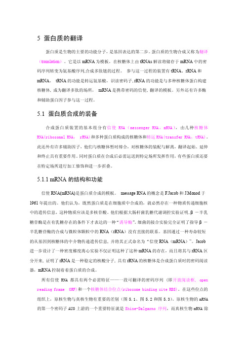
proteins,r-proteins)组成,rRNA 组成总分子量的 60%~65%。核糖体的相对大小常常用 沉降系数单位来表示。大肠杆菌的核糖体称为 70S 核糖体,其中的小亚基称为 30S 核糖体, 大亚基称为 50S 核糖体(图 5-8) 。小亚基包含 21 种不同的蛋白质(被称为 S1 一 S21)和 16SrRNA。大亚基由 33 种蛋白质(被命名为 L1~L33)和 23S 及 5SrRNAs 组成(图 5-9)。真核 核糖体称为 80S 核糖体,其中 40S 小亚基包含 33 种蛋白质和 18SrRNA,而 60S 大亚基包含 50 种蛋白质(图 5-9)和 3 种 rRNA (28S, 5.8S 和 5S) 。 真核 5.8SrRNA 与细菌 23SrRNA 的 5SrRNAs 部分同源(表 5.1) 。古细菌核糖体类似于细菌核糖体,但有些包含与真核相同的特别亚基。
组织上,原核生物与真核生物有重要的差别(图 5.1、图 5.2 和图 5.3) 。原核生物的 mRNA 的第一个密码子 AUG 上游的一个重要特征就是 Shine-Dalgarno 序列,而真核生物 mRNA 除
第一个密码子 AUG 的上游是核糖体小亚基扫描 AUG 的信号序列(CCACC)外, 5’端非翻译区 上游为帽子结构, 3’端非翻译区内有多聚腺苷化的信号 AAUAAA 以及其下游的多聚 A 尾巴。 mRNA 是由 DNA 的模板链转录而来, 其序列与编码链相同与模板链互补。 mRNA 的 5’ →3 ’ 三联体密码子序列与蛋白质 N 端到 C 端的氨基酸序列线形相关。原核生物 mRNA 的转录和翻 译发生在时间与空间上具有相对的同一性,其 mRNA 通常不稳定,在合成后的几分钟内翻译 成蛋白质。 真核 mRNA 的合成与成熟都在核内, 成熟的 mRNA 被运往胞质, 作为模板翻译蛋白 质 , 其 稳 定 性 相 对 较 高 , 达 几 小 时 。
蛋白质的翻译过程
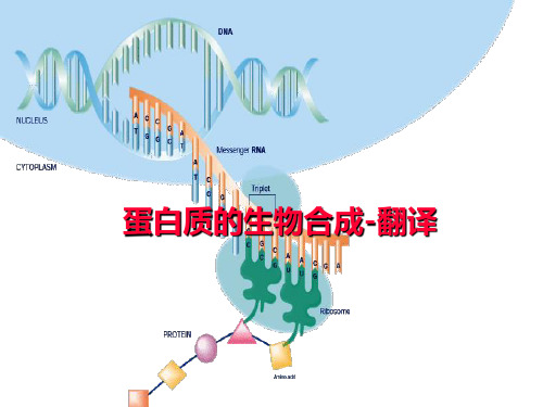
起始密码
➢肽链合成的起始
❖30s起始复合物形成
1.核糖体亚基的拆离
2.mRNA在小亚基上就位 3.fmet-tRNAfmet的结合
起始序列(SD 序列)
30S小亚基与mRNA识别、结 合
IF1、IF3协助 fmet-tRNAfmet -IF2-GTP 通 过
其反密码与mRNA上的起始密
码
AUG相配对
蛋白质的生物合成-翻译
分子生物学的中心法则(central dogma)
复制 DNA
RNA复制
转录
RNA
翻译
蛋白质
逆转录
2
翻译(蛋白质的生物合成)
蛋白质生物合成体系
➢以氨基酸为原料
➢以mRNA为模板 ➢以tRNA为运载工具 ➢以核糖体为合成场所
➢起始、延长、终止各阶段蛋白因子 参与合成后加工成为有活性蛋白质
❖氨基酰tRNA合成 酶
❖催化反应
❖氨基酰tRNA
氨基酰tRNA合成酶
A.A+特异tRNA
氨基酰tRNA
ATP AMP+PPi
氨基酸 பைடு நூலகம் ATP-E 氨基酰-AMP-E + PPi
氨基酰-AMP-E+tRNA 氨基酰tRNA+AMP+E (-COOH) (3’-CCA-OH)
16
2 、氨基酰tRNA合成酶 的高度专一性
➢核糖体与特异蛋白质、mRNA、tRNA的反应 部位
➢新技术 低温电子显微镜技术 中子散射技术
14
第二节 蛋白质合成的过程
原核生物 氨基酸的活化与转运 肽链合成的起始 肽链的延长 “核糖体循环” 肽链合成的终止 蛋白质的加工、修饰
蛋白质翻译()

CAA 翻译
在肝中剪接后的 mRNA 编码了 4563aa 的载脂蛋白
UAA 翻译
肠中的 mRNA 经编辑 产生了终止密码子,在 2153aa 处终止合成
图 13-42 载脂蛋白的基因 ApoB 在肠中经过编辑, 引入终止密码子,不能翻译成完整的载脂蛋白。 (参考 B.Lewin:《GENES》Ⅵ,1997,Fig31.14)
上次讲解内容
一、顺式作用元件与反式作用因子(重点) 二、真核生物RNA的转录过程 三、真核生物RNA转录后加工(重点) 1. 5’加帽; 2. 3’加尾; 3. 选择性剪接; 4. RNA编辑 四、RNA编辑
碱基的突变
C变为U
ApoB 基因有 29 个外显子
CAA
第 2153 个密码子编码 Glu 编辑
上次讲解内容
一、顺式作用元件与反式作用因子(重点) 二、真核生物RNA的转录过程 三、真核生物RNA转录后加工(重点) 四、RNA编辑
(一)转 录起始
TATA
TFIIH TFIIH
TFIIB
RNA-polⅡ的转录因子
因子 RNA pol II TFII D TFII A TFII B TFII H TFII E TFII F 功能 依赖模板合成RNA 通过TATA-box结合蛋白(TBP)识别TATA-box,与TATA-box 形成稳定复合物,使启动子进入转录起始状态中。 稳定TFII D与TATA-box的结合。 结合于TATA-box的下游,促使RNA酶组装到转录因子复合物内 磷酸激酶活性,可以使RNA酶II的CTD磷酸化,转化为高转录 活性的酶 ATPase,提供能量 解旋酶,解开DNA双螺旋
蛋白质翻译的全过程
一、蛋白质合成的装备
蛋白质翻译(translation)
蛋白质翻译ppt课件
氨基酸的活化形式:氨基酰-tRNA 氨基酸的活化部位:α-羧基 氨基酸与tRNA连接方式:酯键 氨基酸活化耗能:2个~P
33
(二)起始肽链合成的氨基酰-tRNA
真核生物:
Met
Met-tRNAi
原核生物: fMet-tRNAifMet
34
fMet-tRNAifMet的生成:
35
第二节
蛋白质生物合成过程
39
(一)原核生物翻译起始复合物形成
• 核蛋白体大小亚基分离; • mRNA在小亚基定位结合; • 起始氨基酰-tRNA的结合; • 核蛋白体大亚基结合。
40
1. 核蛋白体大小亚基分离
IF-1 IF-3
41
2. mRNA在小亚基定位结合
5' IF-3
AUG
IF-1
3'
42
S-D序列:
在原核生物mRNA起始密码AUG上 游,存在4~9个富含嘌呤碱的一致性序列, 如-AGGAGG-,称为S-D序列。又称为核 蛋白体结合位点(ribosomal binding site ,RBS)
48
• 肽链的延长是在核蛋白体上连续性循环式 进行,又称为核蛋白体循环(ribosomal cycle),每次循环增加一个氨基酸,分为 以下三步:
– 进位(entrance) – 成肽(peptide bond formation) – 转位(translocation)
49
肽链合成的延长因子
原核延 长因子
11
重叠密码
非重叠连续的密码 不连续的密码
12
基因损伤引起mRNA阅读框架内的碱 基发生插入或缺失,可能导致框移突变 (frameshift mutation)。
蛋白质翻译(外文版)
Accepting an aminoacyl-tRNA
Forming the peptidyl bonds
Releasing the deacylated tRNA
A site, P site and E site
Section 2
Protein Synthetic Process
General concepts
Section 1
Protein Synthetic System
Protein synthesis requires multiple elements to participate and coordinate.
• mRNA, rRNA, tRNA • substrates: 20 amino acids • Enzymes and protein factors:
• The complex of the GTP-bound IF-2 and the fMet-tRNA enters the P site.
Initiation 4
• The 50S subunit combines with this complex.
• GTP is hydrolyzed to GDP and Pi.
overlapping
Frameshift
2. Degeneracy
• Except Met and Trp, the rest amino acids have 2, 3, 4, 5, and 6 triplet codons.
• These degenerated codons differ only on the third nucleotide.
• A ribosome is composed of a large subunit and a small subunit, each of which is made of ribosomal RNAs and ribosomal proteins.
生命科学中蛋白质折叠和翻译的研究
生命科学中蛋白质折叠和翻译的研究蛋白质是生命中不可或缺的一种分子。
它们在机体内扮演着极为重要的角色,一个细胞中包含了数百万甚至上亿个蛋白质,其中每一个蛋白质都拥有着特定的生物学功能。
然而,蛋白质的构成并不是固定不变的,而是需要经过折叠和翻译的过程,这也是生命科学中极为关注的领域。
在本文中,我们将探讨生命科学中蛋白质折叠和翻译的研究。
蛋白质折叠蛋白质折叠是指蛋白质线性序列在空间中的构象变化,包括疏水效应、氢键、疏水键、离子键和范德华力等作用。
蛋白质在折叠的过程中,对于变化的构象非常敏感,即使是很小的构象变化也可能导致其生物学功能失调。
因此,蛋白质的折叠错误或失调可能引发众多疾病,如阿尔茨海默氏病、克罗恩病、帕金森病以及囊性纤维化等。
在蛋白质折叠研究方面,科学家们探索了许多方法来研究蛋白质的折叠过程,例如光学光谱学、核磁共振技术、X射线晶体学等技术手段。
此外,分子动力学模拟也成为了近年来蛋白质折叠研究的热点领域。
蛋白质翻译蛋白质翻译是指RNA通过载体mRNA所携带的信息,被翻译为蛋白质的过程。
蛋白质是由氨基酸组成的,翻译过程需要依赖着RNA、核糖体、tRNA等多种分子的协同作用。
蛋白质的翻译速度非常快,每秒钟可以合成数千个氨基酸。
翻译过程的可控性非常重要,翻译的准确性和速度直接影响蛋白质的功能和机能。
在蛋白质翻译的研究方面,科学家们主要关注着RNA的结构和作用,RNA是生命中重要的核酸之一,其在翻译过程中发挥着关键作用。
去年,美国加州大学圣克鲁兹分校(UC Santa Cruz)的科学家们发现,一类新的蛋白质——CCR4-Not复合物,可以调控编码RNA的稳定性和组装过程,这为RNA在生命中发挥作用提供了全新的视角。
结语蛋白质折叠和翻译是生命科学中极为关注的领域,它们的研究不仅涉及着生物学的基础研究,还直接关系到医学上的临床应用。
未来,我们相信新的技术手段和科学家们的不断努力,能够深化我们对蛋白质折叠和翻译这一重要生命现象的理解。
分子生物学课件第七章 蛋白质的生物合成-翻译

2020/10/28
38
2020/10/28
39
原核生物翻译过程中核蛋白体结构模式:
P位:肽酰位 (peptidyl site)
A位:氨基酰位 (aminoacyl site)
E位:排出位 (exit site)
2020/10/28
40
蛋白质生物合成体系
mRNA、tRNA、rRNA n 氨基酸
大多数简并性表现在密码子的第三个核苷酸上,即 第一、二个核苷酸确定后,第三个核苷酸可变。
色氨酸
意义: 简并密码子越多,生物遗传的稳定性越大,
氨基酸出现频率越高
2020/10/28
18
(3)摆动性(wobble)
转运氨基酸的tRNA的反密码需要通过碱基互补与 mRNA上的遗传密码反向配对结合,但反密码与密码间 不严格遵守常见的碱基配对规律,称为摆动配对。
氨基酰-tRNA + AMP +PPi
2020/10/28
31
(2)氨基酰tRNA合成酶(amino acyl-tRNA synthetase,AARS)
存在于胞液中,催化一个特定的aa结合到相应的 tRNA分子上。
每种氨基酰tRNA合成酶对相应氨基酸以及携带氨 基酸的数种tRNA具有高度特异性,保证tRNA能够 携带正确的氨基酸对号入座。
UUU,UUG,UGU,GUU, GGG,GGU,GUG,UGG。 • U和G随机加入到三联体中,这样按比例各个位置 上进入U和G的概率不同,如氨基酸测定结果:
2020/10/28
7
• 如UUU:UGG=(555):(511)
= 25 : 1
• 同理UUU:UUG =5 :1,
• 根据检测结果推测:
蛋白质生物合成及其调控
蛋白质生物合成及其调控蛋白质是构成生命的基本分子之一,是组织和细胞机能的重要组成部分,也是许多生物学过程的关键参与者。
蛋白质的生物合成不仅与基因信息的转录和翻译有关,还牵涉到分子水平的调控与控制。
本文将探讨蛋白质生物合成的基本原理和调控机制。
1.蛋白质生物合成的基本原理蛋白质生物合成需要通过一系列复杂的生化步骤来完成。
首先,DNA中的信息通过转录被转换成为mRNA,然后mRNA在核外被翻译成为氨基酸序列,最终组装成蛋白质。
这个过程涉及到多种分子,如核酸、酶、RNA和蛋白质,需要良好的协调和调控。
2.蛋白质生物合成的起始蛋白质的合成始于mRNA的转录。
在DNA上,RNA聚合酶会匹配到一个启动子区域,开始转录过程。
在真核细胞中,mRNA合成的模板是DNA,而在原核生物中,mRNA合成的模板则是DNA或RNA。
转录是一个高度调控的过程,其中一些转录因子、启动子和辅助因子起到了关键作用。
这些因子的缺失或异常会导致错误的信息转录,进而影响蛋白质的合成。
3.蛋白质的翻译转录后的mRNA离开细胞核,进入细胞质后被翻译成为蛋白质。
这个过程是依靠核糖体完成的,核糖体是由多种RNA和的蛋白质组成的。
核糖体可以在mRNA上滑动,从而识别序列并连接氨基酸。
蛋白质翻译的过程受到多种调控,如启动子、辅助因子和mRNA的稳定性等。
4.蛋白质的折叠和组装蛋白质完成翻译之后,需要进行折叠和组装才能成为功能完整的蛋白质。
折叠是一个高度复杂的过程,其中一些辅助因子和分子可以起到关键作用。
同时,蛋白质折叠的错误或异常将导致其失去正常的功能,并可能导致疾病的发生。
组装则是将不同的蛋白质组合成为功能复杂的蛋白质结构。
这个过程也受到多种调控。
5.蛋白质生物合成的调控蛋白质生物合成的过程涉及多种调控机制。
在转录的过程中,转录因子、启动子和辅助因子可以正或负地调节转录的速率和准确性。
在翻译的过程中,mRNA和核糖体的结合也可以受到多种调控。
此外,细胞还可以通过调节折叠和组装过程来影响蛋白质的迅速和精确性。
蛋白质翻译过程
蛋白质翻译过程
蛋白质翻译是核酸到蛋白质的转录-翻译过程,即将mRNA中的信息转录成蛋白质的过程。
翻译是一个复杂的过程,它包括mRNA的扩增、起始密码子的检测、tRNA的折叠、tRNA的定位、引物的消耗、转录末端的处理、抗性的表达、表达子的结构变化和其他一些步骤。
1. mRNA的扩增:mRNA由转录机在DNA的模板上进行扩增,从而产生新的mRNA。
2. 起始密码子的检测:mRNA中的起始密码子被检测到,识别出蛋白质合成应开始的位置。
3. tRNA的折叠:tRNA被折叠成特定的三维结构,使其能够被转录机识别。
4. tRNA的定位:折叠的tRNA被定位到转录机前端,它可以结合mRNA并且将配对的氨基酸送入到转录机上。
5. 引物的消耗:在翻译过程中,引物会被消耗掉。
6. 转录末端的处理:转录末端会被处理,以便进行蛋白质合成。
7. 抗性的表达:翻译过程中,抗性基因会被表达出来,使得细胞对抗性物质有抵抗力。
8. 表达子的结构变化:表达子的结构会随着翻译过程而变化,从而可以调节翻译速率。
- 1、下载文档前请自行甄别文档内容的完整性,平台不提供额外的编辑、内容补充、找答案等附加服务。
- 2、"仅部分预览"的文档,不可在线预览部分如存在完整性等问题,可反馈申请退款(可完整预览的文档不适用该条件!)。
- 3、如文档侵犯您的权益,请联系客服反馈,我们会尽快为您处理(人工客服工作时间:9:00-18:30)。
ProteinsLu Linrong (鲁林荣)PhDLaboratory of Immune Regulation Institute of Immunology Zhejiang University ,School of Medicine Medical Research Building B815-819Email: Lu.Linrong@ Website: /llrMolecular BiologyWhy study proteins?•Part of the central dogma•Proteins are coded by genes•They play crucial functionalroles in almost everybiological processThe life cycle of a protein•Where does a protein come from?•How is a protein processed, modified, translocated to the proper place and degraded?•How to describe theare the functions?••Protein synthesis (Translation) 蛋白质翻译•Protein maturation (folding, modification) and degradation 蛋白质成熟,降解Structure and function of protein 蛋白质的结构与功能•Methods: protein-protein interaction et al 蛋白-蛋白相互作用Protein SynthesisReference and further readings: Chapter 30: Protein Synthesis, Biochemistry, Reginald H.Garrett & Charles M. GrishamChapter 8: Protein Synthesis, Gene IX, Lewin Benjamin Internet Tools: Google, Wiki ……What is translation?--it is the story about decoding the genetic information contained in messenger RNA (mRNA) into proteins.Translation is a big issue for a living cell In rapid growing bacterial cells, protein synthesis consumes80% of the cell’s energy50% of the cell’s dry weightRNAs as both template and machienary:1.mRNAs(~5% of total cellular RNA)2.tRNAs (~15%)3.aminoacyl-tRNA synthetases (氨酰tRNA合成酶)4.ribosomes(~100 proteins and 3-4 rRNAs--~80%)The genetic code is the set of rules by which information encoded in genetic material (DNA or mRNA sequence) is translated into proteins (amino acid sequences) by living cells.The code defines how sequences of three nucleotides, called codons , specify which amino acid will be added next during protein synthesis.Genetic CodeGeorge Gamow (Russian-born theoretical physicist) postulatedthat a three-letter code must be employed to encode the 20standard amino acids used by living cells to encode proteins.With four different nucleotides, a code of 2 nucleotides couldonly code for a maximum of 42or 16 amino acids. A code of 3nucleotides could code for a maximum of 43or 64 amino acids.The Crick, Brenner, Barnett, Watts-Tobin experiment of 1961 demonstrated that three bases of DNA code for one amino acid in the genetic code. The experiment elucidated the nature of gene expression and frameshift mutations.In the experiment, proflavin-induced mutations of the T4 bacteriophage gene, rIIB, were isolated. Proflavin causes mutations by inserting itself between DNA bases, typically resulting in insertion or deletion of a single base pair.The mutants produced by Crick and Brenner could not produce functional rIIB protein because the insertion or deletion of a single nucleotide caused a frameshift mutation. Mutants with two or four nucleotides inserted or deleted were also nonfunctional. However, the mutant strains could be made functional again by using proflavin to insert or delete a total of three nucleotides. This proved that the genetic code uses a codon of three DNA bases that corresponds to an amino acid.The Crick, Brenner, Barnett, Watts-Tobin experiment of 1961The first elucidation of a codon was done by Marshall Nirenberg(sit) and Heinrich J. Matthaei (stand) in 1961 at the National Institutes of Health. They used a cell-free system to translate a poly-uracil RNA sequence (i.e., UUUUU...) and discovered that the polypeptide that they had synthesized consisted of only the amino acid phenylalanine.Similar approaches were taken to decode other codes.Genetic CodeGenetic Code•Start codon: AUG-methionine•Stop codon: UGA, UAA, UAG •Degeneracy: 61 triplets code for 20 amino acids •UniversalProtein Synthesize Machinery•mRNA: the template•tRNA: the amino acid carrier•Ribosome (rRNA &ribosomal proteins): thetranslation apparatus withcatalyzing activityMessenger RNA:Transcribed from DNA and Carries the genetic information out of the nucleus into cytoplasm for protein synthesistRNA•tRNA contains 60-95 nt, commonly 76.•Deliver amino acids to the translational complex.•Binds to mRNA through 3-base anticodon complementary to codon in mRNA.•In bacteria, there are 30-40 tRNAs with different anticodons.In animal and plant cells, about 50 different tRNAs are found. (Coden preference in bacteria and human)•"Wobble" during reading of the mRNA allows some tRNAs to read multiple codons that differ only in the 3rd base.tRNA secondary structure (cloverleaf )•tRNA secondary structureconsistis of stem& loopdomains.•Double helical stem domainsarise from base pairingbetween complementarystretches of bases within thesame strand.•Loop domains occur wherelack of complementarity or thepresence of modified basesprevents base pairing.tRNA tertiary structure (L-shaped)•RNA tertiary structuredepends on interactionsof bases at distant sites.•These interactionsgenerally involve non-standard base pairingand/or interactionsinvolving three or morebases.•Unpaired adenosinespredominate inparticipating in non-standard interactionsthat stabilize tertiary RNAstructures.Synthesis of Aminoacyl-tRNAClass I Class IICodon-Anticodon Recognition Involves WobblingAnti-parallelThe Components of RibosomeRibosome structure----2009 Nobel PrizeThe Components of RibosomeThe Components of RibosomeTwo-dimensional polyacrylamide gel analysis of ribosomal proteins extracted from 50 S subunitsWower I K et al. J. Biol. Chem. 1998;273:19847-19852Structure of RibosomeStructure of Ribosome http://www.molgen.mpg.de/~ag_ribo/ag_franceschi/franceschi-projects-30S-2.htmlhttp://www.molgen.mpg.de/~ag_ribo/ag_franceschi/franceschi-projects-50S-2.html 30 S subunit 50S subunitRibosome from D. radiodurans (耐辐射球菌)Ribosome Has Several Active CentersA site(acceptor site): exposes thecodon representing the next aminoacid due to be added to the chain.P site(donor site): nascentS1polypeptide chain lies. The codonrepresenting the most recent addedamino acid.E site: a temporary tRNA-binding site.S1 site: has a strong affinity forsingle-stranded nucleic acid, and isresponsible for the initial binding of30S subunits to mRNA and initiatortRNA .Protein Synthesis Occurs by Initiation, Elongation, and Termination•Protein synthesis falls into thethree stages: Initiation,Elongation, Termination.•Different sets of accessory factorsassist the ribosome at each stage.•Energy is provided at variousstages by the hydrolysis ofguanine triphosphate (GTP).•Initiation Involves Base PairingBetween mRNA RBS(ribosome binding site) and rRNA •Initiation Needs Accessory Factors (Initiation Factors, IFs)• A Special Initiator tRNA (fMet-tRNAf) Starts the Polypeptide Chain•Small Subunits Scan for Initiation Sites on Eukaryotic mRNA• A rate-limiting stepCritical event:begin protein synthesis at the start codon, thereby setting the stage for the correct in-frame translation of the entire mRNA.Main mechanisms:Base pairing between mRNA and rRNABase pairing between mRNA and tRNAfMet-tRNA i Met can only bind at the P site to begin synthesis Participants:fMet-tRNA i MetmRNAIFssmall subunitlarge subunitInitiation Involves Base Pairing Between mRNAand rRNA•An initiation site on bacterialmRNA consists of the AUGinitiation codon preceded with agap of -10 bases by the Shine-Dalgarno polypurine hexamer (SDsequence).•The rRNA of the 30S bacterialribosomal subunit has acomplementary sequence thatbase pairs with the SD sequenceduring initiation.Small Subunits Scan for Initiation Sites onEukaryotic mRNA•Eukaryotic 40S ribosomal subunits bind to the 5' end of mRNA and scan the mRNA until they reach an initiation site.•The eukaryotic initiation site consists of a ten nucleotide sequence that includes an AUG codon. •60S ribosomal subunits join the complex at the initiationsite.Cozak sequence•Cozak sequence: NNN(A/G)NN AUG G•IRES (Internal ribosome entry site): Independent of 5’cap; co-expression of two proteinsCozak sequence vsIRESCozak sequence vs IRESInitiation in bacteria needs 30S subunits andaccessory factorsInitiation factors (IF in prokaryotes, eIFin eukaryotes) are proteins that associatewith the small subunit of the ribosomespecifically at the stage of initiation ofprotein synthesis.They are released when the 30S associatewith 50S subunit to form 70S ribosome.Bacterial rRNA plays a direct role in recruitingthe small ribosomal subunit to a translationstart site on the mRNA:Shine-Dalgarno sequence is complementaryto the 3 terminus of the 16S rRNA.IF2 has a ribosome-dependent GTPase activity.Energy is needed to form an active ribosome.The Function of Initiation Factors in Bacteria Initiation factors: IF-1, IF-2, and IF-3.IF-3 is needed for 30S subunits to bind specifically to initiation sites in mRNA.IF-2binds a special initiator tRNA and controls its entry into the ribosome.has a ribosome-dependent GTPase activityIF-1binds to 30S subunit in the A site as a part of the complete initiation complex. Associates Prevents an aminoacyl-tRNA from entering. Modulates IF2 binding to the ribosome.A Special Initiator tRNA Starts the PolypeptideChainThe initiator N-formyl-methionyl-tRNA(fMet-tRNAf) is generated by formylation(甲酰化) of methionyl-tRNA, using formyl-tetrahydrofolate (四氢叶酸脂) as cofactor.fMet-tRNAf has unique features thatdistinguish it as the initiator tRNA.Protein Synthesis ElongationProtein Synthesis Elongation Essentials:1)mRNA: 70S ribosome: peptidyl-tRNAcomplex1)Aminoacyl-tRNAs2)Elongation factors3)GTPThe ribosome has three tRNA-binding sites.An aminoacyl-tRNA enters the A site.Peptidyl-tRNA is bound in the P site.Deacylated tRNA exits via the E site.An amino acid is added to the polypeptide chain by transferring the polypeptide from peptidyl-tRNA in the P site to aminoacyl-tRNA in the A site.The Process of ElongationElongation Steps:1.Binding of incoming aminoacyl-tRNAat A site.2.Peptide bond formation:transfer ofthe peptidyl chain from the tRNAbearing it to the –NH2 group of thenew AA.3.Translocation of the one-residue-longer peptidyl-tRNA to the P siteElongation Factor Tu Loads Aminoacyl-tRNA intothe A Site•EF-Tu is a monomeric Gprotein whose active form(bound to GTP) bindsaminoacyl-tRNA.•The EF-Tu-GTP-aminoacyl-tRNA complex binds to theribosome A site.The Polypeptide Chain Is Transferred toAminoacyl-tRNA•The 50S subunit (mainly 23SrRNA) has peptidyltransferase activity.•The nascent polypeptidechain is transferred frompeptidyl-tRNA in the P siteto aminoacyl-tRNA in the Asite.•Peptide bond synthesisgenerates deacylated tRNAin the P site and peptidyl-tRNA in the A site.Translocation Moves the Ribosome•The hybrid state modelproposes that translocationoccurs in two stages:•First, the 50S moves relativeto the 30S;•Second, the 30S movesalong mRNA to restore theoriginal conformation.Translocation requires EF-G•EF-G is a G protein whichresembles the aminoacyl-tRNA-EF-Tu-GTP complex.•Binding of EF-Tu and EF-G tothe ribosome is mutuallyexclusive.•EF-G binds to the ribosome tosponsor translocation.•The hydrolysis of GTP is neededto release EF-G.PolyribosomesActive protein-synthesizing units consist of an mRNA with several ribosomes attached to it. Such structures are polyribosomes or polysomes.Termination •The codons UAA, UAG , and UGAterminate protein synthesis.•Termination codons are recognized by protein release factors (RF).•The structures of the class 1 release factors resemble aminoacyl-tRNA-EF-Tu and EF-G.•The class 1 release factors respond to specific termination codons and hydrolyze the polypeptide tRNA linkage.The Process of Termination•The RF (release factor)terminates proteinsynthesis by releasing theprotein chain.•The RRF (ribosome recyclingfactor) releases the lasttRNA.•EF-G releases RRF, causingthe ribosome to dissociate.Movie Here!Eukaryotic Translation InitiationEukaryotic 48s initiation complexRegulation of Eukaryotic Translation InitiationRegulation of initiation factor 4ERegulation of initiation factor eIF-2Regulation by microRNAsRegulation of initiation factor 4E4E-BP binds to 4E as a limitingstep for initiationGrowth signals activate mTOR,which will in turn phosphorylate4E-BPPhosphorylated 4E-BPdissociates from 4EFree 4E assemble with othereIF4 members and bind to theCap of mRNATranslation initiatedStress/Starvation will block theactivation of mTOR pathwayRegulation of initiation factor 4E•Closely related tometabolic andenvironmental status•eIFs•eIF-binding protein: 4E-BP1Cell Signaling Technology。
