显微共焦拉曼光谱ppt课件
合集下载
显微共聚焦拉曼光谱

显微共聚焦拉曼光谱
显微共聚焦拉曼光谱(confocal Raman spectroscopy)是一种分析技术,它可用于诊断某一物质的成分,以及检测生物材料表面的化学成分。
它利用共聚焦拉曼散射(CRDS)技术,将激光束集中到采样表面上。
此技术不仅可用于研究三维物体的化学结构,而且可以用于构建显微共聚焦图像,并研究表面的化学成分分布。
显微共聚焦拉曼光谱通常由四个主要组成部分组成,分别是激光源、光学系统、数据收集系统和分析系统。
激光源将激光束集中到指定的采样表面上,而光学系统可以调节激光束的尺寸和强度,从而获得良好的数据质量。
数据收集系统通过一个光电探测器来获取扫描区域的拉曼信号,而分析系统则通过计算机程序对这些信号进行分析。
显微共聚焦拉曼光谱技术使科学家可以以更快的速度来进行复杂物质的密度动力学研究,并获得更清晰的结构信息。
它是实现多尺度研究的重要工具,将大尺度的性质(包括多维表面分布)与小尺度的性能(包括原子结构)结合起来。
显微共聚焦拉曼光谱可以迅速地获取表面化学结构和缺陷的扫描,因此可以有效地消灭大量的假设并准确的引导实验研究。
拉曼光谱原理和特点 ppt课件
• 散射光中的1010光子之一是非弹性散射(拉曼)
• 前…
后…
入射光
分子
• 光损失能量,使分子振动
Slide 4
PPT课件
分子振动
散射光
emission
excitation excit.-vib.
拉曼光谱的优点和特点
对样品无接触,无损伤; 样品无需制备; 快速分析,鉴别各种材料的特性与结构; 能适合黑色和含水样品; 高、低温及高压条件下测量; 光谱成像快速、简便,分辨率高; 仪器稳固,体积适中, 维护成本低,使用简单。
2500
N2
2000
1500
1000
500
1500
2000
2500
3000
3500
CO2
CH4
6000
4000
quartz
3000
H2O
2000 1087
1000
1164 1387 1280
1640
2331
1500
2000
2500
3000
3500
1087 1164
1287 1390
2328 2609 2914 3399 3639
Characteristic vibrational spectrum: 指纹性振动谱
Slide 6
PPT课件
Information obtained from Raman spectroscopy 拉曼光谱的信息
Slide 7
PPT课件
characteristic Raman
frequencies
拉曼频率的确认
changes in frequency of Raman peak
简述拉曼光谱在PPT资料(正式版)

傅立叶变换拉曼光谱技术较旧式拉曼光谱 分析技术,有很大的提高.
一束光
相干仪
两束相同的光
( 一束滞后,光程差为d)
正相干:d=n倍波长
负相干: d=(2n+1)/2倍波长
一种理想的单色光通过相干仪,由于滞后 现象不断的有规律地变化,检测到的信号将是 一个余弦波,这些余弦的总和,就是相干图象。 相干图象实际上是光强度的函数,已知在相干 仪中可移动的镜面,是一个合适的时间范围内 的移动,所以相干图象也是一个时间的函数. 对于光强和时间函数关系所表示的频率分析 过程,是一个纯数学分析过程,也就是” 傅立 叶变换”,它将时间域中的相干图象转化为频 率域中的光谱图象(见图1)。
I
FT
共振拉曼光谱定量分析技术 拉曼光谱定量分析据为:
(a)由一个单色光产生的相干图象 胡继明 胡军(武汉大学分析测试科学系)
拉曼光谱分析技术是以拉曼效应为基础建立起来的分子结构表征技术,与红外光谱相同,其信号来源与分子的振动和转动.
I 图1 相干图象的产生
(b)由两个单色光产生的相干图象 还应注意的是任何一物质的引入都会对被测体系带来某种程度的污染,这等于引入了一些误差的可能性,会对分析的结果产生产生一 定的影响。
(四)几种重要的拉曼光谱分析技术
1.单道检测的拉曼光谱分析技术 2.以CCD为代表的多通道探测器用于拉曼光谱
的检测仪的分析技术 3.采用傅立叶变换技术的FT-Raman光谱分析
技术 4.共振拉曼光谱定量分析技术 5.表面增强拉曼效应分析技术 6.近红外激发傅立叶变换拉曼光谱技术
二.傅立叶变换拉曼光谱和近红外激发 傅立叶变换拉曼光谱新技术的简述
简述拉曼光谱在
一. 拉曼光谱分析的依据和特点
拉曼光谱分析技术是以拉曼效应为基础建 立起来的分子结构表征技术,与红外光谱相同, 其信号来源与分子的振动和转动.
《拉曼光谱》PPT课件

②拉曼活性振动 诱导偶极矩 = E 非极性基团,对称分子;
拉曼活性振动—伴随有极化率变化的振动。 对称分子: 对称振动→拉曼活性。 不对称振动→红外活性
15
4. 红外与拉曼谱图对比
红外光谱:基团; 拉曼光谱:分子骨架测定;
16
红外与拉曼谱图对比
17
选律
1 S C S
拉曼活性
2 S C S
红外活性
1
拉曼散射效应的进展:
拉曼散射效应是印度物理学家拉曼(C.V.Raman)于1928年首次发现 的,本人也因此荣获1930年的诺贝尔物理学奖。
1928~1940年,受到广泛的重视,曾是研究分子结构的主要手段。这 是因为可见光分光技术和照相感光技术已经发展起来的缘故;
1940~1960年,拉曼光谱的地位一落千丈。主要是因为拉曼效应太弱 (约为入射光强的10-6),并要求被测样品的体积必须足够大、无色、 无尘埃、无荧光等等。所以到40年代中期,红外技术的进步和商品化 更使拉曼光谱的应用一度衰落;
Stocks lines
anti-Stockes lines
Δν/cm-1
12
2、产生拉曼位移的条件
拉曼散射不要求有偶极矩的变化,却要求有极 化率的变化,与红外光谱不同,也正是利用它 们之间的差别,两种光谱可以互为补充。 分子在静电场E中所产生的诱导偶极矩P与分子 的极化率α之间有关系:P=αE
13
9
激光拉曼光谱基本原理
principle of Raman spectroscopy
激发虚态
h(0 - )
Rayleigh散射:
E1 + h0
弹性碰撞;无 能量交换,仅改变 方向; Raman散射:
非弹性碰撞; 方向改变且有能量
《拉曼光谱分析法》PPT课件
为了有效收集从小体积发
出的拉曼辐射,多采用一 个90度(较通常)或180 度的试样光学系统。
多重反射槽
图6-34 各种形态样品在拉曼光谱仪中放置方法
(a) 透明固体 (b) 半透明固体 (c) 粉末 (d) 极细粉末 (e) 液体 (f) 溶液
2021/4/25
1—反射镜 2—多通道池 3—锲型镜 4—液体
去偏振度) 表征分子对称性振动模式的高低。
I
I∥
(6-17)
式中I⊥和 I// ——分别代表与激光电矢量相垂直和相平行的谱线的强度。
<3/4的谱带称为偏振谱带,表示分子有较高的对称振动模式; =
3/4的谱带称为退偏振谱带,表示分子的对称振动模式较低,即分子是不对称
的2。021/4/25
8
激光拉曼散射光谱法
拉曼位移:斯托克斯线或反斯托克斯线与入射光频率之差
称为拉曼位移。拉曼位移的大小和分子的跃迁能级差一样。 因此,对应于同一分子能级,斯托克斯线与反斯托克斯线的 拉曼位移应该相等,而且跃迁的几率也应相等。在正常情况 下,由于分子大多数是处于基态,测量到的斯托克斯线强度 比反斯托克斯线强得多,所以在一般拉曼光谱分析中,都采 用斯托克斯线研究拉曼位移。
拉曼位移的大小与入射光的频率无关,只与分子的能级结 构有关,其范围为25~4000cm-1。因此入射光的能量应大于 分子振动跃迁所需能量,小于电子能跃迁的能量。
2021/4/25
6
拉曼散射光谱的基本概念
红外吸收要服从一定的选择定则,即分子振动时 只有伴随分子偶极矩发生变化的振动才能产生红外 吸收。同样,在拉曼光谱中,分子振动要产生位移 也要服从一定的选择定则,也就是说只有伴随分子 极化度α发生变化的分子振动模式才能具有拉曼活性, 产生拉曼散射。极化度是指分子在电场的作用下, 分子中电子云变形的难易程度,因此只有分子极化 度发生变化的振动才能与入射光的电场E相互作用, 产生诱导偶极矩::
拉曼光谱原理和特点 ppt课件

diamond grains
晶体对称性和取向
quality of crystal
晶体质量
e.g. amount of plastic deformation
amount of material
物质总量
e.g. thickness of transparent coating
拉曼光谱的特点和主要困难
• 拉曼散射信号弱(比荧光光谱平均小2-3数量级)。 • 激光激发强。 • 拉曼信号频率离激光频率很近。 • 激光瑞利散射比拉曼信号强1010-1014,对拉曼信号干扰很大。 • 拉曼光谱仪器的设计,必须能排除瑞利散射光,并具有高灵敏度(体现在弱信号检
测的高信噪比 ),才能有效地收集拉曼谱。
Slide 8
composition of material
物质的组成
stress/strain State 张力 / 应力
e.g. MoS2, MoO3
e.g. Si 10 cm-1 shift per % strain
crystal symmetry and e.g. orientation of CVD
orientation
Slide 15
PPT课件
高稳定性、高重复性
圆形编码器控制的光栅转动台
– 技术
• 直接测量转动角度,同时编码器精 密伺服控制其转动,而非采用计量 马达转过多少圈的办法
• 确保光栅转动的精确性和重复性。
Slide 16
PPT课件
*grating and wavelength dependent
高重复性、高稳定性
D Frequency cm-
1
Arbitrary Y
de 18
课件:拉曼光谱
4 包裹体:
矿物中的包 裹体成分的鉴 定。
玻璃中的包 裹体(气泡) 成分的鉴定。
5.7.3 拉曼图谱的表示方法
横坐标: 拉曼位移(Raman Shift),以波数(cm-1)
表示。
Δν=| ν入–ν散 |=ΔE/h
纵坐标: 拉曼(散射)强度,以(Raman Intensity)
表示。
CaCO3 的Raman图谱
Raman Intensity
140000
120000
100000
拉曼谱带, 随单键双键三键谱带强度增加。 2)CN,C=S,S-H伸缩振动在拉曼光谱中是强谱带。 3)环状化合物的对称呼吸振动常常是最强的拉曼谱
带。 4)在拉曼光谱中,X=Y=Z,C=N=C,O=C=O-类键的对
称伸缩振动是强谱,反对称伸缩振动是弱谱带。
5)C-C伸缩振动在拉曼光谱中是强谱带。
6)醇和烷烃的拉曼光谱是相似的: I. C-O键与C-C键的力常数或键的强度
Δν/cm-1
三 拉曼位移(Raman shift)
Δν=| ν入–ν散 |=ΔΕ/h
即入射光(激发光)频率与散射光 频率之差,只与能级差有关。
与入射光波长无关 适用于分子结构分析
四 拉曼光谱与分子极化率
1 分子的极化
在外电场作用,分子变形产生诱导偶极 矩或增大永久偶极矩的现象。 分子的变形:
正电中心与负电中心发生位移(由重合变 为不重合,由偶极长度小变偶极长度大) 。
3 珠宝
鉴定和分析真假宝石(如钻石,石英,红 宝石,绿宝石等)以及对珍珠、玉石及其他珠 宝产品进行分析。
手 镯
100000 80000
1316.89 1589.77
Intensity
《共聚焦拉曼光谱仪》课件

化学反应监测
共聚焦拉曼光谱仪可以实时监测 化学反应过程中物质的变化,有 助于理解反应机理和反应动力学 。
污染物检测
共聚焦拉曼光谱仪能够检测痕量 污染物,如重金属、有机污染物 等,对环境监测和污染治理具有 重要意义。
在生物医学研究中的应用
细胞成像
生物分子相互作用研究
共聚焦拉曼光谱仪能够实现细胞的高分辨 率成像,有助于研究细胞结构和功能。
特点
控制系统是实现智能化和 自动化的关键部分。
Part
03
共聚焦拉曼光谱仪的性能特点
高分辨率
STEP 02
STEP 01
共聚焦拉曼光谱仪采用先 进的共聚焦光学系统,能 够实现高分辨率的拉曼散 射信号采集。
STEP 03
提高了对复杂样品和混合 物的鉴别能力,有助于深 入了解样品的性质和组成 。
高分辨率使得光谱分辨率 更高,能够更好地解析出 样品的分子结构和振动模 式。
定制化服务
国际化合作与交流
加强国际间的技术合作与交流,推动 共聚焦拉曼光谱仪技术的不断创新和 发展。
针对不同行业和应用领域的需求,共 聚焦拉曼光谱仪将提供定制化的解决 方案,满足客户的个性化需求。
THANKS
感谢您的观看
特点
光学系统是共聚焦拉曼光 谱仪的核心部分,其性能 直接影响整个仪器的性能 和稳定性。
共聚焦系统
STEP 01
组成
STEP 02
作用
由透镜和反射镜组成,用 于将激发光聚焦到样品上 ,并收集拉曼散射信号。
STEP 03
特点
共聚焦系统是实现高空间 分辨率和高灵敏度的关键 部分。
将激发光聚焦到样品上, 以提高激发效率和拉曼散 射信号的收集效率。
《拉曼光谱分析》PPT课件
优势:激发波长较长, 可以避免部分荧光产生
局限:黑色样品会产生热背景
>1m
薄膜样品的厚度应
23
光谱范围:5~4000cm-
分析方法
普通拉曼光谱 一般采用斯托克斯分析
反斯托克斯拉曼光谱 采用反斯托克斯分析
24
Raman光谱可获得的信息
Raman 特征频率
Raman 谱峰的改变
Raman 偏振峰
化学反应形成界面层,增强化学结合 物理扩散形成界面层,增强物理结合力
49
ACP(%)
ACP (%)
100
Cr
C
50
0 0
50
O
Cr
C
O
2
4
6
8
sputtering time / min
100 depth profile lines
Cr as Cr2C C as diamond
C as Cr2C
C
Cr 50
原位变温附件
适于分析随温度变化发生的: 相变 形变 样品的降解 结构变化
31
温度范围: 液氮温度(-195℃) 至1000℃
自动设置变温程序
样品制备
溶液样品 一般封装在玻璃毛细管中测定
固体样品 不需要进行特殊处理
32
材料分析中的应用
无机化学研究 无机化合物结构测定,主要利用 拉曼光谱研究无机键的振动方式, 确定结构。
如纳米Ag,Au胶颗粒吸附染料或有机物质,其检测灵敏度可以提高105~109量 级。可以作为免疫检测器。
29
紫外拉曼光谱
为了避免普通拉曼光谱的荧光作用,使用波长较短的紫外激光光源,可以使 产生的荧光与散射分开,从而获得拉曼信息。适合于荧光背景高的样品如催 化剂,纳米材料以及生物材料的分析。
激光共聚焦显微拉曼光谱仪PPT课件

操作步骤
1、打开电脑。 2、开机:插上电源插头,将Laser至On,长按power打开仪器,power蓝色灯亮。 3、打开OMNIC软件,点击实验设置,在光学台下打开激光,激光预热,预热完毕后,出现对话框,点击 休息两次,此时激光灯亮,点击实验设置中ok,实验设置完毕。 4、准直聚焦:如果仪器换激光器,如532nm换成780nm或仪器移动需做准直,方法选择10倍物镜,然后 22目观察,在x轴和y轴方向上移动光学台,旋动至黄色小光斑在十字交叉处。 5、校准仪器(检查光斑是否在中心):如果光斑不在中心,可以通过移动光学台,使光斑在中心。 6、采集样品,采集完成后,点击文件另存为,在图谱文件CVS下保存,采样完成。 7、关机:点击实验设置,在光学台下关闭激光,将Laser至off,长按power关闭仪器,power熄灭,拔 下电源插头,关闭OMNIC软件,关闭电脑。
3、显微镜厂家原装透射、反射照明。附送备用照明灯2个。
4、自动XYZ平台,最小步长不大于0.1 um,可进行分散的多点、 线、面扫描和共焦深度的扫描。系统软件能帮助自动聚焦。系 统无反向间隙,能保证位置原始点的良好重复性。
5、采用真共焦光路设计,空间分辨率方面,100X物镜下,xy 分辨率 <= 1 um ,z轴方向分辨率<= 2微米,共焦深度连续可 调。
4、为适应不同样品测量要求以及防止激光功率过高烧坏样品, 要求激光输出功率可调。同时,激光光斑尺寸可调。
共焦显微镜
1、专业的高端科研型显微镜,10X原装目镜,20X、50X、100X、 长焦50X物镜, 包括可同时安装5个镜头的镜头架。其中长焦 50X的焦距都要求大于或接近10mm。
2、彩色摄像机。
写在最后
经常不断地学习,你就什么都知道。你知道得越多,你就越有力量 Study Constantly, And You Will Know Everything. The More
拉曼光谱基本原理通用课件

峰形与对称性 拉曼光谱的峰形反映了散射过程的对称性。对称 性越高,峰形越尖锐;对称性越低,峰形越宽钝。
CHAPTER
内标法与外标法
内标法
通过在样品中添加已知浓度的标准物质作为内标,利用内标物的峰面积或峰高与浓度之间的线性关系,对未知样 品的浓度进行定量分析。
外标法
通过比较已知浓度的标准样品的拉曼光谱与未知样品的拉曼光谱,根据标准样品的浓度和峰面积或峰高之间的关 系,对未知样品的浓度进行定量分析。
峰面积法与峰高法
要点一
峰面积法
通过测量拉曼光谱中某个峰的面积,利用峰面积与浓度之 间的线性关系,对未知样品的浓度进行定量分析。
要点二
峰高法
通过测量拉曼光谱中某个峰的高度,利用峰高与浓度之间 的线性关系,对未知样品的浓度进行定量分析。
多元光谱分析方法
• 多元光谱分析方法:利用多个拉曼光谱之间的信息,通过统计分析和数学建模,对未知样品的成分和浓度进行 定量和定性分析。例如偏最小二乘法、主成分分析法等。
拉曼散射的物理过程
当光波与介质分子相互作用时,光波吸收或释放能量,导致光波的频率发生变 化。这种变化遵循斯托克斯-拉曼散射定律。
拉曼光谱的峰位与峰强
1 2 3
峰位的确定 拉曼光谱的峰位表示散射光的频率变化,通常用 波数(cm^-1)或波长(nm)表示。
峰强的意义 峰强表示散射光的强度,反映了散射过程的概率 大小。一般来说,峰强越强,表示该频率的散射 过程越容易发生。
拉曼光谱的未来
随着技术的不断进步,拉曼光谱将在未来发挥更 加重要的作用。
拉曼光谱的基本原理及特点
拉曼光谱基本原理
拉曼光谱是一种基于光的散射效 应的技术,通过分析散射光的频 率和强度来推断样品的性质。
拉曼分析测试技术PPT课件
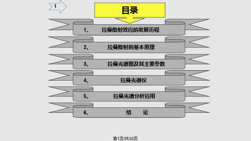
第26页/共32页
27
C)SiC 晶型分布
-25 000
-20 000
-15 000
-10 000
-5 000
Y (祄)
0
5 000
10 000
15 000
20 000 25 000
2000 祄
-20 000
-10 000
0 X (祄)
10 000
20 000
图5-3 SiC 晶型分布图
第27页/共32页
图5-4 不同波长激发波测得的不 同深度的Si薄膜的拉曼光谱图
在制备非晶硅或多晶硅薄过程中, 不同深度处的晶化程度可能不同。利 用不同波长激光在样品中穿透深度不 同,得到各深度层的信息。该样品表 面为多晶硅,往深度方向晶化程度降 低,逐渐变为非晶硅。
第28页/共32页
29
5-2 定量分析
a)蓝宝石衬底上的GaN的应力分布
三原子分子情况——三种振动模式:对称伸缩、弯曲变形 和不对称伸缩。
H2O
CO2
图3-3 三原子分子情况下三种振动模式图
11
第11页/共32页
12
4、拉曼光谱仪
4-1 拉曼光谱仪测量原理
探测器
光栅
滤光片
激光
样品
图4-1 拉曼光谱仪测量基本原理示意图
第12页/共32页
13
激光Raman光谱仪 激光光源:He-Ne激光器,波长632.8nm;
• 得到更好的横向 分辨率 (<1µm)
图4-8 普通显微镜效果
图4-9
针孔共焦显微镜效果
• 有效地减少荧光 干扰
第20页/共32页
21
4)激发波长问题 一般情况,拉曼光谱是不随
《共聚焦拉曼光谱仪》PPT课件

精选课件ppt 10
7
精选课件ppt
校准硅片 首先采用自然光并调节样品台聚 焦,直到视野中看到清晰的正八边形为止。 改用激光测量硅片的拉曼信号,拟合并查看 峰位是否为520±0.02cm-1 ,如不是则用软件 调节校准直到测量信号在520±0.02cm-1 。
测量 重复上述聚焦步骤并设置相应参数开始 测量样品。
8
波滤波器);6.光学显微镜;7.机械平台;8.半波片;9.偏振片;10.狭缝;11.等边棱镜;
12.衍射光栅 (反射光栅);D检测器 (紫外/可见增强型CCD);14.尼克尔棱镜;15.
衍射光栅。
5
精选课件ppt
传统的共焦光路的设计有一个针孔,作为 空间滤波器。而Renishaw使用新的共焦设计通 过狭缝对焦平面的一维限制和通过对最终在 CCD 上的读取信号时对另一维度的信号的限制 ,同样达到选择样品上相应的小体积的信号。 其共焦效果:完全可以达到横向小于1μm ,深度 分辨率约2μm 的空间分辨率。并且由于这样 的光路比针孔式(pinhole) 共焦光路中少两透镜 一个针孔,信号损失减少。
精选课件ppt
共聚焦拉曼光谱仪的特点:
1.灵敏度高 2.快速分析,鉴别各种材料的特性与结构 3.微量样品分析,样品可小于2微米 4.对样品无接触,无损伤,样品无需制备 5.适合黑色和含水样品 6.高、低温及高压测量 7.光谱成像快速、简便,分辨率高 8.仪器稳固,体积适中,维护成本低,使用
简单。
9
精选课件ppt 1
精选课件ppt
◆拉曼光谱系统的组成和工作原理 ◆拉曼光谱仪的操作
2
精选课件ppt
激光器(514.5nm) 样品台 光路系统 探测器 计算机控制部分
3
- 1、下载文档前请自行甄别文档内容的完整性,平台不提供额外的编辑、内容补充、找答案等附加服务。
- 2、"仅部分预览"的文档,不可在线预览部分如存在完整性等问题,可反馈申请退款(可完整预览的文档不适用该条件!)。
- 3、如文档侵犯您的权益,请联系客服反馈,我们会尽快为您处理(人工客服工作时间:9:00-18:30)。
Intensity
m
m
m
mm
mm m tt
t
700oC 500oC 400oC
100 200 300 400 500 600 700 800 900
Raman shift / cm-1
主要为四方 晶相
可见拉曼光谱 的结果和XRD 的结果非常相
似
26
提出的 ZrO2 相变机理
UV Laser
UV Raman Scattering
年代有什么关系,但发现1080cm-1的拉曼峰
的强度与位于高波数的荧光峰强度的比值与
年代有关。
给出一个经验公式:
y=a+bx x= 年代
y=log(I1080/I荧光) a=-25.384 b=0.013
8
Why SERS spectroscopy?
Raman spectroscopy: ▪ high characteristic ▪ good spatial resolution (micro Raman) ▪ minimal sample preparation ▪ all solvents can be used but: ▪ biological samples often show high fluorescence ▪ biological molecules appear often at low concentration level
发光(荧光)的抑制和消除
在拉曼光谱测试中,往往会遇到荧光 的干扰,由于拉曼散射光极弱,所以一旦 样品或杂质产生荧光,拉曼光谱就会被荧 光所淹没。
通常荧光来自样品中的杂质,但有的样 品本身也可发生荧光,常用抑制或消除萤 光的方法有以下几种:
29
(1)纯化样品 (2)强激光长时间照射样品
虽然无法解释为什么用激光长时间照射样品 能够有效的消除荧光干扰,但在很多情况下用 这种方法确实能达到消除荧光干扰的效果。
Visible Laser
Visible Raman Scattering
X-ray
XRD
Amorphous Zr(OH)4 Tetragonal ZrO2
紫外拉曼光谱与XRD,可见拉曼光 谱结果的不同表明氧化锆四方相到 单斜相的相变首先是从表面开始, 接着逐步发展到体相。
Monoclinic ZrO2
m
m mm m
m
m m 700oC
400oC: 混合晶相 500oC: m-ZrO2
tt t
500oC 400oC
100 200 300 400 500 600 700 800 900
Raman shift/cm-1
700oC 焙烧之后仍能观 察到四方晶相ZrO2.
25
ZrO2样品不同温度焙烧后的可见 拉曼光谱图和XRD图谱
显微共焦拉曼光谱及应用
左健
中国科学技术大学 理化科学实验中心
1
拉曼光谱是以光子为探针,它对 样品的结构和成分极为敏感并有很强 的特征性,就像人的指纹一样。
特别是显微拉曼光谱可进行空间分 辨、原位无损的光谱分析。
2
Raman Spectrum of CCl4
435.8 nm
(Hg-line)
anti-Stokes
Barbillat, Dhamelincourt, Delhaye, Da Silva,
J. Raman Spectrosc. 1994, 25, 3-11. 20
选择激发波长——穿透深度
785nm 633nm 488nm 325nm
5
Si
4
Ge
3 k
2
1
0 250 300 350 400 450 500 550 600 650 700 750 800 Wavelength (nm)
1l
Dp
4k
D 为激发波长在; Dp为激发波长在样品中的穿透深度;
k为p消光系数. 21
变换激发波长-分析样品不同深度的信息
利用不同波长穿透深度不同,可以分析样品不同层的信息
22
ZrO2 的晶相结构
monoclinic
tetragonal
cubic
Temperature for phase transformation
NH2
N
N
N H
N
Raman 10-1 M
SERS
Raman Intensity Raman Intensity
10-2 M 10-3 M
10-5 M 10-6 M
10-7 M
1500
1000
500
Wavenumber / cm-1
10-8 M
1500
1000
500
Wavenumber / cm-1
(3)加荧光淬灭剂
有时在样品中加入少量荧光淬灭剂,如硝 基苯,KBr, AgI等 ,可以有效地淬灭荧光干 扰。
30
(4)利用脉冲激光光源 当激光照射到样品时,产生荧光和拉
曼散射光的时间过程不同,若用一个激光 脉冲照射样品,将在10-11~10-13S内产生拉曼 散射光,而荧光则是在10-7∽10-9S后才出现
Making the Microscope Confocal: Introducing an Aperture
Focal length of the lense Effective diameter at the lense
Beam waist of diameter (Gaussian intensity profile)
Focal volume (cylindrical)
Baldwin, Batchelder, Webster: “Raman Microscopy: Confocal and Scanning Near-Field“,
in: Handbook of Raman Spectroscopy
19
Confocal Raman Microspectroscopy
Stokes
Spectrum taken by Raman in 1929; Resolution ca. 10 cm-1
Sample Volume: ca. 1 liter
Exposure time: ca. 40 hours
Isotopic (35,37Cl) splitting of n1vibration
461.5-CCl435
455.1-CCl335Cl37
453.4-CCl235Cl237
Spectrum taken with a modern Raman set-up; Resolution ca. 0.5 cm-1 Sample Volume: ca. 1 ml Accumulation time: ca. 1 s
Focal volume (cylindrical)
Baldwin, Batchelder, Webster: “Raman Microscopy: Confocal and Scanning Near-Field“,
in: Handbook of Raman Spectroscopy
17
Confocal Raman Microspectroscopy
stress/strain state Crystal size
quality of crystal(crystal size)
crystal (molecule) symmetry and orientation
4
不同的物质,其拉曼谱是不同的,就象人的指纹 一样,因此拉曼光谱可用于物相的分析与表征。
11
Typical SERS media
12
Resonance with electronic states
w0
f i
Virtual state
r
w0 = wir
wStokes
wR
பைடு நூலகம்
wir
f i
13
Continuum Resonance Raman Scattering in Iodine Excited with l0 = 488 nm
9
SERS quenches fluorescence
Raman silver colloids
M. x piperita 514.5 nm
Raman Intensity
10 mm
essential oil
2000
1500
1000
500
Wavenumber / cm-1
10
SERS improves the detection limit: Adenine
5
水拉曼特征峰随NaCl 浓度变化趋势图,曲线A,
B,C,D , E,F的盐度分别
为:0.05 ,0.20 ,0.50 ,1.00 ,2.00 ,5.00mol/ L
6
W/Si多层膜
7
年代估计
Bertoluzza等对28个年代在1750~1940年之
间的工艺玻璃杯 进行了拉曼光谱分析,仅从
拉曼峰的位置和强度并不能反映出与样品的
3
从拉曼光谱获取的信息
characteristic Raman peak
changes in frequency of Raman peak
width of Raman peak
polarization of Raman peak
Composition and structure of material
Principle of Confocal Microscopy and Depth Discrimination:
Barbillat, Dhamelincourt, Delhaye, Da Silva, J. Raman Spectrosc. 1994, 25, 3-11.
