中山大学细胞生物学英文题库
10细胞生物学真题中山大学研究生入学考试
象,将对于细胞有什么影响?
4、你用纯化的微粒体研究蛋白质的翻译会转运但是你发现输入的效率非常之低。下
面提供的5种物质中预期有一种能够提高输入效率,方法是将该物质添加到蛋白
质和微粒体的混台物中,请指出是哪一种物质,并加以解释
A BiP B细胞质Hsp70 C游离核糖体
D Sec6l复合物 E SRP
5、培养具有下列缺陷的突变细胞将会发生什么情况?
A不能降解M期细胞周期蛋白
B持续高水平表达p2l蛋自
六、综合问答题
1、细胞生物学是实验科学,研究方洁的创新对推动细胞生物学的发展具有至关重要
的作用。根据你对细胞生物学的了解,请列举三种不同的细胞生物学研究方法对
细胞生物学的影响(简要介绍方法的原理和应用)
氨酸激酶细胞外结构域特异性结合的抗体,如果细胞与这种抗体接触,请你预测
受体酪氨酸激酶将被激活、失活,抑或不受影响?并予以解释。
2、新生蛋白在ER中进行糖基化修饰时,首先是将一个含有l4糖的预制件(糖链)
加到肽链特定氨基酸残基上,请问这一方式具有哪些优越性?
3、有时可在一个子细胞中看到染色体中的两个姐妹染色单体未分开的现象,请推测
6、胞质溶胶、内质网、内体、高尔基体、溶酶体、线粒体、细胞核、过氧化物酶体等是动物细胞的重要结构组分,其中,占有最大空间的是,而数量最多的则是。
7、能够抑制细胞编程性死亡的外源信号,被称为,它是通过与细胞质膜受体的结合而起作用。
二、选择分析题
l、以下四种物质中,除( )外,其他几种物质添加到体外培养的动物细胞
胞。
四、简答题
1、“构成过氧化物酶体的蛋白质是从细胞质中转运进来的,这些蛋白质的定位引导
中山大学细胞生物学英文题库

中山大学细胞生物学英文题库1. You think you might have discovered a new organelle using electron electron microscopy, but you are not sure if it's real or an artifact of your technique. What might you do to prove it is real?a. Look for the structure in many similarly prepared specimens from the same source.b. Look for the structure in many similar prepared specimens from different sources.c. Look for the same structure in specimens prepared using different staining protocols.d. Look for the same structure in specimens prepared using different (or no) fixatives.2. Traditional genetic approaches to problems of cell biology have followed the path of information from gene to protein. Biochemical approaches have traced proteins back lto the DNA that encodes them, i.e., from protein to gene. This chapter has given you the basics to follow either path. Assume you are studying a problem relating to cell function. Briefly outline the basic steps you would take if you were starting with a gene and trying to deduce the function of the protein. Now outline the steps that you would take to get from a specific protein to a sequenced, isolated gene.1. Indicate whether the following statement is true or false. If it is false, expolain why in Nuclease-chypersensitive sites in chromation are located in the linker DNA between nucleosomes.(F)2. Short answera. Evolutionary conservation of histone proteins.b. nuclear matrix3. Suppose you discover a temperature-sensitive nutant whose nucleus failed to accumulate certain nuclear protein atan elevated (restrictive) temperature, but continued to accumulate other species. What conclusions might you draw about nuclear localization and the nature of this mutation?Cells possess more than one NLS. receptor that recongnize different localization sequences, and that the mutation was occuring in a gene that encoded one of these NLS receptor species.1. Fill in the blanks.A functional chromosome regrires the DNA sequence element at least one_______ to permit the chromosome to copied one to facilitate proper segregation of its two copies at mitosis and two ________ to allow the chromosome to be maintianed betwiin cell generations.(ARS; CEN)2. Which statemint best characterizes the nuclear localization signal?a. The NLS is tupically a small molecular weight metabolic intermediate.b. The NLS is a shetch of hydrophobic amino acids on a proteins N terminus.c. The NLS is one or two streches of basic amino acids on a protein'sC-terminus.d. The NLS is a steroid that binds to DNA.(C)3. You suspect that a new hormone you are testing functions to stimulate myosin synthesis by acting at the transcriptioned level. What type of experimental evidence would support this contention.1. Indicate whether the following statement is ture or false. If a statement is false, exlain why.a. Both myosin-! and myosin-!! have a rollike tial that allows them to polymerize into bipolar filaments, which is crucial for moving groups of oppositely oriented actin filaments past each other.b. Sarcomere shortening is caused by the myosin filaments sliding past the actin filaments with no charge in the length of either type of filaments.2. If you treated cells with a drug that interferes with microtubuls such as colchrarne, which of the following would result?a. Cell shape would be disruptedb. Mitosis and merosis would not occurc. The intracellular location of organelles would be disruptedd. All of the above would result3. Some antibodies capable of binding to kinesin are able to block organelle movement, whereas other anti-kinesin antibodies seem to have bittle effect: How can you explain this obsenation?1.Would you agree with the statement that the centrosome plays a key role in determining that rates of lengthening and shortening of the microtubules of an animal cell? Why or why not?pare and contrast the following cytoplasmic dynein v.s. Kinesin3.Differences in microtubular stability and function between different cells (or different locations within a cell) may be due to which of the following?a. the expression of different tubulin isformsb. the number of protofilaments composing a single microtublec. the presence of different MAPsd. all of the above1.What type of antibody could you use to distinguish between isolated coated vesicles that had formed at the TGN from those that had formed at the plasma membrane?2.Which of the body would you probably be the richest in HDL receptors?a. stomach cellsb. liver cellc. muscle celld. epitholial cells(D)3.A certain integral protein of the plasma membrane has two transmembrane domains in wihich both the C-terminals and the N-terminals are directed toward the cell extenor in the mature protein. Where are the C-termini located when the proteinis newly translocated into the RER membrane? Into the Golgi? Into the vesicles?The nicotinic acetylcholine receptor is membrane-bound protein that also functions as a ligand-gated ion channel. Upon binding acetyleholine, the channel opens and sodi1. Fill in the blanks in the following statements:In cells treated with ______ the Golgi appearatus largely disappears and the Golgi proteins end up in the ER, where they intermix with ER proteins.2. Indicate whether the following is true or false. (T)If a statement is false, explain why.All of the glycoprotions and glycolipids in intracellular membranes have their oligosaccharides facing the luminal side, whereas those in the plasma membrane have their oligosaccharides facing outside the cell.3. You have labeled the lipids on a patch of rough ER membrane with a fluorescent probe. After a few minutes, the probe shows up in the membranes of the cis Golgi. Now you treat the cells with the drug brefieldin A where might the fluoresent probe show up next?a. trans Golgi networkb. endoplasmic reticulumc. plasma membraned. secretory vesicles1. Fill in the following blanks:The amino-terminal ER signal poptide of a soluble protein itself have two functions. In addition to directing the protein to the ER mambrane, it also serve as a ________.(起始跨膜信号)2. True of false: if false, explain why.In mammalian cells, the import of protein into ER begins before the polypeptide chain is synthesized, that is, it occures to co-translationality.3. Which of the following lack a signal sequence?a) acid hydrolase enzymes synthesized in macrophage cells.b) glycolytic enzymes synthesized in liver cells.c) polypeptide gormones synthesized in endocrine cells.d) antibody hormones synthesized in plasma cells.1.Which of the following explain why microsomes can't be seen viewed with eletron microscope?a) They are far too small.b) They are antifats of homogenization and centrifugation.c) They are transparent to electrons.d) They actually can be seen in election micrographics of cells.2.If you were to add a drug to interfere with the ability of ribosome to bind to mRNA, what effect would this be expected to have to the structure of the RER?3.Which, if any, of the following polypeptides would be expected to look, a signal peptide antibodies, acid phosphatase, hemoglobin, ribosomal protein, glycophorn.1.Would you expect the import of substances such as ADP or Pi to lead to a decrease in the proton-motive force? Why?Yes, because they occur at the expense of either the potential difference acress the membrane(in the case of the ATP/ADP exchange) or of the proton gradient (in the case of the uptake of Pi)2.Why would you expect the movement of protons into the intermembrane space to have an effect on the enzymes o fthe cytosol?Because the outer membrane is freely permeable to protons, therefore the protons concentration in the intermembrane space would rapidly epuilibrate with that of the cytosol, lowing the cytosolic pH.ing differential centrifugationm, you are trying to isolate peroxisomes and glyoxysomes from a mixture of cellular organelles. After a few centrufugation steps, you think you may have a relkatively pure suspension. How might you determine that your suspension does, indeed, have there organelles?a) Do an assay for the enzyme catalase.b) Do an assay for the enzyme succinate debydrogenase.c) Look in the microscope for a double -membrane bound organelle.d) Assay you suspension for nucleic acid.(A)1.Which of the following statements about the outer mitochondrial membrane is not true?a) The outer membrane is impermeable to hydrogen ions.b) The outer membrane is highly permeable to substances of small molecular weight.c) The outer membrane is about 50% lipid and 50% protein by weight.d) The outer membrane contains proins.2.Which of the follwing is not an electron carrier class in the electron transport chain?a) flavoproteinsb) cytochromesc) iron-sulfur proteind) cytochrome coxidase3.Short answer: Na-k ATPase from red blood cells can be made to synthesis ATP1. In most cases, RNA acts as an intermediary molecule between the genetic information and the final product of that information, proteins.What are the advantages of having an intermediary as opposed to translating the DNA directly into protein?2.The textbook states that the nucleotide sequence of splice sites is "ancient origin". How do we know this sequence is ancient?a. It is widespread and occurs in even the most primitive eukaryotes.b. The number of mutations that have accumulated in the sequence indicates that it must have been in cells for millions of years.c. It is only found in organisms that are highly evolved.d. It codes for ancient proteins.3. If cultured cells are incubated with 14^C-merhionine the radiolab will appear:a. in all the rRNAs simultaneously.b. first in the 45S RNA, followed by the 32S RNA, followed by the 28S RNA.c. first in the 45S RNA, followed by the 28S RNA, followed by the 5S RNA.d. first in the 28S RNA, followed by the 32S RNA, followed by the 45S RNA.pare and contrast the folowing:(1) "one gene-one enzyme" vs "one gene-one transcription unit".(2) "upstream promoters" vs "internal promoters"2.The signification of methylation of pre-rRNA is probablya. To ensure that the final rRNAwill assume the correct tertiary structre.b. To prevent the pre-rRNA from being processed.c. To prevent degredation of the parts destined to become mature rRNA.d. None of the above.3.The following complex of macromolecules is found in a cell: DNA attached to RNA polymerase, attached to a growing chain of RNA attached to a ribosome, attached to two tRNAs, attached to a growing chain of amino acids. The cell must be:a. A plant cell.b. An animal cell.c. A virus.d. A prokaryotic cell.4.The 5s rRNA differs from the other cellular rRNAs in which of the following ways?a. The genes are located outside the nucleolus.b. The genes are transcribed by RNA polymerase Ⅲ.c. The promoter region of the 5s rRNA gene is internal to the gene.d. All of the above.1. Ca^2+, IP3, and cAMP are very different second messagers. In what ways are their mechanisms of action similar? In what ways are they different?2. Suppose that epinephrine and nonepinephrine could initate a similar response in a particular target cell. How could you show that the two compounds act by binding to the same cell-suface-receptor?3. If you break liver cells b homogenization separate the broken cell membranes (particulate) from the cytoplasm (sohuble) and add epinephrine to the soluble portion, the result will bea. the production of cAMPb. binding of the epinephrine to its receptorc. activeation of adenylyl cyclased. none of the aboveA gene encoding growth hormone in rats was introduced into mice. The construst used consisted in an open reading frame encoding rat growth hormone fused to a mouse promoter A. the growth hormone sequence was obtained from a rat cDNA library the only available source of sequence information. Fertilized mouse egg cells were transformed with the rat growth hormone construst and expression of the rat growth hormone was detected. Transformation of fertilized egg cells with rat growth hormone cDNA alone did not result in expression.1. Why was it necessary to fuse the rat growth hormone sequence to a promoter to obtain expression?a. The rat sequence was obtained from a cDNA library and therefore did not include a promoter sequence.b. No factors that recognized the rat promoter sequence were present in the mouse.c. The rat promoter was contained with the rat sequence but was inactivated in the mouse.d. Rat growth hormone is inactivated in mice.2. When the eggs were placed under conditions conducive to activation of the mouse promoter one of the resulting off spring grew large than the expected size. The appearance of the large mouse is consistent with one the following statements:a. That mouse grew bigger because it produced more mouse growth hormone.b. That mouse grew bigger because it was more aggressive and ate more mouse chaw.c. That mouse grew bigger because the mouse promoter A was activated result in increased production of rat growth hormone.d. The mouse grew bigger because his nose was longer.1. Neleic acid hybridization can be used as a measure of evolutionary relationships between species of the following statement about the DNA of related species, which is truea. Closely related species from hybrid DNAs with relatively low melting temperatures.b. Closely related species from hybrid DNAs with relatively high melting temperatures.c. There is no correlation between DNA hybrid meltiong temperature and relatedness of species.d. One cannot creat DNA hybrid molecules from DNAs of closely related species. techniques you might use to answer each of the following questions.a. Does the DNA on the hair found at the scene of a crime match that of the defendant in a criminal trial?b. Where in a cell are the carbobydrates added to the glycoprotein hormones that are exported?1. Which type of the cell would you expect to achieve the largest volume: a highly flattened cell or a splherical cell? Why?2. All of the following individuals contributed to cell theory except:a. Robert Hookeb. Matthias Schleidenc. Theodor Schwannd. Rudolf Virchow3. Cell theory includes all of the following expecta. All organisms are composed of ome or more cells.b. The cell is the most primitive form of life.c. The cell is the structual unit of life.d. Cells arise by division of preexisting cells.4. Given that cell structure reflects cell function, what structual features would you predict in the following?a. Cell that line the digestive tract and take up nutrients across the wall of the intestine into the blood.b. Cells that synthesize and secrete protein such as an insulin producing cell from the pancreas or an antibody-producing lymphocyte.1. Fill in: A specific tripeptide, sequence, the _____________ is found in one of the repeats and is a central feature of the site responsible for cell binding by fibronectin.2. True or false:Although most types of collagen assemble into fibrilsl type**** collagen assembles into a shoetlike network that forms the core of all basal lamina.3. The addition of peptide within RGD sequence would probably ________ the binding of cultured cells to a fibronectin coated dish.a. enhanceb. inhibitc. have no effect ond. none of the above。
细胞生物学题库-04年11月17日期末
中山大学生科院细胞生物学期末试卷任课教师∶王金发2004年11月17日姓名∶专业∶一、简要回答下列名词或短语对的生物学意义(每小题3分, 共15分):1.原癌基因与肿瘤抑制基因(Proto-oncogenes / tumor suppressor genes);2.细胞分化与细胞全能性(Cell differentiation / cell totipotency);3.G蛋白偶连受体与酶偶连受体(G protein-linked receptors / Enzyme-linked receptors);4.微管组织者中心与核仁组织者区域(MTOC / NOR);5.有丝分裂与减数分裂(Mitosis / Meiosis).二、填空题(每空1分,共20分):1.真核细胞三大结构体系是_____________________、______________________和___________________。
2.动物细胞一般靠钠钾泵工作产生的跨膜Na+电化学梯度摄取营养物,而植物细胞、真菌和细菌等细胞主要靠______________电化学梯度摄取营养物。
3.胶原(collagen)是一种分泌蛋白,其合成通路和其它分泌蛋白类似,先在__________切除信号肽,然后在__________装配形成三股螺旋的前胶原分子(procollagen),同时在__________被糖基化等加工修饰,最后在细胞外形成成熟的胶原分子(collagen molecule),之后装配成胶原。
胶原能够赋予组织______能力。
4.上皮细胞能被表皮生长因子(EGF)刺激而分裂。
细胞可通过内吞方式将其连同受体一道内化,最后在___________降解,从而终止信号作用。
5.细胞质基质中,一些变性或错误折叠的蛋白质在无法修复时,将与_______分子共价结合,而被________复合物降解。
6.在线粒体中,高能电子经呼吸链传递后产生的跨内膜质子电化学梯度主要有两方面作用,即___________和________________。
大学细胞生物学考试练习题及答案1311

大学细胞生物学考试练习题及答案131.[单选题]真核细胞中应力纤维的组分是( )A)微管蛋白(tubulin)B)肌动蛋白(actin)C)角蛋白质(keratin)D)中间纤维(intermediate filament)答案:D解析:2.[单选题]肾上腺素可诱导一些酶将储藏在肝细胞和肌细胞中的糖原水解,第一个被激活的酶是()。
A)蛋白激酶AB)糖原合成酶C)糖原磷酸化酶D)腺苷酸环化酶答案:D解析:3.[单选题]属于染色体的异染色质区的结构是( )A)中心粒B)端粒C)动粒D)基粒答案:B解析:4.[单选题]相差显微镜是将相位差转变成( )A)明暗差B)焦距差C)景深差D)色差答案:A解析:5.[单选题]下列关于细胞分裂与细胞分化之间的关系描述错误的是( )A)细胞分裂与分化在机体整个生命过程中常常伴随但不完全平行B)细胞分裂是细胞分化的基础C)细胞分化不可以独立于细胞分裂解析:6.[单选题]下列信号通路中,由G蛋白介导的信号通路主要包括 ( )A)cAMP信号通路B)磷脂酰肌醇信号通路C)受体酪氨酸蛋白激酶信号通路D)cGMP信号通路答案:B解析:7.[单选题]核仁的功能是( )A)合成DNAB)合成mRNAC)合成tRNAD)合成rRNA答案:D解析:8.[单选题]第一个观察到活细胞有机体的是( )。
A)Robert HookeB)Leeuwen HoekC)GrewD)Virchow答案:B解析:9.[单选题]最简单的糖脂分子是 ( )A)脑苷脂B)神经节苷脂C)单半乳糖基甘油二酯D)三半乳糖基甘油二酯答案:A解析:10.[单选题]下列哪项不是细胞分裂末期的特点( )A)染色体去浓缩化B)核膜重新装配,核仁重新形成C)子代细胞核重新装配形成D)出现收缩环答案:D11.[单选题]载体蛋白可以看作是结合在生物膜上的酶,但下列有关载体蛋白与酶的比较错误的是( )。
A)都有特异的结合位点,可同特异的底物(溶质)结合B)载体蛋白转运过程具有类似于酶与底物作用的饱和动力学曲线C)都可以被底物类似物竞争性抑制,都有对pH的依赖性D)都能改变反应平衡点,加快物质沿自由能减少的方向跨膜运动的速率答案:D解析:酶不能改变反应的平衡点,只能增加达到反应平衡的速率。
细胞生物学 期末复习 Cell Biology 英文

1.I n te rmedia te Filaments (~10 nm)1.Intro1.Main function: enable cells to withstand the mechanical stress that occurs when cells are stretched.2.toughest and most durable of the three types of cytoskeletal filaments.3.Can be found in cytoplasm as well as the nucleus. A mesh of intermediate filaments the nuclear lamina,underlies and strengthens the nuclear envelope in all eucaryotic cells.2.Intermediate filaments are strong and ropelike1.Elongated fibrous protein, each composed of an N-terminal globular head, a C-terminal globular tail, and acentral elongated rod domain, is the subunit of intermediate filaments. The rod domain consists of anextended α-helical region that enables pairs of intermediate filament proteins to form stable dimers bywrapping around each other in a coiled-coil configuration. Two of these coiled-coiled dimers then associateby noncovalent bonding to form a tetramer. The tetramers then bind to one another end to end and side byside, and also by noncovalent bonding, to generate the final ropelike intermediate filament. Eight tetramersare twisted into a ropelike filament.2.the globular head and tail regions, which are exposed on the surface of the filament, allow it to interact withother components of their cytoplasm. The globular domains vary greatly in both size and amino acidsequence from one intermediate filament protein to another.3.Intermediate filaments strengthen cells against mechanical stress1.Present in large numbers: along the length of nerve cell axons, providing essential internal reinforcement tocell extensions; in muscle cells and in epithelial cells.2.In all these cells, intermediate filaments, by stretching and distributing the effect of locally applied forces,keep cells and their membranes from breaking in response to mechanical shear.3.Intermediate filaments can be grouped into four classes: (1)keratin filaments in epithelial cells, hair andnails; (2)vimentin and vimentin-related filaments in connective-tissue cells, muscle cells, and supportingcells of the nervous system (glial cells); (3)neurofilaments in nerve cells; and (4)nuclear lamins, whichstrengthen the nuclear membrane of all animal cells4.Keratin filaments typically span the interiors of epithelial cells from one side of the cell to the other, andfilaments in adjacent epithelial cells are indirectly connected through cell-cell junctions called desmosomes.This cabling of high tensile strength, formed by the filaments through the epithelial sheet, distributes thestress that occurs when the skin is stretched.5.Many intermediate filaments are further stabilized and reinforced by accessory proteins, such as plectin, thatcross-link the filament bundles into strong arrays. In addition to holding together bundles of intermediatefilaments (particularly vimentin), these proteins link intermediate filaments to microtubules, to actinfilaments, and to adhesive structures in the desmosomes.4.The nuclear envelope is supported by a meshwork of intermediate filaments1.Intermediate filaments lining and strengthening the inside surface of the inner nuclear membrane areorganized as a two-dimensional mesh. The intermediate filaments within this tough nuclear lamina areconstructed from a class of intermediate filament proteins called lamins.2.Intermediate filaments of the nuclear lamina disassemble and re-form at each cell division, when the nuclearenvelope breaks down during mitosis and then re-forms in each daughter cell.3.Disassembly and reassembly of the nuclear lamina are controlled by the phosphorylation anddephosphorylation of the lamins by protein kinases. When the lamins are phosphorylated, the consequentconformational change weakens the binding between the tetramers and causes the filament to fall apart.Dephosphorylation at the end of mitosis causes the lamins to reassemble.4.Defects in a particular nuclear lamin are associated with certain types of progeria - rare disorders that causeaffected individuals to appear to age prematurely.2.M icrotubule s1.Intro1.Microtubules are long and stiff hollow tubes of protein that can rapidly disassemble in one location andreassemble in another.2.In a typical animal cell, microtubules grow out from a small structure near the center of the cell calledcentrosome.3.Extending out toward the cell periphery, they create a system of tracks within the cell, along which vesicles,organelles, and other cell components are moved. These and other systems of cytoplasmic microtubules arethe part of the cytoskeleton mainly responsible for anchoring membrane-enclosed organelles within the celland for guiding intracellular transport.4.When a cell enters mitosis, the cytoplasmic microtubules disassemble and then reassemble into an intricatestructure called the mitotic spindle. The mitotic spindle provides the machinery that will segregate thechromosomes equally into the two daughter cells just before a cell divides.5.Microtubules can also form permanent structures called cilia and flagella as a means of propulsion or tosweep fluid over the cell surface. The core of a eucaryotic cilium or flagellum consists of a highly organized and stable bundle of microtubules.2.Microtubules are hollow tubes with structurally distinct ends1.Microtubules are built from subunits-molecules of tubulin-each of which is itself a dimer composed of twovery similar globular proteins called α-tubulin and β-tubulin, bound tightly together by noncovalentbonding.2.The tubulin dimers stack together, again by noncovalent bonding, to form the wall of the hollow cylindricalmicrotubule. This tubelike structure is made of 13 parallel protofilaments, each a linear chain of tubulindimers with α- and β-tubulin alternating along its length. Each protofilament has a structural polarity, with α-tubulin exposed at one end and β-tubulin at the other, and this polarity is the same for all theprotofilaments giving a structural polarity to the microtubule as a whole. β-tubulin end = plus end; α-tubulin end = minus end.3.In vitro, tubulin dimers will add to either end of a growing microtubule, although more rapidly to the plusend than the minus end. This polarity is crucial both of the assembly of microtubules and for their role once they are formed. If they had no polarity, they could not serve their function in defining a direction forintracellular transport, for example.3.The Centrosome is the major microtubule-organizing center in animal cells1.Microtubules are formed by outgrowth from centrosome, which is typically close to the cell nucleus whenthe cell is not in mitosis. It organized the array of microtubules that radiates outward from it through thecytoplasm. Centrosomes contain hundreds of ring-shaped structures formed from another type of tubulin, γ-tubulin ring serves as the starting point, or nucleation site, for the growth of one microtubule. The αβ-tubulin dimers add to the γ-tubulin ring in a specific orientation, with the result that the minus end of each microtubule is embedded in the centrosome and growth occurs only at the plus end.2.The centrosome in most animal cells also contains a pair of centrioles, each made of a cylindrical array ofshort microtubules.3.It is much harder to start a new microtubule from scratch, by first assembling a ring of αβ-tubulin dimers,than to add such dimers to a preexisting microtubule structure. By providing organizing centers containing nucleation sites, and keeping the concentration of free αβ-tubulin dimers low, cells can thus control where microtubules form.4.Growing microtubules show dynamic instability1.Dynamic instability stems from the intrinsic capacity of tubulin molecules to hydrolyze GTP. Each freetubulin dimer contains one tightly bound GTP molecule that is hydrolyzed to GDP (still tightly bound)shortly after the subunit is added to a growing microtubule.2.When polymerization is proceeding rapidly, tubulin molecules add to the end of the microtubule faster thanthe GTP they carry is hydrolyzed. The end of a growing microtubule is therefore composed entirely of GTP-tubulin subunits, forming what is know as a GTP cap. In this situation, the growing microtubule willcontinue to grow. Because of the randomness of chemical processes, however, it will occasionally happen that tubulin at the free end of the microtubule hydrolyzes its GTP before the next tubulin has been added, so that the free ends of protofilaments are now composed of GDP-tubulin subunits. This change tips thebalance in favor of disassembly. Because the rest of the microtubule is composed of GDP-tubulin, oncedepolymerization has started, it will tend to continue, often at a catastrophic rate; the microtubule starts to shrink rapidly and continuously, and may even disappear.5.Microtubules are maintained by a balance of assembly and disassembly1. A microtubule growing out from the centrosome can be prevented from disassembling if its plus end issomehow permanently stabilized by attachment to another molecule or cell structure so as to prevent tubulin depolymerization. If stabilized by attachment to a structure in a more distant region of the cell, themicrotubule will establish a relatively stable link between that structure and the centrosome.2.If a cell in mitosis is exposed to the drug colchicine, which binds tightly to free tubulin and prevents itspolymerization into microtubules, the mitotic spindle rapidly disappears and the cell stalls in the middle of mitosis, unable to partition its chromosomes into two groups.3.The drug taxol has the opposite action at the molecular level. It binds tightly to microtubules and preventsthem from losing subunits. Because new subunits can still be added, the microtubules can grow but notshrink.6.Microtubules organize the interior of the cell1.As cells enter mitosis, microtubules become more dynamic to enable them to disassemble rapidly and thenreassemble into the mitotic spindle. When a cell has differentiated into a specialized cell type and taken on a definite fixed structure, the dynamic instability is often suppressed. Stabilized microtubules serve tomaintain the organization of the cell.2.In the nerve cell, all the microtubules in the axon point in the same direction, with their plus ends toward theaxon terminal.7.Motor proteins drive intracellular transport1.Mitochondria and the smaller membrane-enclosed organelles and vesicles move in small, jerky steps. Thissaltatory movement is more sustained and directional than Brownian movement.2.Both microtubules and actin filaments are involved in saltatory movement. In both cases, the movements aregenerated by motor proteins, which use the energy derived from repeated cycles of ATP hydrolysis to travelsteadily along the actin filament or the microtubule in a single direction. At the same time, these motorproteins also attach to other cell components and thus transport this cargo along the filaments.3.The kinesins move toward the plus end of a microtubule (away from the centrosome), while the dyneinsmove toward the minus end (toward the centrosome). These kinesins and dyneins are both dimers with twoglobular ATP-binding heads and a single tail. The heads interact with microtubules in a stereospecificmanner, so that the motor protein will attach to a microtubule in only one direction. The tail of a motorprotein generally binds stably to some cell component, such as a vesicle or an organelle, and therebydetermines the type of cargo that the motor protein transports.anelles move along microtubules1.As the cell develops and the endoplasmic reticulum grows, kinesins attached to the outside of theendoplasmic reticulum membrane (via receptor proteins) pull it outward along microtubules, stretching itlike a net. Dyneins, similarly attached to the Golgi membranes, pull the Golgi apparatus the other way along microtubules inward toward the cell center.9.Cilia and flagella contain stable microtubules moved dynein1. A single cilium contains a core of stable microtubules, arranged in a bundle, that grow from a basal body inthe cytoplasm; the basal body serves as the organizing center for the cilium.2.Cilia move fluid over the surface of a cell or propel single cells through a fluid.3.The flagella are much like cilia in structure but are usually much longer.4. A cross section through a cilium shows nine doublet microtubules arrange in a ring around a pair of singlemicrotubules.5.The movement of a cilium of a flagellum is produced by the bending of its core as the microtubules slideagainst each other. Ciliary dynein generates the bending motion of the core.3.A c ti n filaments (~7 nm)1.Actin filaments are thin and flexible1.Each filament is a twisted chain of identical globular actin molecules, all of which “point” in the samedirection along the axis of the chain.2.Actin filament has a structural polarity, with a plus end and a minus end.2.Actin and tubulin polymerize by similar mechanisms1.Actin filaments can grow by the addition of actin monomers at either end, but the rate of growth is faster atthe plus end than at the minus end. A naked actin filament is unstable, and it can disassemble from bothends. Each free actin monomer carries ATP which is hydrolyzed to ADP soon after the incorporation of theactin monomer into the filament. Hydrolysis of ATP to ADP in an actin filament reduces the strength ofbinding between monomers and decreases the stability of the polymer. Nucleotide hydrolysis promotesdepolymerization, helping the cell to disassemble filaments after they have formed.2.Toxins such as cytochalasins prevent actin polymerization; toxins such as phalloidin stabilize actinfilaments against depolymerization.3.Many proteins bind to actin and modify its properties1.Cells contain small proteins, such as thymosin and profilin, that bind to actin monomers in the cytosol,preventing them from adding to the ends of actin filaments. These proteins play a crucial role in regulatingactin polymerization.2.When actin filaments are needed, proteins called formins and actin-related proteins (ARPs) both controlactin assembly at the advancing front of a migrating cell.4.An actin-rich cortex underlies the plasma membrane of most eucaryotic cells1.Actin is highly concentrated in cell cortex, a layer just beneath the plasma membrane. In this region, actinfilaments are linked by actin-binding proteins into a meshwork that supports the outer surface of the cell and gives it mechanical strength.5.Cell crawling depends on actin1.Cell crawling includes three essential processes: (1) the cell pushes out protrusions at its “front,” or leadingedge; (2) these protrusions adhere to the surface over which the cell is crawling; and (3) the rest of the celldrags itself forward by traction on these anchorage points.2.In the first step, the leading edge of a crawling fibroblast in culture regularly extends thin, sheetlikelamellipodia, which contain a dense meshwork of actin filaments, oriented so that most of the filamentshave their plus ends close to the plasma membrane. Many cells also extend thin, stiff protrusions calledfilopodia, both at the leading edge and elsewhere on their surface. Both lamellipodia and filopodia areexploratory, motile structures that form and retract with great speed. Both are thought to be generated by the rapid local growth of actin filaments, which assemble close to the plasma membrane and elongate by theaddition of actin monomers at their plus ends. In this way the filaments push out the membrane withouttearing it.3.Actin-related proteins (ARPS) promotes the formation of a web of branched actin filaments in lamellipodia.Formins attach to the growing ends of actin filament and promote the addition of new monomers to formstraight unbranched filaments.4.When the lamellipodia and filopodia touch down on a favorable patch of surface, they stick. Integrinsadhere to molecules in the extracellular matrix that surrounds cells or on the surface of a neighboring cellover which the moving cell is crawling. On the intracellular face of the crawling cell’s membrane, integrinscapture actin filaments, thereby creating a robust anchorage for the system of actin filaments inside thecrawling cell.6.Actin associates with myosin to form contractile structures1.Myosin bind to and hydrolyze ATP, which provides the energy for their movement along actin filamentsfrom the minus end of the filament toward the plus end.2.Myosin-I and myosin-II are most abundant. Myosin-I molecules have only one head domain and a tail.1.Overview of th e ce ll cycl e1.The eucaryotic cell cycle is divided into four phases1.2 most dramatic events: mitosis and cytokinesis. These two processes together constitute the M phase of thecell cycle.2.The period between one M phase and the next is called interphase. It consists of three phases: S phase (thecell replicates its nuclear DNA) and G1 and G2 phase (between S and M). During the gap phases, the cellmonitors the internal and external environments to ensure that conditions are suitable and its preparations are complete before it commits itself to S or M.3.During all of interphase, a cell generally continues to transcribe genes, synthesize proteins, and grow in mass.2.A cell-cycle control system triggers the major processes of the cell cycle1.The cell-cycle control system guarantees that the events of the cell cycle occur in a set sequence and that eachprocess has been complete before the next one begins. The cell-cycle control system achieves this bymolecular brakes that can stop the cycle at various checkpoints.2.3 checkpoints: one checkpoint operates in G1 and allows the cell to confirm that the environment is favorablefor cell proliferation before committing to S phase. If extracellular conditions are unfavorable, cells can delay progress through G1 and may even enter a specialized resting state know as G0. Another checkpoint operates in G2 and ensures that cells do not enter mitosis until damaged DNA has been repaired and DNA replication is complete. A third checkpoint operates during mitosis and ensures that the replicated chromosomes areproperly attached to a cytoskeletal machine, called the mitotic spindle, before the spindle pull thechromosomes apart and distributes them into the two daughter cells.2.Th e ce ll-cycle con tr ol sys tem1.The cell-cycle control system depends on cyclically activated protein kinases called Cdks1.The protein kinases at the core of cell-cycle control systems are present in proliferating cells throughout thecell cycle. The activity of each of these kinases rises and falls in a cyclical fashion.2.Switching the kinases on and off at the appropriate times is partly the responsibility of another set of proteinsin the control system-the cyclins. Cyclins have to bind to the cell-cycle kinases before the kinases canbecome enzymatically active. Kinases of cell-cycle are therefore called cyclin-dependent protein kinases (or Cdks).2.The activity of Cdks is also regulated by phosphorylation and dephosphorylation1.Cyclin concentrations increase gradually, but the activity of associated cyclin-Cdk complexes tends to switchon abruptly at the appropriate time in the cell cycle.2.For a cyclin-Cdk to be maximally active, the Cdk has to be phosphorylated at one site by a specific proteinkinase and dephosphorylated at other sites by a specific protein phosphatase.3.Different cyclin-cdk complexes trigger different steps in the cell cycle1.The cyclin that acts in G2 to trigger entry into M phase is called M cyclin, and the active complex it formswith its Cdk is called M-Cdk. Distinct cyclins, called S cyclins and G1/S cyclins, bind to a distinct Cdkprotein late in G1 to form S-Cdk and G1/S-Cdk, respectively, and trigger S phase. Other cyclins, called G1cyclins, act earlier in G1 and bind to other Cdk proteins to form G1-Cdks, which help drive the cell throughG1 toward S phase.4.The cell-cycle control system also depends on cyclical proteolysis1.The concentration of each type of cyclin rises gradually but falls sharply. This abrupt fall results from thetargeted degradation of the cyclin protein. Specific enzyme complexes add ubiquitin chains to the appropriate cyclin, which is then directed to the proteasome for destruction.5.Proteins that inhibit Cdks can arrest the cell cycle at specific checkpoints1.Some of the molecular brakes rely on Cdk inhibitor proteins that block the assembly or activity of one ormore cyclin-Cdk complexes.2.As a general rule, mammalian cells will multiply only if they are stimulated to do so by extracellular signalscalled mitogens produced by other cells. If deprived of such signals, the cell cycle arrests at a G1 check-point; and, if the cell is deprived for long enough, it will withdraw from the cell cycle and enter the non-proliferating state G0.3.The G1 checkpoint is sometimes called Start.3.S phas e1.S-Cdk initiates DNA replication and helps block re-replication1.DNA replication begins at origins of replication, nucleotide sequences that are scattered along eachchromosome. These sequences recruit specific proteins that control the initiation and completion of DNAreplication. One multiprotein complex, the origin recognition complex (ORC) remains bound to origins ofreplication throughout the cell cycle, where it serves as a sort of landing pad for additional regulatory proteins that bind before the start of S phase.2.One of the regulatory proteins, called Cdc6, is present at low levels during most of the cell cycle, but itsconcentration increases transiently in early G1. When Cdc6 binds to ORCs in G1, it promotes the binding of additional proteins to form a pre-replicative complex. Once the pre-replicative complex has been assembled, the replication origin is ready to “fire”. The activation of S-Cdk in late G1 then “pulls the trigger”, initiating DNA replication.3.S-Cdk also helps prevent re-replication of the DNA. Activated S-Cdk helps phosphorylate Cdc6, causing itand other proteins in the pre-replicative complex to dissociate from the ORC after an origin has fired. Thisdisassembly prevents replication from occurring again at the same origin. In addition to promotingdissociation, phosphorylation of Cdc6 by S-Cdk marks it for degradation, ensuring that DNA replication isnot reinitiated later in the same cell cycle.2.Cohesins help hold the Sister Chromatids of each replicated chromosome together1.The sister chromatids are held together by protein complexes called cohesins. Cohesins form protein ringsthat surround the two sister chromatids, keeping them united.3.DNA damage checkpoints help prevent the replication of damaged DNA1.DNA damage checkpoint in G1 is especially well understood. DNA damage causes an increase in both theconcentration and activity of a protein called p53, which is a transcription regulator that activates thetranscription of a gene encoding a Cdk inhibitor protein called p21. The p21 protein binds to G1/S-Cdk andS-Cdk, preventing them from driving the cell into S phase. The arrest of the cell cycle in G1 gives the celltime to repair the damaged DNA before replicating it. If the DNA damage is too severe to be repaired, p53can induce the cell to kill itself by undergoing apoptosis.2.If p53 is defective, the unrestrained replication of damaged DNA leads to a high rate of mutation and theproduction of cells that tend to become cancerous.3.The activity of cyclin-Cdk complexes is inhibited by phosphorylation at particular sites. For the cell toprogress into mitosis, M-Cdk has to be activated by the removal of these inhibitory phosphates by a specific protein phosphatase. When DNA is damaged, this activating protein phosphatase is itself inhibited, so theinhibitory phosphates are not removed from M-Cdk. As a result, M-Cdk remains inactive and M phase cannot be initiated until DNA replication is complete and any DNA damage is repaired.4.M phas e1.M-Cdk drives entry into M phase and mitosis1.M-Cdk triggers the condensation of the replicated chromosome into compact, rod-like structures, readingthem for segregation, and ti also induces the assembly of the mitotic spindle that will separate the condensed chromosomes and segregate them into the two daughter cells.2.M-Cdk activation begins with the accumulation of M cyclin. Synthesis of M cyclin starts immediately after Sphase. M-Cdk complexes, when they first form, are inactive. The sudden activation of a protein phosphatase (Cdc25) that removes the inhibitory phosphates holding M-Cdk activity in check.3.Once activated, each M-Cdk complex can indirectly activate more M-Cdk, by phosphorylating and activatingmore Cdc25. In addition, activated M-Cdk also inhibits the inhibitory kinase Wee1, further promoting theactivation of M-Cdk. The overall consequence is that, once the activation of M0Cdk begins, there is anexplosive increase in M-Cdk activity that drives the cell abruptly from G2 into M phase.2.Condensins help configure duplicated chromosome for separation1.Protein complexes, called condensins, help carry out this chromosome condensation. The M-Cdk thatinitiates entry into M phase triggers the assembly of condensin complexes onto DNA by phosphorylatingsome of the condensin subunits.2.Both cohesins and condensins form ring structures, and, together, the two types of protein rings help toconfigure the replicated chromosomes for mitosis.3.The cytoskeleton carries out both mitosis and cytokinesis1.The mitotic spindle is composed of microtubules and the various proteins that interact with them, includingmicrotubule-associated motor proteins.2.Contractile ring consists mainly of actin filaments and myosin filaments arranged in a ring around the equatorof the cell. It starts to assemble just beneath the plasma membrane toward the end of mitosis.3.Both structures rapidly disassemble after they have performed their tasks.4.M phase is conventionally divided into six stages1.The first five stages of M phase (prophase, prometaphase, metaphase, anaphase, and telophase) constitutemitosis. Cytokinesis constitutes the sixth stage, and it overlaps in time with the end of mitosis.2.Prophase=replicated chromosomes condense and mitotic spindle begins to assemble outside the nucleus.Prometaphase=the nuclear envelope breaks down, allowing the spindle microtubules to bind to thechromosomes. Metaphase=the mitotic spindle gathers all of the chromosomes to the center (equator) of thespindle. Anaphase=the two sister chromatids in each replicated chromosome synchronously split apart, andthe spindle draws them to opposite poles of the cell. Telophase=a nuclear envelope reassembles around each of the two sets of separated chromosome to form two nuclei. Cytokinesis begins in anaphase and continuesthrough telophase. It is when nucleus and cytoplasm of each of the daughter cells return into interphase,signaling the end of M phase.1.E x tr ace ll ular ma tr ix and connec ti ve ti ssue s1.Intro1.The strength of plant tissue comes from the cell walls, formed like boxes, that enclose, protect, and constrainthe shape of each of its cells.2.The cell wall is a type of extracellular matrix that the plant cell secretes around itself.2.Plant cells have tough external walls1.Osmotic swelling of the cell, limited by the resistance of the cell wall, can keep the chamber distended, and amass of such swollen chambers cemented together forms a semirigid tissue.2.Most newly formed cells in a multicellular plant initially make thin primary cell walls that can slowly expandto accommodate cell growth, the driving force for growth is the swelling pressure, called the turgor pressure, that develops as the result of an osmotic imbalance between the interior of the cell and its surroundings. Once growth stops and the wall no longer needs to expand, a more rigid secondary cell wall is often produced,either by thickening of the primary wall or by deposition of new layers with a different compositionunderneath the old ones.3.Cellulose microfibrils give the plant cell wall its tensile strength1.Plant cells derive their tensile strength from long fibers oriented along the lines of stress. In higher plants, thelong fibers are generally made from the polysaccharide cellulose, the most abundant organic macromolecule on Earth. These cellulose microfibrils are interwoven with other polysaccharides and some structuralproteins, to resists compression as well as tension. In woody tissue, a highly cross-linked network of ligninassociated with cellulose is deposited to make the matrix more rigid and waterproof.2.Because the cellulose microfibrils resist stretching, their orientation governs the direction in which thegrowing cell enlarges.3.Cellulose synthesis is mediated by cellulose synthase complexes. Cellulose is synthesized outside the cell, onthe outer surface of the cell by enzyme complexes embedded in the plasma membrane. These complexestransport sugar monomers across the membrane and incorporate them into a set of growing polymer chains at their points of membrane attachment. Each set of chains forms a cellulose microfibril.4.Just beneath the plasma membrane, microtubules are aligned exactly with the cellulose microfibrils outsidethe cell. These microtubules are thought to serve as tracks to guide the movement of the enzyme complexes.In this way, the cytoskeleton controls the shape of the plant cell and the modeling of plant tissues.4.Animal connective tissues consist largely of extracellular matrix1.4 major types of tissues in animals: connective, epithelial, nervous, and muscular.2.In connective tissues, extracellular matrix is plentiful and carries the mechanical load. In other tissues,extracellular matrix is scanty, and the cells are directly joined to one another and carry the mechanical loadthemselves.3.Animal connective tissues are enormously varied in strength and location. They can be tough and flexible liketendons and ligaments or dermis of the skin, hard and dense like bone, resilient and shock-absorbing likecartilage, or soft and transparent like the jelly that fills the interior of the eye.4.The tensile strength is chiefly provided by collagen.5.Collagen provides tensile strength in animal connective tissues1.Collagen molecule has a long, stiff, triple-stranded helical structure, in which three collagen polypeptidechains are wound around one another in a ropelike superhelix. These molecules in turn assemble into ordered polymers called collagen fibrils. These can pack together into still thicker collagen fibers.2.Connective-tissue cells that manufacture and inhabit the matrix go by various names: in skin, tendon, andmany other connective tissues they are called fibroblasts; int bone they are called osteoblasts. They makeboth the collagen and other organic components of the matrix. Almost all of these molecules are synthesized intracellularly and then secreted in the standard way, by exocytosis.3.Collagen avoids premature assembly before secreting by secreting procollagen, with additional peptides ateach end that obstruct assembly into collagen fibrils. Extracellular enzymes called procollagen proteinases。
大学细胞生物学考试练习题及答案1831

大学细胞生物学考试练习题及答案181.[单选题]细胞中RNA大量合成是在细胞周期中的( )A)G1期B)S期C)G2期D)M期E. G0期答案:C解析:2.[单选题]下列由奢侈基因编码的蛋白是( )。
A)肌动蛋白B)膜蛋白C)组蛋白D)血红蛋白答案:D解析:3.[单选题]心脏细胞必须同步收缩,才能产生有效的跳动,而通知各细胞收缩的电信号必须同时到达心脏的每一个细胞。
据此推测,下列哪种细胞连接存在于心脏组织中?( )[中山大学2017研]A)间隙连接(gap junctions)B)紧密连接(tight junctions)C)桥粒(desmosome)D)黏着连接(adherens junctions)答案:C解析:4.[单选题]真核生物中,不定位在核仁组织者区NORs的rRNA基因是( )A)28SB)18SC)5.8SD)5S答案:D解析:5.[单选题]正常Rb蛋白的功能是( )。
A)抑制RNA转录B)抑制DNA复制C)促进RNA转录解析:6.[单选题]细胞变形足(1amellipodia)的运动主要是通过什么所引起( )A)微管的动态变化B)肌动蛋白的装卸C)肌球蛋白丝的滑动D)微绒毛的伸缩答案:B解析:7.[单选题]核仁的纤维中心实际上是( )A)装配的核糖体亚基B)转录的rRNA分子C)rDNA袢环D)以上都不是答案:C解析:8.[单选题]中心粒的复制发生在( )。
A)G1期B)S期C)G2期D)M期答案:A解析:9.[单选题]下列哪种信号通路是通过MAPK级联放大反应实现的 ( )A)cAMP信号通路B)磷脂酰肌醇信号通路C)受体酪氨酸蛋白激酶信号通路D)cGMP信号通路答案:A解析:10.[单选题]细胞骨架分子装配中没有极性的是( )A)微丝B)微管C)中间纤维D)包被小泡答案:C11.[单选题]在芽殖酵母中,细胞通过起始点后( )。
A)DNA开始复制B)RNA开始转录C)蛋白质开始合成D)都不对答案:A解析:在芽殖酵母中,细胞的起始点为G1期的一个特定时期,通过这个时期,细胞进入S期,进行DNA复制。
中山大学_细胞生物学_含答案

序《细胞生物学》题目与答案章号节1.细胞生物学的研究内容有哪几个方面、包含哪几个层次?01细胞生物学CellBiology是研究细胞结构、功能及生活史的一门科学。
可分为三个层次,即:显微水平、超微水平和分子水平。
2. 简述细胞学说的主要内容01①.有机体是由细胞构成的;②.细胞是构成有机体的基本单位;③.新细胞来源于已存在细胞的分裂。
3. 举例说明细胞生物学与医学的关系01没有参考答案,请提供一些例证说明。
4. 原核生物有什么主要特征?02①.没有核膜,遗传物质集中在一个没有明确界限的低电子密度区,称为拟核;②.DNA为单个裸露的环状分子,通常没有结合蛋白;③.没有恒定的内膜系统。
5. 病毒(Virus)基本特征有哪些?02①.个体微小,可通除滤菌器,大多数病毒必须用电镜才能看见;②.仅具有一种类型的核酸,DNA或RNA;③.专营细胞内寄生生活。
6. 什么是蛋白质感染因子(prion)?02是一种变异的蛋白质,可引起同类蛋白质发生构象改变,从而使变异蛋白数量增多,在细胞中积累,引起细胞病变,所以也叫朊病毒。
羊瘙痒病、疯牛病都是由蛋白质感染因子引起的。
7. 什么是电镜冰冻蚀刻(freeze-etching)技术03亦称冰冻断裂(freeze-fracture)。
标本置于干冰或液氮中,进行冰冻。
然后用冷刀骤然将标本断开,升温后,冰在真空条件下迅即升华,暴露出了断裂面的结构。
冰升华暴露出标本内部结构的步骤称为蚀刻(etching)。
蚀刻后,再向断裂面上喷涂一层蒸汽碳和铂。
然后将组织溶掉,把金属薄膜剥下来,此膜即为复膜(replica)。
复膜显示出了标本蚀刻面的形态,可置于电镜下观察。
电镜下的影像即代表标本中细胞断裂面处的结构。
8. 如何提高光学显微镜的分辨能力?03①•增大镜口率;②.使用波长较短的光线;③.增大介质折射率。
9. 生物膜的基本结构特征是什么?04①.磷脂分子以疏水尾部相对,极性头部朝外,形成磷脂双分子层,组成生物膜的基本骨架。
生物专业细胞生物学课程试题5

生物专业细胞生物学课程试题(5)一、选择填空(每小题2分,共60分,请做在答题卡上)1.The first man that described the cell structures isA.Robert Hook.B.van Leeuwenhoek.C.W. Flemming.D.J.E. Pukinye.2.Phase contrast microscope can be used to observeA.Alive cell.B.Stained cell.C.Not stained cell.D.All of the above.3.If you wanted to separate eukaryote ribosomal subparticles by size, which of the following techniques would you use?A.Gel electrophoresis.B.Radiolabelling.C.Polymerase chain reaction.D.Density gradient centrifugation.4.Which of the following description about prion in not true?A.Prion is a sort of protein that causing some severe diseases, such asbovine sponge-form encephalopathies (BSM, mad cow disease).B.Prion can replicate itself by change the conformation of the proteinswhich are similar to prion.C.Prion is a transcription regulatory protein.D.Prion was discovered by Prusiner in 1982.5.In general, glycoproteins and glycolipids:A.Are on the outer leaflet of the unit membrane, facing the environment ofa cell.B.Are on the inner leaflet of the unit membrane, facing the cytosol.C.Are equally distributed on the inner and outer surface of the unitmembrane.D.Occur only in internal membranes.6.Which of the following is not the function of the bio-membrane?A.Signal transduction.B.Transportation.C.Transcription.partmentalization.7.In human intestine glucose are absorbed by the way ofA.Na+ powered antiport.B.Na+ powered symport.C.Channel proteins.D.ABC transporter.8.The Na+/K+ ATPase of eukaryotic cellsA.Is a P-type pump.B.Is a V- type pump.C.Is an F- type pump.D.Is an ATP-binding cassette.9.Following organelles are all surrounded by double bio-membranes exceptA.MitochondrionB.Chloroplast.C.ER.D.Nucleus.10."Cis," "medial "and "trans " specify different compartments of the...A.Golgi body.B.Endoplasmic reticulum.C.Mitochondrion.D.Nucleus.11.The PH value in lysosome is lower than that in cytoplasm because lysosome membrane existA.P-type H+ pump.B.F-type H+ pump.C.V-type H+ pump.D.P-type Na+-K+ pump.12.Which of the following statements about signal sequences is false?A.Signal sequences are removed from protein by signal peptidase afterthe sorting process has been completed (except nuclear localization sequence (NLS) and signal patch).B.Each signal sequence specifies a particular destination in the cell.C.NLS direct proteins into cytoplasm from nucleus.D.Any protein with a proper signal sequence will be transferred into theperoxisome.13.Which of the following about peroxisomes is trueA.They are formed by binary fission of existing peroxisomes.B.They contain their own DNA.C.They contain oxidative enzymes.D.Both A and C14.PSI absorb light of wavelength atA.680nm.B.700nmC.280nmD.Both A and B15.Which of the following is not encoded by the mtDNA?A.Cytochrome c.B.Cytochrome b.C.F0.D.Keratin.16.Cyclic photophosphorylation is accomplished byA.Photosystem I.B.Photosystem II.C.Photosystem III.D.Both photosystems I and II.17.G-protein linked receptors areA.Single-pass transmembrane proteins.B.Seven-pass transmembrane proteins.C.Three-pass transmembrane proteins.D.Not a transmembrane protein.18.Which of the following is NOT a second messenger?A.NO.B.cGMP.C.cAMP.D.IP3.19.Dyneins are motor proteins that move from alongA.–end to +end; microtubules.B.+end to –end; microtubules.C.–end to +end; microfilaments.D.+end to –end; microfilaments.20.Microtubules function in cellsA.As structural supports, maintaining the shape of the cell.B.To form the spindle during mitosis and meiosis.C.As a part of the system that causes movement in cilia and flagella.D.All of the above.21.Functions of tight junctions include all the following exceptA.Separation of extracellular fluid.B.Sealing of body cavities.C.Prevention of diffusion of membrane proteins and lipids between apicaland basolateral regions.D.Tight communication and extrange of small molecules betweenneighboring cells.22.Which of the following cytoskeleton structures links to desmosomes and hemidesmosomes?A.Microtubules.B.Intermediate filaments.C.Microfilaments.D.all of the above.23.Cell binding domain in fibronectins containA.RGD sequences.B.KDEL sequences.C.Gly-XY sequences.D.PEST sequences.24.The core proteins in nucleosome areA.H2A, H2B, H3, H4.B.H1A, H1B, H3, H4.C.H1A, H2A, H3, H4.D.H1, H2, H3, H4.25.Eukaryotic chromosomes contain two general domains which relate to thedegree of condensation. These two regions areA.Uniform in the genetic information they contain.B.Called heterochromatin and euchromatin.C.Separated by large stretches of repetitive DNA.D.Both A and B26.What is the main function of cyclin proteins?A.They initiate a signal transduction cascade that leads to changes in thecell’s behavio r.B.They regulate the cell division by binding and activating CDK enzymes.C.They are the main protein components of the kinetochore.D.They block cell cycle progression when conditions are not appropriatefor growth.27.MPF consists ofA.CDC2 and cyclin DB.CDC2 and cyclin EC.CDC2 and cyclin BD.CDK1 and cyclin C28.NecrosisA.May be mediated by FAS signal pathway.B.Characterized by DNA degraded into about 200bp fragment.C.Is a genetically controlled process.D.Occurs when cell is injured.29.Which of the following mutations in a tumor suppressor gene will most likely not result in cancer?A.Point mutant.B.Deletion.C.Gene rearrangement.D.All of the above.30.Which of the following about apoptosis is not true?A.Is accompanied by random DNA degradation.B.Is a sort of "cellular self-destruction" that takes place in normal tissues.C.Is characterized by nuclear fragmentation and cellular breakdown intoapoptotic vesicles. And does not induce inflammation.D.Differs from necrosis because necrosis occurs when cell is injured.二、简答题(20分)31.用细胞松弛素B处理分裂期的动物细胞将会产生什么现象?为什么?(5分)关键词:Myosin II, Actin32.什么是Hayflick极限?有什么理论依据?(5分)关键词:成纤维细胞端粒端聚酶33.说明减数分裂的遗传学意义。
[中山大学]细胞生物学资料(习题+答案+重点+总结)细胞生物学习题答案.docx
![[中山大学]细胞生物学资料(习题+答案+重点+总结)细胞生物学习题答案.docx](https://img.taocdn.com/s3/m/22fbef372cc58bd63086bd80.png)
细胞生物学第1讲自学测试题参考答案一. 名词解释(每题2分,共20分)1.原核细胞(prokaryotic cell)是组成原核生物的细胞,没有明显可见的细胞核,同时也没有核膜和核仁,只有拟核,进化地位低。
2.自组装(self assembly)是指生物大分子借助本身的力量自行装配成高级结构,不需要模板和酶系的催化。
3.原生质(protoplasm)被细胞膜包裹在细胞内的所有生活物质。
4.结构域(domain)蛋白质分子中两个或两个以上结合紧密的区域构成的功能区域。
5.模板组装(template assembly)指由模板指导,在一系列酶的催化下,合成新的与模板完全相同的分子。
6.原生质体(protoplast)脱去细胞壁的植物、微生物细胞。
7.细胞生物学(cell Biology)以细胞为研究对象,从细胞的整体水平、亚显微水平、分子水平三个层次,以动态的观点研究细胞和细胞器结构和功能、细胞生活史和细胞各种生命活动规律的学科。
8.类病毒(viroid)仅含RNA成分、但具感染性的病毒。
9.肮病毒(prion)仅含蛋白质成分、但具感染性的病毒。
10.酶效应组装(enzymatic assembly)相同单体分子经不同酶系作用生成不同的产物.二. 填空题1.答:能量守恒定律、细胞学说、达尔文进化论2.答:3. 5—5万3.答:原生质体,原生质,胞质溶胶,细胞质基质4.答:原生质体5.答:膜,一个核的一团原生质6.答:定形的核,拟核7.答:细胞壁8.答:生命活动,质膜,一团原生质,核膜,质膜,植物细胞有细胞壁(动物细胞没有细胞壁)。
9.答:分开,偶联10.答:都有遗传物质,都有核糖体,都有细胞质膜,都是分裂法繁殖.11.答:细胞生物学分子生物学神经生物学生态学12.答:细胞是有机体生长与发育的基础,细胞是构成有机体的基本单位,细胞是代谢与功能的基本单位,细胞是遗传的基本单位13.答:原核生物、真核生物、古核生物。
细胞生物学英文试卷
细胞生物学英文试卷三(开卷)Each question has one correct answer only!1. Programmed cell death (PCD)A.Occurs by a morphologically defined process known as apoptosis.B.Is the type of cell death that occurs naturally as part of the normal development.C.Is a process regulated at the genetic level.D.All of the above2. Which of the following mutations in a tumor suppressor gene will most likely not result in cancer?A.Point mutant.B.Deletion.C.Gene rearrangement.D.All of the above.3. Cellular proto-oncogenes (c-onco)A.Are involved in the regulation of normal cellular growth and differentiation.B.Are negative regulators of cell proliferation because they are inactive in normal cells.C.Are derived from the oncogenes of tumor viruses.D.Are expressed in tumor cells but not in normal cells.4. At anaphase BA.Microtubules shorten at kinetochore.B.Motor proteins pull chromosomes towards pole.C.Two poles begin to separate from each others.D.Both A and B.5. Movement of chromosomes at mitosis requiresA.Kinetochores.B.Microtubules.C.Microtubule-based molecular motors.D.All of the above.6. What is the main function of cyclin proteins?A.They initiate a signal transduction cascade that leads to changes in the cell’s behavior.B.They regulate the cell division by binding and activating CDK enzymes.C.They are the main protein components of the kinetochore.D.They block cell cycle progression when conditions are not appropriate for growth.7. Polytene chromosomesA.Are the chromosome during meiotic prophase I.B.Consist of thousands of copies of each chromosome aligned with one another.C.Are found in salivary gland cells of the drosophila (fruit fly).D.Both B and C.8. When about 200bp of DNA wraps around a core of histone proteins, the structure is referred to as aA.Nucleosome.B.Chromosome.C.Chromatin.D.Heterochromatin.9. Which of the following is not the basic element of chromosomes?A.Autonomously replicating DNA sequence, ARS.B.Centromere DNA sequence, CEN.C.Telomere DNA sequence, TEL.D.Homogenously stained region, HSR.10. Cell binding domain in fibronectins containA.RGD sequences.B.KDEL sequences.C.Gly-XY sequences.D.PEST sequences.11. The adhesion molecules in desmosomes and adhesion belts areA.Cahesin.B.Selectin.C.Intergerin.D.Both A and C12. What type of cell attachment is most important in passing molecules between adjacent cells?A.Desmosomes.B.Adhesion belt.C.Gap junctions.;D.Tight junctions.13. Which of the following protein structures have polarity?A.Microtubules.B.Intermediate filaments.C.Microfilaments.D.Both A and C.14. Microtubules function in cellsA.As structural supports, maintaining the shape of the cell;B.To form the spindle during mitosis and meiosis.C.As a part of the system that causes movement in cilia and flagella;D.All of the above.15. Kinesins are motor proteins that move from____along___A.–end to +end; microtubules.B.+end to –end; microtubules.C.–end to +end; microfilaments.D.+end to –end; microfilaments.16. In JAK-STAT pathwayA.Trimerized STAT enters the nucleus and acts as a protein kinase.B.Dimerized STAT enters the nucleus and acts as a protein kinase.C.Dimerized STAT enters the nucleus and acts as a protease.D.Dimerized STAT enters the nucleus and acts as a transcription factor.17. Which of the following describes the receptors for small hydrophobic signals such as steroid hormones?A.The receptors are multi-pass transmembrane proteins.B.The receptors function as transcription factors when bound by ligand.C.The receptors stimulate second messengers.D.They are protein kinases.18. G protein linked receptors areA.Single-pass transmembrane proteins.B.Seven-pass transmembrane proteins.C.Not a transmembrane protein.D.Lipoprotein.19. Noncyclic photophosphorylation in chloroplasts functions to:A.Synthesize ATP.B.Deliver electrons to NAD+ to make NADH.C.Deliver electrons to NADP+ to make NADPH.D.Syntesize ATP and deliver electrons to NADP+ to make NADPH.20. The inner folded membranes of mitochondria are calledA.Cristae.B.Thylakoids.C.Golgi body.D.Endoplasmic reticulum.21. In the respiratory chain of mitochondrion, the electron transfer complex contain Cu2+ isplex I, NADH dehydrogenase complex.plex II, succinate dehydrogenase.plex III, cytochrome reductase complex.plex IV, cytochrome oxidase complex.22. PSI absorb light of wavelength atA.680nm.B.700nmC.280nmD.Both A and B23. In an attempt to isolate chloroplasts, you treat an extract of plant leaves with a series of centrifugation runs. After each run, you test the subcellular fractions for the presence of chlorophyll, and centrifuge the material floating on the surface at a higher speed. What method are you using?A.Homogenization.B.Differential centrifugation.C.Density gradient centrifugation.D.Equilibrium density centrifugation.24. The signal recognition particle (SRP) recognizes which of the following signals?A.M6P(mannose-6-phosphate ).B.KDEL.C.The mitochondrion signal peptide.D.The ER signal peptide.25. Which of the following organelles does not contain nucleic acid (either RNA or DNA)A.Ribosome.B.Mitochondrion.C.Chloroplast.D.Peroxisome.26. The PH value in lysosome is lower than that in cytoplasm because lysosome membrane existA.P-type H+ pump.B.F-type H+ pump.C.V-type H+ pump.D.P-type Na+-K+ pump.27. Proteins destined to mitochondrion are transported by the way ofA.Gated transport.B.Transmembrane tansport.C.Vesicular transport.D.Default pathway.28. Molecule chaperones areA.Proteins help other protein fold properly.B.Proteins that guide the nascent proteins to target organelles.C.Recognition proteins on the cell surface.D.Receptor proteins on the cell surface.29. Which of the following transporter does not consume energy?A.Ion channels.B.H+ pump.C.Bond III protein in human red cell membrane.D.ABC transporter.30. The Na+/K+ ATPase of eukaryotic cellsA.Is a P-type pump.B.Is a V- type pump.C.Is an F- type pump.D.Is an ATP-binding cassette.31. The active movement of 2 types of molecules in the reverse direction acrossa membrane is an example ofA.symport.B.antiport.C.uniport.D.Facilitated diffusion.32. Acetylcholine (ACH) receptor is a (an)A.Kinase.B.Ion channel.C.G protein linked receptor.D.Ion carrier.33. The predominant components of the lipid bilayer in biomembranes areA.Lipoproteins.B.Glycolipids.C.Phospholipids.D.Glycerols.34. In general, glycoproteins and glycolipids:A.Are on the outer leaflet of the unit membrane, facing the environment of a cell.B.Are on the inner leaflet of the unit membrane, facing the cytosol.C.Are equally distributed on the inner and outer surface of the unit membrane.D.Occur only in internal membranes.35. Generally each phospholipid molecule has two lipid tails exceptA.Phosphotidylcholine, PC.B.Phosphotidylserine, PSC.Phosphotidylinostole, PID.Diphosphotidylglycerol, DPG36. Which of the following would be visible with an electron microscope but not a light microscope?A.Ribosomes.B.Bacteria.C.Skin cell.D.Mitochondrion.37. PCR (polymerase chain reaction) techniques can be used toA.Amplify DNA in vivo.B.Amplify DNA in vitro.C.Amplify DNA in situ.D.Both B and C.38. Phase contrast microscope can be used to observeA.Alive cell.B.Stained cell.C.Not stained cell.D.All of the above.39. Which of the following statements about cell theory is true?A.Cell is the basic units of an organism.B.In multi-cell organism ever cell have specific function.C.New cells are produced from the pre-existing cells.D.All of the above. D EF40. In 1997 cloned sheep Dolly was born in Roslin Institute, Scottland. This discovery showed that:A.Animal somatic cells contain the whole genome information for embryo development.B.Any animal somatic cell can develope into an embryo.C.The fusion of a somatic cell and an oocyte can make a zygote.D.Cytoplasm in somatic cell is required for embryo development.正确答案1. D;2. C;3. A;4. C;5. D;6. B;7. D;8. A;9. D;10. A;11. A;12. C;13. D;14. D;15. A;16. D;17. B;18. B;19. D;20. A;21. D;22. B;23. B;24. D;25. D;26. C;27. B;28. A;29. A;30. A;31. B;32. B;33. C;34. A;35. D;36. A;37. D;38. D;39. D;40. A;。
细胞生物学习题库(附参考答案)
细胞生物学习题库(附参考答案)一、单选题(共50题,每题1分,共50分)1、能抑制微管解聚的药物是()。
A、诺可脞B、秋水仙素C、长春花碱D、紫杉醇正确答案:D2、在caspases家族中,起细胞凋亡执行者作用的是()。
[浙江师范大学2011研]A、caspase-1,-4,-11B、caspase-3,-6,-7C、caspase-2,-8,-9D、caspase-3,-5,-10正确答案:C3、下列关于细胞分裂的描述,不正确的是()。
A、有丝分裂包括核分裂和胞质分裂两个过程B、减数分裂发生于有性生殖中C、无丝分裂中不涉及纺锤体的形成及染色质凝集形成染色体D、无丝分裂仅存在于低等生物体内,而有丝分裂和减数分裂存在高等生物体内正确答案:D4、核糖体上具有一系列与蛋白质合成有关的结合位点与催化位点,其中P位点为()。
A、与新掺入氨酰-tRNA的结合位点B、与延伸中肽酰-tRNA的结合位点C、肽酰转移后与即将释放的tRNA的结合位点D、肽酰转移酶的催化位点正确答案:B5、关于核孔复合体的运输错误的是()。
[军事医学科学院2006研]A、主动运输和被动运输都需要核定位序列B、分为主动运输和被动运输C、主动运输的有效孔径比被动运输大D、小GTP酶Ran在核质间穿梭正确答案:A6、植物细胞特有的细胞器是()。
[中国科学院大学2016研]A、线粒体B、叶绿体C.高尔基体D.核糖体正确答案:B7、对于核膜的装配机制目前尚无定论,就已研究的成果,下列结论错误的是()。
A、在真核细胞的细胞周期中,核被膜有规律地解体与重建B、旧核膜解体后被完全降解,并不参与新核膜的重建C、核被膜的解体与重建的动态变化受细胞周期调控因子的调节D、核被膜的去装配不是随机的,具有区域特异性正确答案:B8、真核生物中RNA聚合酶有三种类型,下列哪种分子是由RNA聚合酶Ⅲ催化合成?()A、rRNAB、5S rRNAC、mRNAD、hnRNA正确答案:B9、下列哪种因素可使细胞膜流动性增加?()A、增加鞘磷脂的含量B、增加不饱和脂肪酸的含量C、降低温度D、增加脂肪酸链的长度正确答案:B10、人体肝细胞属于()。
中山大学生科院 细胞生物学 习题课 (2)
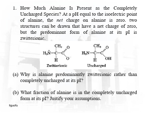
liguofu
(1) 非变性分子筛层析蛋白质亚基不分开,因此蛋白 整体的分子量是240kDa;第二次层析用了5M的尿 素,亚基分离,亚基的分子量是60kDa.因此结论 是:蛋白由4个相同的亚基(或部分)构成 (240kDa=4×60kDa)。
(2) 根据SDS-PAGE电泳相对迁移率作图大致估算, 只出现了约2,500Da和3,500Da两条带(不需要 计算,从图上大概估算就行),这说明在层析中 得到的60kDa的亚基是由二硫键连着的一大一小两 条多肽链构成。因此,这个蛋白的结构为: 2500Da多肽链与3500Da多肽链通过二硫键相连构 成一个单位,再由4个这样的单位(单位间没有二 硫键)构成这个蛋白。
盐析的饱和硫酸氨浓度 MW (kDa) pI
1
45%
38
3.7
2
80%
22
4.8
3
65%
14
5.3
4
20%
75
6.8
5
30%
55
9.5
α-半乳糖苷酶
45%
115
5.3
liguofu
4细胞环境与互作 中山大学研究生入学考试细胞生物学真题各章节专项整理
40.标出下图各部分细胞连接的名称(00年)
41.下面是对一些具有特殊功能的组织的描述.推测是哪种细胞-细胞连接方式使这些细胞连接在一起,为什么?(01年)
a.消化道周围的平滑肌。
这些肌肉必须同步收缩产生蠕动,推动内
容物在肠中移动.
b.胰腺的腺泡是一群围在一个空腔周围的细胞,它们分泌有功能的消
化酶到肠腔内。
分泌的酶后段切除能严重地破坏
腺泡周围的组织.
c.子宫的平滑肌在怀孕期间必须能承受巨大的张力而不被撕破.
d.青蛙的表皮能够主动地将周围环境的盐转运到体内,在动物内环境和池塘的水间产生一个渗透压和电势梯度;。
[中山大学]细胞生物学资料(习题+答案+重点+总结)细胞生物学习题.docx
![[中山大学]细胞生物学资料(习题+答案+重点+总结)细胞生物学习题.docx](https://img.taocdn.com/s3/m/72f78225c1c708a1294a4481.png)
细胞生物学第1讲自学测试题一. 名词解释(每题2分,共20分)1.原核细胞(prokaryotic cell)2.自组装(self assembly)3.原生质(protoplasm)4.结构域(domain)5.模板组装(template assembly)6.原生质体(protoplast)7.细胞生物学(cell Biology)8.类病毒(viroid)9.肮病毒(prion)10.酶效应组装(enzymatic assembly)二. 填空题(每空格1分,共40分)1.、、被誉为19世纪自然科学的三大发现。
2.人的细胞基因组中约有个功能基因。
3.没有细胞壁的细胞称为,细胞中含有细胞核及其它细胞器的部分称为,将细胞内的物质离心后得到的可溶相称为, 或。
4.脱去细胞壁的植物、微生物的细胞叫 o5.细胞是由包围着含有所组成o6.真核生物与原核生物最主要的差别是:前者具有,后者只有。
7.植物中多糖作为细胞结构成分主要是参与的形成。
8.细胞是的基本单位,是由包围着所组成。
与之间的部分叫细胞质。
动物细胞和植物细胞在表面结构上的主要差别是。
9.由于真核生物具有核膜,所以,其RNA的转录和蛋白质的合成是进行的;而原核生物没有核膜,所以RNA转录和蛋白质的合成是的。
10.细胞的共性包括、、、.11.生命科学的四大基础学科是①;②;③;④O12.细胞是生命活动的基本单位,因为细胞是的基础,是的基本单位,是__________________ 基本单位,是的基本单位。
13.世界上所有的生物可归纳为三界,它们是生物、生物和生物。
14.体现所有生命特征的单位是。
15.生命同一性和生命多样性的分子基础分别是和 O三. 判断题(每题1分,共14分)1.从细胞内生物大分了的组装方式看,DNA和RNA都是复制组装,即都是以自身为模板合成一个完全相同的分子。
2.胞质溶胶含有膜结合细胞器,如溶酶体。
3.原生动物是复杂的生物,它有一套特化细胞形成的组织,如鞭毛、口部、刺射器以及足状附器。
中山大学2004级细胞生物学期末试卷

04级细胞期末考题填空真核生物DNA损伤后最重要的反应是——细胞程序性死亡可以——、——、——Golgi体形成的两个模型:膜泡转运模型、潴泡成熟模型目前没有发现在中间纤维上运动的发动机蛋白,可能是因为:中间纤维无极性True/False一个含有核输入信号和核输出信号的蛋白质合成后将会成为穿梭蛋白 (T)在分裂中……只有星微管是旁观者Deacetylation of histone tails allows nucleosomes to pack together into tighter arrays, which usually reduces gene expression. (T)没有内膜系统,真核细胞不能生存myosinII 的唯一作用是肌收缩(F 还有胞质分裂)选择Which of the following proteins would not be found in the smooth endoplasmic reticulum?()a. Ca2+-pumping enzymesb. cytochrome P450c. glucose 6-phosphatased. signal peptidaseIf you compared the proteins in a cis Golgi compartment with those in a trans Golgi compartment, you would find:()a. the proteins in the two compartments are identical.b. the proteins in the cis compartment are glycosylated and contain modified amino acids, whereas those in the trans compartment are not modified.c. the proteins in the cis compartment are glycosylated, whereas those in the trans compartment are glycosylated and contain modified amino acids.d. the proteins of the cis compartment are shorter than those of the trans compartment.Which type of vesicle of the trans Golgi network would be most likely to carry hormones destined for regulated secretion?()a. lysosomal vesiclesb. clathrin-coated vesiclesc. non-clathrin-coated vesiclesd. all of the aboveIf you treated cells with a drug that interferes with microtubules, such as colchicine, which of the following would result?()a. Cell shape would be disrupted.b. Mitosis and meiosis would not occur.c. The intracellular location of organelles would be disrupted.d. All of the above would result.First you dissolve the membrane from an intact flagellum, using the detergent Triton X-100. Next you soak the axoneme in a solution containing EDTA, which removes the Mg2+. What remains of the axoneme after these treatments?()a.peripheral tubules onlyb. peripheral tubules and central tubules, but no side arms or ATPase activityc. peripheral tubules, central tubules, side arms, and ATPase activityd. peripheral tubules, central tubules, side arms, ATPase activity, and a Membrane 简答:好像是答糖基化的作用(列举3个)什么是APC,它在cell cycle的调控中起什么作用如何理解着丝粒是细胞的保护神Nocodazole阻止微管的聚合,这样可以得到同步化的细胞。
中山大学至第一学期细胞生物学期中考试试题

3. Water has a high specific heat.答:4. There is no fundamental distinction between signaling molecules that bindto cell-surface receptors and those that bind to intracellular receptors.答:5. How is it that different cells can respond in different ways to exactlythe same signaling molecule even when they have identical receptors?答:6. Protein kinase A itself is different in different cell types, whichexplains why the effects of cyclic AMP vary depending on the target cell.答:7. It is thought that extracellular ligand binding to a receptor tyrosinekinase activates the intracellular catalytic domain by propagatinga conformational change across the lipid bilayer through the singletransmembrane a helix.答:8. Atrial natriuretic peptides(心钠肽) bind to a receptor that activatesa G protein,which in turn activates guanylyl cyclase(鸟苷环化酶) to producecyclic GMP which then activates a cyclic GMP-dependent protein kinase (PKG).答:9. Because prokaryotic cells have neither mitochondria nor chloroplasts,they cannot carry out either ATP synthesis or photosynthesis.答:10.电镜的波长与电压有关,电压越高,波长越短,分辨率越高。
中山大学XXX学年细胞生物学复习题及答案
中山大学XXX学年细胞生物学复习题及答案—、名词说明细胞生物学cell biology细胞生物学是从细胞、亚细胞、分子三个水平来探讨各种生命活动的科学。
生物膜biology membrane细胞膜和细胞内膜的统称。
膜受体membrane receptor是细胞表面的一种或一类分子,它们能识别、结合专一的生物活性物质(称配体),生成的复合物能激活和启动一系列物理化学变化,从而导致该物质的最终生物效应。
信号识别颗粒signal recognition particle由6个蛋白质和1个小RNA分子组成的复合体。
介导新生肽链与内质网膜结合,在核糖体没有和内质网膜结合的情形下阻止肽链延长。
细胞表面cell surface细胞表面是指包围在细胞质外层的结构复合体系和功能体系。
内膜系统endomembrane system内膜系统是指细胞内那些在形状结构功能上相互紧密相关的膜性结构细胞器的统称,要紧有内质网,高尔基体,溶酶体,各种襄泡等。
主动运输active transport主动运输是一种由特走载体蛋白介导的逆浓度梯度的运输,需要消耗能呈。
膜泡运输vesicu怙r transport襄泡运输是指襄泡以出芽的形式,从一种细胞器膜上产生、脱落并定向地与另一细胞器膜锚走、融合的过程。
细胞呼吸cellu怙r respiration细胞呼吸是指糖类、月旨肪、蛋白质在细胞内被完全氧化分解为CO2和H2O,开釋出能呈储存在ATP中的过程。
细胞骨架cytoskeleton细胞骨架是由微管、微丝和中间纤维构成的细胞质中的蛋白质纤维网状结构。
踏车运动treadmilling微管、微丝在体外组装过程中,两端的聚合和解聚达到平稳,微管和微丝的总长不变,称为踏车。
巴尔小体Barr body雌性哺乳类细胞内的X染色体在间期凝集固缩成异染色质,称为巴尔小体。
核纤层nuclear lamina核纤层是附着在内核膜上的纟干维蛋白网,与中间纤维和核骨架相互连接,构成跨过细胞质和细胞核的骨架体系。
中山大学2003学年第一学期细胞生物学期中考试试题_真题(含答案与解析)-交互(5)

中山大学2003-2004学年第一学期细胞生物学期中考试试题(总分110, 做题时间90分钟)一、填空题(每空0.5分,共5分)1.决定红细胞ABO血型的物质是糖脂,它由脂肪酸和寡糖素链组成。
A型血糖脂上的寡糖链较O型多一个;B型血仅多一个。
SSS_FILL分值: 1答案: N-乙酰半乳糖胺残基,半乳糖残基2.根据题意, 将与A、B、C、D相对应的词用连线连起来:**municate in ways that are analogous to **munication.Decide which of the following forms of **munication are analogousto autocrine, paracrine, endocrine, and synaptic signaling by cells.A. A telephone conversation: autocrineB. Talking to people at a cocktail party: endocrineC. A radio announcement: synaptic signalingD. Talking to yourself : paracrineSSS_FILL分值: 13.What is the major factor that determines the limit of resolution of a light microscope?SSS_FILL分值: 1答案:波长4.编码载脂蛋白B的基因在肠组织中转录成mRNA后,第2153位的C被形成U,使CAA密码转变成终止密码UAA, 使翻译提前终止。
SSS_FILL分值: 1答案:脱氨5.是细胞外基质的受体蛋白。
在结构上,它们是异二聚体,。
这种跨膜蛋白的细胞外部分有一个球形的头部,可与细胞外基质蛋白结合,而细胞质结构域则可与相结合。
细胞生物学英文试卷(I)
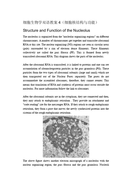
细胞生物学双语教案4(细胞核结构与功能)Structure and Function of the NucleolusThe nucleolus is organized from the "nucleolar organizing regions" on different chromosomes. A number of chromosomes get together and transcribe ribosomal RNA at this site. The nuclear organizing (NO) regions are seen as circular areas (pale) surrounded by a rim of electron dense filaments. These filaments collectively are called the pars fibrosa (PF). This is formed from newly transcribed ribosomal RNA. This diagram shows the parts of the nucleolus.After the ribosomal RNA is transcribed, it is linked to proteins and one can see accumulation of ribonucleoprotein particles in the pars granulosa (PG). These particles form the two types of ribosomal subunits (large and small) which are then transported out of the Nuclear Pores separately. The pores do not accommodate the assembled ribosomes, therefore, they cannot reenter. This means that translation of RNA and synthesis of proteins must occur outside the nucleolus. For more information follow the link to ribosomesAfter the ribosomal subunits are in the cytoplasm, they are connected and then, they may attach to endoplasmic reticulum . They provide an attachment and "code reading" site for the messenger RNA. If they attach to rough endoplasmic reticulum, they form a pore that moves the newly synthesized proteins into the cisterna of the rough endoplasmic reticulum .The above figure shows another electron micrograph of a nucleolus with the nuclear organizing region, the pars fibrosa and the pars granulosa. Nucleoliincrease in number and enlarge when the cell is stimulated to produce proteins. This is a sign that the cell has been stimulated or is actively involved in protein synthesis. Nucleoli disappear during cell division and then reform at the chromosomal nucleolar organizing centers.One can study synthesis of ribosomal RNA with the electron microscope. A preparation of nucleoli is dissociated and spread on a liquid surface (in a manner similar to the spreading of chromatin). Then, the synthesis of ribosomal RNA is stimulated and after a period of time, the DNA from the nucleolar organizing region begins to look like a Christmas tree. The top of the tree is the start site. As you move down the "tree", the branches appear longer. Each branch is a growing strand of ribosomal RNA. The DNA code is being transcribed and the nucleotides added to the growing RNA strand.For more information about the process, see the web site describing ribosomal functions.A Microscopists' View of Chromosome OrganizationThe purpose of this presentation is to show how cytologists and cytochemists contribute to our understanding of the organization of nuclear chromatin. Chromatin is the name that describes nuclear material that contains the genetic code. In fact, the code is stored in individual units called "chromosomes". As we shall see below, these are highly ordered, organized packages designed for the storage of genetic material, its condensation during cell division and regulation of gene expression.If one uses classical transmission electron microscopy (on glutaraldehyde and osmium-tetroxide-fixed tissues and ultrathin plastic sections), there is no hint of the high degree of organization actually found in the nucleus. When one looks at the nucleus, one can find some important clues about the physiological state of the cell. The drawing in this figure shows the two basic cytological patterns that are evident with classical techniques. Two types of chromatin can be described as follows:HeterochromatinThis is the condensed form of chromatin organization. It is seen as dense patches of chromatin. Sometimes it lines the nuclear membrane, however, it is broken by clear areas at the pores so that transport is allowed. Sometimes, the heterochromatin forms a "cartwheel" pattern. Abundant heterochromatin is seen in resting, or reserve cells such as small lymphocytes (memory cells) waiting for exposure to a foreign antigen. Heterochromatin is considered transcriptionally inactive. See Alberts et al, Molecular Biology of the Cell, Garland Publishing, 1994, pages 352 and 353EuchromatinEuchromatin is threadlike, delicate. It is most abundant in active, transcribing cells. (See Alberts et al, Molecular Biology of the Cell, Garland Publishing, 1994, pp 351-352). Thus, the presence of euchromatin is significant because the regions of DNA to be transcribed or duplicated must uncoil before the genetic code can be read.Structure of isolated metaphase chromosomesJust before cell division (and after DNA synthesis), the chromatin condenses further into individual metaphase chromosomes. The dividing chromosomes appear as two chromatids. An electron microscopic view of a chromosome isshown in this photograph. The chromatids appear to be made of coiled loops of 20-30 nm nucleoprotein fibers. Modified from Bloom and Fawcett, A Textbook of Histology, Chapman and Hall, 12th edition, Figure 1-14Can one define individual genes on chromosomes?In the section below, we will show the electron microscopic views that support the organization of DNA in chromosomes. First, we will look at how we can identify specific genes by cytochemistry.As is evident from the foregoing figure, it is difficult to appreciate details of chromosome structure with the electron microscope. However, one can spread these chromosomes on a light microscope slide and label them with dyes that are preferentially taken up by certain regions (for example, modified Giemsa stains). These modifications create a banding pattern that can be used to identify and characterize individual chromosomes. The banding pattern can be used along with shape and length of arms to identify genetic translocations.FISHA more precise way to identify genetic sequences and genes is via a technique called "Fluorescence In situ Hybridization" or FISH. This involves the use of DNA or oligonucleotides complementary to a nucleotide sequence on the chromosomes. These "cDNA probes" are conjugated to fluorescein, rhodamine, Texas red, or another fluorescent compound and then used to detect the sites of the gene on the individual chromosomes. This figure detects a sequence on chromosome 21 in a normal individual (white label). Note, there are two copies of this chromosome, each bearing the sequence. This probe is useful for the detection of Down syndrome which contains an extra chromosome 21. Taken from Boehringer Mannheim Biochemicals "Non Radioactive In Situ Hybridization Application Manual", Figure 10.FISH has also been called "chromosome painting" and there are commercial kits available to detect multiple chromosomes in one section or metaphase spread. A recently developed kit is so colorful, it has been named "Joseph's coat". Each nucleotide probe is attached to a different fluorescent compound and they can be added together because they will attach to different chromosomes, or different regions of the nucleus or chromosomes. In this photograph, FISH was used to detect sequences in chromatin in interphase nuclei. A triple labeling protocol was applied to detect chromosome 2 (red; Texas red), 4 (blue, AMCA) and 8 (green, fluorescein).Taken from Boehringer Mannheim Biochemicals "Non Radioactive In Situ Hybridization Application Manual", Figure 7.Actually, chromosome painting may involve other types of labeling tools, such as autoradiography for radioactive probes, or immunocytochemistry for biotin-labeled or digoxygenin-labeled probes. The immunocytochemical reaction can be detected with gold, peroxidase or alkaline phosphatase labels. Furthermore, the detection of genes or genetic sequences can be done at the electron microscopic level. This figure shows a metaphase spread next to an interphase nucleus. The centromeric heterochromatin has been detected on chromosome 1 with a specific complementary DNA (cDNA) probe.How DNA and Histones are organized in chromosomes.To understand how the DNA and histones are organized in a chromosome, wemust appreciate the fact that the nucleus is only 6 micrometers in diameter. The total length of DNA in the human genome is 1.8 meters. Thus, in order to pack the DNA into the nucleus as in the photograph of the metaphase chromosome , there must be several levels of coiling and supercoiling. There is nearly a 10,000-fold reduction in length in an interphase nucleus. Each chromosome contains 1 long molecule of DNA plus associated histones (basic proteins) which help in the condensation and regulation processes. In the following section, we will show an uncoiled chromosome and also visualize it with specific electron microscopic techniques.Visualize 10-30 nm fibers and fibrilsTo look at chromatin, we can isolate nuclei and disrupt them. This will disperse the chromatin which can be spread on a water surface. This spread can be picked up on a plastic-coated grid and examined with the electron microscope.The first level of organization you see is a tangle of 20-30 nm fibers. These are actually coils of the DNA and histones. The figure on the left shows the tangled chromatin fibers in the left panel. Shearing forces can be used to further uncoil and stretch these fibers and the beaded filaments appear. The strands between the beads are segments of double stranded DNA. These are shown in the right panel.Visualize 4 nm DNA filaments.Another way to look at the chromatin is to remove the histones from isolated chromatin (with dextran sulfate and heparin). This will cause an unraveling of the chromatin from a central protein core. They can be spread on a water surface and collected with a plastic coated grid for electron microscopy. In the EM, they will appear as 4 nm DNA filaments, with no attached histones. This figure shows the unraveled chromatin in the upper inset with the dense protein core. The individual filaments are seen in the main photograph and in the lower inset at higher magnification.This cartoon shows the different levels of uncoiling seen in the chromosome. Start from the bottom of the figure with the DNA helix. Click the hyperlinked text to take you to the electron micrograph illustrating that level of coiling. The first level is the 4 nm DNA filament seen in the above figure (which is DNA stripped of its histones). In this cartoon, the 4 nm DNA filaments are labeled the "DNA helix". In the normal, unstripped chromosome, the double stranded DNA is wrapped around sets of 8 histones to form a 10 nm filament. These sets of 8 histones are separated by spacer regions of 4 nm DNA filament (double stranded DNA) and Histone H1. The sets of 8 histones wrapped by DNA are called "nucleosomes". These are shown in the cartoon. They are the 10 nm nucleoprotein fibrils or "beads on a string" seen in the above electron micrographs. These nucleosomes (beads) are separated by the spacer DNA (seen in the right panel).(注:可编辑下载,若有不当之处,请指正,谢谢!)。
- 1、下载文档前请自行甄别文档内容的完整性,平台不提供额外的编辑、内容补充、找答案等附加服务。
- 2、"仅部分预览"的文档,不可在线预览部分如存在完整性等问题,可反馈申请退款(可完整预览的文档不适用该条件!)。
- 3、如文档侵犯您的权益,请联系客服反馈,我们会尽快为您处理(人工客服工作时间:9:00-18:30)。
中山大学细胞生物学英文题库1. You think you might have discovered a new organelle using electron electron microscopy, but you are not sure if it's real or an artifact of your technique. What might you do to prove it is real?a. Look for the structure in many similarly prepared specimens from the same source.b. Look for the structure in many similar prepared specimens from different sources.c. Look for the same structure in specimens prepared using different staining protocols.d. Look for the same structure in specimens prepared using different (or no) fixatives.2. Traditional genetic approaches to problems of cell biology have followed the path of information from gene to protein. Biochemical approaches have traced proteins back lto the DNA that encodes them, i.e., from protein to gene. This chapter has given you the basics to follow either path. Assume you are studying a problem relating to cell function. Briefly outline the basic steps you would take if you were starting with a gene and trying to deduce the function of the protein. Now outline the steps that you would take to get from a specific protein to a sequenced, isolated gene.1. Indicate whether the following statement is true or false. If it is false, expolain why in Nuclease-chypersensitive sites in chromation are located in the linker DNA between nucleosomes.(F)2. Short answera. Evolutionary conservation of histone proteins.b. nuclear matrix3. Suppose you discover a temperature-sensitive nutant whose nucleus failed to accumulate certain nuclear protein atan elevated (restrictive) temperature, but continued to accumulate other species. What conclusions might you draw about nuclear localization and the nature of this mutation?Cells possess more than one NLS. receptor that recongnize different localization sequences, and that the mutation was occuring in a gene that encoded one of these NLS receptor species.1. Fill in the blanks.A functional chromosome regrires the DNA sequence element at least one_______ to permit the chromosome to copied one to facilitate proper segregation of its two copies at mitosis and two ________ to allow the chromosome to be maintianed betwiin cell generations.(ARS; CEN)2. Which statemint best characterizes the nuclear localization signal?a. The NLS is tupically a small molecular weight metabolic intermediate.b. The NLS is a shetch of hydrophobic amino acids on a proteins N terminus.c. The NLS is one or two streches of basic amino acids on a protein'sC-terminus.d. The NLS is a steroid that binds to DNA.(C)3. You suspect that a new hormone you are testing functions to stimulate myosin synthesis by acting at the transcriptioned level. What type of experimental evidence would support this contention.1. Indicate whether the following statement is ture or false. If a statement is false, exlain why.a. Both myosin-! and myosin-!! have a rollike tial that allows them to polymerize into bipolar filaments, which is crucial for moving groups of oppositely oriented actin filaments past each other.b. Sarcomere shortening is caused by the myosin filaments sliding past the actin filaments with no charge in the length of either type of filaments.2. If you treated cells with a drug that interferes with microtubuls such as colchrarne, which of the following would result?a. Cell shape would be disruptedb. Mitosis and merosis would not occurc. The intracellular location of organelles would be disruptedd. All of the above would result3. Some antibodies capable of binding to kinesin are able to block organelle movement, whereas other anti-kinesin antibodies seem to have bittle effect: How can you explain this obsenation?1.Would you agree with the statement that the centrosome plays a key role in determining that rates of lengthening and shortening of the microtubules of an animal cell? Why or why not?pare and contrast the following cytoplasmic dynein v.s. Kinesin3.Differences in microtubular stability and function between different cells (or different locations within a cell) may be due to which of the following?a. the expression of different tubulin isformsb. the number of protofilaments composing a single microtublec. the presence of different MAPsd. all of the above1.What type of antibody could you use to distinguish between isolated coated vesicles that had formed at the TGN from those that had formed at the plasma membrane?2.Which of the body would you probably be the richest in HDL receptors?a. stomach cellsb. liver cellc. muscle celld. epitholial cells(D)3.A certain integral protein of the plasma membrane has two transmembrane domains in wihich both the C-terminals and the N-terminals are directed toward the cell extenor in the mature protein. Where are the C-termini located when the proteinis newly translocated into the RER membrane? Into the Golgi? Into the vesicles?The nicotinic acetylcholine receptor is membrane-bound protein that also functions as a ligand-gated ion channel. Upon binding acetyleholine, the channel opens and sodi1. Fill in the blanks in the following statements:In cells treated with ______ the Golgi appearatus largely disappears and the Golgi proteins end up in the ER, where they intermix with ER proteins.2. Indicate whether the following is true or false. (T)If a statement is false, explain why.All of the glycoprotions and glycolipids in intracellular membranes have their oligosaccharides facing the luminal side, whereas those in the plasma membrane have their oligosaccharides facing outside the cell.3. You have labeled the lipids on a patch of rough ER membrane with a fluorescent probe. After a few minutes, the probe shows up in the membranes of the cis Golgi. Now you treat the cells with the drug brefieldin A where might the fluoresent probe show up next?a. trans Golgi networkb. endoplasmic reticulumc. plasma membraned. secretory vesicles1. Fill in the following blanks:The amino-terminal ER signal poptide of a soluble protein itself have two functions. In addition to directing the protein to the ER mambrane, it also serve as a ________.(起始跨膜信号)2. True of false: if false, explain why.In mammalian cells, the import of protein into ER begins before the polypeptide chain is synthesized, that is, it occures to co-translationality.3. Which of the following lack a signal sequence?a) acid hydrolase enzymes synthesized in macrophage cells.b) glycolytic enzymes synthesized in liver cells.c) polypeptide gormones synthesized in endocrine cells.d) antibody hormones synthesized in plasma cells.1.Which of the following explain why microsomes can't be seen viewed with eletron microscope?a) They are far too small.b) They are antifats of homogenization and centrifugation.c) They are transparent to electrons.d) They actually can be seen in election micrographics of cells.2.If you were to add a drug to interfere with the ability of ribosome to bind to mRNA, what effect would this be expected to have to the structure of the RER?3.Which, if any, of the following polypeptides would be expected to look, a signal peptide antibodies, acid phosphatase, hemoglobin, ribosomal protein, glycophorn.1.Would you expect the import of substances such as ADP or Pi to lead to a decrease in the proton-motive force? Why?Yes, because they occur at the expense of either the potential difference acress the membrane(in the case of the ATP/ADP exchange) or of the proton gradient (in the case of the uptake of Pi)2.Why would you expect the movement of protons into the intermembrane space to have an effect on the enzymes o fthe cytosol?Because the outer membrane is freely permeable to protons, therefore the protons concentration in the intermembrane space would rapidly epuilibrate with that of the cytosol, lowing the cytosolic pH.ing differential centrifugationm, you are trying to isolate peroxisomes and glyoxysomes from a mixture of cellular organelles. After a few centrufugation steps, you think you may have a relkatively pure suspension. How might you determine that your suspension does, indeed, have there organelles?a) Do an assay for the enzyme catalase.b) Do an assay for the enzyme succinate debydrogenase.c) Look in the microscope for a double -membrane bound organelle.d) Assay you suspension for nucleic acid.(A)1.Which of the following statements about the outer mitochondrial membrane is not true?a) The outer membrane is impermeable to hydrogen ions.b) The outer membrane is highly permeable to substances of small molecular weight.c) The outer membrane is about 50% lipid and 50% protein by weight.d) The outer membrane contains proins.2.Which of the follwing is not an electron carrier class in the electron transport chain?a) flavoproteinsb) cytochromesc) iron-sulfur proteind) cytochrome coxidase3.Short answer: Na-k ATPase from red blood cells can be made to synthesis ATP1. In most cases, RNA acts as an intermediary molecule between the genetic information and the final product of that information, proteins.What are the advantages of having an intermediary as opposed to translating the DNA directly into protein?2.The textbook states that the nucleotide sequence of splice sites is "ancient origin". How do we know this sequence is ancient?a. It is widespread and occurs in even the most primitive eukaryotes.b. The number of mutations that have accumulated in the sequence indicates that it must have been in cells for millions of years.c. It is only found in organisms that are highly evolved.d. It codes for ancient proteins.3. If cultured cells are incubated with 14^C-merhionine the radiolab will appear:a. in all the rRNAs simultaneously.b. first in the 45S RNA, followed by the 32S RNA, followed by the 28S RNA.c. first in the 45S RNA, followed by the 28S RNA, followed by the 5S RNA.d. first in the 28S RNA, followed by the 32S RNA, followed by the 45S RNA.pare and contrast the folowing:(1) "one gene-one enzyme" vs "one gene-one transcription unit".(2) "upstream promoters" vs "internal promoters"2.The signification of methylation of pre-rRNA is probablya. To ensure that the final rRNAwill assume the correct tertiary structre.b. To prevent the pre-rRNA from being processed.c. To prevent degredation of the parts destined to become mature rRNA.d. None of the above.3.The following complex of macromolecules is found in a cell: DNA attached to RNA polymerase, attached to a growing chain of RNA attached to a ribosome, attached to two tRNAs, attached to a growing chain of amino acids. The cell must be:a. A plant cell.b. An animal cell.c. A virus.d. A prokaryotic cell.4.The 5s rRNA differs from the other cellular rRNAs in which of the following ways?a. The genes are located outside the nucleolus.b. The genes are transcribed by RNA polymerase Ⅲ.c. The promoter region of the 5s rRNA gene is internal to the gene.d. All of the above.1. Ca^2+, IP3, and cAMP are very different second messagers. In what ways are their mechanisms of action similar? In what ways are they different?2. Suppose that epinephrine and nonepinephrine could initate a similar response in a particular target cell. How could you show that the two compounds act by binding to the same cell-suface-receptor?3. If you break liver cells b homogenization separate the broken cell membranes (particulate) from the cytoplasm (sohuble) and add epinephrine to the soluble portion, the result will bea. the production of cAMPb. binding of the epinephrine to its receptorc. activeation of adenylyl cyclased. none of the aboveA gene encoding growth hormone in rats was introduced into mice. The construst used consisted in an open reading frame encoding rat growth hormone fused to a mouse promoter A. the growth hormone sequence was obtained from a rat cDNA library the only available source of sequence information. Fertilized mouse egg cells were transformed with the rat growth hormone construst and expression of the rat growth hormone was detected. Transformation of fertilized egg cells with rat growth hormone cDNA alone did not result in expression.1. Why was it necessary to fuse the rat growth hormone sequence to a promoter to obtain expression?a. The rat sequence was obtained from a cDNA library and therefore did not include a promoter sequence.b. No factors that recognized the rat promoter sequence were present in the mouse.c. The rat promoter was contained with the rat sequence but was inactivated in the mouse.d. Rat growth hormone is inactivated in mice.2. When the eggs were placed under conditions conducive to activation of the mouse promoter one of the resulting off spring grew large than the expected size. The appearance of the large mouse is consistent with one the following statements:a. That mouse grew bigger because it produced more mouse growth hormone.b. That mouse grew bigger because it was more aggressive and ate more mouse chaw.c. That mouse grew bigger because the mouse promoter A was activated result in increased production of rat growth hormone.d. The mouse grew bigger because his nose was longer.1. Neleic acid hybridization can be used as a measure of evolutionary relationships between species of the following statement about the DNA of related species, which is truea. Closely related species from hybrid DNAs with relatively low melting temperatures.b. Closely related species from hybrid DNAs with relatively high melting temperatures.c. There is no correlation between DNA hybrid meltiong temperature and relatedness of species.d. One cannot creat DNA hybrid molecules from DNAs of closely related species. techniques you might use to answer each of the following questions.a. Does the DNA on the hair found at the scene of a crime match that of the defendant in a criminal trial?b. Where in a cell are the carbobydrates added to the glycoprotein hormones that are exported?1. Which type of the cell would you expect to achieve the largest volume: a highly flattened cell or a splherical cell? Why?2. All of the following individuals contributed to cell theory except:a. Robert Hookeb. Matthias Schleidenc. Theodor Schwannd. Rudolf Virchow3. Cell theory includes all of the following expecta. All organisms are composed of ome or more cells.b. The cell is the most primitive form of life.c. The cell is the structual unit of life.d. Cells arise by division of preexisting cells.4. Given that cell structure reflects cell function, what structual features would you predict in the following?a. Cell that line the digestive tract and take up nutrients across the wall of the intestine into the blood.b. Cells that synthesize and secrete protein such as an insulin producing cell from the pancreas or an antibody-producing lymphocyte.1. Fill in: A specific tripeptide, sequence, the _____________ is found in one of the repeats and is a central feature of the site responsible for cell binding by fibronectin.2. True or false:Although most types of collagen assemble into fibrilsl type**** collagen assembles into a shoetlike network that forms the core of all basal lamina.3. The addition of peptide within RGD sequence would probably ________ the binding of cultured cells to a fibronectin coated dish.a. enhanceb. inhibitc. have no effect ond. none of the above。