乳腺数字X射线摄影系统质量控制检测操作细则
放射科数字化X线检查技术的质量控制
放射科数字化X线检查技术的质量控制
数字化X线摄影检查,包括CR、DR、造影、乳腺钼靴等检查,临床应用广泛,涉及每个系统的日常诊疗,为临床诊疗发挥重要作用。
为了做好这些X线检查,提高检查质量,保证检查安全,技师在工作中必须认真执行影像学检查的相关规章制度,严把质量关。
科室质控小组要从以下方面进行工作质量控制和监督。
1.严格执行各部位X线检查规范、操作规程。
2.要求操作人员每日进行设备的使用记录。
3.按照设备要求定期关机、开机和磁盘清理记录。
4.每天对各种检查的第1例患者的图像或胶片质量,技师进行自查并记录。
5.图像转存或传输,技师每日自查和确认记录。
6.设备配有专职的维护保养工程技术人员。
7.每台设备有专门的设备故障维修和零配件更换记录。
8.各种检查,要求部位正确,使用曝光参数合适。
9.完成的图像符合诊断要求。
10.对检查部位之外的身体敏感部位采取屏蔽防护辐射。
11.技术质控小组组织技师定期进行图像质量分析。
数字化图像或照片质量控制内容可以从以下几方面进行分析。
①胶片或数字图像外观:胶片无划痕、无水迹、无指纹、无漏光、无静电阴影;无IP板或DR探测器等影像设备原因的伪影。
②影像密度值测量,抽查10张胶片,测量以下指标:胶片基础灰雾密度值D≤0.30;诊断区域密度值D=0.25~2.0;空曝射区密度值DW2.4。
③数字图像曝光剂量:抽查10张数字图像,检查数字图像曝光剂量(kV、mAs,或体表入射剂量mGy)与设备规定的参数范围是否相符合(提供参照设备操作手册)。
乳腺机X射线性能检测操作规程
乳腺机X射线性能检测操作规程1. 目的为了规范乳腺机X射线性能检测操作程序,保证正确使用仪器,保证检测工作的顺利进行和设备安全。
2. 适用范围适用于从事乳腺X射线摄影系统质量控制性能检测的专业技术人员常规操作,质量控制人员检测、校正仪器,有关职能部门负责人监督仪器的使用维护。
3. 检测所用仪器及模块表3-1 仪器及型号4.质量控制性能检测项目与技术要求表4-1 检测项目与技术要求1.质量控制性能检测操作方法4.1. 标准照片密度4.1.1. 将4cm 厚的专用检测模体置于乳腺摄影乳房支撑台上。
将装有胶片的暗盒插入乳房支撑台的暗盒匣中。
4.1.2. 在自动曝光条件下曝光,冲洗胶片,测量距胸侧边沿4cm 处照片长轴中心的光密度,并与基线值进行比较,基线值的光密度在1.4D~1.8D 范围内。
4.2. 胸壁侧射野的准直4.2.1. 将装有胶片的暗盒插入乳房支撑台的暗盒匣中,调整光野与胸侧支撑台边沿对齐,进行曝光,冲洗胶片。
4.2.2. 观察胶片,胸侧胶片边缘应全部曝光。
4.3. 胸壁侧射野与台边的准直4.3.1. 将装有胶片的暗盒于乳房支撑台底部,胸壁侧暗盒超出支撑台边沿4cm 左右,进行曝光,冲洗胶片。
4.3.2. 用刻度为1mm 的钢制直尺测量照片上曝光区域边沿与台边的距离。
4.4. 光野与照射野的一致性4.4.1. 将装有胶片的暗盒插入乳房支撑台的暗盒匣中,调整光野与胸侧支撑台边沿对齐,并在四边作好光野的标记,进行曝光,冲洗胶片。
4.4.2. 用刻度为1mm的钢制直尺测量光野与照射野相应边沿的距离。
4.5. 自动曝光控制4.5.1. 乳房支撑台上分别放置2cm、4cm、6cm 厚的模体,将装有胶片的暗盒分别插入乳房支撑台的暗盒匣中,在自动曝光控制下分别进行曝光。
4.5.2. 测量距胸侧4cm 处照片长轴中心的光密度,2cm 和6cm 模体影像光密度分别与4cm 影像光密度值比较。
4.6. 管电压指示的偏离4.6.1. 应采用非介入方法,如用剂量仪进行检测。
乳腺摄影技术质量控制

乳腺摄影技术质量控制计划一.设定合理的曝光条件:1. 对于经过高X线曝光量曝光的IP一定要在规定时间内消除潜影,不可将还未为完全消除好的IP取出马上投入使用;2. 利用AEC(自动感光控制系统)采取自动曝光条件参数的优化值,千伏值一般采用28KV (不同厂家可能有所不同);3. 实际操作过程中还要利用手工调节系统,根据受检者不同年龄、乳房的不同厚度、实际照射野内不同组织密度来调节千伏值和时间。
二.选择摄影体位:常规采用双侧乳腺头尾位(CC)及内外侧斜位(MLO)投照,发现病变再加照特殊位置,如侧位、乳沟位、局部电压放大等。
三.掌握正确的摆位方法:做到乳腺照片标记、摄影技术员的站法和手法、患者体位均准确合理,摄影台高度、机架倾角、乳房压迫力度均适度而双侧统一。
所获得的钼靶乳房组织显示充分,显影组织真实清晰,病灶位置明确,显影充分。
在满足诊断要求的同时,又降低被检者的辐射剂量。
1. 侧斜位(MLO):正确的侧斜位使所有乳腺组织在单一体位中成像机会最大。
①常规暗盒托盘平面与水平面成45°,根据需要可调节为30~60°(通常高瘦患者为50°~60°,较矮胖患者30~40°,一般身高患者40~50°),使得暗合与胸大肌平行,以利于最大组织成像,双侧乳房体位角度角度应尽量相同。
X线束方向从乳房上内测射向下外侧。
②患者成像乳房侧手放在手柄上,移动患者肩部,使其尽可能靠近滤线栅中心,暗合托盘的角放在胸大肌后面腋窝凹陷的上方,背部肌肉前方;③患者手臂悬在暗合托盘后面,肘弯曲松弛胸大肌,向暗盒托盘的方向旋转患者,使托盘边缘替代技师的手向前衬托乳房组织和胸大肌;④向上向外牵拉乳房,离开胸壁以避免组织影像相互重叠;⑤开始压迫,压迫板经过胸骨后,连续旋转患者使其双臂双足正对乳腺摄影设备,压迫器上角应稍低于锁骨;⑥向下牵拉腹部组织以打开乳房下皮肤褶皱,整个乳房从乳房下褶皱到腋窝都应位于暗合托盘的中心。
[精品]数字化医用X射线摄影系统摄影影像质量要求及试验方法
![[精品]数字化医用X射线摄影系统摄影影像质量要求及试验方法](https://img.taocdn.com/s3/m/8cb5a72d86c24028915f804d2b160b4e767f8119.png)
附件:数字化医用X射线摄影系统摄影影像质量要求及试验方法一、数字化医用X射线摄影系统摄影影像质量要求1、空间分辨率产品标准中应规定在标称有效成像区域下有衰减体模和无衰减体模情况下的空间分辨率及测量时的加载因素组合。
在厚度为20mm 的铝(纯度大于99.5%)衰减体模情况下空间分辨率应不小于2.0lp/mm。
2、低对比度分辨率产品标准中应规定低对比度分辨率的最小值及测量时的空气比释动能和加载因素组合。
在规定的空气比释动能和加载因素组合下,低对比度分辨率的最小值,应不大于规定的最小值。
鉴于许多试验器件都可以有效地测量低对比度分辨率,如果使用的试验器件与本文规定不同,则应将所使用试验器件的说明与低对比度分辨率的测量结果一起记录。
3、影像均匀性产品标准中应规定影像均匀性的最大值及所使用的SID和加载因素。
除非制造商另有声明,影像规定采样点的灰度值标准差R与规定采样点的灰度值均值V之比不应大于2.2%。
即:m%2.2 mV R……………………………………………(1) 式中:R ——灰度值标准差;m V ——灰度值均值。
4、有效成像区域产品标准中应规定所采用的探测器的有效成像区域在x ,y 两个方向上的最大尺寸,实际有效视野尺寸应大于制造商声称有效视野尺寸的95%。
5、残影 无可见残影存在。
6、伪影 无可见伪影存在。
二、数字化医用X 射线摄影系统摄影影像质量试验方法 1、空间分辨率置厚度为20mm 的纯铝衰减体模于射束中心,使之覆盖整个照射野;试验器件采用附录B 中的线对分辨率测试卡或类似的测试卡,测试卡与防散射滤线栅呈45°,置于视野中心位置。
测试卡尽可能靠近影像接收面。
将DR 系统设置到标称有效成像区域。
在有衰减体模的情况下,用70kV,适当的mAs 进行曝光,适当调节影像至最佳,目测观察,确定分辨率。
在未附加衰减体模状态下,用适当的kV和mAs进行曝光,影像不应饱和,适当调节影像至最佳,目测观察,确定分辨率。
乳腺机X射线性能检测操作规程

乳腺机X射线性能检测操作规程1. 目的为了规范乳腺机X射线性能检测操作程序,保证正确使用仪器,保证检测工作的顺利进行和设备安全。
2. 适用范围适用于从事乳腺X射线摄影系统质量控制性能检测的专业技术人员常规操作,质量控制人员检测、校正仪器,有关职能部门负责人监督仪器的使用维护。
3. 检测所用仪器及模块表3-1 仪器及型号4.质量控制性能检测项目与技术要求表4-1 检测项目与技术要求1.质量控制性能检测操作方法4.1. 标准照片密度4.1.1. 将4cm 厚的专用检测模体置于乳腺摄影乳房支撑台上。
将装有胶片的暗盒插入乳房支撑台的暗盒匣中。
4.1.2. 在自动曝光条件下曝光,冲洗胶片,测量距胸侧边沿4cm 处照片长轴中心的光密度,并与基线值进行比较,基线值的光密度在1.4D~1.8D 范围内。
4.2. 胸壁侧射野的准直4.2.1. 将装有胶片的暗盒插入乳房支撑台的暗盒匣中,调整光野与胸侧支撑台边沿对齐,进行曝光,冲洗胶片。
4.2.2. 观察胶片,胸侧胶片边缘应全部曝光。
4.3. 胸壁侧射野与台边的准直4.3.1. 将装有胶片的暗盒于乳房支撑台底部,胸壁侧暗盒超出支撑台边沿4cm 左右,进行曝光,冲洗胶片。
4.3.2. 用刻度为1mm 的钢制直尺测量照片上曝光区域边沿与台边的距离。
4.4. 光野与照射野的一致性4.4.1. 将装有胶片的暗盒插入乳房支撑台的暗盒匣中,调整光野与胸侧支撑台边沿对齐,并在四边作好光野的标记,进行曝光,冲洗胶片。
4.4.2. 用刻度为1mm的钢制直尺测量光野与照射野相应边沿的距离。
4.5. 自动曝光控制4.5.1. 乳房支撑台上分别放置2cm、4cm、6cm 厚的模体,将装有胶片的暗盒分别插入乳房支撑台的暗盒匣中,在自动曝光控制下分别进行曝光。
4.5.2. 测量距胸侧4cm 处照片长轴中心的光密度,2cm 和6cm 模体影像光密度分别与4cm 影像光密度值比较。
4.6. 管电压指示的偏离4.6.1. 应采用非介入方法,如用剂量仪进行检测。
乳腺钼靶X线摄影的质量控制

乳腺钼靶X线摄影的质量控制乳腺X线摄影是目前除MRI检查外应用最广泛、敏感性和特异性较高的诊断早期乳腺癌的重要方法。
本文着重探讨乳腺屏-片系统X线摄影的有关质量控制。
1、材料与方法1.1 仪器设备意大利G10TTO高频智能乳腺机,柯达Min-R2000乳腺专用暗盒(18×24cm)及胶片,柯达洗片机。
1.2 一般材料自2002年2月~2012年5月,我科共做女性乳腺X线检查10240例,年龄16~77岁,平均年龄46.7岁,多数症状为乳腺胀痛,发现肿块,少数为乳头溢液患者。
1.3 方法摄影前由操作技师根据患者主诉,仔细触诊乳腺,常规摄取内外侧斜位(MLO)和头尾位(CC),要确保整个乳腺全部纳入照片中,MLO位中,在保证乳腺组织不丢失的前提下,尽量包含更多的胸大肌和腋窝,使胸大肌呈上宽下尖的楔形,CC位中要包括胸大肌前缘,任何位置均使乳头置于切线位,乳腺展平无皱褶,有乳腺假体置入者,则尽量向前牵拉乳腺组织;同时向后向上推压假体,使乳腺组织与假体最大程度分离。
由于具有自动测量压力和电离室自动控制曝光量的装置,故只要体位摆放正确,两侧乳腺厚度、压力和曝光量是对称的(有乳腺假体时应选手动控制曝光,否则将导致曝光过度)。
当照片灰度增加,对比度下降时,应适当增加曝光量,以补偿显影液消耗所带来的影响。
要确保增感屏及暗盒清洁无污染。
洗片机每次更换显、定影液时务必彻底清洁,以免胶片薄膜被刮掉而误诊,当显、定影液衰竭时及时更换。
2、结果按照美国放射学院有关乳腺摄影质量控制的规范要求和标准,94%的照片完全满足诊断要求。
3、讨论乳腺摄影中严格的质量控制是提高乳腺疾病诊断准确率的基础。
目前有三种途径实现乳腺X线检查:⑴全数字化乳腺X线摄影,为当今最理想的设备,分辨率高,辐射剂量最小,易于保证所有照片质量同一性;⑵乳腺机+乳腺专用IP板及CR系统,无需洗片机,但分辨率较低,曝光剂量较大(由IP板本身结构所决定的);⑶乳腺机屏-片系统:分辨率及曝光剂量居于二者之间,但由于使用洗片机,易造成X线片质量不稳定。
乳腺数字X射线摄影系统质量控制检测操作细则

乳腺数字X射线摄影系统质量控制检测操作细则为规范实施乳腺机性能检测,参照国标《乳腺数字X射线摄影系统质量控制检测规范》(WS 522-2017),结合我单位检测设备,制定《乳腺数字X射线摄影系统质量控制检测操作细则》1.胸壁侧的射野准直调整光野大小至少10cm*15cm,将检测工具放置于乳房撑台上,曝光,冲洗照片,观察胶片。
(只有CR板可做)3.光野与照射野的一致性将乳腺成像准直评估测试工具放置在支撑台上,调整四边的定位标尺,使得标尺的“0”端对准四边的光野边沿,曝光。
5.管电压指示的偏离采用非介入方法,分别在焦点大和焦点小的状态下测量。
选取常用的管电压,在三个不同的KV档条件下曝光,读取每此曝光的管电压值。
7.半值层将探测器放置在支撑台上,将压迫器调至焦点与探测器之间约二分之一处,使用管电压28kv,适当电流曝光条件曝光,读取半值层、曝光时间读数。
8、输出量重复性摘去压迫器,将探测器放置在支撑台上,设置管电压28KV,用临床常用条件,重复5次曝光。
9、特定辐射输出量将探测器放置在支撑台上,记录焦点至探测器的距离,设置管电压28KV,用临床常用条件,重复5次曝光。
记录每次曝光的空气比释动能。
10、影像接收器响应将计量仪紧贴探测器放置在支撑台上,设置28kv,在10mAs-100mAs间取5档值进行手动曝光。
记录数值并获取预处理影像。
移去剂量仪探测器,按相同条件进行手动曝光。
11、影像接收器均匀性将4cm厚的PMMA模体放到乳房支撑台上,设置28kv,临床常用条件进行曝光,获取预处理影像。
12、伪影采用评估影像接收器均匀性时产生的曝光影像进行观察。
12、自动曝光控制重复性将4cm厚的PMMA模体放置在乳房支架台上,模体边沿与乳房支撑台胸壁侧对齐。
用临床常用条件,选择AEC条件重复5次曝光。
13、普通乳腺数字X射线摄影(2D)将4cm厚的PMMA模体放置在乳房支架台上,模体边沿与乳房支撑台胸壁侧对齐。
全数字化乳腺x射线摄影系统质量控制检测方法探讨
梁永刚,付丽媛,钟群,等•全数字化乳腺X射线摄影系统质量控制检测方法探讨[J].医疗卫生装备,2020,41(5):61-64.•6I"Thesis-医械应用与质控・全数字化乳腺X射线摄影系统质量控制检测方法探讨梁永刚,付丽媛,钟群,肖慧,许尚文,陈自谦*(联勤保障部队第900医院医学影像中心,福州350025)[摘要]目的:对全数字化乳腺X射线摄影系统的主要参数进行质量控制检测,以保证设备性能良好。
方法:采用瑞典奥利科Piranha型乳腺机剂量仪、M12乳腺X射线机性能检测模体对全数字化乳腺X射线摄影系统进行胸壁侧照射野准直、光野与照射野的一致性、管电压指示的偏离、半值层、自动曝光控制重复性、探测器均匀性、伪影、高对比分辨力和乳腺平均剂量等检测。
检测完毕根据测算方法计算出检测结果,并判断其是否符合WS522—2017(乳腺数字X射线摄影系统质量控制检测规范》要求。
结果:胸壁侧照射野准直检测中硬币被切割的直径距离为4mm;光野与照射野的一致性检测中光野与照射野相应边沿的偏差为5mm;管电压指示的偏离最大值为0.31kV;靶/滤过为Mo/Mo,半值层为0.34mmAl;自动曝光控制重复性的偏差为2.6%;探测器均匀性检测中图像中心区域与边缘区域兴趣区像素值的偏差为1.60%;无影响临床影像的伪影;高对比分辨力纵向为8lp/mm,横向为10lp/mm;乳腺平均剂量为0.61mGy。
全数字化乳腺X射线摄影系统符合WS522—2017(乳腺数字X射线摄影系统质量控制检测规范》要求,质量控制检测通过,设备运行良好。
结论:通过对进行质量控制检测,可以客观地反映设备的性能,从而保证设备处于良好的运行状态。
[关键词]全数字化乳腺X射线摄影系统;质量控制;检测规范;检测步骤;测算方法[中国图书资料分类号]R318.6;TH774[文献标志码]A[文章编号]1003-8868(2020)05-0061-04DOI:10.19745/j.1003-8868.2020112Study on full field digital mammography system quality control test LIANG Yong-gang,FU Li-yuan,ZHONG Qun,XIAO Hui,XU Shang-wen,CHEN Zi-qian*(Medical Imaging Center,the900th Hospital of Joint Logistics Support Force,Fuzhou350025,China) Abstract O时ective To carry out quality control test of full field digital mammography system to ensure its performances.Methods Full field digital mammography system was tested with Piranha dosimeter and M12phantom on radiation field collimation at chest wall edge,consistency between light field and radiation field,deviation of tube voltage indication,half value layer,repeatability of automatic exposure control,detector uniformity,artifact,high-contrast resolution,mean dose of breast and etc.The results were calculated after detection,and then were verified whether they accorded with WS522—2017 Specificaii onfbr test i ng fquali t y control in digi t al mammography systems,The diameter distance of coin cut was4mm in the detection of radiation field collimation at chest wall edge;the deviation between light field and corresponding edge of radiation field was5mm in the consistency test for light field and radiation field;the maximum deviation of tube voltage indication was0.31kV;target/filter was Mo/Mo,and half value layer was0.34mmAl;the repeatability deviation of automatic exposure control was2.6%;the deviation of pixel value between the center area and the edge area of interest in the detector uniformity detection was1.60%;there was no artifact affecting the clinical image;the high contrast resolution was8lp/mm in the longitudinal direction and10lp/mm in the transverse direction;the mean dose of breast was0.61mGy.The results accorded with WS522—2017SpecficaZion/br testing ofquality control in digital mammography systems,and thus the full field digital mammography system proved its performances,Quality control test of the full field digital mammography system contributes to keeping it in a good running condition.[Chinese Medical Equipment Journal,2020,41(5):61-64]Key words full field digital mammography system;quality control;test specification;test step;calculation method0引言-基金项目:国家重点研发计划数字诊疗装备研发重点专项子课题(2016YFC0103103)作者简介:梁永刚(1984—),男,硕士,主治医师,主要从事医学影像设备管理、质量控制与质量管理方面的研究工作,E-mail:313870625@。
医疗机构医院乳腺数字X射线摄影系统质量控制检测规范
医疗机构医院乳腺数字X射线摄影系统质量控制检测规范医疗机构医院乳腺数字X射线摄影系统质量控制检测规范1 范围本标准规定了乳腺数字X射线摄影系统质量控制检测的要求及方法。
本标准适用于乳腺数字X射线摄影(包括乳腺数字体层合成摄影)系统的质量控制检测。
本标准不适用于乳腺计算机X射线摄影(乳腺CR)的质量控制检测。
2 规范性引用文件下列文件对于本文件的应用是必不可少的。
凡是注日期的引用文件,仅注日期的版本适用于本文件。
凡是不注日期的引用文件,其最新版本(包括所有的修改单)适用于本文件。
GBZ 186 乳腺X射线摄影质量控制检测规范3 术语和定义GBZ 186 界定的以及下列术语和定义适用于本文件。
3.1对比度细节阈值 threshold contrast details在规定测量条件下,从一均匀背景条件中能分辨出来的规定形状和面积的最低的对比度细节,以%表示。
3.2伪影 artifact影像上明显可见的图形,它既不体现物体的内部结构,也不能用噪声或系统调制传递函数来解释。
3.3乳腺平均剂量 average glandular dose;AGD乳腺X射线摄影中所致受检者受均匀压迫乳房的腺体组织中(不包括皮肤和脂肪组织)的平均吸收剂量。
3.4感兴趣区 region of interest;ROI在影像中划定的像素区域(圆形或矩形)。
利用软件工具提供该区域的平均像素值和标准偏差等。
3.5预处理影像 pre-processed image未处理影像 unprocessed image经过像素缺陷校正和平野校正后的影像,但是这种影像并没有经过图像后处理。
3.6乳腺数字体层合成摄影 digital breast tomosynthesis;DBT由一系列从不同角度拍摄所获得的低剂量X射线图像经重建后合成体层图像的数字乳腺摄影方法。
3.7尼奎斯特频率 Nyquist frequency;f Nyquist由采样间距a确定的空间频率,关系式为:f Nyquist=1/(2a)。
富士胶片 AMULET Innovality 数字乳腺X射线摄影系统 质量控制程序 用户手册说明书
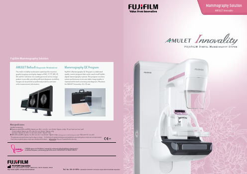
Mammography SolutionAMULET Innovality with measurement information.Main specificationsStandard componentsLasting smiles for women worldwide I n n o v a t i o n a n d q u a l i t y i n m a m m o g r a p h yAMULET Innovality – the result of Fujifilm’s ongoing “innovation” and commitmentto providing top “quality” mammography services. The Innovality utilises Fujifilm’sunique a-Se direct conversion flat panel detector (FPD)* to produce clear imageswith a low X-ray dose. This system makes use of intelligent AEC (i-AEC) combinedwith a image analysis technology to automatically adjust the X-ray dosage for eachbreast type. AMULET Innovality is a highly advanced mammography system whichoffers an extremely fast image interval of just 15 seconds. With this system, Fujifilmfurthers the provision of high quality examinations with superior image quality.*Using a HCP (Hexagonal Close Pattern) TFT array.With its mammography solutions Fujifilm hopes to be an “Amulet” — always there to protect women’s healthOrigin of the nameand allow them to be true to themselves, vibrant and beautiful. The AMULET series aims to provide top-classdigital mammography solutions that can be customised to meet every sites needs.AMULET Innovality hexagonal pixelConventional square pixelThis low-noise and high-speed switching technology allows tomosynthesis exposures with a low X-ray dosage and short acquisition time to be performed. Fast image display is also possible, realizing a smooth mammography workflow from exposure to image display.ISC – Adjusted contrast andlow X-ray dose using a Tungsten Target*Based on Image analysis the appearance is adjusted to emulate the image quality with the simulated “optimal” spectrum.Unique detector for fast, low dose examinationsAMULET Innovality employs a direct-conversion flat panel detector made of Amorphous Selenium (a-Se) which exhibits excellent conversion efficiency in the mammographic X-ray spectrum. The HCP (Hexagonal Close Pattern) detector efficiently collects electrical signals converted from X-rays to realize both high resolution and low noise. This unique design makes it possible to realize a higher DQE (Detective Quantum Efficiency) than with the square pixel array of conventional TFT panels. With the information collected by the HCP detector, AMULET Innovality creates high definition images with a pixel size of 50 μm; the finest available with a direct-conversion detector.Image-based Spectrum Conversion* (ISC) technology can be used to adjust contrast in an image. ISC analyzes images to compensate for variations in contrast due to the density of mammary glands, amount of fat and X-ray spectrum. ISC aims to ensure that images display adequate contrast even with the use of a high energy, low-dose X-ray beam.This technology allows sites that previously exploited the superior contrast of a Molybdenum target to realize the dose advantages offered by the use of Tungsten without having to compromise image contrast.DYN II – Provides high contrast image without saturation in breast regionDynamic Visualization II (DYN II) provides consistent appropriate density of glandular andadipose tissue in each breast type, so the contrast of thick breast and dense breast is improved. Furthermore, it provides high contrast with no saturation in breast region, so the sites are possible to set high contrast parameter.Fujifilm’s unique TechnologySolution to support diagnosis1Close-up (Pixel size 50μm)Unique detector for fast, low dose examinationsIntelligent AEC has advantages in defining the appropriate dose for an examination compared to conventional AEC systems where the sensor position is fixed.Through the analysis of information obtained from low- dose preshot images, Intelligent AEC makes it possible to consider the mammary gland density (breast type) when defining the x-ray energy and level of dose required.Able to be used even in the presence of implants; intelligent AEC enables more accurate calculation of exposureparameters than is possible with conventional AEC systems. By allowing the use of automatic exposure for the implanted breast, Intelligent AEC can further enhance examinationworkflow.Breast areaMammary gland areaConventional AECManual sensor AECRequires manual adjustment of thesetting based on the assured location of mammary glandAutomatic sensor AECAutomatically selects the appropriate sensor from the pre-shot imagesBThe information shown on the displayat the base of the exposure unit can be switched between patientinformation (ID, name, date of birth, etc.) and positioning information (angle of swivel arm, compression force and breast thickness). Positioning information can also be confirmed on the display on the compression arm.A BAAWSHigh definition second monitor• Integrated X-ray controller allows setting and confirmation of exposure conditions on a single screen.• Examination screen can be split and switched between 1, 2, or 4 image display.• Individual images can be immediately output to a PACS, viewer or printer during an examination.• Density and contrast can be easily adjusted while viewing images.• Alignment of left and right images can be adjusted both automatically and manually.• A second, high resolution monitor can be added to the AWS making it possible to display previous images recalled from a PACS to ensure the mammographer has access to previous images at all times.• For Tomosynthesis, reconstructed images can be displayed.Optimal examination workflowHigh definition second monitor (3M/5M: Optional)*When an iodine-based contrast agent is usedWith one compression, it continuously performs low tubevoltage (low energy) imaging close to the ordinary mammo- graphy imaging and high tube voltage (high energy) imaging with a Cu filter, and automatically generates and displays a subtraction image of the obtained images.This subtraction image constitutes an image emphasizing specific tissues.Low energy image High energy imageEnergysubtraction imageAs information for doctors to classify the breast morequantitatively, calculation in the mammary gland area was added to the "mammary gland volume measurementfunction" that automatically calculates the mammary gland volume in the breast area from a mammography image. This mammary gland volume measurement in the breast area/mammary gland area can also be calculated with Tomosynthesis images.Breast Density MeasurementPatient information display7 supported languages1232D mammography image Excellent-m 3DX-ray tubeTomosynthesisHigh quality images for easier diagnosis2In the process of reconstructing the 3D breastarchitecture from multiple 2D images, calcification,mass, spicula, mammary gland and other signalsthat emerge from different depths in the breastarchitecture are selected off to reproduce the breastarchitecture at the focus depth with greater fidelity.2. Suppressing interference of human bodyarchitectures at different depths(as illustrated on the right)The image patterns are recognized to selectivelysuppress the patterns that do not exist in humanbody architectures as noise, to reduce distractivenoises in the event of low-dose tomography.1. Reducing graininess of image inlow-dose tomography*Standard FBP (Filtered back-Projection)The tomosynthesis iterative super-resolution reconstruction (ISR) methodis applied to optimize image quality, achieving significant X-ray dosagereduction.Our super-resolution technology is introduced torestore the fine-structure of calcification and otherphenomena, the visibility of which is impaired bythe movement of the X-ray tube, to facilitateinterpretation of tomosynthesis images.3. Restoring the fine-structureIn breast tomosynthesis, the X-ray tube moves throughan arc while acquiring a series of low-dose X-ray images.The images taken from different angles are reconstructedinto a range of Tomosynthesis slices where the structureof interest is always in focus.The reconstructed tomographic images make it easier toidentify lesions which might be difficult to visualize inroutine mammography because of the presence ofoverlapping breast structures.The Tomosynthesis function on AMULET Innovality issuitable for a wide range of uses, offering two modes tocater for various clinical scenarios. Standard (ST) modecombines rapid exposure timing and efficient workflowwith a low X-ray dose while High Resolution (HR) modemakes it possible to produce images with an even higherlevel of detail, allowing the region of interest to bebrought into clearer focus.Tomosynthesis: making it possible to observethe internal structure of the breastISR ( Iterative Super Reconstruction)Offers significantly lower doses than the conventional methodRadiationdose(mGy)12Conventional processingDose reduced byapprox. 30With combination of 2D and TomosynthesisDose of 2 or less mGy is available*2*1: Equivalent to an image of 40 mm PMMA compared with previous images(Breast thickness of 45 mm, 50% mammary gland, 50% fat)*2: IAEA guidance level: 3 mGy, guidelines of the Japan Association ofRadiological Technicians: 2 mGy*In-house comparisonStatic face guard for Tomosynthesis imaging[Face Guard Comfort (898Y200541)]Fixing the face guard to the device instead of the tube part eliminates movement of the face guard during Tomosynthesis imaging. It will not be reflected at any angle of the ST mode (15 degrees) or HR mode (40 degrees). It can also be used as-is for normal mammography imaging.TomosynthesisHigh quality images for easier diagnosis2Shortens the imaging cycle with a fast display and reconstructionTwo modes suitable for a range of clinical purposesDepth resolutionAdditional imaging for complete checkup,grasping morphology, etc.With a larger acquisition angle the depth resolution is improved. This allows the region of interest to be defined more clearly and brought into clearer focus.HR (High Resolution) mode•Acquisition angle: ±20° •Pixel size: 100/50 μmCheck-up, screening, follow-up, etc.The smaller angular range and fast image acquisition allow Tomosynthesis scans to be quickly performed with a relatively low X-ray dose.ST (Standard) mode•Acquisition angle: ±7.5° •Pixel size: 100/150 μmDepth resolutionAfter a shot, the next shot in either 2D or 3D can be started with a cycle time of approx. 15 seconds.In the case of ST mode*Varies depending on the type and thickness of the breast*2D imaging Cycle timeDisplaying projected imageDisplays 2D imageDisplaying reconstructed imageDisplays projected image immediately after Tomo imagingDisplays 2D imageimmediately after 2D imagingDisplays reconstructed image immediately after Tomo imagingStarts the next shotTomosynthesis imagingapprox.4 secondsapprox.3 secondsapprox.15 secondsStarts a shotStarts positioningAccessoriesVariable image resolution for different needsThe system is designed to support flexible positioning of tube and detector, from -90°to +90°. Ergonomically designed arm rests and disposable soft pads ensure patient comfort and safe positioning.Biopsy – 50 µm image solution(FDR-2000BPY)Irradiation field size can be easily adjusted, depending on breast size and procedure needs. Convenient spacers can be used in order to perform needle positioning in extremely thin breasts, too.AEC full automatic function is available for both scout (2D) and Tomosynthesis exposures.Prior images and studies can be viewed during biopsy, to further improve accuracy.Thanks to the adapter, needle positioning can be performed both vertically and laterally. Accessing to the compressed breast in two directions ensures precise targeting of lesions whichmight be in a difficult position.Both CNB/FNB/Hook wire and VAB needles can be used in a wide range of sizes, for various models and manufacturers.Lateral approach (898Y101490)Supports a variety of needleRefer to technical specifications and to local representatives for further information.+90°-90°69cm(minimum)Both Tomosynthesis and stereotactic support for needle positioningThe highest image quality and workflow efficiencyfor interventional proceduresAdvanced Biopsy SystemSupports a variety of approach for patients3Targeting is supported using both tomosynthesis and stereoscopic images: the choice depends on operator confidence and lesion positioning. Tomosynthesisacquisition can be performed in both ST (Standard) and HR (High Resolution) modes, according to desired accuracy and lesion size.Using a tomosynthesis image, it makes it possible to target the lesion which cannot be found on 2D image.Thanks to easier lesion position identification, tomosynthesis targeting results in a more efficient workflow and more simple operation.ST: ±7.5°HR: ±20°2D imageReconstructed images show overlapping structures separately Tomosynthesis BiopsyEasier to locate a target than with the conventional methodOverlapping breast structures make lesions less visible Difficult to identify a particular regionStereo imagingTomosynthesis50μmAccessoriesAccessoriesAMULET HarmonyEasy operation and patient comfort4Shift Compression PaddleThis small compression paddle can be positioned in the middle, right or left side of the detector at any time of examination according to the positioning of the patient.When this compression paddle is used with 18 24 cm radiation field, the radiation field remains in the center for the CC position, while shifting to the upper portion of the detector when the C-arm is rotated to a MLO or ML position.18 24cm24 30cmWarm indirect lighting is used to illuminate the exposure stand, helping patients to relax and allowing examinations to be performed with minimal stress.AMULET Harmony incorporates a range of mammography solutions specifically designed to maintain a harmonious examination environment and foster an atmosphere of trust between mammographers and their patients.This type of compression paddle fits to the shape of the breast, allowing pressure to be evenly applied while holding the breast securely andensuring the breast tissue is adequately separated. Models with the lateral shift function are also available in the lineup.Fit Sweet Paddle(401Y100131, 401Y120033, 401Y200001, 401Y100130)Mood lighting to ease patient anxietyFive different stand labels are available to add a gentle ambience. Each site can choose a stand appearance that best suits theexamination environment, thus relieving patient stress and anxiety.Decorative labels adaptable to each room environmentThis function will reduce the compressionpressure within a range (within + 3 mm) in which the thickness of the breast does not change after normal breast compression is completed for the purpose of alleviating the patient's pain. For breast compression, there is a phenomenon (hysteresis*) where the thickness of the breast becomes thinner during decompression after compression than during compression even with the same pressure. By utilizing this phenomenon, it is possible to automatically decompress it so that the breast condition remains almost the same even if the duration of maximum compression pressure is reduced.* Hysteresis: A phenomenon where the state of a substance or system depends on the course of force added in the past.L. Han, M. Burcher, and J.A. Novle. Non-invasive Measurement of Biomechanical Properties of in vivo Soft Tissues. MICCAI 2002, LNCS 2488, pp. 208-215, 2002.Automatic decompression ONX-ray irradiationBreast thicknessRetentionDecompression (compression release)ConventionalConventionalComfort Comp29kV 44mAs 0.83mGy33mm 102N 29kV 44mAs 0.83mGy34mm 62.8N120N 60NCompression forceCompression (positioning)DecompressionAutomatic compression reduction control (Comfort Comp)(401Y120038, 401Y120046, 401Y200001)(401Y120025, 401Y100124)AccessoriesAccessoriesAccessories。
医疗机构医院医用数字X射线摄影(DR)系统质量控制检测规范
医疗机构医院医用数字X射线摄影(DR)系统质量控制检测规范1 范围本标准规定了医用数字X射线摄影(DR)系统质量控制检测的一般要求、检测项目和检测方法及其技术要求。
本标准适用于医用数字X射线摄影(DR)系统的质量控制检测,包括采用线阵或面阵扫描影像探测器的DR系统。
本标准不适用于乳腺数字X射线摄影(乳腺DR)、牙科数字X射线摄影、数字双能摄影和数字体层合成摄影的质量控制检测。
2 规范性引用文件下列文件对于本文件的应用是必不可少的。
凡是注日期的引用文件,仅注日期的版本适用于本文件。
凡是不注日期的引用文件,其最新版本(包括所有的修改单)适用于本文件。
WS 76 医用常规X射线诊断设备影像质量控制检测规范3 术语和定义下列术语和定义适用于本文件。
3.1医用数字X射线摄影系统 medical digital radiography system采用数字化X射线影像探测器技术实现X射线摄影的一种医学成像装置。
它的影像直接从影像探测器读出,通常由X射线发生装置、数字化X射线影像装置和机械辅助装置组成。
简称数字X射线摄影(digital radiography,DR)。
3.2信号传递特性 signal transfer property;STPDR系统的影像探测器入射面影像中心区域测量的平均像素值和影像探测器接受的入射空气比释动能之间的一种相互关系的描述。
对于不同生产厂的影像探测器两者之间有不同的相互关系,如线性、对数或方根关系。
3.3探测器剂量指数 detector dose index曝光指数exposure index用于表示DR系统剂量性能指标的一个参数量。
3.4探测器剂量指示 detector dose indicator;DDI为建立和显示影像探测器剂量指数而采用的一种方法。
3.5感兴趣区 region of interest;ROI在影像中划定的像素区域(圆形或矩形)。
利用软件工具提供该区域的平均像素值和标准偏差等。
医用数字摄影系统X射线辐射源质控操作细则
医用数字摄影(DR、CR)系统X射线辐射源质控质控操作细则为规范实施医用数字摄影(DR、CR)系统X射线辐射源质控,参照国标《医用数字摄影(CR、DR)系统X射线辐射源》JJG1078-2012、《医用数字X射线摄影(DR)系统质量控制检测规范》WS 521-2017 、《计算机X射线摄影(CR)质量控制检测规范》WS 520-2017,结合我单位检测设备,制定《医用数字摄影(DR、CR)系统X射线辐射源质控操作细则》。
一、检查、检验、记录1.设备运行1.1稳定的电源供给系统,独立的变压器供电,足够大的电缆截面积,电源电压稳定。
1.2合理的设备外部运行环境,远离大功率用电设备,与强磁设备隔离,避免无线电干扰。
1.3正确的设备运行内部环境,适宜的房间温湿度,无环境灰尘。
1.4设备维修更换球管、探测器等重要部件后需进行性能检测。
2.设备操作2.1合格的放射技术人员。
2.1熟悉设备的性能及功能。
2.3时刻保证设备安全。
2.4高压注射器的正确使用。
3.图像质量3.1合理调节球管、检查床二者的相对距离。
3.2训练病人在平静状态下屏气。
3.3对四肢的检查应积极使用步进或旋转采集功能。
3.4根据患者体型合理使用曝光条件。
4.设备消耗和人员防护4.1合理使用曝光条件。
4.2透视尽量使用脉冲透视。
4.3摄片、透视时患者及医护人员佩戴防护用品。
二、保养、维护、记录1.保养维护与清洁1.1每日开机前首先检查控制室、手术室和控制柜间的温度(20-25摄氏度)和湿度(40-70%)是否正常。
1.2定期校准设备(时间、日期)等检查各运动部件有无松动及时紧固。
1.3润滑运动部件,定期进行保养,先用干毛巾擦干净轨道上的油污,再用合适的润滑油均匀的涂抹到相关的部件。
1.4工作结束后及时清洁设备上的血渍与污物以免渗入设备内部引起故障。
查看机器是否有损坏或者异常等。
1.5清洁设备前请先关闭设备,有必要等待设备冷却后再进行清洁。
1.6设备表面可用湿毛巾和中性洗涤剂清洁。
乳腺X线摄影技术质量控制述要
乳 腺 X线 摄 影 技 术 质 量 控 制 述 要
曹厚德 , 赵泽华
【 摘要 】乳腺 X线摄影至今仍为乳腺疾病检查 的首选技术方法 。本文以国际权威机构发布的相关质控标 准为依 据, 结合我国国情 , 就乳腺 x线摄 影质量管理 的主要 内容作简要介绍 , 以期在相关工作开展时有所借鉴 。由于乳
【 ywo d 】 Ma Ke r s mmo r h R do r h ; u l yc nrl ga y; a ig a y Q ai o to p p t
引 言
专业系列, 且缺乏有关的专业学科进行系统的人才培养。 所以文中涉及的相关工作 只能 由放射技师担任。
放射 技师 职责
美国放射学院 ( m r a o eeo a i oy A ei nC l g f d l , c l R og AR C )长期以来被业界公认为制定 、出版各种检查技 术、 质量控制及诊断常规的权威机构。 94 A R出 19 年 C 版 《 m o rp yQ a t C nr n a)( Ma m ga h ulv o t l i o Mau1 简称 )
中国医学计算机成像杂志 。0 7 1 :7 —3 4 20 ,3 3 6 8 作者单位 : 上海市静安 区中心医院放射科 1 2 上海普陀 区中心 医院放射科 联 系地址 : 上海市静安 区蔼康路 2 9 2 0 4 5 号 00 0 通讯作者 : 曹厚德 ( 电子邮箱 :a —h u e ia c r) c o o d @s .o n n
M M) QC 。迄乳 腺摄 影质 量标 准法规 ( mmo rp v Ma gah Qul ySa drsA t 简 称 MQ A)19 a t tn ad c, i S 94年 4月 2 8 日生 效后 ,AC R随 之 出版 《 mmo rp yQuly Ma gah ai t
- 1、下载文档前请自行甄别文档内容的完整性,平台不提供额外的编辑、内容补充、找答案等附加服务。
- 2、"仅部分预览"的文档,不可在线预览部分如存在完整性等问题,可反馈申请退款(可完整预览的文档不适用该条件!)。
- 3、如文档侵犯您的权益,请联系客服反馈,我们会尽快为您处理(人工客服工作时间:9:00-18:30)。
乳腺数字X射线摄影系统质量控制检测操作细则
为规范实施乳腺机性能检测,参照国标《乳腺数字X射线摄影系统质量控制检测规范》(WS 522-2017),结合我单位检测设备,制定《乳腺数字X射线摄影系统质量控制检测操作细则》
1.胸壁侧的射野准直
调整光野大小至少10cm*15cm,将检测工具放置于乳房撑台上,曝光,冲洗照片,观察胶片。
(只有CR板可做)
3.光野与照射野的一致性
将乳腺成像准直评估测试工具放置在支撑台上,调整四边的定位标尺,使得标尺的“0”端对准四边的光野边沿,曝光。
5.管电压指示的偏离
采用非介入方法,分别在焦点大和焦点小的状态下测量。
选取常用的管电压,在三个不同的KV档条件下曝光,读取每此曝光的管电压值。
7.半值层
将探测器放置在支撑台上,将压迫器调至焦点与探测器之间约二分之一处,使用管电压28kv,适当电流曝光条件曝光,读取半值层、曝光时间读数。
8、输出量重复性
摘去压迫器,将探测器放置在支撑台上,设置管电压28KV,用临
床常用条件,重复5次曝光。
9、特定辐射输出量
将探测器放置在支撑台上,记录焦点至探测器的距离,设置管电压28KV,用临床常用条件,重复5次曝光。
记录每次曝光的空气比释动能。
10、影像接收器响应
将计量仪紧贴探测器放置在支撑台上,设置28kv,在10mAs-100mAs间取5档值进行手动曝光。
记录数值并获取预处理影像。
移去剂量仪探测器,按相同条件进行手动曝光。
11、影像接收器均匀性
将4cm厚的PMMA模体放到乳房支撑台上,设置28kv,临床常用条件进行曝光,获取预处理影像。
12、伪影
采用评估影像接收器均匀性时产生的曝光影像进行观察。
12、自动曝光控制重复性
将4cm厚的PMMA模体放置在乳房支架台上,模体边沿与乳房支
撑台胸壁侧对齐。
用临床常用条件,选择AEC条件重复5次曝光。
13、普通乳腺数字X射线摄影(2D)
将4cm厚的PMMA模体放置在乳房支架台上,模体边沿与乳房支撑台胸壁侧对齐。
选用临床所用对4.5cm厚的人体乳房的AEC曝光条
件进行自动曝光。
然后移去PMMA 模体,将剂量仪探测器放置在支撑台上,选用相同条件进行手动曝光。
14、乳腺数字体层合成摄影(3D )
将4cm 厚的PMMA 模体放置在乳房支架台上,模体边沿与乳房支撑台胸壁侧对齐。
将乳腺摄影设置成体层合成摄影模式,获取临床常用的3D 模式时4.5cm 厚的人体乳房的AEC 曝光条件。
然后移去PMMA 模体,将设备调至2D 模式,将电离室探测器放置在乳房支架台上,选用相同条件进行手动曝光。
16、高对比分辨力
将两个高对比分辨力卡分别呈水平和垂直方向放置在乳房支架台上,选用适当条件进行手动曝光。
17、对比度细节阈值
选用乳腺X 射线摄影专用对比度细节模体放于支撑台上,依据模说明条件对体进行曝光。
18、计算公式
偏差(%)=%
100⨯-预设值预设值
测量平均值
%1001
)
(12
⨯--=
∑n K K
K
CV i
2
22
111222
11112t I K t I K t I K t I K L +-=
式中:
CV :变异系数;%
K i :每次空气比释动能读数,mGy ;
k
: n 次空气比释动能测量值的平均值,mGy ;
n :空气比释动能的测量值总次数。
L 12 :相邻两档间的线性度;
K 1、K 2:分别为1/2两档的空气比释动能测量值的平均值 ,mGy ; t 1、、t 2:分别为1、2两档的曝光时间,s ; I 1、I 2:分别为1、2两档的电流值,mA 。
%
1001
)(12
⨯--=
∑n D D D
CV i
式中:
CV :重复性,%;
D :n 次曝光管电流时间积读数值的平均值,mAs ;
i D :第i 次曝光管电流时间积读数值,mAs ; n :曝光读取管电流时间积总次数。
特定辐射输出量
式中:1Y --距离焦点的1d (cm )出的,单位为μGy/mAs.
2
Y--距离焦点的2d(cm)出的,单位为μGy/mAs 影像接收器均匀性
偏差
%
100
x
m
m
-
m
D
e
中心
角
中心
19、技术要求
乳腺数字X射线摄影系统质量控制检测规范要求。
