人体标本图片集锦
人体塑化标本欣赏
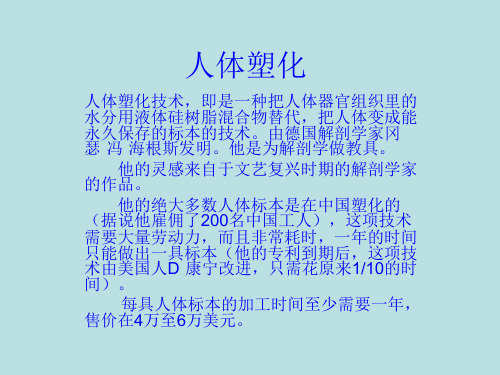
人体塑化技术,即是一种把人体器官组织里的 水分用液体硅树脂混合物替代,把人体变成能 永久保存的标本的技术。由德国解剖学家冈 瑟·冯·海根斯发明。他是为解剖学做教具。 他的灵感来自于文艺复兴时期的解剖学家 的作品。 他的绝大多数人体标本是在中国塑化的 (据说他雇佣了200名中国工人),这项技术 需要大量劳动力,而且非常耗时,一年的时间 只能做出一具标本(他的专利到期后,这项技 术由美国人D·康宁改进,只需花原来1/10的时 间)。 每具人体标本的加工时间至少需要一
人体股三角塑化标本,科普涨知识
人体股三角塑化标本一、什么是人体股三角塑化标本?人体股三角塑化标本是一种用于解剖学教学和研究的特殊制品。
它通过对人体遗体进行特殊处理,使其组织固定并保持原有形态,然后进行注蜡、浸渍等处理,最终制成透明的塑化标本。
这种标本可以展示人体骨骼、血管、神经等结构,帮助学生和研究者更直观地了解人体解剖学知识。
二、科普涨知识:为什么需要人体股三角塑化标本?1.解剖学教学:人体股三角是解剖学中重要的区域之一,包括大腿骨骼、血管和神经。
通过观察和研究这个区域的结构,可以更好地理解整个下肢的解剖关系。
2.医学研究:对于医学研究者来说,深入了解人体股三角的结构对于理解下肢疾病和手术操作具有重要意义。
通过观察和研究人体股三角塑化标本,可以更准确地确定手术切口位置、血管走行和神经分布等。
3.临床应用:在骨科、血管外科等临床领域,人体股三角塑化标本可以作为手术前的辅助教学工具,帮助医生更好地了解患者的解剖结构,从而提高手术成功率和安全性。
4.教育普及:人体股三角塑化标本可以用于公众健康教育,帮助普通人了解自己身体的结构和功能。
通过观察标本,人们可以更直观地了解股骨、肌肉、血管和神经的位置和作用,增强对健康的重视。
三、制作过程1.遗体捐献:制作人体股三角塑化标本首先需要有合适的遗体捐献者。
这些捐献者在生前提出愿望,并经过相关程序进行登记和确认。
2.遗体处理:在捐献者去世后,遗体会被送往专门的实验室进行处理。
首先进行尸检以确认身份,并排除传染病等因素。
然后进行遗体表面的消毒和清洁。
3.组织固定:为了保持遗体组织的形态和结构,需要进行组织固定处理。
常用的方法是使用甲醛等化学物质进行浸泡或注射,使组织固定并防止腐败。
4.注蜡:在组织固定后,需要将遗体进行注蜡处理。
这一步骤可以增加标本的透明度和硬度,使其更加坚固和耐久。
5.浸渍:经过注蜡处理后,遗体会被浸泡在特殊的浸渍液中,以去除甲醛等化学物质,并进一步增强标本的硬度和透明度。
6.干燥和整理:经过浸渍后,标本需要进行干燥处理,并对各个结构进行整理和修剪,使其更加美观和易于展示。
人体寄生虫图片

133
溶组织内阿米巴滋养体
精选ppt
134
溶组织内阿米巴包囊
精选ppt
135
溶组织内阿米巴包囊
精选ppt
136
溶组织内阿米巴包囊
精选ppt
137
溶组织内Байду номын сангаас米巴包囊
精选ppt
138
溶组织内阿米巴包囊
精选ppt
139
溶组织内阿米巴包囊
精选ppt
140
溶组织内阿米巴包囊
精选ppt
141
溶组织内阿米巴包囊
精选ppt
114
肥胖带绦虫成虫(牛带绦虫成虫)
精选ppt
115
肥胖带绦虫头节
精选ppt
116
肥胖带绦虫头节
精选ppt
117
肥胖带绦虫头节
精选ppt
118
肥胖带绦虫成节
精选ppt
119
肥胖带绦虫孕节
精选ppt
120
肥胖带绦虫孕节
精选ppt
121
肥胖带绦虫孕节
精选ppt
122
带绦虫卵
精选ppt
精选ppt
142
结肠内阿米巴滋养体
精选ppt
143
结肠内阿米巴滋养体
精选ppt
55
曼氏迭宫绦虫成节
精选ppt
56
曼氏迭宫绦虫虫卵
精选ppt
57
曼氏迭宫绦虫第一中间宿主:剑水蚤
精选ppt
58
曼氏迭宫绦虫第二中间宿主:蛙
精选ppt
59
华支睾吸虫成虫(肝吸虫成虫)
精选ppt
60
华支睾吸虫成虫(肝吸虫成虫)
精选ppt
人体横切面解剖标本

人体横切面解剖标本是医学教育中非常重要的教学工具,它能够清晰地展示人体的结构和器官。
以下是对人体横切面解剖标本的介绍:一、外观特点人体横切面解剖标本通常是一个透明的塑料容器,里面装满了生理盐水,水中漂浮着人体的横切面模型。
这些模型是由人体的器官和组织切成薄片制成,然后粘贴在塑料容器中。
每个器官和组织都有其特定的颜色和纹理,以帮助区分不同的组织和器官。
二、解剖结构展示人体横切面解剖标本能够清晰地展示人体的解剖结构。
例如,我们可以看到心脏、肺、肝、肾等主要器官的横切面,以及血管、神经、肌肉等组织的分布情况。
通过观察这些模型,学生可以更好地理解人体内部的结构和功能。
三、教学用途人体横切面解剖标本是医学教育中的重要工具,主要用于解剖学、生理学、病理学等课程的教学。
通过观察这些模型,学生可以更好地理解人体结构和功能的关系,以及疾病的发生和发展过程。
此外,这些模型还可以帮助学生更好地理解手术操作的过程和技巧。
四、注意事项在使用人体横切面解剖标本进行教学时,教师需要注意以下几点:1. 尊重人体模型:人体横切面解剖标本是教学工具,而不是用于娱乐或亵渎的物品。
教师需要尊重人体模型,并确保学生在使用时也遵守这一原则。
2. 讲解清晰:教师需要向学生清楚地解释每个器官和组织的名称、位置和功能,以便学生能够更好地理解人体结构和功能的关系。
3. 安全性:由于人体横切面解剖标本包含人体的器官和组织,因此在使用时需要确保安全。
教师需要确保容器密封良好,避免有害物质泄漏。
此外,学生需要遵守实验室的安全规定,如佩戴手套和口罩等。
4. 尊重学生的感受:在使用人体横切面解剖标本进行教学时,教师需要尊重学生的感受,并确保学生在观察时不会感到不适或恐惧。
总之,人体横切面解剖标本是医学教育中非常重要的教学工具,它能够帮助学生更好地理解人体结构和功能的关系。
在使用这些模型进行教学时,教师需要注意尊重人体模型、讲解清晰、确保安全和尊重学生的感受等方面的问题。
另类裸体艺术摄影图片
另类裸体艺术摄影图片裸体艺术是古希腊与文艺复兴两个时期的雕塑与绘画艺术,以下是店铺为大家整理的另类裸体艺术摄影图片,供大家参考!另类裸体艺术摄影图片人体艺术在大多数人的眼里就是情色,然而在艺术家的眼里,人体是最好的创作元素。
来自法国的舞蹈家兼摄影师Arthur Cadre拍摄了一组名为《仙境的生灵》的作品,画面中Arthur Cadre就像一只大自然的精灵,舞动着自己的身体,在大自然的各个角落留下身影。
人体摄影分类古典唯美古典唯美的人体摄影由于人体摄影从一开始就和古典绘画产生了枝枝蔓蔓的纠葛,所以古典氛围笼罩下的人体摄影成为最早的时尚。
同时,在现代乃至当代的人体摄影中,古典氛围作为一种怀旧的情绪,依旧弥漫在诸多大师的镜头底下。
甚至古典在某种意义上并非意味着复辟,而且可以成为一种时尚--时尚向来就是风水轮流转的,多少年以后总会来一次轮回。
从总体的技术手段上看,19世纪末期的画意派摄影家主要通过柔光等技巧,使裸体成为合适的拍摄题材。
一些摄影家还采用一些如今已经失传的古典印相工艺,借助其典雅精致的画面效果,产生古典浪漫的风格特征,直逼绘画中的裸体效果。
而一些经过印象派加工的裸体画面,也逐渐成为国际沙龙中画意摄影的代表作。
大量的摄影家,包括韦斯顿和斯泰肯,在拥抱现代人体摄影之前,就是通过一种古典的、画意的氛围和朦胧的方式完成人体摄影的。
当摄影进入20世纪20年代以后,画意方式的人体摄影渐渐退到了后台,现代人体摄影的表现方式如潮而至,但是也并不妨碍画意方式的"死灰复燃"。
这些摄影家力图回应早期画意式人体摄影的辉煌,进行了许多的努力。
尽管他们的作品无法再一次成为人体摄影中的主流,但还是让我们想到的是,摄影中的古典、唯美的人体永远是一种可以复兴的主题。
比如当代中国摄影家张旭龙的"汤加丽人体摄影系列",就是比较成功的拍摄实践之一。
局部特写局部特写的人体摄影当人体摄影从古典的意境中走出来的时候,摄影家突然发现,人体的思考不一定要借助完美的整体来表现。
人体标本价格表(精)

SL005颅骨矢状切显示:沟、口、板、窦等,可除去颧弓显示翼腭
窝4,850.00
SL006颅骨冠状切显示:沟、口、板、窦等5900.00
SL007额骨弓、孑L、突、沟、嵴、压迹等明显480.00
SL008顶骨角、线、孑L、沟、结节、板障静脉等480.00
SL057整个头部茎突舌骨肌,咽部打开,从后方显示咽部结构
29,700.00
SL058上肢浅层肌肉解剖,显示血管和神经21,000.00
SL059上肢深层肌肉解剖,显示血管和神经21,000.00
SL060上肢表面解剖和深层解剖相结合23,000.00
SL061手肌浅层4,500.00
SL062手肌深层4,500.00
SL063膈肌带部分脊柱、胃、肝脏、脾和胰27,500.00
SL064膈肌不带脏器6,900.00
SL065躯干肌浅层108,500.00
SL066躯干肌深层108,500.00
SL067男性会阴肌表面肌肉解剖,显示血管和神经24,500.00
SL068女性会阴肌表面肌肉解剖,显示血管和神经26,500.00
SL040寰枢关节显示寰椎十字韧带900.00
SL041半个女性骨盆正中矢状切,显示盆腔韧带4,850.00
SL042半个男性骨盆正中矢状切,显示盆腔韧带4,250.00
SL043完整女性骨盆显示盆腔韧带8,500.00
SL044完整男性骨盆显示盆腔韧带7,200.00
SL045脊柱显示相关韧带连接,关节等22,000.00
SL032胸椎一个(带相邻椎间盘)380.00
SL033腰椎一个(带相邻椎间盘)380.00
人体标本 (2)
人体标本简介人体标本是指人类身体的各种组织、器官或部分身体部位,用于医学研究、教学以及临床实践。
这些标本是通过解剖学实验、手术或捐献以及遗体解剖获取的,有助于深入了解人类身体的结构和功能。
人体标本的类型人体标本根据不同的目的和用途可以分为多种类型,包括但不限于以下几种。
解剖标本解剖标本是通过对尸体进行解剖得到的,可以包括整个身体、器官、组织、血管以及神经等。
这些标本广泛应用于医学教育和研究中,用于解剖学实验、病理学研究以及手术模拟训练等。
解剖标本的获取通常需要进行尸体解剖或捐献,因此需要遵守相关的道德和法律规定。
组织标本组织标本是从活体或尸体中取出的组织样本,经过特殊处理后进行保存和展示。
常用的组织标本包括肌肉、骨骼、器官等,可以用来观察细胞结构和病变情况,对疾病的诊断和治疗起到重要的作用。
组织标本通常需要经过固定、切片和染色等处理步骤,以便于显微镜下观察。
血液标本血液标本是指从人体中采集的血液样本,用于检测和分析人体的生理指标和疾病情况。
常见的血液标本检测项目包括血常规、血型、血糖、肝功能、肾功能等。
采集血液标本通常需要在严密的无菌条件下进行,以避免感染和污染。
骨骼标本骨骼标本是从人体中获取的骨头样本,可以用于解剖学研究、骨科手术训练以及人类进化研究等领域。
骨骼标本通常需要经过清洗、消毒和保存等过程,以保持其完整性和稳定性。
标本的保存和利用为了有效保存和利用人体标本,通常需要采取一系列的措施和技术手段。
首先,对于解剖标本和组织标本,常见的保存方法包括冷冻、固定和切片等。
冷冻保存可以保持标本的生物活性,而固定和切片可以使标本在显微镜下观察。
另外,还可以将标本进行脱水、浸泡、封装等处理,以延长标本的保存时间。
其次,对于血液标本,通常需要采用抗凝剂和保存液进行保存。
抗凝剂可以防止血液凝固,而保存液可以保护血液标本的细胞完整性和稳定性。
最后,对于骨骼标本,常见的保存方法包括骨溶解、硬化和干燥等。
骨溶解可以将骨骼标本中的有机物质去除,硬化可以增加标本的硬度和稳定性,干燥可以去除水分并防止腐败。
20年后我重回自然博物馆的人体器官标本禁区试图克服自己的恐惧
20年后我重回自然博物馆的人体器官标本禁区,试图克服自己的恐惧20年后我重回自然博物馆的人体器官标本禁区,试图克服自己的恐惧2015-12-28VICE已收藏文中的某些照片可能会引起不适,请谨慎选择观看。
我从小就喜欢动物,拿起画笔第一个画的也是动物,父亲偶尔会带我去自然博物馆,那是件比过年还高兴的事情。
对我来说,那是个特殊的地方,是一个通过分类博物学展现生命进化和演替的地方,让人由内向外地了解生命,了解自己。
但这个地方也同样给我带来过恐惧。
记得上五年级时又去过一次自然博物馆,正赶上人体展览,那也是我第一次见到正具尸体——展区满是形形色色的参观者,但我却和另一个同学注意到了展区尽头的一个大木门;透过门缝看过去,里面是一个很长的走廊,从门缝中灌到脸上一股冷气和药水的味道。
这是我认为最不同的一次博物馆经历,后来的20年里,我再没光顾过自然博物馆。
人类对尸体的恐惧,或许源于死亡和性,我们在面对这两者时,心理和生理都会有强烈的反映,见到尸体的恐惧和性爱时的兴奋感一样,是种快速的应急反映——虽然那时我还不知道人是怎么诞生的。
后来在我记忆中的噩梦里,也出现过自然博物馆,密密麻麻的恐龙骨架,无数个关着门的房间和标本库,就像是一个很难走出来的迷宫。
即使在梦里,我还是会担心进错一些地方——就像在电影《闪灵》中丹尼骑着玩具车路过的237房间一样。
事隔20年后,我决定到自然博物馆的那个所谓“禁区” 看一看,也算是为了克服自己的恐惧吧,还跟那里的负责人聊了会儿天。
但结果却告诉我,有些东西真不是通过时间或强迫自己就能克服的。
说起来我也不是胆小的人,但在这个房间呆了一小会儿,就感觉自己需要认真考虑下该如何过渡对这个特殊环境恐惧的适应期——恐怕需要一两天的时间才能好吧。
面对尸体,我并不会产生动态的想象,这也是另一部分人产生恐惧的原因之一:在这个集中展示生命科学的地方,进步同样伴随着恐惧;无论在人类文明里的哪个领域,都会存在这样一个特殊环境中的特殊空间,让人不愿意去触碰。
人体解剖图片

据国外媒体报道,下面这十五张令人惊异的人体图片,都是用扫描电子显微镜(SEM)拍摄的,通过它们你可以更近地观察人体的内部情况。
下面将从头部开始,穿过胸腔,一直到达腹腔,经过这次自我发现之旅,让你切身体验到扫描电子显微镜的非凡影响力。
在这个过程中,你将看到当细胞受到肿瘤侵扰时,会出现什么情况,以及卵子第一次与精子相遇时的情景。
1。
红血球从这张图片上看,它们很像肉桂色糖果,但事实上它们是人体里最普通的血细胞—-红血球。
这些中间向内部凹陷的细胞的主要任务,是将氧气输送到我们的整个身体。
在女性体内,每立方毫米血液中大约有400万到500万个红血球,男性每立方毫米血液中有大约500万到600个红血球。
居住在海拔较高的地区的人,体内的红血球数量更多,因为他们生活的环境氧气相对更少。
2。
头发分叉经常修剪和良好的护理,可避免像这张图片上出现发梢分叉的现象。
3. 普尔基涅神经元在大脑里的1000亿个神经元中,普尔基涅神经元是体积最大的.这些细胞是小脑皮层里的运动协调大师。
接触酒精、锂等有毒物质、患有自身免疫性疾病、存在孤独症和神经退行性疾病(Neurodegenerative disease)等遗传变异,都会对人类的普尔基涅神经元造成消极影响。
4.耳毛细胞这张图片看起来好像是在耳朵里面对耳毛细胞进行近距离观察时拍摄的。
耳毛细胞的主要功能是发现对声震作出反应时产生的机械运动。
5。
从视神经中伸出的血管这张照片显示的是血管从黑色视盘中伸出.视盘是个盲点,因为视网膜的这个区域没有光感细胞,视神经和视网膜血管从眼睛后面的这个部位伸出去。
6。
舌头上的味蕾这张彩色图片上显示的是舌头上的一个味蕾。
人舌上大约拥有10000个味蕾,味蕾所感受的味觉可分为甜、酸、苦、咸四种。
其他味觉,如涩、辣等都是由这四种融合而成的。
7。
牙釉质要有一口亮丽牙齿,经常刷牙非常有必要,因为牙齿表面的牙釉质看起来就像“煮熟的老玉米”。
8.血液凝块还记得你刚刚看到的形状统一的红血球图片吗?这张图看起来像是红血球粘在了粘性网上,形成血液凝块。
实验报告躯干骨标本

一、实验目的1. 熟悉人体躯干骨的组成和结构;2. 掌握躯干骨标本的观察方法;3. 了解躯干骨在人体运动和生理功能中的作用。
二、实验器材1. 躯干骨标本;2. 显微镜;3. 线条纸;4. 铅笔;5. 实验记录本。
三、实验步骤1. 观察躯干骨标本的整体结构,记录标本的长度、宽度、厚度等基本特征;2. 分别观察椎骨、胸骨、肋骨、骶骨和尾骨的结构特点;3. 利用显微镜观察骨骼的内部结构,如骨皮质、骨松质、骨髓等;4. 将观察结果绘制成图,并标注相关结构名称;5. 记录实验过程中发现的问题和疑问。
四、实验结果与分析1. 椎骨:椎骨是脊柱的主要组成部分,包括颈椎、胸椎、腰椎、骶骨和尾骨。
颈椎椎体较小,棘突末端分叉,横突根部有横突孔;胸椎椎体两侧和横突末端有肋凹,棘突呈叠瓦状排列;腰椎椎体粗大无肋凹,棘突呈板状水平位后伸;骶骨前面有4对骶前孔,后面有四对骶后孔;尾骨呈三角形,由4-5尾椎合成。
2. 胸骨:胸骨是胸廓的前壁,分为胸骨柄、胸骨体和剑突三个部分。
胸骨柄与锁骨相连,胸骨体与肋骨相连。
3. 肋骨:肋骨是胸廓的侧壁,共有12对。
肋骨前端与胸骨相连,后端与脊柱相连。
4. 骶骨:骶骨由5块骶骨合成的骶骨,上连腰椎,下联尾骨,呈倒三角形,尖向下,底向上,前凹,背侧后凸。
5. 尾骨:尾骨呈三角形,由4-5尾椎合成,上端为底,尾骨下端尖。
五、实验讨论1. 躯干骨在人体运动和生理功能中起着重要作用。
脊柱是人体的支柱,支撑着人体的重量,保护脊髓和内脏器官。
胸廓保护心脏、肺等重要器官,维持呼吸功能。
骨盆是人体的重要骨盆支架,支持体重,保护内脏器官,参与下肢运动。
2. 骨骼的生长发育与人体生长发育密切相关。
在儿童期,椎骨的数目是33-34,成人的椎骨数目通常是26。
这表明骨骼在生长发育过程中会发生一定程度的改变。
六、实验总结本次实验通过对躯干骨标本的观察,了解了人体躯干骨的组成和结构,掌握了躯干骨标本的观察方法。
通过实验,我们认识到躯干骨在人体运动和生理功能中的重要作用。
人体标本价格表

SR011肺门显示一侧肺的形态及结构4,850.00
SR012肺左、右各一7,250.00
SR013肺左、右各一,带心脏9,950.00
SR014纵隔显示左、右纵横结构22,500.00
85600.00元
泌尿生殖系统
SU001肾、输尿管和膀胱的位置膈以下躯干部,保留主动脉和下腔静脉;睾丸动静脉;
SU006女性盆腔上面观显示直肠、子宫及膀胱的位置26,500.00
SU007肾外形一侧600.00
SU008肾冠状切两种颜色灌注700.00
SU009膀胱显示膀胱三角750.00
SU010精囊、前列腺带膀胱,显示膀胱三角2,500.00
SU011男性生殖器3,500.00
SU012睾丸和附睾显示三层被膜1200.00
SL004颅骨水平切显示:孔、沟、口、板、隆凸、压迹等6,900.00
SL005颅骨矢状切显示:沟、口、板、窦等,可除去颧弓显示翼腭窝4,850.00
SL006颅骨冠状切显示:沟、口、板、窦等5900.00
SL007额骨弓、孔、突、沟、嵴、压迹等明显480.00
SL008顶骨角、线、孔、沟、结节、板障静脉等480.00
SN007小脑完整或剥离出齿状核2,600.00
SN008海马2,200.00
SN009穹隆5,000.00
SN010脑岛一侧,剖开外侧裂、显示岛叶5,800.00
SN011大脑投射纤维剥离上纵、下纵及沟束纤维8,800.00
SN012外囊和豆状核一侧,剖开外侧裂,分离岛叶显示外囊及豆状核6,500.00
SC004心脏两种颜色灌注,剖开4,500.00
SC005心脏两种颜色灌注,四周开窗4,500.00
人体标本模型大全

S E I TSomso Modelle Artificial Bone Models Extremities and JointsM ARCUS S OMMER S OMSO M ODELLES E I TFriedrich-Rueckert-Straße 54, D-96450 CoburgPhone 0049 9561 85740, Fax 0049 9561 857411e-mail: somso@somso.de, Internet: www.somso.deA NATOMY14 Extremities and Joints T HE HUMAN FOOT- A MARVEL COM AND19 MUSCLESNS 1 · N ORMAL F OOTNatural size, in SOMSO-Plast. Showing the anatomical structure and the distal end of tibia. In one piece. Length (Pternion-Akropodion): 24 cm., height: 13 cm., width: 26 cm., depth: 10 cm., weight: 450 gNS 2 · F LAT F OOTNatural size, in SOMSO-Plast. Showing the anatomical structure and the distal end of tibia. In one piece. Height: 13 cm., width: 26 cm., depth: 9 cm., weight: 450 gNS3 · A RCHED F OOTNatural size, in SOMSO-Plast.Showing the anatomicalstructure and the distalend of tibia. In one piece.Height: 16 cm.,width: 24 cm.,depth: 10 cm.,weight: 450 gNS4 · C LUB FOOTNatural size, in SOMSO-Plast.The model had been developedin cooperation withDr. Urs Schneider, Tübingen.Showing the pathologicalanatomy of the footand distal tibia.Height: 13 cm.,width: 20,5 cm.,depth: 10,5 cm.,weight: 410 gNS9 ·M USCLES OFTHE F OOTNatural size, in SOMSO-Plast. Showing the network ofnerves and vessels, the layers of the muscles of the sole of the foot are removable (M. flexor digitorum brevis, M. quadratus plantae, M. extensor digitorum longus, Tendo calcaneus (Achillis), M. abductor digiti minimi, M. flexor hallucis bre-vis, M. adductor hallucis (caput obliquum) and M. abductor hallucis), ligamentous apparatus is shown. Altogether in 9 parts. On a stand with base. Height: 18 cm., width: 33 cm., depth: 18 cm., weight: 1.1 kgNS7 · N ORMAL F OOTNatural size, in SOMSO-Plast.Showing the surface muscles. Inone piece. On a stand with base.Height: 29 cm., width: 32 cm., depth:17.5 cm., weight: 600 gNS8 · N ORMAL F OOTNatural size, in SOMSO-Plast.Sagittal section through the insideof the foot. Showing the surfacemuscles at the right half of thefoot. In one piece. On a standwith base. Height: 28 cm., width:29 cm., depth: 17.5 cm., weight:98S E I TPRISING26 BONES , 107 LIGAMENTS99Nature is our ModelSomso ModelleNS 10 · M USCLES OF THE L EG WITH B ASE OF P EL VISA little under natural size, in SOMSO-Plast. Showing the most important blood vessels and nerves in the left leg. The following muscles are removable: the greater gluteal muscle, tensor muscle of the broad fascia, sartorius muscle, straight muscle of the femur, semimembranous muscle, semitendinous muscle, biceps muscle of the femur, digitorum longus muscle, triceps muscle of the calf. Separates altogether in 10 parts. Standing upright, revolving on a stand with base. Height: 108 cm., width:39 cm., depth: 26 cm., weight: 5 kgNS5 ·H ALLUX VALGUS MODELIn cooperation withDr. Urs Schneider, TübingenNatural size, in SOMSO-Plast. The purchased hallux valgus model is a frequent orthopaedic clinical picture as an accompanying aspect of splay feet or pes phanovalgus. On the one hand the model shows the "first ray" pathology with abduction of the first metatarsal bone, adduction and pronation of the proximal phalanx as well as the faculta-tive flexed distal phalanx. On the other hand the significance of the pathological muscle pull for the ethiology and thera-py of the deformity is shown. The role of a pathological muscle pull direction in the progression of the hallux valgus can be studied as an example for many other deformities. Height: 13 cm., width:25 cm., depth: 10 cm., weight: 430 gThis leaflet is an over-worked extract from our complete catalogue A 74. On page 98-104 you will find the extremities and joints and on page 110-139 the artificial bone preparations are shown. Our models are protected by copyright. ©2004 by Marcus Sommer ,SOMSO Modelle.Duplication andreprinting in any form by any method, whether digital or conventional processes, are prohi-bited.A NATOMY 14Extremities and JointsA RM AND LEG MODELS SHOW THE A BONES , TENDONS , JOINTS AND BLOO100NS 15 · M USCLES OF THE A RM WITH S HOULDER G IRDLENatural size, in SOMSO-Plast. Showing the network of blood vessels and nerves in the right arm. The following muscles are removable: deltoid muscle, lateral head of the triceps muscle of the arm, short and long extensor muscle of the radial wrist with brachioradial muscle, round pronator muscle - flexor muscle of the radial wrist - long palmar muscle, superficial flexor muscle of the fingers. Altogether in 6 parts. Standing upright and revolving on a stand with base. Height: 105 cm.,width: 39 cm., depth: 26 cm., weight: 4.6 kgNS 13 ·M USCLES OF THE H AND WITH B ASE OF F ORE -A RMNatural size, in SOMSO-Plast. Aponeu-rosis of the inner hand with the superfici-al muscles removable in layers (Mm. lum-bricales, M. abductor pollicis brevis, M.abductor digiti minimi und M. flexor di-giti minimi brevis). Showing the network of blood vessels and nerves as well as liga-mentous apparatus. Altogether in 5 parts.On a stand with base. Height: 34 cm.,width: 14 cm., depth: 12 cm., weight: 500 gS E I TNATOMY OF THE MUSCLES ,D VESSELSNature is our ModelSomso Modelle101NS 17 · S HOULDER J OINTNatural size, in SOMSO-Plast. With ligaments and synovial capsule. In one piece. On a stand with base. Height:23 cm., width: 19 cm., depth: 19 cm.,weight: 500 gNS 18 · E LBOW J OINTNatural size, in SOMSO-Plast. Showing the ligaments. In one piece. On a base.Height: 21 cm., width: 13 cm., depth:12 cm., weight: 200 gNS 19 · K NEE J OINTNatural size, in SOMSO-Plast. Showing the ligaments and menisci. In one piece.On a base. Height: 24 cm., width: 12 cm.,depth: 14 cm., weight: 300 gNS 20 · H IP J OINTNatural size, in SOMSO-Plast. Showing the ligaments. In one piece. On a base.Height: 28 cm., width: 18 cm., depth:18 cm., weight: 600 gNS 50 · FUNCTIONALM ODELOFTHE KNEE J OINTNatural size, in SOMSO-Plast. Thefollowing movements are possible:flexion, extension, inner and outer rota-tion. On a base. Height: 34 cm., width:18 cm., depth: 18 cm., weight: 1 kgThe advantages of SOMSO functional models1. Authentic repro-duction of the arti-cular anatomy2. Top quality,tough and durable flexible plastic for the ligaments3. Use of screw connectionswherever possible 4. Practical to hand-le by removal from the stand5. Key on base6. 5-year warrantyFFF102A NATOMY 14Extremities and JointsS OMSO FUNCTIONAL MODELS ALLOWNATURAL LOCOMOTIVE PROCESSESFFF103Bending movements of the toe jointsS E I TA DETAILED PRESENTATION OFNature is our ModelSomso ModelleNS 37 ·L IGAMENTSTHE A NKLEO PEN T ALONAVICULAR J OINTNS21 · A NKLE J OINTSWITH L IGAMENTSNatural size, in SOMSO-Plast. Consi-sting of the bones of the foot and thedistal ends of tibia and fibulawith ligamentous apparatus.Length(Pternion-Akropodion):21.5 cm. Inone piece. On astand with base.Height: 38 cm.,width: 18 cm.,depth: 18 cm.,weight:400 g104A NATOMY14Extremities and JointsT HE SECTIONS OF JOINT IN SOMSO-P LAST, DOCUMENTED IN A SERIES OF MODELSNS 43 - NS 48. C ASTS FROM NATURAL BONE SECTIONS WITH TOPOGRAPHY OFMUSCLES, LIGAMENTS, VESSELS AND NERVES. E ACH WITH EXPLANATION ON THE BASEPLATE. U NDER REMOVABLE TRANSPARENT COVERNS 43 · S ECTION THROUGHTHE K NEE J OINTNatural size, in SOMSO-Plast. Sagittalsection. In one piece. Height: 26 cm.,width: 32 cm., depth: 4 cm., weight:800 gNS 44 · S ECTION THROUGHTHE H IP J OINTNatural size, in SOMSO-Plast. Frontalsection. In one piece. Height: 26 cm.,width: 32 cm., depth: 4 cm., weight:900 gNS 45 · S ECTIONTHROUGH THE H ANDNatural size, in SOMSO-Plast.Sagittal section. In one piece. Height:26 cm., width: 32 cm., depth: 4 cm.,weight: 800 gNS 47 · S ECTIONTHROUGH A N ORMAL F OOTNatural size, in SOMSO-Plast.Sagittal section. In one piece. Height:26 cm., width: 32 cm., depth: 4 cm.,weight: 800 gNS 48 · S ECTION THROUGHTHE S HOULDER J OINTNatural size, in SOMSO-Plast.Frontal section. In one piece. Height:26 cm., width: 32 cm., depth: 4 cm.,weight: 900 g.NS 46 · S ECTIONTHROUGH THE E LBOWNatural size, in SOMSO-Plast.Sagittal section. In one piece. Height:26 cm., width: 32 cm., depth: 4 cm.,weight: 800 gSOMSO - a fullfive-year guaranteefive-year warranty - on nearly all models - that covers both durabilityand workmanship.Nature is OurModelEach and every model in the rangedemonstrates SOMSO’scommitment to the highest standards of scientific accuracy and artistry.From concept through prototype to limited or series production, only specialist scientists,model makers and technicians are employed to produce the highestquality models, accurate down to the finest detail.SOMSO MODELLE - subject to stringent quality controlsSOMSO’s primary concern is for quality.Quality that passes the tests for scientific accuracy, paintwork, function, durability and materials. Genuine SOMSOMODELLE reflect these quality criteria,and their base material is virtually un-breakable SOMSO-PLAST.World-wide appreciation from the science and teaching professions and from museumsSOMSO MODELLE are indispensable for practical teaching of general biology in schools. The …Nature is Our Model“ range is superbly instructive, particularly in accuracy, quality and colour, enabling students to experience nature in an incomparable, hands-on manner.Appropriately proportioned SOMSO MODELLE are in use in science labora-tories and lecture halls of universities and colleges throughout the world, making an important contribution to the efficient instruction of trainee doctors and nurses.For many decades, SOMSO MODELLE have been permanently displayed in private collections and public museums,and are of unique interest to specialists and lay visitors alike.The family members are personallyresponsible for the production of every model, and the guaranteed quality oftheir products.Hand assembly and finishing by GermancraftsmenSOMSO MODELLE are produced only in Sonneberg or Coburg - nowhere else -by highly qualified and skilled craftsmen.Some components are now machine-made, but all models are assembled and painted entirely by hand so that each is a unique work of art.Over 1,000anatomical, zoological and botanical modelsApplying to nearly every one of these models is the …SOMSO SUN“,the in-stantly recognisable and world famous registered trade mark.To produce the teaching aids for studying anatomy ,zoology and botany the com-pany has a quite simple, but demanding philosophy: …Nature is Our Model“.SOMSO SUN, the symbol of qualitySOMSO was founded in Sonneberg,Thuringia more than 125 years ago. Since then, SOMSO MODELLE have proved to be the benchmark to which others aspire, recognised by the most discerning experts as the ultimate for teaching aids and scientific demonstration. For the Sommer family this is the motivation that drives them to contribute now, and in the future, to training and teaching in the service of science.A family-run firm founded in 1876The company has been owned and managed by five generations of the Sommer family since it was first established in 1876.The Sommer family110QS 2 · A RTIFICIAL H UMAN S KULLNatural cast, in SOMSO-Plast. Remo-vable vault, the lower jaw is movable and modelled to show the roots of the teeth and their network of vessels. Base of the skull and roof with markings in colour of the venous sinus of the dura mater of the brain and the arteries.Separates into 3 parts. Length: 17.5 cm.,width: 14.1 cm., size: 51.2 cm., weight:800 gA NATOMY 16Artificial Bone ModelsN ATURAL BONE STRUCTURE IS THE FOR S OMSO ARTIFICIAL BONE PREPA111ESSENTIAL YARDSTICK RATIONSNature is our ModelSomso ModelleQS 7/T ·A RTIFICIAL TRANSPARENTHUMANS KULLNatural cast, in SOMSO-Plast. Remova-ble vault, lower jaw movable. Life-like reproduction of the bony structure. Sepa-rates into 3 parts. Weight: 800 gQS 7/1 ·A RTIFICIAL H UMAN S KULLNatural cast, in SOMSO-Plast. As QS 7,but with notation and explanation in English and Latin. Separates into 3parts. Weight: 800 gQS 7/E · A RTIFICIAL H UMAN S KULLNatural cast, in SOMSO-Plast. Remova-ble vault, lower jaw movable. Separates into 3 parts. Length: 17.5 cm., width:14.1 cm., size 51.2 cm., weight: 800 gQS 7/2 · A RTIFICIAL B ASE OF THE S KULLNatural cast, in SOMSO-Plast. Designed for medical students'studies. In one piece. Length: 17.5 cm., width: 14.1 cm., size:51.2 cm., weight: 530 gQS 7 · A RTIFICIAL H UMAN S KULLNatural cast, in SOMSO-Plast. Removable vault, lower jaw movable. Life-like reproduction of the bony skull. Separates into 3 parts. Length. 17.5 cm., width: 14,1 cm., size: 51.2 cm., weight:800 gIllustration of the skullbase from below.The structure of the bones is identical to the models QS 2, QS 2/1, QS 7 and QS 7/1S E I TA NATOMY 16Artificial Bone ModelsT HE H UMAN CRANIUM CLASSICALL YLAST DETAIL112QS 7/6-1 · A RTIFICIAL H UMAN S KULL , F EMALENatural cast, in SOMSO-Plast. As QS 7/6, but with notation. Explanation in English and Latin. Separates into 3 parts. Weight: 700 gQS 7/6 · A RTIFICIALH UMAN S KULL , F EMALENatural cast, in SOMSO-Plast. Remo-vable vault, lower jaw movable. Life-like reproduction of the bony structure.Separates into 3 parts. Length: 18.3 cm.,width: 12.8 cm., size: 50.8 cm., weight:700 gQS 3 · A RTIFICIALS KULL OF A N EWBORNModeled according to nature, in SOMSO-Plast. Upper and lower jaw are open. Altogether in 2 parts. Length:12.1 cm., width: 9.6 cm., size: 33.9 cm.,weight: 180 gQS 3/3 ·A RTIFICIAL S KULL OF A F ETUSNatural cast, in SOMSO-Plast. In one piece. Length: 10.5 cm., width: 8.5 cm.,size: 29.7 cm., weight: 130 gQS 3/2 ·A RTIFICIAL S KULL OF C HILD (A BOUT 6 Y EARS O LD )Natural cast, in SOMSO-Plast. Lower jaw movable. Upper and lower jaw are open to show the emergent second den-tition. Altogether 2 parts. Length:16 cm., width: 11.5 cm., size: 44 cm.,weight: 380 gQS 7/5 ·A RTIFICIALH UMAN S KULLNatural cast, in SOMSO-Plast. As QS 7/1, but showing the areas of origin and insertion of the most important muscles of the head. Separates into 3 parts.Length: 17.5 cm., width: 14.1 cm., size:51.2 cm., weight: 800 gQS 1 ·A RTIFICIAL H UMAN S KULLNatural cast, in SOMSO-Plast. With closed vault, lower jaw removable. Sepa-rates into 2 parts. Weight: 700 gQS 7/3 ·A RTIFICIAL H YOID B ONENatural cast, in SOMSO-Plast. In one piece. On a stand with base. Height:13 cm., width: 12 cm., depth: 12 cm.,weight: 130 gQS 8 ·T RANSPARENTD USTPROOF C OVERSuitable for the artificial human skulls.Height: 21 cm., width: 32 cm., depth:19 cm., weight: 600 gS E I TPREPARED RIGHT DOWN TO THENature is our ModelSomso Modelle113QS 8/10 ·A RTIFICIAL S KULL OF AN A DULTNatural cast, made of SOMSO-Plast.Designed to be separated into 10 parts.Design as QS 8/11, but without repre-sentation of the blood vessels and nerves. Weight: 1 kgQS 8/11 ·A RTIFICIAL D EMONSTRATION S KULL OF AN A DULTNatural cast, made of SOMSO-Plast.With representation of the blood vessels and nerves (N. trigeminus and N. opti-cus etc.). Designed to be separated into 10 parts as follows:1. Cranium with coloured vessels and blood supply of the hard meninx2. Base of the skull, sectioned through median line into two halves3. Nasal septum detachable. Theparanasal sinuses and turbinate bones are shown4. The frontal sinus can be opened5. The maxillary sinus can be opened6. The right temporal bone can be taken out and opened. Representation of the bony labyrinths, the semicircular canals,the eardrum and the chain of auditory ossicles. A radial mastoidectomy is shown on the left temporal bone.7. Detachable lower jaw and roots of the teeth are exposed (flap). Complete set of teethLength: 18 cm., width: 13.1 cm., size:50.4 cm., weight: 1 kgQS 8/11-S · A RTIFICIALD EMONSTRATION S KULLOF ANADULTNatural cast, in SOMSO-Plast. As QS 8/11, but with notation. Key in Englishand Latin. Weight: 1 kg114QS 8/2 ·14-P IECE M ODEL OF THE S KULLNatural size, made from SOMSO-Plast after Prof.Dr. Dr. J. W . Rohen, De-partment of Anatomy,University of Erlangen.The model is construc-ted from 14 individual parts, which can easily be dismantled and put back together by way of interconnecting plugs.The sphenoid bone, oc-cipital bone and the two temporal bones form the basis of the skull;the two parietal bones and the frontal bone attach to the anterior of the sphenoid bone. The facial part of the skull is then completed through attachment of the right and left maxilla, each of which also includes the lacrimal, nasal and pala-tine bones. Facial and cranial bones are con-nected to each other on each side by the zygo-matic bone, which in the model is a separate element that can be in-dividually removed. The mandible is fixed into sockets on either side of the skull through a hin-ge-joint. Weight: 700 gA NATOMY 16Artificial Bone Models C OMPLEX CRANIAL ANATOMY - 14P115QS 8/3 · 14-P IECE M ODEL OF THE H UMAN S KULLNatural size, made from SOMSO-Plast after Prof. Dr. Dr. J. W . Rohen, Department of Anatomy, University of Erlangen. The same model as QS 8/2, but in colour. Here, the individual bones are identified by different colours. This version of the model eases learning of the shape and size of the individual bones and thereby assists in the under-standing of the mosaic-like structure of the human skull. Weight: 700 gQS 8/4 ·T RANSPARENT C ASEHinged and made out of transparent synthetic material. Suitable for SOMSO skulls. Weight: 900 gQS 69 · T HE T HREE A UDITORY O SSICLESCast from natural specimen 1 : 1, in SOM-SO-Plast. Malleus, incus and stapes mo-unted under "Plexiglas" cover, removable.On a base plate. Height: 3 cm., width:12 cm., depth: 12 cm., weight: 80 gQS 69/1 · T HET HREE A UDITORY O SSICLESNatural cast 1 : 1, in SOMSO-Plast. Mal-leus, Incus and Stapes mounted in natural position under "Plexiglas cover". Can be removed, on stand. Height: 3 cm., width:12 cm., length: 12 cm., weight 80 g.QS 70 · A RTIFICIALBONY LABYRINTHCast from natural specimen 1:1, in SOM-SO-Plast. The labyrinth is mounted under "Plexiglas" cover, removable. On a base plate. Height: 3 cm., width: 12 cm., depth:12 cm., weight: 80 gQS 8/51 · A RTIFICIAL T EMPORAL B ONENatural cast, in OMSO-Plast. In one part. On stand with base. Height:17cm., width:12cm., depth:12cm., weight: 150 gQS 8/53 · A RTIFICIAL T EMPORAL B ONENatural cast, in SOMSO-Plast. The opened tympanic cavity shows the tym-panic membrane, the three auditory ossicles, the cochlea and the semicircular canals. Separates into 2 parts. On a stand with base. Height: 17 cm., width: 12 cm.,depth: 12 cm., weight: 150 gS E I TART STEP BY STEP SEPARATIONNature is our ModelSomso ModelleQS 70/1 · T HE T HREEA UDITORY O SSICLES WITHBONY LABYRINTHNatural cast 1 : 1, in SOMSO-Plast.Under "Plexiglas cover". Can be remo-ved, on stand. Height: 3 cm., width:12 cm., length: 12 cm., weight 80 gA NATOMY 16Artificial Bone Models T HE DISMANTABLE S KULL M ODEL A ALSO AVAIABLE IN 18P IECES AND M116QS 8/218 · 18-P IECES M ODEL OF THE S KULLNatural size, made from SOMSO-Plast,after Prof. Dr. Dr. J. W . Rohen, Depart-ment of Anatomy, University of Erlan-gen. The model comprises 18 elements corresponding to the natural bones.Apart from the cranium (frontal, pa-rietal, occipital and sphenoid bones), the bones of the viscero cranium (ethmoid bone, vomer, palatine bone, zygomatic bone, maxilla and mandible) and the in-ferior nasal concha can be removed and reassembled to form the complete skull.Weight: 640 gQS 8/2C+M ·14-P IECES M ODEL OF THE S KULL WITHMUSCLES OF MASTICA -TION AND CERVICAL VERTEBRAL , COLUMN AND HYOID BONENatural size, made from SOMSO-Plast, after Prof. Dr.Dr. J. W . Rohen, Department of Anatomy, University of Erlangen. As QS 8/2, but with the 4 muscles of mastication and cervical vertebral, column and hyoid bone. Weight:1.720 kgMoreover the skull (18-pieces)with muscles of mastication,cervical vertebral column and hyoid bone is available under article number QS 8/218C+M and the coloured versions areavailable with article number QS 8/3C+M (14-pieces skull) as well as QS 8/318C+M (18-pieces skull).QS 8/318 · 18-P IECES M ODEL OF THE S KULLNatural size, made from SOMSO-Plast,after Prof. Dr. Dr. J. W . Rohen, Depart-ment of Anatomy, University of Erlan-gen. The model comprises 18 elements corresponding to the natural bones.Weight: 640 g117S E I TNature is our ModelSomso ModelleFTER P ROFESSOR R OHEN NOW USCLES OF MASTICATIONQS 8/218M · 18-P IECESM ODEL OF THE S KULL WITHMUSCLES OF MASTICATIONNatural size, made from SOMSO-Plast,after Prof. Dr. Dr. J. W . Rohen, Depart-ment of Anatomy, University of Erlan-gen. Version as QS 8/218 but with the 4 masticatory muscles. Weight: 715 g The 14-pieces model of the skull with muscles of mastication has the article number QS 8/2M.QS 8/3M · 14-P IECES M ODEL OF THE S KULL WITH MUSCLESOF MASTICATIONNatural size, made from SOMSO-Plast,after Prof. Dr. Dr. J. W . Rohen, Depart-ment of Anatomy, University of Erlan-gen. Version as QS 8/3 but with the 4masticatory muscles. Weight: 715 gThe 18-pieces model of the skull with muscles of mastication has the articlenumber QS 8/318M.Lower jaw with the 4 muscles of mastication QS 8/1 ·M ETAL S TAND WITH B ASESuitable for the SOMSO skull models.Height: 19 cm., width: 18 cm., depth:18 cm., weight: 300 g; Illustration of the stand with the skull model see QS 8/3CQS 8/3C · 14-P IECESM ODEL OF THE S KULL WITH C ERVICAL V ERTEBRALC OLUMN AND H YOID B ONENatural size, made from SOMSO-Plast,after Prof. Dr. Dr. J. W . Rohen, Depart-ment of Anatomy, University of Erlan-gen. Version as QS 8/3 but with cervical vertebral column and hyoid bone.Weight 1.220 kgQS 8/6 · F ALX C EREBRINatural size, made from SOMSO-Plast,after Prof. Dr. Dr. J. W . Rohen.The 14or 18 piece skull model can also be supplied with a transparent plastic falx cereberi with tentorium cerebelli.Weight: 66 gs e e b a cktit l e s-118QS 9/5 · A RTIFICIALB AUCHENE S KULL OFAN A DULTNatural cast, inSOMSO-Plast. The samemodel as QS 9,but coloured.Height: 40 cm.,width: 26 cm.,depth: 39 cm.,weight: 1.9 kgQS 9/1 ·A RTIFICIALB AUCHENE S KULLOF AN A DULTNatural cast, in SOMSO-Plast. Unmo-unted in a case, altogether 22 parts.Height: 12 cm., width: 42 cm., depth:30 cm., weight: 3 kgQS 9/4 · T RANSPARENTS TORAGE C ASEFitting to unmounted single bonesof the baucheneskull.Height:12cm.,width:42cm.,depth: 30cm.,weight: 2.4kgIllustrationof the individuell bonesQS 9/1, QS 9/2 and QS 9/3119QS 9/2 ·A RTIFICIALB AUCHENES KULL OF AN A DUL TNatural cast, in SOMSO-Plast.Unmounted, each bone individuallypacked in a suitable transparent box,altogether 22 parts. Weight: 2.2 kg.Illustration of the individual bonessee QS 9/1QS 9/3 ·A RTIFICIALB AUCHENES KULL OF AN A DUL TNatural cast, in SOMSO-Plast.All bones loose and unmounted inplastic sacks included in a carton,altogether 22 parts. Weight: 550 g.Illustration of the individual bonessee QS 9/1S E I TTHE ART OF BONE PREPARATION Nature is our Model Somso ModelleThe height and dimensions comply withthe Central European average.Maximumcranium circumference:Female = 50.8 cm.,male = 51.2 cm.Cranium length (Glabel-la-Ophistocranion line):Female = 18.3 cm.,male = 17.5 cm.Cranium width(Euryon distance):Female = 12.8 cm.,male = 14.1 cm.Hand skeleton length(Stylion-Dactylion III):Female = 18 cm.,male = 19 cm.Foot skeleton length(Pternion-Acropodion):Female = 22.2 cm.,male = 25 cm.A NATOMY16Artificial Bone ModelsT HE HUMAN SKELETAL SYSTEM- THE 120QS 10 · A RTIFICIAL H UMANS KELETONNatural cast of the bones of a male adult,in SOMSO-Plast. Showing life-size allthe anatomical details of the bone struc-ture. Skull with removable vault andmandible. Joints mounted and movable,upper and lower extremities removable.The right and left foot can be detachedfrom the leg. Mounted upright on astand. With a dustproof cover. Height:179 cm. (skeleton 170 cm.), width:55 cm., depth: 55 cm., weight: 10 kgQS 10/1 · A RTIFICIALH UMAN S KELETONNatural cast of the bones of a maleadult, in SOMSO-Plast. As QS 10, butwith rollers on the base of the stand.Height: 180 cm. (skeleton 170 cm.),width: 55 cm., depth: 55 cm., weight:10.4 kgQS 10/E · A RTIFICIALH UMAN S KELETON(without ill.)Natural cast of the bones of a maleadult, in SOMSO-Plast. Showing life-size all the anatomical details of thebone structure. Skull with removablevault and mandible. Joints mounted andmovable, upper and lower extremitiesremovable, except of the hands and feet.Mounted upright with rollers on the baseof the stand. With a dustproof cover.Height: 180 cm. (skeleton 170 cm.), width:55 cm., depth: 55 cm., weight: 10 kgMaleSkeletonQS 10FS E I TS OMSO PROGRAMME Nature is our Model Somso Modelle121 Detail QS 10/2 – Muscular functionDetail QS 10/3, QS 10/10, QS 10/11 –Hook for hangingDetail - Standfor hangingQS 10/4QS 10/12QS 10/13QS 10/13GAQS 10/14FemaleSkeletonQS 10/7FF QS 10/2 · A RTIFICIALH UMAN S KELETONNatural cast of the bones of a maleadult, in SOMSO-Plast. As QS 10, buton one arm the flexible muscles of theupper arm are reproduced. By bendingor stretching the arm the flexion orextension of the muscles can be shown.Schematic working model. Height:179 cm. (skeleton 170 cm.), width:55 cm., depth: 55 cm., weight: 10.1 kgQS 10/3 · A RTIFICIALH UMAN S KELETONNatural cast of the bones of a maleadult, in SOMSO-Plast. As QS 10, butwith hook for hanging at the skull(without stand). Height: 170 cm., width:38 cm., depth: 28 cm., weight: 8.8 kgQS 10/4 · A RTIFICIALH UMAN S KELETONNatural cast of the bones of a maleadult, in SOMSO-Plast. As QS 10/3,but with stand for hanging and base.Height: 180 cm. (skeleton 170 cm.),width: 55 cm., depth: 55 cm., weight:11.5 kgQS 10/7 · A RTIFICIALH UMAN S KELETONNatural cast of the bones of a femaleadult, in SOMSO-Plast. Life-like repre-sentation of bone structure with fullanatomical detail. Skull with removablevault and mandible. Joints movable,upper and lower limbs removable. Bothright and left foot can be detached fromthe leg. Mounted upright on stand.Height: 180 cm. (skeleton 171 cm.),width: 55 cm., depth: 55 cm., weight:10.4 kgQS 10/8 · A RTIFICIALH UMAN S KELETONNatural cast of the bones of a femaleadult, in SOMSO-Plast. As QS 10/7,but with rollers on the base of the stand.Height: 180 cm. (skeleton 171 cm.),width: 55 cm., depth: 55 cm., weight:10.7 kgQS 10/10 · A RTIFICIALH UMAN S KELETONNatural cast of the bones of a femaleadult, in SOMSO-Plast. As QS 10/7 butwith hook for hanging on the skull(without stand). Height: 171 cm., width:39 cm., depth: 28 cm., weight: 8.5 kgRollers on the baseof the stand (5 arms)The artificial skeletons are articulatedstanding or suspended, rigid or articulated,with muscular attachments, numbering,articular ligaments or muscle function,according to the customer’s requirements.。
