二甲双胍对胃癌抗肿瘤作用的研究进展
二甲双胍的抗肿瘤作用

险¨ 。临床上治疗 多囊 卵巢综合征患者仍然面 临很 多 挑战 。治疗 主要 针 对调 整月 经周期 、降低血 雄 激素 水平 、改 善胰 岛 素抵抗 及诱 发 排卵 。二 甲双胍 可 抑制 肝脏 合成 葡萄 糖 ,增 加 外 周组 织 对 胰 岛素 的敏 感 性 。 可通过降低胰岛素及雄激素水平 ,同时升高性激素结 合球蛋白的水平 , 从而提高了多囊卵巢综合征妇女的 内分 泌参数 如 葡萄糖 耐量 和排 卵率 ¨ ¨。
二 甲双胍作为双胍类 口服降血糖药的一种 ,通过 提高 胰 岛素 的敏感 性降 低血糖 而 广泛应 用 于 2型糖 尿 病患 者 。近些 年流 行病 学调查 研究 发现 二 甲双胍 有抗 肿瘤 的作 用 ¨ ,在大量 的文献 报 道 中 可 以看 到二 甲 双胍对乳腺癌 、卵巢癌 、子宫 内膜癌 、 肺癌等均能有 效降低疾病患病风险 。本文将就二甲双胍的抗肿 瘤 作用 及其 临床 应用 前景 做一综 述 。
中国 妇 幼 保健 2 0 1 5年 第 3 0卷
・
综述 ・
二 甲双胍 的抗肿 瘤 作用 ①
王静 璐 史 惠蓉 ② 郑州大学第一附属医院妇产科 ( 河南 郑州 ) 4 5 0 0 5 2
中国 图书 分 类 号 R 9 6 9 - 3 文 献标 识 码 A 文章 编 号 1 0 0 1 - 4 4 1 1 ( 2 o l s ) 2 4 - 4 2 4 4 - 0 4 ; d o i : 1 0 . 7 6 2 0 / z g f y b j . J . i s s n . 1 0 0 1 - 4 4 1 1 . 2 0 1 5 . 2 4 . 5 7
二甲双胍用于抗癌治疗的可能性 降糖药二甲双胍愈加表现出抗癌效应

发现 胰 腺癌 的5 年存活 率也 只有
‘
800 81O 1 3 87
. . .
BEI NG Jl SHENG YONG PHA RMAC EUTI LCO . TD CA , L
WW W 。 I UnI I . m . da co cn
3 糖 病 地2 l 2 尿天 o 2 l 0
助 手 在 6 .0 余 名糖 尿 病患 者 中开 289 展 了一 项 回顾 性 队 列 研 究 ,结果 发
展 ,这一发现 将有助于开发更有效
的乳腺癌疗法。
二甲双胍可减少胰腺癌
美 国一 项 新研 究 显 示 ,二 甲
=甲双胍可显著 降低 2 型糖尿
病患者罹患肝细胞癌的风险 肝硬化为糖尿病 常见的晚期并
肺部 由亚硝胺诱发 的肿瘤要 比对照 组少3 % ,而注射二 甲双胍 的实验 4
鼠肺部肿瘤 比对照组少7%。 3 美国 肠 胃病学
— —
《 尿病学》 (i e l i) 糖 Da t o a bo g 今年刊载 的一项研究得 出了二 甲双 胍具 有保护作 用的证据 .英 国卡迪 夫大学 的Ca J C re ri u i g r 博士及其
君力达@
超前 控 血糖 减 少合 并症
— — — — —
■
—
—
—
—
—
—
. .
甲双胍用于出抗癌 效应
胍 可 能 有助 于 预 防 因 吸烟 引发 的肺
今 年 的 欧洲 糖尿 病 研 究学 会 ( S )年会上 ,专家报告称二 甲 E D A 双胍愈加表现 出抗癌效应的结论 .
二甲双胍可抑制乳腺癌扩散
美 国 哈佛 大 学 医学 院 的研 究 人员发现 ,在 化疗 的同时让实验鼠
二甲双胍治疗胃癌 gastric cancer and metformin
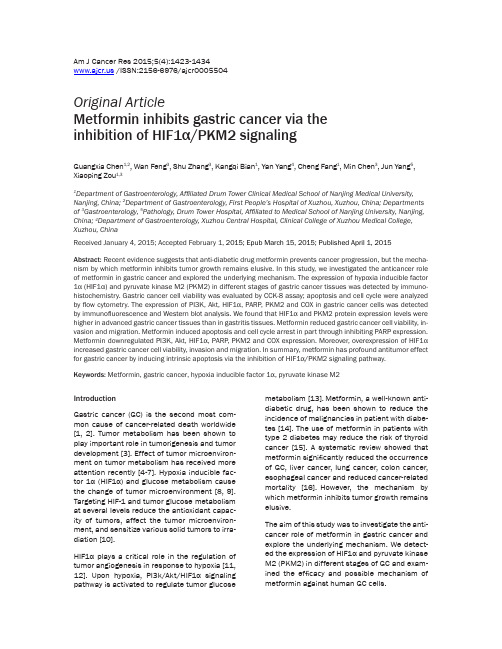
Am J Cancer Res 2015;5(4):1423-1434 /ISSN:2156-6976/ajcr0005504Original ArticleMetformin inhibits gastric cancer via theinhibition of HIF1α/PKM2 signalingGuangxia Chen1,2, Wan Feng3, Shu Zhang3, Kangqi Bian1, Yan Yang4, Cheng Fang1, Min Chen3, Jun Yang5, Xiaoping Zou1,31Department of Gastroenterology, Affiliated Drum Tower Clinical Medical School of Nanjing Medical University, Nanjing, China; 2Department of Gastroenterology, First People’s Hospital of Xuzhou, Xuzhou, China; Department s of 3Gastroenterology, 5Pathology, Drum Tower Hospital, Affiliated to Medical School of Nanjing University, Nanjing, China; 4Department of Gastroenterology,Xuzhou Central Hospital, Clinical College of Xuzhou Medical College, Xuzhou, ChinaReceived January 4, 2015; Accepted February 1, 2015; Epub March 15, 2015; Published April 1, 2015 Abstract: Recent evidence suggests that anti-diabetic drug metformin prevents cancer progression, but the mecha-nism by which metformin inhibits tumor growth remains elusive. In this study, we investigated the anticancer role of metformin in gastric cancer and explored the underlying mechanism. The expression of hypoxia inducible factor 1α (HIF1α) and pyruvate kinase M2 (PKM2) in different stages of gastric cancer tissues was detected by immuno-histochemistry. Gastric cancer cell viability was evaluated by CCK-8 assay; apoptosis and cell cycle were analyzed by flow cytometry. The expression of PI3K, Akt, HIF1α, PARP, PKM2 and COX in gastric cancer cells was detected by immunofluorescence and Western blot analysis. We found that HIF1α and PKM2 protein expression levels were higher in advanced gastric cancer tissues than in gastritis tissues. Metformin reduced gastric cancer cell viability, in-vasion and migration. Metformin induced apoptosis and cell cycle arrest in part through inhibiting PARP expression. Metformin downregulated PI3K, Akt, HIF1α, PARP, PKM2 and COX expression. Moreover, overexpression of HIF1α increased gastric cancer cell viability, invasion and migration. In summary, metformin has profound antitumor effect for gastric cancer by inducing intrinsic apoptosis via the inhibition of HIF1α/PKM2 signaling pathway. Keywords: Metformin, gastric cancer, h ypoxia inducible factor 1α, p yruvate kinase M2IntroductionGastric cancer (GC) is the second most com-mon cause of cancer-related death worldwide [1, 2]. Tumor metabolism has been shown to play important role in tumorigenesis and tumor development [3]. Effect of tumor microenviron-ment on tumor metabolism has received more attention recently [4-7]. Hypoxia inducible fac-tor 1α (HIF1α) and glucose metabolism cause the change of tumor microenvironment [8, 9]. Targeting HIF-1 and tumor glucose metabolism at several levels reduce the antioxidant capac-ity of tumors, affect the tumor microenviron-ment, and sensitize various solid tumors to irra-diation [10].HIF1α plays a critical role in the regulation of tumor angiogenesis in response to hypoxia [11, 12]. Upon hypoxia, PI3k/Akt/HIF1α signaling pathway is activated to regulate tumor glucose metabolism [13].Metformin, a well-known anti-diabetic drug, has been shown to reduce the incidence of malignancies in patient with diabe-tes [14]. The use of metformin in patients with type 2 diabetes may reduce the risk of thyroid cancer [15]. A systematic review showed that metformin significantly reduced the occurrence of GC, liver cancer, lung cancer, colon cancer, esophageal cancer and reduced cancer-related mortality [16].However, the mechanism by which metformin inhibits tumor growth remains elusive.The aim of this study was to investigate the anti-cancer role of metformin in gastric cancer and explore the underlying mechanism. We detect-ed the expression of HIF1α and pyruvate kinase M2 (PKM2) in different stages of GC and exam-ined the efficacy and possible mechanism of metformin against human GC cells.Metformin inhibits gastric cancerMaterials and methods ReagentsMetformin and 4’,6-diamidino-2-phenylindole (DAPI) were purchased from Sigma-Aldrich (St. Louis, MO, USA). Cell counting kit-8 (CCK-8) was purchased from Dojindo Laboratories (Ku- mamoto, Japan). Alexa Fluor 488 conjugated goat anti-rabbit secondary antibody and Trizol were purchased from Invitrogen (Carlsbad, CA , USA). Annexin V Apoptosis Detection kit FITC was purchased from eBioscience (San Diego, CA, USA). Antibodies against PARP (9532), Akt (9272), and β-actin (4967) were purchased from Cell Signal Technology (Boston, MA, USA). Antibody against PI3K (Y467) and HIF1α (K377) were purchased from Bioworld Technology (USA). Antibody against HIF1α (ab113642) and COX (ab33985) were purchased from Abcam (USA). Rabbit secondary antibody was from Cell Signal Technology. PrimeScript™ RT Master Mix and SYBR Premix Ex Taq reagents were pur-chased from Takara Biotechnology (Dalian, China).Samples and immunohistochemistryA total of 20 superficial gastritis, 40 early GC and 40 advanced GC tissues were obtained from patients admitted at The Affiliated Drum Tower Hospital of Nanjing University after the approval of the local ethics committee and informed consent were obtained. Immunohi- stochemical staining of deparaffinized tumoral and gastritis tissues were performed according to standard protocols using HIF1α and PKM2 antibody. The staining intensities were graded as 0, 1, 2, and 3 by two pathologists, respecti- vely.Cell cultureHuman GC lines SGC7901 (moderately differ -entiated) and BGC-823 (poorly differentiated) were purchased from Shanghai Institute of Biochemistry, and cultured in RPMI 1640 medi-um containing 10% fetal bovine serum, 100 ng/L penicillin, and 100 ng/L streptomycin at 37°C in 5% CO 2. HIF1α overexpression plasmid or control plasmid was transfected into BGC823 cells using Lipofectamine 2000 according to the manufacturer’s protocol.Cell viability assayCell viability was detected by cell counting kit-8 (CCK-8) assay. Cells were seeded into 96-well plates at 1×10⁴ cells/well and cultured over-night at 37°C. After treatment with metformin at indicated concentrations for 24, 48, 72 h, 10 µL CCK-8 was added to each well and incubat-ed for 1 h at 37°C. The absorbance was mea-sured at 450 nm. The data were presented as mean ± SD of triplicate samples from at least three independent experiments. The cell viabil-ity was calculated using the following formula: cell viability (%)=(As-Ab)/(Ac-Ab)×100%, where As represents the A value of the experimental well, Ac represents the A value in the control well, and Ab represents the A value of the blank well.Annexin V-FIT C apoptosis assayCells were seeded in six-well plates at 4×10⁵ cells/well and then treated with different con-centrations of metformin for 24 h. Apoptotic cells were detected by flow cytometry using Annexin V-FITC kit according to the instructi- ons.Cell cycle analysisCell cycle distribution was analyzed by flow cy- tometry. After indicated treatments, cells were trypsinized, rinsed with PBS, fixed with 70% ethanol at 4°C overnight, and treated with RNaseA (0.02 mg/ml) in the dark at room tem -perature for 30 min. Cells were resuspended in 0.05 mg/ml propidium iodide and analyzed with flow cytometry. For each sample, at least 1×10⁴ cells were recorded.Cell invasion assayInvasion assay was performed using 24-well Transwell units with 8μm pore size polycarbon -ate inserts. The polycarbonate membranes were cultured at 37°C for 1 h. Cells (1×10⁴) sus-pended in 200 μl of RPMI1640 medium con -taining 1% fetal bovine serum were seeded in the upper compartment of the Transwell unit. 800 μl of RPMI1640 medium containing 10% fetal bovine serum was added into the lower compartment as a chemoattractant. After 24 h incubation, cells on the upper side of the mem-brane were removed, and the cells that migrat-ed through the membrane to the underside were fixed and stained with 0.1% crystal violet. Cell numbers were counted in five separate fields using light microscopy at 400× magnifi -cation. The data were expressed as the meanMetformin inhibits gastric cancervalue of cells in five fields based on three inde -pendent experiments.Cell migration assayMigration assay was performed using 24-well Transwell units with 8 μm pore size polycarbon -ate inserts. The polycarbonate membranes were coated with Matrigel (Becton Dickinson) and cultured at 37°C for 1 h. The next steps were the same as cell invasion assay described above.Quantitative Real-time PCRTotal RNA was extracted using the Trizol Rea- gent and subsequently reverse transcribed using the PrimeScript RT Master Mix according to the manufacturer’s instructions. Quantitative Real-time PCR was performed with the 7500 Real-time PCR System (Applied Biosystems) using SYBR Premix Ex Taq reagents. PCR cycling conditions were: 40 cycles of 5 s at 95°C, 32-34 s at 60°C. Fold-induction was calculated using the formula 2-(ΔΔCt). The specific primers were as follows: HIF1α: sense: 5’-GTAGTGCTG- ACCCTGCACTCAA-3’ antisense: 3’-CCATCGGAA- GGACTAGGTGTCT-5’; β-actin: sense: 5’-ACCGA- GCGCGGCTACA-3’, antisense: 3’-CAGCCGTGG- CCATCTCTT-5’. Western blot analysisCells were lysed in RIPA buffer (50 mM Tris-HCl with pH 7.4, 150 mM NaCl, 0.25% deoxycholic acid, 1% NP- 40, 1 mM EDTA). The proteins in cell lysates were resolved by 8-12% sodium dodecyl sulfate-polyacrylamide gel electropho-resis and transferred to polyvinylidene fluoride membranes. The membranes were blocked by 5% non-fat dry milk in Tris buffered saline con -taining 0.1% Tween-20 for 2 h at room tempera-ture. Then the membranes were incubated with primary antibodies (1:1000 dilutions) over-night, followed by incubation with appropriate HRP-conjugated secondary antibodies (1:5000 dilutions). The blots were detected using Mil- lipore Immobilon Western Chemiluminescent HRP Substrate according to the manufacturer’s instructions. ImmunofluorescenceCells were cultured on 24-well plates, fixed with 4% paraformaldehyde, and blocked for 1 h with 5% normal goat serum, followed by incubation with monoclonal antibodies against HIF1α (1:200) and COX (1:100) overnight at 4°C. Cells were then rinsed with PBS and incubated with Alexa Fluor 488-conjugated goat anti-rabbit or goat anti-mouse secondary antibody. Cells were counter-stained with DAPI (2 μg/ml) and examined by fluorescence microscopy.Statistical analysisAll data were presented as mean ± SD of three independent experiments at least. Statistical analysis was performed using SPSS22.0 and Prism 5 (GraphPad Software Inc., San Diego, USA). Single factor analysis of variance test was used for comparisons among multiple groups, and t test was used for comparisons between two groups. P <0.05 was considered statistically significant.ResultsHigh expression levels of HIF1α and PKM2 in GC tissueWe detected the expression of HIF1α and PKM2 in superficial gastritis, early GC and advanced GC tissues by immunohistochemis-try. The expression level of HIF1α appeared to increase in early GC, but there was no signifi -cant difference compared with superficial gas -tritis. However, the expression level increased significantly in advanced GC comparedwithFigure 2. Metformin inhibits the viability of gastric cancer cells. SGC7901 cells (A) and BGC823cells (B) were treated with metformin (0-50 mM) for 24, 48, 72 h. Cell viability was evaluated by CCK-8 assy.Metformin inhibits gastric cancersuperficial gastritis and early GC tissues (Figure 1A). The expression level of PKM2 increased significantly in early and advanced GC com-pared with superficial gastritis tissue (Figure 1B).Metformin decreases GC cell viabilityWe evaluated the effect of metformin on the viability of two GC cell lines: SGC7901 and BGC823. CCK-8 assay showed significant dose- and time-dependent decrease in the via-bility of SGC7901 and BGC823 cells after met-formin treatment (Figure 2).Metformin induces apoptosis and cell cycle arrest in GC cellsTo elucidate the mechanism by which metfor-min decreases the viability of GC cells, we won-dered whether metformin could induce apopto-sis and cell cycle arrest in GC cells. By Annexin V-FITC and PI staining, we observed that met-formin increased the proportion of apoptotic cells in SGC7901 and BGC823 cells in a dose dependent manner (Figure 3A, 3B). In addi-tion, by flow cytometry analysis, we found that metformin induced cell cycle arrest in SGC7901 Figure 4.Metformin reduces HIF1α, PARP and COX protein expression in gastric cancer cells. (A,B) SGC7901 cells were treated with metformin(0, 40 mM) for 24 h and then analyzed for the expression of HIF1α (A) and COX (B) by immuno-fluorescence. Original magnification 400×. (C) SGC7901 and BGC823 cells were treated with metformin (0, 40, 50 mM) for 24 h and then pro-tein expression of PI3K, Akt, HIF1α, PARP, COX, PKM2 was detected by Western blot analysis.β-actin was loading control.and BGC823 cells in a dose dependent manner (Figure 3C , 3D ).Metformin reduces HIF1α, PARP, COX and PKM2 expression in GC cellsNext we evaluated the effects of metformin on PI3k, Akt, HIF1α, PARP, COX, and PKM2 protein expression in GC cells. Immunofluorescence staining of HIF1α and COX in SGC7901 cells showed significant decrease in HIF1α and COX expression after metformin treatment (Figure 4A , 4B ). Western blot analysis showed that metformin inhibited the expression level of PI3K, Akt, HIF1α, PARP, COX and PKM2 in SGC7901 and BGC823 cells (Figure 4C ).Figure 5. Metformin inhibits the invasion and migration of gastric cancer cells. SGC7901 and BGC823 cells were seeded on transwell for invasion and migration analysis. The numbers of invaded and migrated cells were counted in five separate fields using light microscopy. Original magnification 400×. The data were expressed as the mean value of cells in five fields based on three independent experiments. ***P<0.001.Figure 6. HIF1α over-expression increases the viability, invasion and migration of BGC823 cells. A, C. Lipofectamine, control vector or HIF1α overexpression plasmid were transfected into BGC823 cells, and HIF1α mRNA and protein expression were detected by RT-PCR and Western blot analysis. β-actin was loading control. B. Cell viability was de -tected by CCK-8 assay. D. The numbers of invaded and migrated cells were counted in five separate fields using light microscopy. Original magnification 400×. The data were expressed as the mean value of cells in five fields based on three independent experiments. *P <0.05, ***P <0.001.Metformin inhibits the invasion and migration of GC cellsTo investigate the activity of metformin against tumor metastasis, we examined the effects of metformin on the invasion and migration of GC cells. Transwell assay showed that metformin inhibited the invasion and migration of SGC- 7901 and BGC823 cells in a dose dependent manner (Figure 5).HIF1α over-expression increases the viability, invasion and migration of GC cellsSince metformin inhibited protein expression of HIF1α in GC cells, we wondered whether HIF1α might be the key factor to mediate the effects of metformin on GC cells. We transfected HIF-1α over-expression plasmid into BGC823 cells, and confirmed the expression of HIF1α by RT-PCR and Western blot analysis (Figure 6A, 6C). We found that HIF1 overexpression incr-eased the viability, invasion and migration of BGC823 cells (Figure 6B, 6D).DiscussionIn this study, we revealed the high expression of HIF1α and PKM2 in GC tissues, and found that metformin significantly induced apoptosis, inhibited cell invasion and migration of GC cells. The mechanism by which metformin exhibits anti-tumor activities is through the induction of apoptosis and the inhibition of HIF1α. Recent studies showed that overexpression of HIF1α are implicated in tumorigenesis, tumor chemotherapy resistance, tumor angiogenesis, and tumor glycolysis [17-20]. Increased HIF-1α level is associated with increased risk of mor-tality in many human cancers, including gastric cancer [11]. HIF1α inhibitor inhibited tumor growth and angiogenesis [12].In this study we found that metformin inhibited the expression of HIF1α in GC cells, suggesting that metformin may inhibit tumor cell growth and metastasis via HIF1α inhibition. However, further studies are needed to confirm our conclusion. Targeting of tumor metabolism is emerging as a novel therapeutic strategy against cancer [21]. According to the “Warburg effect”, tumor cells exhibit an increased dependence on glycolytic pathway for ATP generation both in normoxia or hypoxia conditions [22].PKM2 is an important executor downstream of HIF1α signaling and acts as the key enzyme of glycolysis [23]. In this study we found high expression of PKM2 in GC tissues by immunohistochemistry, indicating the important role of glycolysis in the develop-ment of GC. Our study showed that metformin inhibited the expression of PKM2 protein, espe-cially in poorly differentiated BGC823 cells. These data suggest that metformin reduces the energy supply of GC by inhibiting HIF1α/ PKM2 pathway.The most important function of mitochondrial respiratory chain is to generate ATP by oxida-tion phosphorylation (OXPHOS). After sequen-tial electron transfer, two respiratory chains generate ATP through being catalyzed by the respiratory chain enzyme complexes IV- cyto-chrome C oxidase (COX). In energy-rich condi-tions, the mitochondria of tumor cells maintain “well-being” state and effectively shut off apop-totic machinery, resulting in the protection against cell death, even when challenged with toxic drugs. Conversely, when the mitochondria of tumor cells are in the condition of “stress”, they induce the apoptosis of tumor cells [24]. One study showed recently that metformin inhibited mitochondrial complex I of cancer cells to reduce tumorigenesis [25].In this study we found that metformin inhibited the expres-sion level of COX in SGC7901 and BGC823 cells.Poly(ADP)-ribose polymerase (PARP) plays a crucial role in DNA repair and the maintenance of genome stability. The proteolytic degrada-tion of PARP is caused by a variety of stimuli [26]. In the present study, the expression of PARP was decreased significantly in GC cells treated with metformin. At the same time, cell apoptosis ratio increased remarkably.In order to confirm that HIF1α mediates the effects of metformin on GC cell proliferation, apoptosis, invasion, and migration, we trans-fected HIF1α overexpression plasmid into BGC823 cells; and found that cell viability, inva-sion and migration were obviously enhanced in the cells transfected with HIF1α plasmid. These data indicate that metformin inhibits GC cell proliferation, invasion and metastasis by inhib-iting the expression of HIF1α.To the best of our knowledge, this is the first report demonstrating HIF1α/PKM2 signal path-way as a target of metformin in GC cells. Met- formin exhibit potent effects to inhibit malig-nant behaviors of GC cells through decreasingthe expression of HIF1α and PKM2. However, how metformin inhibits HIF1α/PKM2 signal pathway is not clear and needs further explo- ration.In conclusion, our study provides evidence that metformin inhibits GC growth and metastasis. The main mechanism responsible for the anti-tumor effects of metformin might be inducing intrinsic apoptosis and tumor glucose metabo-lism via the inhibition of HIF1α. These findings suggest that metformin is a promising thera-peutic agent for GC.AcknowledgementsThis study was supported by The National Natural Science Foundation of China (No. 81101814, 81272742, 81472756), Jiangsu Provincial Commission of Health and Family Planning (No. Q201413),Medical Youth Talent Reserve of Xuzhou, Xuzhou Science and Technology Plan (No. KC14SH007). Disclosure of conflict of interestNone.Address correspondence to: Xiaoping Zou, Depar- tment of Gastroenterology, Affiliated Drum Tower Clinical Medical School, Nanjing Medical University, Nanjing, China. Tel: 86-25-83304616; E-mail: yji-ang8888@References[1] Jemal A, Bray F, Center MM, Ferlay J, Ward E,Forman D. Global cancer statistics. CA CancerJ Clin 2011; 61: 69-90.[2] Parkin DM, Bray F, Ferlay J, Pisani P. Globalcancer statistics, 2002. CA Cancer J Clin 2005;55: 74-108.[3] Macintyre AN, Rathmell JC. Activated lympho-cytes as a metabolic model for carcinogenesis.Cancer Metab 2013; 23: 1-5.[4] Kumar A, Kant S, Singh SM. Antitumor andchemosensitizing action of dichloroacetate im-plicates modulation of tumor microenviron-ment: a role of reorganized glucose metabo-lism, cell survival regulation and macrophagedifferentiation. Toxicol Appl Pharmacol 2013;273: 196-208.[5] Tavares-Valente D, Baltazar F, Moreira R, Qu-eirós O. Cancer cell bioenergetics and pH regu-lation influence breast cancer cell resistanceto paclitaxel and doxorubicin. J Bioenerg Bio-membr 2013; 45: 467-475.[6] Brauer HA, Makowski L, Hoadley KA, Casbas-Hernandez P, Lang LJ, Romàn-Pèrez E, D’ArcyM, Freemerman AJ, Perou CM, Troester MA.Impact of tumor microenvironment and epithe-lial phenotypes on metabolism in breast can-cer. Clin Cancer Res 2013; 19: 571-585. [7] Carito V, Bonuccelli G, Martinez-OutschoornUE, Whitaker-Menezes D, Caroleo MC, Cione E,Howell A, Pestell RG, Lisanti MP, Sotgia F.Metabolic remodeling of the tumor microenvi-ronment: migration stimulating factor (MSF)reprograms myofibroblasts toward lactate pro-duction, fueling anabolic tumor growth. CellCycle 2012; 11: 3403-3414.[8] Kumar V, Gabrilovich DI. Hypoxia inducible fac-tors in regulation of immune responses in tu-mor microenvironment. Immunology 2014;143: 512-519.[9] Ohashi T, Akazawa T, Aoki M, Kuze B, Mizuta K,Ito Y, Inoue N. Dichloroacetate improves immu-ne dysfunction caused by tumor-secreted lac-tic acid and increases antitumor immunoreac-tivity. Int J Cancer 2013; 133: 1107-1118. [10] Meijer TW, Kaanders JH, Span PN, Bussink J.Targeting hypoxia, HIF-1, and tumor glucosemetabolism to improve radiotherapy efficacy.Clin Cancer Res 2012; 18: 5585-5594. [11] Semenza GL. HIF1 mediates metabolic resp-onses to intratumoral hypoxia and oncogenicmutations. J Clin Invest 2013; 123: 3664-3671.[12] Yu GT, Bu LL, Zhao YY, Liu B, Zhang WF, ZhaoYF, Zhang L, Sun ZJ. Inhibition of mTOR reduceStat3 and PAI related angiogenesis in salivarygland adenoid cystic carcinoma. Am J CancerRes 2014; 4: 764-775.[13] Liu Z, Jia X, Duan Y, Xiao H, Sundqvist KG,Permert J, Wang F. Excess glucose induces hy-poxia-inducible factor-1α in pancreatic cancercells and stimulates glucose metabolism andcell migration. Cancer Biol Ther 2013; 14:428-435.[14] McFarland MS, Cripps R. Diabetes mellitusand increased risk of cancer: focus on metfor-min and the insulin analogs. Pharmacotherapy2010; 30: 1159-1178.[15] Tseng CH. Metformin reduces thyroid cancerrisk in taiwanese patients with type 2 diabetes.PLoS One 2014; 9: e109852.[16] Franciosi M, Lucisano G, Lapice E, Strippoli GF,Pellegrini F, Nicolucci A.Metformin therapy andrisk of cancer in patients with type 2 diabetes:systematic review. PLoS One 2013; 2: e71583.[17] Goscinski MA, Nesland JM, Giercksky KE,Dhakal HP. Primary tumor vascularity in eso-phagus cancer - CD34 and HIF1-α expressioncorrelate with tumor progression. Histol His-topathol 2013; 28: 1361-1368.[18] Tong Y, Li QG, Xing TY, Zhang M, Zhang JJ, XiaQ. HIF1 regulates WSB-1 expression to pro-mote hypoxia-induced chemoresistance in he-patocellular carcinoma cells. FEBS Lett 2013;587: 2530-2535.[19] Liu R, Li Z, Bai S, Zhang H, Tang M, Lei Y, ChenL, Liang S, Zhao YL, Wei Y, Huang C. Mechanismof cancer cell adaptation to metabolic stress:proteomics identification of a novel thyroidhormone-mediated gastric carcinogenic sig-naling pathway. Mol Cell Proteomics 2009; 8:70-85.[20] Semenza GL. HIF1: upstream and downstreamof cancer metabolism. Curr Opin Genet Dev2010; 20: 51-56.[21] Kumar A, Kant S, Singh SM. Antitumor andchemosensitizing action of dichloroacetate im-plicates modulation of tumor microenviron-ment: a role of reorganized glucose metabo-lism, cell survival regulation and macrophagedifferentiation. Toxicol Appl Pharmacol 2013;273: 196-208.[22] Warburg O. On the origin of cancer cells.Science 1956; 123: 309-314.[23] Chaneton B, Gottlieb E. Rocki ng cell metabo-lism: revised functions of the key glycolyticregulator PKM2 in cancer. Trends Biochem Sci2012; 37: 309-316.[24] Martinez-Outschoorn UE, Pestell RG, Howell A,Tykocinski ML, Nagajyothi F, Machado FS,Tanowitz HB, Sotgia F, Lisanti MP. Energy trans-fer in “parasitic” cancer metabolism: Mito-chondria are the powerhouse and Achilles’heel of tumor cells. Cell Cycle 2011; 10: 4208-4216.[25] Wheaton WW, Weinberg SE, Hamanaka RB,Soberanes S, Sullivan LB, Anso E, Glasauer A,Dufour E, Mutlu GM, Budigner GS, Chandel NS.Metformin inhibits mitochondrial complex I ofcancer cells to reduce tumorigenesis. Elife2014; 3: e02242.[26] Ibrahim MY, Hashim NM, Mohan S, AbdullaMA, Kamalidehghan B, Ghaderian M, DehghanF, Ali LZ, Arbab IA, Yahayu M, Lian GE, Ahm-adipour F, Ali HM. α-Mangostin from Cratoxylumarborescens demonstrates apoptogenesis inMCF-7 with regulation of NF-κB and Hsp70protein modulation in vitro, and tumor reduc-tion in vivo. Drug Des Devel Ther 2014; 8:1629-1647.。
二甲双胍对人胃癌细胞株MKN45增殖和迁徙的影响
Ba k r u d Mef r n h s r c nl e n s o o h v oe t la t u ref c, b t h t de n dg sie cg o n : t mi a e e t b e h wn t a e p tn i n i mo f t o y a t e u e s is o ie t t u v s se t mo s e p c al h a t c c n e r ae Ai s y t m u r s e il t e g sr a c rae r r . y i m : T n e t ae t e ef c fme o mi n p o i r t n a d o i v si t h f to f r n o r l eai n g e f o
h m ngs ccll eMK 4 a c bt i 0m ] tr nwt o i o t -u ruaifF )S ri la u a at e n N 5w s nu a dwt 1 moLmeomi i r t u f oorc 一U. uvv t i r li i e h / f h wh 5 s cc n e ell e MKN4 , n o a p a s h o s l o e t lme h n s M e h d : C l r d g t fh ma a t a c rc l i r o i r n 5 a d t p r iet e p s i e p t ni c a im. b a to s ut e u
促 进 细 胞 凋亡
二甲双胍联合化疗药物对人胃癌 AG S细胞的作用
二甲双胍联合化疗药物对人胃癌 AG S细胞的作用吴诗文;张自森;刘谦;王世超;司远方;赵胜男;夏兴洲【摘要】目的:探讨二甲双胍(Met)联合化疗药物对人胃癌AGS细胞的影响。
方法:应用Met及其分别联合顺铂( DDP)、阿霉素( ADM)、紫杉醇( PTX)处理人胃癌AGS细胞,应用CCK-8、Transwell模型、流式细胞术检测细胞增殖、迁移能力、侵袭能力及细胞凋亡。
结果:Met可呈时间-剂量依赖性抑制AGS细胞增殖(P均<0.001),IC50为10 mmol/L。
10 mmol/L Met、2 mg/L 的DDP、0.02 mg/L的ADM、0.02 mg/L的PTX单用均可降低AGS细胞的迁移、侵袭能力,促其凋亡(P均<0.05),Met可协同3种化疗药物促进AGS细胞凋亡(P<0.05)。
结论:Met有望用于胃癌的辅助化疗。
%Aim:To investigate the effect of metformin ( Met) combined with chemotherapeutic agents on human gas-tric cancer cell lineAGS .Methods:AGS cells were treated with Met alone or in combination of cisplatin ( DDP) , doxoru-bicin(ADM) or paclitaxel(PTX).CCK-8, Transwell, and flow cytometry were used to detect cell proliferation , migration, invasion and apoptosis.Results:Met could inhibit cell proliferation in adose-and time-dependent manner(P<0.001), and IC50 was 10 mmol/L.10 mmol/L Met,2 mg/L DDP,0.02 mg/L ADM,0.02 mg/L PTX separate application could de-crease the migration, invasion capabilities and prompt apoptosis of AGS cells (P<0.05), and Met had synergistic effected when being used with DDP, ADM or PTX in cellapoptosis(P<0.05).Conclusion: Met might be used for adjuvant chemotherapy for gastric cancer .【期刊名称】《郑州大学学报(医学版)》【年(卷),期】2017(052)001【总页数】5页(P37-41)【关键词】二甲双胍;胃癌;顺铂;阿霉素;紫杉醇【作者】吴诗文;张自森;刘谦;王世超;司远方;赵胜男;夏兴洲【作者单位】郑州大学第五附属医院消化内科郑州450052;郑州大学第五附属医院肿瘤科郑州450052;郑州大学第五附属医院消化内科郑州450052;郑州大学第五附属医院消化内科郑州450052;郑州大学第五附属医院消化内科郑州450052;郑州大学第五附属医院消化内科郑州450052;郑州大学第五附属医院消化内科郑州450052【正文语种】中文【中图分类】R735.2#通信作者,男,1964年9月生,硕士,主任医师,研究方向:胃癌的临床基础研究,E-mail:****************近年来研究[1-4]发现二甲双胍(metformin,Met)能够抑制多种肿瘤细胞的生长,其抗肿瘤作用得到了广泛关注。
神药”二甲双胍,又有新作用!
神药”二甲双胍,又有新作用!1 二甲双胍可护胃,降低胃癌风险近日,世界顶级科学期刊《CELL》的子刊《Cell Stem Cell》官网更新了有关二甲双胍的最新研究:二甲双胍可以调节胃内干细胞的代谢,促使它们分化为产胃酸的胃壁细胞。
研究显示,AMPK代谢通路促进干细胞生成分泌酸的壁细胞,二甲双胍通过激活AMPK和KLF4减慢祖细胞增殖的同时,也可以通过激活AMPK 和PGC1a诱导壁细胞成熟,这为二甲双胍为何会增加酸分泌并降低人患胃癌的风险提供了潜在的暗示。
据奇点网解读,简单来说,就是二甲双胍可以支持胃内的干细胞分化出更多保护胃的细胞,这就能有一定的护胃效果——公开资料显示,胃壁细胞的大量受损是胃癌发生的重要一步,而二甲双胍激活AMPK时,干细胞分化成胃壁细胞的速度会接近一倍,胃壁细胞的存活时间也明显延长。
此外,2018年一项研究表明:二甲双胍的使用以持续和剂量反应的方式使幽门螺旋杆菌根除的糖尿病患者,胃癌风险降低了51%。
据国际癌症中心统计,2018年全球新增癌症1807.9万例,死亡955.5万例,且2022年全球老年人口将从2015年的12%增加到22%,预计未来几十年癌症发病率将增加70%,肺癌是我国最常见的癌症,其次是结直肠癌、胃癌、肝癌及乳腺癌,前五大癌症占据我国新增癌症患者近60%,新增胃癌是全球相应类别新增病例的44%。
米内网数据显示,二甲双胍作为全球核心糖尿病药物,2018年度在中国公立医疗机构、实体药店等销售终端合计市场规模超过60亿元,作为非专利药,赛柏蓝在国家药监局官网以“二甲双胍”为关键词搜索,共有306个批文,涉及大批企业。
2治疗潜力,20个新发现随着研究深入,神药二甲双胍的治病潜力不断被拓展,但此次研究“护胃”和“降低胃癌风险”的结论还属首次,意味着二甲双胍继抗老、减肥、心血管保护、或可治疗三阴乳腺癌外,又下一城,拓宽在胃病用药领域的新发现。
多个研究显示,二甲双胍可为使用者带来“意外之喜”。
二甲双胍对胃癌抗肿瘤作用的研究进展
Advances in Clinical Medicine 临床医学进展, 2019, 9(7), 831-836Published Online July 2019 in Hans. /journal/acmhttps:///10.12677/acm.2019.97128Research Progress on Anti-Tumor Effect ofMetformin in Gastric CancerLiqin He, Shuangshuang Zhang, Yichao FengYan’an University Affiliated Hospital, Yan’an ShaanxiReceived: Jun. 23rd, 2019; accepted: Jul. 8th, 2019; published: Jul. 15th, 2019AbstractAt present, more and more evidence that metformin has the effect of lowering blood sugar; it also can reduce the cancer risk of diabetes. Previous research has shown that metformin may work alone or in combination with some anticancer drugs synergy, through single adenosine phosphate activated protein kinase (Adenosine 5'-monophosphate (AMP)-activated protein kinase, AMPK) signaling pathway having antitumor effects on various types of cancer. However, metformin re-search on antitumor effect of gastric cancer is less in this paper. Combined with domestic and for-eign literature, this paper introduces the metformin in different mechanisms of antitumor, gastric cancer from genes, signaling pathways to the function of gastric cancer cell lines and gastric can-cer stem cells and the interaction between tumor cells and tumor microenvironment. And they summarized as follows.KeywordsMetformin, Gastric Cancer, Anti-Tumor Effect二甲双胍对胃癌抗肿瘤作用的研究进展贺礼琴,张双双,冯义朝延安大学附属医院,陕西延安收稿日期:2019年6月23日;录用日期:2019年7月8日;发布日期:2019年7月15日摘要目前越来越多的证据表明,二甲双胍除了具有降低血糖的作用外,还可降低糖尿病患者的癌症发生风险,贺礼琴等先前的研究表明二甲双胍可以单独作用或与某些抗癌药物协同作用,通过腺苷单磷酸活化蛋白激酶(Adenosine 5'-monophosphate (AMP)-activated protein kinase,AMPK)信号通路对各种类型的肿瘤产生抗肿瘤作用。
二甲双胍抗肺癌作用及其机制研究概述_辛文秀
二甲双胍对肺癌具有治疗作用。Ashinuma 等[14]研究证 实,体外实 验 中,二 甲 双 胍 可 诱 导 肺 癌 细 胞 RERF-LC-AI、 A549、IA-5 和 WA-HT 等周期阻滞,抑制其生长。Wu 等[15] 研究发现,低剂量二甲双胍 ( 5 mmol·L - 1 ) 可诱导人肺癌细
糖尿病是一类以慢性血糖水平增高为特征的代谢性疾 病,此病的发生与胰岛素分泌及其作用缺陷密切相关。糖尿 病患者中约有 95% 的患者为 2 型糖尿病( T2DM) 。研究显 示,T2DM 除了可引起心血管疾病、神经系统疾病、肾病及视 网膜病变外,还与肺癌、肝癌、肠癌、肾癌及乳腺癌等多种恶性 肿瘤的发生及发展密切相关[1 ~4]。临床试验及流行病学调查 显示,T2DM 与肿瘤相互作用的机制可能与高胰岛素血症及 胰岛素抵抗相关。血清胰岛素水平升高,胰岛素可作用于上 皮细胞,或通过影响其他调节分子如激活胰岛素样生长因子、 性激素和脂肪因子等,直接或间接的促进肿瘤有丝分裂及血 管生成[5 ~7]。越来越多的研究表明,传统降糖药物如胰岛素、 胰岛素增敏药、胰岛素分泌促进药等均可能影响肿瘤的发病 率及死 亡 率。二 甲 双 胍 是 双 胍 类 口 服 降 糖 药,用 于 治 疗 T2DM。据统计,世 界 范 围 内 二 甲 双 胍 的 使 用 人 数 超 过1. 2 亿[8]。近年来,大量临床前、流行病学及临床研究结果显示, 二甲双胍可以抑制肿瘤细胞增长与繁殖。与其他降血糖药物 相比,二甲双胍可预防肿瘤发生,甚至具有改善肿瘤预后的作
二甲双胍潜在用途研究进展

二甲双胍潜在用途研究进展张睿;葛少华【摘要】二甲双胍(metformin,MF)是双胍类降糖药物,主要用于治疗Ⅱ型糖尿病(Type 2 diabetes mellitus,T2DM)和代谢综合征.然而,二甲双胍的实际和潜在的用途已经远远超出了其规定的使用范围.近年来随着对二甲双胍研究的越来越多,该药物在不同情况下的更多作用相继被发现.二甲双胍除了有降低血糖的作用以外还有抗肿瘤作用、抗衰老作用、抗炎作用、菌群改善作用和潜在的成骨作用.该文就二甲双胍在各领域的作用作一综述,为临床和研究人员提供参考.【期刊名称】《口腔医学》【年(卷),期】2018(038)010【总页数】4页(P938-941)【关键词】二甲双胍;抗肿瘤;抗衰老;抗炎;菌群改善作用;成骨作用【作者】张睿;葛少华【作者单位】山东省口腔组织再生重点实验室,山东大学口腔医学院牙周科,山东济南 250012;山东省口腔组织再生重点实验室,山东大学口腔医学院牙周科,山东济南250012【正文语种】中文【中图分类】R977.6糖尿病(diabetes mellitus,DM)是一种慢性非传染性疾病,其患病人群在世界范围内从1980年的1.08亿增长到2014年约有4.2亿(约9%的成年人)患有糖尿病,预计未来十年内这一数字将增加到6.4亿人,现在已成为全球健康与发展面临的最大威胁之一[1],其中Ⅱ型糖尿病(Type 2 diabetes mellitus, T2DM)患者占糖尿病患者人数的95%。
二甲双胍(metformin, MF)作为T2DM的一线治疗药物应用于临床已近60年。
它主要通过降低肠内葡萄糖吸收、改善外周葡萄糖摄取、降低空腹血浆胰岛素水平和增加胰岛素敏感性而起作用,它在降低血糖的同时并不会刺激胰岛β细胞分泌产生胰岛素,所以不会引起低血糖[2]。
除了具有显著的降糖作用,MF在抗肿瘤、抗衰老、促进成骨方面的功能相继被发现,成为了国内外研究的热点,其在降糖以外的潜在作用受到广泛的关注。
二甲双胍药理作用及其机制研究

二甲双胍药理作用及其机制研究二甲双胍(Metformin)是一种世界范围内广泛应用于治疗2型糖尿病的口服降糖药物,其药理作用及机制已经受到广泛的关注和研究。
本文将就二甲双胍的药理作用及其机制进行详细介绍。
一、二甲双胍的药理作用1. 降糖作用二甲双胍是一种胰岛素敏感剂,其主要作用是通过降低血糖、提高胰岛素敏感度来减轻胰岛素抵抗。
二甲双胍能够抑制肝葡萄糖生成,并提高组织对葡萄糖的利用,从而降低血糖水平。
2. 抗氧化作用二甲双胍还具有一定的抗氧化作用,能够减少氧自由基的产生,增强抗氧化酶的活性,减轻细胞膜的损伤,增强组织的抗氧化能力。
二甲双胍也具有一定的抗炎作用,能够抑制炎症介质的释放,减轻炎症反应,从而减少炎症对机体的损害。
4. 促进体重减轻二甲双胍还能够通过抑制食欲、促进脂肪氧化等途径,减少能量摄入,增加能量消耗,从而帮助糖尿病患者减轻体重。
近年来的研究表明,二甲双胍还具有一定的抗肿瘤作用,可以抑制肿瘤细胞的生长,诱导肿瘤细胞凋亡,减少肿瘤的转移和复发。
以上就是二甲双胍的主要药理作用,接下来我们将重点介绍二甲双胍的作用机制。
1. AMPK信号通路二甲双胍的降糖作用主要是通过激活AMPK(AMP-activated protein kinase)信号通路来实现的。
AMPK是一种细胞内能量传感器,能够调节许多能量代谢相关的信号通路,包括葡萄糖代谢、脂质代谢等。
二甲双胍能够通过抑制线粒体的复合体I和GTP酶活性,导致ATP生成减少,AMP/ATP比值升高,从而激活AMPK信号通路,进而促进葡萄糖摄取和利用,抑制肝糖新生,达到降糖的作用。
2. 肠道菌群调节最近的研究表明,二甲双胍的作用还与肠道菌群的调节有关。
二甲双胍能够改变肠道菌群的结构和组成,调节肠道内短链脂肪酸的生成,从而影响全身能量代谢和炎症反应。
3. 肝脏代谢调控二甲双胍还能够通过影响肝脏的葡萄糖生成和利用来实现降糖作用。
二甲双胍能够抑制线粒体呼吸链的复合体I,减少ATP的生成,从而激活AMPK信号通路,抑制糖异生、糖原分解,减少肝葡萄糖的产生。
二甲双胍抗肿瘤作用机制的研究进展

有降血糖作 用, 还有抗 肿 瘤 的生 物效 应。流 行病 学 资料 显
示 , 甲 双胍 能 降低 肿 瘤 的 发 病 率 和 病 死 率 … 。体 外研 究 证 二 明 二 甲双 胍 能通 过 激 活 L B / MP K 1A K通 路 , 抑 制 肿 瘤 细 J来 胞 生长 , 导 肿 瘤 细 胞 细 胞 周 期 停 滞 及 细 胞 凋 亡 J 体 内研 诱 。
化 疗药物合 用时可增强化 疗的敏 感性 , 除肿瘤 干细胞 , 清 激
活肿 瘤 免 疫 。二 甲 双胍 还 能 抑 制 未 折 叠 蛋 白质 应 答 ( no- ufl
ddpoe sos , P , 使 肿 瘤 细 胞 发 生 凋 亡 。 二 甲双 e rt nr pneU R)促 i e
ci E C K l / D 2复 合 体 活 性 , 而 阻 止 细 胞 从 G n 从 。期 进 入 S
核细胞 内与细胞能量代谢有 关的激酶 ,M K的活性 受细胞 中 AP
能 量 变化 的 调 节 , 细 胞 处 于 能 量 缺 乏 或 应 激 状 态 时 , M / 当 A P
A P比 值 升 高 。 导 A P 活 化 。研 究 发 现 , 甲 双 胍 能 T 诱 MK 二
使 D A断裂成 小片断, N 导致细胞凋亡 。
2 降低循环 中的胰 岛素和 I F Gs
高胰 岛素血症 、 岛素抵 抗及细胞膜上胰 岛素 受体 表达 胰 异常可 能导致 细胞增 殖、 凋亡异常 ,导致肿 瘤发 生。I F 是 Gs
一
6 激活免疫系统
肿 瘤 抗 原 经 抗 原 提 呈 细胞 加 工 和 处 理 后 , 激 活 C 8 能 D
T淋 巴细胞 , 而在肿瘤免疫过程 中, 巴细胞起 着关键性 的 T淋
二甲双胍的抗肿瘤作用
二甲双胍的抗肿瘤作用
沈洁;李章芳
【期刊名称】《药品评价》
【年(卷),期】2012(009)010
【摘要】@@ 近一个世纪以来,人们逐渐认识到糖尿病与癌症的发生风险升高有关.多项研究证实[1-3],2型糖尿病患者发生乳腺癌、胰腺癌、大肠癌、子宫内膜癌等恶性肿瘤的风险增加,死亡风险高于非糖尿病患者.二甲双胍是美国糖尿病学会(American Diabetes Association,A D A ) 和欧洲糖尿病研究协会(European Association for the Studyof Diabetes,EASD)共识推荐的糖尿病一线治疗的最佳选择.
【总页数】5页(P28-32)
【作者】沈洁;李章芳
【作者单位】南方医科大学第三附属医院内分泌代谢科;南方医科大学第三附属医院内分泌代谢科
【正文语种】中文
【中图分类】R587.1
【相关文献】
1.二甲双胍联合奥沙利铂抗肿瘤作用 [J], 郭敦伟;王建祥;周锐
2.二甲双胍对胃癌抗肿瘤作用的研究进展 [J], 贺礼琴; 张双双; 冯义朝
3.二甲双胍与二氯乙酸盐对MYCN扩增神经母细胞瘤BE-2 C细胞的协同抗肿瘤
作用 [J], 王文斌; 任飞桦; 胡少洋; 刘欢; 廖清船; 陈红霞; 任平
4.二甲双胍抗肿瘤作用的研究进展 [J], 蒋腾;朱仲玲
5.二甲双胍抗肿瘤作用的分子机制研究进展 [J], 丁秋花;史道华
因版权原因,仅展示原文概要,查看原文内容请购买。
二甲双胍抗肿瘤作用的研究进展

二甲双胍抗肿瘤作用的研究进展冯婷婷;凌孙彬;张阳【摘要】Large numebers of clinical epidemiological studies have recently reported the reduced incidence of cancer and cancer mortality rate in type 2 DM patients treated with metformin,a kind of traditional antidiabetic medicine.Studies have shown that metformin inhibits the growth of several types of cancer cells both in vitro and in vivo.Metformin possibly targets the insulin/IGF-1 axis and AMPK pathways,and regulates the dedifferentiation of CSCs by miRNA.These results indicate metformin may be used as a new therapy for anticancer.%近期大量流行病学研究提示传统降耱药二甲双胍可降低2型糖尿病患者恶性肿瘤的发生率和死亡率,而体外细胞实验及体内动物模型实验多提示二甲双胍具有显著抑制肿瘤生长作用,其可能机制是通过调节胰岛素/IGF-1轴及AMPK通路发挥抗肿瘤作用,并通过诱导miRNA表达发挥抗肿瘤干细胞效应,提示二甲双胍为恶性肿瘤的防治提供了新的可能.【期刊名称】《大连医科大学学报》【年(卷),期】2013(035)004【总页数】5页(P395-399)【关键词】二甲双胍;肿瘤;microRNA【作者】冯婷婷;凌孙彬;张阳【作者单位】大连医科大学,辽宁大连116044;大连医科大学,辽宁大连116044;大连医科大学附属第二医院肿瘤内科,辽宁大连116027【正文语种】中文【中图分类】R73二甲双胍在过去几十年中一直作为2型糖尿病的口服降糖药被广泛应用。
二甲双胍抗肿瘤作用新证据论文

二甲双胍抗肿瘤作用新证据【中图分类号】r 96 【文献标识码】a 【文章编号】1004- 7484(2012)05- 0019- 01 1 使用抗糖尿病药物与胰腺癌风险的病例—对照研究[1]目的:探究二甲双胍或其它抗糖尿病药物使用、糖尿病和胰腺癌风险之间的关联。
方法:使用英国全科医学研究数据库(gprd)资料进行病例—对照研究。
病例组为初诊胰腺癌患者,每例患者以年龄、性别、全科诊疗和gprd活动史年分值等匹配6名对照者。
控制可能的混杂因素,如体重指数、吸烟、酒精消耗和糖尿病持续时间等,进一步以多变量回归分析调整结果。
结果:共确定2763例诊断为胰腺癌患者,平均年龄为69.5±11.0。
总体上,长期使用二甲双胍(≥30处方)与胰腺癌风险显著改变无关联(调整or=0.87,95%ci 0.59—1.29)。
但是这种影响受性别差异的修饰,长期使用二甲双胍与女性胰腺癌风险下降相关联(调整or=0.43,95%ci 0.23—0.80)。
使用磺酰脲类药物(≥30处方,调整or=1.90,95%ci 1.32—2.74)和胰岛素(≥40处方,调整or=2.29,95%ci 1.34—3.92)均与胰腺癌风险升高相关联。
结论:使用二甲双胍可降低女性胰腺癌风险,而使用磺酰脲类药物和胰岛素与胰腺癌风险升高相关。
2 2型糖尿病与使用二甲双胍对卵巢癌进展、生存率和化疗反应性的关系研究[2]目的:判断2型糖尿病卵巢癌患者使用二甲双胍是否与其生存率升高相关联。
方法:以回顾性队伍研究,总结糖尿病和糖尿病药物对卵巢癌治疗和结果的影响。
纳入标准为:国际妇产科联盟病理分期i-iv期卵巢上皮癌、输卵管癌、腹膜癌;排除标准为:非侵袭性病理诊断、非上皮性恶性肿瘤。
主要暴露为2型糖尿病史和糖尿病药物治疗史;主要结果为无进展生存率和总体生存率。
结果:纳入研究341名卵巢癌病人,其中297名无糖尿病,28名为未使用二甲双胍的2型糖尿病患者,16名为使用二甲双胍糖尿病患者。
二甲双胍的抗肿瘤作用

近 一 个 世 纪 以 来 ,人 们 逐 渐 亡 风 险 。
认 识到 糖 尿病 与 癌症 的发 生风 险 升
本 文 旨在 通过 对 二 甲双 胍 抗肿
高 有 关 。 多项 研 究 证 实 ] 型 糖 瘤 作 用 的相 关机 制 和 临 床研 究进 行 。 内分 泌 专业 委 员会 青 年委 员; 中 国医 院协 会 临床检 验
激 酶 ,介 导 二 甲双 胍 AMP K的活 化 。二 甲双胍 通 过 f tr eetr ,H R ) 因 ,定位 于染 色 体 1q 2 a o rcpo- c 2 E 2基 7 1— L KB1 活AMP 激 K信号转 导 通路 。 2 .2 ,具 有酪 氨 酸激 酶 活性 。HE 2 因过表 达 或 13 上 R基
沈 洁 李章 芳
17— 8921 )O 02— 5 2 20 (0 1- 0 8 0 6 2
5 71 8
文献标识码
A
文章编号
二 甲双 胍 i抗癌 ;抗肿 瘤机制 原 癌基因
沈 洁 南 方 医 科 大 学 第三 附属 医院 内 分泌 代谢 科
主 任 ,副 教 授 ,主 任 医 师 ,
MP 不 但 能有 效 降低 血 糖 、改 善 胰 岛素 通路 来 实 现 。A K信号 转 导 通 路
余篇 ,主编 书籍2 ,参 与 部 编写教 材及 专业书籍5 。 部
《 品评 价 》编委 。 药
28 药品评 价 21 [ 年第9 ) 2 卷第l o 期
抵抗和代谢综合征 ,还具有一定的 是组 织 细胞 中最重 要 的 能量 代 谢调
尿 病 患者 发生 乳 腺癌 、胰 腺癌 、大 探 讨 ,为 临 床 医生 的 应 用提 供 更好 肠 癌 、子 宫 内膜 癌 等恶 性 肿瘤 的风 的依据 。
二甲双胍抗炎、抗癌和抗衰老作用及其机制的研究进展

二甲双胍抗炎、抗癌和抗衰老作用及其机制的研究进展黄秀姬【期刊名称】《国际医药卫生导报》【年(卷),期】2018(024)022【摘要】二甲双胍是目前临床应用最为广泛的2型糖尿病治疗药物;其主要通过抑制肝糖输出和改善胰岛素敏感性而发挥降糖作用.此外,越来越多的研究表明,二甲双胍除降糖作用之外,还有抗炎、抗癌和抗衰老作用等.目前有关二甲双胍抗炎、抗癌和抗衰老作用的机制尚未明确,但研究表明,其主要涉及AMPK途径的激活.进一步加强对二甲双胍抗炎、抗癌和抗衰老作用及其机制的研究,将有助于开拓二甲双胍临床应用的新领域.%Currently,metformin is the most-widely clinicall used drug for type 2 diabetes;it improves hyperglycemia primarily by suppressing hepatic glucose production and increasing insulin sensitivity.In addition,accumulating evidences suggest that,beside glucose-lowering effect,metformin also possesses the properties of anti-inflammatory,anti-cancer,and anti-aging effects.Although the underlying mechanisms of anti-inflammation,anti-cancer,and anti-aging currently remain to be elucidated,studies indicate that it primarily involves the activation of AMPK pathway.Further study on the effects and mechanisms will be beneficial to exploiting the new field of the clinical use of metformin.【总页数】4页(P3370-3373)【作者】黄秀姬【作者单位】510120广州,中山大学孙逸仙纪念医院内分泌科【正文语种】中文【相关文献】1.非甾体类抗炎药物抗癌作用的研究进展 [J], 罗云;唐宗山2.二甲双胍抗衰老作用的研究进展 [J], 林意;林金德3.二甲双胍抗癌作用的研究进展 [J], 周侠;缪珩4.非甾体类抗炎药抗癌作用研究进展 [J], 高雪芹;张维东5.蒲公英的抗癌抗炎机制研究进展 [J], 岑丽航;肖瑞琳;李苗;王珍珍;常冰梅因版权原因,仅展示原文概要,查看原文内容请购买。
二甲双胍在抗癌领域的研究进展
二甲双肌在抗癌领域的研究进展熊惠娟,王宇静,李向华,胡春弟,王小波*(湖北科技学院药学院,湖北咸宁437100)摘要:二甲双弧是一种天然产物的衍生物,后被发现具有十分强大的降糖效果,至今仍然被看作是治疗2型糖尿病的一线药物。
随着对二甲双弧的生理活性和作用机制更深入的了解,发现它在抗癌、抗衰老、抗炎等方面也极具潜力。
尤其在抗癌方面的探索已有一些重要进展,为二甲双弧的“老药新用”和“小分子,大功能”开启了序幕。
同时,关于二甲双弧在癌症治疗方面的作用机制还不深入,尚未达成共识。
本文综述了近几年单独使用二甲双弧、与其它抗癌药物联用及与其它抗癌疗法结合等方法在抗癌领域中的研究进展,旨在阐明二甲双弧对不同癌症所发挥的作用效果,并进一步了解它们的作用机理和具备的市场潜力,以期未来能够更深入和全面的理解二甲双弧的抗癌机制与临床应用范围,并由此开发出更有效、毒副作用更小的弧类抗癌药。
关键词:二甲双弧;抗癌;协同治疗中图分类号:R730.53文献标识码:A文章编号:2095-4646(2021)02-0174-05开放科学(资源服务)标识码(OSID):DOI:10.16751/ki.20954646.2021.02.01741922年,Werner等⑴合成了二甲双肌及相关的双肌类化合物,为二甲双肌的广泛应用奠定了基础。
二甲双肌作为小分子药物,它具有廉价、安全、容易被吸收等特点。
多项研究表明,二甲双肌可通过激活腺昔活化蛋白激酶(AMPK),抑制mTOR信号通路和胰岛素样生长因子信号转导来阻碍肿瘤细胞增殖[旳,能活化抑癌基因P53来阻滞肿瘤细胞周期以及诱导细胞凋亡,还可以通过调节细胞线粒体中的能量供需平衡来达到抑制肿瘤的效果,这些功能使二甲双肌能够降低由2型糖尿病引起的肝癌、胰腺癌、子宫内膜癌、结肠癌、乳腺癌和膀胱癌等癌症的风险⑺。
Bowker 等⑻的研究结果表明,接受二甲双肌治疗的糖尿病患者的相关癌症死亡率明显比接受磺类降糖药物或胰岛素患者的相关癌症死亡率低。
二甲双胍的临床应用及研究进展
二甲双胍的临床应用及研究进展摘要:二甲双胍是目前临床上使用最广泛的一种口服降糖药物,与其他口服降糖药物相比,其降糖效果肯定,低血糖发生风险小,价格适中,因而被国内外各大指南推荐为一线降糖药物。
二甲双胍主要通过抑制肝糖原异生,减少肝糖输出,增加机体对胰岛素的敏感性,抑制肠道吸收葡萄糖而降低血糖;此外,二甲双胍还有延缓2型糖尿病的发生、调节血脂、减轻体重、保护心血管的作用;还可用于2型糖尿病患者骨质破坏的防治、改善胰岛素抵抗,同时其还具有抗肿瘤作用。
不良反应方面,二甲双胍最常见的不良反应是消化道反应,最严重的不良反应是乳酸酸中毒,但发生率极低。
关键词:二甲双胍;临床应用;研究进展1.二甲双胍作用机制:①通过抑制糖异生和减少糖原分解来降低肝糖输出,二甲双胍抑制糖异生的主要机制是抑制肝脏对乳酸的摄取。
②增加周围组织的胰岛素敏感性,增加胰岛素介导的葡萄糖利用。
二甲双胍对周围组织的作用主要依赖于胰岛素。
对伴有胰岛素抵抗的患者,通过提高胰岛素的生物作用来降糖;在胰岛素缺乏的情况下,通过促进骨骼肌细胞的糖运输来降糖。
③通过抑制肠道细胞对葡萄糖的吸收来降低餐后血糖,二甲双胍可以减少小肠细胞摄取葡萄糖的作用来降低血糖尤其是餐后血糖。
2.二甲双胍的临床作用大致高括为两类2.1延缓糖尿病发生和降糖作用2.1.1延缓糖尿病发生:糖尿病前期是介于糖尿病和正常血糖之间的一种状态,被认为是糖尿病的必经阶段,是糖尿病发生的预警信号。
糖尿病前期人群的糖调节能力已经遭到破坏,包括空腹血糖受损和糖耐量减低,这部分人群的糖尿病发生风险和未来心脑血管事件的发生风险显著增长[1]。
二甲双胍对于糖尿病的预防和延缓作用首先被证实,它能有效地降低糖尿病前期人群进展为糖尿病的风险,有效性和耐受性良好。
2.1.2降糖作用:本药降糖作用强而有效,单药治疗可以使糖化血红蛋白的水平降低达1.5%-2.0%。
二甲双胍降糖作用主要是减少2型糖尿病患者肝糖原的输出以及改善肌肉组织对胰岛素的敏感性,通过一系列依赖和不依赖胰岛素作用机制拮抗血糖,最终减低循环中葡萄糖和游离脂肪酸水平,缓解糖毒性和脂毒性而改善胰岛素敏感性,从而发挥降糖作用[2]。
- 1、下载文档前请自行甄别文档内容的完整性,平台不提供额外的编辑、内容补充、找答案等附加服务。
- 2、"仅部分预览"的文档,不可在线预览部分如存在完整性等问题,可反馈申请退款(可完整预览的文档不适用该条件!)。
- 3、如文档侵犯您的权益,请联系客服反馈,我们会尽快为您处理(人工客服工作时间:9:00-18:30)。
Advances in Clinical Medicine 临床医学进展, 2019, 9(7), 831-836Published Online July 2019 in Hans. /journal/acmhttps:///10.12677/acm.2019.97128Research Progress on Anti-Tumor Effect ofMetformin in Gastric CancerLiqin He, Shuangshuang Zhang, Yichao FengYan’an University Affiliated Hospital, Yan’an ShaanxiReceived: Jun. 23rd, 2019; accepted: Jul. 8th, 2019; published: Jul. 15th, 2019AbstractAt present, more and more evidence that metformin has the effect of lowering blood sugar; it also can reduce the cancer risk of diabetes. Previous research has shown that metformin may work alone or in combination with some anticancer drugs synergy, through single adenosine phosphate activated protein kinase (Adenosine 5'-monophosphate (AMP)-activated protein kinase, AMPK) signaling pathway having antitumor effects on various types of cancer. However, metformin re-search on antitumor effect of gastric cancer is less in this paper. Combined with domestic and for-eign literature, this paper introduces the metformin in different mechanisms of antitumor, gastric cancer from genes, signaling pathways to the function of gastric cancer cell lines and gastric can-cer stem cells and the interaction between tumor cells and tumor microenvironment. And they summarized as follows.KeywordsMetformin, Gastric Cancer, Anti-Tumor Effect二甲双胍对胃癌抗肿瘤作用的研究进展贺礼琴,张双双,冯义朝延安大学附属医院,陕西延安收稿日期:2019年6月23日;录用日期:2019年7月8日;发布日期:2019年7月15日摘要目前越来越多的证据表明,二甲双胍除了具有降低血糖的作用外,还可降低糖尿病患者的癌症发生风险,贺礼琴等先前的研究表明二甲双胍可以单独作用或与某些抗癌药物协同作用,通过腺苷单磷酸活化蛋白激酶(Adenosine 5'-monophosphate (AMP)-activated protein kinase,AMPK)信号通路对各种类型的肿瘤产生抗肿瘤作用。
然而,二甲双胍对胃癌的抗肿瘤作用研究较少。
本文结合国内外文献,介绍了二甲双胍在胃癌抗肿瘤作用中的不同机制,从基因、信号通路到对胃癌细胞系和胃癌干细胞的功能影响以及肿瘤细胞与肿瘤微环境之间的相互作用,并综述如下。
关键词二甲双胍,胃癌,抗肿瘤作用Copyright © 2019 by author(s) and Hans Publishers Inc.This work is licensed under the Creative Commons Attribution International License (CC BY)./licenses/by/4.0/1. 介绍胃癌(Gastric cancer, GC)是全球发病率第四位的恶性肿瘤,在肿瘤中致死率居第二位[1],由于其恶性程度高,是最常见的癌症类型之一。
胃癌的传统治疗方法有胃切除术和放化疗,即使在根治切除和辅助放化疗后,其复发率和死亡率也很高,5年总生存率(Overall survival,OS) < 25% [2],而且70%以上的胃癌发生在发展中国家,东亚的发病率占世界总量的一半,且主要发生在中国[3]。
因此,在这种背景下开发新的有效的治疗方法是改善GC预后的必要条件。
二甲双胍是一种公认的降糖药物,也被认为是治疗2型糖尿病的一线药物[4][5]。
它通过抑制糖异生而降低肝脏葡萄糖的产生,改善骨骼肌对葡萄糖的摄取,降低胰岛素抵抗[6],与其他降糖药物相比,二甲双胍不会导致体重增加和低血糖风险增加。
除了降血糖特性,流行病学研究表明,接受二甲双胍治疗的糖尿病患者比未接受二甲双胍治疗的患者患癌症的风险明显降低[7]。
Evans等[8]首先假设二甲双胍可以降低罹患癌症的风险,他们对糖尿病患者进行了试点病例对照研究,发现二甲双胍治疗的糖尿病患者的癌症发生率为36.4%,而其他降糖药物治疗糖尿病患者的癌症发生率为39.7%。
Wu等人[9]在2015年的一项荟萃分析中评估了二甲双胍在2型糖尿病患者中的使用情况,结果显示,与未接受二甲双胍治疗的糖尿病患者相比,接受二甲双胍治疗的糖尿病患者的发病率降低了14%,死亡率降低了30%。
此外,其他荟萃分析[10][11][12][13]也得到了类似的结果,表明二甲双胍总体上降低了患癌症的风险。
虽然一些实验室和流行病学研究表明二甲双胍可能在糖尿病患者中发挥普遍的抗肿瘤作用,但二甲双胍是否能降低胃癌等特定类型癌症的风险仍不清楚。
因此,本文综述旨在阐明二甲双胍在胃癌中抗肿瘤作用的研究进展。
2. 二甲双胍对胃癌细胞株具有抗增殖作用Kato等[14]在体内外研究了二甲双胍对不同GC细胞株(MKN1、MKN45、MKN74)的影响。
他们发现,随着二甲双胍剂量的增加及时间的延长降低体外细胞增殖的效应越强,在体内他们对裸鼠皮下注射了MKN74细胞,每日腹腔注射二甲双胍1或2 mg,连续4周,在治疗结束时,治疗组小鼠的肿瘤明显小于对照组小鼠。
这是因为二甲双胍在体内和体外均阻断了G(0)-G(1)的细胞周期,这种阻断伴随着G(1)细胞周期蛋白的大量减少,尤其是在cyclin D1、周期蛋白依赖性激酶(Cdk) 4、Cdk6中,以及视网膜母细胞瘤蛋白(Rb)磷酸化水平的降低。
贺礼琴等3. 二甲双胍抑制上皮细胞向间质转化Shiva等[15]发现二甲双胍通过对上皮–间质转化(Epithelial-mesenchymal transition,EMT)的抑制作用,对胃癌细胞的侵袭和迁移具有较强的抑制作用,其作用随着时间延长而增强,且不受培养基葡萄糖浓度的影响。
单争争等[16]发现IL-6作为肿瘤炎症微环境的重要组成部分,能通过诱导胃癌细胞发生EMT 而增强其侵袭转移能力,加重病情,而经二甲双胍处理后,这种效应能得到明显的抑制。
Huang等[17]发现二甲双胍通过抑制Bmi-1来抑制EMT,Bmi-1是一种促进肿瘤细胞自我更新和上皮向间质转化的转录调控因子,其上调与肿瘤的进展有关。
这种抑制作用是依赖于TNF-ɑ(LITAF)转录因子,LITAF被转移到细胞核中,在细胞核中诱导不同mi-RNA的表达:hsa-miR-15a、hsa-miR-194、hsa-miR-128、和hsa-miR-192,这些mi-RNA降低了Bmi-1的表达。
Li等人[18]的研究通过长链非编码RNA (Long nincoding RNAs,lncRNA)分析了二甲双胍处理的AGS细胞系中长链非编码RNA (lncRNA)的表达水平。
已知lncRNAH19在胃癌组织中过表达。
他们发现,lncRNAH19在二甲双胍存在的情况下显著下调,在二甲双胍存在条件下的lncRNAH19下调可能是AMPK活化和MMP9表达降低的原因。
经二甲双胍处理后lncRNAH19在体外对细胞迁移和侵袭的依赖减少,在体内抑制肿瘤的形成,当把lncRNAH19敲除后的效果与二甲双胍治疗的效果相同,因此lncRNAH19可能是二甲双胍抑制胃癌细胞侵袭过程中的关键成分。
4. 二甲双胍抑制超音hedgehog基因在胃癌细胞中的表达已知超音hedgehog基因(Sonic hedgehog,Shh)信号通路异常激活可导致胃癌[19][20][21],且通路的激活对维持胃(Cancer stem cell,CSC)特性(自我更新和耐药)至关重要。
Song等[22]发现二甲双胍可以调节Shh信号通路,在胃癌细胞株中(HGC 27和MKN 45),Shh经二甲双胍作用后降低,使用小干扰RNA (si-RNA)抑制腺苷酸激活蛋白激酶(AMPK)后,这种效应消失[23]。
因此,二甲双胍是通过AMPK来抑制Shh信号通路。
5. 二甲双胍通过抑制HIF1ɑ/PKM2信号来抑制胃癌的发展Chen等人[24]的研究发现缺氧诱导因子1ɑ (HIF1ɑ)和丙酮酸激酶M2 (PKM2)在胃癌组织中高表达,二甲双胍通过抑制HIF1ɑ和PKM2的表达来抑制恶性行为的GC细胞,从而诱导细胞凋亡,抑制细胞入侵和胃癌细胞的迁移。
6. 二甲双胍通过激活AMPK和抑制mTOR/AKT信号通路,在人AGS胃腺癌细胞中触发固有的凋亡反应LU等[25]研究报道发现二甲双胍对人AGS胃腺癌细胞具有较强的抗增殖作用和诱导凋亡特性。
经二甲双胍处理后增加了腺苷酸活化蛋白激酶(AMPK)的磷酸化,降低了AKT、列帕霉素(mTOR)和p70S6k 的磷酸化,用化合物C(AMPK抑制剂)抑制AMPK磷酸化后,显著消除二甲双胍对AGS细胞活力的影响。
二甲双胍改变凋亡相关信号通路,通过下调AGS细胞中BAD磷酸化和Bcl-2、pro-caspase-9、pro-caspase-3、pro-caspase-7表达,上调BAD、细胞色素c和Apaf-1蛋白水平完成的。
