ECG系统说明书
Midmark IQecg 患者电子卡诊系统说明书
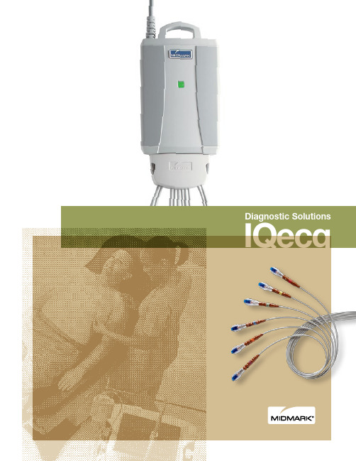
Diagnostic SolutionsIQecgPatient Cable ClipWe’ve improved on our original device with a clip designed to make the connection to thepatient cable even more robust.Integrated LoopDesigned to conveniently hang the IQecg ® in between uses forquick access.At Midmark, we are continuously focused on improving the clinical experience through thoughtful improvements that make your job easier.Clinician-inspired technologyWhy Midmark IQecg ®?Today, healthcare is rapidly evolving. There is one thing that is certain, though—electronic medical records (EMR) are here to stay. And whether that has a positive or negative impact on your practice largely depends on your clinical workflow. Midmark can help.The Midmark IQecg ® system is designed to work with today’s top EMRs in a variety of healthcare IT environments and clinical workflows to save time, reducethe likelihood of errors and improve outcomes.Whether you are using a paper chart, scanning ECG reports or otherwise connected to your EMR, Midmark IQecg ® is designed to improve your workflow.Starting with the compact portability of the IQecg ®, easily move to where the patient is located.Accessing patient information, performing tests and storing reports in the patient chart seamlessly reduces manual data entry and the risk of transcription errors.Unlike typical ECG devices, IQecg ®is designed to let you instantly capture the ECG waveform shown on-screen so you won’t miss an abnormal event when it occurs.Retrieve the report for review at any time from anywhere you can access the patient chart. IQecg ® provides on-screen tools that help you diagnose patients quickly.Real-worldimprovements to theclinical workflowPatient Lead SeparatorThe innovative patient leadseparator (patent pending) helps keep lead wires straight so you can skip the embarrassment of untangling wires and get right down to prepping the patient.IQecg® KitIQecg ® is available as a kit with all the necessary supplies for ECG testing.Easily capture what you seeIf you notice an abnormal event on-screen, capture it in the ECG test with a simple click of the mouse.Arm Lead Reversal detectionAlerts if possible arm lead reversal is detected, allowing you to verify and fix arm lead placements if needed during the test.When we asked clinicians what we could do to make a better ECG product, the answer was obvious. We needed to design a device that is easier to use and improves the clinician workflow. So that is precisely what we did. From accessing the patient chart, hooking up the patient, performing the test and seamlessly saving to the chart, the new interface is designed to make using the Midmark IQecg ® simple and efficient.A new user interface designedfor youLong rhythm stripA simple click takes you to the rhythm screen where you can capture up to six minutes of any preselected three leads.Verify then saveT o further enhance EMR workflow, you can review the quality of the test before saving it to the patient’s chart,giving you the option to retest if desired.Easy access to patient recordsAccessing the electronic patient chart without retyping patient information helps reduce potential data entry errors and the patient’s wait time. (IQmanager® Patient Data screen shown)ECG testing made easy with Midmark IQecg ®— so you will have more time to focus on your patient.The report review screen is designed with a large ECG display area similar to a traditional ECG printout for easier review of tracings.Easy, precise electronic calipersEasy-to-use electronic calipers allow physicians to precisely measure any waveforms in the review screen with a simple click of the mouse.10-second full disclosureAccess the 10-seconds screen to review the full disclosure of all 12 leads so you don’t miss a single beat. The electronic calipers measure all 12 leads with a simple click.On-screen editAdvanced on-screen tools allow you to edit theinterpretation in various ways so you can chose what fits you. You can use free-text or preset diagnostic statements. You can easily setup the more commonly used statements for faster selection during editing. (These editing tools are available on the ECG report review screen and the side-by-side comparison screen.)Side-by-side comparisonA click of the mouse allows you to compare the current ECG side-by-side with any previous ECG of the same patient so you can easily diagnose any changes.We made technology work for you instead of the other way around. The new screen layouts and color scheme, electronic calipers, on-screen side-by-side comparison and editing tools combine a traditional review workflow with technological advancements so you don’t have to abandon what works for you. Everything you need is at your fingertips.Physician workflow improvedMidmark is an ISO 13485 and ISO 9001 Certified Company.For more information or a demonstration, contact your Midmark dealer or call: 1-800-MIDMARK Fax: 1-800-365-2499Outside the U.S.A. call: 1-937-526-3662 Fax: 1-937-526-8392 or visit our website at © 2005 Midmark CorporationMidmark Corporation, Dayton, OH.Products subject to improvement changes without notice Litho in U.S.A. 48-79-0009 Rev. A (8/16)AccessoriesSuppliesUniversal ECG Clips (3 mm, 4 mm & Snap) 3-047-0001Clear ECG Clips (4 mm & Snap) 3-047-0005Disposable EZ-Trode ECG Electrodes 2-100-0205 (Box of 500)2-100-0206 (10 boxes of 500)6231 Care Exchange ® Workstation 6231-001-xxxIQcart ®3-004-1000Minimum System Requirements• Windows ®10, Windows ® 8, Windows ® 7; Professional and Enterprise; 32-bit and 64-bit• Intel ® Core ™ 2 Duo Processor E4300 or 64-bit processor or faster • 2 GB of RAM• 2 GB available hard disk space • USB port• Mouse, keyboard and high resolution display monitor Contact us or visit for more detailed system requirements.Windows is a registered trademark of Microsoft Corporation.Intel Core is a trademark of Intel Corporation in the U.S. and/or other countries.Ordering Information4-000-0062IQecg ® Digital ECG with Lead ManagementMake your practice more efficient with an IQ Digital Diagnostic SystemWorkflow efficiency and improvements in patient care are just a few of the benefits that can result from implementing an IQ Digital Diagnostic System. Call us today at 1-800-MIDMARK and we’ll put together a customized suite of solutions to help grow as your practice grows.IQecg ® Patient Cable with Lead Management 3-100-0203IQconnect™ FrameworkIQconnect™ is a cutting-edge, flexible, scalable integrationframework designed toseamlessly connect Midmark diagnostic solutions, including ECG, Holter, spirometry and vitals with EMR systems orIQmanager ® software. It also enables you to receive future software updates directly from Midmark, shortening the time from release to implementation. The latest Midmark EMR specific connectivity software and IQmanager ® are built on this new IQconnect™ framework.And if you are using a thin-client network, Midmark IQpath ® software reduces latency issues and smooth data flow for uninterrupted ECG, spirometry and vitals testing.Software and License RequiredTo ensure optimal performance of Midmark digital diagnostic products, we encourage you to contact us to see what software you will need, software licensing information and detailed system requirements.Q IQ CONNECT IQCONNECT。
ECG1200G十二道心电图机使用技术说明书

声明本公司拥有此非公开出版的说明书的版权,并有权将其作为保密资料处理。
本说明书只作为操作,保养和维修产品的参考资料。
其他人无权向他人公开此说明书内容。
本说明书包含受版权法保护的专有资料。
版权所有,未经本公司书面同意,不得对本说明书的任何部分进行照相复制、复印或翻译成其他语言。
本说明书的所有内容都被认为是正确的。
本公司对于安装错误和操作不当而造成的偶然或必然损坏不负担任何法律责任。
本公司不向其他方提供专利法所赋予的特许权。
对于违反专利法和任何第三方权利所造成的法律后果,本公司不负担法律责任。
说明书中所含的内容可以不予通知而作出变更。
目录第一部分综述 (1)第二部分安全注意事项 (3)第三部分保修规定 (4)第四部分仪器主要特点 (5)第五部分ECG1200G各界面示意图 (7)第六部分操作前基本注意事项 (10)第七部分使用仪器前的准备工作 (11)第八部分操作中注意事项 (12)第九部分记录纸的使用 (13)第十部分电极安装 (14)第十一部分仪器接地和电源的连接 (16)第十二部分电池操作注意事项 (17)第十三部分控制面板及按键说明 (18)第十四部分常见故障及排除方法 (30)第十五部分仪器维护与保养 (32)第一部分综述1.1 概述ECG1200G十二道心电图机是一种实现对12导联心电信号进行同步采集,采用热阵打印将心电波形打印输出的心电图机。
其具备以下主要功能:以手动/自动的方式记录和显示心电波形;心电波形参数的自动测量以及自动诊断;能够提示电极脱落及缺纸;可切换界面语言(中文/英文);交直流两用供电模式;可任意选择节律导联,便于观察异常的心率;病历数据库管理等功能。
800×600点八寸屏高分辨率液晶显示;1728点宽、十二道心电波形打印。
支持按键及触摸两种方式操作,方便快捷。
1.2 主要技术指标1.2.1 环境条件运行a). 环境温度:+5℃~+35℃b). 相对湿度:≤80%c). 电源:交流: 220V,50Hz直流:14.8V,3700mAh可充电锂电池d). 大气压力:86KPa~106KPa运输和贮存a). 环境温度范围-10℃~55℃;b). 相对湿度范围≤95%;c). 大气压力范围50KPa~106KPa。
MEMRSECG心电网络系统使用说明书

M E M R S E C G心电网络系统使用说明书The pony was revised in January 2021MEMRS-ECG型心电图网络系统使用说明书北京麦迪克斯科技有限公司目录前言.............................................................. 第一章概述........................................................ 第二章软件功能..................................................... 第一节预约........................................................ 第二节登记........................................................ 第三节分诊......................................... 错误!未定义书签。
第四节心电图设备连接匹配.......................................... 第五节电生理设备连接匹配.......................................... 第六节心电图报告分析系统..........................................第一小节报告系统登录 ........................................第二小节回放分析12导心电图 .................................第三小节诊断及报告 ..........................................第四小节打印 ................................................第五小节病历导入导出 ........................................ 第七节电生理系统报告工作站........................................第一小节报告系统登录 ........................................第二小节编写报告 ............................................第三小节测量波形 ............................................第四小节诊断 ................................. 错误!未定义书签。
Burdick Atria 6100 ECG产品说明书

The Burdick Atria 6100 ECG delivers the highest level of clinical performance while incorporating cutting-edge technology to streamline clinic workflow. It offers timely, high-quality, and accurate ECG results critical for patient care.High PerformanceAdvanced technology gives you the ultimate in speed and accuracy.• STABLE baseline filter decreases the level of artifact by assuring that the ECG baseline is stable with no distortion of ST measurements• Storage from 150 patient records improves workflow for busy practices• User definable, customizable output allows reporting flexibilityAtria 6100 ECG Proven performance meets high-tech design to streamline workflow Resting ECG SystemFeatures• Automatic, manual or 12-lead rhythm operation for maximum flexibility • Full color preview screen allows viewing of waveforms and fast menu navigation• Optional, powerful interpretation algorithm based on five clinically significant patient criteria, as well as pediatric analysis and pacemaker enhancement• AccuPrint™restricts ECG printout when leads are not attached correctly, ensuring a clear printout and eliminating retakes • Communications options including both wired Ethernet and wireless 802.11 provide cutting-edge connectivity capabilitiesUser-friendly InterfaceThe Atria 6100 user interface is easy to use and learn. Its structure is intuitive even for infrequent or new users.• Clear, readable color LCD • Ability to preview waveforms before analyzing• Ability to continuously monitor patient throughout procedure • Intuitive menu for fast navigation and quick results• Single button STAT printing • Full size alpha-numeric keyboard with dedicated function keysA Second Opinion That You Can Count OnThe Burdick Atria 6100 utilizes the proven University of Glasgow algorithm for interpretation,providing a silent second opinion.Unlike competitive products, which limit algorithm criteria to age, the University of Glasgow algorithm,under continuous development since the 1970s and introduced by Burdick in 1995, bases its analysis on fiveclinically significant criteria: gender, age,race, medication and classification. This is critical because ECG results for patients with different demographic profiles and medical conditions can vary greatly. For example, subtle T-wave changes that are considered abnormal for a white male may be considered normal for a black male of the same age. An ECG that would represent LVH in a 50-year-old female may be normal for a 50-year-old male. These are just two examples of hundreds of possible clinical issues that may arise when not using an algorithm that takes into account age, gender, race, medication,and classification. Units purchased without interpretation can be upgraded at any time.Atria 6100 ECGLarge color screen enhances viewingexperience. Shown actual sizeUpgradabilityThe Atria 6100 can be easily upgraded at your practice, minimizing the downtime associated with returning the system.• Add Burdick’s proven University of Glasgow algorithm interpretation• Increase ECG storage from 150 to 300 records • Extend communications options by adding Ethernet, wireless 802.11, modem, or Bluetooth3, 6 or 12-lead onscreen preview for maximum viewing flexibilityAtria 6100 menu structure maximizes ECG test efficiencyFor physicians looking for advancedcommunications and storage features, but prefer a traditional ECG, Burdick’s Atria™6100 ECG with advanced communications options combines the best of both worldsFlexible Connectivity OptionsThe Atria 6100 offers cutting-edgecommunications capabilities, including wired Ethernet and wireless 802.11.Advanced encryption tools ensure the highest level of data transmission security.Transmit Test Results to EMRsTest results can be electronicallytransmitted from the exam room to your network and imported by EMRs and cardiology management systems. No more scanning and keeping hard copies.Imagine the convenience of attaching an ECG report to the patient’s electronic medical record, keeping all information in one file. Burdick ECGs can be interfaced to most EMR systems.E-mail Colleagues Test ResultsE-mail capability allows you to send test results directly from the Atria in a PDF format to colleagues for collaborative efforts. The industry-accepted diagnostic quality PDF allows any recipient to review results without purchasing proprietary software.Print Test Results to Plain-paper PrinterSend results wired or wirelessly from the exam room to a network orstandalone plain-paper printer using options 802.11, USB, Bluetooth, orEthernet. The diagnostic-quality printouts contain all the information found on the traditional thermal paper printouts.FAA CompliantAtria 6100 with advancedcommunications includes modem transmissions that comply with FAA system requirements for ECG testing.Wired ConnectionsEthernet (RJ45)•••••S t o r e o n N e t w o r k (P D F )o r S t a n d -a l o n e P C S e n d t o E M R (P D F )P l a i nP a p e r P r i n t E m a i l (P D F )S e n d t o P y r a m i s S e n d t o F A A F ax Basic CommunicationsUpgrade*Advanced Options*Ethernet (RJ45)Fax/Modem (FAA)USB Bluetooth wireless Analog out 802.11 wirelessStandard RS232*Available at additional charge11 lbs (5 kg) (including external power supply)Color LCD; 640 x 480Full alphanumeric keypad plus designated quick keys 150 records standard; Upgrade to 300Network connection Wired:10/100 MBPS Ethernet via RJ45; Wireless:802.11g (compatible with802.11b networks)Power requirements AC operation:115/230 V AC +/-10%, 50/60 Hz at external power source;Battery operation:14.4 V NiMH rechargeable battery pack; Battery duration:300 pages continuous printing without communications optionPrintout device:216 mm thermal dot array; Paper dimensions:8-1/2 x 11(US letter) or A4; Paper type:Thermal sensitive (Burdick Assurance®or Heartline™paper recommended); Chart speeds:12.5, 25, 50 mm/sec; Gain:5, 10, 20 mm/mVChest or Limb (may be split); Printout formats:3-, 4-, 6-, or 12-channel;additional rhythm formatsLead selection:I, II, III, aVR, aVL, aVF, V1, V2, V3, V4, V5, V6, V4R; supportsCabrera and alternate chest lead (chest lead selection V2R thru V9R + V7,V8,V9);Measurement (standard),Interpretation (optional):Diagnosis, measurements andreason statements based on five demographic criteria; Meets IEC 60601-2-51;Pacemaker display capability:meets or exceeds ANSI/AAMI EC11-1991; Modes:Automatic, rhythm or manual; Frequency response:Meets or exceeds ANSI/AAMIEC11-1991 standard; Input impedance:Meets or exceeds ANSI/AAMI EC11-1991standard; Electrode offset tolerance:+/- 300 mV;A/D conversion: 8,000samples/second, 2.5 µV LSB RTI;Artifact filter response:40 Hz, -3db;Data storage resolution:500 samples/second, 2.5 µV LSB RTIEnvironmental Operating temperature: 50°F to 104°F (10°C to 40°C); Operating relativespecifications humidity: 20% to 90%, noncondensing; Operating atmospheric pressure:1060 hPa to 700 hPa (-500 ft to 10,000 ft reference to sea level); Storagetemperature:-4°F to 115°F (-20°C to 45°C); Storage relative humidity:10%to 90%, noncondensing; Storage atmospheric pressure:1060 hPa to 190 hPa(-500 ft to 40,000 ft reference to sea level)Conforms to standards ANSI/AAMI EC11-1991EN 60601-1-2:2001CAN/CSA C22.2 no. 0-M91EN 60601-1-2:2001CAN/CSA C22.2 no. 601.1S1UL 60601-1CAN/CSA C22.2 no. 601.2.25, Amend. 1EN 60601-2-51:2003CAN/USA C22.2 no. 601.1-M90EN 60601-1-4:2000Leakage current:Patient <10 µA, chassis <100 µA; Defibrillation protection:to 5000 V, 360J3 years with return of warranty cardPhone:800-777-1777,608-764-1919•Fax:608-764-7188••****************Due to continuing product innovations, specifications are subject to change without notice. AccuPrint, Atria, Burdick, CardioSens, and HeartLine are trademarks of Cardiac Science Corporation. Other brands and products are trademarks of their respective owners. ISO 13485©2006 Cardiac Science Corporation. All rights reserved.ORDERING INFORMATION Standard ConfigurationA61-1DI016100, interpretive,English, AHA accessoriesA61-1 X X XXD No communicationsE Basic communications, includesEthernet, serial, USB, analog outF Basic communications withfax modemH Basic communications with faxmodem and 802.11gR Basic communications with 802.11gI With interpretationK Measurements only01Accessory kit 1(AHA cable, letter, 120v)02Accessory kit 2(IEC cable, A4, 220v)03Accessory kit 3(AHA cable, A4, 220v)04Accessory kit 4(IEC cable, letter, 220v)Example configuration:A61-1D I01040-1500-00Upgrade Kit, storage 300040-1501-00Upgrade Kit, Bluetooth716-0237-00Assurance paper047029CardioSens Ultra II Electrodes012-0844-00Patient Cable, AHA,10 non-replaceable leads007704Patient Cable, AHA,10 replaceable leadsBurdick helps protect your investment from unforeseen service costs. The Atria 6100 has a 3-year warranty when the warranty card is returned.。
MEMRS-ECG心电网络系统使用说明书

MEMRS-ECG型心电图网络系统使用说明书北京麦迪克斯科技有限公司目录前言 (1)第一章概述 (2)第二章软件功能 (3)第一节预约 (3)第二节登记 (6)第三节分诊 (8)第四节心电图设备连接匹配 (11)第五节电生理设备连接匹配 (14)第六节心电图报告分析系统 (17)第一小节报告系统登录 (17)第二小节回放分析12导心电图 (18)第三小节诊断及报告 (22)第四小节打印 (23)第五小节病历导入导出 (24)第七节电生理系统报告工作站 (25)第一小节报告系统登录 (25)第二小节编写报告 (26)第三小节测量波形 (27)第四小节诊断 (27)第五小节打印 (29)第八节合并病历 (32)第九节查询功能 (34)第三章使用环境和注意事项 (36)前言MEMRS-ECG型心电网络系统软件是一套功能完善、技术先进的心电信号分析系统。
为了正确的使用本仪器,充分发挥它的各项功能,请仔细阅读本说明书。
如果有任何疑问,请与麦迪克斯公司或分销商联系。
本说明书旨在介绍如何正确使用MEMRS-ECG心电网络系统。
麦迪克斯公司执行的是不断发展的政策,因此麦迪克斯公司保留在没有事先通知的情况下,对本手册说明中的产品进行修改变动的权利。
本说明书的修改、解释权属于麦迪克斯公司。
MEMRS-ECG心电网络系统为高技术、高质量的设计实体。
在正确的操作和使用环境下,一般无须过多维护。
如果您在使用中有什么问题,请与麦迪克斯公司直接联系,或与您所购买MEMRS-ECG 心电网络系统的销售商联系。
我们将竭诚为您服务!联系方式:北京麦迪克斯科技有限公司地址:北京市海淀区安宁庄西路9号金泰富地大厦1005室邮编:100085:(010)62974855传真:(010)62978861维修热线:400 8100 559Website: .medextech.E-mail: supportmedextech.第一章概述MEMRS-ECG心电网络系统是一套功能完善、技术先进的心电图分析管理系统,为医院心电检查建立数字化工作平台,实现心电图检查的数字化、流程化管理过程。
ECG 电心电图设备说明书

FLUTTER ATRIALE
L’attività atriale è riconoscibile Le onde P assumono un aspetto a denti di sega Il nodo AV limita il passaggio dei segnali Il ritmo è regolare
3 - Attività Atriale
L’onda P non c’è, l’atrio è spento L’onda P ha un aspetto diverso dal normale NB! Cura il paziente, non il monitor
3 - Attività Atriale
Blocco AV di 2° grado - Tipo 2
Mobitz 2
Blocco AV di 3° grado
Nodo AV
STOP
Blocco AV di 3° grado
Blocco AV di 3° grado
•…Nessuna correlazione - dissociazione atrioventricolare
4 - Rapporto P - QRS
tratto P-QRS
max 0,20 sec (5 quadretti)
NB! Cura il paziente, non il monitor
Blocco AV di 1° grado
Nodo AV
Blocco AV di 1° grado
Blocco AV di 2° grado - Tipo 1
Gli elettrodi
Dove piazzare gli elettrodi adesivi • su una superficie ossea • dove la cute è glabra • dove la cute è meno sudata
Keep-it-Easy ECG System用户指南说明书

Keep-it-Easy ECG System User ManualEdition: Ver1.01. General Introduction............................................................................ - 4 -2. Requirements For Running Keep-it-Easy ECG System .................... - 4 -3. Installation............................................................................................ - 4 -4. Software Operation.............................................................................. - 7 -4.1 Main menu ....................................................................................................................... - 8 -4.2 Main Functions Introduction ...................................................................................... - 9 -4.2.1 Patient Records ............................................................................................................ - 9 -4.2.2 Real Time View............................................................................................................ - 10 -4.2.3 ECG Strip View............................................................................................................ - 13 -4.2.4 Analysis View.............................................................................................................. - 14 -4.2.5 Help .............................................................................................................................. - 18 -4.2.6 Register........................................................................................................................ - 18 -4.2.7 About System.............................................................................................................. - 19 -4.2.8 Close ............................................................................................................................ - 19 -5. Uninstall ............................................................................................. - 19 -Thank you for purchasing the Keep-it-Easy ECG System software which is the data management software for our ECG monitor. Before use, please read this manual carefully.Beijing Choice Electronic Technology Co., Ltd. (hereinafter called Beijing Choice) owns all rights to this unpublished work and intends to maintain this work as confidential. Beijing Choice may also seek to maintain this work as an unpublished copyright. No part of this can be disseminated for other purposes. In the event of inadvertent or deliberate publication, Beijing Choice intends to enforce its right to this work under copyright laws as a published work. Anyone who is not authorized by Beijing Choice cannot copy, use, or disclose the information in this manual. Content of the manual is subject to change without prior notice. All rights reserved.DeclarationAll information contained in this publication is believed to be correct. Choice shall not be liable for errors contained herein nor for incidental or consequential damages in connection with the furnishing, performance, or use of this material. This publication may refer to information and protected by copyrights or patents and does not attorn any license under the patent rights of Beijing Choice, nor the rights of others. Beijing Choice does not assume any liability arising out of any infringements of patents or other rights of third parties.1. General IntroductionKeep-it-Easy ECG System is the application software running on a computer in conjunction with the handheld ECG monitor. You can use the software on your computer to analyze, display, replay, export, import and print data transferred from the ECG monitor.2. Requirements for Running Keep-it-Easy ECG SystemTo run the Keep-it-Easy ECG System, your computer requires a hardware configuration with a minimum of PIII500 CPU, 64M internal memory, CD-ROM drive, a minimum of a 10G hard disc.Requirement for Operating System:Microsoft Windows 2000/XP/Vista operating system.3. Installation3.1 Insert the attached installation CD into your computer’s CD-ROM driver.3.2 Single click the “setup.exe” files to access the installing interface as in Fig.3-1.Fig.3-13.3 Click the “Install Keep-it-Easy ECG System” button to install the software. Refer tosteps 3.3.1 ~3.3.6 for installation instructions:Fig.3-2 Click “Next>”3.3.2Fig.3-3 Click “Next>”Fig.3-4 Click “Next>”3.3.4Fig.3-5 Click “Next>”3.3.5Fig.3-6 Click “CloseIn the interface as in Fig.3-6 click the “Close” on to finish the installation steps.3.3.6 After finishing installation of the software, a shortcut icon “Keep-it-Easy ECGSystem” will appear on your computer’s desktop, refer to Fig.3-7. You can run it by double-clicking the shortcut icon or by clicking the “Keep-it-Easy ECG System” menu item in the “Start” menu of your PC.Fig. 3-74. Software OperationThe Keep-it-Easy ECG System is the application software running on a computer together with the ECG monitor. It will only allow one ECG monitor to be connected to the computer for data transmission at a time.You can use the software for reviewing and analyzing data that was previously uploaded to it without having an ECG monitor attached to your computer.The required computer resolution is 1024×768 or higher.4.1 Main menuDouble-click the “Keep-it-Easy ECG System” icon to run the software; the main menu will be displayed as shown in Fig. 4-1.Fig. 4-1Description of Fig. 4-1::Analysis ViewBy accessing this menu item, you can review data and waveform in continuous measurement mode.:ECG Strip ViewBy accessing this menu item, you can review data and waveform in easy mode.:Real Time ViewBy accessing this menu item, you can view real-time data and waveform.:Patient RecordsBy accessing this menu item, you can manage patients’ records.By accessing this menu item, you can learn some general ECG knowledge.:Register (Optional)By accessing this menu item, you can activate the function of the analysis of arrhythmia type by inputting correct register code.:About SystemBy accessing this menu item, you can get some information about the software system.:CloseBy clicking the “Close” icon, you will exit from the software system.4.2 Main Functions Introduction4.2.1 Patient RecordsClick the icon in the main menu, the window displayed in Fig. 4-2 will pop up. In this window, the user can modify the patient’s information including name, gender and birth date and so on, but the patient ID cannot be modified. The user can also delete any existing patient’s information and add new ones.Fig. 4-2Screen Description:On the Patient Records screen, the user can modify and delete any existing patientlist; the right area of the screen is the detailed patient information for the selected stored patient archive. The following are descriptions of each field:Patient ID:Displays the patient ID, the length of Patient ID is less than 10 characters.Name:Displays the patient’s name; the length of the name should less than 40 characters.Gender:Displays the gender of the patient. Select Male or Female from the drop-down list.Birth Date: Displays the patient birth date, the length of the birthday should less than 40 characters.Height:Displays the patient’s height; the length of the height should less than 10 characters.Weight:Displays the patient’s weight; the length of the weight should less than 10 characters.Telephone:Patient’s telephone number; the length of the phone number should less than 40 characters.Address:Patient’s address; the length of the address should less than 60 characters.Allergies:Description of patient’s allergy symptoms; the length of the allergy field should less than 60 characters.Case History:Description of ECG diagnostic result; the length of words should less than 200 characters.Functional buttons:Add: You can add new patient’s information by pressing this button.Modify: Modify the selected patient’s information from the patient list.Delete: Delete the patient and his/her data selected from the patient list.Back: Exit from the current screen.Note: The Patient ID cannot be changed once saved.4.2.2 Real Time ViewOn the main menu, click the icon to enter the submenu interface, refer to Fig. 4-3. In this interface, the user can view the real-time data measured by the ECG monitor and save them into the PC for reviewing and management.1. Please connect the ECG monitor to the PC well before activating this function.Fig. 4-32. To save the real-time transferred data, be sure to check the “Real-time save”check-box, or else the data will not be saved.3. After checking the “Real-time Save” check-box, click the Start button, “Select theUser” pop-up window will be displayed, refer to Fig. 4-4. Select a user that youwould like to save data for.Note: If you want to save the real-time measured data, please be sure to check the “Real-time save” box in the interface as shown in Fig. 4-4, or else, the data will not besaved.Fig.4-4Select a patient from the table and click the “OK” button, then the data transmission will begin and the transferred data will be saved in the selected user archive. Refer to Fig.4-5.Fig.4-5The real-time data is shown as in Fig. 4-5. “HR” is the heart rate. The area of “Arrhythmia” will display the arrhythmia analysis result if you additionally purchase the register function. “Lead Status” indicates the connection status of the current lead. “ECG Speed” is the scan speed of ECG waveform. The rest items are patient information.Note: if you want to purchase the register function, please contact our company.Functional buttons:There are three functional buttons including Start, Stop and Return.Start: Starts the real-time ECG recording and transmission from the ECG monitor to the software.Stop: This button allows the user to terminate the real-time monitoring.Back: Exit from the current screen.If the real-time monitoring is interrupted because of any other reason, the system may prompt you with the following window as in Fig. 4-6. You can restart the transmission process by clicking the “Start” button after checking the connection between the ECG monitor and your PC.Fig. 4-64.2.3 ECG Strip ViewBefore performing the function, please make sure the monitor has been connected to PC with USB cable and the monitor has been activated and the power of batteries is sufficient.Click the icon in the main interface to access its submenu interface, and then click the “Read Data” button, the software system will begin to read data from the ECG monitor. Refer to Fig. 4-7.Fig. 4-7Note: Please do not disconnect the USB cable or operate the monitor so as to keep the data transferring continually during the data transferring.After finishing data transmission, the system may prompt that the data transmission has been finished successfully! The data that has been transferred into the software will be displayed in the left area of the interface. When the reading is finished, you can select some data item(s) and save them into archive by clicking the “Import ECGs” button. A window as shown in Fig. 4-4 will pop up; you can add new users by clicking the “Add User” button. Itsspecific operations are the same as the introduction in “4.2.1 Patient Records”.Note: Please make sure to save the transferred data items into the software by clicking the “Import ECGs” button, or else, the data may fail to be saved into the software when exiting from the current interface.4.2.4 Analysis View4.2.4.1 Data ReplayClick the “” icon to enter the submenu interface. And then click the “” icon of the toolbar which is on the top of the interface to replay ECG waveform. The interface is as shown in Fig. 4-8.When you want to view someone’s ECG wave, select his/her archive in archives list on the right by clicking its row. And then all the ECG belonging to this user will be displayed on the left area. Every row of contractible ECG wave on the lower left displays ECG wave for 30 seconds.Fig. 4-8From the interface as shown in Fig.4-8, the user may review the ECG wave measured in the continue mode. For example as in Fig.4-8, the user can use a green selecting box toselect a section of ECG wave. Shift mouse and click any dot in ECG wave can pitch on the section of ECG wave, then its amplificatory wave will be displayed on the above wave box and its corresponding record information (its heart rate value and measuring “Time” of the original record) will be shown on the right area of the interface.V-scale Amplify: The user can review the different proportional display of the section of ECG wave by setting the display scale. By clicking the “Last Page” or the “Next Page” button, the user can view the previous or next page data items.Additionally, measurement analysis and patient(s)’ information also may be displayed on the right area.What’ s more, the user can print the current selected record and ECG wave or the current selected page of records and ECG wave by clicking the “Print Report” button.4.2.4.2 Record ManagementClick the “” icon of the toolbar to switch to the “ECG record management” interface, such as in Fig.4-9.Select a patient archive whose data you want to view in the table on the left top of the interface, the data items belonging to this patient will appear in the table on the lower left of the interface. Refer to Fig.4-9.You can also select a data item that you want to view from the list that located in the left area of the interface, and then the corresponding ECG wave will be displayed on the right area. Every data item displays ECG wave for 30 seconds. Refer to Fig.4-9.Fig.4-9When there are too many users in the archive list, the user can use the function of “Query” to search the user archive that is needed. Two query conditions are provided by the system, Patient ID and Name. Input the correct patient ID or name into the edit-box on the right, and then click the “Query” button to search the corresponding patient.Functional buttons:Delete: The user can delete the current record(s) of the current user by clicking this button.Import: By clicking this button, the user can import the record(s) that has been saved on the PC to the software for reviewing and management.Export: By clicking this button, the user can export the current record(s) of the current user to PC with the extension of .cEcg or .bmp.4.2.4.3 ECG waveform analysisClick the “” icon of the toolbar to switch to the ECG waveform analysis interface asshown in Fig.4-10.Fig.4-10Description of Fig.4-10:The patient information table is on the left side of ECG waveform. Select the target in it, and then the right will show the corresponding ECG waveform. According to these waveforms, the ECG waveform analysis will be shown above, including the results of HR, ST, PR, ORS, QT and QTC. You can also choose a specific part of the waveforms to see the analysis by clicking on the waveforms.Functional buttons:Print analysis: The user can print the ECG analysis results by clicking the “” icon in the toolbar.Save analysis as picture: The user can save the analysis as picture to the PC with the extension of .bmp, .jpeg, .gif, .tiff, or .png by clicking the “” icon inthe toolbar.Email picture to: The user can email the analysis as picture to the mailbox you input by clicking the “” icon in the toolbar.Print preview: The user can preview the ECG waveforms and analysis before printing by clicking the “” icon in the toolbar. The preview interface as shown inFig.4-11.Fig.4-114.2.5 HelpYou can obtain the troubleshooting instructions and general knowledge about ECG byclicking the icon in the main menu interface.4.2.6 RegisterThe arrhythmia type analysis function is optional and not included in the standard package. If you have bought the function, you need to apply a register code which is necessary to activate the arrhythmia type analysis function.You can see the following interface in the “Version” menu of the ECG monitor. Refer to Fig.4-12. Return the SN code to our company for Register Code, then you will gain theRegister Code from us.How to activate ECG analysis function of the ECG monitor:Access the dialog box as shown in Fig.4-13 by clicking the icon of “Register”, andinput the Register Code in the editable box, then click the “OK” button.Fig. 4-134.2.7 About SystemClick icon to access the information interface.4.2.8 CloseClick icon to exit from the Keep-it-Easy ECG System.5. UninstallClick the “Start” menu of Windows system to find the Keep-it-Easy ECG System program group, then click the “Uninstall” item and the following prompt will appear as in Fig.5-1. Click the “Yes” button in Fig.5-1, then the uninstall will be finished.Fig. 5-1Manufacturer address:Beijing Choice Electronic Technology Co.,Ltd.Bailangyuan Building BRm. 1127-1128,Fuxing Road, A36100039 BeijingPEOPLE’S REPUBLIC OF CHINA。
ECG系统说明书
差分输入范围:±8mV
输出方式:串行接口,8位数据位,1为停止位,非校验
内置数字滤波:
工频滤波器:关闭或陷波频率50、60Hz可选
高通滤波器:时间常数0.3、0.5、0.8、1.6、3.3s可选
低通滤波器:截止频率26、36、45、54、60、77、150Hz可选
三.应用
心电图机/工作站
五.参数
心电图采样模块的供电应该在下表范围内,否则可能引起永久性损坏。除非另外说明,下列参数的测试条件为:常温、+5V供电、屏蔽驱动和右腿驱动均使用。
参数
条件
最小 典型 最大
单位
模拟通道
8
采样频率
每个通道
2400
Dot
数据输出速率
每个通道、不同导联体系
500/1000
Dot
数据输出位数
每个通道
12
Bit
D8
C4 (0=正常,1=脱落)
D9
C5 (0=正常,1=脱落)
起搏检测导联[D11…D9]
0 1 2 3 4 5 6 7
II III V1 V2 V3 V4 V5 V6
D10
C6 (0=正常,1=脱落)
D11
导联体系(0=Wilson,1=Frank)
D12
基线复位(0=正常,1=复位)
备用
D13
77
60
54
45
36
26
九.数据格式
当发出波形数据传输命令后,模块连续上传采样的数据,每2ms发送一个数据包,每包16字节。
如果模块采用Wilson体系送数,数据包内包括II、III、V1-V6导联的数据,由上位机计算I、avR、avL和avF的数据,计算方法见后面的说明。
心电图(ECG)监测设备操作指南说明书

Types of ECG artifact Motion artifact
Reducing the upper cutoff frequency filter from 150 to 40 Hz reduces muscle artifact
Muscle tremor: High frequency (20-150 Hz) and/or medium frequency (3-5 Hz) If shivering, cover with a blanket Move limb electrode elsewhere on limb to avoid culprit muscle Abrade skin if there is motion artifact
Check for dried gel Abrade skin Have patient lie still
and stop talking
Transport: Medium frequency (3–15 Hz)
Check for dried gel Abrade skin Stop ambulance to get
Caused by dried gel, excessive hair, poor adhesion or when breaks in connectivity occur anywhere between the electrode and the monitor
科诺监护仪使用说明.doc
科诺监护仪使用说明
标识:ECG 心电;SPO2血氧饱和度;NIBP 无创血压TEMP体温;编码器;接地端子功能键;
SETUP;设置选择键;MODE返回键;
MUTE报警开关键;PAUSE冻结键;
PRINT打印键;NIBP无创血压测量键。
软件界面:
ECG心电图;RESP呼吸;TEMP体温;
NIBP无创血压;SPO2血氧饱和度;IBP有创血压;
HR心率;RR呼吸率;PR脉率;ART主动脉压;
PA肺动脉压;LAP左房压;RAP右房压;
ICP颅内压;CVP中心静脉压;P1辅助压力通道1;
P2辅助压力通道2;CV A呼吸心动干扰。
心电导联线:
红R—右锁骨中线与第2肋骨之交点
黄L—左锁骨中线与第2肋骨之交点
绿F—左下腹
黑N—右下最后一根肋骨边缘处
白C—胸电极
编码器:屏幕上随着编码器转动而移动的长方形标志称为光标。
凡是光标可以停留的地方都可以进行操作。
1、把光标移到要操作的项上;
2、按下编码器;
3、系统弹出四种情况:菜单或测量窗口或原来菜单被新菜单替代;带底色的光标变成无底色框,可改动;出现“√”选择标志;立即执行此功能。
报警开关键:按下此键少于2秒,进入报警暂停状态;大于2秒进入静音状态;再次按下恢复所有声音。
ECG系统说明书
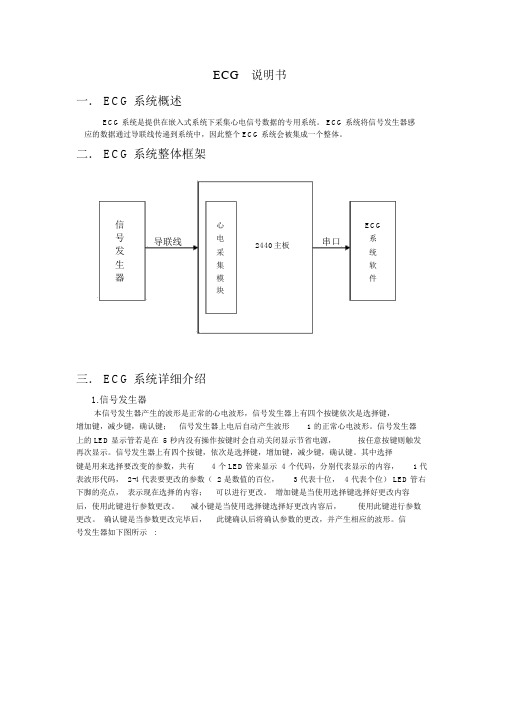
ECG说明书一. ECG 系统概述ECG系统是提供在嵌入式系统下采集心电信号数据的专用系统。
ECG系统将信号发生器感应的数据通过导联线传递到系统中,因此整个 ECG系统会被集成一个整体。
二. ECG 系统整体框架信心ECG号导联线电2440主板串口系发采统生集软器模件块三. ECG 系统详细介绍1.信号发生器本信号发生器产生的波形是正常的心电波形,信号发生器上有四个按键依次是选择键,增加键,减少键,确认键;信号发生器上电后自动产生波形 1 的正常心电波形。
信号发生器上的 LED显示管若是在 5 秒内没有操作按键时会自动关闭显示节省电源,按任意按键则触发再次显示。
信号发生器上有四个按键,依次是选择键,增加键,减少键,确认键。
其中选择键是用来选择要改变的参数,共有 4 个 LED管来显示 4 个代码,分别代表显示的内容, 1 代表波形代码, 2-4 代表要更改的参数( 2 是数值的百位, 3 代表十位, 4 代表个位) LED管右下脚的亮点,表示现在选择的内容;可以进行更改。
增加键是当使用选择键选择好更改内容后,使用此键进行参数更改。
减小键是当使用选择键选择好更改内容后,使用此键进行参数更改。
确认键是当参数更改完毕后,此键确认后将确认参数的更改,并产生相应的波形。
信号发生器如下图所示 :2.导联线导联线是用来将信号发生器产生的数据传送到主板上,由于心电噪声背景比较强, 测量条件比较复杂,因此选择导联线的精度要求较高。
导联线如下图所示:3.心电采集模块采样模块是专门为采样心电图而设计的,它有八个差分信号输入通道,由于模拟输入端具有高达 100M的输入阻抗,所以它可以直接连接高阻信号源,模块以串行方式输出采样的数据。
具体实际参数设置可参见附录。
4.ECG 系统软件4.1 硬件环境运行环境 : 2440开发板(接入电源是12 伏 )运行操作系统 : wince5.0存储位置 : 2G SD卡4.2 软件编程环境Visual Studio 2008专业版4.3 软件编程语言VC++4.4 ECG 系统软件简单介绍其中”Type”下拉列表中是有12 组心电图的波形对应的名称,分别是I,II,III,avR,avL,avF,V1,V2,V3,V4,V5,V6,您在测量显示波形时必须要选择您想看到的波形的名称。
ECGGateway 安装说明书(Win7)-1
ECGGateway安装说明书1基础环境普通PC一台。
日本光电心电机ECG1250或ECG1350。
ECG-1250/1350专用USB-LAN转接口一个或无线网卡一块。
2安装ECGGateway首先必须安装vcredist_x86.exe 。
然后运行ECGGateway.msi,依照画面提示进行安装即可。
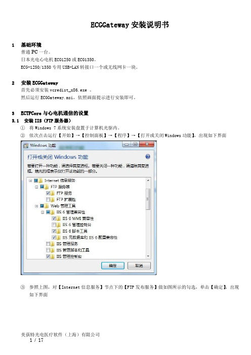
3ECTPCore与心电机通信的设置3.1安装IIS(FTP服务器)①将Windows 7系统安装盘置于计算机光驱内。
②依次点击运行【开始】→【控制面板】→【程序】→【打开或关闭Windows功能】,出现如下界面③参照上图,对【Internet信息服务】节点下的【FTP发布服务】做如图所示的勾选,单击【确定】,出现如下界面④上图所示界面在安装完成后自动退出,此时FTP服务器安装完毕,可取出光驱中的系统安装盘。
3.2设置IIS(FTP服务器)①依次点击运行【开始】→【控制面板】→【系统和安全】→【管理工具】→【Internet信息服务(IIS)管理器】出现如下界面②右击【网站】,弹出菜单中选择【添加FTP站点】,出现如下界面③设定FTP站点名称;(本例中名为“Default FTP Site”)设定物理路径,并确保该目录确实存在。
(本例中使用“C:\inetpub\ftproot”)单击【下一步】,出现如下界面④勾选【自动启动FTP站点】和“SSL”中的【无】,单击【下一步】,出现如下界面。
⑤“允许访问”一栏中,选择【所有用户】,“权限”中,勾选【读取】和【写入】两项。
单击【完成】,退出新建向导。
⑥回到步骤一所示的界面中,如下图面⑧在【防火墙的外部IP地址】一栏中,写入当前计算机在局域网中的IP地址。
输入完毕后,单击界面右侧“操作”栏中的【应用】。
⑨展开界面左侧“连接”栏中的“网站”节点,选定前文第一步至第五步所新建的FTP站点。
在中间的视图区中点击运行【FTP防火墙支持】,出现如下界面⑩在【防火墙的外部IP地址】一栏中,写入当前计算机在局域网中的IP地址。
ECG1200G十二道心电图机使用技术说明书

声明本公司拥有此非公开出版的说明书的版权,并有权将其作为保密资料处理。
本说明书只作为操作,保养和维修产品的参考资料。
其他人无权向他人公开此说明书内容。
本说明书包含受版权法保护的专有资料。
版权所有,未经本公司书面同意,不得对本说明书的任何部分进行照相复制、复印或翻译成其他语言。
本说明书的所有内容都被认为是正确的。
本公司对于安装错误和操作不当而造成的偶然或必然损坏不负担任何法律责任。
本公司不向其他方提供专利法所赋予的特许权。
对于违反专利法和任何第三方权利所造成的法律后果,本公司不负担法律责任。
说明书中所含的内容可以不予通知而作出变更。
目录第一部分综述 (1)第二部分安全注意事项 (3)第三部分保修规定 (4)第四部分仪器主要特点 (5)第五部分ECG1200G各界面示意图 (7)第六部分操作前基本注意事项 (10)第七部分使用仪器前的准备工作 (11)第八部分操作中注意事项 (12)第九部分记录纸的使用 (13)第十部分电极安装 (14)第十一部分仪器接地和电源的连接 (16)第十二部分电池操作注意事项 (17)第十三部分控制面板及按键说明 (18)第十四部分常见故障及排除方法 (30)第十五部分仪器维护与保养 (32)第一部分综述1.1 概述ECG1200G十二道心电图机是一种实现对12导联心电信号进行同步采集,采用热阵打印将心电波形打印输出的心电图机。
其具备以下主要功能:以手动/自动的方式记录和显示心电波形;心电波形参数的自动测量以及自动诊断;能够提示电极脱落及缺纸;可切换界面语言(中文/英文);交直流两用供电模式;可任意选择节律导联,便于观察异常的心率;病历数据库管理等功能。
800×600点八寸屏高分辨率液晶显示;1728点宽、十二道心电波形打印。
支持按键及触摸两种方式操作,方便快捷。
1.2 主要技术指标1.2.1 环境条件运行a). 环境温度:+5℃~+35℃b). 相对湿度:≤80%c). 电源:交流: 220V,50Hz直流:14.8V,3700mAh可充电锂电池d). 大气压力:86KPa~106KPa运输和贮存a). 环境温度范围-10℃~55℃;b). 相对湿度范围≤95%;c). 大气压力范围50KPa~106KPa。
GE Healthcare MAC 2000 ECG 分析系统简介说明书

Expert GE Healthcare service and support are always just a click or call away—our extensive network of trained service pros is ready so you don’t have to wait for help. Many issues can even be resolved remotely, keeping downtime to a minimum. It’s another way the MAC 2000 embraces simplicity to give you a hassle-free experience.
• Easy-to-read 7" color display is configurable to meet your needs
• Multiple connectivity and export options for easy data access and storage
• Available in exercise testing configuration
†Marquette 12SL ECG Analysis Program Physician’s Guide. 2036070-006 Revision A. 2010. GE Healthcare: Milwaukee, WI.
ECG-1000 心电管理系统用户手册(单机版)

注意 您应当了解的在使用过程中,软件使用设备或其他设备可能遭受的损害,如设备故障、失灵或损坏等。
MEMRS-ECG心电网络系统使用说明书

MEMRS-ECG型心电图网络系统使用说明书麦迪克斯科技目录前言 (1)第一章概述 (2)第二章软件功能 (3)第一节预约 (3)第二节登记 (6)第三节分诊 (8)第四节心电图设备连接匹配 (11)第五节电生理设备连接匹配 (14)第六节心电图报告分析系统 (17)第一小节报告系统登录 (17)第二小节回放分析12导心电图 (18)第三小节诊断及报告 (22)第四小节打印 (23)第五小节病历导入导出 (24)第七节电生理系统报告工作站 (25)第一小节报告系统登录 (25)第二小节编写报告 (26)第三小节测量波形 (27)第四小节诊断 (27)第五小节打印 (29)第八节合并病历 (32)第九节查询功能 (34)第三章使用环境和注意事项 (36)前言MEMRS-ECG型心电网络系统软件是一套功能完善、技术先进的心电信号分析系统。
为了正确的使用本仪器,充分发挥它的各项功能,请仔细阅读本说明书。
如果有任何疑问,请与麦迪克斯公司或分销商联系。
本说明书旨在介绍如何正确使用MEMRS-ECG心电网络系统。
麦迪克斯公司执行的是不断发展的政策,因此麦迪克斯公司保留在没有事先通知的情况下,对本手册说明中的产品进行修改变动的权利。
本说明书的修改、解释权属于麦迪克斯公司。
MEMRS-ECG心电网络系统为高技术、高质量的设计实体。
在正确的操作和使用环境下,一般无须过多维护。
如果您在使用中有什么问题,请与麦迪克斯公司直接联系,或与您所购买MEMRS-ECG 心电网络系统的销售商联系。
我们将竭诚为您服务!联系方式:麦迪克斯科技地址:市海淀区安宁庄西路9号金泰富地大厦1005室邮编:100085:(010)62974855传真:(010)62978861维修热线:400 8100 559Website: .medextech.E-mail: supportmedextech.第一章概述MEMRS-ECG心电网络系统是一套功能完善、技术先进的心电图分析管理系统,为医院心电检查建立数字化工作平台,实现心电图检查的数字化、流程化管理过程。
MEMRS-ECG心电网络系统使用说明书

MEMRS-ECG型心电图网络系统使用说明书麦迪克斯科技目录前言 (1)第一章概述 (2)第二章软件功能 (3)第一节预约 (3)第二节登记 (6)第三节分诊 (8)第四节心电图设备连接匹配 (11)第五节电生理设备连接匹配 (14)第六节心电图报告分析系统 (17)第一小节报告系统登录 (17)第二小节回放分析12导心电图 (18)第三小节诊断及报告 (22)第四小节打印 (23)第五小节病历导入导出 (24)第七节电生理系统报告工作站 (25)第一小节报告系统登录 (25)第二小节编写报告 (26)第三小节测量波形 (27)第四小节诊断 (27)第五小节打印 (29)第八节合并病历 (32)第九节查询功能 (34)第三章使用环境和注意事项 (36)前言MEMRS-ECG型心电网络系统软件是一套功能完善、技术先进的心电信号分析系统。
为了正确的使用本仪器,充分发挥它的各项功能,请仔细阅读本说明书。
如果有任何疑问,请与麦迪克斯公司或分销商联系。
本说明书旨在介绍如何正确使用MEMRS-ECG心电网络系统。
麦迪克斯公司执行的是不断发展的政策,因此麦迪克斯公司保留在没有事先通知的情况下,对本手册说明中的产品进行修改变动的权利。
本说明书的修改、解释权属于麦迪克斯公司。
MEMRS-ECG心电网络系统为高技术、高质量的设计实体。
在正确的操作和使用环境下,一般无须过多维护。
如果您在使用中有什么问题,请与麦迪克斯公司直接联系,或与您所购买MEMRS-ECG 心电网络系统的销售商联系。
我们将竭诚为您服务!联系方式:麦迪克斯科技地址:市海淀区安宁庄西路9号金泰富地大厦1005室邮编:100085:(010)62974855传真:(010)62978861维修热线:400 8100 559Website: .medextech.E-mail: supportmedextech.第一章概述MEMRS-ECG心电网络系统是一套功能完善、技术先进的心电图分析管理系统,为医院心电检查建立数字化工作平台,实现心电图检查的数字化、流程化管理过程。
Glasgow ECG 分析程序及 LIFEPAK 15 监测器 抑制器产品说明书
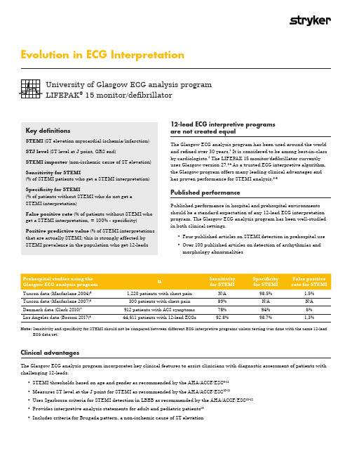
Evolution in ECG InterpretationKey definitionsSTEMI (ST elevation myocardial ischemia/infarction)STJ level (ST level at J point, QRS end)STEMI imposter (non-ischemic cause of ST elevation)Sensitivity for STEMI(% of STEMI patients who get a STEMI interpretation)Specificity for STEMI(% of patients without STEMI who do not get a STEMI interpretation)False positive rate (% of patients without STEMI who get a STEMI interpretation, = 100% - specificity)Positive predictive value (% of STEMI interpretations that are actually STEMI; this is strongly affected by STEMI prevalence in the population who get 12-leadsUniversity of Glasgow ECG analysis program LIFEPAK ® 15 monitor/defibrillatorTuscon data (Macfarlane 2004)51,220 patients with chest pain N/A 98.5% 1.5%Tuscon data (Macfarlane 2007)6300 patients with chest pain 89%N/A N/A Denmark data (Clark 2010)7912 patients with ACS symptoms 78%94%6%Los Angeles data (Bosson 2017)844,611 patients with 12-lead ECGs92.8%98.7%1.3%Note: S ensitivity and specificity for STEMI should not be compared between different ECG interpretive programs unless testing was done with the same 12-leadECG data set.Clinical advantagesThe Glasgow ECG analysis program incorporates key clinical features to assist clinicians with diagnostic assessment of patients with challenging 12-leads.• STEMI thresholds based on age and gender as recommended by the AHA/ACCF/ESC 9-11• Measures ST level at the J point for STEMI as recommended by the AHA/ACCF/ESC 10-11• Uses Sgarbossa criteria for STEMI detection in LBBB as recommended by the AHA/ACCF/ESC 10-12• Provides interpretive analysis statements for adult and pediatric patients 13• Includes criteria for Brugada pattern, a non-ischemic cause of ST elevation12-lead ECG interpretive programs are not created equalThe Glasgow ECG analysis program has been used around the world and refined over 30 years.1 It is considered to be among best-in-class by cardiologists.2 The LIFEPAK 15 monitor/defibrillator currently uses Glasgow version 27.3,4 As a trusted ECG interpretive algorithm, the Glasgow program offers many leading clinical advantages and has proven performance for STEMI analysis.3-8Published performancePublished performance in hospital and prehospital environments should be a standard expectation of any 12-lead ECG interpretation program. The Glasgow ECG analysis program has been well-studied in both clinical settings.• Four published articles on STEMI detection in prehospital use • Over 100 published articles on detection of arrhythmias and morphology abnormalitiesthe true J point measurement for STEMI0.2mV (2 mm)overestimated ST elevationSgarbossa criteria for STEMI analysis in left bundle branch block (LBBB)• LBBB can increase the risk of a false negative STEMI interpretation• LBBB is also a “STEMI imposter” and increases the risk of false positive STEMI interpretation • It occurs in approximately 1 in 2,000 patients• A distinct coved-type ST elevation occurs in the right precordial leads • It is also a “STEMI imposter” and increases the risk of false positive STEMI interpretation• The Glasgow program uses Brugada pattern criteriaaccording to the Second Consensus Conference on the Brugada Syndrome 4,1412-lead ECG interpretive algorithm Glasgow v27.0Inovise 12L v1.00DXL vPH100BPediatric interpretation Yes No Yes LBBB criteria for STEMIYes No Yes ST measurement taken at the J point Yes No Yes Published results from testing with prehospital ECGs4 studies1 studyNo12-lead comparison for a pediatric patientPediatric interpretation for a 10-year-old patient• The Glasgow ECG analysis program gives an appropriate pediatric interpretationAdult interpretation for the same 10-year-old patient• Same 10-year old pediatric patient, but taken after entering an adult age of 18 years• Interpreting a pediatric 12-lead using criteria for adults can produce inappropriate interpretative statements • At least one ECG analysis program is contraindicated for pediatric interpretation15,1612-lead with interpretative statement for STEMI with LBBB• Glasgow ECG analysis program uses Sgarbossa criteria for STEMI detection in a patient with a LBBB©2018 Physio-Control, Inc. All names herein are trademarks or registered trademarks of their respective owners. GDR 3335513_CAll claims valid as of June 2018.Physio-Control is now part of Stryker.For further information, please contact Physio-Control at 800.442.1142 (U.S.), 800.668.8323 (Canada) or visit our website at Physio-Control Headquarters 11811 Willows Road NE Redmond, WA 98052 Customer Support P. O. Box 97006Redmond, WA 98073Toll free 800 442 1142Fax 800 426 8049Physio-Control CanadaPhysio-Control Canada Sales, Ltd.45 Innovation Drive Hamilton, ON L9H 7L8CanadaToll free 800 668 8323Fax 877 247 792512-lead with Brugada interpretative statement•The ST elevation is correctly attributed to the Brugada patternReferences1. Macfarlane P, Devine B, Clark E. The University of Glasgow (Uni-G) ECG analysis program. Computers in Cardiology. 2005;32:451-454.2. Willems J, Abreu-Lima C, Arnaud P, et al. The diagnostic performance of computer programs for the interpretation of electrocardiograms. N Engl J Med.1991;325:1767-73. 3. Glasgow 12-lead ECG Analysis Program: Statement of Validation and Accuracy. Redmond, WA: Physio-Control; 2009. 4. Glasgow 12-lead ECG Analysis Program: Physician’s Guide. Redmond, WA: Physio-Control; 2009.5. Macfarlane P, Browne D, Devine B, et al. Modification of ACC/ESC criteria for acute myocardial infarction. J Electrocardiol. 2004;37(suppl):98-103.6. Macfarlane P, Hampton D, Clark E, et al. Evaluation of age and sex dependent criteria for ST elevation myocardial infarction. Computers in Cardiology.2007;34:293-6. 7. Clark E, Sejersten M, Clemmensen P, et al. Automated electrocardiogram interpretation programs versus cardiologists’ triage decision making based onteletransmitted data in patients with suspected acute coronary syndrome. Am J Cardiol. 2010;106:1696-702. 8. Bosson N, Sanko S, Stickney R, et al. Causes of prehospital misinterpretations of ST elevation myocardial infarction. Prehosp Emerg Care. 2017;21:283-290. 9. Macfarlane P. Age, sex, and the ST amplitude in health and disease. J Electrocardiol. 2001;34(suppl):235-41.10. Wagner G, Macfarlane P, Wellens H, et al. AHA/ACCF/HRS recommendations for the standardization and interpretation of the electrocardiogram: part VI:acute ischemia/infarction. Circulation . 2009;119;e262-70.11. Thygesen K, Alpert J, Jaffe A, et al. Joint ESC/ACCF/AHA/WHF Task Force. Third universal definition of myocardial infarction. Circulation . 2012;126:2020-35.12. Sgarbossa E, Pinsky S, Barbagelata A, et al. Electrocardiographic diagnosis of evolving acute myocardial infarction in the presence of left bundle branch block.N Engl J Med. 1996;334:481-7.13. Macfarlane P, Coleman E, Devine B, et al. A new 12-lead pediatric ECG interpretation program. J Electrocardiol. 1990;23(suppl):76-81.14. Antzelevitch C, Brugada P, Borggrefe M, et al. Brugada syndrome: report of the second consensus conference. Circulation . 2005;111;659-70.15. Inovise 12L Interpretive Algorithm Physician’s Guide. (9650-001357-01 Rev. C). Chelmsford, MA: ZOLL Medical Corp; 2015.16. X Series ®Operator’s Guide, 9650-002355-01-50 Rev 9, 2017. Chelmsford, MA: ZOLL Medical Corp.。
- 1、下载文档前请自行甄别文档内容的完整性,平台不提供额外的编辑、内容补充、找答案等附加服务。
- 2、"仅部分预览"的文档,不可在线预览部分如存在完整性等问题,可反馈申请退款(可完整预览的文档不适用该条件!)。
- 3、如文档侵犯您的权益,请联系客服反馈,我们会尽快为您处理(人工客服工作时间:9:00-18:30)。
2、电源
77
60
54
45
36
26
九.数据格式
当发出波形数据传输命令后,模块连续上传采样的数据,每2ms发送一个数据包,每包16字节。
如果模块采用Wilson体系送数,数据包内包括II、III、V1-V6导联的数据,由上位机计算I、avR、avL和avF的数据,计算方法见后面的说明。
如果模块采用Frank导联体系送数,数据包内包括前后两个点的X、Y、Z波形值,X1、Y1、Z1在前,X2、Y2、Z2在后,因此在此方式下,采样点是1000dot/s
23
NC
必须悬空,不做任何连接
10
XC2
连接C2电极(胸电极)
24
NC
必须悬空,不做任何连接
11
XRA
连接RA电极(右手)
25
NC
必须悬空,不做任何连接
12
SH
导联线屏蔽驱动
26
NC
必须悬空,不做任何连接
13
AGND
模拟地
27
NC
必须悬空,不做任何连接
14
AGND
模拟地
28
NC
必须悬空,不做任何连接
V1 = 数据包中的C1导联值 - 2048
V2 = 数据包中的C2导联值 - 2048
V3 = 数据包中的C3导联值 - 2048
V4 = 数据包中的C4导联值 - 2048
V5 = 数据包中的C5导联值 - 2048
V6 = 数据包中的C6导联值 - 2048
十.注意事项
1、屏蔽
心电图采样模块的输入阻抗很高,容易受到噪声的干扰,因此应该把输入回路尽可能屏蔽,包括模块外面裸露的小信号部分,否则噪声将窜入信号输入模块,使得波形带有较多的噪声。
对应“命令描述”中的附加字节,对滤波器的控制采用如下方法:
数字滤波器
控制方法
缺省
工频滤波器
第一附加字节
0
12
10
12
凹口频率(Hz)
关闭
50Hz
60Hz
低频滤波器
第二附加字节
1
2
4
6
10
1
时间常数(s)
3.3
1.6
0.8
0.5
0.3
高频滤波器
第三附加字节
0
7
9
10
12
15
21
0
截止频率(Hz)
150
如下图所示。
七.命令描述
主机向心电模块的命令共20字节,不需要的附加字节用填充0。
AIKD(0x41,0x49,0x4b,0x44)+命令(1)+附加字节(14)+校验和(1)
(0) (1) (2) (3) (4) (5-18) (19)
命令分类
命令字节
命令描述
波形数据传输命令
(送数命令发出后,模块开始连续送数据)
0
D13
0
第14字节
0
D14
0
第15字节
0
D15
0
状态位的定义:(SEL决定了其它位的定义)
数据位
SEL=0数据位表示的内容
SEL=1数据位表示的内容
D1
RA (0=正常,1=脱落)
工频滤波器[D2…D1]
0=关闭,1=50Hz,2=60Hz
D2
LA (0=正常,1=脱落)
D3
RF (0=正常,1=脱落)
二.性能
同步12导联采样
单5V电源供电
信号地:浮地,等效于右腿启动之直流电平
模拟通道:8通道
模拟输入:差分输入
共模驱动:提取共模信号,反向驱动
高共模抑制比:110dB(典型值)
高输入阻抗:100MΩ(典型值)
低等效输入噪声:15uV(最大值)
采样频率:2400点/通道
数据输出:500点/通道或1000点/通道
0
D8
C3高字节
第9字节
0
D9
C3低字节
第10字节
0
D10
C4高字节
第11字节
0
D11
C4低字节
第12字节
0
D12
C5高字节
第13字节
0
D13
C5低字节
第14字节
0
D14
C6高字节
第15字节
0
D15
C6低字节
心电模块向主机的数据帧由16个字节组成(Frank导联)
高2位
低6位
第0字节
SEL
X1高字节
D8
C4 (0=正常,1=脱落)
D9
C5 (0=正常,1=脱落)
起搏检测导联[D11…D9]
0 1 2 3 4 5 6 7
II III V1 V2 V3 V4 V5 V6
D10
C6 (0=正常,1=脱落)
D11
导联体系(0=Wilson,1=Frank)
D12
基线复位(0=正常,1=复位)
备用
D13
下表叙述了数据包的组成和含义。
心电模块向主机的数据帧由16个字节组成(Wilson导联)
高2位
低6位
第0字节
SEL
II高字节
第1字节
0
D1
II低字节
第2字节
0
D2
III高字节
第3字节
0
D3
III低字节
第4字节
0
D4
C1高字节
第5字节
0
D5
C1低字节
第6字节
0
D6
C2高字节
第7字节
0
D7
C2低字节
第8字节
五.参数
心电图采样模块的供电应该在下表范围内,否则可能引起永久性损坏。除非另外说明,下列参数的测试条件为:常温、+5V供电、屏蔽驱动和右腿驱动均使用。
参数
条件
最小 典型 最大
单位
模拟通道
8
采样频率
每个通道
2400
Dot
数据输出速率
每个通道、不同导联体系
500/1000
Dot
数据输出位数
每个通道
12
Bit
0=150Hz,截至频率控制,缺省=0
起搏检测导联
(对带起搏检测的模块有效)
0X26
第一附加字节:导联编号
0-7表示II、III、C1-C6导联
复位命令
(此命令让波形快速复位)
0X35
不使用附加字节
握手命令
(用此命令识别模块)
0X45
不使用附加字节
模块应答15字节
注意:校验和不对的命令将被忽略。
八.滤波器控制
起搏信号(0=无起搏,1=有起搏)
备用
D14
备用
备用
D15
备用
备用
Wilson导联体系下其它导联的计算方法
I = II - III
II = 数据包中的II导联值 - 2048
III = 数据包中的III导联值 - 2048
avR = III/2 - II
avL = II/2 - III
avF = (II+III)/2
电源电压
4.85 5 5.15
V
电源电流
57
mA
工频数字滤波器在50Hz和高频数字滤波器滤波器在截止频率45Hz下的模拟结果如下图所示,低频滤波器的测试方法符合GB1139的要求,在此不重复叙述。
50Hz下的工频数字滤波器ft=45Hz时候的高频数字滤波器滤波器
可以看出:工频数字滤波器在凹口频率点的衰减接近∞,在强工频干扰下,对噪声信号有很好的抑制作用,下图说明了实际心电图的滤波效果,其中上图为带有工频干扰的心电图波形,下图为滤波后的波形。
基漂滤波器[D5…D3]
0 1 2 3 4
3.3 1.6 0.8 0.5 0.3 (秒)
D4
LF (0=正常,1=脱落)
D5
C1 (0=正常,1=脱落)
D6
C2 (0=正常,1=脱落)
肌电滤波器[D8…D6]
0 1 2 3 4 5 6
关26 36 45 54 60 77 (Hz)
D7
C3 (0=正常,1=脱落)
附录
心电采集模块手册
一.模块简介
AIKD812-256心电图采样模块是专门为采样心电图而设计的产品,它有八个差分信号输入通道,由于模拟输入端具有高达100M的输入阻抗,所以它可以直接连接高阻信号源,模块以串行方式输出采样的数据。他是使用精密运算放大器、24位模数转换器和高速处理器,配合有效的数字滤波器算法,使得本产品非常容易在强噪声背景、高输出阻抗环境下获取微小的心电图信号。且整个电路被封装在1.5×1.5×0.381英寸的模块内。
ECG说明书
一.ECG系统概述
ECG系统是提供在嵌入式系统下采集心电信号数据的专用系统。ECG系统将信号发生器感应的数据通过导联线传递到系统中,因此整个ECG系统会被集成一个整体。
二.ECG系统整体框架
导联线串口
三.ECG系统详细介绍
1.信号发生器
本信号发生器产生的波形是正常的心电波形,信号发生器上有四个按键依次是选择键,增加键,减少键,确认键;信号发生器上电后自动产生波形1的正常心电波形。信号发生器上的LED显示管若是在5秒内没有操作按键时会自动关闭显示节省电源,按任意按键则触发再次显示。信号发生器上有四个按键,依次是选择键,增加键,减少键,确认键。其中选择键是用来选择要改变的参数,共有4个LED管来显示4个代码,分别代表显示的内容,1代表波形代码,2-4代表要更改的参数(2是数值的百位,3代表十位,4代表个位)LED管右下脚的亮点,表示现在选择的内容;可以进行更改。增加键是当使用选择键选择好更改内容后,使用此键进行参数更改。减小键是当使用选择键选择好更改内容后,使用此键进行参数更改。确认键是当参数更改完毕后,此键确认后将确认参数的更改,并产生相应的波形。信号发生器如下图所示:
