血液循环系统 英文.ppt
血液循环系统ppt课件

5
血管
保护和运输血液 使血液循环正常进行
血管非常多,非常长,非常精细
最易受损伤,最易出故障 ----堵塞 破裂
一个大人,大大小小的血管 有1000多亿条,如果它们首 尾相接,长达10万多公里, 可绕地球两圈半。
血管破了或堵了,都 会给人体造成严重损害, 甚至危及生命。
心脏毛细血管非常丰 富,2500根/mm2,为心 肌细胞的供氧提供了充分 的保障,但也为 心肌供血 不足或心肌梗塞 埋下了巨 大的潜在的危险。
.
17
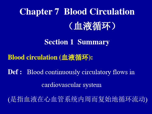
血液循环
四 微循环
是指微动脉和微静脉之间的血液循环 是血液与组织细胞进行物质交换的场所
微循环的血管
血液成分改变 血管管腔狭窄 血液流速变慢 血栓形成
细长如丝 数量极大 分布极广 极易受损
泵 出 的 血 液---动脉血(营养血)
心脏收缩----泵出血液 所产生的血压----收缩压
正常:110—140mmHg
心脏松驰----充盈血液 所产生的血压----舒张压 正常:70—90 mmHg
.
3
心脏
正常心跳 非常有节律
不规则的心跳称为心律失常
正常心跳 70次/分左右
超过 90次/分 称为心动过速
右心房 左心房 右心室 左心室
左心房→左心室→主动脉→中小动脉→毛细动脉 右心室←右心房←腔静脉←中小静脉←毛细静脉
15
血液循环
二 肺循环
通过肺部进行O2 和CO2交换, 使血液由污染血变为营养血。
右心房→右心室→肺动脉→肺泡
体循环
左心室 左心房
肺静脉
.
16
血液循环
三 冠状动脉循环
血液的英文单词

血液的英文单词全文共四篇示例,供读者参考第一篇示例:血液是人体内一种重要的液态组织,它通过血管系统流动在全身,将氧气和养分输送到各个组织和器官中,同时排出代谢产物和二氧化碳。
血液中含有红细胞、白细胞、血小板和血浆等成分,每种成分都扮演着不同的角色,维持着人体内部的平衡。
下面将介绍一些关于血液的英文单词,帮助大家更好地了解这个神奇的液体。
1. Blood:血液,人体内循环系统中运输氧气和营养物质的液体。
2. Red blood cells:红细胞,负责携带氧气到身体组织和器官中的细胞。
3. White blood cells:白细胞,是免疫系统的一部分,负责消灭病原体和维持免疫功能的细胞。
4. Platelets:血小板,起着止血和修复损伤血管的作用。
5. Plasma:血浆,是血液中含有水、盐和蛋白质等物质的液体部分。
6. Hemoglobin:血红蛋白,是红细胞中负责结合氧气并将其输送到身体各个部位的蛋白质。
7. Blood vessel:血管,是血液通过的管道,包括动脉、静脉和毛细血管等。
8. Circulation:循环,指血液在人体内部不断流动的过程。
9. Coagulation:凝血,是血液在受伤时形成血块的过程,起着止血的作用。
10. Blood type:血型,根据红细胞表面的抗原种类将血液分为A 型、B型、AB型和O型等。
11. Hematopoiesis:造血,是骨髓中生产血液细胞的过程。
12. Anemia:贫血,是由于缺乏血红蛋白或红细胞数量不足导致的一种疾病。
13. Leukemia:白血病,是一种骨髓造血系统恶性肿瘤,导致白细胞异常增生。
14. Thrombosis:血栓,是在血管内形成的血块,可能导致血管堵塞和缺血。
15. Transfusion:输血,是将捐赠者的血液输送到接受者体内的过程,用于治疗失血等情况。
以上就是关于血液的一些英文单词,希望能够帮助大家更好地理解血液的结构和功能,保持身体健康。
生理学血液循环双语PPT讲稿

力的变化,不需神经体液参与
Starling机制的意义──通过对搏出量的精细调节,防止心室 舒张末期压力和容积的过久、过度改变,维
持心室射血与静脉回流量的平衡
• 心室功能曲线
–正常 左室充盈压为5~~6 mmHg –最适前负荷 12~~15 mmHg –充盈压在15~~20 mmHg、甚至大于20mmHg时,搏出量不变或略微下
四、影响心脏泵血功能的因素
心输出量=每搏输出量×心率
(一)搏出量
取决于心室肌收缩的强度和速度
影响因素:(1) 前负荷(初长、心室舒张末期容积、
异长自身调节)
(二)心率
(2)后负荷(动脉血压) (3) 心室肌的收缩性能(等长自身调节)
❖ 心室舒张末期容积/压力
心肌初长( initial length) ;前负荷( preload)
(二)心率(heart rate,HR)
➢ 生理差异 ➢ 心率变异性(HRV) ➢ 窦性心动过速/过缓
动脉瓣开 房室瓣关
心房收缩0.1s 心房舒张0.7s
心室收缩0.3s 动脉瓣关 房室瓣开
心室舒张0.5s
全心舒张期0.4s
心动周期中心房、心室活动顺序与时间关系
二. 心脏泵血——射血与充盈过程
两个重要因素:①心脏舒缩→压力变化→形成压力差→动力 ②瓣膜的启闭→控制血流的方向
降(心肌细胞抗伸展的特性)。
❖ 心肌收缩能力(cardiac contractility)
──心肌不依赖于前、后负荷而能改变其力学活动的一种
内在特性 • 决定于心肌细胞兴奋-收缩耦联各环节
(如:肌凝蛋白ATP酶的活性、被活化的横桥数目等 ) • 神经、体液及药物都可通过改变心肌收缩力来调节SV • 等长自身调节
生理学名词中英文对照

生理学名词中英文对照第一章绪论1.机体的内环境(internal environment)2.稳态(homeostasis)3.反射(reflex)4.反射弧(reflex arc)5.旁分泌调节(paracrine regulation)6.自身调节(autoregulation)7.负反馈(negative feedback) 8.正反馈(positive feedback)9.前馈(feed-forward)第二章细胞的基本功能10. 被动转运(passive transport) 11. 单纯扩散(simple diffusion)12. 经载体易化扩散(facilitated diffusion via carrier)13. 经通道易化扩散(facilitated diffusion via ion channel)14. 电压门控通道(voltage gated ion channel)15. 离子通道(ion channel)16. 化学门控通道(chemically gated ion channel)17. 机械门控通道(mechanically gated ion channel)18. 原发性主动转运(primary active transport)19. 继发性主动转运(secondary active transport)20. 同向转运(symport) 21. 反向转运(antiport)22. 出胞(exocytosis) 23. 入胞(endocytosis)24. 跨膜信号转导(transmembrane signal transduction)25. 刺激(stimulation) 26. 兴奋(excitation)27. 兴奋性(excitability) 28. 阈值(threshold)29. 极化(polarization) 30. 去极化(depolarization)31. 超极化(hyperpolarization) 32. 复极化(repolarization)33. 内向电流(inward current) 34. 外向电流(outward current)35. 电化学驱动力(electrochemical driving force)36. 静息电位(resting potential) 37. K+平衡电位(K+ equilibrium potential)38. 动作电位(action potential) 39. 超射(overshoot)40. “全或无”现象(“all or none”phenomenon)41. 阈电位(threshold potential) 42. 局部电位(local potential)43. 时间性总和(temporal summation) 44. 空间性总和(spatial summation)45. 量子释放(quantal release)46. 兴奋-收缩耦联(excitation-contraction coupling)47. 等长收缩(isometric contraction) 48. 等张收缩(isotonic contraction)49. 前负荷(preload) 50. 后负荷(afterload)51.肌肉收缩能力(contractility) 52.不完全强直收缩(incomplete tetauns)53.完全强直收缩(complete tetanus) 54.运动单位(motor unit)第三章血液55. 血细胞比容(hematocrit) 56. 血浆渗透压(plasma osmotic pressure)57. 血浆晶体渗透压(plasma crystal osmotic pressure)58. 血浆胶体渗透压(plasma colloid osmotic pressure)59. 悬浮稳定性(suspension stability)60.红细胞沉降率(erythrocyte sedimentation rate)61.红细胞渗透脆性(osmotic fragility) 62.血小板黏附(platelet adhesion)63.血小板聚集(platelet aggregation) 64.生理性止血(hemostasis)65.出血时间(bleeding time) 66.凝血时间(clotting time)67.血液凝固(blood coagulation) 68.血清(serum)69.凝血因子(clotting factor) 70.内源性凝血途径(intrinsic pathway)71.外源性凝血途径(extrinsic pathway) 72.纤维蛋白溶解(fibrinolysis)73.血型(blood group) 74.红细胞凝集(agglutination)75.凝集原(agglutinogen)76.凝集素(agglutinin)77.交叉配血试验(corss-match test)第四章血液循环78. 心动周期(cardiac cycle)79. 等容收缩期(period of isovolumic contraction)80. 等容舒张期(period of isovolumic relaxation)81. 每搏输出量(stroke volume)82. 射血分数(ejection fraction)83. 心输出量(cardiac output)84 心指数(cardiac index)85. 异长调节(heterometric regulation)86.心室功能曲线(ventricular function curve)87.心肌收缩能力(myocardial contractility)88.等长调节(homometric regulation)89.快反应细胞(fast response cell) 90.慢反应细胞(slow response cell)91.期前收缩(premature systole) 92.代偿间歇(compensatory pause)93.自动节律性(autorhythmicity) 94.正常起搏点(normal pacemaker)95.潜在起搏点(latent pacemaker) 96.异位起搏点(ectopic pacemaker)97.房室延搁(atrioventricular delay)98.膜反应曲线(membrane responsiveness curve)99.钙触发钙释放(calcium-induced Ca2+ release)100.外周阻力(peripheral resistance)101.动脉血压(arterial blood pressure)102.收缩压(systolic pressure)103.舒张压(diastolic pressure) 104.平均动脉压(mean arterial pressure)105.中心静脉压(central venous pressure)106.微循环(microcirculation) 107.压力感受性反射(baroreceptor reflex)第五章呼吸108.肺通气(pulmonary ventilation) 109.肺换气(gas exchange in lungs)110.呼吸运动(respiratory movement) 111.弹性阻力(elastic resistance)112.顺应性(compliance) 113.肺的静态顺应性(static compliance)114.比顺应性(specific compliance) 115.肺表面活性物质(pulmonary surfactant)116.气道阻力(airway resistance) 117.潮气量(tidal volume)118.余气量(residual volume)119.功能余气量(functional residual capacity)120.肺活量(vital capacity) 121.用力肺活量(forced vital capacity) 122.用力呼气量(forced expiratory volume)123.肺通气量(pulmonary ventilation)124.解剖无效腔(anatomical dead space) 125.肺泡无效腔(alveolar dead space) 126.肺泡通气量(alveolar ventilation)127.通气/血流比值(ventilation/perfusion ratio)128.血氧容量(oxygen capacity) 129.血氧含量(oxygen content)130.血氧饱和度(oxygen saturation) 131.氧解离曲线(oxygen dissociation curve) 132.肺牵张反射(pulmonary stretch reflex)第六章消化与吸收133.消化(digestion) 134.机械性消化(mechanical digestion)135.化学性消化(chemical digestion) 136.吸收(absorption)137.慢波(slow wave) 138.胃肠激素(gastrointestinal hormone)139.脑-肠肽(braingut peptide)140.黏液-碳酸氢盐屏障(mucus bicarbonate barrier)141.胃黏膜屏障(gastric mucosal barrier)142.容受性舒张(receptive relaxation)143.紧张性收缩(tonic contraction) 144.胃的排空(gastric emptying)145.肠-胃反射(entero-gastric reflex) 146.分节运动(segmentation contraction)147.胆盐的肠-肝循环(enterohepatic circulation of bile salt)第七章能量代谢与体温148. 能量代谢(energy metabolism)149. 食物的热价(thermal equivalent of food)150. 氧热价(thermal equivalent of oxygen)151. 呼吸商(respiratory quotient) 152. 基础代谢率(basal metabolic rate) 153. 体温(body temperature) 154.温热性发汗(thermal sweating)155.精神性发汗(mental sweating 156.热敏神经元(warm-sensitive neuron) 157.冷敏神经元(cold-sensitive neuron)第八章尿的生成和排出158. 管-球反馈(tubuloglomerular feedback)159. 肾小球滤过率(glomerular filtration rate)160. 滤过分数(filtration fraction)161. 有效滤过压(effective filtration pressure)162. 肾糖阈(renal threshold for glucose)163. 葡萄糖吸收极限量(transfer maximum of glucose)164. 渗透性利尿(osmotic diuresis)165. 球-管平衡(glomerulotubular balance) 166. 水利尿(water diuresis) 167.清除率(clearance)第九章感觉器官的功能168. 感受器(receptor) 169. 感觉器官(sense organ)170. 适宜刺激(adequate stimulus) 171. 换能作用(transducer function)172. 瞳孔对光反射(pupillary light reflex)173.互感性对光反射(consensual light reflex)174. 瞳孔近反射(near reflex of the pupil)175.视敏度(visual acuity)176.暗适应(dark adaptation) 177.明适应(light adaptation)178.视野(visual field) 179.听阈(hearing threshold)180.耳蜗内电位(endocochlear potential)181.耳蜗微音器电位(cochlear microphonic potential)182.眼球震颤(nystagmus)第十章神经系统的功能183.突触(synapse) 184.经典的突触(classical synapse)185.兴奋性突触后电位(excitatory postsynaptic potential)186.抑制性突触后电位(inhibitory postsynaptic potential)187.非定向突触(non-directed synapse) 188.电突触(electric synapse)189.神经递质(neurotransmitter) 190.神经调质(neuromodulator)191.受体(receptor) 192.激动剂(agonist)193.拮抗剂(antagonist) 194.上调(up regulation)195.下调(down regulation) 196.胆碱能纤维(cholinergic fiber)197.肾上腺素能纤维(adrenergic fiber) 198.突触后抑制(postsynaptic inhibition)199.传入侧支性抑制(afferent collateral inhibition)200.回返性抑制(recurrent inhibition) 201.突触前抑制(presynaptic inhibition)202.特异投射系统(specific projection system)203.非特异投射系统(nonspecific projection system)204.牵涉痛(referred pain) 205.脊休克(spinal shock)206.牵张反射(stretch reflex) 207.腱反射(tendon reflex)208.肌紧张(muscle tonus) 209.去大脑僵直(decerebrate rigidity)210.α僵直(α-rigidity) 211.γ僵直(γ-rigidity)212.自发脑电活动(spontaneous electric activity of the brain)213.脑电图(electroencephalogram)214.脑干网状结构上行激动系统(ascending reticular activating system) 215.慢波睡眠(slow wave sleep) 216.异相睡眠(paradoxical sleep)第十一章内分泌217. 激素(hormone) 218. 允许作用(permissive action) 219. 长反馈(long-loop feedback) 220. 短反馈(short-loop feedback)221. 超短反馈(ultra-short-loop feedback)222. 下丘脑调节肽(hypothalamic regulatory peptides)223. 应激反应(stress reaction) 224.应急反应(emergency reaction)第十二章生殖225. 月经(menstruation) 226. 月经周期(menstrual cycle)227. 增生期(proliferative phase) 228. 黄体期(luteal phase)。
第四章 血液循环1[可修改版ppt]
![第四章 血液循环1[可修改版ppt]](https://img.taocdn.com/s3/m/9d9912e383c4bb4cf6ecd10d.png)
衡量心脏泵血功能的基本指标
心指数:在安静状态下心输出量与动物体表面积成正比,因 此将每平方米体表面积、每分钟的心输出量叫心指数。
意义:比较不同个体的心脏功能 在劳动、运动和妊娠时增加 应在静息心指数进行比较
第四章 血液循环1
血液循环
一、概 述:
机体的循环系统是由心脏、血管构成的封闭的管 道系统,血液在循环系统中按照一定的方向循环往复 的流动,称为血液循环(Bloo位于胸腔的中部,偏左下 方,夹在两肺之间,包被于心包膜中。 形状像桃子,大小跟本人的拳头差不多, 重约400g。
右心室
肺动脉
肺毛细血管
肺静脉
左心房
血液循环
血液循环的功能: 完成体内物质运输(代谢原料、产物) 维持机体的内环境稳态(组织液) 参与机体的体液调节
血液循环
心动周期和心脏射血:
心动周期( Cardiac cycle ) 心率(Heart rate) 心脏泵血压力容积变化 心输出量(Cardiac output) 心音(Heart sound)
血液循环
心动周期——心脏每收缩、舒张一次所构成的活动周期。 心房收缩0.1s 心房舒张0.7s 心室收缩0.3s 心室舒张0.5s
血液循环
心率(heart rate)——为心搏频率的简称,以每
分钟心搏次数(次/min)为单位。 心率可因动物的种类、年龄、性别和生理状况的
不同而有差异。总的来说,代谢越旺盛,心率越快; 代谢越低,心率越慢。
和往复泵一样心脏泵血也是间 歇式的,这是脉搏的成因。
心脏的泵血原理
血液循环
1、心房收缩 2、心室收缩
等容收缩期 快速射血期 减慢射血期 3、心室舒张 等容舒张期 快速充盈期 减慢充盈期
血液循环
已激活慢Ca2+通道
+
激活I↓K 通道 Ca2+缓慢内流与 K+外流处于平衡状
态↓ 缓慢复极化 (2期=平台期)
K+ K+ N Ca2+
2期
按任意键显示动画2
慢 Ca2+ 通 道 : 激 活 与 失 活 比 Na+ 通 道 慢 , 特 异 性 不 高 : Ca2+ 和K+ 都通透,阻断剂: Mn2+和多种Ca2+阻断剂(异搏 定)。
周期变化 对应位置 机 制 新AP产生能力 有效不应期 去极相→复极相-60mV 不能产生 绝对不应期: ↓ Na+通道处于
-55mV 完全失活状态 局部反应期: ↓ Na+通道
-60mV 刚开始复活 相对不应期 ↓ Na+通道 能产生(但0期
-80mV 大部复活 幅度、传导、时程 超 常 期 ↓ Na+通道基本 等较正常小)
泰山医学院 基础医学部
生理教研室
康颂建
循环系统
第四章 血液循环 Blood circulation
第一节 心脏生物电活动 第第二二节节 心脏泵血功能 第三节 血 管 生 理 第四节 心血管活动的调节 第五节 器 官 循 环
循环系统(circulatory system):由心血
管系统(cardiovascular system)和淋巴系 统(lamphatic system)构成的封闭管道系统。
抢 先 占 领 超 速 抑 制
2、影响自律性的因素
(1)4期自动去极化速度 a.自动去极化速快→达到
阈电位的时间短→自律性高。 b.自动去极化速慢→达
循环系统CirculatorySystemPPT课件
26
右半心的血流方向:
肺动脉 肺动脉口 上腔静脉口 下腔静脉口 右心房 右Biblioteka 室口 右心室 冠状窦口27
28
(三)左心房left atrium 位于右心房的左后方。左心耳 四个入口:两对肺静脉口。 一个出口:左房室口。
(四)左心室left ventricle 位于右心室的左后方。 一个入口:左房室口。口周围有二尖瓣(前瓣、后瓣)。 一个出口:主动脉口。口周围有主动脉瓣。主动脉窦(左、右、后窦)
25
(二)右心室right ventricle 位于右心房的左前下方。
一个入口:右房室口。口周围有三尖瓣环,其上附有三尖瓣tricuspid valve(借腱索连于乳头肌)。
三尖瓣复合体:三尖瓣环、三尖瓣、腱索、乳头肌共同构成,防止血液 逆流。
一个出口:肺动脉口。周围有肺动脉瓣,心室舒张时关闭,阻止血液逆 流入心室。
肺动脉
肺循环
主动脉
左心室 右心房
组织毛 细血管
上、下腔静脉 冠状窦
体循环
血液循环示意图 10
血管吻合及侧支循环
概念:血管吻合vascular anastomosis是指动脉与动脉、静脉与静脉 或动脉与静脉之间藉吻合支相互连接。
血管吻合的方式
动脉间吻合 静脉间吻合 动、静脉间吻合 侧支吻合
侧支循环collecteral circulation
11
12
13
淋巴系统 lymphatic system
组成:淋巴系统由淋巴管道、淋巴组织和淋巴器官组成。 淋巴管道:为静脉的辅助管道,流动着无色透明的淋巴液。 淋巴组织 淋巴器官
人体结构的功能与疾病的关系
一些免疫、自身免疫或代谢性的全身性疾病,如结节病,系统性红斑狼疮、类风湿性关节炎、皮肌炎、硬皮病等都可累及肺部.肺还具有非呼吸性功能,如肺癌异位性激素的产生和释放所产生内分泌综合证。
常见疾病一哮喘病哮喘病英文:bronchial asthma,简称哮喘,俗称“吼病”,祖国医学称“哮证”,是由多种细胞特别是肥大细胞、嗜酸性粒细胞和T淋巴细胞参与的慢性气道炎症;在易感者中此种炎症可引起反复发作的喘息、气促、胸闷和咳嗽等症状,多在夜间或凌晨发生;此类症状常伴有广泛而多变的呼气流速受限,但可部分地自然缓解或经治疗缓解;此种症状还伴有气道对多种刺激因子反应性增高。
根据权威机构济南哮喘病医院哮喘病可分类为:慢性支气管炎哮喘、过敏性哮喘、药物性哮喘、老年性哮喘、咳嗽变异性哮喘、慢性哮喘、运动性哮喘、儿童性哮喘等十几类。
位,这与冠心病、心肌炎发病率与收治率例数增高有关。
在气候严寒的北方及潮湿的西南地区则为首位.六肺结核肺结核:pulmonary tuberculosis(简称TB)结核病是由结核杆菌(tubercle bacillus)引起的慢性传染病,可累及全身多个器官,但以肺结核最为常见. 本病病理特点是结核结节和干酪样坏死,易形成空洞.临床上多呈慢性过程,少数可急起发病。
常有低热、乏力等全身症状和咳嗽、咯血等呼吸系统表现。
二血液循环系统血液循环系统是血液在体内流动的通道,分为心血管系统和淋巴系统两部分。
淋巴系统是静脉系统的辅助装置.而一般所说的循环系统指的是心血管系统。
血液循环系统由血液、血管和心脏组成。
如果分为两大部分,即为血管和心脏组成。
心血管系统是由心脏、血管、毛细血管及血液组成的一个封闭的运输系统.由心脏不停的跳动、提供动力推动血液在其中循环流动,为机体的各种细胞提供了赖以生存的物质,包括营养物质和氧气,也带走了细胞代谢的产物二氧化碳。
同时许多激素及其他信息物质也通过血液的运输得以到达其靶器官,以此协调整个机体的功能,因此,维持血液循环系统于良好的工作状态,是机体得以生存的条件,而其中的核心是将血压维持在正常水平。
血液循环PPT课件

免疫功能
心脏能够产生一些免疫细 胞和分子,参与免疫应答 和炎症反应,对维护身体 健康具有重要作用。
心脏的功能
泵血功能
心脏通过收缩和舒张运动, 将血液泵入全身血管,维 持血液循环和氧气、营养 物质的输送。
调节血压
心脏通过改变心输出量和 血管阻力来调节血压,保 持血液循环的稳定。
免疫功能
心脏能够产生一些免疫细 胞和分子,参与免疫应答 和炎症反应,对维护身体 健康具有重要作用。
血压的调节
血压的调节对于维持血液循环的正常分配至关重要。通过 神经调节和体液调节机制,可以保持血压的稳定。
血液的分布
血液在血管中的分布是不均匀的,根据组织器官的需要, 血液会优先供应给重要器官和组织,以满足其生理需求。
血液的回收
淋巴系统
淋巴系统是回收血液的重要途径之一,通过淋巴管将组织间隙中 的液体回收到血液循环中。
糖尿病
糖尿病可引起血管病变,影响血液循环,增加心 血管疾病的风险。
贫血
贫血可导致血液携氧能力下降,影响血液循环, 引发缺氧症状。
07 血液循环的疾病与防治
07 血液循环的疾病与防治
高血压与血液循环
1 2
高血压对血液循环的影响
高血压会导致血管内压力增加,使血管壁变硬、 变厚,进而影响血液循环。
高血压对心脏的影响
01
02
03
心脏
负责推动血液循环的器官, 通过收缩和舒张运动将血 液泵出。
血管
由动脉、静脉和毛细血管 组成,负责输送血液到全 身各组织。
血液
含有红细胞、白细胞和血 小板等成分,负责运输营 养物质、氧气和激素等。
组成部分
01
02
03
心脏
血液循环障碍英文课件:PATHOPHYSIOLOGY OF HEART FAILURE
Causes leading to changes of number and size of cardiomyocytes
Preload
Stretching the myocardial fibers during diastole by increasing enddiastolic volume force of contraction during systole = Starling´s law
functions of the heart
Cardiac output (CO) = heart rate (HR) x stroke vol.(SV)
- changes of the heart rate - changes of stroke volume
• Control of HR:
- autonomic nervous systeop
• It is the relation between ventricular volume and pressure
• This loop provides a convenient framework for understanding the response of individual left ventricular contractions to alterations in preload, afterload, and contractility
• Ventricular hypertrophy
– increased mass of contractile elements strength of contraction
血循环系统 - 英文版
Circulatory System∙ Organ system that passes nutrients (such as amino acids, electrolytes and lymph), gases,hormones blood cells, etc. to and from cells in the body to help fight diseases, stabilize body temperature and pH, and maintain homeostasis. ∙ Anything and everything about circulation the body ∙ Composition:o Cardiovascular system: distributes blood: Circulation that happens in the heart and blood vessels ∙ Lymphatic system: returns excess filtered blood plasma from interstitial fluid (between cells as lymph): protection∙ Principal functions of vertebrate circulatory systemof all the substances essential for cellular metabolism▪Respiratory –transports O 2to the tissue cells through the RBC ▪ Nutritive – carries absorbed nutrients through the liver and to the cells ▪ Excretory – removes metabolic wastes , excess water and other substances▪Hormones – part of endocrine systemo Regulation – transport regulatory hormones & temperature regulation▪ Hormone transport – blood carries hormones and carry them to distant targetorgans they regulate▪ Temperature regulation – maintenance of a constant body temperature inwarm-blooded animals.o Protection – protection against injury and foreign microbes▪ Blood clotting – protects against blood loss when vessels are damaged▪ Immune defense – WBCs (leukocytes) provide immunity against many disease-causing agents∙ Basic Typeso Gastrovascular cavity∙ Hydras have 1 opening; water entersand circulates∙ Oxygen/ nutrients are absorbed byChoanocytes∙ Consists of a epidermis andChoanocyteso Open Circulatory System▪Arthropods and some mollusks▪Basic components ofcardiovascular systems∙Hemolympho Limits sizeo Blood + lymph; goesBack into the heart∙Blood vessels- terminate in aopening∙One or more hearts∙Hearts are just musculartubes ▪There is no distinction between the circulating fluid (blood) & the extracellular fluid of the body tissues (interstitial fluid or lymph)▪Fluid in vessels and interstitial fluid mingle in 1 compartment as hemolymph▪Nutrients & waste exchanged by diffusion between hemolymph and body cells ▪Energetically inexpensive▪Limitation: hemolymph can’t be selectively delive red to different tissueso Closed Circulatory System▪Blood and interstitial fluid arephysically separated▪Larger, more active animals need ahigher pressure to pump blood to allbody cells (more efficient bloodpumping)▪Found in earthworm, cephalopods,and all vertebrates▪Advantages:∙Animals can grow larger withmore efficient supply∙Blood flow can be selectivelycontrolled▪TYPES∙Single Circulationo Fishes∙Double Circulationo Crocodiles,birds,Mammals∙Amphibians andmost Reptileshave systems withfeatures of bothCLOSED CIRCULATORY SYSTEMSingle Circulation System – fish (most primitive)∙ Blood passes through the heart only once in a full circuit∙ Heart → Gills (have thin membrane that’s why arteries have lowpressure) → Body → Heart∙ Single atrium collects blood from tissues∙ Single ventricle pumps blood out of the heart ∙ Arteries carry blood away from the heart∙ Blood picks up O 2 and drops CO 2 and goes on through arteries toother body tissues∙ Limitation: pressure lowers; big fishes have to breathe continuouslyDouble Circulation System∙ Blood passes through the heart twice in one full circuit ∙ Heart →Lungs →Heart → Body → Heart∙ Amphibianso Unique in that they can breathe through theirlungs and skino Heart Pumps blood to either▪ Pulmocutaneous circulation – carriesdeoxygenated blood to both the lung and skin▪ Systemic circulation – body tissueso Heart has▪ 3 chambers▪ 2 atria to collect bloodo Right atrium – blood from the body (NOTLUNGS) and is low in O 2 (except oxygenated blood from skin)o Left atrium - blood from lungs (O 2 rich whenair is breathing)o Single ventricle – mixture of oxygenated anddeoxygenated bloodo Internal recesses – separates oxygenatedand deoxygenated (not perfect)o Both atria dump into 1 ventricleo Internal structure keeps 2 O 2 oxygenated and deoxygenated blood mostly separated o Some mixing does occur reducing efficiencyo Noncrocodalian reptiles also have 2 atria and 1 ventricleo Ventricle is partially divide – higher effieciencyo Both must use low moderated pressure systems to minimize pressure flowing through lung tissue∙Crocodiles, birds and mammalso Reptiles have transitional heartso Oxygenated and deoxygenated blood separates into 2 distinct circuitso Systemic circulation – to the bodyo Pulmonary circulation – to the lungso 2 atria and 2 ventriclesThe Human Heart∙Hollow, cone-shaped muscle located between the lungs and behind the sternum (breastbone), tilted at to the left∙About the size of a human fist∙2/3 is located to the left of the midline of the body and 1/3 to the right∙The apex (pointed end) points down and to the left∙Ave. weight between 250-350 grams∙ 4 chambers:o 2 superior atria; the receiving chamberso 2 inferior ventricles; the discharging chambers∙(interatrioventicular) septum – separated the left atrium and ventricle from the right atrium and ventricle, dividing the heart into 2 functionally separate and anatomically distinct units.∙Layerso Endocarium– smooth, inside lining of the heart▪In contact with the blood that the heart pumps▪Protects the cavityo Merges with the inner lining (endothelium)of blood vessels and cover heart valveso Myocardium - middle layer of heart muscle▪Layer that contractso Epicardium or visceral pericardium– outer layer of the heart▪ A fluid sac that surrounds the heart∙Valves:o Atrioventricular (AV) valves - found between the atria and ventricles.▪tricuspid valve, or right atrioventricular valve▪ Between the right atrium and the right ventricle.▪Usually has three papillary muscles.▪Prevents blood from the right ventricle to go to the right atriumo Mitral valve or bicuspid valve or left atrioventricular valve▪ A dual-flap valve that lies between the left atrium and the left ventricle.▪Prevents blood from the left ventricle to go to the left atriumo Semilunar valves - separate the left and right ventricle from the pulmonary artery and the aorta, respectively▪aortic valve∙found between the left ventricle and aorta▪pulmonary valve∙lies between the right ventricle and the pulmonary artery ∙Blood Vessels∙Aortao largest artery in the bodyo arises from the left ventricle of the heart, goes up(ascends) a little ways, bends over (arches), thengoes down (descends) through the chest andthrough the abdomen to where ends by dividinginto 2 common iliac arteries that go to the legs.o Anatomically, it is divided into the ascending aorta,the aortic arch, and the descending aorta.o Can accommodate the greatest pressureo It serves to supply oxygenated blood to the majororgans of the body.o the central conduit from the heart to the body.∙Superior Vena Cava:o A large vein that receivesblood from the head, neck,upper extremities, and thoraxand delivers it to the rightatrium∙Inferior Vena Cava:o A large vein that receivesblood from the lowerextremities, pelvis andabdomen and delivers it tothe right atrium of the heart ∙Pulmonary Artery:o begins at the base of the right ventricle.o it delivers deoxygenatedblood to the lung.∙Pulmonary Vein:o large blood vessels that carry oxygenated blood from thelungs to the left atrium of theheart.o In humans there are fourpulmonary veins, two fromeach lung.∙Myogenic Hearto Electrically excitable, generates own action potentialo Contains auto-rhythmic fibers- can initiate periodic action potential w/o neural activation▪SA Node – most important group of auto-rhythmic cellso Nervous input can increase or decrease rate∙Sino-Atrial Nodeo Found at the upper part of the right atrium of the hearto Acts as the hearts natural pacemakero Triggers a sequence of electrical events in the heart that control the regular muscle contractions (every 0.6 seconds or 100/min) that pump blood out of the heart with a rhythm of about 60-70 beats/min (resting heart)o Depolarization is transmitted through 2 pathways▪Cardiac muscles of the left atrium▪Cardiac muscles of the right atrium and AV Nodeo Depolarization spread quickly among the muscles of the left and right atriasimultaneously▪ Possible because of gap junctions in intercalated diskso AV node provides the only pathway for conduction of depolarization from atria toventricleso Delays ventricular contraction by 0.1 sec (part where atrium transfers blood to ventricles) o Permits atria to finish contraction and emptying of contentso Wave of depolarization is conducted to ventricles by AV bundle or bundle of His▪ Relays depolarization to Purkinje Fibers∙ Stimulates contraction of myocardial cells of the L and R ventricle almostsimultaneouslyo Contraction of the heart is controlled by Ca and troponin/tropomyosin system similar toskeletal muscleso Pattern of voltage change produced by SA node can be measured with electrodes onthe skin▪ Voltage measurements on the skin of the chest are called electrocardiogram(ECG)∙ Record of electrical impulses generated during the cardiac cycle ∙ Monitor electrical activity produced by SA node ∙ Examine fornormal frequency, strength,duration and direction of signals∙ P wave - begins when SA node fires; coincides with depolarization of the atria and, therefore, associatedwith atrial systole ∙ QRS Complex – 3 waves – AV node excites ventricle∙ T wave- repolarization of ventricles back to resting state FLOW OF BLOOD∙ Functioning:o Flow of blood through the heart: one direction▪ from the atria to theventricles, and out of the arteries.▪ Blood is prevented fromflowing backwards by the valves.∙From the left atrium the blood moves to the left ventricle which pumps it out to the body (via the aorta).∙From the R atrium blood moves to the R ventricle which pumps the blood to the lungs∙On both sides, the lower ventricles are thicker and stronger than the upper atria.∙The muscle wall surrounding the left ventricle is thicker than the wall surrounding the right ventricle due to the higher force needed to pump the blood through the systemiccirculation.∙IT ONLY TAKES ABOUT 20 SECONDS TO PUMP BLOOD TO EVERY CELL OF OUR BODYCARDIAC CYCLE (heart beat)∙Filling of atrium, pumping of ventricle∙the sequence of events that occurs when the heart beats.∙ two phases:o diastole phase, the heart ventricles are relaxed and the heart fills with blood.o systole phase, the ventricles contract and pump blood to the arteries.∙One cardiac cycle is completed when the heart fills with blood and the blood is pumped out of the heart.∙Diastole Phase – R sideo Atria and ventricles are relaxed and AV valves are openo Deoxygenated blood from the superior and inferior vena cavae flows into the right atriumo Open AV valves allow blood to pass through to the ventricleso SA node contracts triggering the atria to contracto R atrium empties its contents into R ventricleo Tricuspid Valve closes ∙Systole Phase – R sideo R ventricle receives impulses from the Purkinje Fibers and contracts o SL (pulmonary) valve openso Deoxygenated blood is pumped in to the pulmonary artery. SLvalve closeso Pulmonary artery carries blood to lugs to pick up oxygeno Blood is returned to the L atrium by pulmonary veins∙Diastole Phase – L sideo Blood from pulmonary veins fill left atrium (blood from venacava is also filling the R Atrium) o SA node contracts triggering left atrium to contracto AV (mitral) valves openso L atrium empties its content to L ventricle ∙Systole Phase – L sideo L ventricle receives impulses from the Purkinje fibers and contracts o SL (aortic) valve openso Oxygenated blood is pumped into the aorta. Aorta providesoxygenated blood to the body o Oxygen depleted blood isreturned to the heart via venacavae∙CARDIAC OUTPUT∙Amount of blood the heart pumpsper unit time in L/min∙Depends in the size of the heart and how often it beats∙Stroke volume - amount of blood a heart ejects at each beat∙Higher heart rates of smaller animals gives them a greater cardiac outputthat would be predicted based onthe size of their hearts (must meethigh metabolic demands) HEART SOUNDS∙Lub-dubo the 1st heart sound (lub) is causedby the vibration of the heart atthe time of the closure of thetricuspid and mitral valves.o the 2nd heart sound (dub) iscaused by the vibrations at thetime of closure of the pulmonicand aortic valvesHEART BEAT∙Regular Heart Beat: 70-80 beats a minute∙When you run around a lot your heart pumps more blood into yout body—maybe up to 200 times a minute∙As people grow older their heart rates changeo A newborn baby has a heart rate of about 130o 3 year-old: about 100o18 year-old: about 90 times a minuteo Adult: 70-80o The older you get the slower your heart beats∙An average heart pumps about 10 mililiters of blood into your body with every beat∙That’s about 5L every minute or about 7200L everydayPULSE∙ Represents the tactile aterial palpitationof the heartbeat through the fingertips ∙ May be palpated in any place wherean artery is compressed against a bone, such as at the neck (carotid artery), at the wrist (radial artery), behind the knee (popliteal artery), on the inside of the elbow (brachial artery), and near the ankle joint (posterior tibial artery)BLOOD∙ Specialized bodily fluid in animals thatdelivers necessary substances such as nutrients and oxygen to the cells and transports metabolic waste products away from those some cells ∙ Amount in adults: 4.5-6 quarts ∙ Functionso Transport of gases, nutrients,waste products, and hormones o Maintenance of bodytemperatureo Protection from substance o Clot formationCOMPONENTS OF BLOOD∙ Erythrocytes (Red Blood Cells)o Life span: 120 dayso Function: to carryoxygen from the lungs to every cell in the bodyo Makeup almost 99.9%of formed elements in bloodo Composition: proteinand iron compound, called HEMOGLOBIN , that captures oxygen molecules as the blood moves through the lungsUnites readily with oxygen to form oxyhemoglobin (found in arteries)∙ Form of oxygen as it is transported in the blood stream ∙ Together with iron, it gives blood a bright red coloro Membrane is flexibleo Able to bend in many directions w/o breaking o Biconcave disc shape o No nuclei o About 4.5M in female and 5M in males per cubic mm of blood o Formed in the reticulo-endothelial tissue of the bone marrow o Discharged into the blood capillaries after losing nuclei o Remain in the blood for 3-5 weeks o Destroyed by spleen and liver∙ Leukocytes(White blood cells)o With nucleo Average from 6 thousand – 10Tcells per cubic mm of blood, or about 1 for every 700 RBC o Primary defense mechanismagainst bacteria, viruses, fungi, and parasiteso Produce antibodies, which arereleased into the circulating blood to target and attach to foreign organismso WBC are classified according to their size, shape of nucleus & reaction to dyes used in staining them∙ Thrombocytes (Platelets)o Small irregularly shaped clearcell fragments (i.e. cells that do not have a nucleus o 2-3 m in diametero Derived from fragmentation ofprecursor megakaryocytes o Average lifespan: 5-9 dayso Natural source of growth factor▪ Play a significant role inthe repair and regeneration of connective tissueso Involved in hemostasis(process that stops the flow ofblood), leading to the formation of blood clots▪ Ability of the system to prevent excessive blood loss upon injuryo Normal platelet count: 150,000-450,000/ of blood▪ If too low, excessive bleeding can occur▪ If too high, blood clots/thrombosiscan form∙ May obstruct blood vesselsand result in stroke, myocardial infarction, pulmonary embolism or the blockage of blood vessels to other parts of the body, such as extremities of the arms or legsBLOOD CLOT FORMATION∙ Platelets (cell fragments) in the bloodstream come into contact w/ damaged blood vesselFirst to defendMacrophages∙Platelets and vessel wall release enzyme thrombokinase∙Conversion of inactive enzyme prothrombin into active thrombin∙Thrombin catalyses the conversion of fibrinogen to insoluble fibrin∙Fibrin forms a over the wound that traps RBCS and seals the wound∙Resulting jelly clot like exposure to air to form a scab∙Calcium, vitamin K, and a variety of enzymes called factors are also necessary for efficient blood clotting∙HEMOPHILIAo Genetic defect in clotting factorso Inherited deficiency of a specific clotting factor causes thiso Most common form is X-linked recessive mutationo Treatment requires transfusions of purified clotting factors from donors or genetically engineered organismso Attempting gene therapyBLOOD TYPE∙The most common blood type classification systemis the ABO system discovered by Karl Landsteiner.o observed two distinct chemical moleculeson the surface of the red blood cells and labeledmolecules "A" and "B."o If the red blood cell had only "A" moleculeson it, that blood was called type A.o If the red blood cell had only "B" moleculeson it, that blood was called type B.BLOOD VESSELS∙Arterieso Conduct blood away from the hearto contain a high percentage of smooth muscle.o The artery walls consist of three layers:▪Tunica Adventitia: strong outer covering of arteries and veins which consists of connective tissues, collagen and elastic fibers.▪Tunica Media: the middle layer and consists of smooth muscle and elastic fibers.This layer is thicker in arteries than veins.▪Tunica Intima: the inner layer which is in direct contact with the blood flowing through the artery. It consists of an elastic membrane and smooth endothelialcells.∙Arterioleso Most highly regulated blood vessels in the bodyo Contribute the most to overall blood pressureo Respond to a wide variety of chemical &electrical messages and are constantly changing size to speed up or slow down blood flow∙Capillarieso Site of gas and nutrient/waste exchangeo Single-celled layer of endothelium on a basement membraneo Smallest and narrowest vessels in the body∙Blood enters capillary on arteriole end under pressure∙Pressure forces some fluid out of the blood (not RBC or large proteins)∙Most of the fluid that leaves will be recaptured by the venule end of the capillary∙Venuleso Small, thin extensions of capillaries∙Veinso Conduct blood back to the hearto Thinner and less muscular than arteries o Need help returning blood to the heart▪Smooth musclecontractions help propelblood▪Valves inside veinssqueezed by skeletalmusclesDISORDERS∙Systemic hypertension (High Blood Pressure)o High BP in the systemic arterieso Usually caused by the constriction of the small arteries (arterioles)o Causes: obesity, smoking, aging, etc.∙Congestive heart failureo Condition in which the heart’s function as a pump is inadequate to meet the body’s needo Leads to a buildup of fluid in the lungs and surrounding body tissueso Results from pulmonary hypertension- blood backs up in the lungs, raising pressure, and forcing fluid out into lung tissue∙Atherosclerosiso Disease of the arteries characterized by the deposition of plaques of fatty material on their inner wallso Usual cause of heart attack, strokes and peripheral vascular diseaseo Coronary artery disease results from plaque forming in the coronary arteries supplying the heart muscle∙Myocardial infarction (MI) /Heart Attacko Occurs when blood flow stops to a part of the heart causing damage to the heart muscleo Localized regions of the heart muscle dieo Cardiac angioplasty can detect narrowing of coronary vesselso Balloon angioplasty can widen the lumen of narrowed vesselso Coronary artery bypasses take a healthy blood vessel and use it to replace a blockedcoronary artery。
Chapter7BloodCirculation血液循环.ppt
Cardiac Output = Heart Rate×Stroke Volume
CO=HR×SV =75 × 70 =5250(ml)
Cardiac reserve (心力贮备)
CO (Cardiac output) quiet (安静时):5L/min movement (运动时): 180-200 150-170 25-30L/min
Mechanism of AP :
0期: 去极,钠内流(快通道) 1期: 快速复极初期,钾通过瞬时性外向钾外流通
道外流, 膜内电位降至0mv左右
2期: 缓慢复极期又叫平台期,钙和少量钠通过慢 通道的
内流与少量钾外流相持
3期: 快速复极末期,膜对钾的通透性 升高,钾外流 膜电位由0mv 降至-90mv
(自动兴奋的频率)
2.Functional Synplasm(机能合胞体)
Structure basis : Intercalated disc (闰盘) 心肌的机能合体性
The phenomenon of ‘ all or none ’ in the myocardial
(心肌收缩的“全或无”现象)
Sino-artrial node (S-A node, 窦房结) Artrio-ventricular node (A-V node ,房室结) atrioventricular bundle 0.05m/s and bilateral bundles (房室束及左右束支) 2-5m/s
Purkinje fiber(浦肯野氏纤维) 2-5m/s
1.Def. Cardiac cycle (心动周期) : The whole cycle of systole and
diastole (心脏由收缩到舒张的整个过程)
血液循环PPT课件
定义与功能
定义
血液循环系统是生物体内的运输 系统,负责将氧气和营养物质输 送到身体各部位,同时将废物和 二氧化碳运输到排泄器官。
功能
维持内环境稳态,提供能量,参 与免疫反应等。
循环系统的组成
心脏
血液
作为动力泵,推动血液在全身循环。
含有红细胞、白细胞和血小板等重要 成分。
血管
包括动脉、静脉和毛细血管,负责输 送血液。
循环系统的生理意义
01
02
03
物质运输
确保氧气和营养物质能够 快速到达各个组织。
内环境稳态
通过调节血液中的成分, 维持内环境的稳定。
免疫功能
白细胞在血液循环中巡逻 ,帮助身体抵御感染和疾 病。
CHAPTER 03
血液的组成与特性
血液的组成
血小板
参与血液凝固,有助于止血。
血浆
血液中呈淡黄色的透明液体,约占血液总 量的55%。血浆中含有大量的水分、无机 盐、葡萄糖、氨基酸、酶等营养物质。
心脏的电信号控制系统是心脏 工作的指挥中心,它控制着心 脏的收缩和舒张。
当电信号传到心房和心室时, 心肌细胞会按照一定的顺序收 缩和舒张,推动血液进入心室 并泵出心脏。
心脏的收缩和舒张运动是周期 性的,大约每分钟60-100次, 因人而异。
CHAPTER 05
血管系统
血管的分类与功能
01
02
03
血细胞
包括红细胞、白细胞和血小板,是血液中 最重要的组成部分。血细胞在血液中发挥 着不同的功能。
白细胞
负责免疫功能,帮助身体抵御感染和疾病 。
红细胞
负责输送氧气和养分,主要存在于血液中 。
血液的理化特性