Mcl1-IN-1_COA_13871_MedChemExpress
复方苦参注射液协同索拉非尼提高晚期肝细胞癌应答

复方苦参注射液协同索拉非尼提高晚期肝细胞癌应答李先晨1,徐秋燕2,盛文飞2,李栋梁3,吴庆庆1,陶忠义1*(1. 安徽医科大学附属六安医院药学部,安徽六安 237000;2. 安徽医科大学附属六安医院肿瘤内科,安徽六安237000;3. 安徽医科大学附属六安医院普外科,安徽六安 237000)摘要目的:采用临床队列、网络药理学框架及分子对接技术阐明复方苦参注射液(CKI)协同索拉非尼治疗晚期肝细胞癌(HCC)的物质基础和分子机制。
方法:依据入组标准选取安徽医科大学附属六安医院2019年7月至2022年6月间治疗的134例晚期HCC 患者,其中CKI-索拉非尼组和索拉非尼组各67例。
分析两组患者应答率和总体生存率。
通过网络药理学框架结合中药系统药理学数据库与分析平台及GeneCards、OMIM,PharmGkb、TTD 和 DrugBank 数据库鉴定CKI 治疗晚期HCC 的靶基因。
通过酶联免疫吸附测定验证两组患者治疗前后FBJ 骨髓瘤病毒癌基因同源物(FOS)和表皮生长因子受体(EGFR)表达。
结果:CKI-索拉非尼组患者治疗应答率和生存率均高于索拉非尼组患者(应答率:74.6%∶66.2%,P =0.032;生存率:38.8%∶23.9%,P =0.013)。
FOS 和EGFR 是CKI 治疗晚期HCC 的核心靶基因。
治疗3个月后,CKI-索拉非尼组患者外周血FOS 和EGFR 浓度较索拉非尼组患者显著降低[FOS 浓度:(8.41±2.17)ng/ml ∶(9.85±2.47)ng/ml,P <0.001;EGFR 浓度:(5.47±2.14)ng/ml ∶(6.28±2.04)ng/ml,P =0.027]。
分子对接模型证实槲皮素能与FOS 和EGFR 相互作用,结合能分别为-8.2 kcal/mol 和-7.9 kcal/mol。
结论:CKI 协同索拉非尼提高晚期HCC 患者应答率和生存率,其FOS 和EGFR 是介导其应答的潜在机制。
AR-A014418_DataSheet_MedChemExpress

Inhibitors, Agonists, Screening Libraries Data SheetBIOLOGICAL ACTIVITY:AR–A014418 is a selective and effective GSK3β inhibitor with an IC 50 value of 104 nM, and has no significant inhibition on 26 other kinases.IC50 & Target: IC50: 104 nM (GSK3β)In Vitro: AR–A014418 inhibits tau phosphorylation at a GSK3–specific site (Ser–396) in 3T3 fibroblasts expressing human four–repeat tau protein with IC 50 of 2.7 μM, and protects cultured N2A cells from death induced by blocking PI3K/PKB pathway. In hippocampal slices, AR–A014418 inhibits neurodegeneration mediated by beta–amyloid peptide [1]. While in NGP and SH–5Y–SY cells,AR–A014418 reduces neuroendocrine markers and suppresses neuroblastoma cell growth [2].In Vivo: In ALS mouse model with the G93A mutant human SOD1, AR–A014418 (0–4 mg/kg, i.p.) delays the onset of symptoms,improves motor activity, slows down disease progression, and postpons the endpoint of the disease [3]. In addition, AR–A014418produces inhibition effect on acetic acid– and formalin–induced nociception in mice by modulating NMDA and metabotropic receptor signaling as well as TNF–α and IL–1β transmission in the spinal cord [4].PROTOCOL (Extracted from published papers and Only for reference)Kinase Assay:[1]The competition experiments are carried out in duplicate with 10 concentrations of the inhibitor inclear–bottomed microtiter plates. The biotinylated peptide substrate, biotin–AAEELDSRAGS(PO3H2)PQL, is added at a final concentration of 2 μM in an assay buffer containing 6 milliunits of recombinant human GSK3 (equal mix of both α and β), 12mM MOPS, pH 7.0, 0.3 mM EDTA, 0.01% β–mercaptoethanol, 0.004% Brij 35, 0.5% glycerol, and 0.5 μg of bovine serumalbumin/25 μL and preincubated for 10–15 min. The reaction is initiated by the addition of 0.04 μCi of [γ–33P]ATP and unlabeled ATP in 50 mM Mg(Ac)2 to a final concentration of 1 μM ATP and assay volume of 25 μL. Blank controls without peptide substrate are used.After incubation for 20 min at room temperature, each reaction is terminated by the addition of 25 μL of stop solution containing 5mM EDTA, 50 μM ATP, 0.1% Triton X–100, and 0.25 mg of streptavidin–coated SPA beads corresponding to appr 35 pmol of binding capacity. After 6 h the radioactivity is determined in a liquid scintillation counter. Inhibition curves are analyzed by non–linear regression using GraphPad Prism.Cell Assay: AR–A014418 is dissolved in DMSO.[1]Cell viability is assessed by calcein/propidium iodide uptake. Calcein AM is taken up and cleaved by esterases present within living cells, yielding yellowish–green fluorescence, whereas PI is only taken up by dead cells,which become orange–red fluorescent. In brief, N2A cells are cultured for 2 days in vitro and then treated with 50 μM LY–294002 in the presence of AR–A014418 or vehicle (DMSO) for 24 h. Subsequently, N2A cells are incubated for 30 min with 2 μM PI and 1 μM calcein–AM. The cultures are then rinsed three times with Hanks' buffered saline solution containing 2 mM CaCl 2, and the cells are visualized by fluorescence microscopy using a Zeiss Axiovert 135 microscope. Three fields (selected at random) are analyzed per well (appr 300 cells/field) in at least three different experiments. Cell death is expressed as percentage of PI–positive cells from the total number of cells. In every experiment, specific cell death is obtained after subtracting the number of dead cells present inProduct Name:AR–A014418Cat. No.:HY-10512CAS No.:487021-52-3Molecular Formula:C 12H 12N 4O 4S Molecular Weight:308.31Target:GSK–3; GSK–3Pathway:Stem Cell/Wnt; PI3K/Akt/mTOR Solubility:10 mM in DMSOvehicle–treated cultures.Animal Administration: AR–A014418 is formulated in normal saline.[3]First, to examine the effects of GSK–3 inhibition on the clinical symptoms, life span, and motor behavior function of ALS, 56 Tg mice are divided into four groups. In each group, 0.5 mL of normal saline is mixed with either 0 μg (control group), 1 μg (group A), 2 μg (group B) or 4 μg (group C) of AR–A014418 per gram of mouse, and injected intraperitoneally into 14 animals per group 5 days a week beginning 60 days after birth. The mice are sacrificed at the endpoint described below.References:[1]. Bhat R, Xue Y, Berg S, Structural insights and biological effects of glycogen synthase kinase 3–specific inhibitor AR–A014418. J Biol Chem. 2003 Nov 14; 278(46):45937–45.[2]. Carter YM, et al. Specific glycogen synthase kinase–3 inhibition reduces neuroendocrine markers and suppresses neuroblastoma cell growth. Cancer Biol Ther. 2014 May;15(5):510–5.[3]. Koh SH, et al. Inhibition of glycogen synthase kinase–3 suppresses the onset of symptoms and disease progression of G93A–SOD1 mouse model of ALS. Exp Neurol. 2007 Jun;205(2):336–46.[4]. Martins DF, et al. The antinociceptive effects of AR–A014418, a selective inhibitor of glycogen synthase kinase–3 beta, in mice. J Pain. 2011 Mar;12(3):315–22.Caution: Product has not been fully validated for medical applications. For research use only.Tel: 609-228-6898 Fax: 609-228-5909 E-mail: tech@Address: 1 Deer Park Dr, Suite Q, Monmouth Junction, NJ 08852, USA。
Mcl-1和IAPs家族蛋白介导肝癌细胞抗凋亡作用及其分子机制的研究
Mcl-1和IAPs家族蛋白介导肝癌细胞抗凋亡作用及其分子机制的研究凋亡是程序性细胞死亡,细胞凋亡主要通过内源性、外源性2个信号传导通路。
许多研究证明,肝细胞肝癌(HCC)细胞内源性凋亡缺陷是导致化疗失败的原因之一。
Bcl-2家族蛋白是控制细胞生存与死亡的关键蛋白,在调节细胞凋亡信号中发挥重要作用。
髓细胞白血病基因-1(myeloid cell leukemin-1,Mcl-1)属于Bcl-2家族独特的成员,由于Mcl-1半衰期短(1-4小时),对于各种刺激应答快,其表达在转录、转录后以及蛋白等水平受多种信号的严密调控,以及由于Mcl-1在肝癌等多种恶性肿瘤中普遍高表达,Mcl-1一直是肿瘤分子靶向治疗研究的热点。
ABT-737、 ABT-263(Navitoclax)是一种人工合成的小分子BH3模拟物,体内、外临床前期研究显示,ABT-737、ABT-263(Navitoclax)对多种类型的白血病和实体肿瘤(小细胞肺癌等)有很好的治疗作用,已经进行Ⅰ/Ⅱ期临床试验。
ABT-737、ABT-263(Navitoclax)同时作为小分子Bcl-2和Bcl-xL抑制剂,对Bcl-2、Bcl-xL具有高的亲和力,但对Mcl-1的亲和力极低。
HCC和其它高表达Mcl-1的肿瘤细胞和组织对这个化合物诱导的凋亡抵制,这成为阻碍ABT-737、ABT-263(Navitoclax)进入临床应用的一个主要问题。
有研究证明,联合其他化疗药物能有效增强肿瘤细胞对ABT-737、ABT-263(Navitoclax)治疗的敏感性。
去甲斑蝥素(NCTD)是一个小分子抗肿瘤药物,它衍生于传统中药斑蝥。
一些研究显示,NCTD的抗肿瘤作用可能和Bcl-2家族成员的作用相关。
在前期研究中,我们注意到NCTD极大地抑制了Mcl-1在HCC细胞中的表达。
阿斯匹林(乙酰水杨酸)是一种常见的非类固醇抗炎药物(NSAID),它具有退热、镇痛和抗炎作用。
Expression, Purification and Crystallization of
Expression, Purification and Crystallization of the Mycobacterium Tuberculosis HSP16.3 Molecular Chaperone Background of Mycobacterium Tuberculosis HSP16.3HSP16.3, a 16.3 kDa protein from Mycobacterium Tuberculosis, was originally identified as a prominent antigen (Kingston et al., 1987). During the stationary phase, HSP16.3 is maximally expressed and becomes a main protein of the latent phase (Yuan et al., 1996). Previous studies showed that HSP16.3 can make the cell structure stable and prevent stationary Mycobacterium Tuberculosis from autolysing (Cunningham et al., 1998). In previous studies, HSP16.3 was found as one of theα-crystallin-related small heat shock proteins (sHSP) with molecular chaperone activity. Experiments in vitro revealed that HSP16.3 can suppress the thermal aggregation of citrate synthase at 39.5˚C, without consumption of A TP (Chang et al., 1996).Now the Mycobacterium Tuberculosis HSP16.3 gene was cloned to the plasmid pSTE-HSP16.3, and transformed to E.Coli. BL21(DE3) strain.Material and MethodExpressionThings to have ready before Starting.-Plate or glycerol culture-Sterile LB 25ml in a 50mL shaker flasker, 250ml in a 500mL shaker flasker, all together autoclaved, antibiotic added afterword.- antibiotic and sterile water- TipsPrepare the LB and autoclave:Fomula of the LB medium for 1 Liter:Bacto Tryptone (BT) 10 gBacto Y east Extract (BYE) 10 gNaCl 10gThe LB medium, dd H2O and the tips all together autoclaved at 121 ˚C for 20 minutes.Method:1 Innoculate 25 ml LB Medium ( containing 100 ug) and grow culture overnight(37˚C, 200rpm).2 Next morning inoculate 250 ml prewarmed LB Medium ( containing 100 ug) with the 25 ml overnight culture and grow at 37 ˚C, 200rpm, HSP16.3 was overexpressed in soluble form intracellularly without IPTG induction.3 Incubate the Culture for 10 hours before havesting the cell at 4000 g for 20 minutes.4 Resuspend the cell pellet in 30 ml Butter A and freeze the Sample in -80˚C refigerator.PurificationDE52 Ion-Exchange columnThings to have ready before Starting.-Butter A: 50 mM Imidazole pH 6.5 (1 liter)-Butter B: 50 mM Imidazole pH 6.5 , 300mM NaClall together Fitrate with 0.2 um membrane.- DE52 medium , column ,Gradient maker, UV-monitor and Fractioner- TipsMethod:1 Thaw the cell pellet and vortex .2 Add 0.4ml 100 mM PMSF and sonicate (400kw, 4s-6s 50 cycle* 5 )3 Centrifuge 15000 rpm, 30 minutes to pellet debris4 Transfer supernatant to a 50 ml conicale tube and discard the pellet.5 The supernatant dilute to 50 ml with Buffer A and then load to DE52 ion-exchange columns (20ml), which was pre-equibrated with 100ml Buffer A. And then wash the unbound proteins with 100 ml Buffer A.6 Elute the protein with a linear gradient : 200ml buffer A plus 200ml buffer B, 2ml/min, 6ml each fraction.7 Run 15% SDS-PAGE to determine the HSP16.3 peak.Desalting by dialysis1 Preparation of the dialysis tubeCut the tube in a suitable length (20-30 cm)Boil the tube in solution containing 10 mM NaHCO3 for a few minutes.Boil the tube in solution containing 10 mM EDTA for a few minutes.Rasin the tube with de-ion water2 Pool the HSP16.3 peak and dialysis the Sample against 1000ml Buffer A for more than 6hours.Q-Separose (HP) Ion-Exchange Column1 load the sample to Q-Separose (HP) Ion-Exchange column (20ml), which was pre-equibrated with 100ml Buffer A. And then wash the unbound proteins with 100 ml Buffer A.2 Elute the protein with a linear gradient : 200ml buffer A plus 200ml buffer B, 2ml/min, 6ml each fraction.3 Run 15% SDS-PAGE to determine the purity of the HSP16.3 peak.Gel filtration ColumnThe HSP peak was a final volumn 0.3ml and then run though a Superdex75 (HR, 10/30mm) gel filtration column in 150mM NaCl and 5mM Imdazole, pH6.5. Crystallization1 The purified HSP16.3 was solvent-exchanged to water and concentrated to 20mg/ml before crystallization trails (Bradford). All the crystallization trials were carried out using the hanging-drop vapor-diffusion method at 291K: drops consisted of2 microlitres of HSP16.3 protein solution plus 2 microlitres of the precipitant. The drops were equilibrated against 0.2 ml precipitant at room temperature. The crystallization conditions were investigated with a PEG4000 Kit.Result and discussionThe purity of the final HSP16.3 was over 95% by SDS-PAGE. The crystallization trials of HSP16.3 yielded Cubic crystals with a size of 0.8*0.8*0.6mm in a few days.20040060080010001200mAUBuffer Tris-HCL pH 8.5 Precipitant PEG 4000 MethodV apor Diffusion Temperature 293 K Size0.8*0.8*0.6mmReferencesChang Z., Primm, T.P., Jakana J., Lee H. I., Serysheva I., Chiu W., Gilber H. F., Quiocho F. A., (1996) J Biol Chem 271:7218-7223Cunningham A. F., Spreadbury C. L., (1998) J. Bacteriol. 184:801-808Kingston A. E., Salgame P. R., Mitchison N.A., Colston M. J. (1987) Infect. Immun 55,3149-3154Yuan Y., Crane D. D., Barry C. E. III (1996) J Bacteriol178: 4484-4492。
Keap1

非小细胞肺癌(non-small cell lung cancer,NSCLC)发病率占据肺癌的75%~80%。
肿瘤细胞进展快且易扩散转移,临床常采用手术、放化疗等进行治疗,但5年生存率低于60%[1-2]。
氧化应激是由活性氧(ROS)生成量增加所致,ROS积累可诱导肺癌细胞凋亡,清除ROS 可阻止癌细胞凋亡,即肺癌细胞存活依赖于癌细胞自身抗氧化能力[3]。
Kelch样环氧氯丙烷相关蛋白-1 (kelch-like epichlorohydrin-associated protein-1,Keap1)/核因子E2相关因子2(nuclear factor E2related factor 2,Nrf2)信号通路在癌症中发挥重要调控作用,氧化应激可激活Keap1,促使Keap1-Nrf2复合物裂解,Nrf2转移至细胞核内,可激活下游靶基因表达,参与肺癌发生发展过程[4]。
Nrf2可维持氧化还原稳态,ROS侵袭细胞时,Nrf2可进入细胞核,结合抗氧化反应元件(ARE)转录编码各种抗氧化蛋白、代谢酶基因,抑制氧化应激反应[5-6]。
目前氧化应激、Keap1/Nrf2信号通路在NSCLC发生过程中的机制尚未明确。
基于此,本研究尝试分析Keap1/Nrf2信号通路与临床病理参数、氧化应激指标的相关性,探讨其在NSCLC氧化应激机制中的作用,为临床研制新药提供参考依据。
1资料与方法1.1一般资料选取2017年4月至2020年4月郑州市第三人民医院收治的100例NSCLC患者为研究对象。
纳入标准:符合NSCLC诊断标准[7];术前未接受放化疗、免疫治疗者;预计生存期≥6个月;符合手术适应证、禁忌证;Karnofsky功能状态评分≥70分;签署知情同意书。
排除标准:合并凝血功能障碍、肝肾功能障碍、其他恶性肿瘤者;伴有急/慢性感染者;伴有精神疾病者;既往腹部相关外科手术史者。
所有患者均行肺癌根治性切除术,术中收集癌组织、癌旁组织(距离癌组织5cm范围内正常组织),其中男性63例,女性37例;年龄46~67岁,平均(56.32±3.16)岁;体质量指数(BMI)17~30kg/m2,平均(23.16±2.03)kg/m2;病理类型:鳞癌58例、腺癌42例;病理分级[8]:Ⅰ~Ⅱ级51例、Ⅲ级49例;T分期[9]:T1~T253例、T3~T447例;N分期:N055例、N1~N245例。
细胞蛇的研究进展
2007年,英国牛津大学的刘骥陇等在研究果蝇U 小体和P 小体(U 小体和P 小体是真核生物细胞质中的无膜细胞器)的功能关系时,用4种针对Cup (P 小体中的一种蛋白质)的抗体,对雌性果蝇的卵巢组织进行免疫组织化学染色,染色结果除了预期标记上的P 小体外,还标记出了长条形的丝状结构[1]。
这种结构的形状和数量与纤毛很相似,导致当时以为在果蝇中找到了有纤毛的新细胞类型。
但后来的一系列实验表明,该结构与纤毛没有关系,于是将其命名为“细胞蛇”。
最初是抗Cup 抗体不纯产生假象,意外发现的细胞蛇,而采用亲和层析纯化后的抗Cup 抗体无法再DOI:10.16605/ki.1007-7847.2020.10.0258细胞蛇的研究进展收稿日期:2020-10-22;修回日期:2020-11-19;网络首发日期:2021-07-27基金项目:宁夏自然科学基金项目(2020AAC03179);国家自然科学基金资助项目(31560329)作者简介:李欣玲(1999—),女,广西贵港人,学生;*通信作者:俞晓丽(1984—),女,宁夏银川人,博士,副教授,主要从事干细胞与生殖生物学研究,E-mail:********************。
李欣玲,张樱馨,李进兰,潘文鑫,王彦凤,杨丽蓉,王通,俞晓丽*(宁夏医科大学生育力保持教育部重点实验室临床医学院基础医学院,中国宁夏银川750000)摘要:细胞蛇是近年来细胞生物学研究的热门方向之一,由于其在细胞的增殖、代谢和发育上具有一定的生物学功能,因此,对一些疾病如癌症等的临床诊断或治疗具有一定的指导意义。
细胞蛇是由三磷酸胞苷合成酶(cytidine triphosphate synthetase,CTPS)聚合而成的无膜细胞器,其形成过程及功能在不同类型的细胞中不尽相同。
例如:细胞蛇能促进癌细胞增殖,并使患者病情恶化;过表达的细胞蛇可抑制神经干细胞增殖,影响大脑皮层发育;在卵泡细胞中,细胞蛇相当于CTPS 的存储库,在卵子发生过程起到促进细胞增殖和代谢的作用。
Alda-1_DataSheet_MedChemExpress
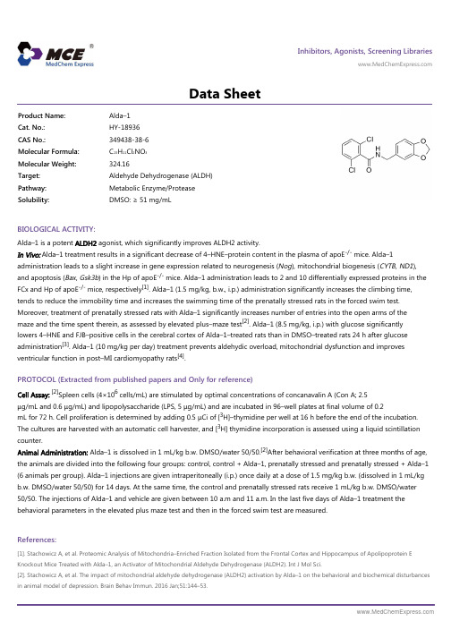
Inhibitors, Agonists, Screening Libraries Data SheetBIOLOGICAL ACTIVITY:Alda–1 is a potent ALDH2 agonist, which significantly improves ALDH2 activity.In Vivo: Alda–1 treatment results in a significant decrease of 4–HNE–protein content in the plasma of apoE -/- mice. Alda–1administration leads to a slight increase in gene expression related to neurogenesis (Nog ), mitochondrial biogenesis (CYTB , ND1),and apoptosis (Bax , Gsk3b ) in the Hp of apoE -/- mice. Alda–1 administration leads to 2 and 10 differentially expressed proteins in theFCx and Hp of apoE -/- mice, respectively [1]. Alda–1 (1.5 mg/kg, b.w., i.p.) administration significantly increases the climbing time,tends to reduce the immobility time and increases the swimming time of the prenatally stressed rats in the forced swim test.Moreover, treatment of prenatally stressed rats with Alda–1 significantly increases number of entries into the open arms of the maze and the time spent therein, as assessed by elevated plus–maze test [2]. Alda–1 (8.5 mg/kg, i.p.) with glucose significantly lowers 4–HNE and FJB–positive cells in the cerebral cortex of Alda–1–treated rats than in DMSO–treated rats 24 h after glucose administration [3]. Alda–1 (10 mg/kg per day) treatment prevents aldehydic overload, mitochondrial dysfunction and improves ventricular function in post–MI cardiomyopathy rats [4].PROTOCOL (Extracted from published papers and Only for reference)Cell Assay:[2]Spleen cells (4×10⁶ cells/mL) are stimulated by optimal concentrations of concanavalin A (Con A; 2.5μg/mL and 0.6 μg/mL) and lipopolysaccharide (LPS, 5 μg/mL) and are incubated in 96–well plates at final volume of 0.2mL for 72 h. Cell proliferation is determined by adding 0.5 μCi of [3H]–thymidine per well at 16 h before the end of the incubation.The cultures are harvested with an automatic cell harvester, and [3H] thymidine incorporation is assessed using a liquid scintillationcounter.Animal Administration: Alda–1 is dissolved in 1 mL/kg b.w. DMSO/water 50/50.[2]After behavioral verification at three months of age,the animals are divided into the following four groups: control, control + Alda–1, prenatally stressed and prenatally stressed + Alda–1(6 animals per group). Alda–1 injections are given intraperitoneally (i.p.) once daily at a dose of 1.5 mg/kg b.w. (dissolved in 1 mL/kg b.w. DMSO/water 50/50) for 14 days. At the same time, the control and prenatally stressed rats receive 1 mL/kg b.w. DMSO/water 50/50. The injections of Alda–1 and vehicle are given between 10 a.m and 11 a.m. In the last five days of Alda–1 treatment the behavioral parameters in the elevated plus maze test and then in the forced swim test are measured.References:[1]. Stachowicz A, et al. Proteomic Analysis of Mitochondria–Enriched Fraction Isolated from the Frontal Cortex and Hippocampus of Apolipoprotein E Knockout Mice Treated with Alda–1, an Activator of Mitochondrial Aldehyde Dehydrogenase (ALDH2). Int J Mol Sci.[2]. Stachowicz A, et al. The impact of mitochondrial aldehyde dehydrogenase (ALDH2) activation by Alda–1 on the behavioral and biochemical disturbances in animal model of depression. Brain Behav Immun. 2016 Jan;51:144–53.Product Name:Alda–1Cat. No.:HY-18936CAS No.:349438-38-6Molecular Formula:C 15H 11Cl 2NO 3Molecular Weight:324.16Target:Aldehyde Dehydrogenase (ALDH)Pathway:Metabolic Enzyme/Protease Solubility:DMSO: ≥ 51 mg/mL[3]. Ikeda T, et al. Effects of Alda–1, an Aldehyde Dehydrogenase–2 Agonist, on Hypoglycemic Neuronal Death. PLoS One. 2015 Jun 17;10(6):e0128844.[4]. Gomes KM, et al. Aldehydic load and aldehyde dehydrogenase 2 profile during the progression of post–myocardial infarction cardiomyopathy: benefits of Alda–1. Int J Cardiol. 2015 Jan 20;179:129–138.Caution: Product has not been fully validated for medical applications. For research use only.Tel: 609-228-6898 Fax: 609-228-5909 E-mail: tech@Address: 1 Deer Park Dr, Suite Q, Monmouth Junction, NJ 08852, USA。
Mcl1-IN-1-SDS-MedChemExpress
Inhibitors, Agonists, Screening LibrariesSafety Data Sheet Revision Date:Oct.-03-2018Print Date:Oct.-03-20181. PRODUCT AND COMPANY IDENTIFICATION1.1 Product identifierProduct name :Mcl1-IN-1Catalog No. :HY-16669CAS No. :713492-66-11.2 Relevant identified uses of the substance or mixture and uses advised againstIdentified uses :Laboratory chemicals, manufacture of substances.1.3 Details of the supplier of the safety data sheetCompany:MedChemExpress USATel:609-228-6898Fax:609-228-5909E-mail:sales@1.4 Emergency telephone numberEmergency Phone #:609-228-68982. HAZARDS IDENTIFICATION2.1 Classification of the substance or mixtureNot a hazardous substance or mixture.2.2 GHS Label elements, including precautionary statementsNot a hazardous substance or mixture.2.3 Other hazardsNone.3. COMPOSITION/INFORMATION ON INGREDIENTS3.1 SubstancesSynonyms:NoneFormula:C23H18ClN3O4Molecular Weight:435.86CAS No. :713492-66-14. FIRST AID MEASURES4.1 Description of first aid measuresEye contactRemove any contact lenses, locate eye-wash station, and flush eyes immediately with large amounts of water. Separate eyelids with fingers to ensure adequate flushing. Promptly call a physician.Skin contactRinse skin thoroughly with large amounts of water. Remove contaminated clothing and shoes and call a physician.InhalationImmediately relocate self or casualty to fresh air. If breathing is difficult, give cardiopulmonary resuscitation (CPR). Avoid mouth-to-mouth resuscitation.IngestionWash out mouth with water; Do NOT induce vomiting; call a physician.4.2 Most important symptoms and effects, both acute and delayedThe most important known symptoms and effects are described in the labelling (see section 2.2).4.3 Indication of any immediate medical attention and special treatment neededTreat symptomatically.5. FIRE FIGHTING MEASURES5.1 Extinguishing mediaSuitable extinguishing mediaUse water spray, dry chemical, foam, and carbon dioxide fire extinguisher.5.2 Special hazards arising from the substance or mixtureDuring combustion, may emit irritant fumes.5.3 Advice for firefightersWear self-contained breathing apparatus and protective clothing.6. ACCIDENTAL RELEASE MEASURES6.1 Personal precautions, protective equipment and emergency proceduresUse full personal protective equipment. Avoid breathing vapors, mist, dust or gas. Ensure adequate ventilation. Evacuate personnel to safe areas.Refer to protective measures listed in sections 8.6.2 Environmental precautionsTry to prevent further leakage or spillage. Keep the product away from drains or water courses.6.3 Methods and materials for containment and cleaning upAbsorb solutions with finely-powdered liquid-binding material (diatomite, universal binders); Decontaminate surfaces and equipment by scrubbing with alcohol; Dispose of contaminated material according to Section 13.7. HANDLING AND STORAGE7.1 Precautions for safe handlingAvoid inhalation, contact with eyes and skin. Avoid dust and aerosol formation. Use only in areas with appropriate exhaust ventilation.7.2 Conditions for safe storage, including any incompatibilitiesKeep container tightly sealed in cool, well-ventilated area. Keep away from direct sunlight and sources of ignition.Recommended storage temperature:Powder-20°C 3 years4°C 2 yearsIn solvent-80°C 6 months-20°C 1 monthShipping at room temperature if less than 2 weeks.7.3 Specific end use(s)No data available.8. EXPOSURE CONTROLS/PERSONAL PROTECTION8.1 Control parametersComponents with workplace control parametersThis product contains no substances with occupational exposure limit values.8.2 Exposure controlsEngineering controlsEnsure adequate ventilation. Provide accessible safety shower and eye wash station.Personal protective equipmentEye protection Safety goggles with side-shields.Hand protection Protective gloves.Skin and body protection Impervious clothing.Respiratory protection Suitable respirator.Environmental exposure controls Keep the product away from drains, water courses or the soil. Cleanspillages in a safe way as soon as possible.9. PHYSICAL AND CHEMICAL PROPERTIES9.1 Information on basic physical and chemical propertiesAppearance White to off-white (Solid)Odor No data availableOdor threshold No data availablepH No data availableMelting/freezing point No data availableBoiling point/range No data availableFlash point No data availableEvaporation rate No data availableFlammability (solid, gas)No data availableUpper/lower flammability or explosive limits No data availableVapor pressure No data availableVapor density No data availableRelative density No data availableWater Solubility No data availablePartition coefficient No data availableAuto-ignition temperature No data availableDecomposition temperature No data availableViscosity No data availableExplosive properties No data availableOxidizing properties No data available9.2 Other safety informationNo data available.10. STABILITY AND REACTIVITY10.1 ReactivityNo data available.10.2 Chemical stabilityStable under recommended storage conditions.10.3 Possibility of hazardous reactionsNo data available.10.4 Conditions to avoidNo data available.10.5 Incompatible materialsStrong acids/alkalis, strong oxidising/reducing agents.10.6 Hazardous decomposition productsUnder fire conditions, may decompose and emit toxic fumes.Other decomposition products - no data available.11.TOXICOLOGICAL INFORMATION11.1 Information on toxicological effectsAcute toxicityClassified based on available data. For more details, see section 2Skin corrosion/irritationClassified based on available data. For more details, see section 2Serious eye damage/irritationClassified based on available data. For more details, see section 2Respiratory or skin sensitizationClassified based on available data. For more details, see section 2Germ cell mutagenicityClassified based on available data. For more details, see section 2CarcinogenicityIARC: No component of this product present at a level equal to or greater than 0.1% is identified as probable, possible or confirmed human carcinogen by IARC.ACGIH: No component of this product present at a level equal to or greater than 0.1% is identified as a potential or confirmed carcinogen by ACGIH.NTP: No component of this product present at a level equal to or greater than 0.1% is identified as a anticipated or confirmed carcinogen by NTP.OSHA: No component of this product present at a level equal to or greater than 0.1% is identified as a potential or confirmed carcinogen by OSHA.Reproductive toxicityClassified based on available data. For more details, see section 2Specific target organ toxicity - single exposureClassified based on available data. For more details, see section 2Specific target organ toxicity - repeated exposureClassified based on available data. For more details, see section 2Aspiration hazardClassified based on available data. For more details, see section 212. ECOLOGICAL INFORMATION12.1 ToxicityNo data available.12.2 Persistence and degradabilityNo data available.12.3 Bioaccumlative potentialNo data available.12.4 Mobility in soilNo data available.12.5 Results of PBT and vPvB assessmentPBT/vPvB assessment unavailable as chemical safety assessment not required or not conducted.12.6 Other adverse effectsNo data available.13. DISPOSAL CONSIDERATIONS13.1 Waste treatment methodsProductDispose substance in accordance with prevailing country, federal, state and local regulations.Contaminated packagingConduct recycling or disposal in accordance with prevailing country, federal, state and local regulations.14. TRANSPORT INFORMATIONDOT (US)This substance is considered to be non-hazardous for transport.IMDGThis substance is considered to be non-hazardous for transport.IATAThis substance is considered to be non-hazardous for transport.15. REGULATORY INFORMATIONSARA 302 Components:No chemicals in this material are subject to the reporting requirements of SARA Title III, Section 302.SARA 313 Components:This material does not contain any chemical components with known CAS numbers that exceed the threshold (De Minimis) reporting levels established by SARA Title III, Section 313.SARA 311/312 Hazards:No SARA Hazards.Massachusetts Right To Know Components:No components are subject to the Massachusetts Right to Know Act.Pennsylvania Right To Know Components:No components are subject to the Pennsylvania Right to Know Act.New Jersey Right To Know Components:No components are subject to the New Jersey Right to Know Act.California Prop. 65 Components:This product does not contain any chemicals known to State of California to cause cancer, birth defects, or anyother reproductive harm.16. OTHER INFORMATIONCopyright 2018 MedChemExpress. The above information is correct to the best of our present knowledge but does not purport to be all inclusive and should be used only as a guide. The product is for research use only and for experienced personnel. It must only be handled by suitably qualified experienced scientists in appropriately equipped and authorized facilities. The burden of safe use of this material rests entirely with the user. MedChemExpress disclaims all liability for any damage resulting from handling or from contact with this product.Caution: Product has not been fully validated for medical applications. For research use only.Tel: 609-228-6898 Fax: 609-228-5909 E-mail: tech@Address: 1 Deer Park Dr, Suite Q, Monmouth Junction, NJ 08852, USA。
Mcl-1抑制剂UMI-77诱导胃癌细胞MGC-803凋亡的机制研究
Mcl-1抑制剂UMI-77诱导胃癌细胞MGC-803凋亡的机制研究朱雪萍;吴萍;张林杰【期刊名称】《安徽医科大学学报》【年(卷),期】2016(051)004【摘要】目的:探讨髓样细胞白血病-1( Mcl-1)分子选择性抑制剂UMI-77诱导胃癌细胞MGC-803凋亡的机制。
方法采用Annexin V/ PI 染色流式细胞术观察不同浓度UMI-77作用后MGC-803细胞凋亡率;JC-1染色流式细胞术分析线粒体膜电位的改变;Western blot 法检测UMI-77作用后半胱天冬氨酸蛋白酶9( Caspase-9)、半胱天冬氨酸蛋白酶3(Caspase-3)和聚腺苷二磷酸核糖聚合酶(PARP)的活化情况以及B 淋巴细胞瘤2(Bcl-2)、Bcl-2蛋白1亚型1(Bcl-XL)和Mcl-1蛋白的表达情况;转染Mcl-1 siRNA 特异性沉默 Mcl-1基因,Westernblot 验证基因沉默效果,Annexin V/ PI 染色流式细胞术检测转染对UMI-77诱导的细胞凋亡的影响。
结果 UMI-77能诱导胃癌细胞MGC-803发生凋亡,随着药物浓度升高,细胞凋亡率逐渐增加;UMI-77作用后,线粒体膜电位下降;Caspase-9、Caspase-3和PARP 在UMI-77处理24 h 显著活化(P <0.05);Bcl-2、Bcl-XL 在UMI-77作用前后的蛋白表达水平没有明显改变,而Mcl-1在12 h 起即明显下调(P <0.05);与阴性对照比较,转染Mcl-1 siRNA 后MGC-803细胞中Mcl-1蛋白表达水平显著下降(P <0.05),且转染后再用UMI-77处理,细胞凋亡率降低(P <0.05)。
结论 UMI-77可通过内源性凋亡途径诱导胃癌细胞MGC-803发生凋亡,下调Mcl-1表达会阻止UMI-77诱导的凋亡。
%Objective To investigate the mechanism of apoptosis induced by UMI-77 , a novel selective inhibitor ofMcl-1, in gastric cancer MGC-803 cells. Methods MGC-803 cells were treated with UMI-77 in different concen-trations for 24 h, apoptotic rates were determined by Annexin V/PI method using flow cytometry. Mitochondrial membrane potential was determined by JC-1 staining on a flow cytometer. The activation of Caspase-9, Caspase-3 and cleavage of PARP were measured by Western blot analysis. The protein level of Bcl-2, Bcl-XL and Mcl-1 was monitored by Western blot as well. Chemically synthesized Mcl-1 siRNA was transfected into MGC-803 cellsusing Lipofectamine 2000 Reagent. The efficacy of gene silencing was confirmed by Western blot analysis, and opoptotic rates before and after transfection was measured by flow cytometry using Annexin V/PI staining. Results UMI-77 was effective in induction of apoptosis in gastric cancer MGC-803 cells, apoptotic rates were increased in a dose-de-pendent manner. Mitochondrial membrane potential was collapsed after UMI-77 treatment. Activation of Caspase-9, Caspase-3 and cleavage of PARP occurred at 24 h (P<0. 05). The expression level of Bcl-2 and Bcl-XL were not altered after exposure to UMI-77 , while Mcl-1 was down-regulated after 12 h ( P<0. 05 ) . Transfection with Mcl-1 siRNA successfully decreased the expression level of Mcl-1 in MGC-803 cells ( P<0. 05 ) and blocked apoptosis induced by UMI-77 ( P<0. 05 ) . Conclusion UMI-77 induces apoptosis through activation of the intrinsic path-way in gastric cancer MGC-803 cells, and knocking down Mcl-1 expression abrogates apoptosis by UMI-77.【总页数】6页(P506-510,511)【作者】朱雪萍;吴萍;张林杰【作者单位】安徽医科大学免疫学教研室,合肥230032;安徽医科大学免疫学教研室,合肥230032;安徽医科大学免疫学教研室,合肥230032【正文语种】中文【中图分类】R735.2【相关文献】1.Mcl-1 siRNA对TRAIL诱导胃癌细胞凋亡的影响 [J], 金玮;吴萍2.ABT-737增强Mcl-1小分子抑制剂UMI-77诱导的胃癌细胞凋亡 [J], 吴萍;李佳佳;裴新茹;陈坤;胡汪来3.芒柄花黄素诱导胃癌细胞株MGC-803凋亡机制研究 [J], 刘红兵;徐成飞;张皓渝;宋强;林兆博;谭令;青廉4.MLN4924诱导胃癌细胞MGC-803发生线粒体自噬及细胞凋亡的初步研究 [J], 邱志兵;邱冬妮;丁伟群5.Mcl-1抑制剂UMI-77诱导胆囊癌细胞GBC-SD凋亡的机制研究 [J], 张生彬;刘宝琴;董长城;李冰因版权原因,仅展示原文概要,查看原文内容请购买。
经动脉灌注蜂毒素-聚乳酸/羟乙酸微球治疗大鼠肝肿瘤
经动脉灌注蜂毒素-聚乳酸/羟乙酸微球治疗大鼠肝肿瘤凌昌全;李琦;刘晓华;陈庆华;彭永海;罗若茵;黄雪强【期刊名称】《世界华人消化杂志》【年(卷),期】2003(11)7【摘要】目的:观察蜂毒素-聚乳酸/羟乙酸微球(M-MS)经动脉给药对大鼠肝肿瘤的疗效.方法:采用改良的复乳-液中蒸发法制备蜂毒素-聚乳酸/羟乙酸微球,建立大鼠移植性肝癌模型并随机分为对照组,蜂毒素组,空白微球组和蜂毒素微球组,每组16只.分别经肝动脉注入生理盐水(NS,1.5mL/kg)、蜂毒素(Melittin,0.35 mg/kg)、空白聚乳酸/羟乙酸微球(B-MS,10mg/kg)和蜂毒素-聚乳酸/羟乙酸微球(M-MS,10mg/kg).比较治疗后各组大鼠的肿瘤生长情况、肿瘤坏死程度和生存时间.结果:治疗后,与对照组比较,蜂毒素组和空白微球组肿瘤生长率均显著降低(12.4±7.1,10.1±8.2vs 28.3±13.6,P<0.01),肿瘤坏死程度以轻中度为主,但两组动物生存时间均未能明显延长(15.8±2.0 d,16.5±3.0 d vs 13.7±2.2 d,P>0.05).M-MS组肿瘤生长率(1.1±1.1)明显低于其他3组(P<0.01),肿瘤坏死更广泛,更彻底,且与对照组比较经蜂毒素微球治疗的大鼠生存期(31.0±3.9 d)显著延长(P<0.01).结论:蜂毒素以药物微球的剂型经肝动脉给药,抗肿瘤效果明显优于单纯的蜂毒素和空白微球.【总页数】4页(P900-903)【关键词】动脉灌注;蜂毒素;聚乳酸;羟乙酸微球;治疗;大鼠;肝肿瘤;疗效;生理盐水【作者】凌昌全;李琦;刘晓华;陈庆华;彭永海;罗若茵;黄雪强【作者单位】中国人民解放军第二军医大学附属长海医院中医科,上海市,200433 上海医药工业研究院,上海市,200437【正文语种】中文【中图分类】R735.7【相关文献】1.经动脉灌注5-FU缓释微球治疗兔VX2肝肿瘤 [J], 关键;胡道予;卢凌;徐涛;潘初2.005 用166Ho-聚乳酸微球治疗大鼠肝脏肿瘤:生物学分布情况的研究 [J], 鞠永健3.抗肝肿瘤天然活性成分的聚乳酸-羟基乙酸共聚物纳米粒/微球制备及应用研究进展 [J], 孙为刚;施峰;孙从永;徐希明;余江南4.阿苯达唑微球肝动脉灌注对大鼠肝泡状棘球蚴病的治疗作用 [J], 樊玉祥;任伟新;迪力木拉提.巴吾冬;顾俊鹏;许晓东;张海潇;纪卫政;姜涛;温浩5.^(32)磷-玻璃微球肝动脉灌注治疗肝肿瘤的毒副作用及并发症观察 [J], 李立;严律南;陈晓理;吴言涛;孙文豪;李茂良因版权原因,仅展示原文概要,查看原文内容请购买。
中链甘油三酯在鼠注射内毒素后肝肠的保护作用
中链甘油三酯在鼠注射内毒素后肝肠的保护作用KonoH;曹国海
【期刊名称】《国外医学:外科学分册》
【年(卷),期】2003(030)004
【总页数】1页(P239)
【作者】KonoH;曹国海
【作者单位】无
【正文语种】中文
【中图分类】R631.3
【相关文献】
1.链霉亲合素-DTPA-Gd经腹腔和静脉注射后对荷瘤裸鼠磁共振肿瘤靶向成像的强化效应 [J], 叶靖;许乙凯;刘岘;吕国士
2.HPLC-ELSD测定呋喃二烯脂肪乳注射液中大豆油和中链甘油三酯的含量 [J], 王艳;杨秀春;韩晓;杨磊;孙晓岩;王东凯;
3.丙泊酚中/长链脂肪乳注射液细菌内毒素检查法的干扰试验 [J], 丁丽燕
4.凝胶法测定10%中/长链脂肪乳注射液(C8-24Ve)和30%脂肪乳注射液(C14-24)中细菌内毒素 [J], 曾博雅
5.脂肪甘油三酯脂酶(PNPLA2)在脂肪性肝炎和内毒素血症小鼠模型中对肝脏炎症起保护作用 [J], Jha P;王静云;李婉玉
因版权原因,仅展示原文概要,查看原文内容请购买。
