X-ray properties of the Composite SeyfertStar-forming galaxies
激光熔覆马氏体
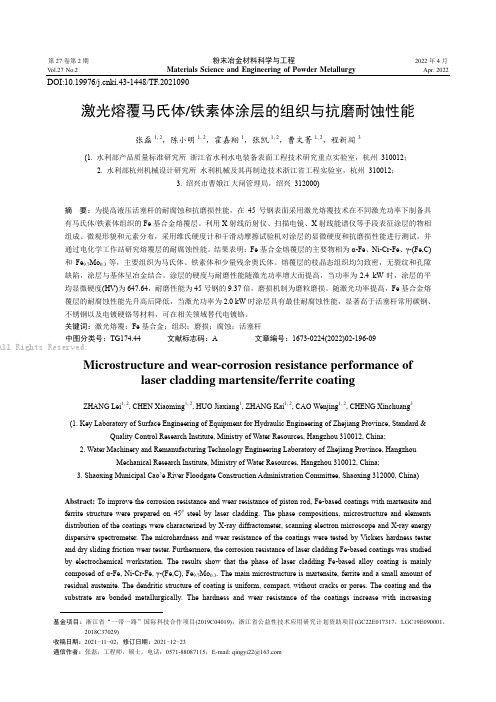
第27卷第2期粉末冶金材料科学与工程2022年4月V ol.27 No.2 Materials Science and Engineering of Powder Metallurgy Apr. 2022DOI:10.19976/ki.43-1448/TF.2021090激光熔覆马氏体/铁素体涂层的组织与抗磨耐蚀性能张磊1, 2,陈小明1, 2,霍嘉翔1,张凯1, 2,曹文菁1, 2,程新闯3(1. 水利部产品质量标准研究所浙江省水利水电装备表面工程技术研究重点实验室,杭州 310012;2. 水利部杭州机械设计研究所水利机械及其再制造技术浙江省工程实验室,杭州 310012;3. 绍兴市曹娥江大闸管理局,绍兴 312000)摘要:为提高液压活塞杆的耐腐蚀和抗磨损性能,在45号钢表面采用激光熔覆技术在不同激光功率下制备具有马氏体/铁素体组织的Fe基合金熔覆层。
利用X射线衍射仪、扫描电镜、X射线能谱仪等手段表征涂层的物相组成、微观形貌和元素分布,采用维氏硬度计和干滑动摩擦试验机对涂层的显微硬度和抗磨损性能进行测试,并通过电化学工作站研究熔覆层的耐腐蚀性能。
结果表明:Fe基合金熔覆层的主要物相为α-Fe、Ni-Cr-Fe、γ-(Fe,C)和Fe9.7Mo0.3等,主要组织为马氏体、铁素体和少量残余奥氏体。
熔覆层的枝晶态组织均匀致密,无裂纹和孔隙缺陷,涂层与基体呈冶金结合。
涂层的硬度与耐磨性能随激光功率增大而提高,当功率为2.4 kW时,涂层的平均显微硬度(HV)为647.64,耐磨性能为45号钢的9.37倍,磨损机制为磨粒磨损。
随激光功率提高,Fe基合金熔覆层的耐腐蚀性能先升高后降低,当激光功率为2.0 kW时涂层具有最佳耐腐蚀性能,显著高于活塞杆常用碳钢、不锈钢以及电镀硬铬等材料,可在相关领域替代电镀铬。
关键词:激光熔覆;Fe基合金;组织;磨损;腐蚀;活塞杆中图分类号:TG174.44文献标志码:A 文章编号:1673-0224(2022)02-196-09All Rights Reserved.Microstructure and wear-corrosion resistance performance oflaser cladding martensite/ferrite coatingZHANG Lei1, 2, CHEN Xiaoming1, 2, HUO Jiaxiang1, ZHANG Kai1, 2, CAO Wenjing1, 2, CHENG Xinchuang3(1. Key Laboratory of Surface Engineering of Equipment for Hydraulic Engineering of Zhejiang Province, Standard &Quality Control Research Institute, Ministry of Water Resources, Hangzhou 310012, China;2. Water Machinery and Remanufacturing Technology Engineering Laboratory of Zhejiang Province, HangzhouMechanical Research Institute, Ministry of Water Resources, Hangzhou 310012, China;3. Shaoxing Municipal Cao’e River Floodgate Construction Administration Committee, Shaoxing 312000, China)Abstract: To improve the corrosion resistance and wear resistance of piston rod, Fe-based coatings with martensite andferrite structure were prepared on 45# steel by laser cladding. The phase compositions, microstructure and elementsdistribution of the coatings were characterized by X-ray diffractometer, scanning electron microscope and X-ray energydispersive spectrometer. The microhardness and wear resistance of the coatings were tested by Vickers hardness testerand dry sliding friction wear tester. Furthermore, the corrosion resistance of laser cladding Fe-based coatings was studiedby electrochemical workstation. The results show that the phase of laser cladding Fe-based alloy coating is mainlycomposed of α-Fe, Ni-Cr-Fe, γ-(Fe,C), Fe9.7Mo0.3. The main microstructure is martensite, ferrite and a small amount ofresidual austenite. The dendritic structure of coating is uniform, compact, without cracks or pores. The coating and thesubstrate are bonded metallurgically. The hardness and wear resistance of the coatings increase with increasing基金项目:浙江省“一带一路”国际科技合作项目(2019C04019);浙江省公益性技术应用研究计划资助项目(GC22E017317,LGC19E090001,2018C37029)收稿日期:2021−11−02;修订日期:2021−12−23通信作者:张磊,工程师,硕士。
x射线大纲要求

目的:掌握X-ray的性质要求:掌握X-ray的基本性质,连续、特征X-ray谱产生的机理,以及X-ray与物质的相互作用。
重点、难点:掌握X-ray的基本性质,连续、特征X-ray谱产生的机理,以及X-ray与物质的相互作用。
了解X-ray发展历史及防护,了解X-ray在现代材料分析中的重要用途。
英语词汇:diffraction\wave’length\target\intensity\filter\Auger effect\思考题:X-ray与物质的相互作用能产生哪些信号?连续、特征X-ray谱的强度、波长与哪些因素有关?X-ray产生的基本条件?作业题:课后习题。
参考资料:《X射线晶体学导论》[英]M.M.乌尔福逊著,科学出版社,1981年。
《X射线结构分析材料性能表征》藤风嗯、王熠明、姜小龙,科学出版社,1997年。
《材料评价的分析电子显微方法》[日]进藤、及川等著,冶金工业出版社,2001年。
《电子衍射图在晶体中的应用》郭可信、叶恒强、呈玉琨著,科学出版社,1983年。
目的:掌握X-ray衍射方向的基本原理—布拉格方程。
要求:在学习晶体几何学的基础上,掌握X-ray衍射原理以及衍射方向与布拉格方程的关系。
重点、难点:复习、掌握晶体几何学基础,理解X-ray衍射的条件与原理----布拉格方程的导出,掌握布拉格方程的意义与应用,理解不同X-ray衍射方法的特点,了解X-ray衍射发展历史。
英语词汇:amorphous\lattice point\space lattice\unit lattice\unit cell\crystallographicaxis\lattice parameter\incident angle\wave frant\Bragg’s law\reflection\angle of diffraction\structrue analysislaue method\rotating-crystal method\power method思考题:布拉格方程的意义与应用?不同X-ray衍射方法的区别?作业题:课后习题。
X-RAY测量厚度的原理

X-RAY荧光测厚法原理X-RAY是原子内层电子在高速运动电子的冲击下产生跃迁而发射的光辐射,可分为连续X-RAY和特征X-RAY两种,常用波段为0.1-20埃(A0)。
X-RAY分析法按照产生的机理可以划分为X-RAY荧光法(X-RAY fluorescence analysis)和X-RAY吸收光谱法(X-RAY absorption spectroscopy)和X-RAY衍射分析法(X-RAY diffraction analysis)等。
在PCB行业,对于金属层厚度一般采用X-RAY荧光法(X-RAY fluorescence analysis)。
X-RAY荧光测厚法原理是利用X-RAY射击到待测量的物体表面上,而反射出荧光,利用皮膜反射的荧光与基材反射荧光的不同性质,与基材反射回来的荧光量的多少,得以计算出皮膜厚度。
当一种物料受到X-RAY的撞击(Bombardment)时,原子中的某些电子在获得足够的能量而脱离(Spin Off)各原子正常轨道的制约后,在原来脱离的价层(Shell)中便产生一个“空洞”(Void)。
当另外有其他的电子从高价层中落下来填补该空洞的时候,其多余的能量便以X-RAY能量的光子释放出来,此X-RAY又在射击到其他物质上,并再度产生第二次的X-RAY荧光(X-RAY fluorescence),参见图2。
各种荧光X-RAY的发射能阶(Limitted Energy Lenel,也就是波长)与其再原子序(Atomic Number)成正比,而且和该物质的特性有关系,其谱线的数量(Quantity,也就是强度)是与该物质的厚度有关。
通过这样的机理,可以对物质进行定性和定量分析。
也就是如果能够采用适当的仪器(Instrumentaition),通过计算机便可以很快利用X-RAY去测量该材料的厚度。
X-RAY测量仪器的基本结构包括X-RAY光管/光源(X-RAY Tude)、准直器(Collimator,或叫瞄准仪)及一个比例记数器(Propertional Counter),参见图3。
x射线荧光光谱法 英文
x射线荧光光谱法英文X-Ray Fluorescence Spectrometry (XRF)。
X-ray fluorescence spectrometry (XRF) is an analytical technique used to determine the elemental composition of materials by measuring the X-rays emitted by the material when it is exposed to a high-energy X-ray beam. This method is widely used in various fields, including geology, environmental science, forensic science, archaeology, and materials science.Principle of Operation.XRF is based on the principle that when a material is irradiated with high-energy X-rays, electrons in the atoms of the material are excited and ejected from their orbits. The resulting vacancies are filled by electrons from higher energy levels, releasing X-rays with energiescharacteristic of the elements present in the material.The energy of the emitted X-rays is specific to each element, and the intensity of the X-rays is proportional to the concentration of the element in the material. By measuring the energies and intensities of the emitted X-rays, it is possible to identify and quantify the elements present in the sample.Instrumentation.A typical XRF spectrometer consists of the following components:X-ray source: Generates high-energy X-rays that bombard the sample.Sample chamber: Holds the sample to be analyzed.Detector: Converts X-rays into electrical signals.Multichannel analyzer (MCA): Digitizes and analyzes the electrical signals from the detector.Types of XRF Spectrometers.There are several types of XRF spectrometers, each with its own advantages and limitations:Energy-dispersive XRF (EDXRF): Uses a solid-state detector to measure the energies of the emitted X-rays. EDXRF is relatively inexpensive and easy to operate, but it has lower energy resolution compared to other types of XRF spectrometers.Wavelength-dispersive XRF (WDXRF): Uses a crystal monochromator to separate the emitted X-rays by wavelength. WDXRF offers higher energy resolution than EDXRF, but it is more complex, expensive, and time-consuming to operate.Total reflection XRF (TXRF): Utilizes total reflection conditions to enhance the sensitivity for analyzing trace elements in liquids. TXRF is highly sensitive, but it requires sample preparation and is not suitable for solid samples.Applications of XRF.XRF is a versatile analytical technique with a wide range of applications:Geochemistry: Determining the elemental composition of rocks, minerals, and soils.Environmental science: Monitoring pollutants in air, water, and soil.Forensic science: Analyzing trace evidence, such as gunshot residue and paint chips.Archaeology: Studying the composition of artifacts and ancient materials.Materials science: Characterizing the elemental composition of metals, alloys, and other materials.Advantages of XRF.Nondestructive: Does not damage the sample being analyzed.Multi-elemental: Can identify and quantify multiple elements simultaneously.Rapid: Provides real-time analysis results.Sensitive: Can detect elements at trace levels.Versatile: Can be applied to various sample types, including solids, liquids, and powders.Limitations of XRF.Limited sensitivity: Cannot detect elements present in very low concentrations.Matrix effects: The presence of other elements in the sample can affect the accuracy of the analysis.Sample preparation: May require sample preparation,such as grinding or homogenization.Cost: XRF spectrometers can be expensive, especially WDXRF systems.Conclusion.X-Ray Fluorescence Spectrometry is a powerful analytical technique that provides valuable information about the elemental composition of materials. It is widely used in various fields and offers advantages such as non-destructiveness, multi-elemental analysis, and rapid results. However, it has limitations in sensitivity and potential matrix effects, which should be considered when selecting this technique for specific applications.。
Spectral and temporal properties of X-ray emission from the ultra-luminous source X-9 in M8

We have analysed the spectra and the variability of individual X-ray sources in the M-81 field using data from the available ROSAations of this nearby spiral galaxy.
2 The data
We analysed the available archival data from ROSAT PSPC (12 observation, with 8 pointed on M 81 nucleus for a total exposure time of 146 ksec) and HRI (7 observations pointed on M 81 nucleus for a total exposure time of 135 ksec). We also used a SAX observation pointed on M 81 and one ASCA observation pointed on X-9.
Figure 3: Radial profiles of the calibration source HZ43 (stars), compared to X9 profile (diamonds). Left panel: external sector. Right panel: internal sector. Data points are normalized to the total number of counts within 2 arcmin. Extraction annuli are 2 arcsec wide (1 detector pixel = 0.5 arcsec)
X射线衍射和小角X射线散射

晶体的X射线衍射特征
[Crystal Structure Analysis, 3rd Edition, p. 48]
晶体结构及其晶胞类型
[Methods of Experimental Physics Volume 16: Polymers, Part B Crystal Structure and Morphology, p. 5]
X射线衍射需要在广角范围内测定,因此又 被称为广角X射线衍射(Wide-Angle X-ray Scattering, WAXS)。
小角X射线散射
如果被照射试样具有不同电子密度的非周 期性结构,则次生X射线不会发生干涉现象, 该现象被称为漫射X射线衍射(简称散射)。
X射线散射需要在小角度范围内测定,因此 又被称为小角X射线散射(Small-Angle Xray Scattering, SAXS)。
晶面指数与晶胞参数
[Fundamentals of Powder Diffraction and Structural Characterization of Materials, 2nd Edition, p. 9]
Bragg方程
设晶体的晶面距为 d,X射线以与晶面间交
角为 的方向照射,从晶面散射出来的X射
粉末衍射条纹摄制及处理
[Fundamentals of Powder Diffraction and Structural Characterization of Materials, 2nd Edition, p. 265]
粉末衍射平板图案摄制
[Fundamentals of Powder Diffraction and Structural Characterization of Materials, 2nd Edition, p. 153]
X-ray Absorption Spectroscopy (XAS)

X-ray Absorption Spectroscopy (XAS)When the x-rays hit a sample, the oscillating electric field of the electromagnetic radiation interacts with the electrons bound in an atom. Either the radiation will be scattered by these electrons, or absorbed and excite the electrons.xA narrow parallel monochromatic x-ray beam of intensity I 0 passing through a sample of thickness x will get a reduced intensity I according to the expression: ln (I 0 /I) = µ x (1)where µ is the linear absorption coefficient , which depends on the types of atoms and the density ρ of the material. At certain energies where the absorption increases drastically, and gives rise to an absorption edge . Each such edge occurs when the energy of the incident photons is just sufficient to cause excitation of a core electron of the absorbing atom to a continuum state, i.e . to produce a photoelectron. Thus, the energies of the absorbed radiation at these edges correspond to the binding energies of electrons in the K, L, M, etc, shells of the absorbing elements. The absorption edges are labelled in the order of increasing energy , K, L I , L II , L III , M I ,…., corresponding to the excitation of an electron from the 1s (2S ½), 2s (2S ½), 2p (2P ½), 2p (2P 3/2), 3s (2S ½), … orbitals (states), respectively. Bohr Atomic Modeled K L L L Continuumge: 2 S ½2P ½2P 32IIIII IWhen the photoelectron leaves the absorbing atom, its wave is backscattered by the neighbouring atoms. The figure below shows the sudden increase in the x-ray absorption of the platinum Pt L III edge in K 2[Pt(CN)4] with increasing photon energy. The maxima and minima after the edge correspond to the constructive and destructive interference between the outgoing photoelectron wave and backscattered wave. 11300115001170011900121001230012500Energy (eV)µ (E )An x-ray absorption spectrum is generally divided into 4 sections: 1) pre-edge (E < E 0); 2) x-ray absorption near edge structure (XANES ), where the energy of the incident x-ray beam is E = E 0 ± 10 eV; 3) near edge x-ray absorption fine structure (NEXAFS ), in the region between 10 eV up to 50 eV above the edge; and 4) extended x-ray absorption fine structure (EXAFS ), which starts approximately from 50 eV and continues up to 1000 eV above the edge.The minor features in the pre-edge region are usually due to the electron transitions from the core level to the higher unfilled or half-filled orbitals (e.g, s → p , or p → d ). In the XANES region, transitions of core electrons to non-bound levels with close energy occur. Because of the high probability of such transition, a sudden raise of absorption is observed. In NEXAFS, the ejected photoelectrons have low kinetic energy (E-E 0 is small) and experience strong multiple scattering by the first and even higher coordinating shells. In the EXAFS region, the photoelectrons have high kinetic energy (E-E 0 is large), and single scattering by the nearest neighbouring atoms normally dominates.11400.00.51.01.52.001150011600117001180011900Multiple scatteringSingle scatteringHome Research Publications Synchrotron XANES EXAFS XAS Measurement。
Exploring the complex X-ray spectrum of NGC 4051
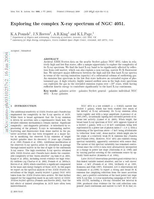
a r X i v :a s t r o -p h /0310257v 2 16 J a n 2004Mon.Not.R.Astron.Soc.000,1–??(2003)Printed 2February 2008(MN L A T E X style file v1.4)Exploring the complex X-ray spectrum of NGC 4051.K.A.Pounds 1,J.N.Reeves 2,A.R.King 1and K.L.Page,11Department of Physics and Astronomy,University of Leicester,Leicester,LE17RH,UK2Laboratory for High Energy Astrophysics,NASA Goddard Space Flight Center,Greenbelt,MD 20771,USAAccepted ;SubmittedABSTRACTArchival XMM-Newton data on the nearby Seyfert galaxy NGC 4051,taken in rela-tively high and low flux states,offer a unique opportunity to explore the complexity of its X-ray spectrum.We find the hard X-ray band to be significantly affected by reflec-tion from cold matter,which can also explain a non-varying,narrow Fe K fluorescent line.We interpret major differences between the high and low flux hard X-ray spectra in terms of the varying ionisation (opacity)of a substantial column of outflowing gas.An emission line spectrum in the low flux state indicates an extended region of pho-toionised gas.A high velocity,highly ionised outflow seen in the high state spectrum can replenish the gas in the extended emission region over ∼103years,while having sufficient kinetic energy to contribute significantly to the hard X-ray continuum.Key words:galaxies:active –galaxies:Seyfert:general –galaxies:individual:NGC 4051–X-ray:galaxies1INTRODUCTIONThe additional sensitivity of XMM-Newton and Chandra has emphasised the complexity in the X-ray spectra of AGN.While there is broad agreement that the X-ray emission is driven by accretion onto a supermassive black hole,the detailed emission mechanism(s)remain unclear.Significant complexity -and diagnostic potential -is introduced by re-processing of the primary X-rays in surrounding matter.Scattering and fluorescence from dense matter in the pu-tative accretion disc has been recognised as a major fac-tor in modifying the observed X-ray emission of bright Seyfert galaxies since its discovery 13years ago (Nandra et al.1989,Pounds et al.1990).Additional modification of the observed X-ray spectra arises by absorption in passage through ionised matter in the line of sight to the continuum X-ray source.The high resolution X-ray spectra obtained with XMM-Newton and Chandra have shown the consider-able complexity of this ‘warm absorber’(eg Sako et al.2001,Kaspi et al.2002),including recent evidence for high veloc-ity outflows (eg Chartas et al.2002,Pounds et al.2003a,b;Reeves et al.2003)which constitute a significant component in the mass and energy budgets of those AGN.In this paper we report on the spectral analysis of two XMM-Newton ob-servations of the bright,nearby Seyfert 1galaxy NGC 4051taken from the XMM-Newton data archive.We find further support for the suggestion made in an early survey of XMM-Newton Seyfert spectra (Pounds and Reeves 2002),that the full effects of ionised absorption in AGN have often been underestimated.NGC 4051is a low redshift (z =0.0023)narrow lineSeyfert 1galaxy,which has been studied over much of the history of X-ray astronomy.Its X-ray emission often varies rapidly and with a large amplitude (Lawrence et al.1985,1987),occasionally lapsing into extended periods of ex-treme low activity (Lamer et al.2003).When bright,the broad band X-ray spectrum of NGC 4051appears typical of a Seyfert 1galaxy,with a 2–10keV continuum being well represented by a power law of photon index Γ∼1.8–2,with a hardening of the spectrum above ∼7keV being attributable to ‘reflection’from ‘cold’,dense matter,which might also be the origin of a relatively weak Fe K emission line (Nandra and Pounds 1994).However,NGC 4051also exhibits strong spectral variability,apparently correlated with source flux.The nature of this spectral variability has remained contro-versial since the GINGA data were alternatively interpreted as a change in power law slope (Matsuoka et al.1990)and by varying partial covering of the continuum source by op-tically thick matter (Kunieda et al.1992).Later ROSAT observations provided good evidence for a flux-linked variable ionised absorber,and for a ‘soft excess’below ∼1keV (Pounds et al.1994,McHardy et al.1995,Komossa and Fink 1997).Extended ASCA observations led Guainazzi et al.(1996)to report a strong and broad Fe K emission line (implying reflection from the inner accretion disc),and a positive correlation of the hard power law slope with X-ray flux.A 3-year monitoring campaign of NGC 4051with RXTE ,including a 150-day extended low interval in 1998,produced clear evidence for the cold reflection com-ponent (hard continuum and narrow 6.4keV Fe K line)re-c2003RAS2K.A.Pounds et al.maining constant,while againfinding the residual power law slope to steepen at higher X-rayfluxes(Lamer et al.2003). More surprisingly,a relativistic broad Fe K line component was found to be always present,even during the period whenthe Seyfert nucleus was‘switched off’(Guainazzi et al.1998, Lamer et al.2003).One other important contribution to the extensive X-ray literature on NGC4051came from an early Chandra observation which resolved two X-ray absorption line systems,with outflowing velocities of∼2300and∼600 km s−1,superimposed on a continuum soft excess with sig-nificant curvature(Collinge et al.2001).Of particular inter-est in the context of the present analysis,the higher velocity outflow is seen in lines of the highest ionisation potential. The Chandra data also show an unresolved Fe K emission line at∼6.41keV(FWHM≤2800km s−1).In summary,no clear picture emerges from a review of the extensive data on the X-ray spectrum of NGC4051, with the spectral variability being(mainly)due to a strong power law slope-flux correlation,or to variable absorption in(a substantial column of)ionised matter.Support for the former view has recently come from a careful study of the soft-to-hardflux ratios in extended RXTE data(Taylor et al.2003),while the potential importance of absorption is underlined by previous spectralfits to NGC4051requiring column densities of order∼1023cm−2(eg Pounds et al.1994, McHardy et al.1995).Given these uncertainties we decided to extract XMM-Newton archival data on NGC4051in order to explore its spectral complexities.After submission of the present paper, an independent analysis of the2002November EPIC pn data by Uttley et al.(2003)was published on astro-ph,reaching different conclusions to those wefind.We comment briefly on these alternative descriptions of the spectral variability of NGC4051in Section9.4.2OBSER V ATION AND DATA REDUCTION NGC4051was observed by XMM-Newton on2001May 16/17(orbit263)for∼117ksec,and again on2002Novem-ber22(orbit541)for∼52ksec.The latter observation was timed to coincide with an extended period of low X-ray emis-sion from NGC4051.These data are now public and have been obtained from the XMM-Newton data archive.X-ray data are available in both observations from the EPIC pn (Str¨u der et al.2001)and MOS2(Turner et al.2001)cameras, and the Reflection Grating Spectrometer/RGS(den Herder et al.2001).The MOS1camera was also in spectral mode in the2002observation.Both EPIC cameras were used in small window mode in thefirst observation,together with the mediumfilter,successfully ensuring negligible pile-up. The large window mode,with mediumfilter,was used in the second,lowflux state observation.The X-ray data were first screened with the latest XMM SAS v5.4software and events corresponding to patterns0-4(single and double pixel events)were selected for the pn data and patterns0-12for MOS1and MOS2.A low energy cut of300eV was applied to all X-ray data and known hot or bad pixels were removed. We extracted EPIC source counts within a circular region of45′′radius defined around the centroid position of NGC 4051,with the background being taken from a similar re-gion,offset from but close to the source.The netexposures Figure 1.Background-subtracted EPIC pn data for the2001 May(black)and2002November(red)observations of NGC4051 available for spectralfitting from the2001observation were 81.7ksec(pn),103.6ksec(MOS2),114.3ksec(RGS1)and 110.9ksec(RGS2).For the2002observation thefinal spec-tral data were of46.6ksec(pn),101.9ksec(MOS1and2), 51.6ksec(RGS1)and51.6ksec(RGS2).Data were then binned to a minimum of20counts per bin,to facilitate use of theχ2minimalisation technique in spectralfitting. Spectralfitting was based on the Xspec package(Arnaud 1996).All spectralfits include absorption due to the NGC 4051line-of-sight Galactic column of N H=1.32×1020cm−2 (Elvis et al.1989).Errors are quoted at the90%confidence level(∆χ2=2.7for one interesting parameter).We analysed the broad-band X-ray spectrum of NGC 4051integrated over the separate XMM-Newton observa-tions,noting the meanflux levels were markedly different, and perhaps representative of the‘high state’and‘low state’X-ray spectra of this Seyfert galaxy.[In fact the2001May X-rayflux is close to the historical mean for NGC4051, but we will continue to refer to it as the‘high state’for convenience].To obtain afirst impression of the spectral change we compare infigure1the background-subtracted spectra from the EPIC pn camera for orbits263and541. The same comparison for the EPIC MOS2data(not shown) is essentially identical.From∼0.3–3keV the spectral shape is broadly unchanged,with the2001flux level being a fac-tor∼5higher.From∼3keV up to the very obvious emission line at∼6.4keV theflux ratio decreases,indicating aflatter continuum slope in the low state spectrum over this energy band.On this simple comparison the∼6.4keV emission line appears essentially unchanged in energy,width and photon flux.We will defer a more detailed comparison of the‘high’and‘low’state data until Section5,afterfirst modelling the individual EPIC spectra.3HIGH STATE EPIC SPECTRUM3.1Power law continuumWe began our analysis of the EPIC data for2001May in the conventional way byfitting a power law over the hard X-ray (3–10keV)band,thereby excluding the more obvious effectsc 2003RAS,MNRAS000,1–??X-ray spectrum of NGC40513 Figure2.Ratio of data to power lawfits over the3–10keV bandfor the pn(black)and MOS(red)spectra in the high state2001May observation of NGC4051.of soft X-ray emission and/or low energy absorption.Thisfityielded a photon index ofΓ∼1.85(pn)andΓ∼1.78(MOS),but thefit was poor with significant residuals.In particularthe presence of a narrow emission line near6.4keV,and in-creasing positive residuals above9keV(figure2),suggestedthe addition of a cold reflection component to refine the con-tinuumfit,which we then modelled with PEXRAV in Xspec(Magdziarz and Zdziarski1995).Since the reflection compo-nent was not well constrained by the continuumfit,we leftfree only the reflection factor R(=Ω/2π,whereΩis thesolid angle subtended at the source),fixing the power lawcut-offat200keV and disc inclination at20◦,with all abun-dances solar.The outcome was an improvedfit,with∆χ2of40for R=0.8±0.2.The power law indexΓincreased by0.1for both pn and MOSfits.In all subsequentfits we thenset R=0.8(compatible with the strength of the6.4keVemission line).Based on this broad bandfit we obtained a2-10keVflux for the2001May observation of NGC4051of2.4×10−11erg s−1cm−2corresponding to a2-10keVluminosity of2.7×1041erg s−1(H0=75km s−1Mpc−1).3.2Fe K emission and absorptionThe power law plus reflection continuumfit at3–10keVleaves several residual features in both pn and MOS data,the significance of which are indicated by the combinedχ2of2068for1740degrees of freedom(dof).Visual examinationoffigure2shows,in particular,a narrow emission line near6.4keV and evidence of absorption near∼7keV and between∼8–9keV.To quantify these features we then added further spec-tral components to the model,beginning with a gaussianemission line with energy,width and equivalent width asfree parameters.This addition improved the3–10keVfit,toχ2/dof of1860/1735,with a line energy(in the AGN restframe)of6.38±0.01keV(pn)and6.42±0.03keV(MOS),rms width≤60eV and lineflux of1.6±0.4×10⁻⁵photons−1cm−2(pn)and1.4±0.6×10⁻⁵photon s−1cm−2(MOS),corresponding to an equivalent width(EW)of60±15eV.Next,wefitted the most obvious absorption feature near7keV with a gaussian shaped absorption line,again withenergy,width and equivalent width free.The best-fit ob-served line energy was7.15±0.05keV(pn)and7.05±0.05keV(MOS)in the AGN rest-frame,with an rms width of150±50eV,and an EW of100±20eV.The addition of thisgaussian absorption line gave a further highly significantimprovement to the overallfit,withχ2/dof=1802/1730.Fitting the less compelling absorption feature at∼8–9keVwith a second absorption line was not statistically signifi-cant.However,an absorption edge did improve thefit toχ2/dof=1767/1728,for an edge energy of8.0±0.1keV andoptical depth0.15±0.05.In summary,the3-10keV EPIC data from the highstate2001May observation of NGC4051is dominated by apower law continuum,with a photon index(after inclusionof cold reflection plus an emission and absorption line)of1.90±0.02(pn)and1.84±0.02(MOS).The narrow emissionline at∼6.4keV is compatible withfluorescence from thesame cold reflecting matter,while-if identified with reso-nance absorption of FeXXVI or FeXXV-the∼7.1keV lineimplies a substantial outflow of highly ionised gas.Wefindno requirement for the previously reported strong,broad FeK emission line,the formal upper limit for a line of initialenergy6.4keV being70eV.3.3Soft ExcessExtending the above3–10keV continuum spectralfit downto0.3keV,for both pn and MOS data,shows very clearly(figure3)the strong soft excess indicated in earlier observa-tions of NGC4051.To quantify the soft excess we againfitted the com-bined pn and MOS data,obtaining a reasonable overallfit with the addition of blackbody continua of kT∼120and270eV,together with absorption edges at∼0.725keV(τ∼0.24)and∼0.88keV(τ∼0.09).Based on this broad bandfit we deduced soft X-rayflux levels for the2001May ob-servation of NGC4051of2.9×10−11erg s−1cm−2(0.3–1keV),with∼61percent in the blackbody components,and1.1×10−11erg s−1cm−2(1–2keV).Combining these re-sults with the higher energyfit yields an overall0.3–10keVluminosity of NGC4051in the‘high’state of7×1041ergs−1(H0=75km s−1Mpc−1).4LOW STATE EPIC SPECTRUMThe above procedure was then repeated in an assessment ofthe2002November EPIC data,when the X-rayflux fromNGC4051was a factor∼4.5lower(figure1).Fitting the hard X-ray continuum was now more un-certain since the spectrum was more highly curved in thelowflux state(comparefigs4and2),making an underlyingpower law component difficult to identify.To constrain thefitting parameters we therefore made two important initialassumptions.Thefirst,supported by the minimal changeapparent in the narrow Fe K line,was to carry forward thecold reflection(normalisation and R)parameters from the‘high state’spectralfit(in fact,as noted above,appropri-ately at aflux level close to the historical average for NGC4051).The second assumption was that the power law con-tinuum changed only in normalisation,but not in slope(as c 2003RAS,MNRAS000,1–??4K.A.Pounds etal.Figure3.Extrapolation to0.3keV of the3–10keV spectralfit(detailed in section3.2)showing the strong soft excess in both pn(black)and MOS(red)spectra during the2001May observationof NGC4051.found in the extended XMM-Newton observation of MCG-6-30-15,Fabian and Vaughan2003).This is in contrast to theconclusions of Lamer et al.(2003)but-as we see later-isconsistent with the difference spectrum(figure8),whichfitsquite well at3–10keV to a power law slope ofΓ∼2,whilealso showing no significant residual reflection features.With these initial assumptions,the3–10keVfit to thelow state spectrum yielded the data:model ratio shown infig-ure4.A visual comparison withfigure2shows a very similarnarrow emission line at∼6.4keV,but with strong curvatureto the underlying continuum,and significant differences inthe absorption features above7keV.These strong residualsresulted in a very poorfit at3–10keV,withχ2of1610/990.We note the spectral curvature in the3–6keV band is rem-iniscent of an extreme relativistic Fe K emission line;how-ever,since our high state spectrum showed no evidence forsuch a feature,and it might in any case be unexpected whenthe hard X-ray illumination of the innermost accretion discis presumably weak,we considered instead a model in whicha fraction of the power law continuum is obscured by anionised absorber.We initially modelled this possibility withABSORI in Xspec,finding both the3–6keV spectral cur-vature and the absorption edge at∼7.6keV were wellfittedwith∼60percent of the power law covered by ionised matterof ionisation parameterξ(=L/nr2)∼25and column densityN H∼1.2×1023cm−2.The main residual feature was then the narrow Fe Kemission line.4.1The narrow Fe K emission lineA gaussian linefit to the emission line at∼6.4keV in thelow state EPIC data was again unresolved,with a meanenergy(in the AGN rest frame)of6.41±0.01keV(pn)and6.39±0.02keV(MOS),and linefluxes of1.9±0.3×10⁻⁵pho-ton s−1cm−2(pn)and2.0±0.4×10⁻⁵photon s−1cm−2(MOS),corresponding to an EW against the unabsorbedpower law component of500±75eV.The important pointis that,within the measurement errors,the measuredfluxesof the∼6.4keV line are the same for the twoobservations.Figure4.Ratio of data to power law plus continuum reflectionmodelfit over the3–10keV band for the pn(black)and MOS(red)spectra in the low state2002November observation of NGC4051.Figure5.Partial covering model spectrumfitted over the3–10keV band for the2002November observation of NGC4051.Alsoshown are the separate components in thefit:the unabsorbedpower law(green),absorbed power law(red)and Gaussian emis-sion line(blue).See Section4.1for details.For clarity only thepn data are shown.This lends support to our initial assumption that both EPICspectra include a‘constant’reflection component,illumi-nated by the long-term average hard X-ray emission fromNGC4051.With the addition of this narrow emission linethe overall3–10keVfit obtained with the partial coveringmodel was then good(χ2/dof=1037/1037).Figure5illus-trates the unfolded spectrum and spectral components ofthisfit.4.2Soft ExcessExtrapolation of the above partial covering3–10keV spec-tralfit down to0.3keV shows a substantial soft X-ray excessremains(figure6),with a similar relative strength to thepower law component seen in the high state data.We notethat the‘soft excess’,ie relative to the power law component,c 2003RAS,MNRAS000,1–??X-ray spectrum of NGC40515 Figure6.Partial covering modelfits over the3–10keV bandextended to0.3keV,for the pn(black)and MOS(red)data fromthe low state2002November observation of NGC4051.would have been extremely strong(data:model ratio∼8)had we taken the simple power lawfit(Γ∼1.4)to the lowstate3–10keV data.Extending the partial covering model to0.3keV,with the addition-as in the high state-of a black-body component of kT∼125eV(the hotter component wasnot required),gave an initially poorfit(χ2of2348for1265dof for the pn data),with a broad deficit in observedfluxat∼0.7-0.8keV being a major contributor(figure6).Theaddition of a gaussian absorption line to the partial coveringmodel gave a large improvement to the broad-bandfit(toχ2of1498for1262dof),for a line centred at0.756±0.003keV,with rms width50±15eV and EW∼40eV.We show thiscomplex spectralfit infigure7,and comment that the modeldependency of unfolded spectra is relatively unimportant inillustrating such strong,broad band spectral features.Sig-nificantly,the broad-band spectralfit remains substantiallyinferior to the similarfit to the high state data.Examina-tion of the spectral residuals shows this is due to additionalfine structure in the soft band of the low state spectrum,structure that is also evident infigures6and7.We examinethe RGS data in Section6to explore the nature(absorptionor emission)of this structure.The deduced soft X-rayflux levels for the2002Novem-ber observation of NGC4051were6.3×10−12erg s−1cm−2(0.3–1keV),with∼53percent in the blackbody component,and1.8×10−12erg s−1cm−2(1–2keV).Combining these re-sults with a2-10keVflux of5.8×10−12erg s−1cm−2yieldedan overall0.3–10keV luminosity of NGC4051in the‘low’state of1.5×1041erg s−1(H0=75km s−1Mpc−1).5COMPARISON OF THE HIGH AND LOWSTATE EPIC DATAThe above spectralfitting included two important assump-tions,that the cold reflection was unchanged between thehigh and lowflux states,and the variable power law com-ponent was of constant spectral index.We now compare theEPIC data for the two observations to further explore thenature of the spectral change.Figure8illustrates the dif-ference spectrum obtained by subtracting thebackground-Figure7.Extrapolation to0.3keV of the3–10keV partial cov-eringfit offig5showing the strong soft excess modelled by ablackbody component(blue),and a broad absorption trough at∼0.76keV.For clarity only the pn data are shown.subtracted low state data from the equivalent high state data(corrected for exposure).To improve the statistical signifi-cance of the higher energy points the data were re-groupedfor a minimum of200counts.The resulting difference spec-trum is compared infigure8with a power lawfitted at3–10keV.Several points are of interest.First,the power law indexof the difference spectrum,Γ∼2.04(pn)andΓ∼1.97(M2),is consistent with the assumed‘constant’value in the indi-vidual spectralfits.Second,the narrow Fe K emission lineand high energy data upturn are not seen,supporting ourinitial assumption of a‘constant’cold reflection component.The narrow feature observed at∼7keV corresponds to theabsorption line seen(only)in the high state spectrum,whilewe shall see in Section6that the deficit near0.55keV inthe MOS data(which has substantially better energy reso-lution there than the pn)is probably explained by a strongand‘constantflux’emission line of OVII.Finally,the smallpeak near8keV can be attributed to the absorption edgeshifting to lower energy as the photoionised gas recombinesin the reduced continuum irradiation.While the arithmetic difference of two spectra providesa sensitive check for the variability of additive spectral com-ponents,a test of the variability of multiplicative compo-nents is provided by the ratio of the respective data sets.Figure9reproduces the ratio of the high and low state data(pn only)after re-grouping to a minimum of500counts perbin.From∼0.3–3keV theflux ratio averages∼5,as seen infigure1,falling to higher energies as the mean slope of thelow state spectrum hardens.The large positive feature at∼0.7–0.8keV is of particular interest,indicating a variablemultiplicative component,almost certainly corresponding toenhanced absorption in the low state spectrum.In fact thatfeature can be clearly seen in the low state EPIC data infigures6and7.We suggest the broad excess at∼1–2keVcan be similarly explained by greater absorption affectingthe low state spectrum,lending support to our overall inter-pretation of the spectral change.Finally,we note that thenarrow dip in the ratio plot at∼6.4keV is consistent withthe Fe K emission line having unchangedflux,but corre-spondingly higher EW in the low state spectrum.c 2003RAS,MNRAS000,1–??6K.A.Pounds etal.Figure8.High minus low state difference spectral data(pn-black,M2-red)compared with a simple power law,as describedin Section5.Figure9.Ratio of high state to low state spectral data(pn only),as described in Section5.6SPECTRAL LINES IN THE RGS DATABoth EPIC spectra show a strong soft excess,with the lowstate(2002)spectrum also having more evidence offinestructure.To study the soft X-ray spectra in more detailwe then examined the simultaneous XMM-Newton gratingdata for both observations of NGC4051.Figures10and11reproduce thefluxed spectra,binned at35m˚A,to showboth broad and narrow features.The continuumflux level ishigher in the2001data(consistent with the levels seen in theEPIC data),with a more pronounced curvature longwardsof∼15˚A.Numerous sharp data drops hint at the presence ofmany narrow absorption lines.In contrast,the2002Novem-ber RGS spectrum exhibits a lower andflatter continuumflux,and a predominance of narrow emission lines.We began an analysis of each observation by simultane-ouslyfitting the RGS-1and RGS-2data with a power lawand black body continuum(from the corresponding EPIC0.3–10keVfits)and examining the data:model residuals byeye.For the2001May observation the strongest featureswere indeed narrow absorption lines,most beingreadilyFigure10.Fluxed RGS spectrum from the XMM-Newton ob-servation of NGC4051in2001May.Figure11.Fluxed RGS spectrum from the XMM-Newton ob-servation of NGC4051in2002November.identified with resonance absorption in He-and H-like ionsof C,N,O and Ne.In contrast,the combined RGS data forthe low state data from2002November showed a mainlyemission line spectrum,more characteristic of a Seyfert2galaxy(eg Kinkhabwala et al.2002).Significantly,the NVI,OVII and NeIX forbidden lines are seen in both high andlow state RGS spectra at similarflux levels.Taking note ofthat fact we then analysed the low state(2002)datafirst,and subsequently modelled the RGS high-minus-low differ-ence spectrum,to get a truer measure of the absorption linestrengths in the high state(2001)spectrum.6.1An emission line spectrum in the low statedataTo quantify the emission lines in the2002spectrum weadded gaussian lines to the power law plus blackbody contin-uumfit in Xspec,with wavelength andflux as free parame-ters.In each case the line width was unresolved,indicating aFWHM≤300km s−1.Details of the8strongest lines therebyidentified are listed in Table1.The statistical quality of thec 2003RAS,MNRAS000,1–??X-ray spectrum of NGC40517fit was greatly improved by the addition of the listed lines,with a reduction inχ2of251for16fewer dof.When ad-justed for the known redshift of NGC4051all the identified lines are consistent with the laboratory wavelengths indi-cating that the emitting gas has a mean outflow(or inflow) velocity of≤200km s−1.Figure12illustrates the OVII triplet,showing the dom-inant forbidden line and strong intercombination line emis-sion,but no residual resonance line emission(at21.6˚A).Theline ratios,consistent with those found in the earlier Chandra observation(Collinge et al.2001),give a clear signature of a photoionised plasma,with an electron density≤1010cm−3(Porquet and Dubau2000).A similarly dominant forbidden line in the NVI triplet yields a density limit a factor∼10 lower.We note the absence of the OVII resonance emission line may be due to infilling by a residual absorption line ofsimilar strength.After removal of the emission lines listed in Table1,sev-eral additional emission features(seefigure11)remained. Although narrow and barely resolved,the wavelength of these features allows them to be unambiguously identifiedwith the radiative recombination continua(RRC)from the same He-and H-like ions of C,N,O and(probably)Ne. Table2lists the properties of these RRC as determined byfitting in Xspec with the REDGE model.While the RRC of CV,CVI,NVI and OVII are well determined,wefixed the other threshold energies at their laboratory values to quan-tify the measured equivalent widths.What is clear is that the RRC are very narrow,a combinedfit yielding a mean temperature for the emitting gas of kT∼3eV(T∼4×10⁴K).We note this low temperature lies in a region of thermal stability for such a photoionised gas(Krolik et al.1981). Furthermore,the low temperature indicates collisional ion-isation and excitation will be negligible,and radiative re-combination should be the dominant emission process.Additional constraints on the emitting gas in NGC4051can be derived by noting that the2002November XMM-Newton observation took place some20days after the source entered an extended lowflux state.Furthermore,the emis-sion line strength of the OVII forbidden line is essentially the same as when NGC4051was much brighter in2001 May.This implies that the emission spectrum arises fromionised matter which is widely dispersed and/or of such low density that the recombination time is>∼2×10⁶s.At a gas temperature of∼4×10⁴K,the recombination time for OVIIis of order150(n9)−1s,where n9is the number density of the ionised matter in units of109cm−3(Shull and Van Steenberg 1982).The persistent low state emission would therefore in-dicate a plasma density≤105cm−3.Assuming a solar abundance of oxygen,with30per-cent in OVII,50percent of recombinations from OVIII di-rect to the ground state,and a recombination rate at kT ∼3eV of10−11cm3s−1(Verner and Ferland1996),we deduce an emission measure for the forbidden lineflux oforder2×1063cm−3.That corresponds to a radial extent of >∼3×1017cm for a uniform spherical distribution of pho-toionised gas at the above density of≤105cm−3.Coinci-dentally,the alternative explanation for a constant emission lineflux,via an extended light travel time,also requires an emitting region scale size of>∼1017cm.We note,furthermore, that these values of particle density and radial distance from the ionising continuum source are consistent with theioni-Figure12.Emission lines dominate the2002November RGS data.The OVII triplet is illustrated with only the forbidden and intercombination lines clearly visible.The gaussian linefits in-clude only the RGS resolution showing the emission lines are in-trinsically narrow.See Section6.1for details.sation parameter derived from our XSTARfit to the RGS absorption spectrum(Section7).The scale of the soft X-ray emitting gas is apparently much greater than the BLR, for which Shemmer et al.2003find a value of3.0±1.5light days(∼3−10×1015cm).In fact it has overall properties,of density,temperature and velocity consistent with the NLR in NGC4051.The above emission lines and RRC provide an accept-ablefit to the RGS data for the2002November observation of NGC4051.However a coarse binning of the data:model residuals(figure13)shows a broad deficit offlux remaining at∼15−17˚A.It seems likely that this feature is the same as that seen in the broad bandfits to the EPIC data for 2002November(Section4)and tentatively identified with an unresolved transition array(UTA)from Fe M-shell ions (Behar et al.2001).Whenfitted with a gaussian absorp-tion line wefind an rms width ofσ=∼30eV and EW of 25eV against the low state continuum,consistent with the absorption trough required in the partial coveringfit to the low state EPIC data(section4.2).6.2Absorption lines in the high state differencespectrum.The observed wavelengths of the main emission lines in the 2002spectrum and their equivalent absorption lines in the 2001spectrum are the same within the resolution of our gaussian linefitting.(At higher resolution the absorption lines appear to have a mean outflow velocity of∼500km s−1,while the emission lines are close to the systemic veloc-ity of NGC4051.)Furthermore,from our analysis in Section 6.1it seems clear that the emission line spectrum represents an underlying component that responds to some long-term averageflux level of the ionising continuum of NGC4051. We thereforefirst subtracted the2002RGS spectrum from the2001spectrum with the aim of obtaining a truer mea-sure of the absorption line strengths in the high state data. Quantifying the main absorption lines by adding gaussianc 2003RAS,MNRAS000,1–??。
XGT-9000 XRF分析微观显微镜说明书

XGT-9000XRF Analytical MicroscopeScreen, Check, Map and MeasureThe combination of elemental images and transmission images allows one to detect hidden defects.Large working distance and coaxial vertical optics provide a clear transmission image without the shadow effect in undulating electronic boards.with elemental image only)and identifiedLine profile of blue part What is the XGT-9000?Screen, check, map and measureThe XGT-9000 is an X-ray Fluorescence Analytical Microscope, which provides non-destructive elemental analysis of materials.Incident X-ray beam is guided towards thesample placed on the mapping stage.X-ray fluorescence spectrum and transmission X-ray intensity are recorded at each point.Information available: Qualitative & quantitative elemental analysis/Mapping/Hyperspectral imaging.123Optical image Elemental imagesTransmission image Elemental imagesTransmission imageTransmission imageFull spectrum at each pixelfil f blXYThe XGT-9000 can detect anddetermine the composition of foreign particles, and therefore track the source of contamination.X-ray Fluorescence photons can be partially absorbed by theencapsulated material and will not show in the spectrum. The X-ray transmission image provides a complete picture.XGT-9000 with a wide range of applicationsOptical imageTiCrFeX-ray backscatter imageX-ray transmission imageAu thicknessOptical imageMapping areaLayered imageAu patternThe combination of microbeam and thickness measurement capability makes the XGT-9000 a useful tool for the QC of semiconductors,which feature thin and narrow patterns. Thickness sensitivity depends on elements traced, but can be at the Angstrom level.Biological samples contain water or gas, and will be heavily modified or damaged if measured in a vacuum. The unique partial vacuum mode of the XGT-9000 keeps the sample in ambient conditions while the detection is in a vacuum for optimum light elements measurement.Archeological artifacts are valuable materials and can only be analyzed by non-destructive methods.Dragonfly eye: XGT-9000 measurement has helped to ascertain the Dragonfly eye found in China actually originated Egypt/Middle East during the 2nd century B.C.Sample: Foreign matter in thecapsuleSample: Fly5 c mAlCaCu ComImage processing for mappingStandard GUI RoHS mode GUI Raw imageFloating viewQueue functionMultiple measurements including mapping /multi pointsResult list viewOptical imageParticle detectionFe image Particle detectionEdited GUIProcessed imageThe user interface offers a flexible way to measure multiple samples or areas in unattended mode (queue function),display the analytical results, present the data, and edit reports. Advanced treatments include image processing, particle finder, colocalization measurement and multivariate analysis (refer to "Combination of XRF and Raman Spectroscopies").XGT-9000 Software SuiteThe particle finding function is available from all the 3 images in the XGT-9000 (Optical, Fluorescence X-ray and Transmission). The particle finding function automatically detects particles and marks their position for multi-point measurement, classification and analysis.Coordinates of detected particles are automatically stored and transferred to the multi-point analysis modeViewbaTeh t s ak c a t S dn a p x ELabSpec linkCombination of XRF9 samples For 2”/4” wafersLow backgroundXGT-9000SLThe XGT-9000SL provides a non-destructive analysis of your most valuable pieces, which may be large or fragile.MESA-50 seriesElemental analysis and RoHS characterizationSLFA seriesThe reference instrument for sulfur-in-oil analysisIn/On-line solutionsReal time analysis forthickness and compositionDo more with your HORIBA XRFHORIBA XRF family* The sample chamber of the XGT-9000SL complies with the radiation safety requirement. The sample is measured in ambient conditions, while the detector operates at ambient or vacuum modes.XRF and Raman spectroscopies are complementary techniques.XRF provides information about elemental composition of the material, whereas Raman spectroscopy offers molecular information.Co-localized measurements between the XGT-9000 and HORIBA Raman spectrometers provide more information about the sample.Transfer of the XGT-9000 data to the advanced LabSpec Suite software using LabSpec link.Various sample holders areprovided to fit different shapes and types of samples.Fast and easy change between holders with HORIBA's modularstage design.Customization examplesTransfer vessel:Measurement of samples isolatedfrom airDimensionsXGT-9000SLXGT-9000(Unit: mm)(134)(476)(38)(9)(50)(1500)(2640)(1090)(1837)(1616)(74)(12)(16)(159)(769)74(2400)1800)003()003()A E R A E C N A N E T N A M()A E R A E C N A N E T N A M((3)(MANTENANCEAREA)DOOR OPENED914.5Bulletin:HRE-3764Ba Printed in Japan 2002SK62 The specifications, appearance or other aspects of products in this catalog are subject to change without notice.Please contact us with enquiries concerning further details on the products in this catalog.The color of the actual products may differ from the color pictured in this catalog due to printing limitations.It is strictly forbidden to copy the content of this catalog in part or in full.The screen displays shown on products in this catalog have been inserted into the photographs through compositing.All brand names, product names and service names in this catalog are trademarks or registered trademarks of their respective companies.3 Changi Business Park Vista #01-01, Akzonobel House,Singapore 486051Phone: 65 (6) 745-8300 Fax: 65 (6) 745-8155Unit D, 1F, Building A, Synnex International Park, 1068 WestTianshan Road, 200335, Shanghai, ChinaPhone: 86 (21) 6289-6060 Fax: 86 (21) 6289-5553Beijing Branch12F, Metropolis Tower, No.2, Haidian Dong 3 Street, Beijing,100080, ChinaPhone: 86 (10) 8567-9966 Fax: 86 (10) 8567-9066Guangzhou BranchRoom 1611 / 1612, Goldlion Digital Network Center,138 Tiyu Road East, Guangzhou, 510620, ChinaPhone: 86 (20) 3878-1883 Fax: 86 (20) 3878-1810Head Office2 Miyanohigashi-cho, Kisshoin, Minami-ku, Kyoto, 601-8510, JapanPhone: 81 (75) 313-8121 Fax: 81 (75) 321-5725HORIBA, Ltd.HORIBA Instruments (Singapore) Pte Ltd.HORIBA (China) Trading Co., Ltd.JapanSingaporeChina HORIBA India Private LimitedHORIBA (Thailand) LimitedIndiaTaiwanThailandPT HORIBA Indonesia Indonesia9755 Research Drive, Irvine, CA 92618, U.S.A.Phone: 1 (949) 250-4811 Fax: 1 (949) 250-0924HORIBA New Jersey Optical Spectroscopy Center20 Knightsbridge Rd, Piscataway, NJ 08854, U.S.A.Phone: 1 (732) 494-8660 Fax: 1 (732) 549-5125Via Luca Gaurico 209-00143, ROMAPhone: 39 (6) 51-59-22-1 Fax: 39 (6) 51-96-43-34Neuhofstrasse 9, D_64625, BensheimPhone: 49 (0) 62-51-84-750 Fax: 49 (0) 62-51-84-752016-18, rue du Canal, 91165, Longjumeau Cedex, FrancePhone: 33 (1) 69-74-72-00 Fax: 33 (1) 69-09-07-21HORIBA FRANCE SASGermanyFranceHORIBA Jobin Yvon GmbHItalyHORIBA ITALIA SrlHORIBA Instruments Incorporated USA246, Okhla Industrial Estate, Phase 3 New Delhi-110020, IndiaPhone: 91 (11) 4646-5000 Fax: 91 (11) 4646-5020Bangalore OfficeNo.55, 12th Main, Behind BDA Complex, 6th sector, HSR Layout,Bangalore South, Bangalore-560102, IndiaPhone: 91 (80) 4127-3637393, 395, 397, 399, 401, 403 Latya Road, Somdetchaopraya,Klongsan, Bangkok 10600, ThailandPhone: 66 (0) 2-861-5995 ext.123 Fax: 66 (0) 2-861-5200East Office850 / 7 Soi Lat Krabang 30 / 5, Lat Krabang Road, Lat Krabang,Bangkok 10520, ThailandPhone: 66 (0) 2-734-4434 Fax: 66 (0) 2-734-4438Jl. Jalur Sutera Blok 20A, No.16-17, Kel. Kunciran, Kec. PinangTangerang-15144, IndonesiaPhone: 62 (21) 3044-8525 Fax: 62 (21) 3044-852125, 94-Gil, Iljik-Ro, Manan-Gu, Anyang-Si, Gyeonggi-Do,13901, KoreaPhone: 82 (31) 296-7911 Fax: 82 (31) 296-7913HORIBA KOREA Ltd.KoreaRua Presbitero Plinio Alves de Souza, 645, LoteamentoMultivias, Jardim Ermida II - Jundiai Sao Paulo - CEP13.212-181 BrazilPhone: 55 (11) 2923-5400 Fax: 55 (11) 2923-5490HORIBA Instruments Brasil, Ltda.BrazilKyoto Close Moulton Park Northampton NN3 6FL UKPhone: 44 (0) 1604 542500 Fax: 44 (0) 1604 542699HORIBA UK Limited UK8F.-8, No.38, Taiyuan St. Zhubei City, Hsinchu County 30265,Taiwan (R.O.C.)Phone: 886 (3) 560-0606 Fax: 886 (3) 560-0550HORIBA Taiwan, Inc.Lot 3 and 4, 16 Floor, Detech Tower II, No.107 Nguyen Phong SacStreet, Dich Vong Hau Ward, Cau Giay District, Hanoi, VietnamPhone: 84 (24) 3795-8552 Fax: 84 (24) 3795-8553HORIBA Vietnam Company Limited Vietnam。
X-Rays

Target Filament
Production of X-rays (2)
Electrons from a hot element are accelerated onto a target anode. When the electrons are suddenly decelerated on impact, some of the kinetic energy is converted into EM energy, as X-rays. Less than 1 % of the energy supplied is converted into X-radiation during this process. The rest is converted into the internal energy of the target.
X-Ray Tube
The tube is designed with a cooling system to prevent the target from melting.
A motor rotates the anode to keep it from melting. A cool oil bath surrounding the envelope also absorbs heat.
X-Rays
X-rays are electromagnetic radiation of very much short wavelength. The wavelengths of X-rays are in the range 5 – 25 nm.
vray中英对照及各种材质参数设置

下面是我对VARY的总结,包含了中英文对照,及各类材质参数的设置,还有草图参数设置及最终效果图设置。
十分齐全,对VARY初学者及不知道如何设置材质球的同志们有很大的帮助。
VR材质参数Diffuse (漫反射)- 材质的漫反射颜色。
能够在纹理贴图部分(texture maps)的漫反射贴图通道凹槽里使用一个贴图替换这个倍增器的值。
Reflect(反射)- 反射表要用于石材金属玻璃等材质,一个反射倍增器,通过颜色来控制反射,能够在纹理贴图部分(texture maps)的反射贴图通道凹槽里使用一个贴图替换这个倍增器的值)。
黑色表面没有任何反射,值越大反射越强,白色表面完全反射。
Hilight glossiness-反射出的光点,也就是高光, 控制着模糊高光,只能在有灯光的情况下有效果,值越低越模糊,高光范围越大)Glossiness(光泽度、平滑度)-这个值表示材质的光泽度大小。
值为意味着得到非常模糊的反射效果。
值为,将关掉光泽度,VRay将产生非常明显的完全反射)。
注意:打开光泽度(glossiness)将增加渲染时间。
Subdivs(细分)-控制光线的数量,作出有光泽的反射估算。
当光泽度Glossiness值为时,这个细分值会失去作用,VRay不会发射光线去估算光泽度。
Fresnel reflection(菲涅尔反射)-不勾选(当这个选项给打开时,反射将具有真实世界的玻璃反射。
这意味着当角度在光线和表面法线之间角度值接近0度时,反射将衰减(当光线几乎平行于表面时,反射可见性最大。
当光线垂直于表面时几乎没反射发生。
) Max depth(最大深度)-光线跟踪贴图的最大深度。
光线跟踪更大的深度时贴图将返回黑色(左边的黑块)。
Refract(折射)-一个折射倍增器。
你能够在纹理贴图部分(texture maps)的折射贴图通道凹槽里使用一个贴图替换这个倍增器的值。
Glossiness(光泽度、平滑度)- 这个值表示材质的光泽度大小。
COMPOSITE TARGET AND X-RAY TUBE WITH THE COMPOSITE
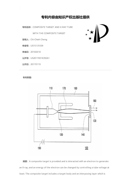
专利名称:COMPOSITE TARGET AND X-RAY TUBEWITH THE COMPOSITE TARGET发明人:Chi-Chieh Cheng申请号:US15131039申请日:20160418公开号:US20170018392A1公开日:20170119专利内容由知识产权出版社提供专利附图:摘要:A composite target is provided and is interacted with an electron to generate an X-ray, and an energy of the electron can be changed by controlling a tube voltage at least. The composite target includes a target body and an interposing layer which isconnected with the target body. The interposing layer moves a highest peak of an energy spectrum of the X-ray toward a high energy direction. The interposing layer may be a single metal or a metal mixture. Not only a low energy photon of the X-ray can be filtered by the interposing layer, but also a distribution of the low energy photon of the X-ray can be increased by increasing a thickness of the interposing layer. As the tube voltage is enhanced, an amount of a high energy photon of the X-ray generated is dramatically increased. An X-ray tube containing the above composite target is also provided.申请人:NanoRay Biotech Co., Ltd.地址:New Taipei City TW国籍:TW更多信息请下载全文后查看。
射线法 金属薄膜厚度
射线法金属薄膜厚度英文回答:X-ray diffraction (XRD) is a versatile non-destructive technique for characterizing the structure and properties of materials, including the thickness of metal films. XRD works by directing a beam of X-rays at the sample and measuring the intensity and pattern of the diffracted X-rays. The thickness of the metal film can be determined by analyzing the intensity of the diffracted X-rays at specific angles.The basic principle behind XRD is Bragg's law, which states that when X-rays interact with a crystalline material, they are diffracted at specific angles that are determined by the wavelength of the X-rays and the spacing between the atoms in the crystal. In the case of a thin metal film, the diffracted X-rays will interfere with each other, producing a pattern of peaks and valleys in the intensity of the diffracted X-rays.The thickness of the metal film can be determined by measuring the distance between the peaks in the diffraction pattern. The distance between the peaks is related to the spacing between the atoms in the metal film, which in turn is related to the thickness of the film.XRD is a highly accurate and precise technique for measuring the thickness of metal films. It is non-destructive, so it does not damage the sample, and it can be used to measure the thickness of films on a variety of substrates.中文回答:射线法测量金属薄膜厚度。
X射线分析技术在涂料分析中的应用

48技术应用与研究2020 ・ 24当代化工研究Modem Chemiad RemearchX 射线分析技术在涂料分析中的应用*孙慧(南京金升华包装材料有限公司江苏210000)摘耍:x 射线分析对于涂料的成分研究具有重要的作用,其中对涂料分析射线技术进行了阐述,其中主要对x 射线衍射光谱、x 射线荧光 光谱和x 射线电子能谱这三种技术在涂料分析中的应用进行了分析和说明.关键词:涂料分析;晶体;X 射线分析技术中图分类号•: T 文献标识码:AApplication of X-ray Analysis Technology in Paint AnalysisSun Hui(Nanjing Jinshenghua Packaging Materials Co., Ltd., Jiangsu, 210000)Abstract z X-ray analysis plays an important role in the composition research of p aint This paper describes the X-ray technology of p aintanalysis, in which the application of X -ray diffraction spectrum, X-ray f luorescence spectrum and X-ray electron spectroscopy in paint analysis is mainly analyzed and explained.Key words : paint analysis i crystals i X-ray analysis technology1. 涂料的定义涂料是一种由高分子材料为主体的,具有保护功能、特 殊功能、装饰功能的固体材料或液体材料的总称,其一般是 通过涂抹的方式将涂料覆盖至物体的表面,使其以被涂物质 为基体从而有机的结合在一起。
X-ray_CT在纤维增强聚合物复合材料中的应用研究进展
㊀第41卷㊀第7期2022年7月中国材料进展MATERIALS CHINAVol.41㊀No.7Jul.2022收稿日期:2020-11-02㊀㊀修回日期:2020-12-17第一作者:李倩倩,女,1994年生,博士研究生通讯作者:李㊀炜,女,1971年生,教授,博士生导师,Email:liwei@DOI :10.7502/j.issn.1674-3962.202011001X-ray CT 在纤维增强聚合物复合材料中的应用研究进展李倩倩1,2,李㊀哲1,2,李㊀炜1,2,3(1.东华大学上海高性能纤维复合材料协同创新中心,上海201620)(2.东华大学纺织学院,上海201620)(3.产业用纺织品教育部工程研究中心,上海201620)摘㊀要:X 射线计算机断层扫描(X-ray computed tomography,X-ray CT)作为一种具有高分辨率㊁非破坏性㊁三维可视化等特征的材料表征测量技术,显示出独特的优势㊂X-ray CT 不仅能得到试样的高分辨率外部特征,还能够获取其详细的内部结构信息,在各个领域已经受到越来越多的关注㊂通过简单比较几种无损检测技术的适用性和局限性,引出了X-ray CT 的独特性,阐述了X-ray CT 的原理以及实验室断层扫描和同步辐射计算机断层扫描(synchrotron radiation computed tomography,SRCT)的特点;从探索纤维增强聚合物(fiber reinforced polymer,FRP)复合材料的内部结构㊁评估制造过程㊁获取损伤失效演变以及X-ray CT 与其他表征手段相结合等几个方面归纳了X-ray CT 在FRP 中的应用,并提出了X-ray CT 在FRP 研究中的挑战㊂关键词:无损检测;纤维增强聚合物复合材料;X-ray CT;结构分析;损伤分析;原位实验中图分类号:TB332;O434㊀㊀文献标识码:A㊀㊀文章编号:1674-3962(2022)07-0525-11引用格式:李倩倩,李哲,李炜.X-ray CT 在纤维增强聚合物复合材料中的应用研究进展[J].中国材料进展,2022,41(7):525-535.LI Q Q,LI Z,LI W.Application Research Progress of X-ray CT in Fiber Reinforced Polymer Composites[J].Materials China,2022,41(7):525-535.Application Research Progress of X-ray CT in Fiber Reinforced Polymer CompositesLI Qianqian 1,2,LI Zhe 1,2,LI Wei 1,2,3(1.Shanghai Collaborative Innovation Center for High Performance Fiber Composites,Donghua University,Shanghai 201620,China)(2.College of Textiles,Donghua University,Shanghai 201620,China)(3.Engineering Research Center of Technical Textile of Ministry of Education,Shanghai 201620,China)Abstract :X-ray computed tomography (X-ray CT),as a high-resolution,non-destructive,3D visualization technology ofmaterial characterization and measurement,shows unique advantages.It can not only provide the high-resolution external characteristics of the sample,but also obtain the detailed internal structure information,which has attracted more and more attention in various fields.This article highlights the uniqueness of X-ray CT by simply comparing the applicability and limi-tations of several non-destructive testing techniques.The principle of X-ray CT and the characteristics of laboratory tomo-graphy and synchrotron radiation computed tomography(SRCT)are mainly described.The application of X-ray CT in fiber reinforced polymer(FRP)composites is summarized from the aspects of exploring the internal structure,evaluating the man-ufacturing process,obtaining the evolution of damage or failure,and combining X-ray CT with other characterization meth-ods.The challenge and development trend of X-ray CT in FRP research are put forward.Key words :non-destructive testing;fiber reinforced polymer;X-ray CT;structure analysis;damage analysis;in-situ experiment1㊀前㊀言纤维增强聚合物(fiber reinforced polymer,FRP)是由纤维增强体和聚合物基体通过成型固化工艺得到的多相中国材料进展第41卷材料,具有轻质高强㊁耐腐蚀㊁抗疲劳㊁可设计性强等优点,广泛应用于建筑㊁交通㊁海洋㊁风电以及航空航天等领域[1-4]㊂但由于材料本身或制造过程所产生的缺陷,在受到外力作用时会产生纤维断裂[5,6]㊁基体开裂[6]㊁分层[7,8]等损伤,逐渐累积就会导致结构的失效,从而降低工件的实际寿命和使用安全性[6]㊂因此,通过有效的损伤评估方式,研究复合材料的损伤演化和破坏机理,进而揭示复合材料力学性能的影响机制,对于材料的安全性和长期稳定服役具有重要意义[6,9]㊂FRP的损伤是一个三维问题,通常使用的光学和电子显微镜只能观察到其损伤后的表面形态[10],无法获得损伤与时间维度的关系,并且有时会因打磨和切割引起伪影或二次损伤,造成误导,使分析更加复杂化[11-13]㊂而无损检测技术既不改变材料的属性,又不会对试样造成损伤,即可获得材料内部或表面的缺陷和损伤[14],可用于试样的单独检测,也可用于整个生产加工过程的监测,既保持了结构的完整性,又可以检测㊁定位并确定损伤的大小㊂常用的无损检测(non-destructive testing, NDT或non-destructive evaluation,NDE)技术包括目视检测(visual testing,VT或visual inspection,VI)㊁射线检测(radiographic testing,RT)㊁超声检测(ultrasonic testing, UT)㊁渗透检测(penetrate testing,PT)㊁涡流检测(eddy current testing,ET),还有红外热成像(infrared thermogra-phy,IRT)和声发射(acoustic emission,AE)等㊂表1列举了几种无损检测方法的适用范围和局限性[6,14-20]㊂由表1可以看出,大部分的无损检测方法只能检测到大裂缝(大于几毫米),而X射线计算机断层扫描(X-ray com-puted tomography,X-ray CT)可以以亚毫米级,甚至是几微米的空间分辨率观察样品中的裂缝,此外,还能实现对纳米级裂纹进行三维观察[21]㊂因此,较其它无损检测方法而言,X-ray CT具有高的空间分辨率和精确捕获多尺度结构的能力,可实现对细节(包括不同的相㊁界面㊁孔隙和裂缝)清晰的可视化,并能够 原位 监控整个制造过程[22]㊂本文首先介绍了X-ray CT的基本原理㊁原位装置和超高温原位拉伸装置,并简单描述了实验室断层扫描(laboratory tomography)和同步辐射计算机断层扫描(synchrotron radiation computed tomography,SRCT)的区别;其次从利用X-ray CT探索内部结构进而辅助建模㊁评估制造过程㊁获取损伤失效过程以及与其他表征手段相结合这4个方面阐述了X-ray CT在FRP中的应用;表1㊀无损检测方法[6,14-20]Table1㊀Methods of non-destructive testing[6,14-20]无损检测方法适用范围特点局限性目视检测(VT或VI)宏观表面缺陷[15]快速㊁成本低㊁不需要设备[14]不精确,小缺陷很难被检测到,也不能看到内部缺陷,人为因素影响较大[16]射线检测(RT)体积性缺陷检测,如孔隙㊁夹杂㊁纤维屈曲[17]结果真实可靠,呈现方式最直观㊁最全面以及后续可追踪性强[17]使用成本较高,检测速度慢,射线会对人体造成伤害以及面积型缺陷的检出率低[17]超声检测(UT)对面积型缺陷的检出率较高,更适用于厚度较大工件的检验,对缺陷在工件厚度方向上的定位较准确,可检测试件内部尺寸很小的缺陷[17]成本低㊁灵敏度高㊁速度快,设备轻便,对人体及环境无害,设备易于携带[6]分辨率有限,跟踪各种缺陷类型的相互作用或检测隐藏缺陷的能力很差[18],不易检测分层缺陷[8]渗透检测(PT)表面开口裂缝[17]简便㊁方便㊁价格低[17]检测前要清理工件,而且渗透液对试样有污染,不适用于疏松多孔结构[6]涡流检测(ET)工件的裂纹㊁孔洞㊁折叠和夹杂等缺陷的检测[17],材料厚度测量㊁涂层厚度测量㊁热损伤检测㊁热处理检测[15]不需与被测物直接接触,可进行高速检测,易于实现自动化不适用于形状复杂的零件,而且只能检测导电材料的表面和近表面缺陷,检测结果也易于受到材料本身及其他因素的干扰,被测信号难以解释,不能用于不导电材料的检测,无法检测出埋藏较深的缺陷,无法从显示信号中直接判断缺陷的性质[17]红外热成像(IRT)适用于厚度较薄的试样,检测材料近表面及表面缺陷[14,19]便捷㊁高效㊁直观㊁探测面积大以及远距离非接触探测[19]要求工件传热性好,需要敏感且昂贵的仪器,需要高技能的检查员来操作仪器,如果缺陷在零件表面下太深,则缺乏清晰度[14,18]声发射(AE)动态加载过程中的各种损伤及其扩展连续㊁实时检测,并能反映出声发射源在载荷作用下的动态响应特性[20],对工件本身的几何形状要求不高不能检测出未扩展的静态缺陷,而且设备价格昂贵[17],对结果解释很困难,未形成统一的评价标准625㊀第7期李倩倩等:X-ray CT 在纤维增强聚合物复合材料中的应用研究进展最后,总结了X-ray CT 广泛应用于FRP 领域的定性和定量评估,并简单归纳了X-ray CT 在应用中可能遇到的挑战㊂2㊀X 射线计算机断层扫描(X -ray CT )X-ray CT 的基本原理是由于试样内部不同相或者多种成分间的密度以及原子序数的不同,当X 射线透过试样时,试件的不同相或不同成分对X 射线的衰减产生差异,从而造成成像的明暗差别[23,24]㊂X-ray CT 关键在于对不同角度获得的X 光片(投影)的计算重建[25],要从重建图像中获取有效特征,其中一个重要参数就是对比度,对比度是由材料成分的线性衰减系数不同引起的,而线性衰减系数却与材料的密度㊁原子序数息息相关㊂同时分辨率也会影响重建图像的细节水平㊂X-ray CT 的原理示意图如图1所示[25]:X 射线照射到固定在旋转控制台的试样上,每旋转一定的角度,探测器就会采集一张照片,对应于试样旋转的N 个角度会有N 张射线照片被探测器(通常是断层扫描仪中的电荷耦合元件CCD)记录下来(这一过程也称为扫描),重构软件利用这些射线照片获得试样内部衰减系数的三维分布,这种分布形成三维图像,可以使用成像软件进行查看[25]㊂多年来对复合材料损伤模式的研究大多局限在对损伤后的表征,而由于一些特殊的实验环境(如高温)或者是探索机械载荷下样品中缺陷的形成与发展,需要对材料进行原位的观察[25]㊂而原位X-ray CT 可以在载荷下同步获得材料内部结构的变化或损伤演变的过程㊂如图2所示[26],在旋转控制台部位增加了加载控制装置,形成原位加载装置㊂还可以在原位加载装置上实现温度图1㊀X 射线计算机断层扫描的原理示意图[25]Fig.1㊀Schematic diagram of X-ray computed tomography (X-ray CT)[25]场的变化,如图3所示[27],图3a 是原位超高温拉伸试验装置的示意图,分为谐波电动机驱动㊁线性执行器平台㊁负载反馈模块㊁在真空/惰性气体环境下控制超高温的样品室4个部分,载荷由步进电机施加到样品上,而力和位移则使用在线称重传感器和线性可变差动变压传感器进行测量㊂图3b 是加热室的截面图,样品通过水冷却拉伸夹具固定在直径约为170mm 的真空密封室的中心,这个密封室可以抽真空并回填选定的气体(通常为惰性气体),150W 卤素灯分布在6个方位作为热源提供热量,每个卤素灯都有一个指向样品室中心的椭圆形反射镜,形成了直径约为5mm 的球形热区㊂通过使用热电偶对灯的功率进行单独校准来确定热区中被测试样品的温度,样品室中有一个圆柱形铝窗(厚度为300μm,高度为7mm),它可以使X 射线照到样品并透过到X 射线成像系统,如图3c 中X 射线照射模式下的装置示意图[27]㊂图2㊀原位X 射线计算机断层扫描的原理示意图[26]Fig.2㊀Schematic diagram of in situ X-ray computed tomography [26]725中国材料进展第41卷图3㊀原位超高温拉伸试验装置[27]:(a)同步辐射计算机断层扫描原位超高温拉伸测试装置示意图;(b)加热室的截面图, X射线透过加热室和样品的传输路径;(c)X射线照射下的装置示意图Fig.3㊀In situ ultrahigh temperature tensile test rig[27]:(a)schematic illustration of in situ ultrahigh temperature tensile test rig for syn-chrotron X-ray computed microtomography;(b)sectional view of the heating chamber,X-ray transmission path through the heat-ing chamber and sample;(c)schematic of the rig in transmission mode for X-ray computed tomography㊀㊀X-ray CT装置的光源分为同步加速器源提供的平行X射线束和实验室断层扫描仪用几微米宽的微聚焦源提供的锥形X射线束㊂前者称为SRCT,后者称为实验室断层扫描㊂SRCT使用相干光源和平行单色光束,可以达到更高的分辨率,采集时间更短,效率更高,对于低对比度的材料同样适用,但尺度受限制;实验室断层扫描使用多色光源和锥形光束,价格相对较低,可以对较大体积的试样进行多个尺度的研究,但是复合材料的图像会受到相位对比度差和采集时间长的影响[18,24,25]㊂总之,X-ray CT以非破坏性的方式对试样进行高精度的三维检查,不仅可以获得试样内部的详细信息,还可以捕捉制造过程中的缺陷或承载过程中材料的变化,对于研究复合材料的损伤机理和破坏过程是一种非常有效的方法㊂3㊀X-ray CT在FRP中的应用3.1㊀探索FRP的内部结构并辅助建模和验证材料的结构决定它的性能,因此要探究纤维增强复合材料的力学性能影响机制,需要对它的内部结构进行详细了解,同时通过建立材料结构的多尺度模型来对力学性能进行分析及预测[28],复合材料被看作宏观尺度,纱线被认为是介观尺度,而纱线中的纤维就作为细观尺度[29]㊂有限元分析的质量取决于初始模型的质量[30],目前大多数的数值模拟或分析都带有人为的假设,导致模型与材料的真实细观结构存在较大出入[28],因此精确㊁详细的内部结构对于改进复合材料的几何模型㊁评估材料内部缺陷是必要的,对模拟预测复合材料的力学性能具有非常重要的意义,而从X-ray CT图像中获得的信息对此有很大的帮助和参考价值㊂Mahadik等[31]利用X-ray CT研究两种不同结构的三维角联锁机织复合材料的结构特征,以及在不断增加的压力下材料内部结构的变化,主要观察了纱线的屈曲以及富含树脂区域的尺寸和形状㊂刘振国等[23]利用显微计算机断层成像(micro-computed tomography,Micro-CT或μ-CT)对三维全五向编织复合材料的内部结构进行分析,为提高对比度,在碳纤维试件中混编入少量玻璃纤维作为示踪纱,并通过三维重建得到纤维束实体模型,结合计算机辅助设计(computer aided design,CAD)对纤维束的825㊀第7期李倩倩等:X-ray CT 在纤维增强聚合物复合材料中的应用研究进展横截面形状和空间走向进行分析,为进一步研究材料的细观结构模型和性能仿真计算奠定了基础㊂Melenka 等[32]提出了一种圆柱体展开算法,将原始的二维编织管状复合材料的μ-CT 图像转换为扁平的编织物结构图像,以简化编织物几何形状内的单根纱线的分割和分析,同时利用一个自定义的MATLAB 图像处理程序确定了每束纱线的质心㊁横截面积㊁纵横比㊁编织角和编织循环周期,准确评估了编织纱轨迹的几何形状和孔隙含量㊂Sencu 等[33]开发了一套图像处理和分割算法,可以有效地从碳纤维复合材料的X-ray CT 图像中识别纤维中心线(轮廓),从而生成具有高保真度的微尺度有限元模型,图4显示了从X-ray CT 图像切片中分析碳纤维增强复合材料(CFRP)几何结构并生成有限元网格的主要步骤㊂Naouar 等[30]则探索了一种基于图像纹理的分割方法来分离μ-CT 图像中的经纱㊁纬纱和接结纱,该方法适用于内部几何形状众多且复杂的三维织物增强体㊂Huang 等[34]通过Micro-CT 的图像,重建连续纤维增强体的细观几何模型,并将该方法称为Micro-CT AGM,同时开发了名为CompoCT 的软件用作该特定建模过程的平台,主要是结合被观察试样的3个视图对织物中的纱线束进行了手动分割,这与通过标准图像处理技术或从纺织品建模软件获得的模型不同,此方法不需要很高的图像分辨率,被扫描的纤维样品的尺寸可以更大,从而包含一个以上的单胞,可将真实材料更具代表性的几何特征与其内在的变化结合起来,实现了在较低的扫描分辨率(22μm /像素)下准确重构二维机织织物和三维正交织物的细观几何模型㊂Liu 等[35]同样基于X-ray CT 图像,用一种统计分析方法来生成三维五向编织复合材料的代表性体积单元,并考虑了织物的压缩和轴向纱线的加捻,从而更接近织物的真实状态㊂Naouar 等[29]阐述了基于μ-CT 的纺织品复合材料的细观建模技术,包括两种分割方法(结构张量和纹理分析)和织物预制体的变形响应模型以及织物复合材料的损伤模型,指出X-ray CT 是一种适用于织物增强复合材料细观分析的工具㊂陈城华[28]将三维编织复合材料试样进行Micro-CT 扫描,获得切片照片后,通过编程实现对该试样图像的三维重构,再提取代表性单胞用于有限元模拟计算,介绍了一套完整的编织复合材料三维重建流程㊂顾伯洪课题组[36,37]也将X-ray CT 与有限元模拟相结合,主要是利用图像来识别材料内部的损伤,再与有限元模拟的结果进行比较验证,从而分析损伤分布或失效机制㊂正如Naresh 等[22]的总结,分析X-ray CT 的图像生成体素几何来运行模拟,进而研究各种复合工艺参数的过程可以分为3个基本步骤,如图5所示:①预处理,包括对X-ray CT 数据进行滤波和平滑,以增强图像质量,再将这些处理后的图像作为输入数据,通过使用软件包来创建三维结构;②分析,包括分割和特征提取(成分分析),分割是数据分析中必不可少的步骤,可根据其灰度值区分FRP 中的不同成分,比如纤维㊁树脂㊁空气等;③数据可视化,包括结果的映射和渲染,然后验证结果[22]㊂相较于其他无损检测技术,X-ray CT 在探索FRP 的内部结构以及辅助建模中具有很大的优势,可以为复杂的几何形状提供详细的三维信息,包括纱线的屈曲㊁纤维的取向㊁各成分的体积分数㊁有关孔隙的形态信息以及纤维㊁纱线路径的轮廓等,但在图像获取后仍需要进行优化,同时对操作人员的熟练程度以及图像处理能力有很高的技术要求,时间和资金成本也很昂贵㊂此外,这些基于X-ray CT 的建模大多是中尺度或者是细观尺度,而用于理解和辅助宏观几何形状的建模方法有限,并且每个扫描标本的文件大小通常为几GB,需要使用大容量的计算机数据存储设备来存储X-ray CT 扫描和重建的数据[18,22]㊂图4㊀从X-ray CT 图像切片中提取碳纤维增强复合材料(CFRP)几何结构并生成有限元网格的主要步骤[33]Fig.4㊀Main steps to extract the CFRP geometry from X-ray CT image slices and generate finite element (FE)meshes [33]925中国材料进展第41卷图5㊀将X-ray CT的图像用于模拟分析的过程[22] Fig.5㊀Process of using X-ray CT images for simulation analysis[22]3.2㊀评估制造过程不仅结构会决定复合材料的性能,制造工艺也会对复合材料的力学性能产生重要的影响,因此可以利用X-ray CT来跟踪成型制造过程[24]㊂在FRP制造过程中,其压力变形响应影响所需的压实力㊁作用在设备上的应力㊁工具要求和成品质量,对压缩变形的良好认识有助于开发更准确的模型来描述和预测制造过程,从而改进所采用的制备方法[38]㊂Somashekar等[38]通过X-ray CT观察了一种双轴缝合玻璃纤维增强材料在不同工艺参数(最终纤维体积分数㊁压实速度㊁压实次数)压缩后的纤维变形㊂孔隙是复合材料制造过程中产生的主要缺陷之一,Plessix等[39]开发了一种专门的装置并安装在欧洲同步辐射光源(ESRF)的一条专用于超快速X-ary CT的光束线上,以获得原位三维图像,并分析孔隙演化的原位图像与时间㊁温度㊁压力㊁初始含水量和树脂转化率之间的关系,这项工作对原位监测热固性复合材料固化过程中孔隙的产生和发展演变做出了开创性的贡献㊂Dilonardo等[3]采用X-ray CT评估层合结构和夹芯结构的CFRP的孔隙率,此外,具体的数据分析还提供了关于孔隙㊁缺陷或纤维错位的尺寸㊁形状和位置的详细信息㊂而了解树脂流动机理对于通过液体模塑制造具有最小孔隙率的复合材料至关重要, Vilà等[40]设计并建造了一个微型真空注入装置,在德国电子同步加速器研究所(DESY)用X-ray CT对真空辅助渗透的微尺度渗透机制进行了原位研究,微观层面的流体传播以及纤维束内的孔隙传输机制与流体和纤维之间的浸润性㊁流体的流变性以及纤维束的局部微结构特征(局部纤维体积分数㊁纤维取向)有关㊂除了孔隙之外,另一个重要的质量评价参数是纤维的排列,纤维与设计对准角度间的偏差是一种制造缺陷,称为纤维错位, Nguyen等[41]提出了一种基于X-ray CT分析碳纤维增强层压板在低压(真空辅助树脂传递模塑)和高压(高压釜)制造过程中纤维错位的方法,结果表明两种不同成型方法的纤维错位角度分别为1ʎ~2ʎ和2ʎ~4ʎ㊂X-ray CT尽管在空间分辨率和样品尺寸之间存在相互制约,尤其对于大型部件的制造过程来说不好实现评估,但仍可以提供详细的三维信息来评估制造质量,在制造过程的不同阶段生成有价值的信息,并且能够以模型的形式提取该信息,从而更好地理解和改进制造过程,提高FRP的质量和制造效率㊂3.3㊀获取损伤失效过程利用X-ray CT捕获复合材料在载荷作用下的损伤失效过程,对于深入理解复合材料的破坏机理和力学影响机制,进而实现结构优化设计具有重要的理论意义和工程应用价值[42]㊂而纤维增强复合材料在载荷下的损伤主要包括层内裂纹㊁层间分层㊁纤维抽拔和纤维断裂等[5,43,44],损伤的开始和随后的扩展是这些机制的复杂相互作用㊂2008年,Wright等[45]首次使用SRCT来实现航空航天级碳纤维/环氧复合材料损伤的亚微米分辨率,并能在以前观察不到的三维尺度上对损伤的结构和损伤机制的相互作用进行可视化,层内裂纹和分层对纤维断裂的关键作用首次得到明确㊂于颂等[46]利用高分辨率Micro-CT 对三维五向编织非周期性结构复合材料和周期性结构复035㊀第7期李倩倩等:X-ray CT在纤维增强聚合物复合材料中的应用研究进展合材料在拉伸载荷下的破坏形貌进行了观测,并得出非周期性三维编织复合材料拉伸强度比相同结构参数周期性材料的低16.84%,损伤主要是因为在减纱处形成了应力集中,而最终破坏模式以纤维束抽拔断裂为主㊂2011年,Scott等[12]利用高分辨率SRCT捕捉碳纤维环氧树脂[90/0]s缺口层合板加载至失效时的纤维损伤进展,首次对高性能材料在载荷条件下的裂纹扩展进行了直接原位测量㊂如图6所示为原位加载的载荷从失效载荷的20%到80%过程中基体损伤的提取和分割,获得的图像能够识别和量化所使用样本的主要损伤机制㊂之后,很多学者利用自己开发的原位拉伸加载装置对试样的加载过程进行原位观察,这些装置必须满足可以安装在X-ray CT仪器的内部,因此都很小,所以对精度的要求很高㊂Hu等[47]开发的原位拉伸试验装置可对小样品施加微小的力,位移精度约为1μm,测力精度约为0.1mN,获得了高分辨率(0.7μm/像素)的原位观察,显示了随机取向短碳纤维/环氧树脂复合材料的断裂过程㊂Saito等[48]也开发了足够小的定制拉伸装置,它的最大行程和最大力分别为220mm和98N(10kgf),加载速率可在0.01~1mm㊃min-1范围内调整㊂Li等[26]则考虑到(拉伸试验中)夹具的难度和(剪切试验中)夹具的X射线吸收,设计了新型试件,并提出了一种将SRCT㊁原位加载框架和新型试件相结合的实验方法,分别对三维机织碳纤维增强复合材料在平面外拉伸和剪切载荷作用下的损伤演化进行了研究㊂Liu等[49]利用高分辨率的SRCT,通过原位准静态拉伸试验研究了短碳纤维增强复合材料从初始状态到试样断裂的内部三维应变演化过程,首次在三维中同时分析材料微观结构和应变值,并得出短碳纤维/环氧树脂复合材料的宏观性能(刚度降低和破坏过程)与微观特征(应变演化和纤维排列)有关的结论㊂冲击试验的典型特征是持续时间非常短,应变率非常高,这意味着通过原位X-ray CT对其进行成像存在困难[13,24],因此,目前大部分的文献都是利用X-ray CT获取复合材料冲击试验前后的图像来分析其损伤情况㊂En-fedaque等[51]对树脂传递模塑(resin-transfer molding, RTM)制造的碳-璃混杂织物层合板进行低速冲击试验,通过X-ray CT研究了不同厚度和不同玻璃纤维含量的试样在30~245J冲击能量范围的变形和断裂机理,并聚焦于未完全穿透试样的分析㊂Bull等[51]用实验室μ-CT和SRCT对颗粒增韧复合材料层合板低速冲击损伤进行了三维评估,两种成像方法的结合使得可以在微观和细观水平上以常规的体素分辨率观察到颗粒增韧的效果,突出了使用X-ray CT将微观力学损伤行为与宏观力学响应联系起来的潜力㊂虽然X-ray CT可对复合材料冲击损伤后的破坏形貌进行三维可视化,但对于弯曲或变形的复合材料板,很难自动将损坏归因于特定层或层间界面, Léonard等[52]开发了一种X-ray CT数据处理方法,这种距离变换方法允许切片近似地遵循复合曲率,允许在三维上逐层地分离㊁可视化和量化冲击损伤以提取损伤在厚度方向上的分布,这对描述复合材料层合板冲击损伤和从X-ray CT数据集提取相关测量数据的能力有很大的提高㊂Zhou等[53]也用X-ray CT对三维编织复合材料圆管在用分离式霍普金森压杆(split Hopkinson pressure bar, SHPB)横向冲击后的损伤进行了研究,通过三维图像分析了冲击速度㊁编织角和编织层数对损伤机理的影响㊂Lu等[13]则通过X-ray CT对热塑性碳纤维/聚醚醚酮复合图6㊀原位加载的载荷从失效载荷的20%到80%过程基体损伤的提取和分割(90ʎ层中的横向层裂纹,从缺口延伸的0ʎ裂口,以及在90/0界面处发生的分层)[12]Fig.6㊀Extraction and segmentation of matrix damage during in situ loading from20%to80%of failure load(transverse ply cracks in the90ʎplies,0ʎsplits extending from the notch,and delamination occurring at90/0interface)[12]135。
骨科英文书籍精读(92)|肘关节脱位(1)

⾻科英⽂书籍精读(92)|肘关节脱位(1)DISLOCATION OF THE ELBOWDislocation of the ulno-humeral joint is fairly common – more so in adults than in children. Injuries are usually classified according to the direction of displacement. However, in 90% of cases the radioulnar complex is displaced posteriorly or posterolaterally, often together with fractures of the restraining bony processes.Mechanism of injury and pathologyThe cause of posterior dislocation is usually a fall on the outstretched hand with the elbow in extension. Disruption of the capsule and ligaments structures alone can result in posterior or posterolateral dislocation. However, provided there is no associated fracture, reduction will usually be stable and recurrent dislocation unlikely. The combination of ligamentous disruption and fracture of the radial head, coronoid process or olecranon process (or, worse still, several fractures) will render the joint more unstable and, unless the fractures are reduced and fixed, liable to redislocation.Once posterior dislocation has taken place, lateral shift may also occur. Soft tissue disruption is often considerable and surrounding nerves and vessels may be damaged. Although certain common patterns of fracture-dislocation are recognized (based on the particular combination of structures involved), highenergy injuries do not necessarily follow any rules. A classic example is the so-called sideswipe injury which occurs, typically, when a car-driver’s elbow, protruding through the window, is struck by another vehicle.The result is forward dislocation with fractures of any or all of the bones around the elbow; soft-tissue damage (including neurovascular injury) is usually severe.Clinical featuresThe patient supports his forearm with the elbow in slight flexion. Unless swelling is severe, the deformity is obvious. The bony landmarks (olecranon and epicondyles) may be palpable and abnormally placed.However, in severe injuries pain and swelling are so marked that examination of the elbow is impossible. Nevertheless, the hand should be examined for signs of vascular or nerve damage.X-rayX-ray examination is essential (a) to confirm the presence of a dislocation and (b) to identify any associated fractures. It is often only when the elbow is screened at the time of surgery that the full extent of the injury can be established.---from 《Apley’s System of Orthopaedics and Fractures》临床特征病⼈⽤肘关节⽀撑前臂,使其轻微弯曲。
X-ray CT在纤维增强聚合物复合材料中的应用研究进展

X- r ay CT 在纤维增强聚合物复合材料中的应用研究进展摘要:纤维增强聚合物复合材料在航空、汽车等领域具有广泛应用,而X- ray CT 技术已成为评估复合材料内部结构的重要手段。
本文综述了X-ray CT 技术在纤维增强聚合物复合材料中的应用研究进展,包括定量分析、损伤评估、微观结构探究等方面,并对未来的发展趋势进行了展望。
关键词:纤维增强聚合物复合材料;X-ray CT 技术;定量分析;损伤评估;微观结构一、引言随着科技的不断发展,纤维增强聚合物复合材料在航空、汽车等领域中得到了广泛应用。
与传统材料相比,纤维增强聚合物复合材料具有轻质、高强度、高模量等突出优点。
为了保证其性能和可靠性,对其内部结构进行评估变得尤为重要。
目前,X-ray CT 技术已成为评估复合材料内部结构的重要手段。
本文将综述X-ray CT 技术在纤维增强聚合物复合材料中的应用研究进展,包括定量分析、损伤评估、微观结构探究等方面,并对未来的发展趋势进行了展望。
二、X-ray CT 技术在纤维增强聚合物复合材料中的应用1.定量分析X-ray CT 技术可以用于复合材料内部结构的定量分析。
从CT 图像中可以获取物体的三维形态、尺寸和形状等信息。
尤其是在分析复合材料内部孔隙率和纤维含量时,CT 技术可以提供更加准确的结果。
同时,基于X-ray CT 数据的模拟技术也可以用于预测材料的力学性能。
2.损伤评估X-ray CT 技术可以用于复合材料损伤评估。
在复合材料中出现的各种损伤类型,如裂纹、孔隙、纤维断裂等都可以通过CT 技术进行检测。
通过分析CT 图像,可以获得材料内部的低密度区域、损伤大小、定位和预测。
3.微观结构探究X-r ay CT 技术可以用于复合材料微观结构的探究。
CT 技术可以提供复合材料内部细枝纤维的分布、大小和取向等信息。
通过对CT 图像的处理,可以获取每个纤维的长度、跨距和连通性等参数。
此外,在对纳米级别的纤维增强聚合物复合材料进行研究时,X-ray CT 技术也表现出了优异的检测表现。
- 1、下载文档前请自行甄别文档内容的完整性,平台不提供额外的编辑、内容补充、找答案等附加服务。
- 2、"仅部分预览"的文档,不可在线预览部分如存在完整性等问题,可反馈申请退款(可完整预览的文档不适用该条件!)。
- 3、如文档侵犯您的权益,请联系客服反馈,我们会尽快为您处理(人工客服工作时间:9:00-18:30)。
a rXiv:as tr o-ph/47585v128J ul24Mem.S.A.It.Vol.73,23c SAIt 2002Memoriedella A.Wolter 1,G.Trinchieri 1,R.Della Ceca 1,F.Panessa 2,3,M.Dadina 2,L.Bassani 2,M.Cappi 2,A.Fruscione 3,S.Pellegrini 4,and G.Palumbo 41Osservatorio Astronomico di Brera via Brera 28,20121MILANO e-mail:anna@brera.mi.astro.it 2INAF/IASF,via P.Gobetti 101,40129Bologna 3Center for Astrophysics,60Garden St.,Cambridge,MASS 4Dipartimento di Astronomia,Universit`a di Bologna,via Ranzani 1,40127Bologna Abstract.An enigmatic,small class of IR and X-ray luminous sources,named “Composite”starburst/Seyfert galaxies,has been defined from IRAS and RASS data.The objects have optical spectra dominated by the features of HII galaxies (plus,in some cases,weak Seyfert signatures)but X-ray luminosities higher than expected from starbursts and more typical of Seyfert nuclei.The true nature of this class of objects is still unknown.We present Chandra data of four of these galaxies that were obtained to investigate the nature of the X-ray source.The X-ray spectrum,the lack of any significant extended component,and the observed variability indicate that the AGN is the dominant component in the X-ray domain.Key words.Galaxies:active –Galaxies:starburst –X-rays:galaxies 1.Introduction An enigmatic class of six low redshift galax-ies has been defined during a spectro-scopic survey of bright IRAS and RASS sources (Moran et al.1996).They werenamed “Composite”to indicate their dualnature:the optical spectrum typical of a star forming galaxy is characterized by ad-24 A.Wolter et al.:“Composite”Seyfert/Star-forminggalaxiesFig.1.The light curve ofIRAS20069+5929,in bins of1000sec,and residuals with respect to a constant(inσ);thefitχ2is46.8/25,indicating thatvariability is present.tailed description of the X-ray data will ap-pear in Panessa et al.(in preparation).2.X-ray dataThe sources were all observed with theACIS-I instrument on board Chandra fora nominal exposure of∼25ksec.Relevantdata for the sources are listed in Table1.Based on the RASSfluxes we had requesteda Chandra configuration that would allowmore frequent read-out of the data by us-ing a smallerfield of view to avoid pile-up.The results however indicate that the pre-cautions taken were not necessary,since theobserved count rates(see Table1)are wellbelow the expected value and the pile-up isnot significant.2.1.VariabilityWe compare the total Chandra luminosi-ties to previous RASS data(Moran et al.1996).Wefind consistency only forIRAS20069+5929.The luminosity issmaller by a factor∼10for IRAS04392-0123,and a factor∼2for IRAS01072+4954Fig.2.Profile of IRAS20069+5929(squares)compared to the chart-derivedPSF with the same spectral distribution ofthe source(histogram).X-axis is in arcsec.&IRAS01319-1604,as indicated by R inTable1.A discrepancy of a factor∼25was also reported for IRAS00317-2142(Georgantopoulos et al.2003),whileno information is available so far forIRAS20051-1117that has been observedby Chandra and XMM-Newton.The longterm variability alone already indicatesthat the dominant source of emission isdue to a point sources.We have further investigated short termvariability.We compute light curves by ex-tracting the source counts in a region of4′′radius and background in a larger,sourcefree,annulus around it.We bin light curvesat1000sec to have enough statistics perbin.We show in Figure1the non constantlight curve of source IRAS20069+5929,which has the largest statistics of the4Chandra observations.We highlight thisvariability detection since it indicates thepresence of a point source even if the av-erage luminosity of this source has notchanged since RASS.2.2.Spatial distributionNo extent is evident in any of the foursources,although a low surface brightnesscomponent might be present;more detailedA.Wolter et al.:“Composite”Seyfert/Star-forming galaxies25 Table1.Properties of the4“Composite”galaxies under study.Name Redshift c/s R∗L X(2-10keV)L(FIR)ACIS-I cgs(Chandra)cgs26 A.Wolter et al.:“Composite”Seyfert/Star-forming galaxiesaccording to Persic et al.(2004)(dotted line is contribution from HMXB;dashed line is the total from the Starburst)or David et al.(1992)(solid line).The cur-rently observed X-ray luminosities of the sources are still at least about a factor of 10higher than both estimates.The AGN dominates in X-rays or the starburst com-ponent is anomalous.3.ConclusionsFrom the X-ray Chandra observations we conclude that the sources are:1)point-like in the Chandra images,although we can-not exclude a low surface brightness com-ponent;2)variable-both within the sin-gle Chandra observation(as in the case of IRAS20069+5929)and with respect to older data;3)a thermal component is in general not present,even if in a few cases it might account for part of the soft emis-sion;4)the intrinsic absorption measured in the X-ray band is small or not required by thefit;5)the current L X is still at least an order of magnitude higher than the pre-dictions from the starburst power,which is of the order of SFR=10-100M⊙/yr.Since the emission lines from the AGN are weak in the optical band,and absorp-tion is not detected in X-rays,we cannot suggest obscuration as responsible for hid-ing the optical AGN.Therefore we conclude that an AGN is present and dominant in X rays, but probably of low luminosity:L X(2-10 keV)=3.5×1041-7.×1042erg/s.This could explain the optical faintness observed by Moran et al.(1996). Acknowledgements.We acknowledge partial financial support from Agenzia Spaziale Italiana(ASI).ReferencesDavid L.P.,Jones C.,&Forman W., (1992),ApJ,388,82Dickey J.M.&Lockman F.J.,(1990), ARAA,28,215Georgantopoulos,I.,(2000),MNRAS,315, 77Georgantopoulos,I.,Zezas,A.,Ward,M. J.,(2003),ApJ,584,129Moran, E.,Halpern,Jules P.,Helfand, David J.,(1996),ApJS,106,341 Persic,M.,Rephaeli,Y.,Braito,V.,Cappi, M.,Della Ceca,R.,Franceschini, A., Gruber,D.E.,(2004),A&A419,849。
