肺癌细胞抑制中英文对照外文翻译文献
英文文献+翻译

Characterization of production of Paclitaxel and related Taxanes in Taxus Cuspidata Densiformis suspension cultures by LC,LC/MS, and LC/MS/MSCHAPTER THEREPLANT TISSUE CULTUREⅠ. Potential of Plant cell Culture for Taxane ProductionSeveral alternative sources of paclitaxel have been identified and are currently the subjects of considerable investigation worldwide. These include the total synthesis and biosynthesis of paclitaxel, the agriculture supply of taxoids from needles of Taxus species, hemisynthesis (the attachment of a side chain to biogenetic precursors of paclitaxel such as baccatin Ⅲ or 10-deacetylbaccatin Ⅲ), fungus production, and the production of taxoids by cell and tissue culture. This reciew will concentrate only on the latter possibility.Plant tissue culture is one approach under investigation to provide large amounts and a stable supply of this compound exhibiting antineoplastic activity. A process to produce paclitaxel or paclitaxel-like compounds in cell culture has already been parented. The development of fast growing cell lines capable of producing paclitaxel would not only solve the limitations in paclitaxel supplies presently needed for clinical use, but would also help conserve the large number of trees that need to be harvested in order to isolate it. Currently, scientists and researchers have been successful in initiating fast plant growth but with limited paclitaxel production or vice versa. Therefore, it is the objective of researchers to find a method that will promote fast plant growth and also produce a large amount of paclitaxel at the same time.Ⅱ. Factors Influencing Growth Paclitaxel ContentA.Choice of Media for GrowthGamborg's (B5) and Murashige & Skoog's (MS) media seem to be superior for callus growth compared to White's (WP) medium. The major difference between these two media is that the MS medium contains 40 mM nitrate and 20mM ammonium, compared to 25mM nitrate and 2mM ammonium. Many researchers have selected the B5 medium over the MS medium for all subsequent studies, although they achieve similar results.Gamborg's B5 media was used throughout our experiments for initiation of callus cultures and suspension cultures due to successful published results. It was supplemented with 2% sucrose, 2 g/L casein hydrolysate, 2.4 mg/L picloram, and 1.8 mg/L α-naphthalene acetic acid. Agar (8 g/L) was used for solid cultures.B. Initiation of Callus CulturesPrevious work indicated that bark explants seem to be the most useful for establishing callus. The age of the tree did not appear to affect the ability to initiate callus when comparing both young and old tree materials grown on Gamborg's B5 medium supplemented with 1-2 mg/L of 2,4-dichlorophenoxyacetic acid. Callus cultures initiated and maintained in total darkness were generally pale-yellow to light brown in color. This resulted in sufficient masses of friable callus necessary for subculture within 3-4 weeks. However, the growth rate can decline substantially following the initial subculture and result in very slow-growing, brown-colored clumps of callus. It has been presumed that these brown-colored exudates are phenolic in nature and can eventually lead to cell death. This common phenomenon is totally random and unpredictable. Once this phenomenon has been triggered, the cells could not be saved by placing them in fresh media. However, adding polyvinylpyrrolidone to the culture media can help keep the cells alive and growing. Our experience with callus initiationwas similar to those studies.Our studies have found that callus which initiated early (usually within 2 weeks ) frequently did not proliferate when subcultured and turned brown and necrotic. In contrast, calli which developed from 4 weeks to 4 months after explants were fist placed on initiation media were able to be continuously subcultured when transferred at 1-2 month intervals. The presence of the survival of callus after subsequent subculturing. The relationship between paclitaxel concentration and callus initiation, however, has not been clarified.C. Effect of SugarSucrose is the preferred carbon source for growth in plant cell cultures, although the presence of more rapidly metabolized sugar such as glucose favors fast growth. Other sugars such as lactose, galactose, glucose, and fructose also support cell growth to some extent. On the other hand, sugar alcohols such as mannitol and sorbital which are generally used to raise the sugars added play a major role in the production of paclitaxel. In general, raising the initial sugar levels lead to an increase of secondary metabolite production. High initial levels of sugar increase the osmotic potential, although the role of osmotic pressure on the synthesis of secondary metabolites is not cleat. Kim and colleagues have shown that the highest level of paclitaxel was obtained with fructosel. The optimum concentration of each sugar for paclitaxel production was found to be the same at 6% in all cases. Wickremesinhe and Arteca have provided additional support that fructose is the most effective for paclitaxel production. However, other combinations of sugars such as sucrose combined with glucose also increased paclitaxel production.The presence of extracellular invertase activity and rapid extracellular sucrose hydrolysis has been observed in many cell cultures. These reports suggest that cells secrete or possess on their surface excess amounts of invertase, which result in the hydrolysis of sucrose at a much faster rate. The hydrolysis of sucrose coupled with the rapid utilization of fructose in the medium during the latter period of cell growth. This period of increased fructose availability coincided with the faster growth phase of the cells.D. Effect of Picloram and Methyl JasmonatePicloram (4-amino-3.5.6-trichloropicolinic acid) increases growth rate while methyl jasmonate has been reported to be an effective elicitor in the production of paclitaxel and other taxanes. However, little is known about the mechanisms or pathways that stimulate these secondary metabolites.Picloram had been used by Furmanowa and co-workers and Ketchum and Gibson but no details on the effect of picloram on growth rates were given. Furmanowa and hid colleagues observed growth of callus both in the presence and absence of light. The callus grew best in the dark showing a 9.3 fold increase, whereas there was only a 2-4 fold increase in the presence of light. Without picloram, callus growth was 0.9 fold. Unfortunately,this auxin had no effect on taxane production and the high callus growth rate was very unstable.Jasmonates exhibit various morphological and physiological activities when applied exogenously to plants. They induce transcriptional activation of genes involved in the formation of secondary metabolites. Methyl jasmonate was shown to stimulate paclitaxel and cephalomannine (taxane derivative) production in callus and suspension cultures. However, taxane production was best with White's medium compared to Gamborg's B5 medium. This may be due to the reduced concentration of potassium nitrate and a lack of ammonium sulfate with White's medium.E. Effect of Copper Sulfate and Mercuric ChlorideMetal ions have shown to play significant roles in altering the expression of secondary metabolic pathways in plant cell culture. Secondary metabolites,such as furano-terpenes, have been production by treatment of sweet potato root tissue with mercuric chloride. The results for copper sulfate, however, have not been reported. F. Growth Kinetics and Paclitaxel ProductionLow yields of paclitaxel may be attributed to the kinetics of taxane production that is not fully understood. Many reports stated inconclusive results on the kinetics of taxane production. More studies are needed in order to quantitate the taxane production. According to Nett-Fetto, the maximum instantaneous rate of paclitaxel production occurred at the third week upon further incubation. The paclitaxel level either declined or was not expected to increase upon further incubation. Paclitaxel production was very sensitive to slight variations in culture conditions. Due to this sensitivity, cell maintenance conditions, especially initial cell density, length of subculture interval, and temperature must be maintained as possible.Recently, Byun and co-workers have made a very detailed study on the kinetics of cell growth and taxane production. In their investigation, it was observed that the highest cell weight occurred at day 7 after inoculation. Similarly, the maximum concentration for 10-deacetyl baccatin Ⅲ and baccatin Ⅲ were detected at days 5 and 7, respectively. This result indicated that they are metabolic intermediates of paclitaxel. However, paclitaxel's maximum concentration was detected at day 22 but gradually declined. Byun and his colleagues suggested that paxlitaxel could be a metabolic intermediate like 10-deacetyl baccatin Ⅲ and baccatin Ⅲ or that pacliltaxel could be decomposed due to cellular morphological changes or DNA degradation characteristic of cell death.Pedtchanker's group also studied the kinetics of paclitaxel production by comparing the suspension cultures in shake flasks and Wilson-type reactors where bubbled air provided agitation and mixing. It was concluded that these cultures of Taxus cuspidata produced high levels of paclitaxel within three weeks (1.1 mg/L per day ). It was also determined that both cultures of the shake flask and Wilson-type reactor produced similar paclitaxel content. However, the Wilson-type reactor had a more rapid uptake of the nutrients (i.e. sugars, phosphate, calcium, and nitrate). This was probably due to the presence of the growth ring in the Wilson reactor. Therefor, the growth rate for the cultures from the Wilson reactor was only 135 mg./L while the shake flasks grew to 310 mg/L in three weeks.In retrospect, strictly controlled culture conditions are essential to consistent production and yield. Slight alterations in media formulations can have significant effects upon the physiology of cells, thereby affecting growth and product formation. All of the manipulations that affect growth and production of plant cells must be carefully integrated and controlled in order to maintain cell viability and stability.利用LC,LC/MS和LC/MS/MS悬浮培养生产紫杉醇及邓西佛米斯红豆杉中相关紫杉醇类的特征描述第三章植物组织培养Ⅰ.利用植物细胞培养生产紫杉的可能性紫杉醇的几个备选的来源已被确定,而且目前是全球大量调查的主题。
文献翻译
Low platelet count reduces subsequent complete remission rate despite marrow with <5% blasts after AML induction therapyAfter initial induction chemotherapy for acute myeloid leukemia(AML), convention and guidelines1suggest observation without therapy if a post-treatment bone marrow shows <5% myelo- blasts, even given persistently low absolute neutrophil counts(ANC) and/or platelet counts. Such practice implies that complete remission (CR; ANC ≥1000/μl and platelet count ≥100000/μl in addition to <5% marrow blasts by morphology) 2 might occur without further therapy. Herein we examine the relation between ANC and platelet count in patients with <5% marrow blasts on day 21 of course 1 of chemotherapy and subsequent probabilities of CR without additional therapy and of survival.Our database comprised 343 patients with newly diagnosed AML(acute promyelocytic leukemia excluded) or myelodysplastic syndrome with 15–19% blasts treated here from 2008 to 2011. We included 85 of these in whom a marrow aspirate on post- treatment day 21–28, 29–35, 36–42 or 43–49 contained<5% blasts, as determined morphologically and by multi-parameter flow cytometry (MFC), but in whom blood count criteria for CR had yet to be met. If more than one marrow was examined between days 21 and 49 only the first one was included. We used Cox regression for multivariate analyses to estimate theindependent effects of platelet count and ANC, and time when the marrow associated with these counts was obtained (for example, day 21–28) on probability of course 1 CR and of survival. The 85 patients had a median age of 54 years, with Southwest Oncology Group (SWOG) intermediate, unfavorable and favorable cytogenetics 3in 52%, 38% and 10%, respectively, and with secondary AML (after an antecedent hematologic disorder or therapy related) in 48%. Sixty-six percent received standard ‘3+7’, 21% received higher and 4% lower doses of cytarabine, while 9% received therapy without cytarabine (none with hypomethylating agents). Sixty-four percent achieved CR on the first course of therapy. Median survival was 13.1 months.CR rates were 44% with platelets <30 000/μl (approximately the median value) on day ≥21, 66% with platelets 30 000–100000/μl and 95% with platelets >100000/μl (P<0.001) (Table 1), recalling that the latter patients were not in CR at the time of marrow evaluation because ANC was still <1000/μl. The effect of ANC was qualitatively similar but less striking. In general, a higher platelet count at each of the various times (day 21–28, 29–35 and so on) correlated with a higher CR rate.Thirteen of the 20 patients with platelet counts <30000 on day ≥21 who did not achieve CR following course 1 achieved CRp (CR with platelet count <100000/μl) or CRi (CR with ANC<1000/μl) on that course vs 9/10 for patients with ≥30000 platelets. CR and CRp/CRi rateswere also higher with subsequent therapy in the ≥30000platelet group (Table 2). The remaining seven patients were resistant to therapy.Multivariate analysis accounting for age (numerical), de novo vs secondary AML, cytogenetics (SWOG favorable+intermediate vs unfavorable)3,day when marrow with<5% blasts was obtained (day 21–28 vs later) and percentage of blasts by MFC (numerical) in addition to platelets and ANC indicated that a higher platelet count (considered numerically) was independently associated with a higher CR rate (P=0.046). More practically, dichotomizing platelet count according to its approximate median (30000) gave an odds ratio of 0.37 (P=0.07) at counts <30000. None of the other covariates noted above was predictive of CR. Nor could we demonstrate that the effect of platelet count varied according to when the relevant marrow was obtained, although our ability to do so may have been hampered by small numbers (Table 1).Dated from day 21, the median survival of the 36 patients with <30000 platelets was 6.4 months compared with the median of 10.6 months in the 49 patients with ≥30000platelets (log rank P=0.26). Multivariate Cox regression, considering the same covariates noted above, suggested that a higher platelet count was independently associated with longer survival (P=0.08). Allogeneic hematopoietic cell transplantation (HCT) was eventually performed in 15/36 (42%) patients in the <30000 platelet group vs 29/49 (59%) in the ≥30000 group (P=0.05, Fisher exact test).Although patients with <30000 platelets on day ≥21appear relatively unlikely to enter CR with continued observation despite a marrow with <5% blasts, their likely subsequent outcome, in particular survival, would naturally influence the management. In this context, the improvement in survival in the group with ≥30000 platelets was relatively modest even though patients in the latter group were ultimately more likely to receive HCT. It is debatable whether the survival differences seen here would mark the <30000 platelet group as worthy of a distinctive treatment approach. However, the lower CR rate in these patients and the likelihood that patients in CR have fewer infections and receive fewer transfusions than patients not in CR suggest that the <30000 platelet group is one in which relatively high-risk subsequent therapy such as HCT might be considered rather than continued observation. Although none of our 15 patients in the <30000 platelet group who failed to enter CR on course 1 received HCT as initial salvage therapy, the feasibility of HCT immediately after failure to obtain CR with initial induction therapy despite <5% marrow blasts has been demonstrated 4.Other alternatives to the usual practice of continued observation include a 2nd ‘3+7’ 5or an investigational agent.Both time to count recovery in patients achieving CR after course 1 of induction therapy 6 and levels of counts at CR 7 are independently associated with longer relapse-free survival. The current findings extendthese observations to achievement of CR (although less so to survival) and also lend support to the importance of blood counts as a surrogate for minimal residual disease (MRD) in AML. Thus, among patients whose marrow was collected on day 21–28 postinduction therapy, 10 of 15 (67%) with platelets <30000, 5 of 12 (42%) with platelets 30–100000, but only one of 15 (7%) with platelets 4100000 had MRD as assessed by MFC and/or cytogenetics (P=0.003). However, an association with MRD cannot be the sole explanation for the lower CR rate because while similar proportions of patients with platelets <30000 (19/36,53%) and ANC <100 (19/40, 48%) had MRD, the effect of platelets on CR was greater than that of ANC. This is consistent with recent data suggesting that blood count level and MRD each contribute independent information about probability of relapse. 8Our data suggest consideration of further therapy in patients who despite a marrow with <5% blasts obtained ≥21days after the start of therapy have persistently low platelet counts in contrast to the more usual practice of continued observation. Proof of the superiority of this approach will require a randomized trial.译文:低血小板计数会减少诱导治疗后的完全缓解率,尽管急性髓系白血病诱导治疗后骨髓中原始细胞<5%在急性髓系白血病的初始诱导化疗后,如果一次治疗后的骨髓提示骨髓细胞<5%,会议和指南建议仅观察而不予治疗,即使中性粒细胞绝对计数(ANC)和/或血小板计数持续较低。
医学英文翻译文献
英文文献翻译第1 篇 Effects of sevoflurane on dopamine, glutamate and aspartate release in an vitro model of cerebral ischaemia七氟醚对离体脑缺血模型多巴胺、谷氨酸和天冬氨酸释放的影响兴奋性氨基酸和多巴胺的释放在脑缺血后神经损伤中起重要作用。
在当前的研究中,采用离体脑缺血模型观察七氟醚对大鼠皮质纹状体脑片中多巴胺、谷氨酸和天冬氨酸释放量的影响。
脑片以34℃人工脑脊液灌流,缺血发作以去除氧气和降低葡萄糖浓度(从4mmol/l至2mmol/l)≤30分钟模拟。
多巴胺释放量用伏特法原位监测,灌流样本中的谷氨酸和天冬氨酸浓度用带有荧光检测的高效液相色谱法测定。
脑片释放的神经递质在有或无4%七氟醚下测定。
对照组脑片诱导缺血后,平均延迟166s(n=5)后细胞外多巴胺浓度达最大77.0μmol/l。
缺血期4%七氟醚降低多巴胺释放速率,(对照组和七氟醚处理组脑片分别是6.9μmol/l/s和4.73μmol/l/s,p<0.05),没有影响它的起始或量。
兴奋性氨基酸的释放更缓慢。
每个脑片基础(缺血前)谷氨酸和天冬氨酸是94.8nmol/l和69.3nmol/l,没有明显被七氟醚减少。
缺血大大地增加了谷氨酸和天冬氨酸释放量(最大值分别是对照组的244%和489%)。
然而,4%七氟醚明显减少缺血诱导的谷氨酸和天冬氨酸释放量。
总结,七氟醚的神经保护作用与其可以减少缺血引起的兴奋性氨基酸的释放有关,较小程度上与多巴胺也有关。
第2篇The Influence of Mitochondrial K ATP-Channels in the Cardioprotection of Proconditioning and Postconditioning by Sevoflurane in the Rat In Vivo线粒体K ATP通道在离体大鼠七氟醚预处理和后处理中心肌保护作用中的影响挥发性麻醉药引起心肌预处理并也能在给予再灌注的开始保护心脏——一种实践目前被称为后处理。
英文文献3
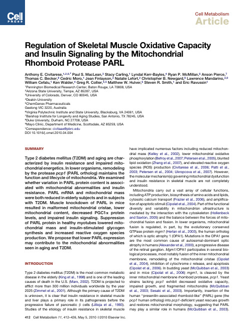
INTRODUCTION Type 2 diabetes mellitus (T2DM) is the most common metabolic disease in the elderly (King et al., 1998) and is one of the leading causes of death in the U.S. (Marx, 2002). T2DM is projected to afflict more than 300 million individuals worldwide by the year 2025 (Zimmet et al., 2001). Although the primary cause of T2DM is unknown, it is clear that insulin resistance in skeletal muscle and liver plays a primary role in its pathogenesis before the progressive failure of pancreatic b cells (Lillioja et al., 1993). Studies of the etiology of insulin resistance in skeletal muscle
MtDNA
6 5 4 3 2 1 Young Elderly T2DM #
Cell Metabolism
Muscle Insulin Resistance and Mitochondria
A
mRNA/RPLPO mRNA (a.u.)
PARL mRNA
mtDNA copy number/nuclei
癌症的免疫治疗和细胞治疗_英文_JeremyCOPP
·FRONTIER VIEWS·Immunotherapy and cell therapy for cancerJeremy COPP1,XIE Wei-dong2,3,Charles ZHANG1,Jon BERGLIN1(1.Theragene Pharmaceuticals,Inc.11555Sorrento Valley Road,Suite A,San Diego,CA,USA 92121;2.Lindan Biotech,Ltd.4-1250Waverly Street,Winnipeg,MB,Canada;3.Institute of Dongguan-Sun Yan-Sen University,Tower9,Xinzhu Center,Songshan Lake,Dongguan523000,China)Jeremy COPP,Ph.D..A native of Colorado.Dr.Copp received his B.S.in Biochemistryfrom Colorado State University(summa cum laude)and his M.S.and Ph.D.in Chemistryfrom the University of California,San Diego.He was an American Cancer Society postdoc-toral fellow in the laboratory of Tony Hunter at the Salk Institute for Biological Studies.Atthe conclusion of his postdoctoral studies he joined Theragene Pharmaceuticals,Inc.,where he is currently the Director of Research and Development.Abstract:Cancer immunotherapies are recently gaining attention as viable therapeutic options. There are two types of immunotherapy:passive and active.The passive immunotherapies include several treatments such as monoclonal antibodies,either alone or as antibody-drug conjugates.The active immunotherapies include cancer vaccines which utilize the patient′s own cells as antigen presenting cells and target specific cancer antigens,and chimeric antigen receptor T-cell(CAR-T)therapy which engineers a patient′s T-cells to recognize cancer antigens through chimeric antigen receptors.Recent successes include the US FDA approval of a number of cancer immunotherapies such as treatments utilizing monoclonal antibodies against immune checkpoint inhibitors,the Provenge cancer vaccine that targets prostrate cancer,and a CAR-T against relapsed/refractory acute lymphoblastic leukemia that was designated with breakthrough drug status,all of which has had drug companies investigating cancer immunotherapies with intense enthusiasm.In this review we discuss where the field of immune-oncology stands today,highlight the latest findings and hypothesize future directions.Key words:cancer;immunotherapy;passive immunotherapy;active immunotherapy;cancer vaccine;chimeric antigen receptor T-cell(CAR-T)CLC number:R730.51Document code:A Article ID:1000-3002(2016)02-0087-08 DOI:10.3867/j.issn.1000-3002.2016.02.001Cancer immunotherapies,or immune-oncology,activate a patient′s immune system to kill tumor cells[1].Recently interest in immunotherapy has been intensified due to its established efficacy and less toxicity when compared to traditional chemo⁃therapy.There are two types of cancer immunotherapies:passive and active.Passive immunotherapies utlilize components of the immune system,such as monoclonal antibodies,that are manufactured outside the body(Fig.1A). Active immunotherapies,such as cancer vaccines and cell therapies,utilize components of the patient′s own immune system to stimulate an immune response against the tumor.Often,the patient′s own immune cells are engineered in some manner and then infused back into the patient to direct the immune system against malignant cells(Fig.1B).It is an exciting time inCorresponding author:Jeremy COPP,Tel:(858)2043104,E-mail:jeremycopp@the field of immune -oncology due to the emer⁃gence of checkpoint blockade inhibitors and engi⁃neered T -cell therapies.Passive and active immunotherapies can be further broken down into 3categories :antibody -based ,cell -based and cytokine -based.Antibody therapy is the most effective and successful treatment for cancer so far ,with proven results in treating a variety of cancers ,especially solid tumors [1].Cell based therapies [2],such as cancer vaccines and chimeric antigen receptor T -cells (CAR -T )therapy ,on the other hand ,are still in the early stage of development but starting to show striking results [3].Thus far ,the US FDA has only approved one cell -based therapeutic drug :Provenge ©(Dendreon/Valeant ,approved in 2012),a drug based ondendritic cells to treat prostate cancer.Currently many types of cells ,including T cells ,natural killer (NK )cells ,and B cells ,are being researched and developed for cell -based therapeutics targeting cancer.Cytokines are an essential part of the immune system that ,by modulating cytokines ,can be activated to target cancer.Currently ,interferon -a and interlukin -2are effective drugs for the treatment hairy -cell -leukemia ,melanoma ,and renal cell carcinoma [4].Antibody and cytokine therapies are passive immunotherapies ,because they are generated outside of the patient′s body as drugs [5].Cell -based therapies are considered active :cells are isolated from the patient′s blood and trained in vitro to recognize cancer cells ,or to help activatespecificFig.1Passive versus active immunotherapy.In passive immunotherapy (Fig.1A ),components of the immune system thattarget specific molecules ,such as monoclonal antibodies against immune system inhibitors like cytotoxic T lymphocyte -associatedantigen 4(CTLA -4)and programmed cell death protein 1(PD -1)and its ligand PD -L1,have proven particularly effective in activating an immune response against cancer.Antibodies targeting CTLA -4are represented in green ,while antibodies targeting PD -1and PD -L1are represented in blue and pink ,respectively.In active immunotherapies (Fig.1B ),a patient′s immune cells ,often antigen presenting cells (APCs )or T cells ,are isolated and manipulated before being put back into the patient.For APCs ,they are engineered to present specific antigens on their cell surface via the major histocompatibility complex (MHC )I.When CD8+T lymphocytes encounter the proper antigen (shown in red )and bind to it through their T -cell receptors ,they become activated ,and are cytotoxic towards tumor cells with that same antigen/MHCⅠcomplex on their surface.For T cells ,the cells are engineered with chimeric antigen receptors (CARs )that recognize specific cancer antigens.This is known as CAR -T therapy ,and it has shown extremely promising results against cancers of the blood.lymphocytes to target cancer cells.The patient′s cells are then infused back into the patient to attack tumors.The process is also known as adoptive cell transfer[6].1ANTIBODY THERAPYAntibodies are proteins,which are generated by the immune system to fight pathogens such as virus,bacteria and other foreign invasion to the host body.The mechanism for the antibody to work is to recognize a specific foreign protein or antigen of the invading virus or bacteria,leading to an antibody-mediated host defence.Antigens are often located on the surface of the virus or bacteria.Cancer arises from abnormal regulation of the signaling pathways that regulate cell division. When this happens,many proteins are over-expressed or upregulated in malignant cells.As such,cancer cells are recognized as different from normal cells in the host body.Furthermore,the transformation of normal cells into tumor cells causes overexpression of key signaling mole⁃cules on the cell surface,leading to potential sur⁃face antigens that the host immune system can recognize via specific antibodies.For antibody ther⁃apy,the specific antibody(drug)binds to the anti⁃gen,resulting in antibody-dependent,cytotoxic T-cell mediated destruction of malignant cells[1]. 1.1Specific cancer antigens and antibodiesThe general principle of antibody therapy for cancer is to develop antibodies to target specific cancer antigens.Ideally,these antigens are not expressed in normal tissues,or at least over-expressed in malignant cells to minimize off-target effects.Many different types of antigens are being studied as potential targets for the treatment of cancer.Several US FDA approved antibodies have achieved clinical and commercial success,including alemtuzumab(aCD52),ipilimumab〔α-cytotoxic T lymphocyte-associated antigen4(CTLA4)〕,of atumumab(aCD20),nivolumab 〔α-programmed cell death protein(PD)-1〕,pembrolizumab(αPD-1),and rituximab(aCD20). Other popular tumor antigens in various stagesof research and development(from currently in clinical trials to FDA-approved drugs on the market are epidermal growth factor receptor(EGFR),ERBB2,vascular endothelial growth factor (VEGF),CD30,CD52,and programmed-death ligands(PDL)-1and PDL-2.There is a great deal of interest and enthusiasm regarding monoclonal antibodies targeting the immune-response checkpoint inhibitors CTLA4 and PD-1.Several years of intense preclinical and clinical studies have led to the development of three checkpoint inhibitor immunotherapy drugs:the anti-CTLA4antibody ipilimumab and two antibodies directed against PD-1,nivolumab and pembrolizumab.Ipilimumab was approved for metastatic melanoma based on a phase3 study where patients treated with ipilimumab had overall survival(OS)rates of23.5%and patients treated with a glycoprotein vaccine had OS rates of13.7%[7].Between2011and2014,the US FDA approved all the three immunotherapy drugs for use in metastatic melanoma,and there is a great deal of interest in studying their efficacy in other indications,such as non-small cell lung,kidney,brain,lymphoma,and prostate cancer[8]. Adverse effects associated with these drugs include rash,diarrhea,colitis,hyper and hypo-thyroidism and pneumonitis.While most of these side effects dissipate after treatment,hypothy⁃roidism can be permanent but treatable.1.2Antibody-drug conjugates(ADCs)LIke antibody therapy,ADCs are another proven and effective method of treating cancer[9]. ADCs are a unique class of highly potent and specific biopharmaceutical drugs that utilize anti⁃bodies targeting specific cancer antigens.ADCs are complex molecules composed of an anti⁃body,usually an antibody fragment such as a single-chain variable fragment(scFv).This frag⁃ment is linked by chemical linker to labile bonds to a biological active,cytotoxic,payload of anti-cancer drug.The purpose of the complex is to utilize the specificity of the antibody to specifically recognize the cancer antigen and deliver a toxic drug in a localized manner to the tumor.Due tothe higher specificity and localization over stan⁃dard chemotherapies,ADCs typically exhibit less side effects and toxicity[10].The history of ADCs is mixed.To date,the US FDA has approved three drugs,with two currently on the market.Gemtuzumab ozogemicin was first approved in2001,but withdrawn from the market in2010at the request of the US FDA due to concerns about safety and effectiveness. Brentuximab vedotin was approved in2011for the use in Hodgkin lymphoma and systemic anaplastic large cell lymphoma,while trastuzumab emtansine was approved in2013for HER2+ breast cancer.The durable clinical response to these drugs has renewed interest in developing novel ADCs and it will be intriguing to witness where this field develops in the future[11].2CELL BASED IMMUNOTHERAPYCell based therapy is considered active immunotherapy,which uses the patient′s own immune system to treat cancers.Cell based immunotherapy usually requires the removal of lymphocytes from the patient′s blood,training of specific immune cells to recognize cancer cells in vitro and subsequent infusion the trained cells back into the patient.The infused cells then either directly kill the patient′s malignant cells or activate other immune cells,such as cytotoxic T cells,to do the killing.The cells are isolated from the patient by a process during a procedure known as leukapharesis,while the process of infusing them back into the patient is called adoptive cell transfer(ACT).The lymphocytes most commonly used for cancer immunotherapy are natural killer(NK)cells,lymphokine-activated killer cells,cytotoxic T lymphocytes(CTLs)and dendritic cells(DCs).These cells may originate from the patient(autologous)or from allogeneic donors.Currently,autologous treatment is much more popular than allogeneic because autologous cells reduce the occurrence graft vs host disease[6].2.1DC based immunotherapyThe first US FDA approved cell therapy,Provenge,is a DC based vaccine against metastatic castration-resistant prostate cancer (mCRPC).Approved in2010,it is currently the only vaccine approved for use as a cancer thera⁃peutic.In this therapy,DCs act as APCs[12]. They are manipulated to present a specific cancer antigen to CTLs,which activates them and directs them to kill cancer cells which present that antigen on their cell surface.In the case of Provenge,the antigen is the prostate tumor anti⁃gen protein acid phosphatase(PAP)fused to granulocyte macrophage colony-stimulating factor (GM-CSF),which acts to fully activate the DCs into mature APCs.Once DCs are isolated from the patient,the synthetic fusion protein combining PAP and GM-CSF is introduced into DCs ex vivo in a laboratory. The DCs are allowed time to mature into APCs and are then infused back into the patient′s body.These activated DCs then present the PAP antigen to T cells to activate them.The activated T cells will destroy prostate cancer cells expressing the cancer cell surface antigen PAP[13].2.2NK cell based immunotherapyNK cells are similar to CTLs,but they do not require the presence of major histocompatibility complex(MHC)or antibodies to be activated as like CTLs do.This unique character of NK cells allows them rapid responses to viral-infected cells,acting at around3days after infection.NK cells are extremely important for host defense against harmful pathogens that are missing MHC classⅠantigens.Because some malignant cells have mechanisms to down-regulate MHC type I mole⁃cules,NK cell manipulation is an effective meth⁃od of cancer treatment[14].In normal circumstances,NK cells are not active or able to recognize and kill cancer cells. Controlling NK cell activation/inactivation is achieved through NK cell activating and inhibito⁃ry receptors.Activating receptors include Ly49,Natural cytotoxic receptors(NCRs),CD49/ NKG2,and CD16(FcyIIIA).Inhibitory receptors include killer-cell immunoglobulin-like receptors (KIRs),leukocyte inhibitory receptors(LIRs),and Ly49(has both activating and inhibitory iso⁃forms).NK cells are inactive while these recep⁃tors are in equilibrium.NK cells are activated upon antibody secretion(binding to CD16),cytokine secretion(IL-2,IL-12,IL-15,IL-18and TNF-αand IFN-γ),and down-regulation of MHC typeⅠ. Under stressful conditions,cells can down-regulate MHC typeⅠexpression,causing NK cells to lose inhibitory signaling and be activated.This process is called″missing-self″recognition,during which NK cells seem to have the ability to compare what receptors the host has versus re⁃ceptors that are missing in response to an″invad⁃ing″pathogen,Therefore,when MHC typeⅠis missing in infected cells,or in cancer cells,then NK cells are activated.Similarly,the activating receptors in NK cells can recognize new mole⁃cules or antigens that the host does not express,which is called″non-self″recognition,or self-expressed antigens/proteins that are up-regulat⁃ed by stressed conditions,called″stress-induced self″recognition.All these conditions can activate NK cells and allow them to recognize and kill infected cells or malignant cells[15-16].Current studies are focused on modifying these three signaling pathways(missing self,non-self and stress-induced self)with NK cells for cancer therapeutic development.For cytokine treatment,NK cells are activated after adminis⁃tration of large amounts of cytokines to patients. However,this can result in severe toxicity due to the cytokine itself.Since NK cells express FcyⅢA/CD16receptor,NK cells can recognize anti⁃body-labeled target cancer cells and they are activated upon binding to these antibodies.This results in antibody-dependent cell-mediated cytotoxicity(ADCC),which can kill antibody-labled malignant cells.Clinical data showed that NK cells play a critical role in cancer treatment by anti-cancer antibodies,such as anti-CD20(rituxumab)and anti-HER2(Herceptin/trastuxu⁃mab)treatments.There are modified proteins or antibodies being developed for this purpose,such as AFM13,a genetic modified antibody developed by Affimed NV(Heidelberg,Germany).AFM13is specifically designed to bind CD16a and activate NK cells.Nantkwest(Culver City,CA USA)is developing genetically modified NK cells that do not express KIRs(inhibitory receptors)for cancer treatment.Although there is a great deal of interest in developing cancer treatments that target NK cells,much more data on toler⁃ance and efficacy are needed.2.3CAR-T technologyPossibly the most intense interest in devel⁃oping an active cell therapy lies in the area of CAR-T therapy.This technology is a promising new approach to cancer.In July2014,US FDA granted breakthrough therapy designation(BTD)to Novartis Pharmaceuticals′investigational CAR-T therapy CTL019for treatment of adult and pediatric relapsed/refractory acute lympho⁃blastic leukemia(r/r ALL).This is the first CAR-T technology to receive the FDA′s BTD.The CAR-T technology for CTL019employs a viral vector carrying a small gene encoding the CD19 cancer antigen binding protein fused to specific T-cell activation domains.T cells from the patient are obtained via leukapharesis and engineered in vitro with the vector so that the patient′s T cells will destroy cancer cells expressing the CD19 antigen once infused back into the patient′s body. Many biopharmaceutical companies,such as Juno Therapeutics,Kite Pharmaceuticals and Celgene,are developing CAR-T technology for cancer therapy.The CAR-T approach,like other adoptive cell therapy methods,relies on the removal of cells from the patient′s body.The T cells are manipulated via transfection by a viral vector that expresses a chimeric receptor specific to a cell-surface antigen.The cells are cultured so that the modified T cells mature and multiply,and then they are infused back into the patient.The infused T cells can recognize and kill malignant cells located in the patient[17].The approach usually employs autologous cells to reduce host defense.Allogeneic cells from other donors are also under investigation,but not very practical at the moment.The structure of chimeric antigen receptors are composed of an extracellular domain with an antigen recognition region to binds to a specific cancer antigen fused to an intracellular T cell activation domain.The antigen recognition region is usually derived from single chain variable fragment(scFv)from an antibody,which can recognize and bind to a specific cancer antigen (such as CD19).Upon binding of the antigen,the intracellular T cell activation domain transduces a signal to activate the T cells.The most common activation domain is CD3-zeta,containing3 immunoreceptor tyrosine-based activation motifs (ITAMs)for better signal transduction.In addition,more signal enhancers can be incorporated for more potent T cell activation.The second gener⁃ation of CAR-Ts is incorporated with CD28,41BB or iCOS receptor at the cytoplasmic tail of the CAR to enhance activation of T cells,while the third generation has combined several of the receptors such as CD3z-CD28-41BB or CD3z-CD28-OX40to further increase the efficacy of treatment[18-19].Several biopharmaceutical com⁃panies,including Juno Therapeutics,are con⁃ducting research on the next generation of CARs,which includes the addition of a second antigen recognition/binding domain for enhanced speci⁃ficity of cancer cell identification and recognition.CAR-T technology is a promising form of cancer immunotherapy supported by data obtained in several preclinical and clinical trials. Research and studies are focusing on engineering improved chimeric receptors to achieve higher efficacy and low toxicity for patients.The current clinical results show that CAR-T is only effective in treating blood related cancers,such as many types of leukemia.Treating solid tumors is still early in research and development,and many more studies are needed to explore this area.Although CAR-T has achieved great success in clinical trials to treat leukemia,there is a high level of toxicity associated with this treatment. The most severe events include tumor lysis syndrome,cytokine release syndrome and organ specific toxicity.These toxicities can be life threatening[20].Tumor lysis syndrome is a group of metabolic abnormalities after treatment of lymphomas and leukemia.It is most likely caused by the release of cellular contents into the blood upon cancer-cell lysis.This leads to higher blood potassium,phosphate,uric acid and BUN,but low blood calcium,changing blood electrolyte and metabo⁃lite levels,and can be fatal[21].Cytokine release syndrome is due to T cell activation and release of inflammatory cytokines into the blood stream,including IL-6,IFN-γand TNF-α.This can cause severe hypotension,hypoxia,fever,multi-organ failure and coagulation disorder,and can also lead to fatality[22].Organ specific toxicity is due to antigen specific toxicity,CNS depres⁃sion,liver function change and pulmonary prob⁃lems that are specific to anti-CD19products[23].3PROSPECTIVECancer therapy is a complicated and long-term approach.In today′s standard care,one is treated by surgery,chemotherapy,or radiation therapy alone,or by combination of the three. After these treatments,the patient is considered cancer free,but in many cases relapse often occurs within a short period of time.Upon recur⁃rence,the prognosis is very poor mainly due to metastasis of the disease to other tissues or organs.At this point,the cancer is much more difficult to treat,and the survival rate is very low. Immunotherapy is particularly designed to treat cancers after they escape from standard-of-care treatments,and both passive and active immu⁃notherapies have shown great efficacy for this purpose.However,more and more immunother⁃apies are proving to be effective as first line treat⁃ments,while others are promising as second line,adjuvant or combination therapies with oth⁃er anticancer drugs.The future of the field may lie in the utilization of therapies targeting check⁃point inhibitors in combination with some type of active immunotherapeutic(vaccine or CAR-T)to lower the toxicity of the former while increasingthe efficacy of the latter.With whole genome sequencing,it is conceivable that these therapies will become part of personalized medicine,attempting to target the exact cancer antigens that are involved in the pathology of a particular patient′s disease.In general,cancer immuno⁃therapy defines a new class of treatments designed to be more specific to a patient′s particular cancer,thus making them a highly effective,less toxic,treatment option.REFERENCES:[1]Scott AM,Wolchok JD,Old LJ.Antibody therapy of cancer[J].Nat Rev Cancer,2012,12(4):278-287.[2]Srinivasan R,Van Epps DE.Specific active immu⁃notherapy of cancer:potential and perspectives[J].Rev Recent Clin Trials,2006,1(3):283-292.[3]Curran KJ,Pegram HJ,Brentjens RJ.Chimeric antigen receptors for T cell immunotherapy:currentunderstanding and future directions[J].J GeneMed,2012,14(6):405-415.[4]Lee S,Margolin K.Cytokines in cancer immuno⁃therapy[J].Cancers(Basel),2011,3(4):3856-3893.[5]Schuster M,Nechansky A,Kircheis R.Cancer immunotherapy[J].Biotechnol J,2006,1(2):138-147.[6]Rosenberg SA,Restifo NP.Adoptive cell transfer as personalized immunotherapy for human cancer[J].Science,2015,348(6230):62-68.[7]Hodi FS,O'Day SJ,McDermott DF,Weber RW,Sosman JA,Haanen JB,et al.Improved survivalwith ipilimumab in patients with metastatic melanoma[J].N Engl J Med,2010,363(8):711-723.[8]Jacob JA.Cancer Immunotherapy Researchers Focus on Refining Checkpoint Blockade Therapies[J].JAMA,2015,314(20):2117-2119.[9]Bouchard H,Viskov C,Garcia-Echeverria C.Anti⁃body-drug conjugates-a new wave of cancer drugs[J].Bioorg Med Chem Lett,2014,24(23):5357-5363.[10]Zolot RS,Basu S,Million RP.Antibody-drug con⁃jugates[J].Nat Rev Drug Discov,2013,12(4):259-260.[11]Wang Z,Guravaiah N,Ning C,He Y,Yao L,Wang J,et al.Antibody-drug conjugates:theforefront of targeted chemotherapy for cancertreatment[J].J Drug Des Res,2015,2(3):1016-1025.[12]Palucka K,Banchereau J.Cancer immunotherapy via dendritic cells[J].Nat Rev Cancer,2012,12(4):265-277.[13]PROVENGE®(sipuleucel-T)immunotherapy is designed to target and attack prostate cancer cells[EB/OL].[2016-01-12]http://www.provengehcp.com/extendsurvival/mechanism-of-action.aspx [14]Cheng M,Chen Y,Xiao W,Sun R,Tian Z.NK cell-based immunotherapy for malignant diseases[J].Cell Mol Immunol,2013,10(3):230-252.[15]Bruno A,Ferlazzo G,Albini A,Noonan DM.A think tank of TINK/TANKs:tumor-infiltrating/tumor-associated natural killer cells in tumor progressionand angiogenesis[J].J Natl Cancer Inst,2014,106(8):dju200.[16]Zamai L,Ponti C,Mirandola P,Gobbi G,Papa S,Galeotti L,et al.NK cells and cancer[J].J Immunol,2007,178(7):4011-4016.[17]Lipowska-Bhalla G,Gilham DE,Hawkins RE,Rothwell DG.Targeted immunotherapy of cancerwith CAR T cells:achievements and challenges[J].Cancer Immunol Immunother,2012,61(7):953-962.[18]Srivastava S,Riddell SR.Engineering CAR-T cells:Design concepts[J].Trends Immunol,2015,36(8):494-502.[19]Maus MV,Grupp SA,Porter DL,June CH.Anti⁃body-modified T cells:CARs take the front seat forhematologic malignancies[J].Blood,2014,123(17):2625-2635.[20]Kalaitsidou M,Kueberuwa G,Schütt A,Gilham DE.CAR T-cell therapy:toxicity and the relevance ofpreclinical models[J].Immunotherapy,2015,7(5):487-497.[21]Davidson MB,Thakkar S,Hix JK,Bhandarkar ND,Wong A,Schreiber MJ.Pathophysiology,clinicalconsequences,and treatment of tumor lysissyndrome[J].Am J Med,2004,116(8):546-554.[22]Breslin S.Cytokine-release syndrome:overview and nursing implications[J].Clin J Oncol Nurs,2007,11(1Suppl):37-42.[23]Dotti G,Gottschalk S,Savoldo B,Brenner MK.Design and development of therapies using chimericantigen receptor-expressing T cells[J].ImmunolRev,2014,257(1):107-126.癌症的免疫治疗和细胞治疗Jeremy COPP1,谢伟东2,3,张朝杰1,Jon BERGLIN1(1.Theragene Pharmaceuticals,Inc.11555Sorrento Valley Road,Suite A,San Diego,CA,USA 92121;2.Lindan Biotech,Ltd.4-1250Waverly Street,Winnipeg,MB,Canada;3.东莞中山大学研究院,广东东莞523000)摘要:最近,癌症免疫治疗作为可行性的新治疗法而得到广泛青睐。
原发性支气管肺癌中英文对照

– At 15 years, 80-90% risk reduction
– Never gets to “never smoker” risk
5
20 Year Lag
6
If what happened on your inside happened on your outside, would you still smoke?
原发性支气管肺癌 Primary bronchogenic
carcinoma
呼吸内科
Respiratory Department
熊维宁
Xiong, Weining
1
定义 Definition
• 原发性支气管肺癌简称肺癌,是起源于 支气管粘膜或腺体的肿瘤。
• Primary bronchogenic carcinoma is abbreviated to lung cancer, it derives from bronchi mucosa or gland.
tumor local expanding
• 1 胸痛
• 1 Chest pain
• 2 呼吸困难
• 2 Dyspnea
• 3 吞咽困难
• 3 Dysphagia: esophageal compression
• 4 声音嘶哑
• 4 Hoarse voice: laryngeal nerve paralysis
• 5 上腔静脉阻塞综合征
• 5 Superior vena cava obstruction syndrome
• 6 Horner综合征,肺上沟瘤
• 6 Horner’s syndrome, Pancoast’s tumor: Cervical/thoracic nerve invasion
肺癌医学英语
肺癌在医学英语中通常被称为"Lung Cancer"。
根据不同的病理类型,肺癌可以分为非小细胞肺癌(Non-Small Cell Lung Cancer, NSCLC)和小细胞肺癌(Small Cell Lung Cancer, SCLC)。
以下是肺癌相关的一些医学英语词汇:
Lung Cancer: 肺癌
Non-Small Cell Lung Cancer (NSCLC): 非小细胞肺癌
Small Cell Lung Cancer (SCLC): 小细胞肺癌
Adenocarcinoma: 腺癌
Squamous Cell Carcinoma: 鳞状细胞癌
Large Cell Carcinoma: 大细胞癌
Bronchial Carcinoma: 支气管肺癌
Pulmonary Carcinoma: 肺癌
Lung Nodule: 肺结节
Central Lung Cancer: 中央型肺癌
Peripheral Lung Cancer: 周围型肺癌
Staging: 分期
Metastasis: 转移
Lymph Node Metastasis: 淋巴结转移
Chemotherapy: 化疗
Radiation Therapy: 放疗
Targeted Therapy: 靶向治疗
Immunotherapy: 免疫治疗
Palliative Care: 姑息治疗
Prognosis: 预后
这些词汇只是肺癌相关医学英语的一部分,具体的词汇和表达可能会根据上下文和特定的医学文献而有所不同。
在医学领域,准确和专业的术语使用非常重要,以确保信息的准确传递和理解的准确性。
原发性支气管肺癌中英文对照课件

精选课件
7
• 2 职业致癌因子
• 2 Occupation carcinogenic factor: Asbestos, Radon 3 空气污染
• 3 Air pollution • (1)室外大环境污染 • (1)Outdoor environment pollution • (2)室内小环境污染 • (2)Indoor environment pollution • 4 电离辐射
• (2)intermediate cell type
• (3)混合型
• (3)mixed type
精选课件
14
• Small cell carcinoma
– Rare in non-smokers – Large hilar mass – 70% present with overt metastasis – Very chemo-responsive… – Worst prognosis
精选课件
18
Squamous Cell Carcinoma
精选课件
19
Squamous Cell Carcinoma
精选课件
20
鳞癌
精选课件
21
Lung Cancer Pathology
• Adenocarcinoma
– Most common pathology(2nd in China) – “Non-smoker’s lung cancer” – Women – Peripheral (75%) – Aggressive metastases
•
*p53
• Dominant oncogenes
•
*Kras
•
*Her-2/neu
非小细胞肺癌表皮生长因子受体酪氨酸激酶抑制剂(英文)[]精品PPT课件
![非小细胞肺癌表皮生长因子受体酪氨酸激酶抑制剂(英文)[]精品PPT课件](https://img.taocdn.com/s3/m/a286e499aa00b52acfc7caad.png)
Ligand EGFR
PTEN
PI3-K pY
pY
STAT3
AKT
pY GRB2
EGFR-TK
SOS RAS RAF
MEK
Proliferation/ maturation
Gene transcription Cell-cycle progression
PP
DNA
Myc
Cyclin
JunFos
D1
Myc
IDEAL 2 (n=216) >2 prior regimens
Gefitinib 250 mg/day
Gefitinib 500 mg/day
Primary endpoints
Objective tumour response Symptom improvement
(IDEAL 2) Safety (IDEAL 1)
Epidermal growth factor receptortyrosine kinase inhibitor in non-
small-cell lung cancer
Yuh-Min Chen, MD, PhD. Chest Dept., Taipei VGH
2020/10/14
YMC
1
Activation of the epidermal growth factor receptor tyrosine kinase (EGFR-TK): a pivotal driver of carcinogenesis
Cyclin D1
MAPK
Survival (anti-apoptosis)
2020/10/14
Chemotherapy/
生物文献翻译——《Antileukemic activity of the HSP70 inhibitor pifithrin-u in acute leukemia》
Antil eukemic activity of the HSP70 inhibitor pifithrin-uin acute l eukemia热休克蛋白70在不同恶性肿瘤中特别表达并且已经成为一种抗癌治疗的很有前景的靶点。
这里,我们分析了体外抗白血病PFT-u的效果,PFT-u是一种诱导HSP-70的抑制剂,在急性髓细胞白血病(AML)及急性淋巴系统白血病(ALL)细胞株,以及在初级AML爆发期。
在所有细胞株测试中在低微摩尔的浓度水平上PFT-u明显地抑制细胞活力,IC50值范围从2.5到12.7uM。
并且在初级AML爆发期用IC50中间值8.9LM(范围5.7-37.2)测试发现PFT-u 是高度表达的。
重要的是,相对较高的IC50值在正常的造血干细胞中也能被发现。
在AML 和ALL中,以剂量依赖的方式PFT-u能够诱导细胞凋亡和阻遏细胞周期。
在NALM-6细胞株中PFT-u也能导致caspase-3活性的增加和减少细胞内AKT和ERK1/2的浓度。
此外,在NALM-6、TOM-1和KG-1a细胞中,PFT-u能够增强阿糖胞苷、17-(allylamino)-17-desmethoxygeldanamycin和索拉菲尼的细胞毒性。
这是首次研究表现出热休克蛋白70抑制剂PFT-u在抗白血病中的显著效果,单独使用和结合不同的抗肿瘤药物在AML和ALL中。
我们的结果表明PFT-u在急性白血病中扮演了一种潜在的治疗角色。
背景尽管依赖于风险的治疗策略,只有大约35%左右60岁以下的成人急性髓细胞白血病(AML)能够被治愈。
同样,在急性淋巴细胞白血病(ALL)中,成年患者的治疗效果仍然不佳,幸存率为30-50%。
进一步增加化疗剂量结果表现出有限的抗白血病效果和高毒性,增加了过早死亡的风险。
因此,我们努力开发一种新的与传统化疗相结合的分子治疗方法。
在BCR-ABL 阳性的ALL中,通过引进甲磺酸伊马替尼结果得到大大的改善。
肺癌英文版[荟萃知识]
![肺癌英文版[荟萃知识]](https://img.taocdn.com/s3/m/c061811c51e79b8968022699.png)
专业知识
6
Pathology And Classification
专业知识
3
Etiology
The cause of lung cancer is unknown.It is believed that there are following related factors.
1. Excessive cigarette smoking:Smoking index(Brinkman Index) is equal to cigarettes per day smoking time(years).
Bronchogenic Carcinoma (Lung Cancer)
Respiratory department
专业知识
1
Definition
Bronchogenic carcinoma refers to the malignant tumor which grows in the bronchus. Originating from mucus or gland of bronchus.
pleura.
专业知识
11
Clinical Features
(4).Horner’s syndrome.It is caused by invading the cervical sympathetic ganglia on the involved side the pupil is small ptosis of the up eyelids,retraction of the eyeball and no sweat of the face.
介绍肺癌的英文作文

介绍肺癌的英文作文英文:Lung cancer is a type of cancer that affects the lungs, which are responsible for bringing oxygen into the body. It is one of the most common types of cancer and is often caused by smoking. Other risk factors include exposure to secondhand smoke, air pollution, and radon gas.Symptoms of lung cancer include persistent coughing, chest pain, shortness of breath, and coughing up blood. However, these symptoms may not appear until the cancer has advanced, which is why early detection is crucial.There are different types of lung cancer, includingnon-small cell lung cancer and small cell lung cancer. Treatment options depend on the type and stage of the cancer, but may include surgery, chemotherapy, radiation therapy, and targeted therapy.It is important to quit smoking and avoid secondhand smoke and other risk factors to reduce the risk of developing lung cancer. Regular screenings for those at high risk can also help with early detection and treatment.中文:肺癌是一种影响肺部的癌症,肺部负责将氧气带入身体。
让人类不再生病癌细胞杀死作文
让人类不再生病癌细胞杀死作文英文回答:Finding a cure for cancer and preventing diseases is a topic that has been discussed and researched for many years. As a human, I believe that it is possible to eliminate diseases and find a way to stop cancer cells from spreading.To begin with, advancements in medical technology and research have already made significant progress in treating and preventing diseases. For example, vaccines have been developed to prevent diseases like polio and measles, significantly reducing the number of cases worldwide. Additionally, the development of antibiotics has revolutionized the treatment of bacterial infections,saving countless lives.Furthermore, understanding the genetic factors that contribute to diseases and cancer can lead to targeted therapies and preventive measures. With the advancements ingenetic testing, individuals can now identify their risk factors for certain diseases and take proactive steps to prevent them. For instance, individuals with a family history of breast cancer can undergo regular screenings and take preventive medications to reduce their risk.Moreover, lifestyle changes can play a crucial role in preventing diseases and reducing the risk of cancer. Adopting a healthy diet, exercising regularly, and avoiding harmful habits like smoking and excessive alcohol consumption can significantly lower the chances of developing diseases. As the saying goes, "prevention is better than cure," and making these lifestyle changes can greatly improve overall health and well-being.In addition to the above approaches, early detection and regular screenings can also contribute to the prevention and treatment of diseases. Regular check-ups and screenings can help identify potential health issues at an early stage when they are more manageable or even curable. For example, mammograms and Pap smears are essential for detecting breast and cervical cancers early on, increasingthe chances of successful treatment.In conclusion, while it may not be possible to completely eradicate diseases and cancer, significant progress has already been made in preventing and treating them. Through advancements in medical technology, genetic research, lifestyle changes, and early detection, we can continue to improve our understanding and management of diseases. By taking proactive measures and making the necessary lifestyle changes, we can work towards a healthier future for humanity.中文回答:找到治愈癌症和预防疾病的方法是一个已经被讨论和研究多年的话题。
治疗肿瘤英文作文
治疗肿瘤英文作文1. Cancer treatment is a tough battle, but with the right combination of surgery, chemotherapy, and radiation therapy, many people are able to beat this disease.2. The side effects of cancer treatment can be harsh, from hair loss to nausea to fatigue. It's important for patients to communicate with their healthcare team about how they're feeling so that adjustments can be made totheir treatment plan.3. In recent years, immunotherapy has emerged as a promising new approach to treating cancer. By harnessing the body's own immune system to target and kill cancer cells, immunotherapy has shown great potential in improving outcomes for patients with certain types of cancer.4. Supportive care is also a crucial aspect of cancer treatment, helping patients manage symptoms and maintain their quality of life. This can include pain management,nutritional support, and emotional counseling.5. It's important for patients to stay informed about their treatment options and to advocate for themselves throughout the process. By being proactive and engaged in their care, patients can play an active role in their recovery.6. Research into new cancer treatments is ongoing, with scientists constantly exploring innovative approaches to target cancer cells more effectively and with fewer side effects. This dedication to advancing cancer care gives hope to patients and their loved ones.。
吉非替尼合成路线英文文献
吉非替尼合成路线英文文献全文共四篇示例,供您参考第一篇示例:吉非替尼(Gefitinib),也被称为吉非替尼龙(Iressa),是一种用于治疗非小细胞肺癌的口服靶向药物。
它通过抑制表皮生长因子受体(EGFR)的激酶活性,从而阻断癌细胞的增殖和生长。
吉非替尼作为一种靶向治疗药物,已被广泛应用于临床,对肺癌患者的治疗产生了显著的影响。
合成吉非替尼的路线是指从原料化合物到最终产品的整个合成过程,通常包括多个步骤的有机合成反应。
吉非替尼的合成路线一直受到化学研究人员的关注,不断地进行优化和改进。
本文将介绍吉非替尼的合成路线,并探讨其中的关键步骤和反应。
合成吉非替尼的路线通常以具有芳香环和氮杂环的结构为起点,通过一系列反应逐步构建目标化合物的结构。
在整个合成过程中,化学研究人员需要考虑产率、反应选择性、废弃物处理等多个因素,以确保合成路线的可行性和经济性。
下面将介绍吉非替尼合成路线的关键步骤。
吉非替尼的合成通常从对苯二酚出发,经过若干步反应得到对苯二酚的衍生物,然后将其转化为苯胺类化合物。
随后,化合物经过多步骤的氨基化和烯丙基化反应,逐步构建了目标化合物的结构骨架。
在这一过程中,化学研究人员需要克服各种挑战,包括底物的选择、反应条件的优化、中间体的稳定性等难点问题。
通过不断的尝试和改进,研究人员逐步找到了有效的合成策略,使得整个合成路线更加高效和可行。
在合成路线的后期,研究人员需要进行目标化合物的结构修饰和官能团的引入,以提高吉非替尼的药物活性和选择性。
这一阶段通常涉及多步反应和复杂的合成操作,需要化学研究人员具备较高的实验技能和丰富的合成经验。
通过精密的合成规划和严谨的操作,研究人员最终得到了满足临床需求的吉非替尼产品。
吉非替尼的合成路线是一个复杂而又具有挑战性的有机合成过程。
通过不断的探索和创新,化学研究人员逐步优化了吉非替尼的合成方法,提高了产率和产物质量,并且减少了环境负荷。
吉非替尼的成功合成为肺癌治疗领域的发展提供了有力支持,同时也为类似结构化合物的合成提供了宝贵的借鉴和经验。
非小细胞肺癌表皮生长因子受体酪氨酸激酶抑制剂(英文)
Other (218)
Asian (53) Other (374) Yes (311)
4.1
18.9 7.5 3.8
No (93)
Unknown (23)
24.7
13.0
<0.001
*Significance between subgroups **Data collected retrospectively In multiple logistic-regression analyses, only never having smoked (p<0.001) and adenocarcinoma histology (p=0.01) were associated with response 2019/1/21 YMC 15 Shepherd et al. NEJM 2005;353:123
2019/1/21 YMC
IDEAL 1
Probability 1.0 0.8 0.6 0.4 0.2 0.0
IDEAL 2
0 2 4 6 8 10 12 14 16 18 20 Months from randomisation Patients (n) 44 58 Deaths (n) 26 56 Median (months) 13.6 3.7
18
12
Months YMC
J Chemother 2005;17:679
Survival according to response or not
Fig. 1 100 90 80
Complete or partial response (n=12) median 20.1M
Stable or progressive disease (n=24) median 4.7M
肺癌研究报告Lung cancer(英文)
Other symptoms that are associated with lung cancer include: include: Weakness
Chills Swallowing difficulties Speech difficulties or changes (e.g., hoarseness) Finger/nail abnormalities (e.g., "clubbing," or overgrowth of the fingertip tissue) Skin paleness or bluish discoloration
Lung cancer
Main pointer
Clinical manifestation Diagnosis procedure Differential diagnosis
Malignant tumors are cancer. They can invade and damage nearby healthy tissues and organs. Cancer cells can also break away from the tumor and enter the bloodstream or the lymphatic system. That is how cancer spreads and forms tumors in other parts of the body. The spread of cancer is called metastasis.
Symptoms
People often decide to visit the doctor only after they have been bothered by certain complaints over a period of time. Individuals who have lung cancer frequently experience symptoms such as:
- 1、下载文档前请自行甄别文档内容的完整性,平台不提供额外的编辑、内容补充、找答案等附加服务。
- 2、"仅部分预览"的文档,不可在线预览部分如存在完整性等问题,可反馈申请退款(可完整预览的文档不适用该条件!)。
- 3、如文档侵犯您的权益,请联系客服反馈,我们会尽快为您处理(人工客服工作时间:9:00-18:30)。
肺癌细胞抑制中英文对照外文翻译文献(文档含英文原文和中文翻译)介导性shRNA能抑制肺癌细胞中livin沉默基因的表达从而促进SGC-7901细胞凋亡背景—由于肿瘤细胞抑制凋亡增殖,特定凋亡的抑制因素会对于发展新的治疗策略提供一个合理途径。
Livin是一种凋亡抑制蛋白家族成员,在多种恶性肿瘤的表达中具有意义。
但是, 在有关胃癌方面没有可利用的数据。
在本研究中,我们发现livin基因在人类胃癌中的表达并调查了介导的shRNA能抑制肺癌细胞中livin沉默基因的表达,从而促进SGC-7901细胞凋亡。
方法—mRNA及蛋白质livin基因的表达用逆转录聚合酶链反应技术及西方吸干化验进行了分析。
小干扰RNA真核表达载体具体到livin基因采用基因重组、测序核酸。
然后用Lipofectamin2000转染进入SGC-7901细胞。
逆转录聚合酶链反应技术和西方吸干化验用来验证的livin基因在SGC-7901细胞中使沉默基因生效。
所得到的稳定的复制品用G418来筛选。
细胞凋亡用应用流式细胞仪(FCM)来评估。
细胞生长状态和5-FU的50%抑制浓度(IC50)和顺铂都由MTT比色法来决定。
结果—livin mRNA和蛋白质的表达检测40例中有19例(47.5%)有胃癌和SGC-7901细胞。
没有livin基因表达的是在肿瘤邻近组织和良性胃溃疡病灶。
相关发现在livin基因的表达和肿瘤的微小分化和淋巴结转移一样(P < 0.05)。
4个小干扰RNA真核表达矢量具体到基因重组的livin基因建立。
其中之一,能有效地减少livin基因的表达,抑制基因不少于70%(P < 0.01)。
重组的质粒被提取和转染到胃癌细胞。
G418筛选所得到的稳定的复制品被放大讲究。
当livin基因沉默,胃癌细胞的生殖活动明显低于对照组(P < 0.05)。
研究还表明,IC50上的5-Fu和顺铂在胃癌细胞的治疗上是通过shRNA减少以及刺激这些细胞(5-Fu proapoptotic和顺铂)(P < 0.01)。
结论—livin基因在胃癌中的过分表达与肿瘤分化与淋巴结转移建立联系,建议了治疗胃癌病例分子预后因素之一。
ShRNA可以抑制在SGC-7901细胞中的livin基因表达,诱导细胞凋亡。
Livin可以作为治疗胃癌凋亡的新目标。
1.介绍胃癌是世界上最常见的恶性肿瘤之一。
大多数患者被诊断为这个疾病的阶段,在最佳时间的机会错失了手术治愈。
尽管有很大改善,但处于晚期胃癌的化疗患者的总体存活率仍然很低。
癌症细胞化疗耐抗性可能导致手术失败。
在耐药的原因中,抑制细胞凋亡的过程会起重要作用。
癌细胞常有抗凋亡增长的特征[1],介导其增加的阻力不同来刺激的细胞凋亡,如DNA损伤、缺氧、营养损失[2、3]。
此外, 在临床实践中细胞凋亡抵抗被认为是肿瘤手术失败的主要原因,因此许多化疗药物和/或放射线疗法都是通过诱导凋亡肿瘤死亡实现的[4]。
酶抑制剂(IAPs),是一种新型的凋亡蛋白抑制基因家族[5,6],包括病毒感染,化疗药物, ,生长因子和肿瘤坏死因子-α(TNF-α凋亡信号通路)/ Fas信号通路[7 - 9]。
IAPs是由一组有凋亡特性的结构相关的蛋白质构成[10],在预防肿瘤细胞凋亡方面可能扮演一个重要的角色,并已成为近年来研究的热点。
这个家庭的新成员是ML-IAP/livin/KIAP/BIRC7 (以下称为livin)有两个种类、livinα和livinβ[11-14]。
有证据表明, livin的过分表达能阻止由多种刺激诱导的细胞凋亡[12]。
有趣的是, livin基因被发现在肿瘤细胞中限制性表达,但是不存在或是很少数量存在于正常成人体组织中[11-15],并且通过允许恶性细胞,以避免凋亡细胞死亡的方式导致肿瘤形成,。
所以抑制livin基因表达可能会呈现出一个有趣的治疗策略。
在目前的研究中,我们调查了livin的表达在胃cancinomas及其邻近组织。
livin 的表达和临床病理参数之间的关系进行了分析。
此外,我们探索了在抑制livin基因表达的shRNA可行性和胃癌易感性的凋亡细胞由shRNA介导的livin沉默基因。
2患者和方法2.1 患者和肿瘤样本胃癌中四十个病例及接受胃切除手术的患者收集到的胃癌组织中13个病例(患者年龄从29~77岁)。
其中良性胃溃疡的13个病例(慢性浅表胃炎)患者在接受了胃内视镜检查(患者年龄从33~77岁)。
这些病例均来自南京医科大学第一附属医院。
胃癌患者被诊断为TNM级的1~4阶段(UICC ,2002)。
手术之后肿瘤标本就立即被冻结在液态氮中,储存在-80℃直到使用为止。
这是在所有的病人知情同意的情况下获得的。
2.2逆转录聚合酶链反应技术程序总共RNA(2毫克)提取冷冻组织反转录进行,最后的体积2微升是用100 pmol of oligo(dT)15和200U M-MLV与逆转录酶(promega、美国) ,根据制造商的说明。
Aliquots对应的250微升cDNA被放大在PCR缓冲容器中,在最终的50微升中含25pmol / ml处理剂和1U聚合酶。
每一个放大了35周期,一个周期的变性曲线在30 s内到达94℃。
热处理在30s内59℃(livin and b-actin)扩大到30s内72℃。
没有RNA的病例作为阴性对照物包含在RT–PCR中。
一系列常用的的livin和β-actin处理剂如下:livinα / βupstream,5'-TCCACAGTGTGCAGGAGACT-3';livinα/ βdownstream;5'-TCCACAGTGTGCAGGAGACT-3';β-actin upstream,5'-ACGGCACAAAGACGATGGAC-3'β-actin downstream,5'-AGCGCAAGTACTCCGTGTG-3'。
产品的尺寸分别为livinα/β是312/258 bp ,β-actin 是501bp。
2.3 西方吸干技术分析病变同质性与冲力缓冲50mM Tris-HCl (pH 7.5), 250 mM NaCl,0.1% NP40和5mM EGTA包含50mM氟化钠,60mM β-丙三醇-磷酸盐,0.5mM钒酸钠,0.1mM苯甲基磺醯化氟10μg/ml亮抑蛋白酶肽。
用考马斯亮蓝微盘比色法测定蛋白质含量。
蛋白质样品电泳10% 变性SDS凝胶并转移到PVDF膜(Roche、美国)。
膜是培养特定的主要的抗体,与过氧化物酶继发性抗体反应(细胞信号技术、美国),最后通过增强化学荧光达到可视化(细胞信号技术、美国)。
Alexesis(美国) 和散塔克鲁兹生物技术(美国)购买的单克隆抗体livin (1:250) and actin (1:400)24细胞系和细胞培养我们选了一个人类胃腺癌细胞系在这一研究中。
SGC-7901(上海细胞研究所,中国上海,)是一种附着中度分化胃腺的人类细胞株。
线是胃癌细胞上皮细胞,并成长为附着细胞RPMI 1640 (Hyclone Inc, USA)含10%FCS (Life Technologies, Inc.),每毫升100个单位的青霉素和毫升100微克的链霉素(BioWhittaker)。
在含有5%CO2空气的条件下的37℃湿润培养器中保存SGC-7901细胞。
溶解顺铂和氟尿嘧啶(齐鲁制药厂、中国)在DMSO并且4℃储存。
2.5。
ShRNA的合成和PGPU / GFP /Neo/livin质粒的制造通过siRNASequence-Selector软件设计并合成了Livin的ShRNA序列(上海生物技术有限责任公司。
集团公司、中国)。
序列如下(表1),然后被插入pGPU/GFP/Neo(上海GenePharma股份有限公司。
中国) BbsI and BamH地址产生pGPU/GFP/Neo/livin and pGPU/GFP/Neo/Control质子。
2.6SGC-7901的建立稳定表达pGPU/GFP/Neo/livin and pGPU/GFP/Neo/Control转染实验,SGC-7901细胞被镀成6孔板(3×105孔密度), , 96孔板(1 ×10⁴孔密度)和12孔板(1.5 ×10⁵孔密度)转染之前培养24小时。
按照制造商的说明这些细胞被调控子用Li-pofectAMINE 2000转染4毫克/孔空的pGPU/GFP/Neo/vector,pGPU/GFP/Neo/livin或pGPU/GFP/Neo/Control 质粒(生命技术公司、大岛屿,NY)。
转染48小时后,这些细胞被转移在1:15 (v/v)并用Geneticin (G418) 1000克/毫升培养4周。
稳定转染的克隆体取出并保存在媒介容器400 g/ml G418用作另外的研究。
2.7依赖贴壁细胞细胞生长的测定亲本细胞和细胞稳定表达pGPU/GFP/Neo/vector, pGPU/GFP/Neo/livin or pGPU/GFP/Neo/Control被种到6孔盘子中。
每隔一天收集三孔的细胞。
使用计数器确定细胞数目(Coulter Electronics, Miami, FL).。
种植一些天数后用平均SD记录每孔细胞的数量。
2.8MTT测定通过MTT对细胞毒性进行了测量。
呈几何数增长的细胞被镀在密度为10000细胞/孔的96孔板上作用24小时。
接下来这些细胞被以不同浓度的药物治疗48小时。
每孔加入100微升MTT溶液(1毫克/毫升),并且这些细胞放在37℃下培养四小时。
上层清液用异丙醇代替溶解有色甲品。
用micro-ELISA测量到吸光率的波长为595nm(ClinBio-128 SLT,,奥地利)。
治疗细胞的吸光率相当于计算控制细胞的吸光率并用细胞死亡百分率显示出来。
2.9流式细胞术细胞被收集并加入冰冷的70%乙醇于PBS缓冲液中储存在-4摄℃暖直到使用。
悬浮后后,100 ml核糖核酸酶I (1 mg/ml) and 100 ml 的聚酰亚胺(400 μg/ml) 37℃下培养细胞并用流式细胞术(BD,美国)进行分析。
2.10统计分析数据的展现要用至少三个不同实验±SD的方法。
实验结果用学生的t检验来分析和当P < 0.05时被认为是具有统计学显著性。
3结果3.1。
livin在胃肠癌中的表达在目前的研究中,我们第一次验证了逆转录聚合酶链反应技术和西方吸干技术的存在在40胃癌中,13 癌组织和13良性病变胃粘膜损伤。
在癌组织和良性病变胃粘膜损伤中, 每个mRNA亚型不可见水平被发现后,在肿瘤组织中,19/40(47.5%)显示出mRNA及livinaα和livinβ蛋白质表达(Figs. 1 and 2). 。
