Staining Intracellular Antigens for Flow Cytometry
抗胰岛细胞抗体检测试剂盒(化学发光免疫分析法)产品技术要求新产业
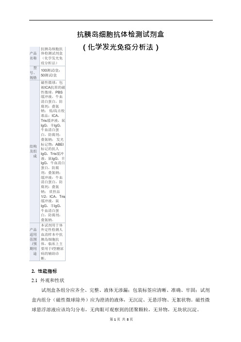
产品名称抗胰岛细胞抗体检测试剂盒(化学发光免疫分析法)型号、规格100测试/盒;50测试/盒结构及组成磁性微球:包被ICA抗原的磁性微球,PBS 缓冲液,牛血清白蛋白,防腐剂:叠氮钠;低/高点校准品:ICA,Tris缓冲液,鼠IgG,羊IgG,牛血清白蛋白,防腐剂:叠氮钠;发光标记物:ABEI 标记的抗人IgG,Tris缓冲液,鼠IgG,羊IgG,牛血清白蛋白,防腐剂:叠氮钠;缓冲液:牛血清白蛋白,防腐剂:叠氮钠;质控品1/2:ICA,Tris 缓冲液,鼠IgG,羊IgG,牛血清白蛋白,防腐剂:叠氮钠。
产品适用范围/预期用途本试剂用于体外定性检测人血清样本中抗胰岛细胞抗体,临床上主要用于I型糖尿病的辅助诊断。
2.性能指标2.1外观和性状抗胰岛细胞抗体检测试剂盒(化学发光免疫分析法)试剂盒各组分应齐全、完整、液体无渗漏;包装标签应清晰、准确、牢固;试剂盒内组分(磁性微球除外)应为澄清的液体,无沉淀、无悬浮物、无絮状物。
磁性微球悬浮溶液应该均匀分布,无肉眼可观察到的团聚颗粒,无异物,无块状沉淀。
第1 页共8 页2.2批内精密度检测抗胰岛细胞抗体企业参考品中精密度参考品,批内变异系数(CV)应≤8%。
2.3批间精密度检测抗胰岛细胞抗体企业参考品中精密度参考品,批间变异系数(CV)应≤15%。
2.4准确性(阳性符合率)检测抗胰岛细胞抗体企业参考品中阳性参考品,不得出现阴性反应(10/10)。
2.5特异性(阴性符合率)检测抗胰岛细胞抗体企业参考品中阴性参考品,不得出现阳性反应(15/15)。
2.6最低检出限检测抗胰岛细胞抗体企业参考品中最低检出量参考品,EL1~EL3 应检出阳性,EL4 应检出阴性。
2.7校准品2.7.1校准品准确度相对偏差应在±10%范围内。
2.7.2校准品瓶内重复性校准品瓶内重复性(CV)应≤8%。
2.7.3校准品均一性)应≤5%。
校准品均一性(CV均一性2.8质控品预期结果质控品1 每次测定结果应在(14.8~27.4)U/mL 范围内,质控品2 每次测定结果应在(36.3~67.3)U/mL 范围内。
人血管内皮细胞生长因子(VEGF)定量检测试剂盒(ELISA)

本试剂盒只能用于科学研究,不得用于医学诊断。
人血管内皮细胞生长因子(VEGF)定量检测试剂盒(ELISA)使用说明书【试剂盒名称】人血管内皮细胞生长因子(VEGF)定量检测试剂盒(ELISA)【试剂盒用途】定量检测人子血清、血浆及相关液体样本中血管内皮细胞生长因子(VEGF)的含量。
【检测原理】本试剂盒采用双抗体两步夹心酶联免疫吸附法(ELISA)。
将标准品、待测样本加入到预先包被人血管内皮细胞生长因子(VEGF))多克隆抗体透明酶标包被板中,温育足够时间后,洗涤除去未结合的成分,再加入酶标工作液,温育足够时间后,洗涤除去未结合的成分。
依次加入底物A、B,底物(TMB)在辣根过氧化物酶(HRP)催化下转化为蓝色产物,在酸的作用下变成黄色,颜色的深浅与样品中人血管内皮细胞生长因子(VEGF))浓度呈正相关,450nm波长下测定OD值,根据标准品和样品的OD值,计算样本中人血管内皮细胞生长因子(VEGF))含量。
【试剂盒组成】1 酶标包被板12孔×8条7 底物夜A6mL2 标准品:1600pg/ml0.6mL 8 底物夜B6mL3 20倍浓缩洗涤液20mL9 终止液6mL4 标准品稀释液6mL10 说明书1份5 样本稀释液6mL 11 封板膜1张6 酶标试剂6mL12 密封袋1个备注:标准品用标准品稀释液依次稀释为:1600、800、400、200、100、50pg/ml【需要而未提供的试剂和器材】1、37℃恒温箱2、标准规格酶标仪3、精密移液器及一次性吸头4、蒸馏水5、一次性试管6、吸水纸【操作步骤】1、准备:从冰箱取出试剂盒,室温复温平衡30分钟。
2、配液:用蒸馏水将20倍浓缩洗涤液稀释成原倍的洗涤液。
3、加标准品和待测样本:取足够数量的酶标包被板,固定于框架上,分别设置标准品孔、待测样本孔和空白对照孔,记录各孔位置,在标准品孔中加入标准品50μL;待测样本孔中先加入待测样本10μL,再加样本稀释液40μL(即样本稀释5倍);空白对照孔不加。
BD细胞固定液

BD Cytofix™Technical Data SheetFixation BufferProduct InformationMaterial Number:554655Size: 100 mlDescriptionBD Cytofix™ Fixation Buffer is comprised of a neutral pH-buffered saline (i.e., Dulbecco's Phosphate-Buffered Saline) that contains 4% w/v paraformaldehyde. This fixation buffer is intended to preserve human and rodent lymphoid cells for the subsequent immunofluorescentstaining of intracellular cytokines. BD Cytofix can also be used to preserve the light-scattering characteristics and fluorescence intensities ofhuman and rodent hematopoietic cells that have been stained by immunofluorescence for subsequent flow cytometric analysis.Preparation and StorageStore at 4° C and protected from prolonged exposure to light.Application NotesRecommended Assay Procedure:BD Cytofix can be used to fix unstained cells for subsequent immunofluorescent staining of intracellular cytokines. The suitability of fixing cellsfor immunofluorescent staining depends on whether the fluorescent antibodies can specifically detect their cognate antigens in a fixed form. Withrespect to intracellular cytokines, Pharmingen offers a large panel of conjugated anti-cytokine antibodies that can be successfully used to stainfixed and permeabilized cells. For the staining of antigens expressed on the surface of fixed cells, several fluorescent antibodies directed againstmouse cell surface antigens have been identified to be useful.BD Cytofix can also be used to fix cells after immunofluorescent staining in order to preserve the light-scattering signals and fluorescentintensities of cells for analysis at a later time. Cell Fixation Buffer may be useful to avoid the capping or shedding of fluorescent antibodies and/orsurface antigens during the period before flow cytometric analysis.Procedure for fixing cells with BD Cytofix™:1. Pellet 10e6 suspended cells (e.g., cytokine-producing cells generated by stimulatory culture) by centrifugation (250 - 300 x g) and carefullyremove supernatants to avoid cell loss.2. Add either 200 µl (for microwell plates) or 500 µl (for tubes) aliquots of cold DPBS containing protein and NaN3, gently resuspend cells,pellet, and remove supernatants.3. Repeat step 2.4. Add either 100 µl (for microwell plates) or 250 µl (for tubes) aliquots of fixation buffer to each cell pellet and resuspend the cells by eitherpipetting or vortexing. Incubate the cells with fixation buffer for 15 to 30 min at 4°C. (Cell aggregation can be avoided by vortexing prior to theaddition of the fixation buffer.)5. Fixed cells should be washed and suspended in a buffer that contains protein and NaN3, e.g., either Stain Buffer (FCS) [Cat. No. 554656] orStain Buffer (BSA) [Cat. No. 554657]. Store the fixed cells at 4°C (protected from light) for subsequent immunofluorescent staining ofintracellular cytokines. It is recommended that fixed cell samples be read as soon as possible, i.e., within one week.For the immunofluorescent staining of intracellular cytokines, cells that have been previously fixed with BD Cytofix™ can be washed two timesin a buffer that contains protein and NaN3 followed by incubating the cells for at least 10 minutes (4°C) in a buffer containing thecell-permeabilizing agent, saponin. BD Perm/Wash™ buffer (Cat. No. 554723) is ideally suited for this purpose. The fixed and permeabilizedcells can then be stained for intracellular cytokines as described in detail in the Immune Function handbook (BD Biosciences. 2003. Techniquesfor Immune Function Analysis, Application Handbook 1st Edition), available:/pdfs/manuals/02-8100055-21A1rr.pdf.Procedure for fixing immunofluorescently-stained cells with BD Cytofix™:Cells stained by immunofluorescence for cell surface antigens can be fixed as described above and stored (4°C, protected from light) forsubsequent analysis by flow cytometry (or fluorescence microscopy).NOTE : BD Cytofix/Cytoperm™ solution (Cat. No. 554722) and the BD Perm/Wash™ buffer (Cat. No. 554723) are included in BDCytofix/Cytoperm Kit (Cat. No. 554714) as well as the BD Cytofix/Cytoperm Plus Kit with GolgiStop™ (containing monensin; Cat. No. 554715)and BD Cytofix/Cytoperm Plus Kit with GolgiPlug™ (containing brefeldin A; Cat. No. 555028).Warnings and Precautions: BD Cytofix Buffer contains formaldehyde.R40Limited evidence of a carcinogenic effect.R43May cause sensitization by skin contact.S2Keep out of reach of children.S13 Keep away from food, drink and animal feedingstuffs.S23Do not breathe gas/fumes/vapour/spray.S36/37Wear suitable protective clothing and gloves.S46If swallowed, seek medical advice immediately and show this container or label.S56Dispose of this material and its container at hazardous or special wasteSuggested Companion ProductsCatalog Number Size CloneName554656Stain Buffer (FBS)500 ml(none) 554657Stain Buffer (BSA)500 ml(none) 554723Perm/Wash Buffer250 tests(none) 554714BD Cytofix/Cytoperm Fixation/Permeablization Kit250 tests(none) Product Notices1.Since applications vary, each investigator should titrate the reagent to obtain optimal results.2.Please refer to /pharmingen/protocols for technical protocols.ReferencesAlaverdi N, Waters JB. Pharmingen's Hotlines. 1997:6-15.(Methodology)BD Biosciences. Techniques for Immune Function Analysis, Application Handbook 1st Edition. 2003; Available:/pdfs/manuals/02-8100055-21A1rr.pdf 2007, Jan. 25.(Methodology)Lanier LL, Warner NL. Paraformaldehyde fixation of hematopoietic cells for quantitative flow cytometry (FACS) analysis. J Immunol Methods. 1981; 47(1):25-30.(Methodology)Sander B, Andersson J, Andersson U. Assessment of cytokines by immunofluorescence and the paraformaldehyde-saponin procedure. Immunol Rev. 1991;119:65-93.(Methodology)。
免疫学名词解释
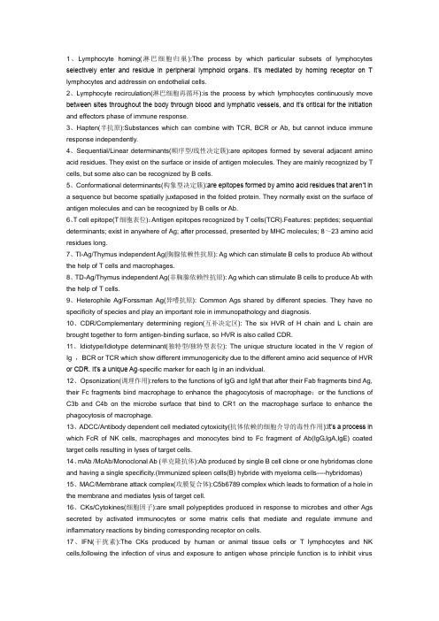
1、Lymphocyte homing(淋巴细胞归巢):The process by which particular subsets of lymphocytes selectively enter and residue in peripheral lymphoid organs. It’s mediated by homing receptor on T lymphocytes and addressin on endothelial cells.2、Lymphocyte recirculation(淋巴细胞再循环):is the process by which lymphocytes continuously move between sites throughout the body through blood and lymphatic vessels, and it’s critical for the initiation and effectors phase of immune response.3、Hapten(半抗原):Substances which can combine with TCR, BCR or Ab, but cannot induce immune response independently.4、Sequential/Linear determinants(顺序型/线性决定簇):are epitopes formed by several adjacent amino acid residues. They exist on the surface or inside of antigen molecules. They are mainly recognized by T cells, but some also can be recognized by B cells.5、Conformational determinants(构象型决定簇):are epitopes formed by amino acid residues that aren’t ina sequence but become spatially juxtaposed in the folded protein. They normally exist on the surface of antigen molecules and can be recognized by B cells or Ab.6、T cell epitope(T细胞表位):Antigen epitopes recognized by T cells(TCR).Features: peptides; sequential determinants; exist in anywhere of Ag; after processed, presented by MHC molecules; 8~23 amino acid residues long.7、TI-Ag/Thymus independent Ag(胸腺依赖性抗原): Ag which can stimulate B cells to produce Ab without the help of T cells and macrophages.8、TD-Ag/Thymus independent Ag(非胸腺依赖性抗原): Ag which can stimulate B cells to produce Ab with the help of T cells.9、Heterophile Ag/Forssman Ag(异嗜抗原): Common Ags shared by different species. They have no specificity of species and play an important role in immunopathology and diagnosis.10、CDR/Complementary determining region(互补决定区): The six HVR of H chain and L chain are brought together to form antigen-binding surface, so HVR is also called CDR.11、Idiotype/Idiotype determinant(独特型/独特型表位): The unique structure located in the V region of Ig ,BCR or TCR which show different immunogenicity due to the different amino acid sequence of HVR or CDR. It’s a unique Ag-specific marker for each Ig in an individual.12、Opsonization(调理作用):refers to the functions of IgG and IgM that after their Fab fragments bind Ag, their Fc fragments bind macrophage to enhance the phagocytosis of macrophage;or the functions of C3b and C4b on the microbe surface that bind to CR1 on the macrophage surface to enhance the phagocytosis of macrophage.13、ADCC/Antibody dependent cell mediated cytoxicity(抗体依赖的细胞介导的毒性作用):It’s a process in which FcR of NK cells, macrophages and monocytes bind to Fc fragment of Ab(IgG,IgA,IgE) coated target cells resulting in lyses of target cells.14、mAb /McAb/Monoclonal Ab (单克隆抗体):Ab produced by single B cell clone or one hybridomas clone and having a single specificity.(Immunized spleen cells(B) hybride with myeloma cells----hybridomas) 15、MAC/Membrane attack complex(攻膜复合体):C5b6789 complex which leads to formation of a hole in the membrane and mediates lysis of target cell.16、CKs/Cytokines(细胞因子):are small polypeptides produced in response to microbes and other Ags secreted by activated immunocytes or some matrix cells that mediate and regulate immune and inflammatory reactions by binding corresponding receptor on cells.17、IFN(干扰素):The CKs produced by human or animal tissue cells or T lymphocytes and NK cells,following the infection of virus and exposure to antigen whose principle function is to inhibit virusreplication or activate macrophage in both innate immunity and adaptive immunity.18、CAMs /Ams/cell adhesion molecules (黏附分子):The cell surface proteins involved in the interaction of cell-cell or cell-extracellular matrix. They play a crucial role in cell interaction, recognition, activation and migration by binding of receptor and ligand.19、CD/cluster of differentiation (分化簇):It is a group of cell surface molecules associated with the development and differentiation of immune cells.20、MHC/major histocompatibility complex(主要组织相容性复合体):A large cluster of linked genes located in some chromosomes of humanity or other mammals that encode major histocompatibility antigen and relate to allograft rejection, immune response and cell-cell recognition.21、HLA/Human leukocyte antigen(人类白细胞抗原):The major histocompatibility antigens for humanity which are associated with histocompatibility and immune response. They are alloantigens which are specific for each individual.22、HLA complex(HLA复合体):The MHC of humanity, a cluster of genes which encode for HLA and related to histocompatibility and immune response.23、MHC restriction(MHC 限制性):In interaction of T cell and APC or target cells, T cells not only recognize specific antigen but also recognize polymorphic residues of MHC molecules.24、PAMP/pathogen associated molecular pattern( 病原相关分子模式): The distinct structures or components that are common for many pathogens ,such as LPS, dsRNA of viruses etc.25、PRR/ pattern recognition receptor (模式识别受体): The receptors on macrophage that can recognize and bind PAMP on some pathogen, injured or apoptotic cells, including mannose receptor, scavenger receptor , toll like receptor etc.26、APC/Antigen presenting cells/Accessory cells/A cells(抗原递呈细胞): A group of cells which can uptake and process antigen and present antigen-MHC-Ⅰ/Ⅱcomplex to T cells, playing an important role in immune response.27、Cross-priming/Cross-presentation (交叉递呈): A mechanism by which a professional APC activates, a naïve CD8 CTL specific for the antigens of a third cell (e.g. a virus-infected or tumor cell)28、ITAM /immunoreceptor tyrosine-based activation motif(免疫受体酪氨酸活化基序): ITAM transduces activation signals from TCR, composing of tyrosine residues separated by around 18 aas. When TCR specially bind to antigen, the tyrosine becomes phosphorylated by the receptor associated tyrosine kinases to transduct active signals.29、TCR complex(TCR复合物): A group of membrane molecules on T cells that can specifically bind to antigen and pass an activation signal into the cell, consisting of TCR(αβ,γδ),CD3 (γε,δε)andδ-δ。
APC及抗原递呈(英文)
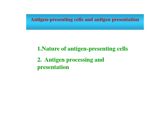
Cell surface
Exogenous antigens
Fusion Late endosome
Early endosome
Antigen degradation Golgi complex
MHC class 2 molecule Endoplasmic reticulum Ii chain
Processing and presentation of exogenous antigens
DC are the most powerful APC in vivo, which can effectively stimulate naï T cells to ve proliferate. Yet M and B cells can only stimulate activated or memory T cells.
(1) Production of endogenous antigenic peptide Ubiquitination of endogenous antigens Degradation of endogenous antigens in proteosome Peptides containing 8-12 amino acid residues
Professional APC
Dendritic cells, mononuclear phagocyte system, B cells, and APC that constitutively express MHC II molecules Non-professional APC
Dendritic cells
1 Antigen uptaking
DC capture antigens :
干细胞活性因子让诗丹尼奥逆时动能原液逆转时光

干细胞活性因子让诗丹尼奥逆时动能原液逆转时光近几年,在护肤行业内常见到干细胞护肤这个概念,事实上干细胞护肤早已不是一个概念了,国内不少朋友已经享受到了干细胞护肤带来的好处。
在我国,干细胞护肤这一方面做得比较好的主要还是国健菁华(深圳)精准健康科技有限公司的旗下品牌诗丹尼奥。
毕竟他们软、硬件设施都比较完备,并且已经在该领域钻研了三十余年。
诗丹尼奥皮肤工程实验室是以清华大学吴耀炯教授为首席科学家,并荟聚了中国中医科学院、新西兰坎特伯雷大学等27位教授、博士团队在内的高、精、尖人才,团队完成了多项科研,并将获得的研究成果运用到护肤领域上。
诗丹尼奥产品的价值所在就是“干细胞活性因子”,这个活性因子对于修复肌肤有着极大的作用,而它也是能够让肌肤实现“逆转时光”的重要角色。
除了在干细胞领域取得了伟大的研发成果,诗丹尼奥也革新了在提取干细胞活性因子需要用到的技术。
首先是无血清培养,无血清培养可除去动物血清等过敏源又不影响细胞的生长;其次是3D培养,3D培养模拟细胞在体内的生长环境,增加细胞间的相互交流,提高间充质干细胞的干性和分泌功能,产出更多活性更强大的细胞因子。
通过这两大培养技术,能够更快更好地分离出活性更好且无致敏源的细胞因子,为后期产品的研发生产提供了更好的原料。
诗丹尼奥利用干细胞活性因子研发了逆时动能原液(面部)和逆时修护原液(妊娠纹)这两大明星产品。
两款产品均由清华大学教授带领团队研发,并且有多重专利支持,主要有效成分均为人体胎盘干细胞活性因子提取物。
逆时修护原液(妊娠纹)是针对妊娠纹、肥胖纹等萎缩纹研发的修复产品,它从根源性排除过敏源,打造最佳人体生理浓度配比,可修复皮肤纤维断裂,重塑光滑迷人肌肤,再生、修复二合一。
逆时动能原液(面部)是针对面部修护的护肤产品,除了有人体胎盘干细胞活性因子提取物外,它还包含了100多种活性小分子,让肌肤重获新生,逆转时光。
除此之外它还具有美白淡斑、抗氧化抗衰老、除皱紧致、促进伤口愈合、红润肌肤等功效。
安瓶幻灯

iStelle-术后极速修复系列
安
瓶
美国·艾思黛尔
目录
1. 美国iStelle(艾思黛尔)公司介绍
2. 中国总代理—万润时代公司介绍 3. 安瓶主要成分--干细胞信息素的来源及作用机理 4. 微脂囊包裹技术 5. 安瓶的主要成分、适用范围及使用方法。 6. 五年大样本临床观察及效果对比图。 7. iStelle(艾思黛尔)安瓶与竞品的比较 8. iStelle(艾思黛尔)系列介绍 9. 各类证书 10.小结
常见问题解答
iStelle(艾思黛尔)安瓶容量怎么才1.7ml,量太少了吧?
答: iStelle(艾思黛尔)安瓶的核心成份为高浓度的细胞信息素及玻尿酸,
具备高活性,同时应用了最先进的微脂囊技术,具有很强的透皮吸收功效, 使用1—2瓶完全可使术后肌肤得到很好的修复,如需要抑制瘢痕形成。需要 使用3个月左右。
术后第一天
40岁
女性
当日同时操作:水光针、5D
童颜术、自体血清填充三项. 术后第一天
美国·艾思黛尔
安瓶--同时做微整形三项效果
术后第三天
女士,40岁 同时操作:水光针、5D童颜术、 自体血清填充三项; 术后第三天。 术后7天淡妆出镜,接受采访
美国·艾思黛尔
安瓶—对儿童皮肤挫伤的修复对比
小朋友, 4岁,意外摔伤, 使用iStelle安瓶,每6小时涂 抹创面一次 48h完全结痂, 第5天结痂脱落,皮肤基本恢复 第6天皮肤完全恢复,没有一点
疤克:只能使用在完全愈合的伤口上,起到淡化疤痕的作用; 愈肤宁:主要为几聚糖(Chitosan),为含氨多糖,干燥后可形 成透明保护薄膜。 金扶宁 :主要成分重组人粒细胞巨噬细胞刺激因子,主要为消炎的功效,没有促 进伤口愈合及细胞再生的作用;
免疫荧光步骤

Immunocytochemistry and immunofluorescence protocolProcedure for staining of cell cultures using immunofluorescenceICC and IF protocol Preparing the slide1.Coat coverslips with polyethylineimine or poly-L-lysine for 1 h at room temperature.2.Rinse coverslips well with sterile H2O (three times 1 h each).3.Allow coverslips to dry completely and sterilize them under UV light for at least 4 h.4.Grow cells on glass coverslips or prepare cytospin or smear preparation.5.Rinse briefly in phosphate-buffered saline (PBS).For wash buffer we recommend 1x PBS 0.1% Tween 20.FixationThe cells may be fixed using one of two methods:1.Incubating the cells in 100% methanol (chilled at -20°C) at room temperature for 5mining 4% paraformaldehyde in PBS pH 7.4 for 10 min at room temperatureThe cells should be washed three times with ice-cold PBS.Antigen retrieval (optional step)Certain antibodies work best when cells are heated in antigen retrieval buffer. Checkthe product information for recommendations for each primary antibody being used.1.Preheat the antigen retrieval buffer (100 mM Tris, 5% [w/v] urea, pH 9.5) to 95°C.This can be done by heating the buffer in a coverglass staining jar which is placedin a water bath at 95°C.ing a small pair of broad-tipped forceps, place the coverslips carefully in theantigen retrieval buffer in the cover glass staining jar, making note of which side ofthe coverslips the cells are on.3.Heat the coverslips at 95°C for 10 min.24.Remove the coverslips from the antigen retrieval buffer and immerse them, withthe side containing the cells facing up, in PBS, in the 6-well tissue culture plates.5.Wash cells in PBS three times for 5 min.PermeabilizationIf the target protein is intracellular, it is very important to permeabilize the cells.Acetone fixed samples do not require permeabilization.1.Incubate the samples for 10 min with PBS containing either 0.1–0.25% Triton X-100(or 100 μM digitonin or 0.5% s aponin). Triton X-100 is the most popular detergent forimproving the penetration of the antibody. However, it is not appropriate formembrane-associated antigens since it destroys membranes.2.The optimal percentage of Triton X-100 should be determined for each protein ofinterest.3.Wash cells in PBS three times for 5 min.Blocking and immunostaining1.Incubate cells with 1% BSA, 22.52 mg/mL glycine in PBST (PBS+ 0.1% Tween 20) for30 min to block unspecific binding of the antibodies (alternative blocking solutionsare 1% gelatin or 10% serum from the species the secondary antibody was raisedin: see antibody datasheet for recommendations).2.Incubate cells in the diluted antibody in 1% BSA in PBST in a humidified chamberfor 1 h at room temperature or overnight at 4°C.3.Decant the solution and wash the cells three times in PBS, 5 min each wash.4.Incubate cells with the secondary antibody in 1% BSA for 1 h at room temperaturein the dark.5.Decant the secondary antibody solution and wash three times with PBS for 5 mineach in the dark.3ICC and IF protocol Multicolor immunostaining(optional step)To examine the co-distribution of two (or more) different antigens in the same sample,use a double immunofluorescence procedure. This can be performed eithersimultaneously (in a mixture) or sequentially (one antigen after another).Ensure you have antibodies for different species and their corresponding secondaryantibodies. For example, rabbit antibody against antigen A, mouse antibody againstantigen B. Alternatively, you can use directly conjugated primary antibodiesconjugated to different fluorophores.Simultaneous incubation1.Incubate cells with blocking solution for 30 min.2.Incubate cells with both primary antibodies in 1% BSA in PBST in a humidifiedchamber for 1 h at room temperature or overnight at 4°C.3.Decant the solution and wash the cells three times in PBS, 5 min each wash.4.Incubate cells with both secondary antibodies in 1% BSA for 1 h at roomtemperature in the dark.5.Decant the secondary antibody solution and wash three times with PBS for 5 minseach in the dark.Sequential incubation1.First blocking step: incubate cells with the first blocking solution (10% serum fromthe species that the secondary antibody was raised in) for 30 min at roomtemperature.2.Incubate cells with the first primary antibody in 1% BSA or 1% serum in PBST in ahumidified chamber for 1 h at room temperature or overnight at 4°C, 1% gelatinor 1% BSA.3.Decant the first primary antibody solution and wash the cells three times in PBS, 5min each wash.4.Incubate cells with first secondary antibody in 1% BSA in PBST for 1 h at roomtemperature in the dark.5.Decant the first secondary antibody solution and wash three times with PBS for 5min each in the dark.6.Second blocking step: incubate cells with the second serum, (10% serum from thespecies that the secondary antibody was raised in) for 30 min at roomtemperature in the dark.47.Incubate cells with the second primary antibody in 1% BSA or 1% serum in PBST in ahumidified chamber in the dark for 1 h at room temperature, or overnight at 4°C.8.Decant the second primary antibody solution and wash the cells three times inPBS, 5 min each wash in the dark.9.Incubate cells with second secondary antibody in 1% BSA for 1 h at roomtemperature in the dark.10.Decant the second secondary antibody solution and wash three times with PBSfor 5 min each in the dark.If you have to detect more than two antigens, continue following steps 1–5 for therest of the antibodies.Counter staining1.Incubate cells with 0.1–1 μg/mL Hoechst stain or DAPI (DNA stain) for 1 min.2.Rinse with PBS.Mounting1.Mount coverslip with a drop of mounting medium.2.Seal coverslip with nail polish to prevent drying and movement under microscope.3.Store in dark at -20°C or +4°C.5。
化学专业英语文献翻译
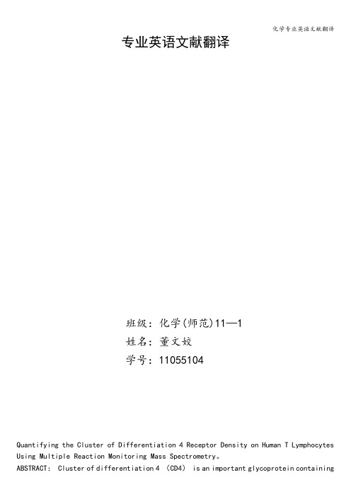
专业英语文献翻译Quantifying the Cluster of Differentiation 4 Receptor Density on Human T Lymphocytes Using Multiple Reaction Monitoring Mass Spectrometry。
ABSTRACT: Cluster of differentiation 4 (CD4) is an important glycoprotein containingfour extracellular domains, a transmembrane portion and a short intracellular tail. It locates on the surface of various types of immune cells and performs a critical role in multiple cellular functions such as signal amplification and activation of T cells。
It is well-known as a clinical cell surface protein marker for study of HIV progression and for defining the T helper cell population in immunological applications。
Moreover,CD4 protein has been used as a biological calibrator for quantification of other surface and intracellular proteins. However,flow cytometry, the conventional method of quantification of the CD4 density on the T cell surface depends on antibodies and has suffered from variables such as antibody clones, the ommatophore and conjugation chemistries, the fixation conditions, and the flow cytometric quantification methods used. In this study, we report the development of a highly reproducible na no liquid chromatography−multiple reaction monitoring mass spectrometry-based quantitative method to quantify the CD4 receptor density in units of copy number per cell on human CD4+ T cells. The method utilizes stable isotope—labeled full—length standard CD4 as an internal standard to measureendogenous CD4 directly, without the use of antibodies. The development of the mass spectrometry-based approach of CD4 protein quantification is important as a complementary strategy to validate the analysis from the cytometry-based conventional method。
人转化生长因子β1(TGF-β1)ELISA试剂盒说明书

人转化生长因子β1(TGF—β1)ELISA试剂盒说明书产品名称:人转化生长因子β1(TGFβ1)ELISA试剂盒英文名称:TGFβ1 ELISA Kit检测原理:ELISA试剂盒采纳抗体夹心法:将抗某蛋白抗体包被于酶标板上,标本和标准品中的某蛋白与抗体结合,加入生物素化的抗某蛋白抗体,再加入SABC复合物与生物素抗体结合,形成免疫复合物,然后加入TMB显色底物,显色剂显蓝色,最后加停止液变黄色,游离的成分被洗去。
在450 nm处测OD值,某蛋白浓度与OD值之间呈正比,可通过绘制标准曲线计算出标本中某蛋白的浓度。
试剂盒组分: (保管温度4℃)名称规格(48 T)规格(96 T)预包被酶标板8×6条8×12条1支1支标准品/样品稀释液10ml15ml生物素化检测抗体(100×)60ul120ul生物素化检测抗体稀释液6ml12mlSABC复合物6ml12mlTMB显色液(A/B)各3ml停止液6ml12ml20×浓缩洗涤液30ml60ml封板胶纸2张4张产品说明书1份1份本试剂盒用于血清、血浆、组织匀浆、细胞培育上清液及其它生物体液。
标本收集与试剂准备:1. 血清、血浆样本收集应使用一次性的无热原,无内毒素试管(EDTA、柠檬酸盐、肝素抗凝均可),血清、血浆躲避使用溶血,高血脂标本,标本悬浮物应离心去除,使标本清亮透亮。
待测样本应尽早检测,28℃保管48小时;更长时间须冷冻(20℃或80℃)保管,躲避反复冻融。
2. 洗涤液配置:用蒸馏水1:20稀释(示例:1ml浓缩洗涤液加入19ml的蒸馏水)3. 标准品配制:取7个1.5ml离心管,分别标注1/2,1/4,1/8,1/16,1/32,1/64,blank。
从第一至七管中分别加入标准品/样品稀释液200ul。
在第一管中加入标准品溶液200ul,置于漩涡混合器上混匀后用加样器吸出200ul,移至第二管。
如此反复作对倍稀释,从第六管中吸出200ul弃去,第七管为空白对比。
Golgi 体染料 说明书

Product InformationRevised: 25–July–2003 NBD- and BODIPY Dye–Labeled SphingolipidsIntroductionFluorescent sphingolipids are versatile probes of lipid traf-f ick i ng in living cells.1,2 In addition, ceramide analogs produce se l ec t ive staining of the Golgi complex for visualization by flu o-res c ence microscopy. Molecular Probesʼ fluorescently la b eled an-a l ogs of ceramides, sphingomyelins and glycosylceramides are prepared from D-erythro-sphingosine and therefore have the same stereochemical conformation as natural biologically active sphin-golipids. Applications of these flu o r es c ent lipid probes in c lude: Ceramides• Fluorescent structural marker for Golgi Complex 3,4• Outlining cellular boundaries to allow observation of mor p ho -ge n et i c movements by confocal microscopy 5• Tracing lipid metabolism and trafficking in living cells 6-8• Measuring rates of lipid synthesis by Schwann cells 9 Sphingomyelins• Tracing lipid endocytosis pathways by fluorescence mi c ros -co p y 7,10,11• Analysis of acid sphingomyelinase expression by flow cytom-etry 12Glycosylceramides• Cellular-level identification of lysosomal lipid storage dis-order phentotypes13,14• Visualization of lipid domains associated with T cell an t i g en receptor activation15The BODIPY FL and NBD fluorophores incorporated in Molecular Probesʼ fluorescent sphingolipids (Figure 1) havesig n if i c ant l y different spectroscopic properties. The BODIPYFL fluorophore produces greater fluorescence output than NBD due to its higher molar absorptivity and fluorescence quantum yield.16 The BODIPY FL fluorophore is also more photostable than NBD. The photostability of NBD is sensitive to the pres e nce of cho l es t er o l, resulting in weak labeling of the Golgi complex in cho l es t er o l-deficient cells.17 The most practically impor t ant spec-t ro s cop i c property of the BODIPY FL fluorophore is an aggregation-de p en d ent shift from green- to red-flu o r es c ence emis s ion due to in t er m o l ec u l ar excimer formation. Con s e q uent l y, struc t ures that ac c u m u l ate large amounts of BODIPY FL–la b eled sphingolipids, such as the Golgi complex 3 or the ly s o s o mesof sph i n g olip i d stor a ge disease fibroblasts,7,13 exhibit red flu o-res c ent stain i ng that is clearly dis t inct from low-abun d ance green flu o r es c ent stain i ng. The BODIPY TR fluorophore ex h ib i ts red flu o r es c ence (~617 nm) in its normal mo n o m er i c state; aggrega-tion-dependent emission shifts cor r e s pond i ng to those of the BODIPY FL fluorophore have not yet been detected.The transport and metabolism of labeled sphingolipids is some w hat dependent on the attached fluorophore.6 In some cas e s, significant differences have been observed in the met a b ol i c prod-u cts de r ived from homologous BODIPY FL– and NBD-la b eled sphingolipids.3,18 NBD-labeled sphingolipids have higher rates of transfer through aqueous phas e s than their BODIPY FL–labeled counterparts.19 Analysis of lipid in c or p o r a t ion and re c y c ling by quantitative removal of labeled lipids from the plasma membrane outer surface (“back-exchange”) is more readily ac c om p lished using NBD-labeled sphingolipids.2Storage and HandlingWith the exception of BODIPY FL C12-sphingomyelin,all flu o r es c ent sphingolipids are supplied in solid form and should be stored at –20°C, desiccated and protected from light. Cer a mide analogs can be dissolved in DMSO or chloroform at concentrations of at least 1 mM. Sphingomyelins and glycosyl-ceramides can be dissolved in DMSO or ethanol to the same extent. De l iv e ry in the form of bovine serum albumin (BSA) com p lex e s is rec o m m end e d for labeling cells (see Ex p er i m en t al Protocols). BODIPY FL C5-ceramide, NBD C6-ceramide and BODIPY TR C5ceramide are also avail a ble as ready-made BSA complexes (B-22650, N-22651, B-34400). Additional BSA-complexed sphingolipids include BODIPY FL C5-ganglioside (B-34401) and BODIPY FL C5-lactosylceramide (B-34402). These complexes are supplied in lyo p hilized form and should be stored at –20°C, desiccated and pro t ect e d from light. Procedures for re c on s ti t u t ion of the lyophilized BSA com p lex e s are described in the Experimental Protocols. BODIPY FL C12-sphingomyelin is supplied as a 1 mg/mL so l u t ion in DMSO which should be stored at –20°C, desiccated and pro t ect ed from light.Figure 1. BODIPY FL– and NBD-labeled ceramide analogs.Experimental ProtocolsPreparation of Sphingolipid–BSA ComplexesFor staining of living and fixed cells, it is efficacious to add flu o r es c ent sphingolipids in the form of complexes with BSA.4 BSA delivery complexes of fluorescent sphingolipids can be pre-p ared as follows:1.1 Prepare an approximately 1 mM sphingolipid stock solution in chloroform:ethanol (19:1 v/v). Molecular weights required for conversion of mass to molarity are printed on the product labels.1.2 Dispense 50 µL of sphingolipid stock solution into a small glass test tube and dry, first under a stream of nitrogen, and then under vacuum for at least 1 hour. Redissolve in 200 µL of ab-s o l ute ethanol.1.3 Measure 10 mL of serum-free balanced salt solution such as Hanks ʼ buffered salt solution + 10 mM HEPES, pH 7.4 (HBSS/HEPES) into a 50 mL plastic centrifuge tube. Add 3.4 mg (0.34 mg/mL) of defatted BSA.1.4 Agitate the tube containing the 10 mL of the BSA solution on a vortex mixer. Inject the sphingolipid solution in eth a n ol (200 µL) into the vortex. Store the resulting solution (5 µM sphingolipid + 5 µM BSA) in a plastic tube at –20°C.Reconstitution of Ready-Made Ceramide–BSA Complexes (B-22650, N-22651, B-34400)2.1 Dissolve 5 mg of the ready-made complex in 150 µL of sterile deionized water. The resulting stock solution contains 0.5 mM sphingolipid and 0.5 mM BSA (1:1 mol:mol). Store unused por t ions of stock solution at –20°C.2.2 Prepare a 5 µM staining solution by diluting the stock so- l u t ion 100-fold (e.g. add 10 µL of stock solution to 1 mL of HBSS/HEPES).Staining the Golgi Complex in Living Cells with Fluorescent Ceramides3.1 Rinse cells grown on glass coverslips in an appropriate medium (such as HBSS/HEPES).3.2 Incubate the cells for 30 minutes at 4°C with 5 µM ceramide–BSA in HBSS/HEPES (prepared in step 1.4 or 2.2).3.3 Rinse the sample several times with ice-cold medium and in c u b ate in fresh medium at 37°C for a further 30 min u tes.3.4 Wash the sample in fresh medium and examine using a fluo-rescence microscope. Prominent labeling of the Golgi apparatus and weaker la b el i ng of other intracellular membranes should be seen.Staining the Golgi Complex in Fixed Cells with NBD C 6-CeramideIt has been reported that NBD C 6-ceramide is suitable for stain i ng of fixed cells, but that BODIPY FL C 5-ceramide and BODIPY TR ceramide are not.4 Conditions for staining appear to be generally noncritical. However, detergents and methanol/acetone fixatives should be avoided.44.1 Rinse cells grown on glass coverslips in HBSS/HEPES and fix for 5–10 minutes at room temperature in 0.5% gl u t a ral d e h yde/10% sucrose/100 mM PIPES, pH 7.0 or 2–4% paraform a l d e h yde in phosphate-buffered saline (PBS).4.2 Rinse the sample several times with ice-cold HBSS/HEPES, transfer to an ice-bath and incubate for 30 minutes at 4°C with 5 µM ceramide–BSA complex (prepared in step 1.4 or 2.2).4.3 Rinse in HBSS/HEPES and incubate for 30–90 minutes at room temperature with 10% fetal calf serum or 2 mg/ml BSA to enhance golgi staining.4.4 Wash the sample in fresh HBSS/HEPES, mount and ex a m i ne by fluorescence microscopy.Fluorescence MicroscopySpectral characteristics of NBD-, BODIPY FL– and BODIPY TR–labeled sphingolipids are summarized in Table 1. Note that because BODIPY FL–labeled sphingolipids may emit either green or red fluorescence depending on their intracellular local-ization, they are generally unsuitable for multicolor imaging applications in which the second probe is a green-fluorescent protein (GFP) chi m e r a or an antibody labeled with FITC (or other spec t ral l y sim i l ar dyes). In applications of this type, BODIPY TR ceramide should be used for Golgi complex staining.20Staining for Electron MicroscopyBODIPY FL Br 2C 5-ceramide (D-7546) is designed for ul- t ra s truc t ur a l localization by electron microscopy (EM). In thisstain i ng pro c e d ure, excitation of the fluorophore is coupled to pho t o o x i d a t ion of diaminobenzidine (DAB) to produce an elec-tron-dense pre c ip i t ate.215.1 Cells for EM staining should be grown in 35 mm–diameter plas t ic tissue culture dishes, not on glass coverslips.5.2 Label living or fixed cells following the protocols described above. Living cells should be fixed after completion of the la-b el i ng procedure.5.3 Prepare fresh diaminobenzidine (DAB) staining solution (15 mg of DAB dissolved in 10 mL of 0.1 M Tris, pH 7.6). Keep ice-cold until required for use.5.4 Wash the cells in 0.1 M Tris, pH 7.6 and add 0.9 mL of DAB staining solution. Cover the culture dish and incubate in the dark for at least 10 minutes.5.5 Irradiate the sample for 30 minutes on a fluorescence mi c ro -scope using a low-magnification objective (10X or below).Label Abs *Em *BODIPY FL 505511†BODIPY TR 589617NBD466536* Absorption and fluorescence emission maxima, in nm, determined in methanol.Values in labeled cells are similar. † BODIPY FL-labeled sphingolipids also emit red fluorescence (~620 nm) in cells.Table 1.Spectral characteristics of NBD-, BODIPY FL– and BODIPY TR–labeled sphin-golipids.References1. Frontiers in Bioactive Lipids , J.Y . Vanderhoek, Ed. pp. 203–213, Plenum Press (1996);2. Fluorescent and Lu m i n es c ent Probes for Biological Activity, 2nd Edition , W.T. Mason, Ed., pp. 136–155, Academic Press (1999);3. J Cell Biol 113, 1267 (1991);4. Cell Biology: A Laboratory Handbook, 2nd Edi- t ion , V ol. 2, J.E. Celis, Ed. pp. 507–512, Ac a d em i c Press (1998);5. Methods Cell Biol 59, 179 (1999);6. Methods Cell Biol 38, 221 (1993);7. Ann NY Acad Sci 845, 152 (1998);8. J Cell Biol 144, 673 (1999);9. J Neurosci Res 49, 497 (1997); 10. Biophys J 72, 37 (1997); 11. Chem Phys Lipids 102, 55 (1999); 12. Blood 93, 80 (1999); 13. Proc Natl Acad Sci USA 95, 6373 (1998); 14. Lancet 354, 901 (1999); 15. J Cell Biol 147, 447 (1999); 16. Anal Biochem 198, 228 (1991); 17. Proc Natl Acad Sci USA 90, 2661 (1993); 18. J Biol Chem 271, 2287 (1996); 19. Biochemistry 36, 8840 (1997); 20. J Biol Chem 274, 27726 (1999); 21. J Cell Biol 126, 901 (1994).Prod u ct List Current pric e s may be ob t ained from our Web site or from our Customer Service Department.Cat # Product Name Unit Size B-22650 BODIPY ® FL C 5-ceramide complexed to BSA.................................................................................................................................................. 5 mg B-13950 BODIPY ® FL C 5-ganglioside G M1...................................................................................................................................................................... 25 µg B-34401 BODIPY ® FL C 5-ganglioside G M1 complexed to BSA......................................................................................................................................... 1 mg B-34402 BODIPY ® FL C 5-lactosylceramide complexed to BSA...................................................................................................................................... 1 mg B-34400 BODIPY ® TR C 5-ceramide complexed to BSA ................................................................................................................................................. 5 mg N-22651 NBD C 6-ceramide complexed to BSA.............................................................................................................................................................. 5 mg N-1154 6-((N-(7-nitrobenz-2-oxa-1,3-diazol-4-yl)amino) hexanoyl)sphingosine (NBD C 6-ceramide)......................................................................... 1 mg D-3521 N-(4,4-difluoro-5,7-dimethyl-4-bora-3a,4a-diaza-s-indacene-3-pentanoyl)sphingosine (BODIPY ® FL C 5-ceramide)...................................... 250 µg D-7540 N-((4-(4,4-difluoro-5- (2-thienyl)-4-bora-3a, 4a-diaza-s-indacene-3-yl)phenoxy)acetyl)sphingosine (BODIPY ® TR ceramide)..................... 250 µg D-7546 N-(2,6-dibromo-4,4-difluoro- 5,7-dim e t h y l-4-bora-3a, 4a-diaza-s-indacene-3-pentanoyl)sphingosine(BODIPY ®FL Br 2C 5-ceramide)......................................................................................................................................................................... 250 µg D-13951 N-(4,4-difluoro-5,7-dimethyl- 4-bora-3a,4a-diaza-s-indacene-3-pentanoyl)sphingosyl 1-β-D -lactoside(BODIPY ® FL C 5-lactosylceramide)................................................................................................................................................................. 25 µg D-3522 N-(4,4-difluoro-5,7-dimethyl- 4-bora-3a,4a-diaza-s-indacene-3-pentanoyl)sphingosylphosphocholine(BODIPY ® FL C 5-sphingomyelin)..................................................................................................................................................................... 250 µg N-3524 6-((N-(7-nitrobenz-2-oxa-1,3-diazol-4-yl)amino) hexanoyl)sphingosylphosphocholine (NBD C 6-sphingomyelin)......................................... 1 mg D-7519 N-(4,4-difluoro-5,7-dimethyl-4-bora-3a,4a-diaza-s-indacene-3-dodecanoyl)sphingosyl 1-β-D -galactopyranoside(BODIPY ® FL C 12-galactocerebroside)............................................................................................................................................................. 25 µg D-7547 N-(4,4-difluoro-5,7-dimethyl-4-bora-3a,4a-diaza-s-indacene-3-dodecanoyl)sphingosyl 1-β-D -glucopyranoside(BODIPY ® FL C 12-glucocerebroside)................................................................................................................................................................ 250 µg D-7548 N-(4,4-difluoro-5,7-dimethyl-4-bora-3a,4a-diaza-s-indacene-3-pentanoyl)sphingosyl 1-β-D -glucopyranoside(BODIPY ® FL C 5-glucocerebroside)................................................................................................................................................................. 250 µg D-7711 N-(4,4-difluoro-5,7-dimethyl-4-bora-3a,4a-diaza-s-indacene-3-dodecanoyl)sphingosylphosphocholine(BODIPY ® FL C 12-sphingomyelin) *1 mg/mL in DMSO*................................................................................................................................ 250 µL5.6 Wash the sample at least five times in 0.1 M Tris, pH 7.6. View i ng the specimen under a dissecting microscope, make a mark on the inside of the culture dish using a needle, encircling the location of the brown DAB reaction product.5.7 After rinsing the sample in 0.1 M sodium cacodylate (C 2H 6AsNaO 2), pH 7.4, treat the specimen with 1% osmium tetrox i de (OsO 4) in 0.1 M sodium cacodylate, pH 7.4 for 60 min-u tes at room temperature. 5.8 Wash the specimen in 0.1 M sodium cacodylate, pH 7.4, de- h y d rate and embed in epoxy medium.5.9 Cut out the region of the dish identified by the mark made instep 5.6 and mount for thin-section electron microscopy.Contact InformationFurther information on Molecular Probes' products, including product bibliographies, is available from your local distributor or directly from Molecular Probes. Customers in Europe, Africa and the Middle East should contact our office in Leiden, the Netherlands. All others should contact our Technical As s is t ance Department in Eugene, Oregon. Please visit our Web site — — for the most up-to-date information.Molecular Probes, Inc.29851 Willow Creek Road, Eugene, OR 97402Phone: (541) 465-8300 • Fax: (541) 344-6504Customer Service: 6:00 am to 4:30 pm (Pacific Time)Phone: (541) 465-8338 • Fax: (541) 344-6504 • order@ Toll-Free Ordering for USA and Canada:Order Phone: (800) 438-2209 • Order Fax: (800) 438-0228 Technical Assistance: 8:00 am to 4:00 pm (Pacific Time) Phone: (541) 465-8353 • Fax: (541) 465-4593 • tech@ Molecular Probes Europe BVPoortGebouw, Rijnsburgerweg 102333 AA Leiden, The NetherlandsPhone: +31-71-5233378 • Fax: +31-71-5233419Customer Service: 9:00 to 16:30 (Central Eu r o p e a n Time) Phone: +31-71-5236850 • Fax: +31-71-5233419eurorder@probes.nlTechnical Assistance: 9:00 to 16:30 (Central Eu r o p e a n Time) Phone: +31-71-5233431 • Fax: +31-71-5241883eurotech@probes.nlMolecular Probes’ products are high-quality reagents and materials intended for research pur p os e s only. These products must be used by, or directl y under the super v ision of, a tech n ically qual i f ied individual experienced in handling potentially hazardous chemicals. Please read the Material Safety Data Sheet pro v id e d for each prod u ct; other regulatory considerations may apply.Several of Molecular Probes’ products and product applications are covered by U.S. and foreign patents and patents pending. Our prod u cts are not available for resale or oth e r commercial uses without a specifi c agreement from Molecular Probes, Inc. We welcome inquiries about licensing the use of our dyes, trade m arks or technolo-gies. Please submit inquiries by e-mail to busdev@. All names con t ain i ng the des i g n a t ion ® are reg i s t ered with the U.S. Patent and Trade m ark Offi ce. Copyright 2003, Molecular Probes, Inc. All rights reserved. This information is subject to change without notice.。
北京中航赛维抗胰岛细胞抗体(ICA)检测试剂盒(磁微粒化学发光免疫分析法)产品技术要求

抗胰岛细胞抗体(ICA)检测试剂盒(磁微粒化学发光免疫分析法)结构组成:
预期用途:用于体外定性检测人血清中的胰岛细胞抗体(ICA)的含量。
2.1外观
2.1.1试剂盒各组分应齐全、完整、液体无渗漏;
2.1.2磁分离试剂摇匀后为均匀悬浊液,无明显凝集;
2.1.3液体组分应澄清,无沉淀或絮状物;
2.1.4包装标签应清晰,易识别;
2.2阳性参考品符合率
用企业阳性参考品进行测定,均为阳性。
2.3阴性参考品符合率
用企业阴性参考品进行测定,均为阴性。
2.4临界值及重复性
检测浓度为10AU/mL的弱阴参考品10次,10次检测结果应均为阴性,S/CO值变异系数(CV)应不大于10%;检测浓度为30AU/mL的弱阳参考品10次,10次检测结果应均为阳性,S/CO值变异系数(CV)应不大于10%。
2.5批间差
用三个批号的试剂盒分别检测浓度为10AU/mL的弱阴参考品,检测结果应均为阴性,S/CO值变异系数(CV)应不大于15%;用三个批号的试剂盒分别检测浓度为30AU/mL的弱阳参考品,检测结果应均为阳性,S/CO值变异系数(CV)应不大于15%。
2.6稳定性
试剂盒在2~8℃贮存,有效期为12个月,取到效期后的试剂盒样品进行检测,检测结果应符合2.2、2.3、2.4的要求。
中性粒细胞胞外诱捕网形成的分子机制研究进展

中性粒细胞胞外诱捕网形成的分子机制研究进展①蒋瑶邢艳(川北医学院附属医院检验科,南充637000)中图分类号R392.6文献标志码A文章编号1000-484X(2022)10-1272-06[摘要]中性粒细胞作为固有免疫的一线细胞,其死亡形式-中性粒细胞胞外诱捕网(NETs)过度形成过程中因释放大量胞内成分而参与包括自身免疫性疾病在内的多种疾病发生。
自身免疫性疾病以自身抗原暴露、免疫耐受缺失为特征,且目前治疗仍以改善症状为主,如能抑制自身抗原产生将为其根治提供新的可能。
本文主要对参与NETs形成的分子进行综述,包括ROS、PAD-4、自噬以及相关信号通路,以期促进NETs相关疾病治疗策略的开发。
[关键词]中性粒细胞胞外诱捕网;分子机制;研究进展Progress on molecular mechanism of neutrophil extracellular traps formationJIANG Yao,XING Yan.Department of Clinical Laboratory,Affiliated Hospital of North Sichuan Medical College,Nanchong637000,China[Abstract]As first-line cells of innate immunity,neutrophils release a large number of intracellular components in process of over-formation of neutrophil extracellular traps(NETs),which participates in occurrence of multiple diseases including autoimmune diseases.Autoimmune diseases are characterized by autoantigen exposure and loss of immune tolerance,whose treatment is still mainly to improve symptoms at present.If production of autoantigens can be inhibited,it will provide a new possibility for cure.This article will review molecules involved in formation of NETs,including ROS,PAD-4,autophagy and its related signal pathways,and aim to promote development of treatment strategies for NETs-related diseases.[Key words]Neutrophil extracellular traps;Molecular mechanism;Progress中性粒细胞是人体内含量最多的固有免疫细胞,通过抵御病原体入侵而在固有免疫中起重要作用,经典杀菌方式包括吞噬和脱颗粒。
EDU说明书
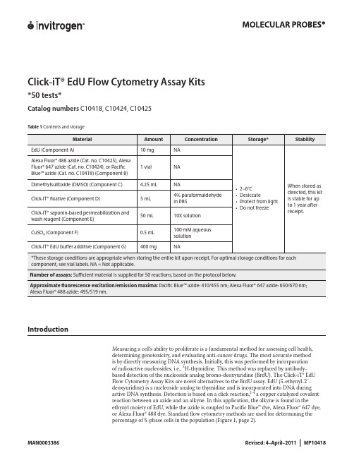
Click-iT® EdU Flow Cytometry Assay Kits*50 tests*Catalog numbers C10418, C10424, C10425Contents and storageTable 1IntroductionMeasuring a cell’s ability to proliferate is a fundamental method for assessing cell health,determining genotoxicity, and evaluating anti-cancer drugs. The most accurate methodis by directly measuring DNA synthesis. Initially, this was performed by incorporationof radioactive nucleosides, i.e., 3H-thymidine. This method was replaced by antibody-based detection of the nucleoside analog bromo-deoxyuridine (BrdU). The Click-iT® EdUFlow Cytometry Assay Kits are novel alternatives to the BrdU assay. EdU (5-ethynyl-2´-deoxyuridine) is a nucleoside analog to thymidine and is incorporated into DNA duringactive DNA synthesis. Detection is based on a click reaction,1-4 a copper catalyzed covalentreaction between an azide and an alkyne. In this application, the alkyne is found in theethynyl moiety of EdU, while the azide is coupled to Pacific Blue™ dye, Alexa Fluor® 647 dye,or Alexa Fluor® 488 dye. Standard flow cytometry methods are used for determining thepercentage of S-phase cells in the population (Figure 1, page 2).Figure 1 Fluorescence signal from Pacific Blue™, Alexa Fluor® 488, and Alexa Fluor® 647 Click-iT® EdU Flow Cytometry Assay Kits. Jurkat (human T-cell leukemia) cells were treated with 10 µM EdU for 2 hours and detected according to the recommended staining protocol. The figures show a clear separation of proliferating cells which have incorporated EdU and nonproliferating cells which have not. Panel A shows data from cells labeled with Pacific Blue™ azide analyzed on an Attune® Acoustic Focusing Cytometer using 405 nm excitation with a 450/40 nm bandpass emission filter; Panel B shows data from cells labeled with Alexa Fluor® 488 azide analyzed on an Attune® Acoustic Focusing Cytometer using 488 nm excitation and a 530/30 nm bandpass emission filter; Panel C shows data from cells labeled with Alexa Fluor® 647 azide analyzed on a flow cytometer using 633 nm excitation and a 660/20 nm bandpass emission filter.The advantage of Click-iT® EdU labeling is that the small size of the dye azide allows for efficient detection of the incorporated EdU using mild conditions. Standard aldehyde-based fixation and detergent permeabilization are sufficient for the Click-iT® detection reagent to gain access to the DNA. This is in contrast to BrdU assays that require DNA denaturation (using acid, heat, or digestion with DNase) to expose the BrdU so that it may be detected with an anti-BrdU antibody. Sample processing for the BrdU assay can result in signal alteration of the cell cycle distribution as well as the destruction of antigen recognition sites when using the acid denaturation method. In contrast, the EdU cell proliferation kit is compatible with cell cycle dyes (Figure 2). The EdU assay can also be multiplexed with antibodies againstsurface and intracellular markers. However, some reagents or antibody conjugates may not be compatible with the Click-iT® EdU detection reaction and may need some additional steps toensure compatibility (see Table 2, page 3 for details).Figure 2 Dual parameter plot of Click-iT® EdU Alexa Fluor® 488 and FxCycle™ Violet. Jurkat (human T-cell leukemia) cells were treated with 10 µM EdU for 2 hours and detected according to the recommended staining protocol. Data was collected and analyzed using an Attune® Acoustic Cytometer using 488 nm excitation and a 530/30 bandpass for detection of the EdU Alexa Fluor® 488 azide and 405 nm excitation and a 450/40 bandpass for detection of the FxCycle™ Violet fluorescence. This figure combines DNA content with EdU; cells that are positive for both labels are in S-phase of the cell cycle.CClick-iT ® EdU Alexa Fluor ® 647 fluorescenceC o u n t5010015020010210103104105A BTable 2 Click-iT® EdU detection reagent compatibilityBefore StartingMaterials Required butNot Provided• 1% bovine serum albumin (BSA) in phosphate buffered saline (PBS), pH 7.1–pH 7.4• Buffered saline solution, such as PBS, D-PBS, or TBS• Deionized water or 18 MΩ purified water• 12 × 75-mm tubes, or other flow cytometry tubesCautions• DMSO (Component C), provided as a solvent in this kit, is known to facilitate the entry oforganic molecules into tissues. Handle reagents containing DMSO using equipment andpractices appropriate for the hazards posed by such materials. Dispose of the reagents incompliance with all pertaining local regulations.• Click-iT® fixative (Component D) contains paraformaldehyde, which is harmful. Use withappropriate precautions.• Click-iT® saponin-based permeabilization and wash reagent (Component E) containssodium azide, which yields highly toxic hydrazoic acid under acidic conditions. Diluteazide compounds in running water before discarding to avoid accumulation of potentiallyexplosive deposits in plumbing.Preparing Reagents1.1 Allow vials to warm to room temperature before opening.1.2 To prepare a 10 mM solution of EdU, add 4 mL of DMSO (Component C) or aqueoussolution (PBS) to Component A and mix well. After use, store any remaining stock solution at≤–20°C. When stored as directed, the stock solution is stable for up to 1 year.1.3 To prepare a working solution of Pacific Blue™ azide (Cat. no. C10418), Alexa Fluor® 647azide (Cat. no. C10424), or Alexa Fluor® 488 azide (Cat. no. C10425), add 130 µL of DMSOto Component B and mix well. After use, store any remaining working solution at ≤–20°C.When stored as directed, this working solution is stable for up to 1 year.1.4 To prepare 500 mL of 1X Click-iT® saponin-based permeabilization and wash reagent, add50 mL of Component E to 450 mL of 1% BSA in PBS. Smaller amounts can be prepared bydiluting a volume of Component E 1:10 with 1% BSA in PBS. After use, store any remainingsolutions at 2–6˚C. When stored as directed, the 1X solution is stable for 6 months and the10X solution is stable for 12 months after receipt.Note: Component E contains sodium azide (see Cautions, page 3).1.5 To make a 10X stock solution of the Click-iT® EdU buffer additive (Component G), add2 mL of deionized water to the vial and mix until the Click-iT® EdU buffer additive is fullydissolved. After use, store any remaining stock solution at ≤–20˚C. When stored as directed,the stock solution is stable for up to 1 year.Experimental ProtocolsThe following protocol was developed with Jurkat cells, a human T cell line, and using anEdU concentration of 10 µM, and can be adapted for any cell type. Growth medium, celldensity, cell type variations, and other factors may influence labeling. In initial experiments,we recommend testing a range of EdU concentrations to determine the optimal concentrationfor your cell type and experimental conditions. If currently using a BrdU based assay for cellproliferation, a similar concentration to BrdU is a good starting concentration for EdU. If usingwhole blood as the sample, we recommend heparin as the anticoagulant for collection.Figure 3 Workflow diagram for the Click-iT® EdU Flow Cytometry Assay Kits.Incubate sample with Click-iT® EdUHarvest cells(Optional) Treat cells with antibodies to cell surface antigensFix and permeabilize cells(Optional) Treat cells with antibodies to intracellular antigensDetection of Click-iT® EdU(Optional) Treat cells with cell cycle stainAnalyze cells by flow cytometryLabeling Cells with EdU2.1 Suspend the cells in an appropriate tissue culture medium to obtain optimal conditions forcell growth. Disturbing the cells by temperature changes or washing prior to incubation withEdU slows the growth of the cells during incorporation.2.2 Add EdU to the culture medium at the desired final concentration and mix well. Werecommend a starting concentration of 10 μM for 1–2 hours. For longer incubations, uselower concentrations. For shorter incubations, higher concentrations may be required. For anegative staining control, include cells from the same population that have not been treatedwith EdU.2.3 Incubate under conditions optimal for cell type for the desired length of time. Altering theamount of time the cells are exposed to EdU or subjecting the cells to pulse labeling with EdUallows the evaluation of various DNA synthesis and proliferation parameters. Effective timeintervals for pulse labeling and the length of each pulse depend on the cell growth rate.2.4 Harvest cells and proceed immediately to step3.1 if performing antibody surface labeling;otherwise continue to step 4.1.Staining Cell-Surface Antigenswith Antibodies (Optional)3.1 Wash cells once with 3 mL of 1% BSA in PBS, pellet cells by centrifugation, and removesupernatant.3.2 Dislodge the pellet and resuspend cells at 1 × 10⁷ cells/mL in 1% BSA in PBS.3.3 Add 100 µL of cell suspension or whole blood sample to flow tubes.3.4 Add surface antibodies and mix well (Table 2, page 3).Note: Do not use PE, PE-tandem, or Qdot® antibody conjugates before performing the clickreaction; wait until step 6.1 for labeling with these fluorophores.3.5 Incubate for the recommended time and temperature, protected from light.3.6 Proceed to step4.1 for cell fixation.Fixation and Permeabilization The Click-iT® saponin-based permeabilization and wash reagent can be used with wholeblood or cell suspensions containing red blood cells, as well as with cell suspensionscontaining more than one cell type. This permeabilization and wash reagent maintains themorphological light scatter characteristics of leukocytes while lysing red blood cells.4.1 Wash the cells once with 3 mL of 1% BSA in PBS, pellet the cells, and remove thesupernatant.4.2 Dislodge the pellet, add 100 µL of Click-iT® fixative (Component D), and mix well.4.3 Incubate the cells for 15 minutes at room temperature, protected from light.4.4 Wash the cells with 3 mL of 1% BSA in PBS, pellet the cells, and remove the supernatant.Repeat the wash step if red blood cells or hemoglobin are present in the sample. Remove allresidual red blood cell debris and hemoglobin before proceeding.4.5 Dislodge the cell pellet and resuspend the cells in 100 μL of 1X Click-iT® saponin-basedpermeabilization and wash reagent (prepared in step 1.4), and mix well. Incubate the cells for15 minutes or proceed directly to step 5.1 for click labeling.Click-iT® Reaction5.1 Prepare 1X Click-iT® EdU buffer additive by diluting the 10X stock solution (prepared instep 1.5) 1:10 in deionized water.5.2 Prepare the Click-iT® reaction cocktail according to Table 3.Table 3 Click-iT® reaction cocktailsNote: Use the Click-iT® reaction cocktail within 15 minutes of preparation.5.3 Add 0.5 mL of Click-iT® reaction cocktail to each tube and mix well.5.4 Incubate the reaction mixture for 30 minutes at room temperature, protected from light.5.5 Wash the cells once with 3 mL of 1X Click-iT® saponin-based permeabilization and washreagent (prepared in step 1.4), pellet the cells, and remove the supernatant. Dislodge the cellpellet and resuspend the cells in 100 μL of 1X Click-iT® saponin-based permeabilization andwash reagent, if proceeding with intracellular antibody labeleing in step 6.1. Otherwise, add500 μL of 1X Click-iT® saponin-based permeabilization and wash reagent and proceed withstep 7.1 for staining the cells for DNA content, or with step 8.1 for analyzing the cells on aflow cytometer.Staining Intracellular or SurfaceAntigens (Optional)6.1 Add antibodies against intracellular antigens or against surface antigens that use RPE,PE-tandem, or Qdot® antibody conjugates. Mix well.6.2 Incubate the tubes for the time and temperature required for antibody staining, protectedfrom light.6.3 Wash each tube with 3 mL of 1X Click-iT® saponin-based permeabilization and wash reagent(prepared in step 1.4), pellet the cells, and remove the supernatant. Dislodge the cell pelletand resuspend the cells in 500 μL of 1X Click-iT® saponin-based permeabilization and washreagent, and proceed with step 7.1 for staining the cells for DNA content, or with step 8.1 foranalyzing the cells on a flow cytometer.Staining Cells for DNA Content(Optional)7.1 If necessary, add Ribonuclease A to each tube and mix (Table 4).Table 4 Click-iT® EdU compatibility with DNA content stains7.2 Add the appropriate DNA stain to each tube, mix well, and incubate as recommended foreach DNA stain.Analysis by Flow Cytometry If measuring total DNA content on a traditional flow cytometer using hydrodynamicfocusing, use a low flow rate during acquisition. If using the Attune® Acoustic FocusingCytometer, all collection rates may be used without loss of signal integrity if the event rate iskept below 10,000 events per second. However, for each sample within an experiment, thesame collection rate and cell concentration should be used. The fluorescent signal generatedby DNA content stains is best detected with linear amplification. The fluorescent signalgenerated by Click-iT® EdU labeling is best detected with logarithmic amplification.8.1 Analyze the cells using a flow cytometer.• For the detection of EdU with Pacific Blue™ azide, use 405 nm excitation with a violetemission filter (450/50 nm or similar).• For the detection of EdU with Alexa Fluor® 647 azide use 633/635 nm excitation with a redemission filter (660/20 nm or similar).• For the detection of EdU with Alexa Fluor® 488 azide, use 488 nm excitation with a greenemission filter (530/30 nm or similar).EdU–Alexa Fluor ® 647 azideB r d U –A l e x a F l u o r ® 488101102103104105102103104105-45-58BrdU –EdU +BrdU –EdU –BrdU +EdU +BrdU +EdU –Figure 4 Dual pulse labeling with EdU and BrdU. Jurkat cells were first treated with 20 mM EdU for 1 hour. Without washing or removal of the EdU, BrdU was added at a 10 μM concentration, and the cells were incubated for 1 hour. The cells were harvested, washed, fixed with 70% ice-cold ethanol and stored at 4°C for 96 hours, followed by an HCl-based denaturation procedure. EdU was detected with Alexa Fluor® 488 azide using the Click-iT® EdU Flow Cytometry Kit (Cat. no. C10420). BrdU was then detected with anti-BrdU, Alexa Fluor® 647 conjugate (Cat. no. A21305). SYTOX® Blue nucleic acid stain (Cat. no. S11348) with RNase was used to detect DNA content. The labeled cells were analyzed by flow cytometry using 488 nm excitation with a 530/30 nm bandpass to detect EdU, 633 nm excitation with a 660/20 nm bandpass to detect BrdU, and 405 nm excitation with a 450/50 nm bandpass to detect DNA content. Four populations of cells are distinguished in the EdU vs BrdU plot: cells which are positive for both (Q2, upper right), cells which are negative for both (Q3, lower left), EdU positive but BrdU negative (Q4, lower right), and cells which are positive for BrdU but negative for EdU (Q1, upper left).Dual Pulse Labeling using EdU and BrdUFollow these guidelines to perform dual labeling of cultured cells by combining EdU with BrdU labeling.• Use EdU for the first pulse and BrdU for the second pulse.• Removal of EdU from the cell culture media is not required when BrdU is added as the second label.• Addition of BrdU to culture media containing EdU results in preferential incorporation of BrdU into the DNA with the exclusion of EdU, while simultaneous addition of EdU with equimolar or half equimolar BrdU to the media results in only BrdU incorporation. This simplifies the dual labeling protocol by eliminating the wash step normally required to remove the first label from the culture media prior to addition of the second label.• Process the cells after dual pulse labeling using a proven BrdU protocol.• After the DNA denaturation step in the BrdU protocol, click label the cells first for the detection of EdU, and then follow with an antibody labeling protocol for the detection of BrdU.• Select a BrdU antibody which does not have cross-reactivity to EdU, such as clone MoBu-1 (Cat. nos. B35129, B35139, B35140, B35141).References1. Chembiochem 4, 1147 (2003);2. J Am Chem Soc 125, 3192 (2003);3. Angew Chem Int Ed Engl 41, 2596 (2002);4. Angew Chem Int Ed Engl 40, 2004 (2001);5. BioTechniques 44, 927 (2008);6. Curr Protoc Cytom 55,7.38.1 (2011).Product List Current prices may be obtained at or from our Customer Service Department.Cat no. Product Name Unit SizeC10418 Click-iT® EdU Pacific Blue™ Flow Cytometry Assay Kit *50 assays*.................................................................. 1 kitC10424 Click-iT® EdU Alexa Fluor® 647 Flow Cytometry Assay Kit *50 assays* .............................................................. 1 kitC10425 Click-iT® EdU Alexa Fluor® 488 Flow Cytometry Assay Kit *50 assays* .............................................................. 1 kit Related productsC10419 Click-iT® EdU Alexa Fluor® 647 Flow Cytometry Assay Kit *100 assays*............................................................. 1 kitC10420 Click-iT® EdU Alexa Fluor® 488 Flow Cytometry Assay Kit *100 assays*............................................................. 1 kitA10027 Click-iT® EdU Alexa Fluor® 488 High-Throughput Imaging (HCS) Assay *2-plate size*............................................... 1 kitA10028 Click-iT® EdU Alexa Fluor® 488 High-Throughput Imaging (HCS) Assay *10-plate size*.............................................. 1 kitA10044 EdU (5-ethynyl-2´-deoxyuridine)................................................................................................. 50 mg A10208 Click-iT® EdU Alexa Fluor® 647 High-Throughput Imaging (HCS) Assay *2-plate size*............................................... 1 kitA10209 Click-iT® EdU Alexa Fluor® 594 High-Throughput Imaging (HCS) Assay *2-plate size*............................................... 1 kitB35129 BrdU mouse monoclonal antibody (Clone MoBU-1) - Pacific Blue™ *for flow cytometry* *100 tests*................................ 1 eachB35139 BrdU mouse monoclonal antibody (Clone MoBU-1) Alexa Fluor® 488 *for flow cytometry* *100 tests*.............................. 1 eachB35140 BrdU mouse monoclonal antibody (Clone MoBU-1) Alexa Fluor® 647 *for flow cytometry* *100 tests*.............................. 1 eachB35141 BrdU mouse monoclonal antibody (Clone MoBU-1) unconjugated *for flow cytometry* *100 tests*................................ 1 eachF10347 FxCycle™ Violet Stain *for flow cytometry* *500 assays* .......................................................................... 1 kitF10348 FxCycle™ Far Red Stain *for flow cytometry* *500 assays* ........................................................................ 1 kitH3570 Hoechst 33342, trihydrochloride, trihydrate *10 mg/mL solution in water* ........................................................ 10 mLP3566 Propidium Iodide – 1.0mg/ml solution in water .................................................................................. 10 mLS10349 SYTOX® AADvanced™ dead cell stain *for 488 excitation* *for flow cytometry* *100 tests* ........................................ 1 kitV35003 Vybrant® DyeCycle™ Violet stain *5 mM in water* *200 assays*.................................................................... 200 µL12091-021 RNase A (20 mg/mL)............................................................................................................. 10 mL14190-144 Dulbecco’s Phosphate Buffered Saline 1X without Calcium Chloride without Magnesium Chloride................................. 500 mL14190-250 Dulbecco’s Phosphate Buffered Saline 1X without Calcium Chloride without Magnesium Chloride.............................10 x 500 mLContact InformationCorporate Headquarters5791 Van Allen WayCarlsbad, CA 92008USAPhone: +1 760 603 7200Fax: +1 760 602 6500Email: techsupport@ European HeadquartersInchinnan Business Park3 Fountain DrivePaisley PA4 9RFUKPhone: +44 141 814 6100Toll-Free Phone: 0800 269 210Toll-Free Tech: 0800 838 380Fax: +44 141 814 6260Tech Fax: +44 141 814 6117Email: euroinfo@Email Tech: eurotech@ Japanese HeadquartersLOOP-X Bldg. 6F3-9-15, KaiganMinato-ku, Tokyo 108-0022JapanPhone: +81 3 5730 6509Fax: +81 3 5730 6519Email: jpinfo@ Additional international offices are listed at These high-quality reagents and materials must be used by, or directl y under the super v ision of, a tech n ically qualified individual experienced in handling potentially hazardous chemicals. Read the Safety Data Sheet provided for each product; other regulatory considerations may apply.Web ResourcesVisit the Invitrogen website at for:• Technical resources, including manuals, vector maps and sequences, application notes, Meds, FAQs, formulations, citations, handbooks, etc.• Complete technical support contact information• Access to the Invitrogen Online Catalog• Additional product information and special offersSDSSafety Data Sheets (SDSs) are available at /sds.Certificate of AnalysisThe Certificate of Analysis provides detailed quality control and product qualification information for each product. Certificates of Analysis are available on our website. Go to /support and search for the Certificate of Analysis by product lot number, which is printed on the product packaging (tube, pouch, or box).Limited WarrantyInvitrogen (a part of Life Technologies Corporation) is committed to providing our customers with high-quality goods and services. Our goal is to ensure that every customer is 100% satisfied with our products and our service. If you should have any questions or concerns about an Invitrogen product or service, contact our Technical Support Representatives.All Invitrogen products are warranted to perform according to specifications stated on the certificate of analysis. The Company will replace, free of charge, any product that does not meet those specifications. This warranty limits the Company’s liability to only the price of the product. No warranty is granted for products beyond their listed expiration date. No warranty is applicable unless all product components are stored in accordance with instructions. The Company reserves the right to select the method(s) used to analyze a product unless the Company agrees to a specified method in writing prior to acceptance of the order.Invitrogen makes every effort to ensure the accuracy of its publications, but realizes that the occasional typographical or other error is inevitable. Therefore the Company makes no warranty of any kind regarding the contents of any publications or documentation. If you discover an error in any of our publications, please report it to our Technical Support Representatives.Life Technologies Corporation shall have no responsibility or liability for any special, incidental, indirect or consequential loss or damage whatsoever. The above limited warranty is sole and exclusive. No other warranty is made, whether expressed or implied, including any warranty of merchantability or fitness for a particular purpose.Limited Use Label License No. 358: Research Use OnlyThe purchase of this product conveys to the purchaser the limited, non-transferable right to use the purchased amount of the product only to perform internal research for the sole benefit of the purchaser. No right to resell this product or any of its components is conveyed expressly, by implication, or by estoppel. This product is for internal research purposes only and is not for use in commercial services of any kind, including, without limitation, reporting the results of purchaser’s activities for a fee or other form of consideration. For information on obtaining additional rights, please contact outlicensing@ or Out Licensing, Life Technologies Corporation, 5791 Van Allen Way, Carlsbad, California 92008.The trademarks mentioned herein are the property of Life Technologies Corporation or their respective owners.©2011 Life Technologies Corporation. All rights reserved.For research use only. Not intended for any animal or human therapeutic or diagnostic use.。
Trypsin Antigen Retrieval Solution (ab970) 使用说明书
Product nameTrypsin Antigen Retrieval Solution Tested applicationsSuitable for: IHC-P General notes Trypsin-based antigen retrieval solution for Protease-Induced Epitope Retrieval (PIER) (ab970).Contains colored pH indicator for easy pH confirmation.Protocol Notes: 1) Deparaffinize tissues and hydrate to water. If necessary perform a hydrogenperoxide block, wash in water, and rinse in PBS or TBS. The tissue section is ready for trypsindigestion. 2) Combine 1 part trypsin concentrate with 1 part buffer (working solution stable for 5working days at 2-8ºC). Incubate section for 5-10 minutes at 37°C or 15 minutes at roomtemperature. Then, rinse well in PBS or TBS buffer and proceed with immunostaining procedure.This product utilises a color-coded pH indicator (rose) for easy identification. The end-user canvisually inspect the solution and see that the mixture or buffer is at the proper pH. If the mixedsolution is purple, the pH is too high for optimum digestion. If the buffer or trypsin solution turnsorange or yellow, the pH is too low for optimum digestion.Trypsin is a commonly used digestive enzyme. In formalin-fixed, paraffin-embedded tissues,certain antibody protocols required enzyme pretreatment for proper immunohistochemicalstaining.Other products for IHCOther enzymes for antigen retrieval include: Proteinase K Antigen Retrieval Solution ab64220 and Pepsin Solution ab64201.Find more kits and reagents for antigen retrieval, blocking, signal amplification, visualization,counterstaining, and mounting in the IHC kits and reagents guide .FormLiquid Storage instructionsStore at +4°C.Storage bufferPreservative: 0.05% Sodium azide Constituents: PBS, Carrier protein Reagent notes Trypsin is a commonly used digestive enzyme. In formalin-fixed, paraffin-embedded tissues,certain antibody protocols required enzyme pretreatment for proper immunohistochemicalstaining.Product datasheetTrypsin Antigen Retrieval Solution ab9701 Abreviews 24 ReferencesOverview PropertiesThe Abpromise guarantee ApplicationsOur Abpromise guarantee covers the use of ab970 in the following tested applications.The application notes include recommended starting dilutions; optimal dilutions/concentrations should be determined by the end user.ApplicationAbreviews Notes IHC-P Use at an assay dependent dilution. Ratio for 25ml concentratedtrypsin and 25 ml buffer is 1:1 - 1:3.Please note: A ll products are "FOR RESEA RCH USE ONLY . NOT FOR USE IN DIA GNOSTIC PROCEDURES"Our Abpromise to you: Quality guaranteed and expert technical support Replacement or refund for products not performing as stated on the datasheet Valid for 12 months from date of delivery Response to your inquiry within 24 hours We provide support in Chinese, English, French, German, Japanese and Spanish Extensive multi-media technical resources to help youWe investigate all quality concerns to ensure our products perform to the highest standardsIf the product does not perform as described on this datasheet, we will offer a refund or replacement. For full details of the Abpromise,please visit https:///abpromise or contact our technical team.Terms and conditions Guarantee only valid for products bought direct from Abcam or one of our authorized distributors。
BD细胞固定液

BD Cytofix™Technical Data SheetFixation BufferProduct InformationMaterial Number:554655Size: 100 mlDescriptionBD Cytofix™ Fixation Buffer is comprised of a neutral pH-buffered saline (i.e., Dulbecco's Phosphate-Buffered Saline) that contains 4% w/v paraformaldehyde. This fixation buffer is intended to preserve human and rodent lymphoid cells for the subsequent immunofluorescentstaining of intracellular cytokines. BD Cytofix can also be used to preserve the light-scattering characteristics and fluorescence intensities ofhuman and rodent hematopoietic cells that have been stained by immunofluorescence for subsequent flow cytometric analysis.Preparation and StorageStore at 4° C and protected from prolonged exposure to light.Application NotesRecommended Assay Procedure:BD Cytofix can be used to fix unstained cells for subsequent immunofluorescent staining of intracellular cytokines. The suitability of fixing cellsfor immunofluorescent staining depends on whether the fluorescent antibodies can specifically detect their cognate antigens in a fixed form. Withrespect to intracellular cytokines, Pharmingen offers a large panel of conjugated anti-cytokine antibodies that can be successfully used to stainfixed and permeabilized cells. For the staining of antigens expressed on the surface of fixed cells, several fluorescent antibodies directed againstmouse cell surface antigens have been identified to be useful.BD Cytofix can also be used to fix cells after immunofluorescent staining in order to preserve the light-scattering signals and fluorescentintensities of cells for analysis at a later time. Cell Fixation Buffer may be useful to avoid the capping or shedding of fluorescent antibodies and/orsurface antigens during the period before flow cytometric analysis.Procedure for fixing cells with BD Cytofix™:1. Pellet 10e6 suspended cells (e.g., cytokine-producing cells generated by stimulatory culture) by centrifugation (250 - 300 x g) and carefullyremove supernatants to avoid cell loss.2. Add either 200 µl (for microwell plates) or 500 µl (for tubes) aliquots of cold DPBS containing protein and NaN3, gently resuspend cells,pellet, and remove supernatants.3. Repeat step 2.4. Add either 100 µl (for microwell plates) or 250 µl (for tubes) aliquots of fixation buffer to each cell pellet and resuspend the cells by eitherpipetting or vortexing. Incubate the cells with fixation buffer for 15 to 30 min at 4°C. (Cell aggregation can be avoided by vortexing prior to theaddition of the fixation buffer.)5. Fixed cells should be washed and suspended in a buffer that contains protein and NaN3, e.g., either Stain Buffer (FCS) [Cat. No. 554656] orStain Buffer (BSA) [Cat. No. 554657]. Store the fixed cells at 4°C (protected from light) for subsequent immunofluorescent staining ofintracellular cytokines. It is recommended that fixed cell samples be read as soon as possible, i.e., within one week.For the immunofluorescent staining of intracellular cytokines, cells that have been previously fixed with BD Cytofix™ can be washed two timesin a buffer that contains protein and NaN3 followed by incubating the cells for at least 10 minutes (4°C) in a buffer containing thecell-permeabilizing agent, saponin. BD Perm/Wash™ buffer (Cat. No. 554723) is ideally suited for this purpose. The fixed and permeabilizedcells can then be stained for intracellular cytokines as described in detail in the Immune Function handbook (BD Biosciences. 2003. Techniquesfor Immune Function Analysis, Application Handbook 1st Edition), available:/pdfs/manuals/02-8100055-21A1rr.pdf.Procedure for fixing immunofluorescently-stained cells with BD Cytofix™:Cells stained by immunofluorescence for cell surface antigens can be fixed as described above and stored (4°C, protected from light) forsubsequent analysis by flow cytometry (or fluorescence microscopy).NOTE : BD Cytofix/Cytoperm™ solution (Cat. No. 554722) and the BD Perm/Wash™ buffer (Cat. No. 554723) are included in BDCytofix/Cytoperm Kit (Cat. No. 554714) as well as the BD Cytofix/Cytoperm Plus Kit with GolgiStop™ (containing monensin; Cat. No. 554715)and BD Cytofix/Cytoperm Plus Kit with GolgiPlug™ (containing brefeldin A; Cat. No. 555028).Warnings and Precautions: BD Cytofix Buffer contains formaldehyde.R40Limited evidence of a carcinogenic effect.R43May cause sensitization by skin contact.S2Keep out of reach of children.S13 Keep away from food, drink and animal feedingstuffs.S23Do not breathe gas/fumes/vapour/spray.S36/37Wear suitable protective clothing and gloves.S46If swallowed, seek medical advice immediately and show this container or label.S56Dispose of this material and its container at hazardous or special wasteSuggested Companion ProductsCatalog Number Size CloneName554656Stain Buffer (FBS)500 ml(none) 554657Stain Buffer (BSA)500 ml(none) 554723Perm/Wash Buffer250 tests(none) 554714BD Cytofix/Cytoperm Fixation/Permeablization Kit250 tests(none) Product Notices1.Since applications vary, each investigator should titrate the reagent to obtain optimal results.2.Please refer to /pharmingen/protocols for technical protocols.ReferencesAlaverdi N, Waters JB. Pharmingen's Hotlines. 1997:6-15.(Methodology)BD Biosciences. Techniques for Immune Function Analysis, Application Handbook 1st Edition. 2003; Available:/pdfs/manuals/02-8100055-21A1rr.pdf 2007, Jan. 25.(Methodology)Lanier LL, Warner NL. Paraformaldehyde fixation of hematopoietic cells for quantitative flow cytometry (FACS) analysis. J Immunol Methods. 1981; 47(1):25-30.(Methodology)Sander B, Andersson J, Andersson U. Assessment of cytokines by immunofluorescence and the paraformaldehyde-saponin procedure. Immunol Rev. 1991;119:65-93.(Methodology)。
快速冰冻免疫组化染色流程

快速冰冻免疫组化染色流程英文回答:Rapid Immunohistochemistry Staining Protocol.Immunohistochemistry (IHC) is a powerful technique for visualizing the presence and localization of specific proteins in tissue sections. The traditional IHC protocol can be time-consuming, often taking several hours or even days. Rapid IHC protocols have been developed to reduce staining time without compromising accuracy or sensitivity.Materials:Tissue sections.Primary antibody.Secondary antibody.DAB substrate.Hematoxylin counterstain.Microscope.Procedure:1. Deparaffinization and rehydration: Heat tissue sections at 60-70°C for 10 minutes. Immerse in xylene for 5 minutes, then in 100% ethanol for 3 minutes, 95% ethanol for 3 minutes, 70% ethanol for 3 minutes, and finally distilled water for 3 minutes.2. Antigen retrieval: Heat tissue sections in an appropriate antigen retrieval buffer at 95-100°C for 20 minutes. Allow to cool for 20 minutes.3. Blocking: Incubate tissue sections in 1-5% bovine serum albumin (BSA) or goat serum for 30 minutes at room temperature.4. Primary antibody incubation: Apply primary antibody to tissue sections and incubate for 30-60 minutes at room temperature or overnight at 4°C.5. Secondary antibody incubation: Apply secondary antibody to tissue sections and incubate for 15-30 minutes at room temperature.6. DAB staining: Apply DAB substrate to tissue sections and incubate for 5-10 minutes at room temperature.7. Counterstaining: Apply hematoxylin counterstain for 1-2 minutes, then wash with water.8. Dehydration and mounting: Dehydrate tissue sections by immersion in 70% ethanol for 3 minutes, 95% ethanol for 3 minutes, and 100% ethanol for 3 minutes. Mount on glass slides and allow to dry.Tips:Use high-quality antibodies and reagents.Optimize the incubation times and temperatures for the primary and secondary antibodies.Ensure complete antigen retrieval to maximize antibody binding.Use appropriate controls, such as negative controls without primary antibody and positive controls with known antigen expression, to validate staining results.中文回答:快速免疫组化染色流程。
流式细胞术胞内染色方法(GSK公司课件)

Multicolor staining in CytometryQualification for clinical immunology Michel JanssensQualification of a multicolor staining in CytometryOutlineIntroduction Development of the ICS test (Intracellular Cytokine Staining) Optimization of test Qualification/Validation of test Results ConclusionsIntroductionVaccinology and ImmunologyVaccine CandidateImmunogenicityEfficacy Clinical TrialsMeasurement of the Immune ResponseHumoralCellularIntroductionImmunological MemoryIntroductionWhich component do we need to measure cellular response ?PREPOSTCell FrequencyCell FunctionsIntroductionIn vitro restimulation of lymphocytes : Intracellular cytokine staining assay (1-day)Cytokine productionDevelopment of ICS testIntracellular cytokine staining : 4-color stainingNo activation0.04 0.00 0.10Antigen0.140.002.89CD8IL-20.050.002.061.05CD40.01 1.41!IFNγDevelopment of ICS testIntracellular cytokine staining : From 4 colors to 7 colorsInformative and sensitive read-outArticles: – "Nine-color flow cytometry for accurate measurement of T cell subsets and cytokine responses. Part I: Panel design by an empiric approach. Part II: Panel performance across different instrument platforms. (Roederer et al - Cytometry A. 2008;73A(5):400-410;410-420) – "Precision and linearity targets for validation of an IFNγ ELISPOT, cytokine flow cytometry, and tetramer assay using CMV peptides (Maecker et al - BMC Immunol. 2008;9:9.)In addition to IL2 and IFNγ, other markers were added :– CD3: discrimination of CD8+ vs NK cells – TNFα: pro-inflammatory cytokine, cytotoxicity, anti-viral – CD40L: T-cell help to B- and CD8+ T-lymphocytesFrom detection of: CD4, CD8, IL-2, IFNγ TO CD3, CD4, CD8, IL-2, IFNγ, CD40L, TNFαOptimization of ICS testWhat ?Which antigen to restimulate PBMCs?– Protein? Peptide pools? – Max number of peptides per pool? Size? Peptide concentration? – Antigen toxicity?In vitro stimulation period?– 6 hours vs 20 hoursAntibodies?– Concentration ? At what time? Extra/Intra cellular?Data : how to analyse? Capture of data? Automation?Optimization of ICS testResultsWhich Antigen to restimulate PBMCs: – 15-mer peptide pools (overlap 11 aa)Article: – "Use of overlapping peptide mixtures as antigens for cytokine flow cytometry." (Picker et al - J Immunol Methods. 2001;255(1-2):27-40)– Up to 210 peptides – At 1.25 µg/ml/peptide – Antigen toxicity assess using spiking with SEB or model Ag In vitro stimulation period : – 20 hours (Brefeldin A added after the two first hours)Optimization of ICS testResultsAntibodies used for staining : – matrix analysis [ ] A [ ] B [ ] CStep of stainingAt thawing Extracellular IntracellularAntibody concentration– Optimized results : (staining in plates - 106 cells-50 µl/well)Extracellular : 20 min RT– <> CD3, <> CD4 and <>CD8Intracellular : 20 min RT (after cytofix/cytoperm step)– <> IFNγ, <> CD40L, <> IL2,<> TNFα γ αOptimization of ICS testResultsData analysis:– focus on “double positives”– Articles : “Duration of antiviral immunity after smallpox vaccination” Erika Hammarlund (Nature Med 2003; 9 (9) -1131-7) "Live-cell assay to detect antigen-specific CD4+ T-cell responses by CD154 expression. " Chattopadhyay PK. (Nat Protoc 2006;1(1):1-6) "Direct access to CD4+ T cells specific for defined antigens according to CD154 expression." Frentsch M. (Nature Med 2005; 11(10):1118-24)– Internal validationCD40L (CD154) expressed on all activated CD4+ T cells Same magnitude of the results in "single" vs "double positive CD4+T cells Background is quite lower in double <> singleOptimization of ICS testResultsData analysis:– focus on “doubles positives"! #" "CD3+/CD4+ or CD3+/CD8+ : – – – – – All double: cells expressing at least two different cytokines (CD40L, IL-2, TNFα, or IFNγ) d-CD40L: cells expressing CD40L and another cytokine (IL-2, TNFα, or IFNγ) d-IL-2: cells expressing IL-2 and another cytokine (CD40L, TNFα, or IFNγ) d-TNFα: cells expressing TNFα and another cytokine (CD40L, IL-2, or IFNγ) α d-IFNγ: cells expressing IFNγ and another cytokine (IL-2, TNFα, or CD40L) γOptimization of ICS testResultsData analysis:– Diva software - batch analysis (1)Optimization of ICS testResultsData analysis:– Diva software - batch analysis (2)Analysis template double cytokine (Bkg response)CD3+/CD4+CD3+/CD8+Optimization of ICS testResultsData analysis:– Diva software - batch analysis (2)Analysis template double cytokine (Antigenic response)CD3+/CD4+CD3+/CD8+Optimization of ICS testGSK Standard ICS Method : protocol (SOP)Peripheral Blood mononuclear cells 1. Short term in vitro stimulation :106 cells/well w versus w/o antigen + CD28 and CD49d AB for 2 hours 37° C + Brefeldin A O/N 37° C Extracellular staining: CD3,CD8 and CD4 Fixation and permeabilisation Intracellular staining: IFNγ, IL-2, TNFα, CD40Lgiug2. Staining :3. Acquisition & Analysis : 4. Results :FACSCantoII /FACS LSRII instrument Diva softwareExpressed as frequencies of double cytokines CD4+ or CD8+ T cells responding to the antigenValidation of the ICS test used for Cellular Immune MonitoringWhy ?Rules? – Does this fall under regulation : GCP, GMP, GLP? – Is it a diagnostic assay (IVD)?NO NO(But ensure confidentiality)– Do we know if the assay is a correlate for efficacy NO – Is it an FDA/EMEA requirement? Recommendations We have to : – Be confident in the integrity of data – Generate reliable infoAs Product development & Critical decisions are guided by the assay dataValidation of the ICS test used for Cellular Immune MonitoringWhat is required?– – – – – –Sample integrity (confidentiality) A GLP-like environment to conduct experiments (documented) A qualified SOP for the method and documented Qualified and calibrated instruments Qualified operators Interpret FDA Guidance and ICH Q(R1)* parameter definitions for the assay:And design experiments to address the specific parameters– Defined statistical analysis for relevance and quality of the data* ICH = International Conference on Harmonization - guidelines part Q(R1)Validation of the ICS test used for Cellular Immune MonitoringAssay validation packageWill consist of : 1st part - Assay validation plan:Description, scope and goal of the assay GLP-like environment documentation Documented test method List of materials and equipments Documented qualification and calibration of critical instruments Documented competency tests to use the validated SOP for operators Procedures for testing each parameter of ICH Q(R1) Acceptance criteria for each performance parameter Definition of the statistical analysisValidation of the ICS test used for Cellular Immune MonitoringAssay validation packageWill consist of: 2nd part - Assay validation report :To :– Proof that validation was approached in a planned and logical manner – Proof that the plan was followed and is reflected in the data – Proof that the SOP used in the clinical trial is based on the validation testsConsisting of :– Concise report (an easily interpreted view of the data)All will be maintained in the QA archives (auditors will ask to see it)Validation of the ICS test used for Cellular Immune MonitoringAssay validation parametersShould fulfil the parameters described in: – The ICH Q(R1) guidelines; – using FDA "Guidance for Industry - Bio-analytical Method Validation"– "Validation involves documenting, through the use of specific laboratory investigations, that the performance characteristics of the method are suitable and reliable for the intended analytical applications"Validation of the ICS test used for Cellular Immune MonitoringAssay validation parametersValidation characteristics to be considered are:Accuracy Precision– Repeatability – ReproducibilitySpecificity Linearity Range Detection and Quantification Limits (LOD-LOQ)It may not be possible to test all parameters: explanation in the validation reportValidation of the ICS test used for Cellular Immune MonitoringAccuracyDefinition: – Comparison between "what's measured" and "true" values – Comparison of the results of the proposed analytical procedure with those of a second well-characterized procedure. In ICS assay for vaccine clinical trials: – Not applicableWhy?– There is no "International standard" available for CMI assay. – There is no "Gold standard assay" available.Validation of the ICS test used for Cellular Immune MonitoringPrecision / RobustnessDefinition: – "Closeness of agreement between a series of measurements for the same samples" = variability of the testUsually expressed as the variance, standard deviation or coefficient of variationR&R plan in GSK: – 10 samplesNot activated = Background Antigen activated– Tested :In duplicates at four different days By at least 2 OperatorsValidation of the ICS test used for Cellular Immune MonitoringPrecision / RobustnessPublished results: – BMC Immunology 2005, 6:13. Standardization of cytokine flow cytometry assays. Maecker H., Rinfret A., D'Souza P. et al.Reproducibility CV's (%) >0,5% 18-24 0,5%<>0.1% 24-57 <0.1% >57GSK results:Reproducibility CV's (%) >0,5% < 25% 0,5%<>0.1% 0.1%<>0.05% 25-36 32-57 <0.05% >57Validation of the ICS test used for Cellular Immune MonitoringPrecision / RobustnessGSK results : – Bridging (Deming's regression) : new operator or interlab validation– At least 40 independent samplesValidation of the ICS test used for Cellular Immune MonitoringSpecificityDefinition: – "Detection of the analyte in the presence of potentially interfering substances"Difficult to test adequately without a purified analyte. No international standard reference for calibrating detection of human T cells Antigen specific is currently available. HVTN (HIV Vaccine Trial Network) : not applicable in ICSIn GSK: to assess the specificity, we study the specific T cell responses before and after vaccination with the candidate vaccines.Validation of the ICS test used for Cellular Immune MonitoringSpecificityGSK results:4000 Frequency of c y t+ ac tiv ated CD4 (Cy t+ Ac t CD4/MioCD4) 3500 3000 2500 2000 1500 1000 500 0 D0 D60– –Pre-vaccinationLow/no cyt+ CD4+ T detectedPost-vaccinationSignificant CD4+ T cells detectedValidation of the ICS test used for Cellular Immune MonitoringLinearity and rangeDefinitions: – "Linearity is the ability to obtain results that are directly proportional to the concentration of analyte in the sample" – "Range is the range of analyte concentration over which the proportionality is maintained with good precision" In GSK: to demonstrate linearity & range : – What to use : Dilution "Post vac." into "Pre vac." PBMCs – Test a minimum of 5 levels (in triplicates) – Demonstration using linear regression and R²Validation of the ICS test used for Cellular Immune MonitoringLinearity and rangeGSK results:10 % CD4 specific T cells (estimated)Linearity1R2 = 0.99870.10.010.001 0.0010.010.1110% CD4 specific T cells (true)– –R² ≅ 0.999 ≅ linearity Specific response seems linear from ≅ 0.02% to …Difficult to define an upper limit for the range– Reported in literature : typical T cell responses range from 0.1 to 10 %Validation of the ICS test used for Cellular Immune MonitoringDetection and quantification limits = LOD - LOQDefinitions: – LOD : "the lowest concentration that can be detected but not necessarily quantified"In GSK :– As "stopped" gate put at 75000 of CD4+ Tcells – And as technically, cytometry is able to detect 1 CD4-specific T cell – LOD for CD4+ Tcells = 1/75000 = 0.0013%– LOQ : "the lowest concentration that can be quantified with acceptable precision"Two approaches:– + 2 SD over geomean of log of the background values ≈0.03% – Or more conservative: decide of an acceptable % of precision and determine the LOQ as the minimal concentration with at least this % of precision (f.i. if 30% ≈0.1%)In GSK: under evaluationValidation of the ICS test used for Cellular Immune MonitoringRobustness : some other aspectsQuality criteria : – Viability of samples at thawing must be upper than 80% – Acquisition of at least 4000 CD4+ and CD8+ T cells – Background response must be lower than 0.1% in CD4 and CD8 double cytokine positive population.Validation of the ICS test used for Cellular Immune MonitoringRobustness : internal standardsQuality controls : – QC standard controls: samples with a specific positive response introduced in each testing run. – ROLLING controls: 2 samples tested in previous testing run retested in the next one.S1 S2 S3 S4 S5 S6 S7… S2 S4 S10 S11 S12 S13… S11 S13 S20 S21 S22 S23…$%– Calibration of instruments by Calibration particles : mixture of particles dyed to eight different fluorescent intensitiesValidation of the ICS test used for Cellular Immune MonitoringRobustness : some other aspectsQuality controls: QC Chartscontrol4 3.8 Log(Events) 3.6 3.4 3.2 3 2.8 2.6 0 20 40 60 Day 80 100 120Validation of the ICS test used for Cellular Immune MonitoringRobustness : some other aspectsTechnical aspects / automation – Instruments : FACS BD LSRIITM or CANTOTM II + HTS:Auto-connection Instrument-PC Automatic fluidic start-up and shut-down Check of pressure and all fluid level– Software: BD FACSDivaTM (version 5)Automatic compensations Template of acquisition Template for batch analysis– Statistical analysis:Raw data charged automatically into clinical databases– (IT application developed for data capture & transfer)BD, BD FACSCanto, BD LSR II, BD FACSDiva are trademarks of Becton Dickinson and Company.ICS test in GSK vaccine clinical trialsResultsCD4 response measured after 2 vaccinations (rec.Ag +/- Adj.)4000 nbre of specific CD4 T cells per Moi of CD4 T cells 3500 3000 2500 2000 1500 1000 500 0 D0 D30 Ag D60 D0 D30 Ag + adj 1 group/tim epoint D60 D0 D30 Ag + adj 2 D60ICS test in GSK vaccine clinical trialsConclusionIn GSK Intracellular cytokine staining has been Developed Optimized & Validated To measure the cellular immune response induced by our candidate vaccinesAcknowledgments& – –' ( ! ! * * , ) ' ' ! ) + ( $ + ' . / -'。
- 1、下载文档前请自行甄别文档内容的完整性,平台不提供额外的编辑、内容补充、找答案等附加服务。
- 2、"仅部分预览"的文档,不可在线预览部分如存在完整性等问题,可反馈申请退款(可完整预览的文档不适用该条件!)。
- 3、如文档侵犯您的权益,请联系客服反馈,我们会尽快为您处理(人工客服工作时间:9:00-18:30)。
oduction
A modification of the basic immunofluorescent staining and flow cytometric analysis protocol can be used for the simultaneous analysis of surface molecules and intracellular antigens at the single-cell level. In this protocol, cells are first stained for surface antigens as in the surface antigen staining protocol, then fixed with formaldehyde to stabilize the cell membrane and permeabilized with the detergent saponin to allow anti-cytokine antibodies to stain intracellularly. In vitro stimulation of cells is usually required for detection of cytokines by flow cytometry since cytokine levels are typically too low in resting cells. Stimulation of cells with the appropriate reagent will depend on the cell type and the experimental conditions. For example, to stimulate T cells to produce IFN-γ, TNFα, IL-2, and IL-4, a combination of PMA (a phorbol ester, protein kinase C activator) and Ionomycin (a calcium ionophore) or anti-CD3 antibodies can be used. To induce IL-6, IL-10 or TNF-α production by monocytes, stimulation with lipopolysaccharide (LPS) can be used. Note: The optimal stimulation condition for induction of a given cytokine is variable and must be determined empirically. For example, the best time for detection of IL-6-producing cells by human LPS-activated monocytes is 6 hours, whereas detection of IL-10 requires stimulation for at least 24 hours. In contrast to detection of secreted cytokines by ELISA, it is necessary to block secretion of cytokines with protein transport inhibitors, such as Monensin or Brefeldin A, during the last few hours of the stimulation for detection of intracellular cytokines by flow cytometry. It is advised that investigators evaluate the use and efficacy of different protein transport inhibitors in their specific assay system. The fixation and permeabilization buffers used for intracellular staining can have varying effects. eBioscience antibodies are optimized for use with the Foxp3 Staining Buffer Set (eBioscience Cat. No. 00-5523) or IC Fixation Buffer (eBioscience Cat. No. 00-8222) and Permeabilization Buffer (10X) (eBioscience Cat. No. 00-8333). Please contact Technical support for more information.
General Notes
1. 2. 3. 4. 5. For optimal performance of fluorochrome-conjugated antibodies, store vials at 4°C in the dark. Do not freeze. Prior to use, quick spin the antibody vial to recover the maximum volume. We do not recommend vortexing the antibody vial. Except where noted in the protocol, all staining should be done on ice or at 4°C with minimal exposure to light. Modifications relevant to staining with eFluor® nanocrystal (NC) reagents are noted in the general protocol by bold print. (Protocol B is recommended for better signal intensity) The fixation and permeabilization steps that are required for detection of intracellular antigens alter the light scatter properties and may increase non-specific background staining. Including extra protein such as BSA or FCS in the staining buffer can help reduce the non-specific background.
Protocol A: Two-step protocol for intracellular (cytoplasmic) proteins
The following protocol allows the simultaneous analysis of cell surface molecules and intracellular antigens at the single-cell level. In this protocol, fixation is followed by permeabilization. This results in the creation of pores in the cellular membrane that require the continuous presence of the permeabilization buffer during all subsequent steps to allow antibodies to have access to the cytoplasm of the cell. This mandates that all intracellular staining be done in the presence of the permeabilization buffer. This protocol is recommended for detecting cytoplasmic proteins, cytokines or other secreted proteins in individual cells following activation in vitro or in vivo. For cytokine detection, the appropriate stimulation conditions and kinetics of cytokine production will vary depending on the cell type and the particular cytokine being assayed. For in vitro stimulation of cells, it is necessary to block secretion of cytokines with protein transport inhibitors, such as the Monensin Solution (eBioscience Cat. No. 004505) or Brefeldin A Solution (eBioscience Cat. No. 00-4506), during the final hours of the stimulation protocol. For the detection of nuclear proteins such as transcription factors, please see Protocol B below. Materials
