斑马鱼综述写作8
斑马鱼(Danio rerio)的资源管理:综述

斑马鱼(Danio rerio)的资源管理:综述Christian Lawrence摘要斑马鱼最近成为了一个卓越的生物医学研究的模型的脊椎动物。
它作为人类疾病和发展的模型,这个同样令人喜欢的特征为它受欢迎做出了贡献;即繁殖力高,体积小,迅速的繁衍周期,在早期胚胎发育早期的光学透明性,也有许多其他学科的研究者长期致力于它的研究,包括动物行为,鱼类生理,水产毒理学。
尽管如此,严谨的饲养斑马鱼的技术还是不够发达。
虽然斑马鱼有一个相当大的身体,都和畜牧业有直接或间接的关系,这个信息有许多不同的来源,而且它很少被应用到发展中国家的标准协议。
这项综述是尝试把可利用的与斑马鱼生物学和文化相关的科学资料整合到一个这项领域的概述,可以用于研究中的这个重要的动物模型的使用效率的改善。
这个综述还强调了在那些领域需要做进一步的研究。
目录1.简介2.斑马鱼的自然历史2.1 喜好的栖息地及分布2.2 繁殖和行为2.3 寿命2.4 食性3.斑马鱼文化3.1 水化学3.1.1 温度3.1.2 PH值3.1.3 硬度3.1.4 盐度3.1.5 溶氧3.1.6 含氮废物4.营养,食性和饲养方法4.1 营养需求4.2 食性4.3 饲养5.繁殖和养殖技术5.1 繁殖5.2 养殖技术5.3 产卵率6.幼体培育6.1 幼体生物学6.2 食性和营养6.3 水质6.4 生长率和存活率7.成体培育7.1 保持密度7.2 遗传育种计划8.总结致谢参考文献1.简介在过去的二十年里,斑马鱼已成为一个研究遗传学和发展的重要的脊椎动物模型,近来,也包括人类疾病和筛查治疗药物。
大量的有利的性质,包括它体积小,快速的发展和繁殖速度,早期发展过程中的光学透明性,比较容易的遗传选育和与人类相似的遗传特点,而且它将很有可能在其他领域的研究中刺激经济的增长,特别是它的基因组草案的进一步完善和令人振奋的用来做扩展研究的可利用的工具和方法。
鉴于斑马鱼作为一个相当重要的实验模型,伴随着的是它大量使用和相关培育设施的建立和维修的大额的经济耗费,在一定程度令人惊讶的是它的畜牧业不发达。
模式生物斑马鱼在药物毒性研究中的应用进展综述

2019.26科学技术创新模式生物斑马鱼在药物毒性研究中的应用进展综述钱星宇(江苏省徐州市第三中学,江苏徐州221000)模式生物是指在研究生命现象的过程中,长期重复使用的非人类物种。
它们通常体积小、结构简单、繁殖快、后代多、成本低、基因组小,可以代表一个大的群体。
大鼠、小鼠、爪蟾、河豚、斑马鱼、海胆、果蝇、秀丽隐杆线虫、线虫、玉米、拟南芥、盐芥、流感嗜血杆菌、酿酒酵母和大肠杆菌是常用的模式生物[1]。
模式生物在生物学研究中有很多应用。
目前,主要应用于人类基因组研究、发育的基因调控的研究、遗传学研究、动物行为范式分析、学习记忆与某些认知行为的研究、衰老与长寿研究、各类神经疾病的研究、帕金森氏病研究、药物生理机制研究、药物有效性研究、药物成瘾和酒精中毒研究、药物毒性研究等方面。
斑马鱼是药物毒性研究中的常见模式生物,在药物的发育毒性与致畸性、心血管毒性、肝毒性、行为毒性和肾毒性等一系列评价实验当中应用较广。
本文就模式生物斑马鱼在药物毒性研究中的应用进展进行综述。
1关于斑马鱼斑马鱼是鲤科的一种骨质小鱼,俗称蓝条纹鱼、条纹鱼和斑马坦尼鱼。
它是一种产于印度、孟加拉和其他水域的小型热带鱼[2]。
斑马鱼具有体积小、饲养成本低、繁殖周期短、产卵率高、体外受精、药物用量少、卵大、易于观察、与人类基因同源性高达87%等优点[3],是理想的模式生物。
可用于发育毒理学、病理毒理学、药物毒理学[4]、正反遗传学、药物研究与开发以及人类疾病研究。
在药物毒性研究的多个领域中,斑马鱼都有着广泛应用,如药物的发育毒性与致畸性、心血管毒性、肝毒性、肾毒性和行为毒性等[5]。
2斑马鱼在药物毒性研究中的应用2.1斑马鱼在药物的发育毒性与致畸性研究中的应用以斑马鱼作为模式生物的药物发育毒性实验的主要内容是根据斑马鱼胚胎的畸形率和死亡率等指标,评价测试药物的发育毒性,以指导临床合理用药,对开发新药也有一定的意义。
利用斑马鱼进行药物发育毒性的评价,具有操作简易、实验周期短等优点,只要短短的1~2天,就可以评价药物是否具有毒性。
《BUVSs在斑马鱼体内的代谢特征及其毒性效应》范文

《BUVSs在斑马鱼体内的代谢特征及其毒性效应》篇一一、引言近年来,BUVSs(一种新型化学物质)在环境中的广泛存在引起了科学家的关注。
由于其在生物体内的潜在影响,研究其代谢特征及毒性效应变得尤为重要。
斑马鱼作为一种常用的模式生物,具有与人类相似的生理和代谢机制,因此常被用于研究化学物质的生物效应。
本文旨在探讨BUVSs在斑马鱼体内的代谢特征及其毒性效应。
二、BUVSs在斑马鱼体内的代谢特征1. 吸收与分布BUVSs进入斑马鱼体内后,通过消化系统被吸收,随后分布于各组织器官。
在肝脏和肾脏中,BUVSs的浓度较高,这可能与肝脏和肾脏对BUVSs的代谢和排泄有关。
2. 代谢途径BUVSs在斑马鱼体内的代谢途径主要包括氧化、还原、水解等反应。
其中,氧化反应是BUVSs的主要代谢途径之一,产生的代谢产物可能具有不同的生物活性。
此外,BUVSs也可能与体内的其他物质发生结合反应,形成新的化合物。
3. 代谢动力学BUVSs在斑马鱼体内的代谢动力学特征包括吸收、分布、代谢和排泄等过程。
通过测定BUVSs在斑马鱼体内的浓度随时间的变化,可以了解其代谢动力学特征,为评估其潜在风险提供依据。
三、BUVSs对斑马鱼的毒性效应1. 对生长与发育的影响BUVSs对斑马鱼的生长与发育具有一定的抑制作用。
长期暴露于BUVSs环境下,斑马鱼的生长速度减慢,体型变小。
此外,BUVSs还可能影响斑马鱼的繁殖能力,导致后代数量减少。
2. 对生理功能的影响BUVSs可能影响斑马鱼的生理功能,如影响神经系统、内分泌系统和免疫系统等。
具体表现为行为异常、激素水平紊乱和免疫反应等。
此外,BUVSs还可能对斑马鱼的感官器官造成损害,如影响视觉和嗅觉等。
3. 对基因表达的影响研究表明,BUVSs可能影响斑马鱼的基因表达,导致基因突变和基因组不稳定。
这可能对斑马鱼的生存和繁衍产生长期影响,并可能对人类健康构成潜在风险。
四、结论通过对BUVSs在斑马鱼体内的代谢特征及其毒性效应的研究,我们了解到BUVSs在生物体内的吸收、分布、代谢和排泄等过程。
斑马鱼描写作文

斑马鱼描写作文“哇,这小鱼也太漂亮了吧!”我和小伙伴一起趴在水族馆的玻璃前,眼睛直勾勾地盯着里面游来游去的斑马鱼。
周末,我和几个好朋友相约来到水族馆。
一走进这里,仿佛进入了一个神奇的水下世界。
五彩斑斓的灯光打在巨大的玻璃缸上,各种各样的鱼儿在水中欢快地游弋着。
我们几个像好奇宝宝一样,一会儿跑到这个缸前看看,一会儿又跑到那个缸前指指点点。
当我们来到斑马鱼的鱼缸前时,一下子就被它们吸引住了。
这些斑马鱼就像一群小精灵,身上的条纹黑白相间,整齐而又醒目。
它们在水中灵活地穿梭着,时而快速游动,时而停下来摆摆尾巴,那模样真是可爱极了。
“嘿,你说这些斑马鱼像不像马路上的斑马线呀?”一个小伙伴笑着问道。
“还真挺像呢!”我回应道。
“它们游得好自在呀,要是我们也能像它们一样无忧无虑就好了。
”另一个小伙伴感叹道。
看着这些斑马鱼,我不禁陷入了沉思。
它们在这个小小的鱼缸里,却依然充满活力,尽情地享受着自己的生活。
而我们呢,有时候总是为了一些小事烦恼,为了考试成绩担忧,为了和父母的矛盾不开心。
和这些斑马鱼比起来,我们是不是有点太“矫情”了呢?这些斑马鱼就像是生活中的一面镜子,让我看到了自己的不足。
它们用自己的行动告诉我们,无论生活在什么样的环境里,都要保持乐观积极的心态,勇敢地面对一切。
我们在水族馆里待了很久,直到闭馆才恋恋不舍地离开。
回家的路上,我的脑海里还一直浮现着那些可爱的斑马鱼。
它们虽然渺小,却有着强大的生命力和乐观的精神。
我想,以后我也要像斑马鱼一样,无论遇到什么困难,都要勇敢地向前游,永不放弃。
本人原创声明:创作不易,请体谅作者的辛苦工作,谢谢!。
华中农大:斑马鱼养殖综述

斑马鱼(Danio rerio)养殖综述Christian Lawrence布莱根妇女医院卡帕研究实验室马萨诸塞州美国收稿日期:2007年7月16日;改修日期:2007年8月24日;接受日期:2007年8月25日。
摘要:斑马鱼(Danio rerio)是脊椎动物研究的模式生物。
斑马鱼的优良性状有利于研究人类疾病的发生与传播。
较强的繁殖力、短小的体型、繁殖周期短、胚胎早期光学透明性强等特性使得斑马鱼成为许多研究者研究其他项目青睐的对象,包括研究动物行为、鱼类生理机能和水产养殖毒性试验。
尽管如此,科学严谨的斑马鱼养殖技术还是很不发达。
尽管斑马鱼有很大的需求量,包括直接或者间接养殖。
斑马鱼的养殖缺乏应用养殖标准。
本文试图将已有的关于斑马鱼生物学和养殖的科学信息进行综述,可以用来提高这一重要的模型动物使用检索的效率。
本研究还强调了该领域还需要进一步研究的内容。
关键词:斑马鱼;Danio rerio;饲养;水产养殖;管理目录:1、引言 (2)2、斑马鱼自然生活史 (3)2.1、分布和栖息地 (3)2.2、生殖和行为 (3)2.3、寿命 (5)2.4、食物 (5)3、斑马鱼养殖 (5)3.1、水化学参数 (5)3.1.1、温度 (6)3.1.1、pH (6)3.1.3、硬度 (7)3.1.4、盐度 (7)3.1.5、溶解氧 (8)3.1.6、含氮废弃物 (8)4、营养、食物、投喂方式 (8)4.1、营养需求 (8)4.2、食物 (9)4.3、投喂 (11)5、繁殖和育种技术 (11)5.1、繁殖 (11)5.2、育种技术 (12)5.3、产卵效率 (13)6、幼鱼饲养 (13)6.1、幼鱼生物学 (13)6.2、食物和营养 (13)6.3、水质 (14)6.4、生长率和存活率 (15)7、成鱼培育 (15)7.1、放养密度 (15)7.2、遗传育种 (16)8、结语 (16)鸣谢 (16)参考文献 (16)1、引言在过去的二十年间,斑马鱼(Danio rerio)广泛作为遗传学和发育学研究的优良模式生物(Fishman,2001),最近斑马鱼还应用于的人类疾病和治疗药物筛选的研究当中(Penberthy et al.,2002;Sumanasa and Lin, 2004)。
实验室新明星——斑马鱼

21世纪实验室新明星——斑马鱼斑马鱼基本简介斑马鱼(Zebrafish, Danio rerio),原产于印度,孟加拉国。
斑马鱼成鱼体长约4 ~ 5公分,体呈纺锤形,稍侧扁。
体侧从头至尾布满多条蓝色条纹,酷似斑马,故得名斑马鱼。
栖息在溪流、沟渠或静止的水中,孵出后约3个月达到性成熟,每2至3天可产卵一次,每次可产约200颗以上的卵,卵子体外受精,体外发育,胚胎发育同步且速度快,胚体透明。
发育温度要求在25-31℃之间。
经过30多年的研究应用和系统发展,已有约20个斑马鱼品系。
斑马鱼作为模式生物的优势斑马鱼基因与人类基因的相似度达到87%,近年来已成为研究脊椎动物发育与人类遗传疾病的新兴模式动物。
研究人员发现,斑马鱼共享了人类70%的蛋白编码基因,而且人类疾病相关基因中有84%可以在斑马鱼中找到对应基因。
这说明斑马鱼作为模式生物,对于人类疾病研究非常重要。
◆斑马鱼是脊椎动物,具有近似人类的各种器官系统,适合用于研究脊椎动物的胚胎及器官发育◆斑马鱼是体外受精的动物,且早期胚胎是透明的,利于观察发育过程中完整形态的变化◆斑马鱼养殖设备比老鼠简单,且花费也低◆斑马鱼成熟快,且繁殖力强,利于遗传学之研究◆斑马鱼可以很容易进行诱发突变及基因转植,利于研究基因功能斑马鱼在基因研究中的应用今年早前,英国桑格研究所完成了斑马鱼的参考基因组,并比较了斑马鱼与人类基因组的异同,进行了系统性的全基因组分析。
研究人员发现,斑马鱼共享了人类70%的蛋白编码基因,且人类疾病相关基因中有84%可以在斑马鱼中找到对应基因。
斑马鱼作为脊椎动物可以用来进行大规模的饱和突变,提供丰富的遗传资源,为人类疾病研究和药物研发开辟新的道路。
近年应用于斑马鱼研究的热门基因组编辑技术(genome editing):■锌指核酸酶锌指核酸酶(ZFNs)不是自然存在的,而是一种人工改造的核酸内切酶,由一个 DNA 识别域和一个非特异性核酸内切酶构成,其中DNA识别域赋予特异性,在DNA特定位点结合,而非特异性核酸内切酶赋剪切功能,两者结合就可在DNA特定位点进行定点断裂。
文献综述-斑马鱼及其应用
斑马鱼及其研究应用作者:杜颖指导老师:张源淑摘要:斑马鱼作为一种新兴的重要模式动物之一,体外受精、胚胎透明,因此可在显微镜下直接观察发育过程及检测药物引起的内脏组织变化,在生命科学领域中应用前景十分广阔。
斑马鱼体型小,适合高通量研究,还具有生长繁殖周期短及其与人类高度相似的基因组等优点,已经广泛用于人类疾病模型的建立、新药研发和药物的筛选,此外,斑马鱼还被应用于毒理学、发育生物学和遗传学等的研究。
因为斑马鱼对污染物反应灵敏,现已用于监测环境污染物及污水检测。
本文主要从几个方面对斑马鱼的研究进展进行了整理和归纳。
关键字:斑马鱼模式动物科学研究发育感染药物Zebrafish and Its ApplicationAbstract:Zebrafish as an important model animal emerging, its in vitro fertilization, transparent embryo, internal organs can be directly observed during the development and testing organ change caused by drugs under the microscope, has very broad application prospects in the field of life sciences. zebrafish also has a live, high-throughput, growth and short reproduction cycles and highly similar to the human genome, etc., it is widely used in modeling human diseases, drug screening, and secondly, zebrafish also is applied in toxicology research, developmental biology and genetics, etc.. Because of its sensitivity, it has been used to monitor environmental pollutants and water testing.This paper mainly from several aspects of zebrafish research progress has been collated and summarizedKey words:zebrafish Animal models Scientific research Development Infection Drug斑马鱼(Danio rerio)又名蓝条鱼、花条鱼、蓝斑马鱼、印度鱼、印度斑马鱼,产于孟加拉、印度东部、巴基斯坦、缅甸、尼泊尔等地,是一种常见的热带淡水硬骨鱼。
介绍一下斑马鱼的作文

介绍一下斑马鱼的作文
朋友们!今天咱们来聊聊一种特别有趣的小鱼——斑马鱼。
斑马鱼,光听名字就知道,它们身上有着像斑马一样的条纹。
这些条纹可
漂亮啦,黑的白的相间,整整齐齐,就像是大自然给它们精心设计的时尚装扮。
斑马鱼个头不大,小小的身体十分灵活。
它们在水里游来游去,那速度,
就像一道道闪电,嗖的一下就从这边窜到那边去了。
别看它们小,精力可旺盛
着呢!
这种小鱼性格还挺活泼,总是一群群地聚在一起,热热闹闹的,好像在开
派对。
而且它们不怎么挑食,给啥吃啥,特别好养活。
在科学研究中,斑马鱼也是大功臣呢!因为它们繁殖快,胚胎透明,科学
家们能很方便地观察它们的生长过程,研究好多生命的奥秘。
总的来说,斑马鱼不仅长得好看,还为科学做出了贡献,真是小鱼界的明
星呀!怎么样,你是不是也觉得斑马鱼很有意思呢?。
《BUVSs在斑马鱼体内的代谢特征及其毒性效应》范文
《BUVSs在斑马鱼体内的代谢特征及其毒性效应》篇一一、引言近年来,BUVSs(一种新型化学物质)在环境中的广泛存在引起了科学家的关注。
由于其在生物体内的代谢过程及其对生物体的潜在毒性效应尚不明确,因此对其在斑马鱼体内的代谢特征及毒性效应的研究显得尤为重要。
本文旨在探讨BUVSs在斑马鱼体内的代谢特征,并分析其可能产生的毒性效应。
二、BUVSs在斑马鱼体内的代谢特征1. 代谢途径与产物BUVSs进入斑马鱼体内后,主要经过肝脏进行代谢。
通过实验研究,我们发现BUVSs在斑马鱼体内主要通过羟基化、甲基化等反应进行代谢。
代谢产物主要包括羟基化BUVSs、甲基化BUVSs等。
2. 代谢速率与影响因素BUVSs在斑马鱼体内的代谢速率受多种因素影响,如个体差异、环境因素等。
实验结果显示,不同年龄、性别和健康状况的斑马鱼对BUVSs的代谢速率存在差异。
此外,环境中的其他化学物质也可能影响BUVSs的代谢过程。
三、BUVSs对斑马鱼的毒性效应1. 对生长与发育的影响实验发现,BUVSs对斑马鱼的生长与发育具有显著影响。
高浓度的BUVSs会导致斑马鱼生长迟缓,体型变小,同时还会影响其生殖系统的发育。
2. 对生理功能的影响BUVSs对斑马鱼的生理功能也具有明显影响。
实验观察到,BUVSs可引起斑马鱼体内酶活性改变,如肝酶、肾酶等,从而影响机体的新陈代谢和解毒功能。
此外,BUVSs还可能对斑马鱼的神经系统和免疫系统造成影响,导致行为异常和免疫力下降。
3. 对基因表达的影响通过基因表达谱分析,我们发现BUVSs可以影响斑马鱼体内基因的表达。
长期暴露于BUVSs环境中可能导致基因突变和染色体损伤,从而对生物体产生长期毒性效应。
四、结论与展望本研究表明,BUVSs在斑马鱼体内的代谢特征及其对生物体的毒性效应不容忽视。
通过实验研究,我们了解了BUVSs在斑马鱼体内的代谢途径、代谢速率及影响因素,并分析了其对生长与发育、生理功能及基因表达的影响。
斑马鱼

Thank you!
Engeszer等通过巧妙的实验设计证明,
斑马鱼的群聚行为由视觉信号介导,即正常情况 下会选择与自己体型、条纹相似的个体作为自己 的同伴或者伴侣。而这种视觉选择是由幼年的社 会经历所决定:如果幼年时与条纹不同的个体一 起饲养,成年后斑马鱼则更喜欢和幼时一起长大 的个体呆在一起,而不是外形上和它本身更像的 个体。 对斑马鱼社会行为的研究有助于我们认识一些人 类社会行为背后的神经机制。
左图. 腔化形成神经管。 Nomarski左侧观(AP轴第9 体节水平)聚焦于中线,背 侧朝上,前侧居左。发育中 的脊髓的底板(f)紧邻脊 索(n)背侧,也在底索(h )背侧。A:开始于18-( 18h)的神经杆腔化底板形 成显著的背侧边界;B:中 央管(c)迅速出现于这一 位置,21-(19.5h)。注意 到同时脊索细胞内空泡的增 大。图34C显示了它们在随 后一个分期的发育。比例尺 =50μm
最近Yu等运用条件性空间偏好以及条件性
空间逃避行为的范式,证明了这两种学习记 忆的能力随着斑马鱼老年化而下降:幼年斑 马鱼中,易于形成稳定的学习记忆,而老年 斑马鱼的这种记忆则极易丢失。 此研究使得我们有可能通过运用斑马鱼强大 的分子遗传操作方法,探寻与年龄相关的认 知衰退的神经机制以及治疗对策。
抑制肿瘤血管生成
A、B为受精后30小时后 的斑马鱼胚胎,黄色箭头 为背大动脉,红色箭头为 尾静脉。
A为对照胚胎,白色箭头 指示的为节间血管。
B为经过药物过夜处理的 胚胎。看不见任何节间血 管。
最新研究进展:
药物成瘾相关研究 空间相关的学习记忆 左右脑不对称性行为 社会行为
肖远 研究举例................... 安洪强 董银松 徐俊 波 斑马鱼的研究现状 林志滨 刘健南 想法 参考
《BUVSs在斑马鱼体内的代谢特征及其毒性效应》范文
《BUVSs在斑马鱼体内的代谢特征及其毒性效应》篇一一、引言近年来,BUVSs(一种新型化学物质)在环境中的广泛存在引起了人们的关注。
由于其在生物体内的潜在影响,研究其代谢特征及毒性效应对于保护生态环境和人类健康具有重要意义。
本文以斑马鱼为研究对象,探讨BUVSs在斑马鱼体内的代谢特征及其毒性效应。
二、材料与方法1. 材料实验所用的BUVSs购自商业供应商,斑马鱼购自专业养殖场。
实验过程中使用的其他试剂均为分析纯。
2. 方法(1)代谢特征研究:将BUVSs暴露于斑马鱼体内,通过高效液相色谱法(HPLC)和质谱法(MS)等方法,分析BUVSs在斑马鱼体内的代谢产物及代谢途径。
(2)毒性效应研究:通过观察斑马鱼的生长情况、行为变化以及生化指标等,评估BUVSs对斑马鱼的毒性效应。
三、BUVSs在斑马鱼体内的代谢特征1. 代谢产物分析通过HPLC和MS等方法,我们发现BUVSs在斑马鱼体内发生了多种代谢反应,生成了多种代谢产物。
其中,主要的代谢途径包括氧化、还原、水解和结合反应等。
2. 代谢途径分析BUVSs在斑马鱼体内的代谢途径主要包括肝细胞内酶的催化作用和生物转化过程。
其中,肝细胞内酶的催化作用是BUVSs代谢的主要途径,生成了多种代谢中间体和最终产物。
生物转化过程则涉及了结合反应,如与葡萄糖醛酸、硫酸等结合,形成极性较强的代谢物,有利于排出体外。
四、BUVSs对斑马鱼的毒性效应1. 生长情况实验结果显示,暴露于BUVSs的斑马鱼生长速度明显减慢,体型变小。
这表明BUVSs对斑马鱼的生长发育具有一定的抑制作用。
2. 行为变化BUVSs暴露导致斑马鱼出现行为异常,如活动减少、游泳速度减慢等。
这可能是由于BUVSs干扰了斑马鱼的神经系统功能。
3. 生化指标通过检测血液中的生化指标,我们发现BUVSs暴露导致斑马鱼体内酶活性发生变化,如肝酶、肾酶等。
这表明BUVSs可能对斑马鱼的肝脏和肾脏等器官造成损伤。
斑马鱼养殖市场分析报告

斑马鱼养殖市场分析报告1.引言1.1 概述概述:斑马鱼是一种颇受欢迎的观赏鱼种,由于其独特的外观和活泼的性格,越来越多的人选择将其养在家中或办公场所。
随着人们对休闲娱乐的需求增加,斑马鱼养殖市场逐渐兴起并呈现出蓬勃发展的态势。
本报告将对斑马鱼养殖市场进行深入分析,为相关行业提供参考和借鉴。
1.2 文章结构文章结构部分的内容:本报告将分为引言、正文和结论三部分。
在引言部分,将概述斑马鱼养殖市场的现状,介绍文章结构并说明报告的目的。
在正文部分,将针对斑马鱼养殖市场的现状、趋势分析以及竞争情况进行详细分析。
最后,在结论部分将对斑马鱼养殖市场的发展前景进行展望,并提出相应的市场策略建议,最终对报告进行总结。
通过这样的文章结构,将全面深入地分析斑马鱼养殖市场的情况,为读者提供全面的市场分析报告。
1.3 目的目的:本报告旨在对斑马鱼养殖市场进行全面分析,包括现状、趋势、竞争情况、发展前景和策略建议,以帮助市场参与者更好地了解市场情况,制定合理的经营策略,提高市场竞争力,促进市场健康发展。
同时,通过本报告的撰写,也可以为相关领域的研究提供一定的参考依据。
1.4 总结总结部分:通过对斑马鱼养殖市场现状、市场趋势分析和市场竞争情况的分析,可以得出以下结论:斑马鱼养殖市场整体呈现出稳步增长的态势,市场需求不断增加。
同时,市场竞争激烈,需要针对竞争对手的优势和劣势制定有效的市场策略。
在未来,斑马鱼养殖市场发展前景广阔,但也存在一定的风险和挑战。
因此,合理制定市场策略和灵活应对市场变化是至关重要的。
希望本报告对斑马鱼养殖市场的相关行业从业者和投资者有所帮助。
2.正文2.1 斑马鱼养殖市场现状斑马鱼养殖市场近年来呈现出不断增长的趋势。
随着人们对斑马鱼的需求不断增加,养殖规模也在逐渐扩大。
目前,斑马鱼被广泛用于科研实验、教学展示及观赏等领域,市场需求持续增长。
同时,由于斑马鱼生长周期短、繁殖力强,养殖成本低,因此养殖行业更加受到青睐。
《BUVSs对斑马鱼仔鱼运动行为及神经炎症通路的影响》范文

《BUVSs对斑马鱼仔鱼运动行为及神经炎症通路的影响》篇一摘要:本文通过实验探究了BUVSs(某种生物活性物质或化学物质)对斑马鱼仔鱼的运动行为以及神经炎症通路的影响。
通过对仔鱼行为模式的观察,结合相关生理学、药理学以及分子生物学方法,对BUVSs的生物学作用进行了深入分析。
实验结果表明,BUVSs在适当浓度下对斑马鱼仔鱼的运动行为有显著影响,同时揭示了其对神经炎症通路的调控作用。
一、引言近年来,关于水生生物尤其是斑马鱼的研究日益受到科学界的关注。
作为一种常用的实验动物模型,斑马鱼在生物学、药理学和神经科学等多个领域具有重要价值。
本文关注的BUVSs是一种尚未完全了解其生物活性的物质,其潜在的药理作用和生理效应尚待研究。
因此,本文旨在探究BUVSs对斑马鱼仔鱼运动行为及神经炎症通路的影响。
二、材料与方法1. 实验材料:斑马鱼仔鱼、BUVSs溶液。
2. 实验方法:(1)将斑马鱼仔鱼分为对照组和实验组,实验组给予不同浓度的BUVSs溶液处理。
(2)通过行为学观察记录法,记录并分析BUVSs处理后仔鱼的运动行为变化。
(3)运用实时定量PCR、蛋白质印迹等方法检测神经炎症通路的基因表达和蛋白质水平变化。
(4)通过组织学切片观察和神经元形态学分析,评估BUVSs对斑马鱼仔鱼神经系统的直接或间接影响。
三、结果与讨论1. 运动行为影响:通过观察记录发现,低浓度的BUVSs能够显著增加斑马鱼仔鱼的活跃度,提高游动速度和活动频率;而高浓度的BUVSs则表现出一定的抑制作用,降低仔鱼的活跃度。
这表明BUVSs在浓度方面对仔鱼的运动行为有双重效应。
该现象可能与BUVSs作用于神经递质或突触相关的生物学机制有关,但具体机制还需进一步研究。
2. 神经炎症通路影响:实验结果显示,BUVSs处理后,斑马鱼仔鱼的神经炎症相关基因表达水平发生显著变化。
在低浓度BUVSs处理下,部分抗炎基因表达上调,而促炎基因表达相对抑制;高浓度处理则表现出相反的效应。
回顾:斑马鱼的养殖
斑马鱼的养殖综述Christian Lawrence *摘要:斑马鱼成为著名的脊椎动物模式研究生物,它有很多与被作为人类疾病和发展研究模型一样深受欢迎的显著地特征:繁殖迅速,体型小,世代时间短,发育早期胚胎透明可见,同时,在其他学科上,包括动物行为学,鱼类生理学,水生生物毒理学,也深受研究者的喜爱。
尽管如此,但斑马鱼科学严谨的养殖技术却发展落后。
虽然有很多来自不同的资源的直接和间接关于斑马鱼养殖的文献信息,但几乎没有用于制定标准文案。
本篇回顾旨在使与斑马鱼的生物学特性和养殖相关的科学信息更加完整,以便提高这个重要的模式生物在科学研究中的效率,以及强调在本领域需要个更长远的研究。
关键词:斑马鱼;斑马鱼类;养殖;水产业;管理.目录1. 前言 (2)2. 斑马鱼的生活史 (3)2.1 分布与生境偏好 (3)2.2 繁殖行为 (3)2.3 生命周期 (5)2.4 食性 (5)3. 斑马鱼的养殖 (5)3.1 水的化学特性 (5)3.1.1 温度 (6)3.1.2 PH (6)3.1.3 硬度 (7)3.1.4 盐度 (7)3.1.5 DO (8)3.1.6 含氮废物 (8)4. 营养,饵料和投喂实践 (8)4.1 营养需求 (8)4.2 饵料 (9)4.3 投喂 (11)5. 繁殖及繁殖技术 (11)5.1 繁殖 (11)5.2 繁殖技术 (12)5.3 产卵率 (13)6. 幼鱼的培育 (13)6.1 幼鱼的生物学特性 (13)6.2 饵料和营养 (13)6.3 水质 (14)6.4 生长率和存活率 (15)7. 成鱼的管理 (15)7.1 饲养密度 (15)7.2 基因育种工程 (16)8.结论 (17)致谢 (17)参考文献 (17)1.前言在过去的20年来,斑马鱼在基因研究与发展(Fishman,2001),以及最近的人类疾病和治疗药物的筛选方面(Penberthy et al.,2002;Sumanasa and Lin,2004),成为著名的脊椎动物模式研究生物。
最新精品作文:介绍可爱的斑马鱼_450字作文

介绍可爱的斑马鱼_450字
今天,我们杭网小记者去传化青教基地参加了一个活动,在里面看到了许多新鲜的东西,也玩了很多游戏。
但给我印象最深的是神奇的斑马鱼。
一听到赵老师说要介绍斑马鱼,我就很好奇。
心想斑马鱼到底是斑马还是鱼呢?所以我迫不及待地想去听关于它的特征。
到了教室赵老师给我们讲解:因为它身上长了一条条蓝色和银色的像斑马一样的条纹,所以叫它斑马鱼。
它的寿命是两年左右,每次可以产下两百多个鱼卵,它的主食是刚孵化的丰年虾,斑马鱼原产于印度和孟加拉国。
斑马鱼对科学研究也有很大的贡献。
因为它百分之八十五的基因和人类很相似,所以在它身上可以做各种各样的实验。
比如:喝了酒的斑马鱼会长成什么样子、抽了烟的斑马鱼会长成什么样子、铅中毒的斑马鱼会长成什么样子、咖啡因摄入过量的斑马鱼会长成什么样子、被电池污染的斑马鱼会长成什么样子,另外斑马鱼还可以用来监测环境。
后来赵老师让我们进入实验室仔细观察斑马鱼。
我们戴上帽子和口罩,穿上白大褂,套上鞋套,我觉得自己像个牙医。
我们用显微镜仔细观察了刚生下的鱼卵,看了在鱼箱里的成年斑马鱼和它们的主食丰年虾等。
这次活动我玩得很开心,
可爱的斑马鱼真是太神奇了,也让我增长了很多知识。
斑马鱼描写作文

斑马鱼描写作文篇一《可爱的斑马鱼》我家养了几条斑马鱼,那可真是一群有趣的小家伙。
它们身体小小的,就像一个个游动的小逗号。
身上的条纹那叫一个特别,就跟斑马似的,一道白一道黑的,整整齐齐,好像是被哪位高明的画家精心画上去的。
我特别喜欢看它们在鱼缸里游来游去。
你看,它们游水的时候,尾巴一摆一摆的,这尾巴可灵活了,就像是船上的小桨在有规律地划动着,轻轻一甩就能够改变游动的方向。
有的时候好几条斑马鱼会排成一行,就像一个小小的舰队在执行什么秘密任务一样。
有一次我给它们喂食的时候可有意思了。
我刚把鱼食撒下去,那些斑马鱼就像疯了一样。
它们在水里横冲直撞的,也不管什么队形了,全都朝着鱼食的方向冲过去。
有一条小斑马鱼可能是太心急了,一头撞到了玻璃上,那小模样就好像在说“哎呀,这怎么有堵墙啊”,然后摇晃了一下脑袋,又赶紧去抢食了。
还有的斑马鱼特别机灵,趁着其他鱼在抢浮在水面上的鱼食的时候,它偷偷地去吃那些沉到水底的。
我在一旁看着都忍不住笑了起来。
斑马鱼还特别喜欢钻到水草里面去。
它们会在水草的缝隙间穿来穿去,感觉就像是在玩捉迷藏一样。
有时候只看到它们的尾巴在水草外面摆动,身子却完全被水草遮住了,就像是水草突然长出了黑白条纹的尾巴似的。
这些斑马鱼啊,给我的生活带来了不少乐趣,看着它们就觉得特别开心。
篇二《调皮的斑马鱼小分队》我家的鱼缸里有一支斑马鱼小分队。
这些斑马鱼真的是调皮得很呢。
斑马鱼的眼睛又大又圆,像两颗亮晶晶的黑宝石,转来转去的,透着一股机灵劲儿。
它们的身体很纤细,这纤细的身体配上那有力的尾巴,在水里游起来超级迅速。
它们游动的时候,整个身体就如同灵动的丝带,在水中翩翩起舞。
记得有一天,我想给鱼缸做个大扫除。
我先把斑马鱼都捞到一个小桶里去。
这可把它们给急坏了。
它们在小桶里窜来窜去的,那速度就像是闪电。
有的斑马鱼不停地跃出水面,腮帮子一鼓一鼓的,好像在对我表示抗议呢。
在我清洗鱼缸的时候,有个斑马鱼还从桶里蹦到了桌子上。
斑马鱼
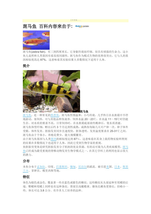
正在查询……斑马鱼百科内容来自于:斑马鱼(zebra fish),有三到四厘米长,它身躯玲珑而纤细,却具有顽强的生命力,这小鱼儿竟然和人类基因有着高度同源性,斑马鱼作为模式生物的优势很突出,它与人的基因相似度高达87%,这意味着其实验结果大多数情况下适用于人体。
简介斑马鱼斑马鱼,是一种常见的热带鱼。
斑马鱼性情温和,小巧玲珑,几乎终日在水族箱中不停地游动,易饲养,可与其他品种鱼混养。
饲养水温20~23℃,在水温11~15℃时仍能生存,对水质的要求不高。
日常饲养时,在水族箱底部放些鹅卵石,使水质清澈。
斑马鱼体型纤细,孵出后约3个月达到性成熟,成熟鱼每隔几天可产卵一次。
卵子体外受精,体外发育,胚胎发育同步且速度快,胚体透明。
发育温度要求在25-31℃之间。
斑马鱼由于个体小,养殖花费少,能大规模繁育。
由于斑马鱼基因与人类基因的相似度达到87%,这意味着在其身上做药物实验所得到的结果在多数情况下也适用于人体,因此它受到生物学家的重视。
鱼胚胎突变体是研究胚胎发育分子机制的优良资源,有的还可做为人类疾病模型,斑马鱼已经成为最受重视的脊椎动物发育生物学模式之一,在其它学科上的利用也显示很大的潜力。
分布本鱼分布于孟加拉、印度、巴基斯坦、缅甸、尼泊尔的溪流。
被引进美国、日本、斯里兰卡、菲律宾、模里西斯等地。
特征体色为银色或金色,覆盖著一些名蓝色或紫色的横纹,这些横纹从头部延伸至尾鳍的后端,臀鳍和尾鳍上同样也有这种条纹,背部呈浅橄榄黄。
雄鱼比雌鱼更修长,但略小一些。
体长可达3.8公分。
有许多人工培养的品种。
品种斑马鱼斑马鱼黄金斑马长鳍黄金斑马大帆斑马豹纹斑马长鳍豹纹斑马电光斑马樱花斑马大斑马金线大斑马珍珠斑马蓝珍珠斑马生态斑马鱼栖息在溪流、沟渠或静止的水中,每2-3天可产卵一次,每次可产约200颗以上的卵,属杂食性,以昆虫、小型甲壳类等为食。
性情温和,喜群游,通常数尾成一群。
发育斑马鱼的发育分为6个阶段:卵裂期,囊胚期,原肠胚期、分裂期、成形期和孵化期。
斑马鱼的软骨发育及其矿化过程
清华大学毕业设计论文摘要斑马鱼,作为发育生物学研究的最佳脊椎动物模型,为人类的相关研究提供了重要的参考依据。
本文主要讨论了斑马鱼幼体的软骨,特别是脊椎软骨的发育过程及其软骨的超微结构特点。
为转基因、基因突变鱼类的研究提供了对比依据。
本文主要讨论了斑马鱼脊椎软骨的发育进程,以及软骨细胞特别是脊索旁软骨的排列,形貌,大小及其显微结构特点和中胚层分化的显微形貌。
斑马鱼脊椎软骨在配卵后2~3天开始发育,围绕在脊索周围,并逐渐向神经管和血管方向延伸。
斑马鱼脊椎软骨沿体节发育,腹部以前的体节在6天已基本分化为软骨细胞,并且随着时间的推移,沿体节向尾部方向的生骨节逐步分化为软骨细胞,在1个半月左右的时间,软骨组织的分化基本完成。
斑马鱼的软骨组织排列整齐,大多数细胞大小在5-20μm范围内。
斑马鱼的软骨组织为透明软骨,胶原纤维为II型胶原,没有典型的周期结构,含量很少。
在TEM的观察中发现,斑马鱼的肌肉组织分化较早,在3天已基本分化完成。
而体干部的脊索旁仍为中胚层的间叶细胞,为软骨细胞的原基,在分化成软骨细胞的过程中,首先是在形状和排列上趋于整齐和规则。
斑马鱼的骨化方式为软骨骨化。
软骨细胞的细胞质中富含大量的线粒体和内质网。
在细胞质中还有大量直径为100-400nm的囊泡,紧密排列在一起,起传输物质的作用。
此外,脊髓处的细胞,其细胞核较大,细胞质很少,有别于软骨细胞。
并且该处细胞有部分已死亡,被其它细胞所代替。
关键词:软骨细胞,发育,显微结构- 1 -清华大学毕业设计论文AbstractZebrafish provides an important referenced foundation for the relative research as the best pattern of vertebrate in developing biology. In this paper the process of development and the ultrastructure of the cartilage especially the parachordal cartilage in zebrafish larva are investigated . And that supply the contradistinctive basis for the research about transgenic and gene mutation fishes.Here we investigate the developing process of parachordal cartilage and chondrocyte arrangements,shapes ,size,and ultrastrcture .In zebrafish parachordal cartilage begin to form at 2-3 days postfertilization ,and surround the notochord and extend gradually to spinal cord and dorsal aorta. Parachordal cartilage develop along somite and at 6 days postfertilization the somite before venter have differentiated and all accomplish differentation on the whole about at one and a half month postfertilization .The chontrocytes arrange regular .The size of chontrocytes is between 5-20μm mostly. In zebrafish the cartilage is hyaline cartilage and the collagen fibrils is predominantly II type fibrils. And that are relatively thin and do not display the characteristic 67nm banding.We discovered that the differentiation of musculature is more earlier than cartilage by TEM and that accomplished basically at 3 days postfertilization. During the differentiation of cartilage the shape and arrangement of mesenchymal cell in mesoblast go to orderliness and regular gradually at first. With the electron microscope ,the active cartilage cell is seen to contain numerous slick endoplasmic reticulum,mitochondria and vesicles which arrange together in order and the diameter of these vesicles is about 100-400nm, transmitting the substance. Furthermore, the nucleolus of spinal cord cells is more larger and we also can observe that some cells have dead.Keywords : cartilage ,development, ultrastrcture- 2 -清华大学毕业设计论文第一章. 前言由于软骨细胞所致的成骨活动发生较早,所以对斑马鱼软骨发育的研究在骨损伤修复过程中对骨力学功能恢复有较大意义。
- 1、下载文档前请自行甄别文档内容的完整性,平台不提供额外的编辑、内容补充、找答案等附加服务。
- 2、"仅部分预览"的文档,不可在线预览部分如存在完整性等问题,可反馈申请退款(可完整预览的文档不适用该条件!)。
- 3、如文档侵犯您的权益,请联系客服反馈,我们会尽快为您处理(人工客服工作时间:9:00-18:30)。
Molecular Evolution of Keap1TWO Keap1MOLECULES WITH DISTINCTIVE INTERVENING REGION STRUCTURES ARE CONSERVED AMONG FISH*□SReceived for publication,October22,2007,and in revised form,December4,2007Published,JBC Papers in Press,December5,2007,DOI10.1074/jbc.M708702200 Li Li‡§,Makoto Kobayashi‡§1,Hiroshi Kaneko§,Yaeko Nakajima-Takagi§,Yuko Nakayama‡,and Masayuki Yamamoto‡§¶From the‡Environmental Response Project,Japan Science and Technology Agency,and the§Institute of Basic Medical Sciences, Graduate School of Comprehensive Human Sciences,University of Tsukuba,1-1-1Tennodai,Tsukuba305-8575,Japan,and the¶Department of Medical Biochemistry,Tohoku University Graduate School of Medicine,2-1Seiryo-cho,Aoba-ku,Sendai980-8575,JapanKeap1is a BTB-Kelch-type substrate adaptor protein of the Cul3-dependent ubiquitin ligase complex.Keap1facilitates the degradation of Nrf2,a transcription factor regulating the induc-ible expression of many cytoprotective genes.Through compar-ative genome analyses,we found that amino acid residues com-posing the pocket of Keap1that interacts with Nrf2are highly conserved among Keap1orthologs and related proteins in all vertebrates and in certain invertebrates,including flies and mosquitoes.The interaction between Nrf2and Keap1appears to be widely preserved in vertebrates.Similarly,cysteine resi-dues corresponding to Cys-273and Cys-288in the intervening region of mouse Keap1,which are essential for the repression of Nrf2activity in cultured cells,are conserved among Keap1 orthologs in vertebrates and invertebrates,except fish.We found that fish have two types of Keap1,Keap1a and Keap1b.To our surprise,Keap1a and Keap1b contain the cysteine residue corresponding to Cys-288and Cys-273,respectively.In our analysis of zebrafish Keap1a and Keap1b activities,both Keap1a and Keap1b were able to facilitate the degradation of Nrf2pro-tein and repress Nrf2-mediated target gene activation.Individ-ual mutation of either residual cysteine residue in Keap1a and Keap1b disrupted the ability of Keap1to repress Nrf2,indicat-ing that the presence of either Cys-273or Cys-288is sufficient for fish Keap1molecules to fully function.These results provide an important insight into the means by which Keap1cysteines act as sensors of electrophiles and oxidants.The transcription factor Nrf2induces the expression of phase2detoxifying and antioxidant proteins in response to electrophilic insults(1).These induced proteins contribute to the prevention of oxidative damage and chemically induced cancer in animals.The importance of Nrf2in this induction and the resulting chemoprevention has been demonstrated by a number of experiments using Nrf2-deficient mice(2).The elec-trophile response is regulated through a cis-acting element called the antioxidant-or electrophile-responsive element within the regulatory region of each gene(3).Nrf2binds to the antioxidant/electrophile-responsive element sequence as a het-erodimeric complex with small Maf proteins through a basic region leucine zipper domain(4).Under normal homeostatic conditions,Nrf2protein is targeted for proteasomal degrada-tion and has a short half-life.This degradation is positively con-trolled by Keap1,a member of the BTB(Broad complex/ Tramtrack/Bric-a-brac)-Kelch protein family(5,6).Keap1 binds to Nrf2and promotes its degradation as a substrate-spe-cific adaptor protein for the Cul3ubiquitin ligase complex(7). When oxidative/electrophilic stress signals disrupt the Nrf2-Keap1-Cul3complex,ubiquitination of Nrf2is blocked,and Nrf2becomes stable(8).Consequently,the expression of a bat-tery of cytoprotective genes is induced as Nrf2accumulates in the nucleus.Keap1is composed of three major domains:a BTB domain,a double glycine repeat(DGR)2domain,and an intervening region(IVR)domain(1).The BTB domain functions to dimer-ize Keap1(9),whereas the DGR domain serves as a binding site for Nrf2(5)and actin(10).Our group(11)and Hannink and co-workers(12)have determined the crystal structure of the Keap1DGR domain and identified its interface with Nrf2. Involvement of the Keap1IVR domain in the ubiquitination of Nrf2has been demonstrated(8,13).In cultured cells,mutation of Cys-273or Cys-288in the IVR domain to alanine or serine reduces Keap1-dependent ubiquitination and increases Nrf2 stability,suggesting that these residues are crucial for the Nrf2-repressing activity of Keap1(13–15).We previously isolated homolog genes of Nrf2and Keap1in zebrafish and established that the Nrf2-dependent induction of cytoprotective genes is conserved among vertebrates(16,17). We thus speculated that the Nrf2-Keap1system of cytoprotec-*This work was supported by grants-in-aid from the Japan Science and Tech-nology Corp.(Exploratory Research for Advanced Technology)(to M.Y.) and from the Ministry of Education,Science,Sports,and Culture of Japan (to M.K.and M.Y.).The costs of publication of this article were defrayed in part by the payment of page charges.This article must therefore be hereby marked“advertisement”in accordance with18U.S.C.Section1734solely to indicate this fact.□S The on-line version of this article(available at )contains supplemental Figs.1–3and Table1.The nucleotide sequence(s)reported in this paper has been submitted to the Gen-Bank TM/EBI Data Bank with accession number(s)AB271119.1To whom correspondence should be addressed:Inst.of Basic Medical Sci-ences,Graduate School of Comprehensive Human Sciences,University of Tsukuba,1-1-1Tennodai,Tsukuba305-8575,Japan.Tel.:81-29-853-8457; Fax:81-29-853-5977;E-mail:makobayash@md.tsukuba.ac.jp.2The abbreviations used are:DGR,double glycine repeat;IVR,intervening region;GFP,green fluorescent protein;HA,hemagglutinin;RT,reverse transcription;IC,IVR cysteine.THE JOURNAL OF BIOLOGICAL CHEMISTRY VOL.283,NO.6,pp.3248–3255,February8,2008©2008by The American Society for Biochemistry and Molecular Biology,Inc.Printed in the U.S.A.3248JOURNAL OF BIOLOGICALCHEMISTRY VOLUME283•NUMBER6•FEBRUARY8,2008 at HARBIN INSTITUTE OF TECHNOLOGY on August 8, 2015/ Downloaded fromtion is also conserved in vertebrates.To our surprise,zebrafish Keap1protein does not contain a cysteine residue correspond-ing to Cys-273in mouse Keap1,yet it still represses the activity of Nrf2in zebrafish embryos(16).In this work,we compared the amino acid sequences of the Keap1-related proteins of var-ious vertebrates and invertebrates by comparative genome analysis.Critical amino acids in the Nrf2-interacting surface of the DGR domain are highly conserved among these proteins, but are completely different in other mouse BTB-Kelch pro-teins.This indicates that Keap1is the only BTB-Kelch protein that regulates Nrf2activity and also implies the presence of the Nrf2-Keap1system in invertebrates.Interestingly,fish have two Keap1genes,which we refer to as Keap1a and Keap1b. Keap1a has a cysteine residue corresponding to Cys-273,but not to Cys-288,in mouse Keap1,whereas the case is the reverse for Keap1b.We analyzed the activities of zebrafish Keap1a and Keap1b using zebrafish embryos and demonstrated that either protein can promote Nrf2degradation;both Cys-273and Cys-288are important for Keap1activity,but either one is enough in fish.EXPERIMENTAL PROCEDURESIsolation of cDNA—A partial cDNA fragment encoding zebrafish Keap1b was prepared by PCR using specific primers designed based on genomic DNA information.AZAP-II 15–19-h-stage cDNA library(18)was screened to isolate a full-length Keap1b cDNA clone using the partial cDNA clone as a probe.The probe was labeled using an AlkPhos Direct DNA labeling kit,and positive plaques on the membrane filters were detected with CDP-Star as substrate according to the manufac-turer’s instructions(GE Healthcare).Radiation Hybrid Mapping—Radiation hybrid mapping using panel LN54was performed as described by Hukriede et al.(19)using specific primers for each Keap1gene.The sequences of each primer were as follows:keap1a,5Ј-AGGAT-TTCTCCGCCATTGTG and5Ј-CCTTGAAGTTGCTGGTG-AAC;and keap1b,5Ј-ATGACGGAGTGTAAGGCGG and5Ј-CAGGCCGTTGGTGAACATG.Plasmid Construction—The plasmid pCS2keap1b was con-structed by subcloning the open reading frame of zebrafish keap1b into the BamHI and XbaI sites of the vector pCS2.To construct pSPkeap1aC,cDNA encoding the C-terminal region (amino acids353–601)containing the3Ј-untranslated region of zebrafish keap1a was inserted into the NotI and SalI sites of the vector pSPORT1.The plasmid pKSkeap1bN was generated by inserting cDNA encoding the N-terminal region(amino acids8–188)of keap1b into the BamHI and XhoI sites of pBluescript II KS.To construct pCS2nrf2NTnGFP,cDNA encoding the N-terminal region(amino acids1–305)of zebrafish nrf2plus two repeats of SV40nuclear localizing signal (DPKKKRKV)were subcloned into the BamHI site of pCS2eGFP.The cDNA fragments for3ϫFLAG tag(MDYKD-HDGDYKDHDIDYKDDDDK)and3ϫhemagglutinin(HA)tag (MEYPYDVPDYAAEYPYDVPDYAAEYPYDVPDYAAKLE) were subcloned into the BamHI and EcoRI sites of pCS2to generate pCS2FL and pCS2HA,respectively.The plasmids pCS2FLkeap1a,pCS2FLkeap1b,and pCS2FLnrf2were con-structed by inserting the open reading frames of keap1a,keap1b,and nrf2,respectively,into the HindIII and XbaI sites of pCS2FL.pCS2HAkeap1a and pCS2HAkeap1b were prepared by inserting the open reading frames of keap1a and keap1b, respectively,into the HindIII and XbaI sites of pCS2HA.The constructs pCS2FLkeap1aC264S and pCS2FLkeap1bC247S were made by introducing Cys-to-Ser point mutations by PCR into pCS2FLkeap1a and pCS2FLkeap1b,respectively. pKSgstp1N was constructed by subcloning the cDNA for the N-terminal region(amino acids1–135)of gstp1into the BamHI and SalI sites of pBluescript II KS.All constructs were verified by DNA sequencing.Plasmids pCS2nrf2,pCS2keap1a(previ-ously named pCS2Keap1),and pCS2eGFP were described pre-viously(17,20).Expression Analysis—Zebrafish embryos and larvae were obtained by natural mating.All experiments were carried out using a wild-type AB strain.The expression of keap1a,keap1b, and gstp1genes was analyzed by reverse transcription(RT)-PCR and whole mount in situ hybridization.For RT-PCR anal-ysis,total RNA was prepared from adult tissues or the whole bodies of embryos and larvae using QIAzol(Qiagen).First-strand cDNA was synthesized by incubation at25°C for15min and at42°C for45min with murine leukemia virus reverse transcriptase(SuperScript II,Invitrogen)and random hexamer oligonucleotide primers.From the20-l first-strand reaction, 0.025–0.1was used for PCR using the following primers: keap1a,5Ј-ATGATATGTCCAAGAAAGAAG and5Ј-TCAT-GAGGAAATCGCAGCAG;keap1b,5Ј-ACGGAGTGTAAG-GCGGAG and5Ј-ACCTGGCTGAAGTTCATG;gstp1,5Ј-CTAGGAGCAGCTTTGAAACGCAC and5Ј-TGGCCAGA-ACATTTTCAAAGC;and ef1␣,5Ј-GCCCCTGCCAATGTA and5Ј-GGGCTTGCCAGGGAC.The expression of ef1␣was used to standardize the amount of cDNA.Real-time RT-PCR was performed to quantitate gstp1expression using an ABI Prism7700(Applied Biosystems)and probes labeled with a reporter fluorescent dye(TaqMan probe)as described pre-viously(21).TaqMan probes,primers,and cDNAs were added to the master mixture containing the reagents for PCR (Eurogentec).The sequences of the specific primers and probes were as follows:gstp1,5Ј-CAACGCCATGCTGAGACATC (sense),5Ј-GAAGATCTTCAACGCCGTCG(antisense),and 5Ј-6-carboxyfluorescein-AACATGCTGCATATGGCAAAA-ACGACAGT-6-carboxytetramethylrhodamine(probe);and ef1␣,5Ј-CGTGGTAATGTGGCTGGAGA(sense),5Ј-CTGAG-CGTTGAAGTTGGCAG(antisense),and5Ј-6-carboxyfluoresc-ein-AGCAAGAACGACCCACCCATGGAG-6-carboxytetra-methylrhodamine(probe).Whole mount in situ hybridization was performed as described previously(22)using RNA probes tran-scribed from pSPkeap1a,pKSkeap1b,and pKSgstp1N. Microinjection of Zebrafish Embryos—Synthetic capped RNA was made with an SP6mMESSAGE mMACHINE in vitro tran-scription kit(Ambion)using linearized DNA of the pCS2deriva-tives described above.For expression in whole bodies,RNA was injected into yolk at the one-cell stage using an IM300microinjec-tor(Narishige).GFP expression was examined under the GFP Plus filter(480nm excitation,505nm emission)of a MZFLIII micro-scope(Leica)equipped with a600CL-CU digital camera(Pixera). In Vitro Translation and Co-immunoprecipitation—HA-and FLAG-tagged Keap1proteins were in vitro translated sep-Molecular Evolution of Keap1FEBRUARY8,2008•VOLUME283•NUMBER6JOURNAL OF BIOLOGICAL CHEMISTRY3249at HARBIN INSTITUTE OF TECHNOLOGY on August 8, 2015/Downloaded fromarately by T N T coupled wheat germ extract systems(Promega) using pCS2derivatives as DNA templates.In vitro translated Keap1proteins were mixed in binding buffer(50m M Tris-HCl, pH7.5,50m M NaCl,and0.1%Nonidet P-40)and incubated with an affinity matrix-immobilized anti-HA antibody(3F10, Roche Diagnostics)at4°C for4h with gentle mixing on a rota-tor.The beads were collected by centrifugation at12,500ϫg for 5s and washed three times in binding buffer.Precipitated pro-teins were eluted in SDS-sample buffer and resolved by12% SDS-PAGE,followed by immunoblotting using anti-HA (12CA5,Roche Diagnostics)and anti-FLAG(M2,peroxidase conjugate,Sigma)antibodies as described previously(22). RESULTSIdentification of the Second Keap1in Zebrafish—By virtue of recent progress in the zebrafish genome project,we came across a novel Keap1-related gene that shows a higher similarity to mammalian Keap1than previously reported zebrafish Keap1 (16).A partial cDNA was isolated by RT-PCR using specific primers whose design was based on genomic DNA information. We screened a zebrafish cDNA-phage library using this par-tial cDNA as a probe and isolated a full-length cDNA clone. We refer to this gene as keap1b,and the previous keap1was renamed keap1a.The deduced amino acid sequence of the Keap1b cDNA product showed81and78%identities to the BTB and DGR domains,respectively,of mouse Keap1protein (Fig.1A).These values are quite high compared with those of Keap1a,whose identities to the BTB and DGR domains are only 49and55%,respectively.We mapped both Keap1genes using an LN54hybrid panel(19)and found that keap1a and keap1b are localized on zebrafish chromosomes2and6,respectively. The latest information from the zebrafish genome project sup-ported these mapped sites and further demonstrated that syn-teny was found between keap1b and the human KEAP1locus on chromosome19p13.2(supplemental Fig.1).Neh2is the domain in Nrf2that interacts with the DGR domain in Keap1(5).Within the Neh2domain,we found that the motifs ETGE and DLG are critical for the interaction with Keap1(16,23).Recently,we identified the region of the Keap1 DGR domain responsible for binding to the ETGE and DLG motifs by structural analysis of the mouse Keap1protein(11, 24).The amino acid residues important for binding to the ETGE motif have been recognized as Ser-363,Arg-380,Asn-382,Arg-415,Arg-483,Ser-508,Tyr-525,Gln-530,Ser-555, and Ser-602.Those important for binding to the DLG motif are Asn-382,Arg-415,Arg-483,Ser-508,Ser-555,Tyr-572,Phe-577,Ser-602,and Gly-603(Fig.1B,white characters highlighted in black).Mutation analyses of mouse and human Keap1pro-teins have demonstrated that Tyr-334,Gly-364,Gly-430,His-436,and Phe-478(Fig.1B,white characters highlighted in gray), in addition to Arg-380,Asn-382,Arg-415,Arg-483,Tyr-525, and Tyr-572,are critical for inhibiting Nrf2activity(11,12). Interestingly,all these residues,except Asn-382and Tyr-572, are conserved in both zebrafish Keap1a and Keap1b,suggesting that both proteins can interact with Nrf2.Indeed,zebrafish Keap1a has been shown to interact with Nrf2and to inhibit its activity(16).Although Mayven is the protein with the highest homology to Keap1in the DGR domain among mouse BTB-Kelch proteins(25),it possesses only2of the13critical Nrf2-interacting residues in mouse Keap1(Fig.1B,m M).This case is similar to that of KLHL20and KLHL5,two other Keap1-related proteins(supplemental Table1).These results suggest that the activity of Nrf2is regulated by two Keap1proteins,Keap1a and Keap1b,in zebrafish and by a single Keap1protein in mouse, which may be the only BTB-Kelch protein that can facilitate Nrf2degradation.Here,we propose to define Keap1as a BTB-Kelch protein carrying the evolutionarily conserved Nrf2-inter-acting surface.Unlike Keap1,we could not find a second Nrf2gene in the zebrafish genome data base.Nrf2is a member of theCNC FIGURE1.Identification of two Keap1proteins in zebrafish.A,percentage amino acid sequence identities in the BTB,IVR,and DGR domains between zebrafish(z)and mouse(m)Keap1proteins.Nucleotide sequence data of zebrafish keap1b have been deposited in the DDBJ/GenBank TM/EBI Data Bank with accession number AB271119.B,amino acid sequence alignment of the DGR domains of Keap1and Mayven proteins.Amino acid residues located in the interaction surface for Nrf2are highlighted in black.Gly-Gly and Trp sequences conserved among all BTB-Kelch family proteins are highlighted in gray.White characters highlighted in gray indicate amino acid residues whose mutations have been shown to reduce Nrf2-repressing activity.The asterisks donate stop codons.zKa,zebrafish Keap1a;zKb,zebrafish Keap1b;mK,mouse Keap1;mM,mouse Mayven.C,phylogenetic tree of Keap1family proteins. Amino acid sequences in the DGR domains were analyzed.The tree was con-structed by the neighbor-joining method using the ClustalW program.aa,A. aegypti;ag,A.gambiae;c,chicken;ci,C.intestinalis;cs,C.savignyi;d,Drosophila melanogaster;f,fugu;h,human;m,mouse;me,medaka fish;te,T.nigroviridis;st, stickleback;xl,evis;xt,Xenopus tropicalis;z,zebrafish.Molecular Evolution of Keap13250JOURNAL OF BIOLOGICALCHEMISTRY VOLUME283•NUMBER6•FEBRUARY8,2008 at HARBIN INSTITUTE OF TECHNOLOGY on August 8, 2015/ Downloaded from(Cap‘n’collar)protein family,whose members are NF-E2p45, Nrf1,Nrf2,Nrf3,Bach1,and Bach2(1).Among them,genetic loci of mammalian NF-E2p45,Nrf1,Nrf2,and Nrf3genes have been mapped close to those of HoxC,HoxB,HoxD,and HoxA, respectively(34).Interestingly,the zebrafish genome has two copies of HoxA,HoxB,and HoxC clusters,but only one HoxD cluster(35).We assume that the second Nrf2gene in zebrafish had been lost together with the second HoxD cluster during evolution. Keap1Is Present in Vertebrates and in Some Invertebrates—To identify the range of species in which Keap1is present,we searched the Ensemble and DDBJ/GenBank TM/EBI Data Bank for Keap1-related proteins.As well as in mammals,Keap1 genes were found in chicken,frogs(Xenopus laevis and Xenopus tropicalis),fugu,Tetraodon nigroviridis,medaka fish,stickle-back,ascidians(Ciona intestinalis and Ciona savignyi),mosqui-toes(Aedes aegypti and Anopheles gambiae),and Drosophila.A phylogenetic tree based on the amino acid sequences of their DGR domains classified the Keap1proteins into five subgroups: 1)vertebrate Keap1,2)fish Keap1a,3)fish Keap1b,4)ascidian Keap1,and5)invertebrate Keap1(Fig.1C).No Keap1-related genes were found in nematode or yeast.We noted that all these Keap1proteins carry13critical Nrf2-interacting residues,with the exceptions of Asn-382and Tyr-572for fish Keap1a and Tyr-525for invertebrate Keap1(supplemental Table1).The results suggest that Keap1regulates Nrf2or related proteins in these organisms in a manner similar to that in mammals. Keap1a and Keap1b are conserved among fish,but not in other vertebrates,signifying that both proteins are essential to the fish Nrf2-Keap1system.Keap1b rather than Keap1a may represent the ortholog of vertebrate Keap1because conserved synteny was observed between human KEAP1and fish keap1b loci(supplemental Fig.1).No synteny was found between human Keap1and fish Keap1a genes or with ascidian or inver-tebrate Keap1.This implies that Keap1b may be the proper homolog of vertebrate Keap1.Keap1a and Keap1b Repress Nrf2Activity Despite Their Lack of a Cysteine Residue Corresponding to Mouse Keap1Cys-273 and Cys-288,Respectively—All fish Keap1a and Keap1b lack a cysteine residue corresponding to Cys-273and Cys-288, respectively,whereas both these cysteines are conserved even in ascidian and invertebrate Keap1proteins(Fig.2).This find-ing was surprising because both Cys-273and Cys-288in the IVR were demonstrated to be crucial for the Nrf2-repressing activity of mouse Keap1(13–15).To elucidate whether zebrafish Keap1a and Keap1b can repress the inducible func-tion of Nrf2,we tested the extent of their repression on the Nrf2-mediated inducible expression of the endogenous gstp1 gene in zebrafish embryos.The gstp1gene encodes a Pi class glutathione S-transferase and is strongly induced in both elec-trophile-treated larvae and Nrf2-overexpressing embryos(16, 26).Its promoter contains an evolutionarily conserved antiox-idant/electrophile-responsive element sequence that is critical for both Nrf2binding and promoter activity(26).In vitro syn-thesized zebrafish Keap1a or Keap1b mRNA(200pg)was co-injected with Nrf2mRNA(100pg)into zebrafish embryos at the one-cell stage(Fig.3A).At midgastrula,gstp1expression was analyzed by whole mount in situ hybridization analysis. Nrf2-induced expression of gstp1was reduced byco-overex-FIGURE2.Alignments of nine cysteine residues and their adjacent amino acid residues in the IVRs of various Keap1proteins.Note that fish Keap1 proteins lack either IC6(Keap1a)or IC7(Keap1b),whereas Keap1proteins in non-fish species have both IC6and IC7without exception.Cysteine residues are highlighted in black.Basic amino acids adjacent to the cysteines are high-lighted in gray.aa,A.aegypti;ag,A.gambiae;c,chicken;ci,C.intestinalis;cs,C. savignyi;d,D.melanogaster;f,fugu;h,human;m,mouse;me,medaka fish;te, T.nigroviridis;st,stickleback;xl,evis;z,zebrafish.FIGURE3.Effects of Keap1a and Keap1b on Nrf2-mediated gstp1induction. A,experimental scheme.Nrf2and/or Keap1mRNA was co-injected into embryos at the one-cell stage.After8h,gstp1expression in embryos was analyzed by whole mount in situ hybridization(B)or by real-time RT-PCR(C).C,in real-time RT-PCR analysis,FLAG-tagged(FL)Keap1proteins were used instead of non-tagged proteins.The expression levels of gstp1were normalized to those of ef1␣measured in the same cDNA preparations.Values for the Nrf2-overexpressing embryoswerearbitrarilysetat100.TheamountsofinjectedmRNAwere0.12,0.6, 3,15,and75pg for Keap1a and0.32,1.6,8,40,and200pg for Keap1b.Error bars indicate S.D.from three independent experiments.Molecular Evolution of Keap1FEBRUARY8,2008•VOLUME283•NUMBER6JOURNAL OF BIOLOGICAL CHEMISTRY3251at HARBIN INSTITUTE OF TECHNOLOGY on August 8, 2015/Downloaded frompression of either Keap1a or Keap1b (Fig.3B ),indicating that both Keap1a and Keap1b possess the ability to repress Nrf2activity.To confirm this,we used FLAG-tagged Keap1proteins to standardize the protein expression level of each Keap1by immunoblotting (supplemental Fig.2).Seventy-five pg of Keap1a mRNA and 200pg of Keap1b mRNA expressed similar amounts of Keap1proteins in zebrafish embryos.Only full-length proteins were overexpressed in embryos.The FLAG-tagged constructs were used to compare the Nrf2repression activity of Keap1a and Keap1b by real-time RT-PCR analyses (Fig.3C ).Sixty pg of Nrf2mRNA were co-injected with various amounts of Keap1a or Keap1b mRNA (Fig.3C ).The dose effects of Keap1mRNA on Nrf2repression were similar between Keap1a and Keap1b,suggesting that the activities of Keap1a and Keap1b to repress Nrf2activity are comparable,at least in zebrafish embryos.Both Keap1Proteins Promote Nrf2Degradation —Mouse Keap1has been shown to promote the degradation of Nrf2as a substrate-specific adaptor protein for the Cul3ubiquitin ligase complex (7).To elucidate whether zebrafish Keap1proteins also promote Nrf2degradation,we examined the effects of Keap1co-overexpression on the level of Nrf2protein.FLAG-tagged Nrf2protein overexpressed in zebrafish embryos by mRNA injection was detectable by whole mount immuno-staining using anti-FLAG antibody (Fig.4A ).This antibody staining disappeared when we co-overexpressed either Keap1a or Keap1b,advocating the promotion of Nrf2degradation as the means by which these Keap1proteins repress Nrf2.To con-firm this,we overexpressed an Nrf2-GFP fusion protein (Nrf2NTnGFP)in zebrafish embryos and tested its stability in the presence or absence of Keap1a or Keap1b by observing GFP expression (Fig.4B ).Note that the N-terminal domain ofzebrafish Nrf2was used to construct the GFP fusion protein because this region corresponds to the mouse Nrf2protein that was shown to be sufficient for Keap1-dependent degradation in both cultured cells and mouse intestine (27).GFP expression was observed in Nrf2NTnGFP-overexpressing embryos,whereas GFP expression was dramatically lower when either Keap1a or Keap1b was co-overexpressed (Nrf2NTnGFP,53.7%,n ϭ93;Nrf2NTnGFP ϩKeap1a,0%,n ϭ132;Nrf2NTnGFP ϩKeap1b,0%,n ϭ73;no injection,0%,n ϭ100)(Fig.4B ).These results demonstrate that both Keap1a and Keap1b repress Nrf2activity by facilitating its degradation,as is the case for mouse Keap1.Keap1a and Keap1b Can Form Homodimers and Hetero-dimers —We previously found that coexpression of C273A and C288A mutant proteins of mouse Keap1substantially restores repressor activity,whereas each Keap1mutant alone lacks repressor activity (15).This observation implies that C273A and C288A form a heterodimer,and the simul-taneous presence of Cys-273on one monomer and Cys-288on the other is sufficient for the repressor activity.Similarly,it is possible that overexpressed Keap1a or Keap1b in zebrafish embryos forms a heterodimer with endogenous Keap1proteins to share cysteine residues in the same com-plex.To assess this possibility,we carried out pulldown anal-ysis using in vitro translated Keap1a and Keap1b proteins with FLAG and HA tags (Fig.5).Tagged Keap1proteins were mixed and pulled down with anti-HA beads.Precipitated proteins were analyzed by immunoblotting using anti-FLAG and anti-HA antibodies.FLAG-tagged Keap1a protein co-precipitated with both HA-tagged Keap1a and Keap1b.Sim-ilarly,FLAG-tagged Keap1b protein was pulled down with HA-tagged Keap1a and Keap1b.These results demonstrate that Keap1a and Keap1b can form both homodimers andheterodimers.FIGURE 4.Effects of Keap1a and Keap1b on Nrf2protein stability.A ,immunostaining analysis of overexpressed Nrf2protein.FLAG-tagged Nrf2(FL-Nrf2)and/or Keap1mRNA was injected into embryos at the one-cell stage.After 8h,the stability of FLAG-tagged Nrf2protein was ana-lyzed by immunostaining using anti-FLAG antibody.B ,expression analysis of Nrf2-GFP fusion protein.mRNA encoding a fusion protein comprising the N-terminal half of Nrf2protein and enhanced GFP (eGFP )protein con-nected by two copies of SV40nuclear localizing signal (NLS )was injected with or without mRNA encoding Keap1proteins into one-cell stage embryos,and GFP expression was analyzed after 6h.FIGURE 5.Dimerization of zebrafish Keap1proteins.FLAG (FL )-and HA-tagged proteins of Keap1a and Keap1b were examined by immunoblotting (IB )using anti-FLAG (upper panel )or anti-HA (lower panel )antibodies.Mix-tures of FLAG-and HA-tagged proteins were co-immunoprecipitated with anti-HA-conjugated agarose beads.Precipitated proteins were analyzed by immunoblotting.Molecular Evolution of Keap13252JOURNAL OF BIOLOGICALCHEMISTRYVOLUME 283•NUMBER 6•FEBRUARY 8,2008at HARBIN INSTITUTE OF TECHNOLOGY on August 8, 2015/Downloaded fromKeap1a and Keap1b Genes are Coexpressed in Many Tissues —Keap1a and Keap1b require simul-taneous expression to function as heterodimers.To provide insight into the roles of Keap1a and Keap1b in vivo ,we examined the tissue dis-tribution of Keap1mRNA in adult fish (Fig.6A ).Total RNA fractions were prepared from various tissues of 10-month-old zebrafish males and analyzed by RT-PCR.The amount of cDNA was standardized by the expression level of ef1␣.Although both keap1a and keap1b were expressed ubiquitously,the expression of keap1b was relatively abundant in brain and scarce in gut.We also examined the expression levels of the zebrafish Keap1genesduring the embryonic and larval stages (Fig.6B ).RT-PCR analyses demonstrated that keap1b was expressed at every stage tested and at similar levels,whereas keap1aexpression was quite low during the embryonic stages and started to increase around the time of hatching (2.5days).Spa-tial expression profiles of zebrafish Keap1genes were assessed at the embryonic stages by whole mount in situ hybridization (Fig.6C ).Both genes were expressed ubiquitously in the whole body,although some specific regions,such as lens (arrow ),expressed keap1a more strongly than others.Overall,these observations suggest that keap1a and keap1b are coexpressed in many cells.Cysteine Residues Corresponding to Cys-273and Cys-288in Mouse Keap1Are Important for the Nrf2-repressing Activity of Keap1a and Keap1b —The critical cysteine residues in Keap1a and Keap1b must be important for repressing Nrf2if these two proteins function as heterodimers.To verify this,point muta-tions were introduced in these cysteines,and the ability to repress Nrf2was analyzed.In this work,we refer to the cysteine residues in the IVR domain as IVR cysteines (ICs)to ease com-parison among the corresponding cysteines of various Keap1proteins (see Fig.2).Cysteine residues corresponding to Cys-273and Cys-288in mouse Keap1are called IC6and IC7.We introduced Cys-to-Ser point mutations in IC7of Keap1a and in IC6of Keap1b and examined the effects of these mutations on Nrf2-repressing activity (Fig.7A ).We used FLAG-tagged Keap1proteins to standardize the protein expression level of each Keap1by immunoblotting.Mutations in Keap1a IC7and Keap1b IC6strongly abolished the Nrf2-repressing activity(Fig.7B ).IC7in Keap1a and IC6in Keap1b are thus essential for the repression of Nrf2activity.DISCUSSIONThis is the first work referring to the evolutionary aspects of Keap1,as well as to its definition.Stogios and Prive ´(28)pre-dicted that more than 53members of the BTB-KelchproteinFIGURE 6.Expression profiles of keap1a and keap1b .A ,RT-PCR analysis using specific primers for keap1a ,keap1b ,and ef1␣and total RNA from tissues of 10-month-old male fish.Br ,brain;E ,eye;Gi ,gill;L ,liver;Gu ,gut;S ,spleen;H ,heart;K ,kidney;T ,testis;Bl ,bladder.Cyc denotes the number of PCR cycles.B ,RT-PCR analysis using total RNA from the whole bodies of embryos or larvae at the developmental stages indicated.d ,day(s).C ,in situ hybridization analysis.Shown are lateral views of keap1a (upper panels )and keap1b (lower panels )expression atthe developmental stages indicated.Dominant expression of keap1a was observed in lens (arrow).FIGURE 7.Effects of point mutations of the IVR cysteines.A ,Keap1mutants used in the analysis.z ,zebrafish;m ,mouse;WT ,wild type.B ,activities of Nrf2repression of IVR cysteine-mutated Keap1proteins.Nrf2and/or FLAG-tagged Keap1(FL-Keap1)mRNA was co-injected into embryos at the one-cell stage.The amount of Keap1mRNA was standardized by the expression level of FLAG-tagged Keap1protein,which was analyzed by immunoblotting using anti-FLAG antibody (FL-Keap1protein ).After 8h,gstp1expression in embryos was analyzed by RT-PCR analysis.The expression of ef1␣was used to stan-dardize the amount of cDNA.Molecular Evolution of Keap1FEBRUARY 8,2008•VOLUME 283•NUMBER6JOURNAL OF BIOLOGICAL CHEMISTRY3253at HARBIN INSTITUTE OF TECHNOLOGY on August 8, 2015/Downloaded from。
