SABC(兔IgG)-POD Kit使用说明
Protein A G免疫沉淀磁珠使用说明书

Protein A/G免疫沉淀磁珠Figure 1. General Protocol for ImmunoprecipitationcomplexSDS-PAGE loading buffer Neutralize bufferMagnetic Beads antibodyMagnetic Separator Remove supernatant Pipette Repeat45产品组分产品参数:磁珠粒径100 nm,浓度10 mg/mL,结合量>400 μg human IgG/mL2-8℃保存,保质期2年。
储存方法实验步骤1. 抗原样品制备本操作说明书提供以下三种样品处理方法。
2. 磁珠预处理将磁珠漩涡振荡1 min,使其充分混悬;取25~50 µL磁珠悬液置于1.5 mL EP管中。
加入200 µL结合缓冲液洗涤,进行磁性分离(将离心管置于磁力架上,管底对准①卡口压紧,静置2分钟或待磁珠吸附于管壁),吸弃上清。
抽出②磁条,加入200 µL结合缓冲液重复洗涤一次,插回②磁条,磁性分离并吸弃上清。
加入200 µL结合缓冲液重悬磁珠备用。
血清样品处理:若目标蛋白丰度较高, 建议用结合缓冲液稀释血清样品至目标蛋白终浓度为10~100 µg/mL,置于冰上备用(或置于-20℃长期保存)。
悬浮细胞样品处理:离心收集细胞(4℃, 500 g, 10 min),弃上清后称重,按每毫克细胞50 µL的比例用1×PBS洗涤2次;按每毫克细胞5~10 µL的比例加入结合缓冲液,同时加入蛋白酶抑制剂,混匀后置于冰上处理10 min;离心收集上清液(4℃, 14000 g, 10 min),置于冰上备用(或置于-20℃长期保存)。
贴壁细胞样品处理:移去培养基,按每1.0×10⁵个细胞150 µL的比例用1×PBS洗涤两次;用细胞刮棒刮脱细胞,收集至1.5 mL EP管内,按每1.0×10⁵个细胞20~30 µL的比例加入结合缓冲液,同时加入蛋白酶抑制剂,混匀后置于冰上处理10 min;离心收集上清液(4℃, 14000 g, 10 min),置于冰上备用(或置于-20℃长期保存)。
免疫组化sabc法原理

免疫组化sabc法原理免疫组化SABC法原理免疫组化(Immunohistochemistry,IHC)是一种利用抗体和免疫反应来检测组织样本中特定蛋白质的方法。
免疫组化技术的发展为研究细胞和组织中蛋白质的表达和定位提供了一种有效的手段。
在免疫组化技术中,SABC法(Streptavidin-Biotin Complex)是一种常用的放大方法,用于增强目标抗原的检测信号。
SABC法的原理是通过将抗原特异性的一抗与生物素化的二抗结合,再利用亲和力很强的鸡蛋白素-链霉亲和素(Streptavidin-biotin)结合系统,将生物素与酶或荧光染料等标记物连接起来,从而使目标抗原能够被可视化。
具体而言,SABC法分为四个步骤:抗原修复、阻断、一抗孵育和二抗孵育。
首先是抗原修复,组织样本通常需要进行抗原修复处理,以恢复抗原的免疫活性。
抗原修复的方法包括热处理、酶解和酸性处理等。
接下来是阻断步骤,目的是阻止非特异性结合。
通常使用的阻断剂包括牛血清蛋白(BSA)、小鼠血清、马血清和羊血清等。
然后是一抗孵育,将具有特异性的一抗与待检测的抗原结合。
一抗可以是单克隆抗体或多克隆抗体,具体选择取决于实验的需求。
最后是二抗孵育,将生物素化的二抗与一抗结合。
二抗通常是兔源抗小鼠IgG的抗体,也可以是其他动物源的抗体。
生物素化的二抗通过亲和力结合到一抗上,形成免疫复合物。
在SABC法中,生物素与酶或荧光染料等标记物结合,常用的酶标记有辣根过氧化物酶(HRP),荧光标记有荧光素酶(FITC)和罗丹明(Rhodamine)等。
这些标记物能够使抗原产生可见的颜色或荧光信号。
在目标抗原上添加底物,使酶催化底物产生可见的颜色反应,或者直接观察荧光信号。
通过显微镜观察或图像分析系统,可以定量分析目标抗原的表达和定位。
免疫组化SABC法具有高度特异性和敏感性,能够定量检测目标抗原在组织中的表达水平和定位。
它在研究细胞生物学、病理学和分子生物学等领域发挥了重要作用。
支原体 PCR 检测试剂盒使用说明书

支原体 PCR 检测试剂盒使用说明书Product Name: Mycoplasma PCR Detection KitIntended Use:The Mycoplasma PCR Detection Kit is intended for the qualitative detection of Mycoplasma spp. in cell cultures and other biological samples by PCR amplification.Kit Components:- PCR amplification reaction mix and enzyme mix- Positive control DNA template- Negative control DNA template- PCR tubes and caps- User manualStorage:The kit should be stored at -20°C. Thaw the kit components at room temperature before use.Sample Preparation:1. Collect the cell culture sample or other biological sample.2. Extract the DNA from the sample using a DNA extraction kit of your choice.3. Dilute the DNA extract as necessary in molecular grade water or buffer to achieve the optimal concentration for PCR amplification (typically between 10-100 ng/µL).PCR Amplification:1. Prepare the PCR reaction mix (in a sterile tube or plate) by adding the PCR amplification reaction mix, enzyme mix, andtemplate DNA to the appropriate volumes as specified in the table below.Component Volume per Reaction (20 µL)PCR amplification reaction mix 10 µLEnzyme mix 1 µLTemplate DNA 1-5 µLMolecular grade water/buffer To 20 µL2. Close the PCR tubes with the caps provided and mix the contents well by vortexing or pipetting.3. Place the tubes in a thermal cycler and perform PCR amplification according to the following conditions:Initial denaturation: 95°C for 5 minutesDenaturation: 95°C for 30 secondsAnnealing: 55°C for 30 secondsExtension: 72°C for 1 minuteFinal extension: 72°C for 5 minutesHold at 4°C4. Analyze the PCR products by gel electrophoresis or other methods as appropriate.Interpretation of Results:- Positive Result: A distinct band is observed in the gel at the expected size for Mycoplasma spp. This indicates the presence of Mycoplasma DNA in the sample.- Negative Result: No band or only faint bands are observed in thegel. This indicates the absence of Mycoplasma DNA in the sample. - Invalid Result: No bands or smear are observed in the gel or the band size is incorrect. Repeat the experiment with fresh controls and follow the instructions carefully.Limitations:1. This assay is designed for research use only and is not intended for diagnostic purposes.2. False negative results may occur due to low level or degraded Mycoplasma DNA in the sample, PCR inhibitors, or other factors.3. The presence of PCR inhibitors in the DNA extraction and amplification process may affect the sensitivity and reproducibility of the assay.4. The kit components must not be used beyond the expiry date.5. The Mycoplasma PCR Detection Kit may only be used by trained personnel.。
碧云天 Rat IgG ELISA Kit 说明书
Rat IgG ELISA Kit产品编号 产品名称包装 PI482Rat IgG ELISA Kit96次产品简介:碧云天的Rat IgG ELISA Kit (Rat IgG Enzyme-Linked ImmunoSorbent Assay Kit),即大鼠总免疫球蛋白G 酶联免疫吸附检测试剂盒,是一种用于特异性地高灵敏地定量检测大鼠血清、血浆或细胞培养上清液中的IgG 的ELISA 试剂盒。
本产品检测灵敏度高,特异性强,重复性好。
多次重复检测结果表明,最小检出量为5.5ng/ml ,本试剂盒采用特异性抗体识别大鼠IgG ,包括IgG1、IgG2a 、IgG2b 和IgG3,检测值为总的IgG 浓度,板内、板间变异系数均小于10%。
免疫球蛋白G (Immunoglobulin G, IgG)是血清中最主要的免疫球蛋白形式,约占血清中免疫球蛋白总含量的75%,主要参与病原体及有毒物质的识别和清除,具有抗病毒、抗菌剂及免疫调节的功能。
IgG 是一种分子量为150kDa 的大分子糖蛋白,由两条轻链和两条重链组成,每条重链和轻链之间通过二硫键相连。
结构上可以分为可变区域和恒定区域,其中可变区域具有抗原识别位点。
IgG 具有4个亚型,即IgG1、IgG2、IgG3和IgG4。
IgG 也是唯一能够穿过胎盘的抗体,在新生儿抗感染中起到重要作用。
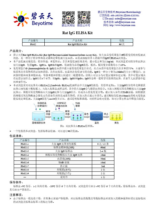
本试剂盒采用双抗体夹心ELISA 法(Sandwich ELISA)检测样品中大鼠IgG 的浓度,其原理见图1。
大鼠IgG 特异的单克隆捕获抗体已预包被于酶标板上,当加入标准品或样品时,其中的大鼠IgG 会与捕获抗体结合。
当加入辣根过氧化物酶标记大鼠IgG 抗体后,辣根过氧化物酶标记大鼠IgG 抗体与大鼠IgG 结合,形成夹心的免疫复合物。
最后加入显色剂TMB 溶液,固相捕获的辣根过氧化物酶就会催化无色的显色剂氧化成蓝色物质,在加入终止液后呈黄色。
通过酶标仪检测450nm 处的吸光度值就能实现定量检测。
Extract-N-Amp Tissue PCR Kit 产品说明书

Product InformationExtract-N-Amp™ Tissue PCR KitXNAT2, XNAT2RProduct DescriptionThe Extract-N-Amp™ Tissue PCR Kit for direct PCR contains the reagents needed to rapidly extract and amplify genomic DNA from mouse tails and other animal tissues, buccal swabs, hair shafts, and saliva. Briefly, the DNA is released from the starting material by incubating the sample with a mixture of the Extraction Solution and the Tissue Preparation Solution at room temperature for 10 minutes. There is no need for mechanical disruption, organic extraction, column purification, or precipitation of the DNA.After adding Neutralization Solution B, the extract is ready for PCR. An aliquot of the neutralized extract is then combined with the Extract-N-Amp™ PCR Reaction Mix and user-provided PCR primers to amplify target DNA. The Extract-N-Amp™ PCR Reaction Mix is a 2X ready mix containing buffer, salts, dNTPs, and Taq polymerase. It is optimized specifically for use with the extraction reagents. It also contains the JumpStart Taq antibody for hot start PCR to enhance specificity but does not contain the inert red dye found in the REDExtract-N-Amp™ PCR Reaction Mix.Reagents Provided Cat. No. XNAT2 100 Preps,100 PCRsXNAT2R 1000 Preps, 1000 PCRsExtraction SolutionE7526 24 mL 240 mL Tissue Preparation Solution T3073 3 mL 30 mL Neutralization Solution BN391024 mL240 mLExtract-N-Amp™ PCR Reaction Mix This is a 2X PCR reaction mix containing buffer, salts, dNTPs, Taq polymerase, and JumpStart™ Taq antibody.E30041.2 mL12 mLReagents and Equipment Required(Not Provided)•Microcentrifuge tubes (1.5 or 2 mL) or multi-well plate for extractions (200 μL minimal well volume) • Small dissecting scissors• Forceps (small to medium in size)• Buccal swab - Sterile foam tipped applicator (Cat. No. WHAWB100032)•Sample collection card - Bloodstain card (Cat. No. WHAWB100014)• Tubes or plate for PCR• Heat block or thermal cycler at 95 °C • PCR Primers (Cat. No. OLIGO) • Thermal cycler•Water, PCR Reagent (Cat. No. W1754)Precautions and DisclaimerThis product is for R&D use only. Not for drug, household, or other uses. Please consult the Safety Data Sheet for information regarding hazards and safe handling practices.StorageThe Extract-N-Amp™ Tissue PCR Kit can be stored at 2 to 8 °C for up to 3 weeks. For long-term storage, greater than 3 weeks, -20 °C is recommended. Do not store in a "frost-free" freezer.ProcedureAll steps are carried out at room temperature unless otherwise noted.DNA Extraction from Mouse Tails, Animal Tissues, Hair, or Saliva1.Pipette 100 μL of Extraction Solution into amicrocentrifuge tube or well of a multi-well plate.Add 25 μL of Tissue Preparation Solution to thetube or well and pipette up and down to mix.Note: If several extractions will be performed,sufficient volumes of Extraction and TissuePreparation Solutions may be pre-mixed in a ratio of 4:1 up to 2 hours before use.2.For fresh or frozen mouse tails: Rinse thescissors and forceps in 70% ethanol prior to useand between different samples. Place a 0.5–1 cm piece of mouse tail tip (cut end down) into thesolution. Mix thoroughly by vortexing or pipetting.Ensure the mouse tail is in solution.Note: For fresh mouse tails, perform extractions within 30 minutes of snipping the tail.For animal tissues: Rinse the scissors or scalpel and forceps in 70% ethanol prior to use andbetween different samples. Place a 2–10 mgpiece of tissue into the solution. Mix thoroughlyby vortexing or pipetting. Ensure the tissue is inthe solution.For hair shafts: Rinse the scissors and forceps in 70% ethanol prior to use and between differentsamples. Trim excess off of the hair shaft leaving the root and place sample (root end down) intosolution. Only one hair shaft, with root, isrequired per extraction.For Saliva: Pipette 10 μL of saliva into thesolution. Mix thoroughly by vortexing or pipetting.For saliva dried on card: Pipette 50 μL of saliva onto collection card and allow the card to dry.Rinse the punch in 70% ethanol prior to use andbetween different samples. Punch a disk(preferably 1/8 inch or 3 mm) out of the cardfrom the area with the dried saliva sample. Place disk into the solution. Tap tube or plate on hardsurface to ensure disk is in solution forincubation period.3.Incubate sample at room temperature for10 minutes.4.Incubate sample at 95 °C for 3 minutes.Note: Tissues will not be completely digested atthe end of the incubations. This is normal and will not affect performance.5.Add 100 μL of Neutralization Solution B to sampleand mix by vortexing.6.Store the neutralized tissue extract at 4 °C oruse immediately in PCR amplification.Note: For long term storage, remove theundigested tissue or transfer the extracts tonew tubes or wells. Extracts may now be storedat 4 °C for at least 6 months without notable loss in most cases.DNA Extraction for Buccal Swabs1.Collect buccal cells on swab and allow theswab to dry. Drying time is approximately10 to 15 minutes.Note: Due to the low volume of solution used for DNA extraction, a foam tipped swab should beused. Swabs with fibrous tips, such as cotton orDacron®, should be avoided because the solution cannot be recovered efficiently.2.Pipette 200 μL of Extraction Solution into amicrocentrifuge tube. Add 25 μL of TissuePreparation Solution to the tube and pipette upand down to mix.Note: If several extractions will be performed,sufficient volumes of Extraction and TissuePreparation Solutions may be pre-mixed ina ratio of 8:1 up to 2 hours before use.3.Place dried buccal swab into solution and incubateat room temperature for 1 minute.4.Twirl swab in solution 10 times and then removeexcess solution from the swab into the tube bytwirling swab firmly against the side of the tube.Discard the swab. Close the tube andvortex briefly.5.Incubate sample at room temperature for10 minutes.6.Incubate sample at 95 °C for 3 minutes.7.Add 200 μL of Neutralization Solution B to sampleand mix by vortexing.8.Store the neutralized extract at 4 °C or useimmediately in PCR. Continue to PCRamplification.Note: Extracts may be stored at 4 °C for at least6 months without notable loss in most cases. PCR AmplificationThe Extract-N-Amp™ PCR Reaction Mix contains JumpStart™ Taq antibody for specific hot start amplification. Therefore, PCR mixtures can be assembled at room temperature without premature Taq DNA polymerase activity.Typical final primer concentrations are approximately 0.4 μM each. The optimal primer concentration and cycling parameters will depend on the system being used.1.Add the following reagents to a thin-walled PCRmicrocentrifuge tube or plate:Reagent VolumeWater, PCR grade VariableExtract-N-Amp™ PCRreaction mix 10 μLForward primer VariableReverse primer VariableTissue extract 4 μL*Total volume 20 μL*The Extract-N-Amp™ PCR Reaction Mix isformulated to compensate for components in the Extraction, Tissue Preparation, and Neutralization Solutions. If less than 4 µL of tissue extract isadded to the PCR reaction volume, use a 50:50mixture of Extraction and Neutralization BSolutions to bring the volume of tissue extract upto 4 μL.2.Mix gently.3.For thermal cyclers without a heated lid, add20 μL of mineral oil on top of the mixture in eachtube to prevent evaporation.4.Perform thermal cycling. The amplificationparameters should be optimized for individualprimers, template, and thermal cycler.Common cycling parameters:Step Temperature Time Cycles InitialDenaturation 94 °C 3 minutes 1 Denaturation 94 °C 30 seconds Annealing 45 to 68 °C 30 seconds 30-35 Extension 72 °C 1-2 minutes(1 min/kb)FinalExtension 72 °C 10 minutes 1 Hold 4 °C Indefinitely5.The amplified DNA can be loaded onto an agarosegel after the PCR is completed with the addition ofa separate loading buffer/tracking dye such as GelLoading Solution, Cat. No. G2526.Note: PCR products can be purified, if desired, fordownstream applications such as sequencing withthe GenElute PCR Clean-Up Kit, Cat. No.NA1020.Troubleshooting GuideProblem Cause SolutionLittle or no PCR product is detected. PCR reaction may beinhibited due tocontaminants in thetissue extract.Dilute the tissue extract with a 50:50 mix of Extractionand Neutralization Solutions. To test for inhibition, includea DNA control and/or spike a known amount of template(100-500 copies) into the PCR along with the tissue extract. Extraction isinsufficient.Incubate samples at 55 °C for 10 minutes instead ofroom temperature.A PCR component maybe missing or degraded.Run a positive control to ensure that componentsare functioning. A checklist is also recommendedwhen assembling reactions.There may be too fewcycles performed. Increase the number of cycles (5-10 additional cycles at a time). The annealingtemperature maybe too high.Decrease the annealing temperature in 2-4 °C increments.The primers may notbe designed optimally.Confirm the accuracy of the sequence information. If theprimers are less than 22 nucleotides long, try to lengthen theprimer to 25-30 nucleotides. If the primer has a GC contentof less than 45%, try to redesign the primer with a GCcontent of 45-60%.The extension timemay be too short.Increase the extension time in 1-minute increments, especiallyfor long templates.Target templateis difficult.In most cases, inherently difficult targets are due to unusuallyhigh GC content and/or secondary structure. Betaine, Cat. No.B0300, has been reported to help amplification of high GCcontent templates at a concentration of 1.0-1.7 M.Multiple products JumpStart™ Taqantibody is notworking correctly.Do not use DMSO or formamide with Extract-N-Amp™ PCRReaction Mix. It can interfere with the enzyme-antibodycomplex. Other cosolvents, solutes (e.g., salts), and extremesin pH or other reaction conditions may reduce the affinity ofthe JumpStart™ Taq antibody for Taq polymerase and therebycompromise its effectiveness.TouchdownPCR maybe needed.“Touchdown” PCR significantly improves the specificity of manyPCR reactions in various applications. Touchdown PCR involvesusing an annealing/extension temperature that is higher thanthe TM of the primers during the initial PCR cycles. Theannealing/extension temperature is then reduced to the primerTM for the remaining PCR cycles. The change can be performedin a single step or in increments over several cycles.Negative control shows a PCR product or “false positive” result. Reagents arecontaminated.Include a reagent blank without DNA template be included asa control in every PCR run to determine if the reagents used inextraction or PCR are contaminated with a template froma previous reaction.Tissue is not digested after incubations. Tissue is not expectedto be completelydigested.The REDExtract-N-Amp™ Tissue PCR Kit does not require thetissue to be completely digested. Sufficient DNA is released forPCR without completely digesting the tissue.Buccal swab absorbed all the solution. The recommended typeof swab was not used.Due to the low volume of solution used for DNA extraction, afoam tipped swab should be used. Swabs with fibrous tips, suchas cotton or Dacron®, should be avoided because the solutioncannot be recovered efficiently.References1.Dieffenbach, C.W., and Dveksler, G.S. (Eds.), PCRPrimer: A Laboratory Manual, 2nd ed., Cold Spring Harbor Laboratory Press, New York (1995).2.Don, R.H. et al., ‘Touchdown' PCR to circumventspurious priming during gene amplification.Nucleic Acids Res., 19, 4008 (1991).3.Erlich, H.A. (Ed.), PCR Technology: Principles andApplications for DNA Amplification, StocktonPress, New York (1989).4.Griffin, H.G., and Griffin, A.M. (Eds.), PCRTechnology: Current Innovations, CRC Press,Boca Raton, FL (1994).5.Innis, M.A., et al., (Eds.), PCR Strategies,Academic Press, New York (1995).6.Innis, M., et al., (Eds.), PCR Protocols: A Guide toMethods and Applications, Academic Press, SanDiego, California (1990).7.McPherson, M.J. et al., (Eds.), PCR 2: A PracticalApproach, IRL Press, New York (1995).8.Newton, C.R. (Ed.), PCR: Essential Data, JohnWiley & Sons, New York (1995).9.Roux, K.H. Optimization and troubleshooting inPCR. PCR Methods Appl., 4, 5185-5194 (1995).10.Saiki, R., PCR Technology: Principles andApplications for DNA Amplification, Stockton, New York (1989). Product OrderingOrder products online at Related Products Cat. No.Ethanol E7148; E7023; 459836 Forceps,micro-dissecting F4267PCR Marker P9577PCR microtubes Z374873; Z374962;Z374881PCR multi-well plates Z374903Precast Agarose Gels P6097Sealing mats & tapes Z374938; A2350TBE Buffer T4415, T6400, T9525The life science business of Merck operatesas MilliporeSigma in the U.S. and Canada.Merck, Extract-N-Amp, REDExtract-N-Amp, JumpStart, GenElute and Sigma-Aldrich are trademarks of Merck KGaA, Darmstadt, Germany or its affiliates. All other trademarks are theproperty of their respective owners. Detailed information on trademarks is available via publicly accessible resources.NoticeWe provide information and advice to our customers on application technologies and regulatory matters to the best of our knowledge and ability, but without obligation or liability. Existing laws and regulations are to be observed in all cases by our customers. This also applies in respect to any rights of third parties. Our information and advice do not relieve our customers of their own responsibility for checking the suitability of our products for the envisaged purpose. The information in this document is subject to change without notice and should not be construed as a commitment by the manufacturing or selling entity, or an affiliate. We assume no responsibility for any errors that may appear in this document. Technical AssistanceVisit the tech service page at/techservice.Terms and Conditions of SaleWarranty, use restrictions, and other conditions of sale may be found at /terms. Contact InformationFor the location of the office nearest you, go to /offices.。
眼科各项标准操作规程SOP

标准操作规程(SOP)目录眼科专业SOP制定和管理的SOPSOP编号:SOP-YK-001-1页数:3制定人:审核人:批准人:(签名、日期)(签名、日期)(签名、日期)生效日期:颁发日期:审查修订登记:Ⅰ目的:统一标准,明确职责,保障临床试验的运行条件,提高临床试验的运行质量。
Ⅱ范围:适用于眼科专业的临床试验的SOP的制定和管理。
Ⅲ规程:1由眼科专业的负责人指定本专业人员完成专业的各项SOP的起草或修订。
该人员必须参加过GCP的培训,并且具有本专业临床经验,从事临床工作3年以上。
2起草人或修订人按照GCP的要求,根据本专业的实际情况起草或修订SOP,修订后签字并注明日期。
然后由眼科专业的负责人审核各专业的具体SOP、审核后签字并注明日期,最后由机构负责人批准、签字并注明日期。
3SOP制定后要有一周的审核时间。
注意批准日期到生效日期中间有三个月的过渡期,以便相关人员学习掌握新的SOP。
4所有制定的SOP按统一格式制定,字体大小及序号使用规则参照机构SOP制定和管理的SOP。
5SOP文件编码原则参照机构SOP制定和管理的SOP。
6眼科专业标准操作规程的编码格式为:“SOP-YK-LL-××-##”,“YK”为眼科专业前两个字的汉语拼音的第一个字母(大写);“LL”为专业试验方案设计、急救预案或仪器管理和使用类的代码;“××”为本专业SOP的顺序号;##为修订的版本号。
例如:眼科专业的第一版的第一个试验方案设计类的SOP的编号为:“SOP-YK-FASJ-001-01”;再如:“SOP-YK-JJYA-001-01”表示:眼科专业的修订的第三版的第一个急救预案SOP。
7专业试验方案设计、急救预案或仪器管理的使用类的代码:试验方案设计-FASJ;急救预案-JJYA;主要疾病—JB;仪器管理和使用-YQGL;其他-QT。
8药物临床试验机构办公室对通过的SOP归档保存,制定内容及修订原因应记录并存档。
免疫组化操作规程

免疫组化操作规程(一)、仪器设备1)18cm不锈钢高压锅或电炉或医用微波炉;2)水浴锅(二)、试剂1)PBS缓冲液(pH7.2~7.4):NaCl 137mmol/L,KCl 2.7mmol/L,Na2HPO4 4.3mmol/L,KH2PO4 1.4mmol/L。
2)0.01mol/L柠檬酸盐缓冲液(CB,pH6.0,1000ml):柠檬酸三钠3g,柠檬酸0.4g。
3)0.5mol/L EDTA缓冲液(pH8.0):700ml水中溶解186.1gEDTA·2H2O,用10 mmol/L NaOH调至pH8.0,加水至1000ml。
4)1mol/L的TBS缓冲液(pH8.0):在800ml水中溶解121gTris碱,用1N的HCl调至pH8.0,加水至1000ml。
5)酶消化液:a、0.1%胰蛋白酶液:用0.1%CaCl2(pH7.8) 配制。
b、0.4%胃蛋白酶液:用0.1N的HCl 配制。
6)3%甲醇-H2O2溶液:用30%H2O2和80%甲醇溶液配制。
7)封裱剂:a、甘油和0.5mmol/L碳酸盐缓冲液(pH9.0~9.5)等量混合;b、油和TBS(或PBS)配制。
8)TBS/PBS pH9.0~9.5,适用于荧光显微镜标本;pH7.0~7.4适合于光学显微镜标本。
(三)、操作流程1、脱蜡和水化脱蜡前,应将组织芯片在室温中放置60分钟或60℃恒温箱中烘烤20分钟。
1)组织芯片置于二甲苯中浸泡10分钟,更换二甲苯后再浸泡10分钟;2)无水乙醇中浸泡5分钟;3)95%乙醇中浸泡5分钟;4)70%乙醇中浸泡5分钟;2、抗原修复用于福尔马林固定的石蜡包埋组织芯片。
1)抗原热修复(1)高压热修复在沸水中加入EDTA(pH8.0)或0.01M枸橼酸钠缓冲溶液(pH6.0)。
盖上不锈钢高压锅的盖子,但不进行锁定。
将玻片置于金属染色架上,缓慢加压,使玻片在缓冲液中浸泡5分钟,然后将盖子锁定,小阀门将会升起来。
免疫组化实验步骤

免疫组化操作步骤一、实验原理与意义免疫组织化学又称免疫细胞化学,是指带显色剂标记的特异性抗体在组织细胞原位通过抗原抗体反应和组织化学的呈色反应,对相应抗原进行定性、定位、定量测定的一项新技术。
它把免疫反应的特异性、组织化学的可见性巧妙地结合起来,借助显微镜(包括荧光显微镜、电子显微镜)的显像和放大作用,在细胞、亚细胞水平检测各种抗原物质(如蛋白质、多肽、酶、激素、病原体以及受体等)。
(一)、仪器设备1. 18cm不锈钢高压锅或电炉或用微波炉.2. 水浴锅(二)、试剂1. PBS缓冲液(ph7.2―7.4):NaC137mmol/L,KCl2.7mmol/L ,Na2HPO4 4.3mmol/L, KH2PO4 1.4mmol/L. 2.0.01mol/L柠檬酸钠缓冲液(CB,ph6.0,1000ml):柠檬酸三钠3g,柠檬酸0.4g。
3.0.5mol/L EDTA缓冲液(ph8.0):700ml水中溶解186.1g EDTA& 8226;2H2O,用10mmol/L NaOH调至ph8.0,加水至1000ml.4. 1mol/L的TBS缓冲液(ph8.0):在800ml水中溶解121gTris碱,用1N的HCl调至pH8.0, 加水1000ml。
5. 酶消化液:a.0.1%胰蛋白酶:用0.1%CaCl 12(ph7.8)配制。
b.0.4%胃蛋白酶液:用0.1N的HCl配制。
6. 3%甲醇―H2O2溶液:用30%H2O2和80%甲醇溶液配制7. 风裱剂:a.甘油和0.5mmol/L碳酸盐缓冲液(pH9.0–9.5)等量混合 b 油和TBS(PBS)配制8.TBS/PBS PH9.0–9.5,适用于荧光纤维镜标本;ph7.0-7.4适合光学纤维标本(三)、操作流程1、脱蜡和水化:脱蜡前应将组织芯片在室温中放置60分钟或60℃恒温箱中烘烤20分钟。
a 组织芯片置于二甲苯中浸泡10分钟,更换二甲苯后在浸泡10分钟b 无水乙醇中浸泡五分钟c 95%乙醇中浸泡五分钟d 75%乙醇中浸泡五分钟2、抗原修复:用于福尔马林固定的石蜡包埋组织芯片:A 抗原热修复a 高压热修复在沸水中加入EDTA(ph8.0)或0.01m枸橼酸钠缓冲溶液(ph6.0).盖上不锈钢锅盖,但不能锁定。
RHCK1兔笼完整套件用户手册说明书

RHCK1 Rabbit Hutch Complete KitEasy to clean, rust resistant• galvanized steel construction Bottom panel with 1/2 x 1” • spacingSide and top panels with • 1 x 2” spacing Spring tension door latch • Vinyl door guard • Stackable frame kit • Dropping pan • Water bottle • Feeder•1 Bottom Panel 1 Back Panel 1 Front Panel 1 Door Panel2 Side Panels 1 Top Panel 4 Vinyl DoorGuards 1 Spring 1 Door Latch 4 Frame Legs 2 Tray Brackets 1 Dropping Pan 1 Water Bottle1 Feeder1 Package Wire Clips1 Wire Clip Pliers 4 Bolts, washers, and nutsWire Clip Pliers (see • instructions at right)Wire Cutter• Straight Blade Screwdriver • T wo 7/16” Wrenches• Each Rabbit Hutch Contains:Tools Required:Features:Assembly Instructions (continued):Assembly Instructions:Miller Manufacturing Company1450 West 13th Street • Glencoe, MN 55336 • Customer Service 800-260-0888 • FAX 651-982-5101For more information visit NOTE: If you want to put the door on the left side of the front panel, reverse the position of the door and feeder hole shown.1. Cut out a hole in the front panel for the feeder using a wire cutter. See Figure 2.2. Lay out the hutch panels as shown in Figure3. You can put the door on the right or left side of the front of the hutch.3. Fasten a clip at each . Do not put clips where appears.4. Raise two adjoining panels so that they meet at the corner.Fasten clips at each shown in Figure 4. Repeat for the other corners.5. Attach the door on the inside of the hutch using clips at each shown in Figure 5.6. Slide the vinyl door guard over the edge of the door opening.7. Place the top panel on top of the side panels. Fasten clips at each shown in Figures 6. Do not put clips where the appears.8. Install a frame leg at each shown in Figure 7.NOTE: Wire clips cannot be used where the legs are to be installed. If necessary, remove any clips in these locations. 9. Ensure that the top wire of the side panel fi ts over the prongs on the legs as shown in Figure 7. If necessary, bend the prongs out slightly with a screwdriver. Install the bolts, washers, and nuts (fi nger tight only at this time).10. Install the pan rails on the wide prongs as shown in Figure 7.11a. When assembling one frame kit (not stacking hutches)complete the assembly by tightening the bolts, washers, and nuts for each leg.11b. When assembling two frame kits (stacking hutches) proceed tostep 12.12 Install another set of legs beneath the fi rst set as shown in Figure 8. Make sure the small tabs on the upper leg fi t over the top of the lower leg. Install the bolts, washers, and nuts (fi nger tight only at this time).1. Place ACC1 Wire Cage Clip in the pliers. See Figure 1.2. Position clip next to wires to be fastened.3. Close the pliers to form clips around wires CAUTION: Avoid pinching skin in pliers.Using the ACP2 Wire Clip Pliers:Figure 313. Position the second hutch on the lower set of legs (anotherperson is helpful when lowering the second hutch into position).14. Install the pan rails on the wide prongs as shown in Figure 7.15. Tighten all fasteners securely.16. Install the feeder, water bottle, and dropping pan as shown inFigure 9.Miller Manufacturing Company1450 West 13th Street • Glencoe, MN 55336For more information visit RHCK 1INSTRUCT I O06/10Wire Cage Clips • (ACC1)Wire Clip Pliers • (ACP2)Small Animal Water Bottles • (AW8, AW16, and AW32)Clear Series Water Bottles • (CPB8, CPB16, and CPB32)Plastic Cage Cups • (ACU1, ACU2, ACU4)Galvanized Cage Cup • (ACU5)Salt Spool and Hanger • (SSH2)Hay Rack • (153171)Frame Kits • (AHFK24, and AHFK30)Small Animal Feeders with metal bottom and lid • (AF3ML, AF5ML, and AF7ML)Small Animal Feeders with sifter bottom • (AF3S, AF5S, and AF7S)Small Animal Feeders with sifter bottom and lid • (AF3SL, AF5SL, and AF7SL)Urine Guard Kits • (AUG2424, AUG3030, and AUG3036)Galvanized Dropping Pans • (ADP2424, ADP3030, and ADP3036)Plastic Dropping Pan • (PDP2424)AccessoriesFigure 7Figure 8Figure 9Dropping PanFeederWater BottleFigure 4ClipCorner ViewFigure 5ClipSpring LatchVinyl Door GuardDoor mounts inside hutchLegLegProngsPan RailWide Prong。
博士德试剂盒说明
产品货号:SA1023产品价格:290元/1/2盒(可以做80张左右的切片),550元/1盒(可以做160张左右的切片)产品编号:SA1023—山羊IgG(适于一抗为山羊多克隆抗体)SA1024—人IgG (适于人自身抗体的检测)SA1025—大鼠IgG(适于一抗为大鼠单克隆抗体)试剂的保存:4℃可保存一年,应避免冷冻。
工作原理:SABC 是专为免疫组化和其他免疫检测而设计的,用以显示组织和细胞中抗原分布。
链霉亲和素是一种从链霉菌中提取的蛋白质,分子量47000。
同亲和素一样,对生物素分子有极高的亲和力,是一般抗原抗体亲和力的一百万倍。
亲和素是一个碱性蛋白质(IP=10),经改造后可以转变成中性蛋白质。
链霉亲和素等电点接近中性,IP=6.0~6.5,对组织和细胞的非特异吸附很低,基于链霉亲和素的免疫组化方法背景很低。
SABC 即StreptAvidin—BiotinComplex,。
根据研究,SABC 大约可形成一百个左右的过氧化物酶和五十个左右的链霉亲和素所构成的复合物。
大量的酶将保证SABC 具有很高的敏感性。
SABC 兼具高敏感性,低背景和操作简便的优点。
试剂盒中内容1.正常兔血清封闭液:12ml。
用于组织切片的封闭。
2.二抗:12ml。
亲和纯化抗体,标记长臂生物素。
兔抗大鼠IgG(或IgM)或明或暗兔抗山羊IgG 或兔抗人IgG。
3.SABC:12ml。
链酶亲和素—过氧化物酶复合物。
用户自备试剂:1.粘片剂APES 或POLY-L-LYSINE(博士德公司有售)。
2.免疫组化专用PBS(pH7.2-7.6)配法:1000ml 蒸馏水中加氯化钠8.5g, Na2HPO4 2.8g,NaH2PO4 0.4g。
如果用的是含水磷酸盐,应加上分子式中水的含量。
3.0.01M 枸橼酸盐缓冲液:1000ml 蒸馏水中加枸橼酸三钠(C6H5Na3O7·2H2O)3g, 枸橼酸(C6H8O7·H2O ) 0.4g。
二抗鼠抗兔IgG单克隆抗体
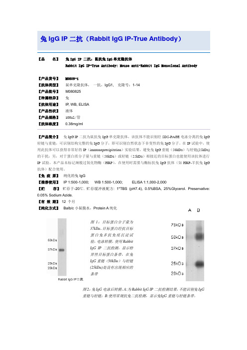
兔IgG IP二抗(Rabbit IgG IP-True Antibody)【品名】兔IgG IP 二抗:鼠抗兔IgG单克隆抗体Rabbit IgG IP-True Antibody: Mouse anti-Rabbit IgG Monoclonal Antibody【产品货号】 M0809-1【抗体类型】鼠单克隆抗体,一抗,IgG1, 克隆号:1-14【产品批号】M080825【种属特异】兔【抗体用途】IP, WB, ELISA【产品性状】液体【产品规格】100ul/管【抗体浓度】0.38mg/ml【产品简介】兔IgG IP二抗为鼠抗兔IgG单克隆抗体,该抗体不能识别经SDS-PAGE电泳分离的兔IgG 轻链与重链,可识别结构完整的兔IgG分子,即可识别自然状态下非变性的兔IgG分子。
在IP试验中,使用此抗体可以获得非常好的IP(immunoprecipitation)实验结果,避免兔IgG重链(50kDa)与轻链(25kDa)的干扰;另,对于蛋白质分子量与重链(50kDa)或轻链(25kDa)相接近的目标蛋白也能使用该抗体进行IP试验。
本产品未标记辣根过氧化物酶(HRP),在使用时需要与酶标抗兔IgG抗体(如HRP-羊抗兔IgG 抗体)配合使用。
【免疫原】纯化的兔IgG【推荐使用】IP 1:500-1,000; WB 1:500-1,000; ELISA 1:1,000-2,000【贮存】贮存于-20℃. 贮存缓冲液配方: 1*TBS (pH7.4), 0.5%BSA, 25%Glycerol. Preservative: 0.05% Sodium Azide.【有效期】12 个月【纯化方式】Balb/c小鼠腹水,Protein A纯化图1:目标蛋白分子量为37kDa..目标蛋白经抗目标蛋白兔多抗免疫沉淀试验,电泳转膜,使用RabbitIgG IP二抗检测,显示特异性目标蛋白条带,在兔IgG重链(50kDa)与轻链(25kDa)处没有出现相应的条带图2:兔IgG电泳后转膜,A:为Rabbit IgG IP二抗检测结果,不能识别兔IgG重链与轻链;B:使用常规抗兔二抗检测,显示兔I gG重链与轻链条带。
XX第X医科大学2023年春季本科教学用低值易耗品采购清单及要求(2024年)

57
三(羟基甲基)氨基甲烷
500g
瓶
1
58
十二烷基硫酸钠-SDS
500g
瓶
1
59
30%丙烯酰胺溶液Acr-Bis(30%,29:1)
500ml
瓶
1
60
彩色预染蛋白(10-180KDa)
250ul
瓶
2
61
高灵敏度化学发光检测试剂盒
50ml
瓶
2
62
10×TBST缓冲液
500mL
瓶
5
63
10%SDS
100ml
5
215
石英砂
500克
瓶
5
216
碳酸钙
500克
瓶
5
217
玻璃试管
12*75
支
50
218
玻璃漏斗
直径6厘米
个
50
219
手术剪刀(组织剪)
14cm
把
10
220
酚酞
500克
瓶
2
221
石蕊
25克
瓶
2
222
氯化三苯四氮唑
20g
瓶
1
223
玻璃量筒
100 ML
支
10
224
玻璃量筒
200 ML
支
10
225
硝酸银
100g/瓶
不锈钢玻片架
26片
个
5
90
伊红染液
500ml
瓶
2
91
苏木素染液
500ml
瓶
2
92
中性树胶
500g
瓶
2
康欣胶囊对兔动脉粥样硬化斑块稳定性的影响

康欣胶囊对兔动脉粥样硬化斑块稳定性的影响目的观察康欣胶囊对兔动脉粥样硬化(AS)血管斑块稳定性的影响,探讨其稳定斑块的可能机制。
方法将40只纯种雄性新西兰大耳白兔随机分为模型组、阿托伐他汀钙组(简称他汀组)、康欣胶囊低剂量组(简称小剂量组)、康欣胶囊高剂量组(简称大剂量组)、正常对照组,每组8只。
除正常组外,采用内膜损伤加高胆固醇饮食法建立兔AS模型,分别予相应处理,6周后进行主动脉血管病理形态学观察,采用免疫组化法检测基质金属蛋白酶-2 (MMP-2)、血管细胞黏附分子-1(VCAM-1)在动脉斑块内的表达。
结果康欣胶囊干预组动物血管腔壁比(W/L)、平均光密度、阳性面积百分比均较模型组小,动脉壁组织MMP-2及VCAM-1均较模型组下调,差异有统计学意义(P<0.05,P<0.01),以大剂量组最为明显。
结论康欣胶囊可减少MMP-2、VCAM-1等炎性介质表达,可能是其稳定斑块的机制。
标签:康欣胶囊;动脉粥样硬化斑块;基质金属蛋白酶-2;血管细胞黏附分子-1;兔动脉粥样硬化(atherosclerosis,AS)是引起冠心病心肌缺血和各种临床症状的基本原因,但临床事件并不都是发生在管腔最狭窄处,而是随着AS的发展,斑块破裂及继发血栓形成。
因此,如何稳定斑块显得尤其重要。
AS斑块的形成是一个局部和系统的炎症过程[1-2]已为人们所公认。
研究表明,AS斑块在由稳定向不稳定发展过程中,炎性标志物血管细胞黏附分子-1(VCAM-1)、基质金属蛋白酶-2(MMP-2)等的表达增多。
据此,我们应用高脂饮食加颈动脉内膜损伤术建立AS模型,探讨康欣胶囊稳定斑块的可能机制。
1 材料与方法1.1 仪器与主要试剂机械组织匀浆机(Polytron-ultraturrax公司,德国),显微镜图像分析仪(Motic image advance 3.0,香港)。
即用型SABC免疫组化试剂盒兔IgG(SA1022),多克隆兔源抗兔MMP-2与VCAM-1抗体(博士德生物工程有限公司);辣根过氧化物酶标记山羊抗兔IgG(H+L),碧云天生物技术研究所。
电针刺激通过调节5-羟色胺和去甲肾上腺素水平治疗雌性大鼠压力性尿失禁
分类号学号M*********学校代码10487 密级硕士学位论文电针刺激通过调节5-羟色胺和去甲肾上腺素水平治疗雌性大鼠压力性尿失禁学位申请人:卢鹏学科专业:外科学指导教师:杨为民教授答辩日期:2013年5月A dissertation submitted to Huazhong Universityof Science and Technology for the Degreeof Master of MedicineAcupuncture treatment for female rats stress urinary incontinence by adjusting the expression ofserotonin and norepinephrineCandidate : Lu PengMajor : UrologySupervisor : Prof. Dr. Yang WeiminDepartement of Urology, Tongji HospitalTongji Medical CollegeHuazhong University of Science and TechnologyWuhan, Hubei, 430030, P.R. ChinaMay, 2013独创性声明本人郑重声明,本学位论文是本人在导师指导下进行的研究工作及取得的研究成果的总结。
尽我所知,除文中已经标明引用的内容外,本论文不包含任何其他个人或集体已经发表或撰写过的研究成果。
对本文的研究做出贡献的个人和集体,均已在文中以明确方式标明。
本人完全意识到本人将承担本声明引起的一切法律后果。
学位论文作者签名:日期:年月日学位论文版权使用授权书本学位论文作者完全了解学校有关保留、使用学位论文的规定,即:学校有权保留并向国家有关部门或机构送交论文的复印件和电子版,允许论文被查阅和借阅。
本人授权华中科技大学可以将本学位论文的全部或部分内容编入有关数据库进行检索,可以采用影印、缩印或扫描等复制手段保存和汇编本学位论文保密□ ,在_____年解密后适用本授权书。
贝恩施糖果兔w1说明书
贝恩施糖果兔w1说明书
1、用户均可直接扫描说明书上的二维码,按提示下载即可。
2、长按中间按键开机,贝恩施糖果兔w1开机后,会发出“叮”声响,等待5-1秒即勃发开机音乐,随后发出语音提示“正在启动中,请稍等哟”,表示开机成功。
3、成功开机后,贝恩施糖果兔w1语音提示“检测网络中,请稍等,没有找到可用的网络,请打开手机APP根据引导帮我联网哟”,此时灯光闪烁。
4、打开APP,依次点击“添加新设备”-“设备联网”-“已经启动设备啦”,进入“配置设备网络”页面。
5、语音提示“请设置设备要连接的无线网络”,悬着WIFI,输入密码,并点击“下一步”,来到“进入联网模式页面”
6、长按设备的配网键,听到语音提示“已经入联网模式”后,即可点击“下一步”,进入“声波联网页面”
7、在安静的环境下,将手机靠近设备,并长按APP上的“开始配网”,等待设备接收声波联网,当设备提示“配网成功”,耳朵灯光停止闪烁,此时点击“成功了,去绑定设备进入”绑定设备页面。
8、按激动键,对设备说话“绑定设备”,输入播报的数字后,点击“绑定”即可完成设备与手机绑定。
深低温冷冻骨的形态学及免疫组织化学检测

深低温冷冻骨的形态学及免疫组织化学检测夏武宪;郭华;李龙腾;韩芬;孙永武;张向军;张帆【摘要】背景:深低温冷冻技术能够降低同种异体骨的免疫原性,对修复骨缺损具有重要意义.目的:观察深低温冷冻对骨材料中骨形态发生蛋白2表达的影响.方法:18只新西兰兔随机分为3组,深低温冷冻3个月组,深低温冷冻6个月组,新鲜骨对照组,取右侧胫骨中上1/3,分别于-80 ℃保存保存3个月,6个月及不保存,进行苏木精-伊红染色和骨形态发生蛋白2免疫组织化学染色.结果与结论:深低温冷冻后骨材料形态学,与新鲜骨差异不大;深低温冷冻3个月及6个月骨组织中骨形态发生蛋白2均有表达,但较新鲜骨降低.%BACKGROUND: Bone morphogenetic protein 2 (BMP-2) in fracture healing and repair process plays an important role indeep-freezing technology.OBJECTIVE: To investigate the effects of deep-freezing technology on BMP-2 expression in the bone materials.METHODS: 18 rabbits were randomly and evenly divided into three groups: deep-frozen 3 months, deep frozen 6 months, freshbone. The middle upper 1/3 right tibia from each group was taken and preserved at -80 ℃ for 3 or 6 months or was not preserved.Then all bone samples were performed hematoxylin-eosin staining and BMP-2 immunohistochemicalstaining.RESULTS AND CONCLUSION: There was no significant difference in morphology between deep frozen bone and fresh bone.BMP-2 was expressed in bone deep-frozen for 3 and 6 months, but the expression was lower than that in the fresh bone.【期刊名称】《中国组织工程研究》【年(卷),期】2011(015)037【总页数】3页(P6863-6865)【关键词】深低温冷冻;移植;形态学;骨形态发生蛋白2;免疫组织化学【作者】夏武宪;郭华;李龙腾;韩芬;孙永武;张向军;张帆【作者单位】郑州铁路职业技术学院,河南省郑州市,450052;郑州铁路职业技术学院,河南省郑州市,450052;郑州铁路职业技术学院,河南省郑州市,450052;郑州铁路职业技术学院,河南省郑州市,450052;郑州铁路职业技术学院,河南省郑州市,450052;郑州铁路职业技术学院,河南省郑州市,450052;郑州铁路职业技术学院,河南省郑州市,450052【正文语种】中文【中图分类】R3180 引言同种异体骨材料来源较多,且因其具有天然结构、形状和强度,并具有一定的诱导活性,在骨移植中有其不可替代的地位。
Pierce crosslink IP kit步骤(中文版)

Pierce crosslink IP kit步骤A 抗体与附着于琼脂糖上的Protein A/G的结合注:以下步骤以10ug抗体为例,实际操作时可结合2-50ug,若抗体量>50ug,则按适当比例增加resin,crosslinker和buffer体积。
1.每个IP反应使用2 ml 1×coupling buffer。
2.缓慢旋转含Protein A/G 的琼脂底部,使之均匀悬浮。
向spin column中加入20 ul resin slurry,将column置入离心管,1000g离心1 min,弃废液。
3.200 ul 1×coupling buffer清洗resin,离心去废液。
重复一次。
4.轻柔的将column底部吸到滤纸上,除去多余液体,换为bottom plug插回column。
5.准备10 ug抗体,用20×coupling buffer与ddw将抗体稀释为100 ul。
将ddw,20×buffer与纯化后的抗体直接加到column的树脂片上。
6.把column帽子盖好,室温下旋转混匀30—60min。
7.取下bottom plug并保留,去掉盖子。
将column置于一收集管中离心,收集流出液,检测抗体有无偶联上。
8.100 ul 1×Coupling Buffer清洗resin,离心,弃废液。
9.300 ul 1×Coupling Buffer清洗resin,离心,弃废液。
重复一次。
B 抗体间臂的生成注:DSS crosslinker吸湿性强,使用后需将DSS保存于铝箔袋中,用前将DSS立即溶解于DSMO或DMF,DSS与氨基的buffer不共存(如Tris,Gly等)。
1.用pipette tip刺穿DSS tube的foil覆盖膜,加入217 ul DMSO/DMF,以制备10×solusion(25 mM),用枪头彻底混匀solusion,直至DSS溶解完全。
应用中性粒细胞迁移距离推断损伤时间

应用中性粒细胞迁移距离推断损伤时间刘起清;郭红民;王磊;鲁翰霖;杜秋香;百茹峰;孙俊红;王英元【摘要】目的探索中性粒细胞迁移距离在大鼠骨骼肌损伤时间推断中的应用,为后续损伤时间推断研究提供方法学依据.方法建立大鼠骨骼肌挫伤模型,设置对照组和损伤后2、4、6h组,采用免疫组织化学染色技术检测损伤后中性粒细胞参与炎症的反应规律,运用TissueFAXS PLUS软件检测中性粒细胞的迁移距离,并探讨其与损伤时间的关系.结果骨骼肌损伤后2~6h中性粒细胞浸润明显,伤后2h中性粒细胞阳性率为(28.75±0.94)%,伤后4 h达到峰值[(45.50±3.63)%],伤后6 h下降为(31.92±1.56)%.中性粒细胞迁移距离随炎症进展逐渐增大,伤后2、4、6 h分别为(124.80±12.32)μm、(229.03±21.45)μm、(335.04±16.75)μm.结论骨骼肌损伤后2~6 h,中性粒细胞的数量及迁移距离与损伤时间之间存在相关性,有望用于骨骼肌早期损伤时间推断.【期刊名称】《法医学杂志》【年(卷),期】2019(035)002【总页数】5页(P166-170)【关键词】法医病理学;创伤和损伤;细胞迁移分析;中性粒细胞;骨骼肌;损伤时间推断;大鼠【作者】刘起清;郭红民;王磊;鲁翰霖;杜秋香;百茹峰;孙俊红;王英元【作者单位】山西医科大学法医学院,山西太原 030001;太原市公安局迎泽分局,山西太原 030002;山西医科大学法医学院,山西太原 030001;山西医科大学法医学院,山西太原 030001;山西医科大学法医学院,山西太原 030001;中国政法大学刑事司法学院,北京 100040;山西医科大学法医学院,山西太原 030001;山西医科大学法医学院,山西太原 030001【正文语种】中文【中图分类】DF795.1损伤时间推断是法医病理学的难点问题之一,其研究方法主要包括形态学、分子生物学等,其中形态学方法具有确凿、稳定、直观可视化等优点。
睾酮对膝关节骨关节炎去势雄兔的软骨修复作用

睾酮对膝关节骨关节炎去势雄兔的软骨修复作用熊华章;刘毅;王胜民;吴术红;桑鹏【摘要】目的探讨睾酮对膝关节骨关节炎(KOA)睾丸切除(去势)雄兔的软骨修复作用.方法将18只雄兔采用改良Hulth法建立右侧KOA模型并去势,8周时随机处死6只,确认模型建立成功.将剩余12只兔随机分为观察组和对照组,每组6只.观察组每隔2周肌注十一酸睾酮6 mg/kg,对照组同时肌注等量生理盐水,共8周(4次).分组处理第8、12、16周,采用电化学发光免疫分析法检测血清睾酮;分组处理第16周末处死,取出右股骨髁,肉眼观察标本大体情况,采用免疫组化法检测软骨组织胰岛素样生长因子1(IGF-1)表达,并进行关节软骨Mankin′s评分.结果两组第8周血清睾酮水平比较差异无统计学意义(P>0.05),观察组第12、16周血清睾酮水平均明显高于对照组(P均<0.05).对照组右膝关节明显肿大,关节软骨面变薄、粗糙,软骨表面色泽变暗,溃疡较深,边缘骨赘形成明显.观察组右膝关节肿大程度轻于对照组,软骨表面有光泽,可见小溃疡和不规则裂隙.观察组软骨组织IGF-1阳性表达率高于对照组,Mankin′s评分低于对照组(P均<0.05).结论睾酮可促进KOA去势雄兔的软骨修复;其机制可能与促进IGF-1表达有关.【期刊名称】《山东医药》【年(卷),期】2017(057)016【总页数】3页(P35-37)【关键词】骨关节炎;膝关节;睾酮;去势法;胰岛素样生长因子1;软骨修复;兔【作者】熊华章;刘毅;王胜民;吴术红;桑鹏【作者单位】遵义医学院附属医院,贵州遵义563003;遵义医学院附属医院,贵州遵义563003;遵义医学院附属医院,贵州遵义563003;遵义医学院附属医院,贵州遵义563003;遵义医学院附属医院,贵州遵义563003【正文语种】中文【中图分类】R684膝关节骨关节炎(KOA)是一种以关节软骨退变、软骨下骨硬化、骨赘形成及滑膜炎为特征的疾病,目前尚无有效的治疗药物,膝关节置换手术效果较好,但价格昂贵,且存在翻修等问题。
- 1、下载文档前请自行甄别文档内容的完整性,平台不提供额外的编辑、内容补充、找答案等附加服务。
- 2、"仅部分预览"的文档,不可在线预览部分如存在完整性等问题,可反馈申请退款(可完整预览的文档不适用该条件!)。
- 3、如文档侵犯您的权益,请联系客服反馈,我们会尽快为您处理(人工客服工作时间:9:00-18:30)。
SABC(兔IgG)-POD Kit使用说明
货号:SA0021
产品内容:
封闭液(5%BSA)10ml
3%H2O210ml
Bio-羊抗兔IgG浓缩液100ul
SABC-POD浓缩液100ul
稀释液30ml
20×DAB显色液A1ml
20×DAB显色液B1ml
保存:
如果长时间不使用,请将所有试剂存放于-20℃,如经常使用,可将封闭液,3%H2O2和20×DAB显色液B存放于2-8℃以方便使用。
产品说明:
本试剂盒适合于一抗为兔IgG的免疫组化实验,DAB显色。
SABC是专为免疫组化和其他免疫检测而设计的,用以显示组织和细胞中抗原分布。
链霉亲和素(StreptAvidin)是一种从链霉菌中提取的蛋白质,同亲和素一样,对生物素有极高的亲和力,亲和素是一个碱性蛋白质(PI=10),经改造后可以转变成中性蛋白质。
链霉亲和素等电点接近中性,对组织和细胞的非特异吸附很低,基于链霉亲和素的免疫组化方法背景很低。
SABC即StreptAvidin—Biotin Complex,SABC大约可形成一百个左右的过氧化物酶和五十个左右的链霉亲和素所构成的复合物。
大量的酶将保证SABC具有很高的敏感性。
操作步骤:(以石蜡切片为例)
1.切片常规脱蜡至水(三次二甲苯,三次乙醇)。
2.3%H2O2室温处理5-10分钟以灭活内源性酶。
蒸馏水洗3次。
3.可选步聚:
a、热修复抗原,将切片浸入0.01M柠檬酸钠缓冲液(PH6.0),电炉或微波炉加热至沸腾后断电,间隔5-10分钟后,反复1-2次。
冷却后PBS(pH7.2-7.6)洗涤1-2次。
b、酶消化,滴加消化液,37℃10分钟,PBS(pH7.2-7.4)洗涤2-3次。
c、跳过此步,直接进入下一步。
4.滴加5%BSA封闭液,室温20分钟。
甩去多余液体,免洗。
5.用稀释液将一抗按一定比例稀释(稀释后的一抗4℃可保存一周),也可另行购买抗体稀释液。
滴加稀释的一抗37℃孵育1小时左右或20℃2小时左右,也可4℃过夜。
PBS (pH7.2-7.4)洗涤3次,每次2分钟(一抗的稀释度、孵育时间和温度与染色强度、背景有直接关系。
一般来说,阳性染色强度不够时,可提高一抗浓度和延长孵育时间;背景过高时,可降低一抗浓度和缩短孵育时间)。
6.根据用量,用稀释液将Bio-羊抗兔IgG浓缩液按1:50-100稀释成工作液(1ml稀释液加入10-20ul Bio-羊抗小鼠IgG浓缩液混匀,此工作液4℃可保存一周)。
滴加Bio-羊抗兔IgG 工作液,20-37℃孵育30分钟。
PBS(pH
7.2-7.4)洗涤3次,每次2分钟。
7.根据使用量,用稀释液将SABC-POD浓缩液按1:100稀释成工作液(1ml稀释液加入10ul SABC-POD浓缩液混匀,此工作液4℃可保存一周)。
滴加SABC-POD工作液,20-37℃,30分钟。
PBS(pH7.2-7.4)洗涤4次,每次5分钟。
8.DAB显色:根据用量,用PBS(pH7.2-7.4)配制,按1ml PBS加入50ul20×DAB显色液A,加入50ul20×DAB显色液B。
充分混匀后滴加到切片上。
室温显色,镜下控制反应时
间,一般在5-30分钟之间。
蒸馏水洗涤终止反应。
9.苏木素轻度复染。
脱水,透明,封片。
显微镜观察。
对于细胞爬片,固定后,PBS漂洗两次,再用0.5%Triton X-100室温孵育20分钟,PBS 漂洗两次,3%H2O2处理15分钟;PBS漂洗两次,接上述第4步。
对于冰冻切片,固定后,PBS漂洗两次,接上述第二步。
注意事项:
如果染色背景过高,在SABC反应之后,DAB显色之前,用加有0.01—0.02%TWEEN-20的PBST(pH7.2-7.4)洗涤切片4次,PBS洗2次,然后DAB显色。
相关试剂:
A1800抗体稀释液(普通型)
A1810一抗稀释液
A1820HRP标记抗体稀释液
P11104%多聚甲醛
P210010×多聚赖氨酸
T8200Triton X-100
P103220×PBS,PH7.2-7.4
P10311×PBST PH7.2-7.40.01M
C1010柠檬酸钠缓冲液0.01mol/L,pH6.0
X10200.1%胰蛋白酶液消化液
G1080Mayer'苏木素染液(免疫组化)。
