ERCP文献快讯创刊号2009年11月
ERCP在胆胰疾病中的内镜诊治进展—博士
ENBD
( 鼻胆管引流术 )
(内镜下胆道支架引 流术 )
ERBD
ERCP ↓ ENBD ↓ 择期EST 取石 外科手术
ERCP ↓ ERBD ↓ 择期EST 取石 外科手术
壶腹部结石嵌顿
针状刀/拉式刀切开 ↓ 取石
胆管stent(内引流管)引流(ERBD)
ERCP ↓ ERBD (内镜下胆道支架引流术 ) ↓ 反复引流更换
ERCP在胆胰疾病
诊治中应用进展
内
科
学
教
研
室
内镜技术在我国胰腺疾病诊治中发展
年代
1973 1983 1985
作者
陈敏章等 于中麟等 鲁焕章等
项目
ERCP(内镜下逆行胰胆管造影) ENBD(鼻胆管引流术 ) EPT(经内镜乳头括约肌切开取石 )
1988
1995 1997
1998
1999
张齐联等 陆星华等 许国铭,李兆申等 任旭,李兆申等 许国铭,陆星华等 李兆申等
胰管镜诊断价值
• Uehara等报道 • 11例胰腺原位癌(手术证实) • 术前CT、EUS、ERCP仅发现胰管扩张或 囊肿,未发现明确占位性病变 • 10例胰管镜发现胰管癌性变化
内镜治疗技术进展
急性胰腺炎 慢性胰腺炎
▪胰管结石
▪胰腺假性囊肿 ▪胰管损伤 ▪胰 漏
▪急性胆源性胰腺炎
▪特发性胰腺炎
急性胆源性胰腺炎(ABP)
ABP内镜治疗适应证
AP(急性胰腺炎)+梗阻性黄疸
AP(急性胰腺炎)+急性胆管炎
内镜治疗时机
轻症ABP 严密观察,不必急于ERCP/EST(内镜下乳头括约肌切开术 ) 重症ABP 尽早ERCP /EST(24h~72 h内)
ERCP在胆胰疾病中的应用进展
– 2、胰头周围、肠系膜根部及后腹膜广泛淋巴结肿大考虑,渗出待排,包绕腹腔干 ,请结合增强扫描。
• ERCP发现乳头可见不断有白色粘液溢出,副乳头位于右上方,亦可见白色粘液溢出
。X光片示,胰管增粗,最大径约2.5cm,胰头部胰管内可见多发充盈缺损影,最大 约1.8cm。行乳头中切开,创面少量渗血,可见大量乳白色混浊脓性粘液溢出,恶臭 。取出少量胰石,大胰石坚硬,取出困难。
ERCP下特殊检查
• 胆管细胞刷检术 • 经内镜胆道镜 • 经内镜胆胰管内超声
(IDUS)
精选ppt
8
ERCP常见治疗介绍
• 内镜下乳头括约肌切开术
(EST)
• 内镜下鼻胆管引流术
(ENBD)
• 内镜下支架内引流术
(ERBD)
精选ppt
9
精选ppt
10
胆管结石的内镜治疗进展
• 内镜下治疗胆管结石优点
22
精选ppt
23
ERCP治疗慢性胰腺炎
• 手术治疗创伤大 • 内镜需要反复治疗,扩张狭窄 • 早期内镜治疗有助于缓解症状,延缓病情发展
精选ppt
24
案例分享
• 刘某,61岁,四川人,2012年3月入住 • 腹部不适30余年,剧烈腹痛伴高热4天 • 白细胞计数:21.2↑中性粒细胞:88.4 (%) ↑糖抗原199:1498.6↑ • CT报告:
– 胆漏 – 胆道狭窄
• 医源性损伤导致胆漏、胆管狭窄 • 全层带膜金属支架的应用
精选ppt
17
精选ppt
18
精选ppt
19
ERCP用于胆管梗阻病人术前减黄
ERCP技术在胰胆疾病治疗中的临床疗效

ERCP技术在胰胆疾病治疗中的临床疗效周昱山东省泰安市妇幼保健院成人内科,山东泰安 271000[摘要] 目的探析ERCP技术在治疗老年与非老年胰胆疾病患者中的临床疗效。
方法随机抽取2011年1月—2014年5月该院收治的100例胰胆疾病患者,男性54例、女性46例,实施ERCP治疗。
结果 100例胰胆疾病患者中,ERCP技术条件下内镜手术治疗成功例数为96例,成功率为96%。
结论 ERCP技术在胰胆疾病临床治疗中操作简单,疗效显著,具有临床推广应用价值。
[关键词] ERCP技术;胰胆疾病;临床疗效[中图分类号] R4 [文献标识码] A [文章编号] 1674-0742(2015)03(a)-0012-03随着医疗技术的不断进步,内镜逆行胰胆管造影技术(ERCP)逐渐成为胰胆疾病诊断和治疗的重要手段。
与传统的诊治手段相比,ERCP 能够清楚地显示胆管形态以及结石位置、形状和数量,并可在准确诊断的基础上行内镜治疗[1]。
ERCP是微创手术,患者术后的痛苦较少、恢复快、易耐受,在胰胆疾病治疗中具有一定优势。
由于老年人是胰胆疾病的高发人群,临床研究也多围绕ERCP技术诊治老年胰胆疾病的疗效等方面进行探讨,如姚谦、黄强等[2]学者通过将ERCP应用于556例老年胰胆疾病患者的临床诊疗中,获得了97.8%的成功率,且并发症发生率较低,为7.7%,无病例死亡,疗效良好,但未对治疗的远期疗效和治疗对患者生活质量的影响进行观察和评估。
该研究旨在探析ERCP技术在治疗囊括老年和非老年胰胆疾病患者中的临床疗效和术后半年的疾病复发情况,以及治疗对患者生活质量的影响,为临床应用,现分析2011年1月—2014年5月间该院收治的100例胰胆疾病患者的临床资料,报道如下。
1 资料与方法1.1 一般资料将该院收治的100例胰胆疾病患者为研究对象,其中男性患者54例,女性患者46例,年龄20~85岁,平均(54±2.5)岁,临床表现为右上腹疼痛、黄疸、乏力、发热、腰背放散痛,经CT、MRI、彩超检查以及肝功能、血常规、血尿淀粉酶等检查证实为胰胆疾病,其中胆总管结石48例,胆总管末段或乳头良性狭窄16例,恶性胆道梗阻22例,慢性胰腺炎伴胰管狭窄4例,急性梗阻性化脓性胆管炎10例。
胆胰疾病内镜下治疗的最新进展

2、ERCP相对简便,对患者术前状态要求相对较低。
3、ERCP可以重复操作,易于反复治疗和复查。
4、 ERCP的主要并发症已经明确,发生率逐渐降低。
对于内镜下难以清除的胆总管结石病例, 尤其是高龄、不适合手术的患者,可在胆 管内留置塑料支架,有助于引流胆汁、控 制感染、减少发作频度,起到一定的姑息 性治疗作用,部分较疏松的结石还有可能 逐步缩小。
塑料/金属支架引流 术ERBD/ENBD
德国Soehendra教授1979年率先报 道
与ENBD比较恢复了胆汁的生理流 向,术后无需特殊护理,适于长 期引流
适应症:
恶性肿瘤所致胆道梗阻 良性胆道狭窄,可在胆道扩张术
后作为狭窄段支撑使用 胆瘘
胆管恶性梗阻
总体而言,金属支架的通畅时效比塑料支 架更高,平均通畅期在9~12个月,总的临 床疗效优于塑料支架,尤其对于预计生存 期超过6个月的病例具有更高的成本效益比
BJ, Kang P, Lee JK, Dig Dis Sci. 2009 Mar 18.
日本学者报道,IDUS鉴别胆管狭窄总体准确率为88.2%,敏感性、特异性 分别为89.7%、84%。 证明IDUS用于鉴别胆管良恶性狭窄具有重要价值。
Inui K, Yoshino J, Myoshi H. Clin Gastroenterol Hepatol 2009;7:S79–S83
胆总管结石的ERCP诊治 胆管良恶性狭窄的ERCP诊治 胰腺疾病的ERCP诊治
治愈率高,残余结石率低
创伤小,恢复快,并发症少
内镜治疗成功后,即使LC中转手术,也可以 避免胆总管探察取石并留置T型管
ERCP在急性胆源性胰腺炎的应用

恢 复正常 时间显著低于非急诊 者 , 而住 院天数和住 院费用比较差异无统计 学意义。结论 E C R P治疗 A P安全且 疗效显 B
著, 并可有效预 防复发。
【 关键词 】 E C ; ; 源性胰腺 炎 R P急性 胆 【 中图分类号 】 R 56 7 【 文献标识 码】 A 【 文章 编号】 10 - 0 (02 0- 4 - 040 12 1)6 93 3 5 0 0
Meh d 9 ae a et wt B e ad mydv e t bevt ngo rn o l idd i oosrao ru n=5 )ad cn o go p ( f o i h e i n i 7 n ot l ru n=4 ) r 1
A pia o f R Pi a e t i ct fayp n rais U u n -i . h l t o ilfC eg uU i p l t no C p t n t aueb ir a ce ti ci E n i w h l t .Y EG agpn Te f i e H s t hn d n- g A a d pao i
率均显著低 于对照组( P均 < .5 。观 察组轻症 患者 中急诊 行 E C 00 ) R P治疗的腹 痛缓 解 时间、 淀粉 酶恢 复正常 时 问、 血 住 院天数和住院 费用与非急诊 E C R P治疗 比较差异 无统计学意义 , 尸均 > .5 0 0 。重症急诊 患者 的腹 痛缓 解时 间、 血淀粉 酶
ERCP在胆胰疾病中治疗价值的研究进展
ERCP在胆胰疾病中治疗价值的研究进展摘要:自从1968年Mccune报道ERCP以来,ERCP逐渐成为了诊断和治疗胆胰疾病的一项重要手段[1]。
随着核磁共振胰胆管成像术等影像技术的进展,ERCP演变为一项胆胰疾病微创治疗技术。
然而,随着ERCP的广泛应用,它的缺点也慢慢显露。
本文通过综述ERCP在胆胰疾病中治疗价值的研究进展为其临床应用提供借鉴。
关键词:ERCP;胆胰疾病;治疗价值;进展ERCP又称经内镜逆行性胰胆管造影术,是将十二指肠镜插至十二指肠降部,找到十二指肠乳头,由活检管道内插入造影导管至乳头开口部,注入造影剂后X 线摄片,以显示胰胆管技术。
随着操作技巧的完善和配件的改进,ERCP在EST (内镜乳头括约肌切开术)、胆道取石术、ENBD(鼻胆管引流术)、ENPD和ERBD(内镜下胆汁内引流术)等技术中取得了较好的临床疗效,有着创伤小、手术时间短等优点。
因此,广受患者欢迎。
但是ERCP术后可能引起胆道感染和出血、急性胰腺炎和穿孔、高淀粉酶血症、胆瘘等并发症,也逐渐受到重视。
现将相关文献综述如下:1.ERCP在临床上取得了较好的效果1.1 经内镜处理胆道疾病通过EST碎石取石术处理胆道结石,可以取得良好的效果。
经内镜处理胆管结石特别是胆囊切除术后胆管残余结石已经得到公认。
恶性胆道梗阻的治疗主要为姑息性治疗,手术前行ENBD或者ERBD,可以达到减黄的效果。
急性梗阻性化脓性胆管炎主要是由胆总管结石引起的,通过ERCP取石可以降低胆道内压力,降低病死率。
可以行ENBD或者ERBD置入支架,并且可以随时检查引流效果,冲洗胆道。
赵秋等研究表明,鼻胆引流联合内支架引流对肝移植术后胆瘘有确切疗效[2]。
1.2经内镜处理胰腺疾病Classen等首先提出了EST治疗胆源性胰腺炎[3],与手术相比,24h内行ERCP治疗的风险将大幅降低。
通过ERCP治疗后,血清淀粉酶水平将在24h内大幅度下降。
Smits、Cremer等论证了内镜治疗慢性胰腺炎的可能性,并且在国外已被广泛应用[4、5]。
ERCP在胆道外科治疗中作用
ERCP在胆道外科治疗中作用王玉德;谢有志;张富喜【期刊名称】《交通医学》【年(卷),期】2009(23)2【摘要】目的:探讨十二指肠乳头逆行胰胆管造影术(ERCP)在治疗肝外梗阻性黄疸中的应用价值.方法:对48例肝外梗阻性黄疸患者在外科手术前后应用ERCP或内镜下乳头Oddi括约肌切开术EST的临床资料进行回顾性分析.结果:48例肝外梗阻性黄疽患者成功应用ERCP或EST,其中23例胆囊结石合并胆总管结石患者实施了ERCP/EST+LC,2例胆管癌患者行ERCP+胆管癌根治术,2例胆囊癌患者行ERCP/EST+胆囊癌根治术,5例伴有合并症的胆囊结石合并胆总管结石患者实施ERCP/EST+开腹手术治疗,16例胆总管继发或复发结石患者行ERCP/EST治疗.48例均痊愈出院,半年内随访无残余结石及复发.结论:ERCP或EST是微创治疗,优于传统外科手术,合理选择适应证是治疗成功的关键.【总页数】2页(P178-179)【作者】王玉德;谢有志;张富喜【作者单位】深圳市南山区人民医院普外科,广东,518000;深圳市南山区人民医院普外科,广东,518000;深圳市南山区人民医院普外科,广东,518000【正文语种】中文【中图分类】R616.2【相关文献】1.ERCP在胆道外科治疗中的应用 [J], 盛红;叶国良;谢韵琴2.ERCP在胆道疾病中的作用 [J], 张晓清;于永立3.ERCP联合PTC治疗ERCP难治性胆道梗阻15例 [J], 王军华;苏树英;费凛;许卓明4.ERCP胆道塑料支架置入术与ERCP胆道取石术治疗老年多发胆总管结石的临床效果比较 [J], 杨甜;王友春;张瑜;赵巧飞5.ERCP胆道金属支架置入在恶性胆道梗阻中的研究进展 [J], 徐小满;赵悦竹;宋吉涛;陈晶因版权原因,仅展示原文概要,查看原文内容请购买。
经内镜逆行胰胆管造影术(ERCP)患者的护理体会
经内镜逆行胰胆管造影术(ERCP)患者的护理体会发表时间:2012-03-15T11:15:45.710Z 来源:《中外健康文摘》2011年47期供稿作者:骆凤娇肖春香邹萍董春英[导读] 若无胆汁或胰液引出,应及时报告医生处理。
骆凤娇肖春香邹萍董春英(广州市花都区人民医院消化内科广东广州 510800)【中图分类号】R473.6【文献标识码】B【文章编号】1672-5085(2011)47-0300-02 【摘要】 ERCP是经内镜逆行胰胆管造影的英文首写字母(EncoscopicRetrogradeCholangio-Pancreatography,ERCP),是在内镜下经十二指肠乳头插管注入照影剂,从而逆行显示胰胆管的造影技术,不用开刀、创伤小,住院时间短,该检查方法对胰胆管病变显示较清楚、直观,可取活组织做病理检查,目前已成为诊断胰腺和胆道疾病的一种重要手段,是目前公认的诊断胰胆管疾病的金标准。
在ERCP的基础上,可以进行十二指肠乳头括约肌切开术(EST)、内镜下鼻胆汁引流术(ENBD)、内镜下胆汁内引流术(ERBD)等介入治疗,由于不用开刀、创伤小,住院时间也大大缩短,深受患者欢迎。
通过对患者的检查及治疗前后的观察和护理,认为术前良好的心理护理和充足的准备,术中准确的配合,术后严格的饮食管理及腹部情况的观察,血尿淀粉酶的监测及加强对并发症的观察,促进胆道系统的充分引流是ERCP成功的重要条件。
【关键词】ERCP 护理成功的条件 1.1 我科自2008年10月~2010年10月实施ERCP 10例, 其中男7例,女3例;年龄24~75岁,平均50岁。
其中单纯诊断性ERCP 4例,ERCP+EST 3例,ERCP+EST+ENDB 2例。
所有患者均顺利完成相应检查,引流时间依病情而定,体温、血象恢复正常,腹痛、腹胀缓解后3天可拔管,平均住院时间8天。
其中1例出现术后高淀粉酶血症,经治疗后好转。
ERCP操作技巧和并发症
Gastrointest Endosc 2008;67:1106-12
慢性胰腺炎
易弯曲多孔胰管支架
回顾性分析13例置入此类胰管支架的慢性钙化性胰腺炎患者
Gastroenterol Clin Biol 2008;32:801-5
术中完全取出结石
EST+大球囊扩张 (53例)
51 (96%)
EST (48例)
41 (85%)
P值 0.057
术中进行机械碎石
3 (6%)
12 (25%) <0.01
ERCP操作时间
31.6±11.3min 40.2±16.3min <0.05
X线透视时间
13.1±6.6min 21.9±14.7min <0.05
EST+大球囊扩张(12-20mm)+胆管取石术 (EST combined with large balloon dilation)
比较53例EST+大球囊扩张和48例EST胆管取石术的回顾性分析
Am J Gastroenterol 2009;104:560-5.
EST+大球囊扩张(12-20mm)+胆管取石术 (EST combined with large balloon dilation)
D, E 10个月拔除支架, 清理胆道
C 狭窄成形, 置入支架
DBE+ERC 狭窄扩张 支架置入 胆道清理
Surg Endosc. 2009 Jul 8 in press
ERCP插管技术
双气囊小肠镜(double balloon enteroscopy) — Roux-en-Y术后ERCP
ERCP在胆道外科治疗中的应用

ERCP在胆道外科治疗中的应用盛红;叶国良;谢韵琴【期刊名称】《胃肠病学和肝病学杂志》【年(卷),期】2007(16)3【摘要】目的探讨ERCP在胆道外科治疗中的应用价值.方法回顾性分析近3年(2003年1月至2006年1月间)胆道术后残余结石及再生结石行乳头括约肌切开取石122例,腹腔镜胆囊切除术(LC)术后胆瘘行鼻胆管引流(ENBD)13例,原位肝移植术后胆管狭窄行胆管球囊扩张,放置胆管内支架或鼻胆管引流6例.结果 122例胆道术后残余结石及再生结石患者经十二指肠镜胆道造影(ERC)成功率95.9%,取石成功率91.5%,其中有5例经2次操作取尽结石.13例胆瘘患者经鼻胆管引流2~3周后,胆瘘处均闭合,无严重并发症发生.6例胆管狭窄患者经ERC胆道介入(球囊扩张、ENBD或内支架)均治愈.结论 ERCP在胆道外科治疗中具有重要应用价值,是术后残余结石或再生结石、术后胆瘘及术后胆管狭窄的有效介入方法.【总页数】3页(P277-279)【作者】盛红;叶国良;谢韵琴【作者单位】象山县第一人民医院消化内科,浙江,象山,315700;象山县第一人民医院消化内科,浙江,象山,315700;象山县第一人民医院消化内科,浙江,象山,315700【正文语种】中文【中图分类】R735.1【相关文献】1.ERCP在胆道外科治疗中作用 [J], 王玉德;谢有志;张富喜2.ERCP联合胆道引流术在胆管癌治疗中的应用效果 [J], 董贾中3.ERCP在原位肝移植术后早期胆道并发症中的应用 [J], 郭亚飞;黄德好;吴维;黄强;刘连新4.ERCP在恶性胆道梗阻中的应用进展 [J], 刘忠涛;刘威;何超5.ERCP在肝移植术后胆管结石合并胆道感染中的应用进展 [J], 吴静怡;吉建梅;龚彪因版权原因,仅展示原文概要,查看原文内容请购买。
ERCP在胆胰疾病中的临床应用(附38例次分析)
手法轻巧等 。 一旦创 面有 活动性 出血 , 可先行镜下止血 , 如微波 、 止血药物喷洒 和黏膜下注射 与钛夹止血等 。术
中疑为肠道穿孔 或术后 腹痛 、 腹胀 明显 , 及 时行 腹平 应
o t :h t tgc a f i a Ca e u da l fr cin tesaei lno t n l ncr o n tbeⅢ , a - a o r p Na o R C n cr 2 0 , 58:6 8 12 , e, 0 2 9 () 1 1 ~ 6 8
【 摘要 】 目的 研究内镜下逆行胰胆管造影术 ( R P 在诊治胆胰疾病中的临床应用。方法 对 EC )
2 0 年 2月至 9月我院应用 E C 05 R P诊治 的 3 5 ( 8例次 ) 3 胆胰疾病 患者 资料进行 回顾性分析 。结果 诊 断性 E C 7例( . , 中胆 总管 狭窄 4例 , RP 20 其 0 %) 胰腺癌 1 , 例 术后胆 总管 瘘 1 , 例 胆总管囊 肿 1 。治 例 疗性 E C2 R P 8例(0 %) 8 . ; 指肠 乳头切 开术(S 1 胆道取石 术是治疗 性 E C 0 十二 E r加 I ) R P中最 多 的术式 , 组 本 共2 3例(21 ; 8 .%) 胆道蛔虫症 1 (6 ; 例 3 %)因恶性狭窄所致 黄疸外科手术前 E C +鼻胆引流术 ( N D) . RP EB 3 1. ; 例(07 因结石过大无 法取石 1 。结论 治疗性 E P对胆胰疾病是有效手段 , %) 例 RC 值得推广应用。
禁忌 , 并做好思想工作 , 患者充分配 合 ; 术前病史询 使 ②
2 吴 子刚 , 吴子光 , 全华斌 , 大肠 良恶性息 肉的『 等, 临床特征及 内镜 病理形态学特 点[ ' J 中华消化内镜杂志 , 9 9 1()1 1 4 , 1 19 , 63 : ~14 4
ERCP在胃大部切除消化道重建毕Ⅱ术后胆总管结石患者的应用
ERCP在胃大部切除消化道重建毕Ⅱ术后胆总管结石患者的应用陈圣雄;郝子佳;金成;屈志刚;段佳悦;王文斌【摘要】目的通过胃大部切除消化道重建毕Ⅱ术后胆总管结石患者行腹腔镜胆道探查取石与内镜逆行胰胆管造影术(endoscopic retrograde cholangiopancreatography,ERCP)取石的对照研究,评价胃大部切除消化道重建毕Ⅱ术后胆总管结石患者行ERCP取石的临床疗效.方法胃大部切除毕Ⅱ式吻合术后胆总管结石61例患者根据行手术取石治疗方式分为腹腔镜组35例、ERCP组26例,比较2组取石成功率、手术操作时间、术后下床活动时间、术后住院时间、并发症发生率、住院费用等指标.结果 ERCP组取石成功率低于腹腔镜组,手术操作时间、术后下床活动时间、术后住院时间短于腹腔镜组,并发症少于腹腔镜组,住院费用低于腹腔镜组,差异均有统计学意义(P<0.05).结论胃大部切除消化道重建毕Ⅱ式吻合术后胆道结石患者行ERCP取石,创伤更小,恢复更快,费用更少,值得推广.【期刊名称】《河北医科大学学报》【年(卷),期】2019(040)004【总页数】4页(P429-432)【关键词】胆总管结石;胰胆管造影术,内窥镜逆行;胃切除术【作者】陈圣雄;郝子佳;金成;屈志刚;段佳悦;王文斌【作者单位】河北医科大学第二医院肝胆外科,河北石家庄050000;河北省石家庄市中医院皮肤科,河北石家庄050051;河北医科大学第二医院肝胆外科,河北石家庄050000;冀中能源峰峰集团有限公司总医院外三科,河北邯郸056200;河北医科大学第二医院肝胆外科,河北石家庄050000;河北医科大学第二医院肝胆外科,河北石家庄050000【正文语种】中文【中图分类】R575.7胆道结石是肝胆外科的常见多发病,其是非恶性胆道梗阻的主要病因,有可能导致患者出现胆管炎、胰腺炎等,重者危及生命。
胆道结石形成的原因是多因素的结果,其中包括胆汁成分改变、胆汁排泄不畅、胆道感染等[1-2]。
急诊ERCP在急性胆源性胰腺炎治疗中的应用
急诊ERCP在急性胆源性胰腺炎治疗中的应用发表时间:2016-04-28T14:50:52.270Z 来源:《心理医生》2015年12期供稿作者:田野1 缪林2[导读] 南京医科大学南京医科大学第二附属医院急性胆源性胰腺炎(acute biliary pancreatitis,ABP)是临床常见的急腹症之一。
田野1 缪林2(1南京医科大学江苏南京 210003)(2南京医科大学第二附属医院江苏南京 210003)【摘要】目的:探讨急诊ERCP在急性胆源性胰腺炎(ABP)急性反应期中的作用与地位。
方法:回顾分析92例ABP患者,根据是否早期接受急诊ERCP分为ERCP组(ERCP组,52例)和非ERCP组(N-ERCP组,40例)。
观察ERCP组中胆总管微小结石或胆泥发生率;比较两组重症胰腺炎发生率、腹痛缓解时间、血清淀粉酶及肝功能变化。
结果:ERCP组中49例急诊ERCP治疗成功,成功率达94.2%。
ERCP组中,胆总管微小结石及胆泥共6例,占胰腺炎病因11.5%(6/52);ERCP组重症胰腺炎发生率[5.8%(3/52)]明显低于N-ERCP组[20%(8/40)](P<0.05)。
ERCP组腹痛缓解时间(3.5±1.1dvs5.0±1.5d)、血清淀粉酶下降速度(50±135U/Lvs201±120U/L)、肝功能(TBIL:125±114μmol/Lvs250±140μmol/L;ALT:210±183U/Lvs452±215U/L;GGT:241±198U/Lvs450±285U/L)改善情况均优于N-ERCP组(P<0.05)。
结论:诊治疗性ERCP 可显著缓解临床症状和降低重症胰腺炎发生率。
【关键词】急性胆源性胰腺炎;急诊 ERCP;微小结石【中图分类号】R45 【文献标识码】A 【文章编号】1007-8231(2015)12-0017-02【Abstract】Objective To explore the function and role of emergency therapeutic endoscopic retrograde cholangio pancreatography (ERCP) on patients with acute biliary pancreatitis. Methods Ninety-two patients withacute biliary pancreatitis were studied and analyzed retrospectively. The patients were divided into two groups; More,the occurrence of severe pancreatitis,the relief time of abdomianl pain and the serum biochemical changes of these patients were also analyzed and compared between the two groups. Results In the ERCP group,49 (94.2%) patients with acute biliary pancreatitis successfully received ERCP combined with endoscopic sphincterotomy and swept the common bile duct by balloon catheter. Six patients (11.5%) in the ERCP group were found with microlithiasis or biliary sludge in common bile duct. The rate of occurrence of severe pancreatitis in the ERCP group was much lower than that in the N-ERCP group,which respectively was 5.8%(3/52) and 20%(8/40). In addition,compared with the N-ERCP group,the relief time of abdomianl pain was shorter in the ERCP group. Consequently,after 3 days' operation in the ERCP group,the serumamylase (AMY),total bilirubin (TBIL), glutamyl transferase (GGT) and alanine aminotransferase (ALT) of 49 patients decreased significantly. Conclusion Emergency therapeutic ERCP can markedly reduce the risk of developing to severe acute pancreatitis.【Key words】Acute biliary pancreatitis; Emergency therapeutic endoscopic retrograde cholangio- pancreatography; Microlithiasis 急性胆源性胰腺炎(acute biliary pancreatitis,ABP)是临床常见的急腹症之一,进展快,并发症多,部分患者很快进展为重症胰腺炎。
ERCP诊疗进展及热点
6年3月第3 7卷第3B期总第515期
ERCP诊疗进展及热点*杨卓①高峰。1麻树人1
摘要:内镜逆行胰胆管造影(ERCP)技术经过40余年的发展,日渐成熟.越来越多的胆胰系统 疾病需要通过内镜微创技术得到有效的诊断和治疗。本文从疾病分类角度出发,在肝外胆管
结石、良恶性胆道梗阻、胆系感染、急慢性胰腺炎、胰腺恶性肿瘤等方面详细介绍了ERCP诊断 和治疗技术的进展及热点问题,并提出了国内ERCP发展面l临的问题。 关键词:胰胆管造影,内镜逆行,进展.热点 中图分类号:R443.8,R57 文献标识码:A
retrograde
cholangiopancreatography,ERCP)技术问世至今已有近40年,
随着医用材料技术及器械的发展,ERCP技术也逐步从诊断性操作转变为治疗性操作,并在疗效性与安全性上取得
极大提升,在某些疾病的诊治上,ERCP已成为首选方案。随着乳头肌切开、扩张技术的应用,ERCP技术在治疗胆 管结石、狭窄等疾病方面已日趋成熟。Spygalss系统的应用更是使胆管的直视下观察及治疗得到实现。同时,ER— CP技术与对胆胰系统具有极强特异性的超声内镜的联合应用,也使ERCP技术的应用得到十足的拓展。ERCP已 成为相对成熟的技术,有较高疗效性的同时ERCP术后胰腺炎、出血、穿孔等并发症的发生率也逐渐降低。但我国 ERCP的发展仍存在很多问题,因此传统对ERCP技术面临着巨大的挑战,亟需更多的创新技术及诊疗理念。 内镜超声检查术(endoscopic uItrasonography,EUS)是消化内镜发展中的又一重要诊疗手段。同样,传统意义 上的单纯以诊断为目的的EUS,特别是消化道黏膜下病变、胰腺、胆系疾病的扫查诊断已广泛得到普及。尤其对于 胆胰系疾病,超声内镜具有独特的优越之处,同时在EUS引导下针吸活检术(EUS—guided
ERCP在胆管疾病的诊断与治疗中的应用
胆胰疾病 是临床上常见的疾病 ,其诊 断依 据主要靠影像学 检查。由于胆胰所处部位解剖关系复杂 , 依靠单一的影像学 检查 如 x线造影 、 、 T 磁共振胰胆管成像术 ( C ) B超 C 、 MR P 等很难准确
2 结果
21 E P诊 断 . RC 3 7例患者 中 ,有 3 3例成功插管 行 E C R P诊断 ,成功率为 8. 92 %。3 例 患者 均行 B超检查 , 中 1 例 同时 c 7 其 8 T检查 ,2例
1 一 般 资 料 . 1
发 生轻症 急性胰腺炎 1 , 例 经保守 治疗一周后 出院 ; 中出 术 血 4例 , 经电凝及去 甲肾上腺素 冲洗 出血停止 ; 高淀粉酶 血症 6 例, 经禁食 、 善宁 等处理 ,~ d均恢复正常。 23
3 例患者中, 2 例 , 1 例 ; 7 男 2 女 5 年龄为 2 ~ 8 平均 5 .岁。 3 7 岁, 8 5
( ) 械 : 用 O y u F v 型 电 子 十 二 指 肠 镜 , 德 国 1器 选 l mp s J_
E B IC 20高 频 电 刀 。造 影 导 管 、导 丝 及 拉 式 切 开 刀 R EC ~ 0 ( l psCo 、 Oy u 、 ok 波士 顿等公 司产 品 ) 取 石 网篮 ( l p s公司 m , Oy u m 产 品 )机械 碎石器 ( l u 公司产 品 ) 取石气囊 ( l p s , Oy s mp , Oy u 公 m 司 产 品 ) 塑料 内支架 ( ok公 司 产 品 ) 鼻 胆 引流 管 ( ok公 , Co , Co
司 产 品 ) 。
( ) 法 : 者 术 前 禁 食 至 少 6 , 查 前 肌 肉 注 射 安 定 2方 患 h检
2023年ERCP在儿童胰腺疾病诊治中的应用及术后并发症(全文)
2023年ERCP在儿童胰腺疾病诊治中的应用及术后并发症(全文)儿童胰腺疾病主要表现为基因突变导致的慢性胰腺炎、创伤所致的胰管断裂、先天性解剖异常等。
随着饮食习惯、生活方式改变,儿童胰腺疾病中急性胰腺炎的发生率呈明显上升趋势。
相较于成人,儿童慢性胰腺炎具有基因突变率高、恶变发生率低、疼痛程度难以评估等特点。
患儿反复发作腹痛、营养吸收不良等对儿童身心健康及智力发育产生严重影响。
因此,寻求安全有效的微创治疗方式对于诊治儿童胰腺疾病具有重要意义。
在1976年,首例经内镜逆行胰胆管造影(ERCP)被成功应用于儿童消化系统疾病诊治[1]。
2017年欧洲胃肠道内镜学会(ESGE)及欧洲儿科胃肠肝病学和营养学会(ESPGHAN)在《儿童消化道内镜指南》中强烈建议,胆道及胰腺疾病是儿童ERCP的主要适应证[2]。
随着内镜硬件设备的发展及内镜技术的进步,在儿童中安全施行ERCP的成功率超过85%[3],逐渐成为儿童胰腺疾病的一线治疗方式。
1、ERCP在儿童胰腺疾病中的应用1.1 急性胰腺炎较成人而言,儿童急性胰腺炎的发生率低,但随着生活方式的改善,其发病率也逐年上升,大多数行保守性治疗即可恢复。
但若未及时处理,可能合并系统或局部并发症,可导致病情加重,需要进一步干预,包括内镜治疗[4]。
2018年版的欧洲儿童胰腺炎诊治指南指出,急性胆源性胰腺炎合并胆道梗阻属于儿童治疗性ERCP的主要适应证。
当儿童存在急性胆源性胰腺炎合并重度胆管炎时,应尽快积极处理,建议在24h内行ERCP;当合并轻度胆管炎时,治疗时限可放宽至72 h[5]。
1.2 慢性胰腺炎慢性胰腺炎是一种主要表现为腹痛的进行性炎症病变过程,可导致胰腺实质的不可逆性损坏,以胰腺内、外分泌功能受损及胰管狭窄为主要特征。
治疗性ERCP的目的在于缓解疼痛、延缓进展、减少复发及治疗并发症,如胰管狭窄及胰腺假性囊肿等。
大部分慢性胰腺炎患儿通过内镜下治疗可显著缓解症状[6]。
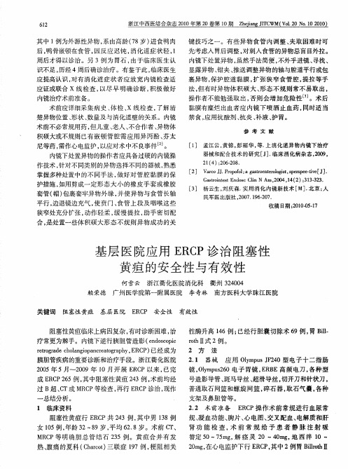
基层医院应用ERCP诊治阻塞性黄疸的安全性与有效性
62 1
浙江 中西医结合杂志 2 1 0 0年第 2 O卷第 1 O期 Z e agJ C hj n T WM( o 2 o 1 0 0 i I V l 0N .02 1 。
其中 1 例为外 源性异 物 , 由高 龄 (8岁 ) 系 7 进食 鸭 肉 后, 鸭骨嵌顿 在食 管 , 因反 应 迟 钝 , 化道 症 状 轻 , 消 1 周后 才得 以诊 治 。另 3例 为 胃石 , 由于 临床 医生认 识 不足 , 历经 4周后确 诊治 疗 。有 鉴 于此 , 临床 医生 应 提高认 识 , 有 消化 道症 状 者 应 放 宽 内镜 检 查适 对 应证 或联合 x线 检 查 , 以尽 早 明确 诊 断 , 积极 做 好
规 、 血功 能 、 片 、 电 图 、 叉 配血 、 凝 胸 心 交 电解质 和肝
1 临床 资料
阻塞性黄疸行 E C R P共 23例, 4 其中男 18例 3 女 15 , 0 例 年龄 3 8 岁 , 2— 9 平均 6 . 岁 。术前 C 、 28 T M C 等 明确胆 总管结石 2 5例。黄疸合 并有发 RP 3
赖荣德 关键词 阻塞性黄疸
广州医学院第一 附属医院
李奇林
南方医科大学珠江医院
基层 医院 E C 安全性 RP
有效性
阻塞性黄疸临床上病因复杂 , 有时诊断困难 , 治
疗 常更为 棘手 。内镜下逆 行胰 胆 管造 影 (nocp edsoi c rt gaec0 nipnra gah , R P 已经成 为 e or hl g acet rpy E C ) r d a o o 胰 胆管疾 病 的重要 诊 断和治疗 手段 。浙 江衢化 医院 20 05年 5月~20 O9年 l 开 展 E C 0月 R P以 来 , 已完
ERCP的历史及临床应用

ERCP的历史及临床应用王雪峰;刘颖斌【摘要】自1968年内镜下逆行胰胆管造影术(ERCP)首次问世以来,临床应用已有半个世纪的历史,国内开展也有40多年的时间.经过50年的发展和进步,ERCP从最初单纯应用于胆胰管造影诊断到如今成为融合影像学、细胞学、组织学诊断及胆管取石、支架引流、肿瘤射频消融治疗的综合诊疗技术,已成为诊治胆胰疾病最重要的手段之一.我院于上世纪90年代开展胆胰疾病的ERCP诊治操作,经过20余年的总结与创新,目前在复杂困难ERCP的开展方面已取得一定的成果,在此我们希望通过分享本中心的经验,与国内同仁共同努力探索,积极创新,与国际ERCP诊治新技术接轨,从而更好的造福患者.【期刊名称】《上海医药》【年(卷),期】2018(039)019【总页数】4页(P20-23)【关键词】内镜下逆行胰胆管造影;乳头肌切开;支架引流【作者】王雪峰;刘颖斌【作者单位】上海交通大学医学院附属新华医院普外科上海 200092;上海交通大学医学院附属新华医院普外科上海 200092【正文语种】中文【中图分类】R657.4;R657.51 ERCP五十年历史回顾ERCP(endoscopic retrograde cholangiopancreatography)即内镜下逆行胰胆管造影,是一种对于肝胆胰系统疾病无创或微创的诊治方法,距离其问世已有50年的历史。
1968年,乔治华盛顿大学的McCune[1]利用侧视纤维十二指肠镜完成了十二指肠乳头的首次插管。
这种内镜下的物镜与目镜不在同一轴线上,而是形成90°角,恰好适合于观察位于侧壁的十二指肠乳头,并能在直视下进行插管操作。
他们组装了一组内窥镜下胆道和胰管插入装置,在Eder型纤维十二指肠镜上放置了导管,并在直视下用球囊对Vater的乳头进行插管,首次报道了该技术的临床应用(图1、2),但当时的插管成功率仅为25%。
1970年,日本学者Oi [2]进行了进一步的研究和改进,报道了60例成功的ERCP操作经验,使ERCP逐渐广泛应用于世界临床,成为胆胰疾病的重要诊断技术。
经ERCP取胆汁检测淀粉酶在胰胆管合流异常诊治中的意义_邹树
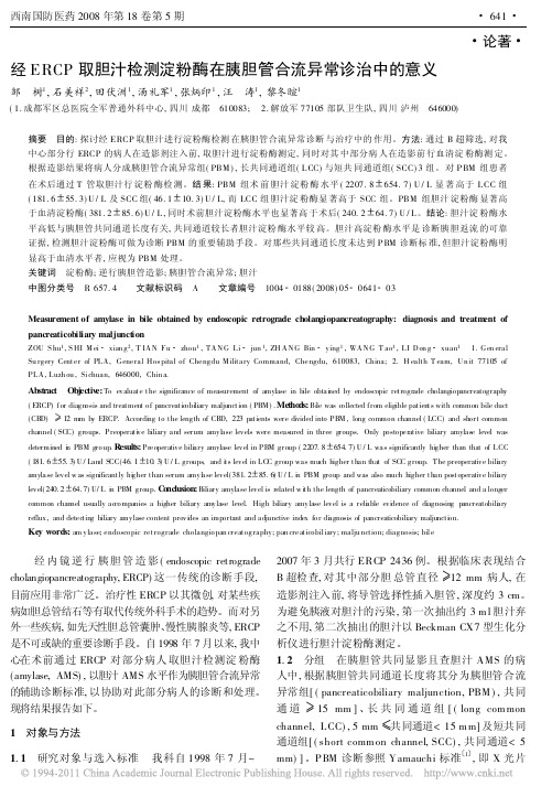
论著 经ERCP取胆汁检测淀粉酶在胰胆管合流异常诊治中的意义邹 树1,石美祥2,田伏洲1,汤礼军1,张炳印1,汪 涛1,黎冬暄1(1.成都军区总医院全军普通外科中心,四川成都 610083; 2.解放军77105部队卫生队,四川泸州 646000)摘要 目的:探讨经ERCP取胆汁进行淀粉酶检测在胰胆管合流异常诊断与治疗中的作用。
方法:通过B超筛选,对我中心部分行ERCP的病人在造影剂注入前,取胆汁进行淀粉酶测定,同时对其中部分病人在造影前行血清淀粉酶测定。
根据造影结果将病人分成胰胆管合流异常组(PBM),长共同通道组(L CC)与短共同通道组(SCC)3组。
对P BM组患者在术后通过T管取胆汁行淀粉酶检测。
结果:PBM组术前胆汁淀粉酶水平(2207.8 654.7)U/L显著高于L CC组(181.6 55.3)U/L及SCC组(46.1 10.3)U/L,而LCC组胆汁淀粉酶显著高于SCC组。
PBM组胆汁淀粉酶显著高于血清淀粉酶(381.2 85.6)U/L,同时术前胆汁淀粉酶水平也显著高于术后(240.2 64.7)U/L。
结论:胆汁淀粉酶水平高低与胰胆管共同通道长度有关,共同通道较长者胆汁淀粉酶水平较高。
胆汁高淀粉酶水平是诊断胰胆返流的可靠证据,检测胆汁淀粉酶可做为诊断PBM的重要辅助手段。
对那些共同通道长度未达到P BM诊断标准,但胆汁淀粉酶明显高于血清水平者,应视为PBM处理。
关键词 淀粉酶;逆行胰胆管造影;胰胆管合流异常;胆汁中图分类号 R657.4 文献标识码 A 文章编号 1004 0188(2008)05 0641 03Measurement of amylase in bile obtained by endoscopic retrograde cholangiopancreatography:diagnosis and treatm ent of pancreaticobiliary maljunctionZOU S hu1,S HI M ei xian g2,T IAN Fu zhou1,TANG Li jun1,ZH ANG Bin ying1,WANG T ao1,LI Don g xuan1 1.Gen eral Su rgery Center of PLA,General Hos pital of Chengdu M ilitary Command,Chengdu,610083,China; 2.H ealth T eam,Un it77105of PLA,Luzh ou,Sichuan,646000,Chin a.Abstract Objectiv e:To evaluate the significance of measurement of amylase in bile obtai ned by endoscopic retrograde cholangiopancreatography (ERCP)for diagnosis and treatment of pancreatic obi liary maljunction(PBM).Metho ds:Bi le was c ollected from eligible patients with common bile duct (CBD) 12mm by ERCP.Accordi ng to the length of CBD,223patients were divided into PBM,long common channel(LCC)and short common channel(SCC)groups.Preoperati ve biliary and serum amyl ase levels were meas ured in three groups.Only postoperative biliary amylas e level was determined i n PBM group.R esults:Preoperative bili ary amylase level in PBM group(2207.8 654.7)U/L was significantly hi gher than that of LCC (181.6 55.3)U/Land SCC(46.1 10.3)U/L groups,and i ts level in LCC group was much higher than that of SCC group.The preoperative bili ary amylase level w as significantly hi gher than serum amyl ase level(381.2 85.6)U/L i n PBM group and was also much higher than postoperative bili ary level(240.2 64.7)U/L i n PBM group.C o nclusion:Biliary amylase level is related w i th the length of pancreaticobiliary common channel and a longer common channel usually ac companies a higher biliary amylase level.High bi liary amylase level is a reli able evi dence of diagnosing pancreatobili ary reflux,and detec ting biliary amylase content provi des an important and adjunctive index for di agnosis of pancreaticobiliary maljuncti on.Key words:am ylase;endoscopic retrograde cholangiopan creatography;pan creaticobiliary;malju nction;diagnosis;bile经内镜逆行胰胆管造影(endoscopic retrograde cholangiopancreatography,ERCP)这一传统的诊断手段,目前应用非常广泛。
胰腺囊性病变的影像表现与临床特点(下)

国际医学放射学杂志IntJMedRadiol2020Nov 鸦43穴6雪胰腺囊性病变的影像表现与临床特点(下)徐建国1唐光健2彭泰松1赵丽丽1于萍1任龙飞1许志高1【摘要】胰腺囊性病变(PCL)是胰腺上皮和间质组织发生囊腔病变的一大类疾病,以胰腺内囊性包块为主要特征,具有不同的病因、临床和组织病理学特点。
本文的前两部分介绍了胰腺炎症相关囊性病变(包括胰腺假性囊肿与胰腺包裹性坏死)与胰腺真性囊肿(包括孤立性胰腺上皮囊肿、von Hippel-Lindau 病、多囊肾和囊性纤维化)以及常见的胰腺浆液性囊腺瘤、胰腺黏液性囊腺瘤和胰腺实性假乳头状瘤的影像表现与临床特点。
本篇为最后一部分,就常见的胰腺导管内乳头状黏液瘤与胰腺少见囊性肿瘤予以介绍和分析,以期为临床诊断与治疗提供重要依据。
【关键词】胰腺囊性病变;体层摄影术,X 线计算机;磁共振成像;导管内乳头状黏液瘤;囊性肿瘤中图分类号:R576;R445.2;R445.3文献标志码:APancreatic cystic lesions:imaging findings and clinical features (Part 3)XU Jianguo 1,TANG Guangjian 2,PENGTaisong 1,ZHAO Lili 1,YU Ping 1,REN Longfei 1,XU Zhigao 1.1Department of Radiology,Third People ’s Hospital of Datong City,Datong 037008,China;2Department of Radiology,Peking University First Hospital【Abstract 】Pancreatic cystic lesions (PCLs)are a broad spectrum of diseases with cystic lesions in pancreaticepithelial and interstitial tissues,characterized by cystic mass,with various etiology,clinical and histopathological characteristics.The first two parts of the article introduces the imaging manifestations and clinical features of 1)pancreatitis related cystic lesionincluding pancreatic pseudocyst and pancreatic encapsulated necrosis,2)pancreatic true cysts including isolated pancreatic epithelial cyst,von Hippel Lindau disease,polycystic kidney and cystic fibrosis,and3)the common cystic tumors of the pancreas including serous cystadenoma,mucinous cystadenoma and solid pseudopapilloma of the pancreas.In the last part of this issue,we introduce another common cystic tumor of pancreas (intraductal papillary mucinous neoplasm of the pancreas)and rare cystic tumors of the pancreas,in order to provide an important basis for clinical diagnosis and treatment of pancreatic cystic lesions.【Keywords 】Cystic disease of pancreas;Tomography,X -ray computed;Magnetic resonance imaging;Intraductalpapillary mucinous neoplasm;Cystic tumorIntJMedRadiol,2020,43(6):716-720作者单位:1大同市第三人民医院医学影像科,大同037008;2北京大学第一医院放射科通信作者:唐光健,E-mail:***************.com DOI:10.19300/j.2020.J18513图文讲座3.4胰腺导管内乳头状黏液瘤胰腺导管内乳头状黏液瘤(intraductal papillary mucinous neoplasm,IPMN )实际上并不是囊性肿瘤,由于肿瘤分泌黏液,引起胰腺导管扩张,大体病理与影像表现为伴有囊的病变,故在胰腺囊性病变内一并讨论。
- 1、下载文档前请自行甄别文档内容的完整性,平台不提供额外的编辑、内容补充、找答案等附加服务。
- 2、"仅部分预览"的文档,不可在线预览部分如存在完整性等问题,可反馈申请退款(可完整预览的文档不适用该条件!)。
- 3、如文档侵犯您的权益,请联系客服反馈,我们会尽快为您处理(人工客服工作时间:9:00-18:30)。
创刊号2009年11月南京大学医学院附属鼓楼医院消化科编者按:由邹主任倡导的ERCP 文献快讯终于正式和大家见面了!本刊的目的是为大家提供ERCP方面最新的文献资讯。
在创刊初期也许还存在许多的不足,敬请大家对本刊的形式和内容提出宝贵的意见,使之不断的完善,更好地为大家服务。
本刊初步确定为每月一期,在每月的月底推出,并适时补充最近三年的文献。
本期导读Coté GA等统计了2345例ERCP患者,针对困难的胆管插管患者放置胰管支架有利于提高胆管插管成功率,可以减少预切开的机会。
病例报道:一例胰腺导管内乳头状粘液瘤(IPMN)同时侵犯胃和十二指肠,良性IPMN同时侵犯相邻两个脏器非常少见。
4. Face and construct validity of a computer-based virtual reality simulator for ERCP. Gastrointest Endosc. 2009 Nov 16. [Epub ahead of print]介绍一种计算机模拟ERCP培训方法。
5. Clinical application of intraductal ultrasound during endoscopic retrograd e cholangiopancreatography. Gastrointest Endosc Clin N Am. 2009 Oct;19(4):615-28.介绍了IDUS在诊断胆管不明原因狭窄、充盈缺损及壶腹部肿瘤方面有重要意义。
6. Can endoscopic palliation of large neoplasm increase the risk of pancreatitis after endoscopic retrograde cholangiopancreatography?讨论了ERCP术后胰腺炎的危险因素,认为预防性胰管支架置入是预防术后胰腺炎的有效方法,但需要更多的随机对照研究来证实。
7. Magnetic resonance cholangio-pancreatography versus endoscopic retrograde cholangio-pancreatography in the diagnosis of common bile duct stones: a prospective comparative study. Minerva Med. 2009 Oct;100(5):341-8.一项MRCP与ERCP在诊断胆总管结石方面的前瞻性对照研究,认为MRCP在诊断小结石方面有局限性,但何种病人采取哪种检查方法仍然难以确定。
8. Pancreaticopleural fistula: a rare complication of ERCP-induced pancreatitis. Sut M, Gray R, Ramachandran M, Diamond T.Ulster Med J. 2009 Sep;78(3):185-6. No abstract available. PMID: 19907687 [PubMed - in process]报道一例ERCP术后胰腺炎并发胰胸膜瘘患者,最终行外科手术分析了在胰管正常的患者为了预防ERCP术后胰腺炎放置胰管支架引起的胰管损伤的情况,提示这类患者放置支架需谨慎小心。
注: 所有文献及摘要均来自Pubmed, 如果需要全文可以申请文献传递。
——编者文献摘要1. Endoscopic Appearance of the Minor Papilla Predicts Findings at Pancreatography.Dig Dis Sci. 2009 Oct 29. [Epub ahead of print]Lawrence C, Stefan AM, Howell DA.Medical University of South Carolina, 25 Courtenay Drive, ART 7100A, Charleston, SC, 29425-2900, USA, Lawrench@.BACKGROUND: The minor papilla serves as a site of alternative pancreatic duct drainage via the accessory pancreatic duct. AIMS: The objectives of this study were to assess the endoscopic appearance of the minor papilla for characteristics that might predict increased accessory pancreatic duct flow and hence suggest pathology of the downstream pancreatic ductal system. METHODS: This was a nonrandomized, prospective analysis of consecutively enrolled patients from a tertiary care medical center (Maine Medical Center, Portland, Maine). The study cohort consisted of consecutive patients presenting for endoscopic retrograde cholangiopancreatography (ERCP) without prior pancreaticobiliary endotherapy or ductography. RESULTS: Sixty-four patients received a minor papilla score prior to ERCP. A normal pancreatogram was found in 37 of 64 (57.8%) patients; the remaining 27 (42.2%) patients had an abnormal pancreatogram. The median minor papilla bulge score was 0.49 (range 0-3) in the normal pancreatogram group and 2 (range 0-3) in the abnormal pancreatogram group (P < 0.0001). The median minor papilla orifice score of those with a normal pancreatogram was 0 (range 0-2) compared to 2 (range 0-3) in the abnormal pancreatogram group (P < 0.001). The median minor papilla cumulative score of 1 (range 0-5) for the normal pancreatogram group was significantly less than that for the abnormal pancreatogram group (3, range 0-6, P < 0.0001), resulting in a sensitivity of 96.3% for an abnormal pancreatogram. The minor papilla orifice was noted to be either gaping or actively dripping pancreatic juice in four out of five patients with pancreas divisum. CONCLUSIONS: A minor papilla without bulging or a visible orifice would suggest a normal pancreatogram at ERP. Conversely, an abnormal minor papilla, particularly a patent minor papilla orifice, should raise suspicion of pancreatic ductal pathology and can help direct pancreatic endotherapy at the major or minor papillae.2. Difficult biliary cannulation: use of physician-controlled wire-guided cannulation over a pancreatic duct stent to reduce the rate of precut sphincterotomy (with video).Gastrointest Endosc. 2009 Nov 16. [Epub ahead of print]CotéGA, Ansstas M, Pawa R, Edmundowicz SA, Jonnalagadda SS, Pleskow DK, Azar RR.Current affiliations: Division of Gastroenterology (G.A.C.), Indiana University, Indianapolis, Indiana, Department of Medicine (M.A., S.A.E., S.S.J., R.R.A.), Division of Gastroenterology, Washington University, St. Louis, Missouri, Department of Medicine (R.P., D.K.P.), Division of Gastroenterology, Beth Israel Deaconess Medical Center, Boston, Massachusetts, USA.BACKGROUND: Successful cannulation of the common bile duct (CBD) remains the benchmark for ERCP. Use of a pancreatic duct (PD) stent to facilitate biliary cannulation has been described, although the majority of patients require precut sphincterotomy to achieve CBD cannulation. OBJECTIVE: To report the performance characteristics of using a PD stent in conjunction with physician-controlled wire-guided cannulation (WGC) to facilitate bile duct cannulation. DESIGN: Retrospective cohort. SETTING: Two tertiary care, academic medical centers. PATIENTS: All undergoing ERCP with native papillae. INTERVENTION: In cases of difficult biliary access in which the PD is cannulated, a pancreatic stent is placed. After this, physician-controlled WGC is attempted by using the PD stent to direct the sphincterotome into the biliary orifice. If cannulation is unsuccessful after several minutes, a precut sphincterotomy is performed over the PD stent or the procedure is terminated. MAIN OUTCOME MEASUREMENTS: Frequency of successful bile duct cannulation and precut sphincterotomy. RESULTS: A total of 2345 ERCPs were identified, 1544 with native papillae. Among these, CBD and PD cannulation failed in 16 (1.0%) patients, whereas 76 (4.9%) patients receiveda PD stent to facilitate biliary cannulation. Successful cannulation was achieved in 71(93.4%) of 76 patients, 60 (78.9%) of whom did not require precut sphincterotomy. Complications included mild post-ERCP pancreatitis in 4 (5.3%) and aspiration in 1 (1.3%). Precut sphincterotomy was complicated by hemorrhage, controlled during the procedure in 2 (13.3%) of 15. CONCLUSIONS: Physician-controlled WGC over aPD stent facilitates biliary cannulation while maintaining a low rate of precut sphincterotomy.PMID: 19922927 [PubMed - as supplied by publisher]3. A case of intraductal papillary mucinous neoplasm of the pancreas rupturing both the stomach and duodenum.Gastrointest Endosc. 2009 Nov 16. [Epub ahead of print]Shimizu M, Kawaguchi A, Nagao S, Hozumi H, Komoto S, Hokari R, Miura S, Hatsuse K, Ogata S.Current affiliations: Department of Internal Medicine, Division of Gastroenterology and Hepatology (M.S., A.K., S.N., H.H., S.K., R.H., S.M.), Departments of Surgery (K.H.) and Clinical Pathology (S.O.), National Defense Medical College, Saitama, Japan.BACKGROUND: Intraductal papillary mucinous neoplasm (IPMN) of the pancreas may extend to other organs. However, it is rare for a histopathologically benign IPMN to rupture other organs, particularly multiple organs. There has been no report of a benign IPMN rupturing both the stomach and duodenum. OBJECTIVE: We experienced a very rare case and make personal remarks based on bibliographical consideration. DESIGN: Case report. SETTING: National Defense Medical College. PATIENT: A patient with IPMN. INTERVENTION: EGD, ERCP, and pancreatoduodenectomy. CONCLUSIONS: We report a case of benign IPMN of the pancreas extending to two adjacent organs. A 77-year-old male who was diagnosed as having IPMN by CT, MRI, upper GIF, and ERCP underwent pancreatoduodenectomy for a mass of 4.2 cm in diameter. Pathological examinations revealed that the IPMN was composed of adenoma. Intraluminal nodular growth was observed in the duodenal gland tissue, and abnormal growth was observed in the fistula to the stomach. According to a literature review based on PubMed data up until March 2009, it is rare for a benign IPMN to penetrate two adjacent organs.PMID: 19922925 [PubMed - as supplied by publisher]4. Face and construct validity of a computer-based virtual reality simulator for ERCP. Gastrointest Endosc. 2009 Nov 16. [Epub ahead of print]Bittner JG 4th, Mellinger JD, Imam T, Schade RR, Macfadyen BV Jr.Current affiliations: Department of Surgery (J.G.B., J.D.M., B.V.M.), the Virtual Education and Surgical Simulation Laboratory (J.G.B., J.D.M., T.I., B.V.M.), and the Department of Medicine, Section of Gastroenterology and Hepatology (R.R.S.), Medical College of Georgia School of Medicine, Augusta, Georgia, Department of Surgery (T.I.), Drexel University College of Medicine, Philadelphia, Pennsylvania, USA.BACKGROUND: Currently, little evidence supports computer-based simulation for ERCP training. OBJECTIVE: To determine face and construct validity of a computer-based simulator for ERCP and assess its perceived utility as a training tool. DESIGN: Novice and expert endoscopists completed 2 simulated ERCP cases by using the GI Mentor II. SETTING: Virtual Education and Surgical Simulation Laboratory, Medical College of Georgia. MAIN OUTCOME MEASUREMENTS: Outcomes included times to complete the procedure, reach the papilla, and use fluoroscopy; attempts to cannulate the papilla, pancreatic duct, and common bile duct; and number of contrast injections and complications. Subjects assessed simulator graphics, procedural accuracy, difficulty, haptics, overall realism, and training potential. RESULTS: Only when performance data from cases A and B were combined did the GI Mentor II differentiate novices and experts based on times to complete the procedure, reach the papilla, and use fluoroscopy. Across skill levels, overall opinions were similar regarding graphics (moderately realistic), accuracy (similar to clinical ERCP), difficulty (similar to clinical ERCP), overall realism (moderately realistic), and haptics. Most participants (92%) claimed that the simulator has definite training potential or should be required for training. LIMITATIONS: Small sample size, single institution. CONCLUSIONS: The GI Mentor II demonstrated construct validity for ERCP based on select metrics. Most subjects thought that the simulated graphics, procedural accuracy, and overall realism exhibit face validity. Subjects deemed it a useful training tool. Study repetition involving more participants and cases may help confirm results and establish the simulator's ability to differentiate skill levels based on ERCP-specific metrics.PMID: 19922914 [PubMed - as supplied by publisher]5. Clinical application of intraductal ultrasound during endoscopic retrograde cholangiopancreatography. Gastrointest Endosc Clin N Am.2009 Oct;19(4):615-28.Kundu R, Pleskow D.Division of Gastroenterology, UCSF Fresno, 2823 Fresno Street, 1st Floor Endoscopy Suite, Fresno, CA 93721, USA.Intraductal ultrasound (IDUS) used during endoscopic retrograde cholangiopancreatography (ERCP) can facilitate reliable evaluation of biliary and pancreatic disorders. The smaller diameter, flexibility, and the image quality offered by IDUS devices makes them ideal for evaluating a variety of difficult biliary and pancreatic diseases, especially in undefined strictures, luminal filling defects, and ampullary neoplasms. This article examines the numerous possible roles for IDUS in the evaluation of biliary and pancreatic conditions, as well as in ampullary neoplasms. IDUS is a simple, easy to learn, and safe technique that should be considered an integral tool in the therapeutic endoscopist's armamentarium.PMID: 19917467 [PubMed - in process]6. Can endoscopic palliation of large neoplasm increase the risk of pancreatitis after endoscopic retrograde cholangiopancreatography?Surg Endosc. 2009 Nov 13. [Epub ahead of print]Fanello G, Fiocca F, Benedetti M, Martino G, Marengo M, Meniconi RL, Papini F, Chirletti P.Surg Endosc. 2009 Nov 13. [Epub ahead of print] No abstract available. PMID: 19911230 [PubMed - as supplied by publisher]Related articlesWhen the main pancreatic duct cannulation is obtained,first, as often happens if the neoplastic lesion does not involve the duct, the clinician should place a pancreatic stent at once before proceeding to the CBD. The use of small-caliber (maximum, 5 Fr) short (2–3 cm) stents and elimination of flaps before insertion allow a spontaneous migration in 2 to 3 weeks without the need for endoscopic removal. Pancreatic stent positioning is recommended for patients with suspected or confirmed sphincter of Oddi dysfunction or a history of PEAP orrecurrent pancreatitis and for patients who have undergone repeated pancreatic duct injection, precut sphincterotomy, pancreatic sphincterotomy, or balloon dilation of the biliarysphincter . Although we agree withthese recommendations, we believe that the risk of PEAP could be higher also with voluminous pancreatic tumors. Therefore, for these patients, prophylactic pancreatic stenting may be considered, if technically feasible, to reduce the risk of thiscomplication. Further randomized controlled studies areneeded to validate our proposal.7. Magnetic resonance cholangio-pancreatography versus endoscopic retrograde cholangio-pancreatography in the diagnosis of common bile duct stones: a prospective comparative study.Minerva Med. 2009 Oct;100(5):341-8.Scaffidi MG, Luigiano C, Consolo P, Pellicano R, Giacobbe G, Gaeta M, Blandino A, Familiari L.Department of Medicine and Pharmacology, University of Messina, Messina, Italy - carmeluigiano@libero.it.AIM: As it is a non-invasive method, magnetic resonance cholangiography (MRCP) has almost completely replaced endoscopic retrograde cholangiography (ERCP) in the diagnosis of pancreato-biliary diseases. The aim of this study was to evaluate sensitivity, specificity, diagnostic accuracy, positive predictive value (PPV) and negative predictive value (NPV) of MRCP in diagnosis of choledocholithiasis using ERCP/endoscopic sphincterotomy (ES) as gold standard. METHODS: For this study 140 individuals, suspected for lithiasis of the common bile duct (CBD), were enrolled. After a clinical and biochemical evaluation, patients underwent upper abdominal ultrasonography, then MRCP and diagnostic and/or operative ERCP. RESULTS: Only 120 out of 140 patients completed the study. MRCP diagnosed lithiasis of CBD in 84. ERCP confirmed the lithiasis in 73/84 patients who were submitted to ES. Eleven were negative after ES. ERCP documented stones in 10 patients among the 36 negative at MRCP; stones were detected only in four patients after ES. In 26 out of 36 patients negative at MRCP, ERCP confirmed this response: only 12 out of 26 patients underwent ES. The sensitivity, specificity, diagnostic accuracy, PPV and NPV of MRCP were: 88%, 72%, 83%, 87%, 72%. CONCLUSIONS: As the MRCP diagnostic yield is still limited with small stones, the question of which patient is the best candidate to ERCP/ES is still unsolved.8. Pancreaticopleural fistula: a rare complication of ERCP-induced pancreatitis. Sut M, Gray R, Ramachandran M, Diamond T.Ulster Med J. 2009 Sep;78(3):185-6. No abstract available. PMID: 19907687 [PubMed - in process]9. Significant clinical implications of prophylactic pancreatic stent placement in previously normal pancreatic ducts.Endoscopy.2009 Nov 10. [Epub ahead of print]Bakman YG, Safdar K, Freeman ML.Division of Gastroenterology, Department of Medicine, University of Minnesota, Minneapolis, Minnesota, USA.Pancreatic duct stent placement is increasingly performed for the prevention of pancreatitis after endoscopic retrograde cholangiopancreatography (ERCP); however stents can result in injury especially in normal ducts. The clinical significance and outcomes of subsequent endoscopic therapy are unknown. This study was a retrospective review of the management of symptomatic stent-inducedpancreatic duct injury following stent placement for prevention of post-ERCP pancreatitis in eight patients with previously normal pancreatic ducts. Subsequent treatment included pancreatic sphincterotomy, balloon dilation of stricture, and placement of multiple 3 - 5-Fr soft polymer pancreatic stents. All patients showed improvement or resolution of pancreatic strictures. Five patients had resolution or substantial improvement of pain, one patient showed a fair response with repeated ERCPs, and two patients failed to respond and underwent total pancreatectomy with islet autotransplantation. Pancreatic duct stent-induced ductal injury with significant clinical consequences can occur with conventional polyethylene stents. Endoscopic therapy is moderately effective but some patients develop irreversible damage. Caution should be used when placing standard polyethylene stents in normal ducts. Further research is required to identify safer materials and configurations of pancreatic stents.。
