Effect of americium-241 on luminous bacteria. Role of peroxides
野莓提取物可增强胰腺癌药物功效

2014年9月17日在《Journal of Clinical Pathology》发表的一项研究指出,美国一种本土野莓,可以增强胰腺癌常用化疗药物的有效性。
生物通报道:2014年9月17日在《Journal of Clinical Pathology》发表的一项研究指出,美国一种本土野莓,可以增强胰腺癌常用化疗药物的有效性。
本研究是由英国伦敦国王学院医院和南安普敦大学等处的研究人员完成,表明在化疗周期中添加保健营养品,可以提高常规药物的疗效,特别是难以治疗的癌症,如胰腺癌。
该研究小组检测了野樱莓(黑果腺肋花楸,Aronia melanocarpa)提取物杀死癌细胞的有效性,可能是通过细胞凋亡(程序性细胞死亡),因为早期细胞凋亡的标志出现在治疗细胞中。
云天团野樱莓是生长在北美东部湿地和沼泽地区的一种野生浆果。
其浆果富含维生素和抗氧化剂,包括各种多酚类化合物,这些化合物被认为可清除正常细胞活动的有害副产品。
研究人员之所以选择研究提取物对胰腺癌的影响,是因为胰腺癌的长期预后较差:患者的五年存活率小于5%。
研究人员在实验室采用著名的胰腺癌细胞株(AsPC-1),评估了单独使用化疗药物吉西他滨、单独使用不同程度的商购野樱莓提取物、以及吉西他滨结合野樱莓提取物进行治疗时,对于细胞株的影响如何。
分析表明,用1 ug/ml的野樱莓提取物对胰腺癌细胞进行48小时治疗后,可诱导细胞死亡。
研究人员检测了野樱莓提取物对正常血管内皮细胞的毒性,发现直到最高水平(50 ug/ml)都没有产生什么影响,表明细胞死亡作用的发生,是通过防止新血管形成方式(抗血管生成——对癌细胞生长很重要的一个过程)以外的方式。
南安普敦大学的Bashir Lwaleed评论说:“这些结果非常令人兴奋。
该当提取物与吉西他滨联合使用时,低剂量的提取物可大大增强吉西他滨药物的有效性。
此外,我们发现,低剂量的常规药物是必需的,表明要么化合物一起协同作用,要么该提取物产生一种‘超相加’效应。
每周只需注射一次,3个月即可轻松减掉10斤肥肉能让你管住嘴的减肥神药真的来了 临床大发现

每周只需注射一次,3个月即可轻松减掉10斤肥肉。
能让你管住嘴的减肥神药真的来了临床大发现“管住嘴,迈开腿”简简单单六个字,就道出了减肥的真谛。
然而,面对那么多的美食诱惑,光这前三个字就足以让无数人的减肥大业半途而废了。
不过,好消息来了!最近,肥胖研究领域中的著名期刊《糖尿病,肥胖和代谢》杂志刊登的一项临床研究[1]显示,诺和诺德公司开发的索马鲁肽,可以抑制食欲,让你轻松“管住嘴”。
只需一周注射1次,连续注射12周后,就可减重10斤!而且,在这减轻的体重中,主要还是体内的脂肪组织,药物对除脂肪以外的去脂体重影响很小。
不光有效,还很安全!这项研究的通讯作者,来自英国利兹大学的John Blundell 教授表示,“索马鲁肽的作用是非常令人惊讶的,我们在12周内就观察到了其他减肥药物需要6个月才能达到的效果。
它减少了饥饿感和食欲,让患者能更好地控制饮食摄入。
”[2] John Blundell教授索马鲁肽(Semaglutide)本身是一款针对2型糖尿病的降糖药,主要成分为胰高血糖素样肽-1(GLP-1)类似物。
GLP-1是一种由小肠分泌的激素,在血液中葡萄糖水平升高时促进胰岛素的合成和分泌。
GLP-1进入人体后很容易被酶降解,天然的GLP-1半衰期仅有几分钟,所以,为了让它更长久的工作,研究人员会对它进行一些结构上的改造,在保留功能的同时不那么容易被酶降解。
这样得到的GLP-1类似物药物,比如大名鼎鼎的利拉鲁肽,可以将注射频率减缓到每天1~2次。
而索马鲁肽可以说是它们的“升级版”,在经过改造后,它的半衰期可延长至大约1周,因此注射一次的效果可以维持大约一周的时间[3],对于患者来说更方便。
在不久前公布的全球大型III期临床试验中,索马鲁肽表现优秀,既能控制血糖,还可以保护心血管,这为它在上周赢得了FDA内分泌及代谢药物专家咨询委员会16:0的支持率,不出意外的话,索马鲁肽上市在即[4]。
不少分析人士预测它未来十年内的销售峰值将超百亿,成为治疗2型糖尿病中最好的降糖药。
机械通气临床应用指南(中华重症医学分会2024)
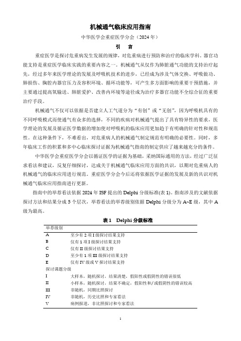
机械通气临床应用指南中华医学会重症医学分会(2024年)引言重症医学是探讨危重病发生发展的规律,对危重病进行预防和治疗的临床学科。
器官功能支持是重症医学临床实践的重要内容之一。
机械通气从仅作为肺脏通气功能的支持治疗起先,经过多年来医学理论的发展及呼吸机技术的进步,已经成为涉及气体交换、呼吸做功、肺损伤、胸腔内器官压力及容积环境、循环功能等,可产生多方面影响的重要干预措施,并主要通过提高氧输送、肺脏爱护、改善内环境等途径成为治疗多器官功能不全综合征的重要治疗手段。
机械通气不仅可以依据是否建立人工气道分为“有创”或“无创”,因为呼吸机具有的不同呼吸模式而使通气有众多的选择,不同的疾病对机械通气提出了具有特异性的要求,医学理论的发展及循证医学数据的增加使对呼吸机的临床应用更加趋于有明确的针对性和规范性。
在这种条件下,不难看出,对危重病人的机械通气制定规范有明确的必要性。
同时,多年临床工作的积累和多中心临床探讨证据为机械通气指南的制定供应了越来越充分的条件。
中华医学会重症医学分会以循证医学的证据为基础,采纳国际通用的方法,经过广泛征求看法和建议,反复仔细探讨,达成关于机械通气临床应用方面的共识,以期对危重病人的机械通气的临床应用进行规范。
重症医学分会今后还将依据医学证据的发展及新的共识对机械通气临床应用指南进行更新。
指南中的举荐看法依据2024年ISF提出的Delphi分级标准(表1)。
指南涉及的文献依据探讨方法和结果分成5个层次,举荐看法的举荐级别依据Delphi分级分为A E级,其中A 级为最高。
表1 Delphi分级标准举荐级别A 至少有2项I级探讨结果支持B 仅有1项I级探讨结果支持C 仅有II级探讨结果支持D 至少有1项III级探讨结果支持E 仅有IV级或V探讨结果支持探讨课题分级I 大样本,随机探讨,结果清楚,假阳性或假阴性的错误很低II 小样本,随机探讨,结果不确定,假阳性和/或假阴性的错误较高III 非随机,同期比照探讨IV 非随机,历史比照和专家看法V 病例报道,非比照探讨和专家看法危重症患者人工气道的选择人工气道是为了保证气道通畅而在生理气道与其他气源之间建立的连接,分为上人工气道和下人工气道,是呼吸系统危重症患者常见的抢救措施之一。
益生菌对阿尔茨海默病作用的研究进展
益生菌对阿尔茨海默病作用的研究进展发布时间:2021-12-14T06:08:15.523Z 来源:《中国结合医学杂志》2021年12期作者:宋鑫萍1,2,李盛钰2,金清1[导读] 阿尔茨海默病已成为威胁全球老年人生命健康的主要疾病之一,患者数量逐年攀升,其护理的经济成本高,给全球经济造成重大挑战。
近年来研究显示,益生菌在适量使用时作为有益于宿主健康的微生物,在防治阿尔茨海默病方面具有积极影响,其作用机制可能通过调节肠道菌群,影响神经免疫系统,调控神经活性物质以及代谢产物,通过肠-脑轴影响该病发生和发展。
宋鑫萍1,2,李盛钰2,金清11.延边大学农学院,吉林延吉 1330022.吉林省农业科学院农产品加工研究所,吉林长春 130033摘要:阿尔茨海默病已成为威胁全球老年人生命健康的主要疾病之一,患者数量逐年攀升,其护理的经济成本高,给全球经济造成重大挑战。
近年来研究显示,益生菌在适量使用时作为有益于宿主健康的微生物,在防治阿尔茨海默病方面具有积极影响,其作用机制可能通过调节肠道菌群,影响神经免疫系统,调控神经活性物质以及代谢产物,通过肠-脑轴影响该病发生和发展。
本文综述了近几年来国内外益生菌对阿尔茨海默病的作用进展,以及其预防和治疗阿尔茨海默病的潜在作用机制。
关键词:益生菌;阿尔茨海默病;肠道菌群;机制Recent Progress in Research on Probiotics Effect on Alzheimer’s DiseaseSONG Xinping1,2,LI Shengyu2,JI Qing1*(1.College of Agricultural, Yanbian University, Yanji 133002,China)(2.Institute of Agro-food Technology, Jilin Academy of Agricultural Sciences, Chanchun 130033, China)Abstract:Alzheimer’s disease has become one of the major diseases threatening the life and health of the global elderly. The number of patients is increasing year by year, and the economic cost of nursing is high, which poses a major challenge to the global economy. In recent years, studies have shown that probiotics, as microorganisms beneficial to the health of the host, have a positive impact on the prevention and treatment of Alzheimer’s disease. Its mechanism may be through regulating intestinal flora, affecting the nervous immune system, regulating the neuroactive substances and metabolites, and affecting the occurrence and development of the disease through thegut- brain axis. This paper reviews the progress of probiotics on Alzheimer’s disease at home and abroad in recent years, as well as its potential mechanism of prevention and treatment.Key words:probiotics; Alzheimer’s disease; gut microbiota; mechanism阿尔茨海默病(Alzheimer’s disease, AD),系中枢神经系统退行性疾病,属于老年期痴呆常见类型,临床特征主要包括:记忆力减退、认知功能障碍、行为改变、焦虑和抑郁等。
腺苷A2A受体(A2AR)拮抗剂ZM241385对视网膜脱离动物模型视网膜光感受器细胞的保护作用

欁欁欁欁欁欁欁欁欁欁欁欁欁欁欁欁欁欁欁欁欁欁欁欁欁欁欁欁欁欁欁欁欁欁欁欁欁欁欁欁欁欁欁欁欁欁欁欁欁欁欁欁欁欁欁欁欁欁欁欁欁欁欁欁欁欁欁欁欁欁欁欁欁欁欁欁欁欁氉氉氉氉引文格式:高莎,沈玺.腺苷A2A受体(A2AR)拮抗剂ZM241385对视网膜脱离动物模型视网膜光感受器细胞的保护作用[J].眼科新进展,2022,42(7):510 513,519.doi:10.13389/j.cnki.rao.2022.0104【实验研究】腺苷A2A受体(A2AR)拮抗剂ZM241385对视网膜脱离动物模型视网膜光感受器细胞的保护作用△高 莎 沈 玺欁欁欁欁欁欁欁欁欁欁欁欁欁欁欁欁欁欁欁欁欁欁欁欁欁欁欁欁欁欁欁欁欁欁欁欁欁欁欁欁欁欁欁欁欁欁欁欁欁欁欁欁欁欁欁欁欁欁氉氉氉氉作者简介:高莎(ORCID:0000 0002 4168 2488),女,1987年10月出生,上海人,博士。
研究方向:玻璃体视网膜疾病。
E mail:gaosha_1343@126.com通信作者:沈玺(ORCID:0000 0001 9669 6521),男,1970年12月出生,上海人,博士,教授,主任医师。
研究方向:玻璃体视网膜疾病。
E mail:carl_shen2005@126.com收稿日期:2022 01 29修回日期:2022 04 27本文编辑:董建军△基金项目:国家自然科学基金项目(编号:81970805)作者单位:200025 上海市,上海交通大学医学院附属瑞金医院眼科【摘要】 目的 探讨腺苷A2A受体(A2AR)拮抗剂ZM241385对视网膜脱离(RD)动物模型视网膜光感受器细胞的保护作用。
方法 选取健康雄性SPF级C57BL/6J小鼠48只作为研究对象。
将实验小鼠随机分为对照+二甲基亚砜(DMSO)组、对照+ZM241385组、RD+DMSO组、RD+ZM241385组,每组12只。
RD模型建立后,实验小鼠立即腹腔注射ZM241385(3mg·kg-1,剂量不超过0.2mL)或同体积DMSO,连续给药3d。
氯化锂对地塞米松诱导的大鼠胰岛β细胞凋亡的影响及其机制
ABSTRACT Objective: To construct the apoptosis model of islet INS-1 cells of the rats induced by dexamethasone (Dex) , and to investigate the effect of lithium chloride (LiCl) on the apoptosis of the islet 4 cclls induced by Dex and its possible mechanism. Methods: The INS-1 cells were divided into control group, 0. 1 gmol • L_lDex group and LiCl+0. 1 gmol • L_l Dex group. TUNEL staining and Annexin V /PI staining methods were used to detect the apoptotic rates of the INS-1 cells in various groups ; Real-time PCR method was used to detect the expression levels of superoxide dismutase (SOD), inducible-nitric oxidesynthase (iNOS) , NADPH oxidase 4 (Nox4), NADPH oxidase (p47phox), and glycogen-synthase tinase-30 (GSK30) mRNA in theINS-1 cclls in various groups; Western blotting method was used to detect the expression levels of GSK-34, p-GSK-30, SOD, iNOS and Nox4
治疗糖尿病的方法和能够治疗糖尿病的组合物[发明专利]
![治疗糖尿病的方法和能够治疗糖尿病的组合物[发明专利]](https://img.taocdn.com/s3/m/faf51be503d8ce2f01662366.png)
专利名称:治疗糖尿病的方法和能够治疗糖尿病的组合物专利类型:发明专利
发明人:亚龙·布拉姆,埃胡德·加齐特
申请号:CN201180037836.3
申请日:20110602
公开号:CN103037891A
公开日:
20130410
专利内容由知识产权出版社提供
摘要:公开了一种物质组合物,其包含分离的人胰岛淀粉样多肽(IAPP)的寡聚体。
也公开了识别其的抗体。
还公开了所述物质组合物及所述抗体的用途。
申请人:雷蒙特亚特特拉维夫大学有限公司
地址:以色列特拉维夫
国籍:IL
代理机构:北京康信知识产权代理有限责任公司
更多信息请下载全文后查看。
利拉鲁肽药物的作用原理及与其他肠促胰素药物的比较

第16页
4、利拉鲁肽与艾塞那肽药品比较
利拉鲁肽药物的作用原理及与其他肠促胰素药物的比较
第17页
基于肠促胰素药品分类
利拉鲁肽药物的作用原理及与其他肠促胰素药物的比较
第18页
利拉鲁肽及艾塞那肽比较
97% 氨基酸与人GLP-1同源
53% 氨基酸与人GLP-1同源
利拉鲁肽药物的作用原理及与其他肠促胰素药物的比较
第9页
两种主要肠促胰素比较
GLP-1
GIP
主要合成部位
L 细胞(回肠和结肠)
K 细胞 (十二指肠和空肠)
2型糖尿病患者中分泌
是
否
餐后胰高糖素
是
否
食物摄入
是
否
延缓胃排空
是
否
促进 β 细胞增殖
是
是
促进胰岛素生物合成
是
是
利拉鲁肽药物的作用原理及与其他肠促胰素药物的比较
利拉鲁肽药物的作用原理及与其他肠促胰素药物的比较
第8页
肠促胰素效应:
口服葡萄糖和静脉注射葡萄糖效应比较
静脉血糖浓度(mg/dL)
时间(分钟)
C肽 (nmol/L)
200
100
0
01
60
120
180
01
60
120
180
0.0
0.5
1.0
1.5
2.0
时间(分钟)
02
02
肠促胰素效应
口服葡萄糖
静脉用葡萄糖
胰岛素抵抗
胰高糖素抑制不足细胞功效失调
胃肠道吸收葡萄糖
慢性β细胞功效衰竭
胰岛素分泌不足β细胞功效异常
反义多重耐药性基因抑制眼睫状体非色素上皮细胞容积激活性氯电流

反义多重耐药性基因抑制眼睫状体非色素上皮细胞容积激活性氯电流陈丽新;王立伟【期刊名称】《中国病理生理杂志》【年(卷),期】2000(016)007【摘要】目的:研究多重耐药性(MDR1)基因及其产物P糖蛋白(P-gp)与容积激活性Cl-电流的关系.方法:用膜片钳全细胞记录技术记录牛眼睫状体非色素上皮(NPCE)细胞容积激活性Cl-电流,反义寡核苷酸阻抑细胞MDR1基因表达,在激光共聚焦显微镜下检测细胞P-gp免疫荧光.结果:P-gp免疫荧光与人源反义MDR1呈剂量依赖性减弱关系,容积激活性Cl-电流被人源反义MDR1特异性地部分阻抑,电流潜伏期延长,峰电流值减少,电流抑制率与人源反义MDR1呈现剂量依赖性增强关系,(r=0.99, P<0.01). P-gp表达抑制率和容积激活性Cl-电流抑制率高度正相关(r=0.99, P<0.01).结论:MDR1基因及其产物P-gp参与NPCE细胞容积激活性Cl-电流的形成.【总页数】5页(P658-662)【作者】陈丽新;王立伟【作者单位】广东医学院生理教研室,广东,湛江,524023;广东医学院生理教研室,广东,湛江,524023【正文语种】中文【中图分类】Q25【相关文献】1.多重耐药基因在睫状体上皮细胞容积调节中的作用 [J], 陈丽新;王立伟2.局部碳酸酐酶抑制剂对睫状体非色素上皮细胞AQP1表达的影响 [J], 李嘉文;李平华;张黎3.人眼睫状体非色素上皮细胞心钠素mRNA的表达 [J], 梁平;陈子庆;苏恩本;卞春及;朱兰兰;周惠英;丁小键4.睫状体色素上皮细胞容积激活性氯电流(英文) [J], 陈丽新;王立伟5.MDR1基因在睫状体色素上皮细胞容积激活性氯电流中的作用(英文) [J], 陈丽新;王立伟;Tim JACOB3因版权原因,仅展示原文概要,查看原文内容请购买。
乳酸脱氢酶241

乳酸脱氢酶241
(实用版)
目录
1.乳酸脱氢酶 241 的概述
2.乳酸脱氢酶 241 的功能和作用
3.乳酸脱氢酶 241 的应用领域
4.乳酸脱氢酶 241 的研究现状和未来发展
正文
乳酸脱氢酶 241(Lactate Dehydrogenase 241,简称 LDH 241)是一种酶,属于乳酸脱氢酶家族的一员,主要功能是催化乳酸(Lactic acid)的氧化脱羧反应,将其转化为丙酮酸(Pyruvic acid)。
乳酸脱氢酶 241 的功能和作用主要体现在生物体内。
在人和动物体内,乳酸脱氢酶 241 参与糖酵解过程,这一过程在细胞进行无氧代谢时产生乳酸。
通过乳酸脱氢酶 241 的作用,乳酸被转化为丙酮酸,丙酮酸进一步参与有氧代谢,产生能量。
因此,乳酸脱氢酶 241 在生物体内扮演着能量代谢的重要角色。
乳酸脱氢酶 241 的应用领域广泛。
首先,在生物医学领域,乳酸脱氢酶 241 可以作为研究糖酵解过程、细胞能量代谢、疾病发生发展机制的重要工具。
此外,在运动生理学领域,乳酸脱氢酶 241 的研究有助于了解运动过程中体内乳酸的生成和清除机制,为提高运动员运动能力和预防运动疲劳提供理论依据。
在食品工业中,乳酸脱氢酶 241 可用于生产乳酸,乳酸作为一种重要的食品添加剂,具有防腐、抗菌、调节酸度等作用。
乳酸脱氢酶 241 的研究现状较为成熟,但仍有许多研究者在不断深入研究,以期发现更多有关乳酸脱氢酶 241 的新机制和新应用。
溶组织内阿米巴分泌的一种降低炎症前期细胞趋化因子的抗炎症寡肽
溶组织内阿米巴分泌的一种降低炎症前期细胞趋化因子的抗炎
症寡肽
佚名
【期刊名称】《国际医学寄生虫病杂志》
【年(卷),期】2005(032)001
【摘要】无
【总页数】2页(P42-43)
【正文语种】中文
【相关文献】
1.溶组织内阿米巴的一种表面蛋白酪氨酸磷酸酯酶通过破坏Hela细胞的肌动蛋白纤维诱导细胞分裂 [J], 郭肖岩;何纳
2.炎症介质通过刺激炎症细胞分泌炎症因子促进晶状体细胞增殖 [J], 赵婷婷;刘红;陈章明;
3.炎症介质通过刺激炎症细胞分泌炎症因子促进晶状体细胞增殖 [J], 赵婷婷;刘红;陈章明
4.溶组织内阿米巴的主要化学元素与抗阿米巴药物疗效观察 [J], 程燕芳;陈金富;刘光英
5.Nitazoxanide治疗小肠贾第鞭毛虫、溶组织内阿米巴/迪斯帕内阿米巴引起的腹泻的随机、双盲、安慰剂对照研究 [J], 邵红霞
因版权原因,仅展示原文概要,查看原文内容请购买。
胰岛素与脱皮激素调控果蝇个体大小的信号机制

胰岛素与脱皮激素调控果蝇个体大小的信号机制
陈苏;李传芬;李子剑;李珍祖;李云龙
【期刊名称】《生命的化学》
【年(卷),期】2006(26)4
【摘要】在动物生长调节和个体大小调控过程中,胰岛素起到了关键作用。
在果蝇的研究中发现,胰岛素主要是通过与类固醇激素—蜕皮激素(ecdysone)的相互作用来进行调节的。
最近认为,蜕皮激素通过控制幼虫的生长速率来调节个体的最终大小。
【总页数】3页(P291-293)
【关键词】胰岛素;FOXO家族转录因子;S6K;蜕皮激素
【作者】陈苏;李传芬;李子剑;李珍祖;李云龙
【作者单位】山东师范大学生命科学学院动物抗性生物学省级重点实验室
【正文语种】中文
【中图分类】Q25
【相关文献】
1.微RNA-30家庭与骨骼肌胰岛素抵抗:磷脂酰肌醇-3-激酶信号通路介导胰岛素抵抗的调控机制 [J], 陈丹;张红梅
2.我国科学家发现Ci在果蝇卵巢体细胞中调控Hippo信号通路的机制 [J], 郑庆伟;
3.黑腹果蝇保幼激素反应区(JHRR)与核蛋白结合能力调控的分子机制 [J], 何倩毓;
张原熙
4.胰岛素样生长因子结合蛋白-3通过细胞外信号调节激酶1/2信号通路调控多胶母细胞瘤增殖的机制 [J], 扈玉华;吴玉鹏;王刘帅;王程业;吴建瓴
5.卵泡膜细胞的胰岛素抵抗对其雄激素合成的影响以及胰岛素增敏剂的调控机制[J], 高磊;李威;吴效科;侯丽辉;J.F.STRAUSS;匡海学
因版权原因,仅展示原文概要,查看原文内容请购买。
瑞香素杀疟原虫裂殖体的作用(英文)
瑞香素杀疟原虫裂殖体的作用(英文)王琴美;倪奕昌;徐月琴;哈淑华;蔡王月【期刊名称】《中国寄生虫学与寄生虫病杂志》【年(卷),期】2000(18)4【摘要】[目的 ]研究中药瑞香素在体外和体内的杀裂殖体作用。
[方法 ]在恶性疟原虫FCCl株常规体外培养中测试瑞香素杀裂殖体活性 ,并按“四天抑制性试验”在感染伯氏疟原虫ANKA株的小鼠中测定不同剂量瑞香素的体内抗疟活性。
[结果 ]体外试验中瑞香素在 1~10 μmol L剂量范围内有明显杀灭裂殖体作用 ,而体内试验中按D4减虫率与感染鼠在 30d内的平均生存天数评价 ,5 0或 10 0mg kg .d- 1 × 4d瑞香素灌胃以及 10 ,5 0或 10 0mg kg.d- 1 × 4d瑞香素腹腔注射给药在伯氏鼠疟原虫ANKA株感染鼠中的抗疟作用与 10mg kg .d- 1 × 4d氯喹(CQ)灌胃的疗效相似。
[结论 ]瑞香素在体外和体内均有一定的杀裂殖体作用。
【总页数】3页(P204-206)【关键词】瑞香素;恶性疟原虫;杀裂殖体活性【作者】王琴美;倪奕昌;徐月琴;哈淑华;蔡王月【作者单位】中国预防医学科学院寄生虫病研究所【正文语种】中文【中图分类】R286.861;R531.305【相关文献】1.双氢青蒿素和奎宁对恶性疟原虫早期配子体作用的随机比较 [J], 陈沛泉;简华香;符林春;李国桥;范梨盛;王炳西2.瑞香素抗红外期疟原虫作用的研究 [J], 刘云光;王琴美;徐月琴;倪齐珍;倪奕昌3.蒿甲醚与瑞香素伍用对感染伯氏疟原虫小鼠的治疗作用 [J], 郭俭;倪奕昌;吴嘉彤;王琴美4.瑞香素体外抗疟作用与其铁螯合能力的关系(英文) [J], 牟凌云;王琴美;倪奕昌5.CPU86017及其旋光异构体对L-甲状腺素致大鼠心肌病异常的calcineurin和NFκB基因表达的抑制作用(英文) [J], 齐敏友;夏绘晶;戴德哉;汤晓赟;苏蔚;张灿因版权原因,仅展示原文概要,查看原文内容请购买。
外源性IL-24基因对大鼠C6胶质瘤细胞的抑制作用的开题报告
外源性IL-24基因对大鼠C6胶质瘤细胞的抑制作用
的开题报告
题目:外源性IL-24基因对大鼠C6胶质瘤细胞的抑制作用
背景:脑胶质瘤是中枢神经系统最常见的原发性恶性肿瘤之一,治疗效果仍然不佳。
因此,寻找有效的治疗方法是当前研究的热点。
IL-24(干扰素样细胞因子24)可以诱导肿瘤细胞凋亡,抑制其生长和增殖,并对恶性肿瘤细胞具有显著的治疗效果。
但是,目前对于外源性IL-24基因对大鼠C6胶质瘤细胞的抑制作用还需要深入探究。
研究方法:选取C6胶质瘤细胞,将其分成对照组和实验组。
实验组细胞将通过转染外源性IL-24基因来观察其对细胞增殖、凋亡以及表达核因子κB(NF-κB)和类风湿因子(TNF-α)等的影响。
同时,还将采用MTT法、流式细胞术和Western blot等方法来检测细胞的生长和凋亡情况,以及关键蛋白表达水平的变化。
研究意义:本研究可以为深入了解外源性IL-24基因对大鼠C6胶质瘤细胞的抑制作用提供理论基础。
结果有助于为脑胶质瘤治疗提供新的治疗方法和手段,促进临床治疗疗效提高。
- 1、下载文档前请自行甄别文档内容的完整性,平台不提供额外的编辑、内容补充、找答案等附加服务。
- 2、"仅部分预览"的文档,不可在线预览部分如存在完整性等问题,可反馈申请退款(可完整预览的文档不适用该条件!)。
- 3、如文档侵犯您的权益,请联系客服反馈,我们会尽快为您处理(人工客服工作时间:9:00-18:30)。
Effect of americium-241on luminous bacteria.Role of peroxidesM.Alexandrova a ,*,T.Rozhko a ,G.Vydryakova b ,N.Kudryasheva a ,ba Siberian Federal University,Svobodny 79,660041Krasnoyarsk,RussiabInstitute of Biophysics SB RAS,Akademgorodok 50,660036Krasnoyarsk,Russiaa r t i c l e i n f oArticle history:Received 25October 2010Received in revised form 12February 2011Accepted 13February 2011Available online 8March 2011Keywords:Luminous bacteria Radiotoxicity Am-241Hormesis Peroxidea b s t r a c tThe effect of americium-241(241Am),an alpha-emitting radionuclide of high speci fic activity,on lumi-nous bacteria Photobacterium phosphoreum was studied.Traces of 241Am in nutrient media (0.16e 6.67kBq/L)suppressed the growth of bacteria,but enhanced luminescence intensity and quantum yield at room temperature.Lower temperature (4 C)increased the time of bacterial luminescence and revealed a stage of bioluminescence inhibition after 150h of bioluminescence registration start.The role of conditions of exposure the bacterial cells to the 241Am is discussed.The effect of 241Am on luminous bacteria was attributed to peroxide compounds generated in water solutions as secondary products of radioactive decay.Increase of peroxide concentration in 241Am solutions was demonstrated;and the similarity of 241Am and hydrogen peroxide effects on bacterial luminescence was revealed.The study provides a scienti fic basis for elaboration of bioluminescence-based assay to monitor radiotoxicity of alpha-emitting radionuclides in aquatic solutions.Ó2011Elsevier Ltd.All rights reserved.1.IntroductionRecent years have seen a change in the approach in radio-ecological studies:biota in toto might be considered as a target of radiation impact,with the human included as a part of biota and integrated into the biosphere by a multiplicity of functional interrelations.Microorganisms are the simplest and perhaps most basic part of the biosphere,and their physiological indices can serve as an indi-cator of the state of the biosphere on the whole.Hence,microor-ganisms can be used as bioassays to monitor environmental radiotoxicity.Generally,the main feature of all bioassays is an integral response that accounts for non-additivity of the effects of numerous environmental pollutants and natural components.In this case,the bioassays indicate media toxicity,considering that toxic effect of a sum of compounds (including radioactive ones)can be higher or lower than the sum of the effects of these compounds.Radiometric analyses per se do not characterize medium hazard to living organisms since they do not consider the integral effect of all substances and difference in sensitivity of various organisms.It is supposed (Kudryasheva et al.,1998)that only a combination ofradiometric,chemical and biological methods can provide complete information on ecological state of a medium.Assay systems based on luminous marine bacteria are good candidates for radiotoxicity monitoring.Bacterial bioluminescent assays are widely used to monitor environmental toxicity for more than forty years (Girotti et al.,2008;Ivask et al.,2009;Kudryasheva et al.,2003;Roda et al.,2009;Thomas et al.,2009),and now they are the traditional and important biotechnological applications of bioluminescence phenomenon.The tested parameter here is the luminescence intensity of bacteria that can be easily measured instrumentally.The advantages of bioluminescent assays are their high sensitivity,simplicity,rapidity (1e 3min),and availability of the devices for toxicity registration (Gitelson and Kratasyuk,2002).Bacterial bioluminescent assays are humane,as they do not apply high organisms.In 1990,the bioluminescent system of enzymatic coupled reactions was suggested as a toxicity assay (Kratasyuk,1990);its applications were developed (Esimbekova et al.,2007,2009).Mechanisms of exogenous compounds interactions with enzyme systems were reviewed in (Kudryasheva,2006a,b ).For the first time,bacterial bioluminescence was used to monitor radiation toxicity in Min et al.(2003).In this work,the effect of gamma-ray irradiation on a recombinant Escherichia coli strain was studied.In our previous works (Rozhko et al.,2007,2008),we studied sensitivity of bioluminescent assay systems to americium-241(241Am),an alpha-emitting radionuclide,the product of pluto-nium radioactive decay.It is known to be increasingly accumulated by the environment.Observations in the waters of the Chernobyl*Corresponding author.Tel.:þ7(391)2494242;fax:þ7(391)2433400.E-mail address:maka-alexandrova@rambler.ru (M.Alexandrova).Contents lists available at ScienceDirectJournal of Environmental Radioactivityjou rnal homepage:www.e lse /locate/jenvrad0265-931X/$e see front matter Ó2011Elsevier Ltd.All rights reserved.doi:10.1016/j.jenvrad.2011.02.011Journal of Environmental Radioactivity 102(2011)407e 411zone contaminated with radioactive fallout demonstrated that aquatic plants accumulate241Am in their biomass in a species-specific manner(Gudkov,2002).Accumulation of241Am by sedi-ments and aquatic plants in the Siberian River Yenisey is currently under study(Bolsunovsky,2010;Zotina et al.,2010).We found that bioluminescent assay systems in vivo and in vitro were shown to be sensitive to241Am in the activity range 0.1e6kBq/L(Rozhko et al.,2007,2008).The addition of241Am to bacterial assay systems was found to result in a50-h activation of bioluminescence intensity,which was followed by inhibition of bioluminescence in all samples of different activity concentrations. The doses of0.25e1.5Gy were delivered to bacterial cells by the time of bioluminescence inhibition.The adaptation of bacterial bioluminescent systems for bioassay purposes requires further basic investigations on the bacteria properties under various irradiation conditions,as well as mecha-nisms of the radionuclide impact on bacterial luminescence as a physiological function of the microorganism.The current paper provides additional data on the influence of alpha radiation on luminous bacteria.We consider chronic effect of 241Am on bacterial growth and bioluminescence.Bioluminescent kinetics at20 C and4 C were analyzed,and relative biolumi-nescence quantum yields were calculated.Accumulation of241Am in cells and DNA was determined.The question of our interest deals with the molecular mecha-nism of bioluminescence activation and inhibition under exposure to radiation.Bioluminescence of bacteria can be considered as a simple model process for understanding activation and inhibition of physiological functions of higher organisms.Radiolysis of water is a known process occurring under ionizing radiation(Fridovich,1998;Halliwell,2003).Secondary products of ionizing radiation(ions and radicals)affect water ecosystems and their inhabitants.Peroxide compounds are an important group among the secondary products of ionizing radiation.With regard to metabolic processes,peroxides are intermediates in a series of metabolic reactions,mainly,oxidation of organic compounds. Depending on concentrations,they can either activate or inhibit the metabolic processes(Kislenko and Berlin,1991;Kudryashov,2004). We hypothesize that peroxide compounds can be responsible for activation and inhibition of the bioluminescence function of lumi-nous bacteria.In our experiments,we monitor peroxides in241Am solutions and compare the effects of hydrogen peroxide and241Am on luminous bacteria P.phosphoreum,thus elucidating the role of peroxides in activation and inhibition of bioluminescence.2.Materials and methodsLuminous bacteria P.phosphoreum from the Collection of the Institute of Biophysics SB RAS(CCIBSO863),strain1883IBSO,were applied.Nutrient media used for bacterial growth included:1L H2O,30g NaCl,1g KH2PO4$12H2O,0.5g(NH4)2HPO4,0.2g MgSO4$7H2O,6g Na2HPO4,3ml glycerol,and5g peptone.Reagents included potassium ferricyanide,3%H2O2solution and luminol (Molecular probes Inc.).Solutions of241Am(NO3)3were used as the source of ionizing radiation.Bacteria were grown in30mL nutrient media with241Am(NO3)3 involved;media activity was0.16,0.67,1.7,3.3,6.7kBq/L.Control (without Am)and tested(with Am)bacterial suspensions were examined and compared.Optical density(D)of suspensions was used to monitor bacterial growth.Number of bacterial cells(N)was calculated as:N¼KD. Here,K is a coefficient of linear dependence calculated in prelimi-nary microscopic experiments with thin-layer suspensions by method of direct enumeration(Bianchi and Giuliano,1996;Kepner and Pratt,1994).Cells were visualized withfluorochrome DAPI.Fig.1presents the examples of bacterial growth in control and radioactive nutrient media.Bioluminescence intensity was measured in bacterial suspen-sions sampled at two growth stages:in15,and22h,Fig.1.The samples were placed into microplates;the time-course of their bioluminescence intensity was measured in solutions of3%NaCl at þ20 C.The samples were kept atþ20 C orþ4 C.The volume ratio of bacterial suspension and NaCl solution in microplate samples was1:30.Specific bioluminescence intensity,I s,was calculated as: I s¼I=NHere,I is a bioluminescent intensity of radioactive sample,N is a number of cells in the sample.Values of I s were plotted vs.time of light emission.Relative quantum yields of the samples were calculated as a ratio of areas restricted by bioluminescence kinetics curves in the presence and absence of241Am.Bioluminescent measurements were carried out in four repli-cations;SD values were calculated.Fig.2presents the examples of time-course of I s values in control and radioactive nutrient media.Relative bioluminescence intensity,I rel s,was calculated as I rel s¼I s=I s contr,where I s is a specific bioluminescent intensity in radioactive solution,I s contr is a specific bioluminescent intensity in a control sample without241Am.To assess the influence of H2O2on the bacteria,the effect of H2O2 on bioluminescence was investigated using the following reaction mixture:50m L bacterial suspension,50m L H2O2in3%NaCl. Concentration of H2O2in the mixture varied up to10À3M.Relative bioluminescence intensity,I rel,was calculated as I rel¼I/I contr,where I is a bioluminescent intensity in H2O2solution,I contr is a biolumi-nescent intensity in a control sample without H2O2.Peroxide component in solutions of241Am(NO3)3was deter-mined by the chemiluminescent method(Komagoe and Katsu, 2006).Luminol chemiluminescence was triggered by addition of potassium ferricyanide.Bioluminescence and chemiluminescence intensity was measured by TriStar Multimode Microplate Reader LB941,Berthold Technologies.Optical density of bacterial suspension was registered by colorimeter KFK-2MP,Russia.The number of cells was counted byfluorescence microscope Zeiss Axioskop40with a Filter Set02, Zeiss,Germany.The DNA was extracted with the IsoCode Paper according to manufacturer instructions(Gupta et al.,2010;Lee et al.,2010). Briefly,from8to10m L aliquots of bacterial cultures werespotted Fig.1.Time-course of bacterial growth in a control sample and in the presence of 241Am,1.7kBq/L.M.Alexandrova et al./Journal of Environmental Radioactivity102(2011)407e411 408directly onto the corner of paper and DNA was eluted in a 100m L volume of sterile water,with 10m L used for electrophoresis.Elec-trophoresis was performed in 1%agarose gel in 1ÂTAE buffer.The s oncentration of DNA was determined by a spectrophotometric method (Sagi et al.,2009)with the UVIKON 943double-beam spectrophotometer,Italy.The 70m g samples of DNA were prepared,their radioactivity was analyzed.To determine the accumulation of 241Am in bacterial cells,the bacterial suspensions were centrifuged (6000Âg ,4 C,15min),bacterial and supernatant fractions were separated.The radioac-tivity of 241Am in bacterial,supernatant and DNA samples was measured by gamma-counter Wallac Wizard 1480,PerkinElmer,Finland.Sensitivity of the counter to 241Am is 0.05Bq per sample.The activity concentrations of 241Am were calculated in nutrient solutions (kBq/L),bacterial fraction (%),and DNA (mBq/m g).3.Results and discussion 3.1.Effect of241Am on luminous bacteriaKinetic curves for bacterial growth in a control sample and in the presence of 241Am,1.7kBq/L,are compared in Fig.1.Fig.3presents a number of bacteria in nutrient media of different radioactivity after 22-h bacterial growth.As is seen,the exposure to 241Am suppresses bacterial growth.Bacterial suspensions were sampled for bioluminescence measurements at two stages of bacterial growth e 15and 22h (shown in Fig.1with vertical arrows).The time-courses of biolu-minescent intensity in the control sample and in the presence of 241Am,1.7kBq/L,at room temperature are presented in Fig.2.Fig.4demonstrates the same result in terms of relative bioluminescent intensity,I rel .It is seen that I rel >1during the whole period of light registration.The radioactive sample demonstrated an increase of luminescent intensity (bioluminescence activation)during the whole period of light registration,as compared to that of the control sample.Similar results were obtained in solutions of different activity concentrations.Luminescence quantum yields were calculated for all radioac-tive solutions;they exceeded that of control sample (Fig.5).The time-courses and quantum yield dependences of 15-h and 22-h samples were similar.To increase time of exposure of the bacteria to the 241Am,we prolonged their luminescence lifetime up to 300h (Fig.6)by keeping the bacterial microplate samples at lower temperature.The Figure demonstrates the time-course of relative biolumines-cent intensity of the radioactive sample kept at 4 C.These condi-tions revealed bioluminescence activation (I rel >1)at t <80h being followed by bioluminescence inhibition (I rel <1)at t >150h in the radioactive sample.The change in bioluminescence time-course under exposure to 241Am shown in Fig.6is similar to those described previously (Rozhko et al.,2007).However,the difference occurs under conditions of the current experiment when the bioluminescence inhibition was observed only at lower temperature after 150h of exposure to 241Am (Fig.3),while in the previous paper (Rozhko et al.,2007)the bioluminescence inhibition took place at room temperature after 50h of exposure to 241Am.Most probably,this difference is related to conditions of exposure to 241Am:in this work the bacteria were grown in radioactive nutrient media,while in Rozhko et al.(2007)the 241Am was added to 15-h bacterial suspension according to standard technique of the bioluminescent assay.This difference is probably a demonstration of bacterial adaption ability to the unfavorable stress-factor.Its relation to genetic ‘adaptive response ’(Volkert,1988;Yu et al.,2006)might be considered in further experiments.Hence,the data demonstrate that the onset time of biolumi-nescence inhibition by 241Am depends on conditions of growing of the bacteria (radioactive nutrient media or not),and temperature.Activation of bioluminescence in Figs 2and 4e 6can be ascribed to the hormesis phenomenon (Calabrese and Baldwin,2001;Kaiser,Fig.2.Effect of 241Am on bacterial luminescence at 20 C,15-h bacterial culture:time-course of speci fic bioluminescence intensity (I s )in control and radioactive media,1.7kBq/L.Fig.3.Number of bacteria vs.241Am activity concentrations,A(Am),kBq/L,in 22-hbacterialculture.Fig.4.Effect of Am-241on bacterial luminescence at 20 C,15-h bacterial culture:time-course of relative bioluminescence intensity (I rel s)in radioactive media,1.7kBq/L.M.Alexandrova et al./Journal of Environmental Radioactivity 102(2011)407e 4114092003;Heinz et al.,2010).Activation of the vital functions of various organisms is a well-known effect;it is a general property of all living organisms.Hormesis is attributed to triggering of cell defenseresponse under the influence of low concentrations of toxic compounds,low dose radiation,and other stressors(Yu et al.,2006; Calabrese and Baldwin,2001).Examples of hormesis are nonspe-cific adaptive syndrome of plants(Pakhomova,1995)and stress reaction in animals(Selye,1980).The molecular mechanism of radiation hormesis is a question of high interest.Accumulation of241Am by bacterial cells was evaluated.It was found that the bacteria accumulated60e70%of the radionuclide from any22h suspension irrespective of its activity.The DNA electrophoretic diagram of all samples showed no visible changes. Radioactivity(1.7mBq/m g)was found only in DNA isolated from the sample of maximal activity.3.2.Role of peroxides in effects on luminous bacteriaTo verify the hypothesis on involvement of peroxides into radionuclide effect on the bacterial cells,we compared the effects of hydrogen peroxide(H2O2)and241Am as model compounds on bacterial luminescence(Fig.7and Fig.6,respectively).Fig.7gives the dependence of bioluminescent intensity on H2O2concentration. It is seen that lower concentrations of the peroxide(210À7e10À4M) activate bacterial luminescence,whereas higher concentrations (>10À4M)inhibit it.Activation reaches w300%at C(H2O2)¼1.710À6M.Bioluminescence activation by H2O2is supposed to deal with the increase of rates of metabolic oxidative reactions.One of them is the bioluminescence reaction which includes a peroxide compound (peroxyhemiacetal)as an intermediate(Nemtseva and Kudryasheva, 2007).The excess amount of reaction components is known to inhibit all the reactions and quench electron-excited states offluo-rofores(Lakowicz,2006),thus inhibiting bioluminescence and other metabolic processes.Previously in Remmel et al.(2003),activation and inhibition of bacterial luminescence by hydrogen peroxide was also reported,with Vibrio harveyi strain as an example.The luminol chemiluminescent method was applied to deter-mine peroxides in water solutions of241Am(NO3)3.Fig.8presents the dependence of peroxide concentration on activity of41Am in water solutions.One can see that the increase of peroxide concentration follows the rise of241Am activity.It was found that at A(241Am)¼5kBq/L,the concentration of peroxides exceeds their background concentration up to18times.It is possible that the increase of peroxide concentration takes place in solutions of all alpha-radionuclides of high specific activity.Hence,the peroxides produced in the solution of241Am can be a reason of the processes shown in Figs.2e5.Thus,the datapresentedFig.6.Effect of Am-241on bacterial luminescence at4 C,22-h bacterial culture:time-course of relative bioluminescence intensity(I rel s)in radioactive media,2kBq/L.Fig.7.Relative bioluminescent intensity of P.phosphoreum(I re l)at different concen-trations of H2O2C(H2O2),mM.Fig.5.Relative bioluminescence quantum yields vs.activity concentration of media,A(Am),kBq/L.Fig.8.Concentration of peroxides,C(H2O2),mM,at different activities of241Am inwater solutions,A(Am),kBq/L.M.Alexandrova et al./Journal of Environmental Radioactivity102(2011)407e411410in Figs.6e8support our hypothesis on the involvement of peroxides, secondary products of alpha-decay of241Am,in water solutions,into functioning of bioluminescent system of luminous bacteria.However,the comparison of H2O2concentrations and241Am radioactivity that either activates or inhibits bioluminescence, suggests that peroxides could not be the sole moiety affecting the bacteria.The argument is supported by the data of our last paper (Alexandrova et al.,2010)demonstrating bioluminescence activa-tion by beta-emitting radionuclide,tritium,which does not produce peroxides in water solutions.Probably,another type of particle,hydrated electrons,can be responsible for the change of bioluminescence intensity,too.They can be involved into electron transfer chain of coupled metabolic redox reactions in organisms, thus increasing or decreasing the rates of metabolic processes exposed to ionizing radiation.The results under consideration call for broad investigations of elementary mechanisms of ionizing radiation impact on living organisms.4.ConclusionWe showed that the presence of241Am in nutrient media (0.16e6.7kBq/L)suppressed the growth of bacteria and activated their luminescence at room temperature.Lower temperature increased the time of bacterial luminescence and revealed a stage of bioluminescence inhibition after150h of bioluminescence registration start.Analysis of241Am effects in the current and previous(Rozhko et al.,2007)experiments brought about conclu-sion that bioluminescence inhibition start depends on conditions of bacterial growth(radioactive nutrient media or not),and temper-ature of bioluminescence registration.The results were explained in terms of adaptive response and hormesis-effect in the bacteria exposed to241Am as to a stress-factor.It was found that bacteria accumulated up to60e70%of the 241Am from the nutrient solutions.We related the effects of241Am on luminous bacteria to peroxide compounds which are generated as products of water radiolysis. The suggestion was confirmed by similarity of the effects of241Am and H2O2on bacterial bioluminescence,and the presence of peroxides compounds in the241Am solutions.The alternative mechanism affecting the bacterial bioluminescent intensity is discussed as well.AcknowledgmentsAuthors are thankful to Prof.Bolsunovsky for discussion of the results,Dr.Dementyev for help with radiometric measurements, and Ms Vasyunkina for experiments with hydrogen peroxide.The work was supported by Grants from RFBR N09-08-98002-Sibir_a; RFBR N10-05-01059-a,the Program“Molecular and Cellular Biology”of RAS;the Grant“Leading Scientific School”N 1211.2008.4;the Grant of Ministry of Education and Science RF N 2.2.2.2/5309;Federal Target Program“Research and scientific-pedagogical personnel of innovation in Russia”for2009e2013 years,contract N02.740.11.0766.ReferencesAlexandrova,M.A.,Rozhko,T.V.,Badun,G.A.,Bondareva,L.G.,Vydryakova,G.A., Kudryasheva,N.S.,2010.Effect of tritium on growth and bioluminescence of bacteria P.Phosphoreum.Radiat.Biol.Radioekol.6,613e618.Bianchi,A.,Giuliano,L.,1996.Enumeration of viable bacteria in the marine pelagic environment.Appl.Environ.Microbiol.62,174e177.Bolsunovsky, A.,2010.Artificial radionuclides in sediment of the Yenisei River.Chem.Ecol.26,401e409.Calabrese,E.J.,Baldwin,L.A.,2001.The frequency of U-shaped dose responses in the toxicological literature.Toxicol.Sci.62,330e338.Esimbekova,E.N.,Kratasyuk,V.A.,Torgashina,I.G.,2007.Disk-shaped immobilized multicomponent reagent for bioluminescent analyses:correlation between activity and composition.Enzym.Microb.Technol.40,343e346. Esimbekova, E.N.,Torgashina,I.G.,Kratasyuk,V.A.,parative study of immobilized and soluble NADH:FMN-oxidoreductase-luciferase coupled enzyme system.Biochem.Mosc.74,695e700.Fridovich,I.,1998.Oxygen toxicity:a radical explanation.J.Exp.Biol.201,1203e1209. Girotti,S.,Ferri,E.N.,Fumo,M.G.,Maiolini,E.,2008.Monitoring of environmental pollutants by bioluminescent bacteria.Anal.Chim.Acta608,2e29. Gitelson,J.I.,Kratasyuk,V.A.,2002.Photobiophysics of ecosystems.Ecol.Biophys.Logos,Moscow.Gudkov,D.I.,Zub,L.N.,Derevets,V.V.,Kuz’menko,M.I.,Nazarov,A.B.,Kaglian,A.E., Savitskii,A.L.,2002.90Sr,137Cs,238Pu,239þ240Pu,and241Am radionuclides in macrophytes within the Krasnenskyflood plain:species specificity of concentration and distribution in phytocenosis components.Radiat.Biol.Radioecol.42,419e428.Gupta,V.,Dorsey,G.,Hubbard,A.E.,Rosenthal,P.J.,Greenhouse,B.,2010.Gel versus capillary electrophoresis genotyping for categorizing treatment outcomes in two anti-malarial trials in Uganda.Malar.J.9,19.Halliwell,B.,2003.Free Radical in Biology and Medicine.Oxford University Press,Oxford. Heinz,G.H.,Hoffman,D.J.,Klimstra,J.D.,Stebbins,K.R.,2010.Enhanced reproduc-tion in mallards fed a low level of methyl mercury:an apparent case of hormesis.Environ.Toxicol.Chem.29,650e653.Ivask,A.,Rolova,T.,Kahru,A.,2009.A suite of recombinant luminescent bacterial strains for the quantification of bioavailable heavy metals and toxicity testing.BMC Biotechnol.doi:10.1186/1472-6750-9-41.Kaiser,J.,2003.Hormesis:sipping from a poisoned chalice.Science302,376e379. Kepner,R.L.,Pratt,J.R.,e offluorochromes for direct enumeration of total bacteria in environmental samples:past and present.Microbiol.Rev.58,603e615. Kislenko,V.N.,Berlin,A.A.,1991.Kinetics and mechanism of hydrogen peroxide oxidation of organic substances.Russ.Chem.Rev.60,949e981.Komagoe,K.,Katsu,T.,2006.Porphyrin-induced photogeneration of hydrogen peroxide determined using the luminol chemiluminescence method in aqueous solution:a structure-activity relationship study related to the aggregation of porphyrin.Anal.Sci.22,255e258.Kratasyuk,V.A.,1990.Principles of luciferase biotesting.In:Jezowska-Trzebiatowska,B., Kochel,B.,Stawinski,J.,Strek,I.(Eds.),Biological Luminescence.World Scientific, Singapore,pp.550e558.Kudryasheva,N.S.,2006a.Bioluminescence and exogenous compounds.Physico-chemical basis for bioluminescent assay.J.Photochem.Photobiol.B-Biol.83,77e86. Kudryasheva,N.S.,2006b.Nonspecific effects of exogenous compounds on bacterial bioluminescent enzymes:fluorescence study.Curr.Enzym.Inhib.4,363e372. Kudryasheva,N.S.,Kratasyuk,V.A.,Esimbekova,E.N.,Vetrova,E.V.,Kudinova,I.Y., Nemtseva, E.V.,1998.Development of the bioluminescent bioindicators for analyses of pollutions.Field Anal.Chem.Technol.5,277e280. Kudryasheva,N.S.,Esimbekova,E.N.,Remmel,N.N.,Kratasyuk,V.A.,Visser,A.J.W.G., van Hoek, A.,2003.Effect of quinones and phenols on the triple-enzyme bioluminescent system with protease.Luminescence18,224e228. Kudryashov,Y.B.,2004.Radiative Biophysics(Ionizing Radiation).Fizmatlit,Moscow. Lakowicz,J.R.,2006.Principles of Fluorescence Spectroscopy,third ed.Springer, New York.Lee,J.H.,Park,Y.,Choi,J.R.,Lee,E.K.,Kim,H.S.,parisons of three auto-mated systems for genomic DNA extraction min a clinical diagnostic laboratory.Yonsei Med.J.51,104e110.Min,V.J.,Lee,C.W.,Gu,M.B.,2003.Gamma-radiation dose-rate effects on DNA damage and toxicity in bacterial cells.Radiat.Environ.Biophys.42,189e192. Nemtseva,E.V.,Kudryasheva,N.S.,2007.The mechanism of electronic excitation in bacterial bioluminescent p.Khimii76,101e112.Pakhomova,V.M.,1995.The main provisions of the modern theory of stress and nonspecific adaptation syndrome in plants.Russ.J.Cytologya.37,66e91. Remmel,N.N.,Titova,N.M.,Kratasyuk,V.A.,2003.Oxidative stress monitoring in biological samples by bioluminescent method.Bull.Exp.Biol.Med.136,209e211. Roda, A.,Guardigli,M.,Michelini, E.,Mirasoni,M.,2009.Bioluminescence in analytical chemistry and in vivo imaging.Trac-Trends Anal.Chem.28,307e322. Rozhko,T.V.,Kudryasheva,N.S.,Kuznetsov,A.M.,Vydryakova,G.A.,Bondareva,L.G., Bolsunovsky,A.Y.,2007.Effect of low-level A-radiation on bioluminescent assay systems of various complexity.Photochem.Photobiol.Sci.6,67e70. Rozhko,T.V.,Kudryasheva,N.S.,Aleksandrova,M.A.,Bondareva,L.G., Bolsunovsky,A.Y.,Vydryakova,G.V.,parison of effects of uranium and americium on bioluminescent bacteria.J.Sib.Fed.Univ.Biol.1,60e64. Sagi,N.,Monma,K.,Ibe,A.,Kamata,K.,parative evaluation of three different extraction methods for rice(Oryza sativa L.)genomic DNA.J.Agric.Food Chem.57,2745e2753.Selye,H.,1980.Changing distress into eustress e selye,hans voices theories on stress.Tex.Med.76,78e80.Thomas,D.J.L.,Tyrrel,S.F.,Smith,R.,Farrow,S.,2009.Bioassays for the evaluation of landfill leachate toxicity.J.Toxicol.Environ.Health12,83e105.Volkert,M.R.,1988.Adaptive response of Escherichia coli to alkylation damage.Environ.Mol.Mutagen.11,241e255.Yu,B.,Edstrom,W.C.,Benach,J.,Hamuro,Y.,Weber,P.C.,Gibney,B.R.,Hunt,J.F., 2006.Crystal structures of catalytic complexes of the oxidative DNA/RNA repair enzyme AlkB.Nature439,879e884.Zotina,T.A.,Bolsunovsky,A.Y.,Bondareva,L.G.,2010.Accumulation of Am-241by suspended matter,diatoms and aquatic weeds of the Yenisei River.J.Environ.Radioactiv.101,148e152.M.Alexandrova et al./Journal of Environmental Radioactivity102(2011)407e411411。
