Influence of Citric Acid on the Formation of Hydroxyapatite Powders by Using Ca(NO3)2-P2O5 Ethanol
lecture 9 Citric acid
1954-1955年,Ramakrishman等发现黑曲霉中存在TCA循环
黑曲霉利用糖类发酵生成柠檬酸,其生物合成途径现普遍认为 是,葡萄糖经EMP、HMP途径降解生成丙酮酸,丙酮酸一方面 氧化脱羧生成乙酰CoA,另一方面经CO2固定化反应生成草酸乙 酸,草酰乙酸与乙酰CoA缩合生成柠檬酸。
Production of citric acid by fermentation
柠檬酸生产菌株
• 很多微生物都能产生柠檬酸,例如黑曲霉(A.niger)、棒曲霉、文 氏曲霉、泡盛曲霉、芬曲霉、丁烯二酸曲霉、淡黄青霉、桔青霉、 二歧拟青霉、绿色木霉及假丝酵母属中的一些种。 • 最具竞争优势:黑曲霉、文氏曲霉和解脂假丝酵母等菌种的深层 发酵法。 • 在现代,使用黑曲酶发酵糖质原料,因为它的柠檬酸产量最高, 且能利用多样化的碳源。
• 发酵培养基同种子培养基,补料液为黄血 盐处理后的糖蜜,含糖250g/L左右,pH6.87.0
• 发酵温度32 ℃,pH<3,溶氧在10-20%以 上,绝对不能停止供氧(甚至是短时间的 停止供氧) • 发酵160-240h
柠檬酸的发酵机制及代谢调控
柠檬酸合成途径的发现
1940年,Krebs: TCA;
种子培养
• 种子培养基(每升中):黄血盐处理过的甜糖 蜜80g(含蔗糖50%),NH4NO3 1.7g, KH2PO4 0.08-0.12g,MgSO4· 7H2O 0.3g, ZnSO4 1g,pH5
• 34-35℃,18-28h • 种子质量检查:镜检、pH2-2.5、酸度1.5 -2%
发酵
生化名词解释
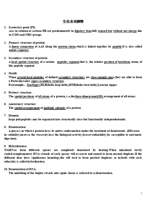
生化名词解释1.Isoelectrrc point (PI):AAs in solution at certaon PH are predominately in dipolary fom,fully ionized but without net charge due to-COO and-NH3+groups.2.Primary structure of protein:A linear connection of AAS along the protein chain,which is linked together by peptide.It is also calledamino sequence.3.Secondary structure of protein:A local spatial structure of a certain peptides segment,that is, the relatice position of backbone atoms ofthis peptide segment.4.Motift:When several local peptides of defined secondary structures are close enough space,they are able to forma Particularsular super-secondary structure.Forexample:Zincfinger,HLH(helix-loop-helix),HTH(helix-turn-helix),Leucine zipper.5.Pertiary structure:The spatial positions of all atoms of a protein, i..e,thethree-dimensional(3D) arrangement of all atoms.6.Auaternary structure:The spatial arrangement of multiple subunits of a protein.7.Domain:large polypeptides may be organized into structurally close but functionally independentunits.8.Denaturation:A process in which a protein loses its native conformation under the treatment of denaturants..(DEcreaes in solubility;incresse the visecosity;lose the biological activity;losecrystalizability;be susceptible to enzymatic digestion).9.Hybridization:TwoDNAs form different spacies are completely denatured by heating.When mixedand slowly plementary DNA strands of each species will associste and anneal to form normal duplexes.If the different dans have significance homology,the will teed to form partiarl duplexes or hybrids with cach other,this is called hybridization.10.Denaturation of DNA:The unfolding of the duplex strsnds into signle chains is referred to asdenaturation.11.Meltion temperature(TM):A260The temperature at which UV adsorption reaches the half of the maximum value as=lsomens that about 50%of the dsDNA is disassociated into the single-stranded DNA.12.Enzymes:A biomolecule. Either protion or nucleic acid, that catalyzes aspecific chemical reaction.They have extraordinary catalytic power and ahigh degree of spaecifity for their substrates.13.Active centers:Some functional groups are close enough in sasce to form a hydrophobic portion where substrates ar e catalyzed into products within the confines of a pocket on the enzyme.14.Isoenzyme:A group of enzymes that catalyze the same reaction but differ form each other in their structure,substrateaffinity, V max, and/of regulatory properties.15.Km:The substrate concentrates at which enzyme-catalyzed reaction proceeds at one-half itsmaximum velocity.16.Ribozymes:Ribonucleic acid(RNA) molecules with catalytic; RNA enzymes.17.Glycolysis:A molecule of glucose is degraded in a series of enzymatic reaction to yield two molecules of or lactate in cyto sol. glucoss(G)→lactic acid (lactic acid(lack of O2)18.Gluconeogenesis:The process of transformation of non-carbohydrates to glucose or glycogen.18.TCA:The reaction procedure is a cycle;form acetyl CoA+oxaloacetic acid→citric acid on,by a few times of dehydrogenesis and decarboxylation→oxaloacetic acid cycle;Also called citrate/Krebs cycle.19.Cori cycle:A metabolic process in which lactate,produced in tissues such as muscle,is transferred to liver where itbecomes a substrate in gluconeogenesis.(prevent acidosis;reduced lactate).20.AEerobic oxidation of glucose:The process of complete oxidation of glucose to CO2 and water with liberation of energy as The form of ATP.glucose(G)→H2O+CO2(O2)21.Biological oxidization:the process in which substance(Carbohydrate,lipid,AAs)are oxidized in living organism.22.Respiratory china(Electron transfer chain,ETC):A chain in the inner membrara of the mitochondria consists of redox carriers for transferring electronsform the substrate to molecular an oxygen ion,which combines with protons to form water.23.P/O ration:is the number of inorganic phosphates incorporates into ATP per oxyfen atom consumed.(Number of mol of ATP/2H)25.Hormone sensitive lipase (HSL):TG lipase is the rate-limiting enzyme in the TG degradation in adipose tissue.It is alsonamed HSL because it is regulated some hormones.26.Oxidative phosphorylation:The enzymatic phosphorylation of ADP to ADP coupled to clectron transfer from a substrate from a substrate to molecular oxygen.27.Substrate level phosphorylation:phosphorylation of ADP or some other nucleoside 5'-diphosphate coupled to the dehydrogenation of an organic substrate; independent of the electron transfer chain.28.Putrefaction of protein:some un-digested proteins and no absorbed are anaerobic by the bacteria in intestine. The products are toxic to body except few viamin and fatty acid.29.Essential fatty acids (EFA):Liinoleic, Linolenic and arechidonic acids are called essential fatty acids,because they cannot be synthesized by the body and must be obtained through diet.30.Fat mobilization:The triacyglycerol stored in the adipocytes are hydrolysized by lipsses,to produce free fatty acids(FFA) and glycerol, which are released to the blood, this process is called fat mobilization.31.Ketone bodies:water-soluble fuels normally exported by the liver but overproduced during fasting or in underated diabetes mellitus, including acetoactate, β-hydroxybutyrate,and acetone.32.Essential amino acids:acids that can not be synthesized by the body and must be obtained from the diet. 8 kinds of essential AAs: Val,Ile,LEU,Phe,Met,Trp,Thr,Lys.33.Union deamination:The α-amino group of most amino acids is transfereed to α-ketoglutarate toform a α-keto acid and glutamate by transaminase,Glutamate is then oxidatively deaminated to yied ammonia and α-ketoglutarate by glutamate dehydrogenase.34.One carbon unit:one carbon units(or groups) are one carbon-containing groups produced in catabolism of some amino acids. They are not free.-CH3,-CH2,-CH=,-CHO,-CH=CN.35.De noyo synthesis:The synthesis of nucleotides begins with their metabolic precursors: amino acids, ribose-5-phosphate,CO2,and one carbon units.36.Salvage pathways:The syntheses of nucleotide by recycie the free bases or nucleosides released from nucleic acid breakdown. 37.Replication:The separation of the two DNA strands,followed by synthesis of two new strands, each one complementary to a parental stand. So that eachnew formed double-helical ADNA contatins one parental strand and one daughter strand.38.Semi-conservative replication:The daughter strands on two parental strands are synthesized differently since the replication process obeys the principle that and DNA synthesis is in thedifferetlu since the replication process obeys the principle that the DNA synthesis is in the 5'-3'directuion.39.Leading strand:On the parental strand 3'-5'the daughter strand is synthesized continously.40.Okazaki fragments:The lagging strand synthesis processds discontinuously. These DNA fragments are called Okzaki fragments.41.Reverse transcription:a processin which ssRNA is used as the template to synthesize dsDNA by RNase. It Plays an important role in cancet-causing theory of viruses.42.Mutation:Mutation is a change of nucleic acids in genomic DNA of an organism,which could occur in the replication process s well as in other steps of life process.43.DNA repairing:a kind response made by the cells after DNA damage occurs, which may resume their natural structures and normal biological functions.44.Transcription:The synthesis of RNA molecules using DNA strands the templates so that the genetic information can be transferred from DNA to rna.45.Translation:the synthesis of protein molecules using mRNA as the template,in other words,to translate the nucleotide sequence of Mrna into the amino acid sequence of protein according to the genetic codon.46.Cis-acting elements:the DNA sequence that can affect the expression of its own gene composed of promoter, enhancer,and silencer.47.Trans-acting factors:the proteins that bind directly to cis-acting elements and regulate their activities.48.Exon:the coding sequence that appear on splice genes and primary transcription,and will be expressed to matured mRNA.49.Intron:the non-coding sequences that are transcripted into primary mRNAs, and will be cleaved out in the later splicing process.50.Activation of amino acid:in the first stage of protein synthesis, which takes place in the cytosol.The 20 differeent amino acids are etherified to their corresponding tRNA by aminoacyl-tRNA synthesis,each of which is specific for one amino acid and one or more corresponding Trna.51.Genetic codon:Three adjacent nucleotides in the 5'-3' direction on mRNA, which codes for an amino acid.52.S-D sequence:In Mrna of prokaryote, 8'-13 'nts prior to AUG, purine rich of 4-9 nts long,which is called ribosomal binding sequence.53.Prmoter:The DNA sequence that RNA-pol can bind to and initate transcription. Is is the key point for the transcription.54.Operon:Each transcription region, which is the basis of regulation of gene expression of prokaryotic systems,composed of structural genes, promoter, operator,and other regulatory sites.55.Gene:A DNA segment that encodes the all genetic information required to produce fanctional biological products.56.Genome:A complete set of genes of given species.57.Gene exptessin:briefly, it is gene transcription and translation. In detail, it is that production of proteins with biological activites according to the gene through transcription and translation.58.Housekeeping genes:The genes are essential and necessary for life, and therefore continuously expressed.such as those enzymes involved in TAC.59.Receptor:Receptor are specific membrane or intracellular proteins, which are able to recognize and bind to corresponding ligand molecules, become activated,and transducer signaling to next signaling molecules. They are usually glycoprotein or lipoprotein.60.Secondary messengers:small molecules synthesized with acell in response to an external signal are the second messenger,which are responsible for intracellular signal transduction. Ca,DG,Cer.IP3,cAMP,cGMP.。
柠檬酸对建筑石膏缓凝作用影响因素的研究
柠檬酸对建筑石膏缓凝作用影响因素的研究*彭家惠,白冷,瞿金东,张建新,吴莉(重庆大学材料科学与工程学院,重庆400045)摘要:研究了柠檬酸对建筑石膏凝结时间与流动度经时性的影响,以及建筑石膏细度、相组成、pH值、水化温度对柠檬酸缓凝性的影响。
结果表明:柠檬酸是建筑石膏高效缓凝剂,可有效抑制其流动度经时损失;Ó型无水石膏和二水石膏削弱柠檬酸的缓凝效果,比表面积增加,凝结时间缩短;柠檬酸在弱碱性条件下缓凝效果最好;柠檬酸的缓凝效果对温度很敏感,温度升高,缓凝性降低。
关键词:石膏;缓凝剂;凝结时间;影响因素中图分类号:T U502文献标识码:A文章编号:1006-7329(2007)02-0110-03Study on the Factors Influencing RetardingEffect of Citric Acid in Building GypsumPENG Jia-hui,BAI Leng,QU Jin-dong,ZHANG Jian-xin,WU Li(College of Materials Science and Engineer ing,Chongqing University,Chongqing400045,China)Abstr act:T he influences of citric acid on the setting time and the time dependency of fluidity of building gyp2 sum were investigated.Furthermore,the factors affecting the r etarding proper ty of citric acid were also studied such as gypsum particles fineness,phase composition,pH value and hydration temperature.T he results indi2 cate that citric acid is highly efficient retardant for building gypsum and it is able to inhibit the fluidity loss of fresh gypsum plaster with time effectively.T he existence ofÓ-type anhydrate and dihydrate gypsum in phase composition would weaken the effect of citric acid.With the incr ease of specific sur face area of gypsum parti2 cles,its setting time is shortened.In alkalescent liquid environment,the citric acid has the best retar ding capa2 bility.In addition,the temperature also has a susceptive influence on its retarding effect which is decreased with the increase of temper ature.Keywor ds:gypsum;retar dant;setting time;influencing factor半水石膏是石膏基材料的主要成分和胶凝相。
酸碱滴定法测定山楂炭中枸橼酸的含量
酸碱滴定法测定山楂炭中枸橼酸的含量王小永1,马 丽1,周维维1,张 征1,赵丽娜2*(1.承德市食品药品检验检测中心,河北承德 067000;2.承德医学院附属医院,河北承德 067000)摘 要:目的:建立山楂炭中枸橼酸含量的测定方法。
方法:以酚酞为指示液,用氢氧化钠滴定液(0.1 mol L-1)滴定,测定山楂炭中总有机酸的含量,滴定结果以枸橼酸计。
结果:收集的山楂炭饮片总有机酸的结果在4.25%~6.32%,方法精密度(RSD)为1.10%,回收率为96.9%。
结论:本方法操作简便易行,精密度良好,可作为山楂炭中枸橼酸含量测定的方法。
关键词:酸碱滴定法;山楂炭;枸橼酸;含量测定Determination of Citric Acid in Crataegus pinnatifida Charcoalby Acid-Base TitrationWANG Xiaoyong1, MA Li1, ZHOU Weiwei1, ZHANG Zheng1, ZHAO Lina2*(1.Chengde City Institute for Food and Drug Control, Chengde 067000, China; 2.The Affiliated Hospital of ChengdeMedical College, Chengde 067000, China)Abstract: Objective: To establish a method for the determination of citric acid content in Crataegus pinnatifida charcoal. Method: The content of total organic acids in Crataegus pinnatifida charcoal was determined by titration with sodium hydroxide titration solution (0.1 mol·L-1) using phenolphthalein as the indicator solution, and the titration results were expressed as citric acid. Result: The results of total organic acids in the collected Crataegus pinnatifida charcoal tablets ranged from 4.25% to 6.32% with a method precision (RSD) of 1.10% and a recovery of 96.9%. Conclusion: The method is simple and easy to operate with good precision, and can be used as a method for the determination of citric acid content in Crataegus pinnatifida charcoal.Keywords: acid-base titration; Crataegus pinnatifida charcoal; citric acid; content determination山楂为蔷薇科植物山里红(Crataegus pinnatifida Bge. var. major N. E. Br.)或山楂(Crataegus pinnatifida Bge.)的干燥成熟果实,主产于山东、河北、河南等省份。
柠檬酸生产过程中碳酸钙的应用
柠檬酸生产过程中碳酸钙的应用Calcium carbonate, also known as chalk or limestone, is a key ingredient in the production of citric acid. It is commonly used as a neutralizing agent during the fermentation process of citric acid production. The calcium carbonate helps maintain the pH balance of the fermentation medium, ensuring optimal conditions for the citric acid-producing microorganisms to thrive. In addition, calcium carbonate serves as a source of calcium ions, which are essential for the enzymatic reactions involved in citric acid biosynthesis.碳酸钙,也称为白垩或石灰石,是柠檬酸生产中的关键成分。
在柠檬酸生产的发酵过程中,碳酸钙常用作中和剂。
碳酸钙有助于维持发酵培养基的pH 平衡,确保柠檬酸生产微生物生长繁殖的最佳条件。
此外,碳酸钙还作为钙离子的来源,这对柠檬酸生物合成的酶促反应至关重要。
Furthermore, calcium carbonate plays a crucial role in the precipitation of impurities present in the fermentation broth. As citric acid is produced, various byproducts and impurities may accumulate in the fermentation medium. Calcium carbonate helps these impurities to precipitate out of the solution, allowing for easierseparation and purification of the citric acid. This process of precipitation and removal of impurities enhances the overall yield and purity of the citric acid product.此外,碳酸钙在沉淀发酵液中存在的杂质方面起着至关重要的作用。
13314760_小麦秸秆有机酸组分的GC-MS_分析及其对牛筋草的化感效应
黄淮海地区是典型的小麦-玉米两熟制种植区,秸秆还田模式在该地区应用相当普遍。
小麦经过联合收割机收获之后,秸秆得以粉碎还田,还田秸秆在土壤中经过水分淋溶或微生物腐解,化感物质得以释放,从而影响杂草种子的萌发及幼苗生长[1,2]。
研究发现,小麦秸秆水浸提液对牵牛花、稗草、黑麦草、牛筋草、白茅等多种杂草,甚至一些抗药性杂草种子的萌发及生长具有抑制作用[3~5]。
化感物质根据结构的不同,可分为水溶性有机酸、直链醇、脂肪族醛和酮,简单不饱和内脂,长链脂肪酸和多炔,醌类,苯甲酸及其衍生物,肉桂酸及其衍生物,香豆素类,类黄酮类,单宁,类萜和甾类,氨基酸和多肽,生物碱和氰醇,硫化物和芥子油刘小民,许贤,李秉华,王贵启*,张焕焕(河北省农林科学院粮油作物研究所,河北石家庄050035)摘要:有机酸类化合物是重要的化感活性物质之一,为了明确小麦秸秆中有机酸类化合物的组分及其对牛筋草的化感效应,探讨其对牛筋草的化感作用机理,利用GC-MS 方法对小麦秸秆不同部位的有机酸组分进行了定性和定量分析,结果表明,从小麦秸秆中共鉴定出22种有机酸组分,包含15种饱和脂肪酸、4种不饱和脂肪酸和3种酚酸,其中,从茎部鉴定出的有机酸种类最多,有17种,从穗部和根部各鉴定出11种。
对水溶性有机酸的进一步分析结果表明,阿魏酸、柠檬酸、肉桂酸是小麦秸秆中含量较高的3种有机酸组分。
利用室内生物测定法研究了外源有机酸标准品对牛筋草种子萌发及幼苗生长的影响,结果表明,阿魏酸和柠檬酸均对牛筋草种子萌发及胚轴生长具有“低促高抑”的浓度效应,肉桂酸对牛筋草种子萌发及胚根生长始终表现抑制作用,并且3种有机酸对牛筋草胚根生长的抑制作用显著高于胚轴。
关键词:小麦秸秆;GC-MS ;有机酸;牛筋草;化感作用中图分类号:S512.1文献标识码:A 文章编号:1008-1631(2016)06-0058-04收稿日期:2016-07-05基金项目:国家科技支撑计划项目(2012BAD19B02)作者简介:刘小民(1981-),男,山东莱阳人,助理研究员,博士,主要从事杂草生态学及除草剂抗性机理研究。
泼尼松原研说明书
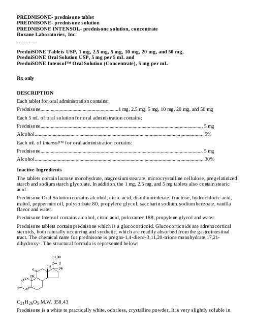
PREDNISONE- prednisone tabletPREDNISONE- prednisone solutionPREDNISONE INTENSOL- prednisone solution, concentrateRoxane Laboratories, Inc.----------PredniSONE Tablets USP, 1 mg, 2.5 mg, 5 mg, 10 mg, 20 mg, and 50 mg,PredniSONE Oral Solution USP, 5 mg per 5 mL andPredniSONE Intensol™ Oral Solution (Concentrate), 5 mg per mLRx onlyDESCRIPTIONEach tablet for oral administration contains: Prednisone.............................................................1 mg, 2.5 mg, 5 mg, 10 mg, 20 mg, and 50 mgEach 5 mL of oral solution for oral administration contains: Prednisone............................................................................................................................... 5 mg Alcohol.................................................................................................................................... 5%Each mL of Intensol™ for oral administration contains: Prednisone............................................................................................................................... 5 mg Alcohol.................................................................................................................................... 30%Inactive IngredientsThe tablets contain lactose monohydrate, magnesium stearate, microcrystalline cellulose, pregelatinized starch and sodium starch glycolate. In addition, the 1 mg, 2.5 mg, and 5 mg tablets also contain stearic acid.Prednisone Oral Solution contains alcohol, citric acid, disodium edetate, fructose, hydrochloric acid, maltol, peppermint oil, polysorbate 80, propylene glycol, saccharin sodium, sodium benzoate, vanilla flavor and water.Prednisone Intensol contains alcohol, citric acid, poloxamer 188, propylene glycol and water. Prednisone tablets contain prednisone which is a glucocorticoid. Glucocorticoids are adrenocortical steroids, both naturally occurring and synthetic, which are readily absorbed from the gastrointestinal tract. The chemical name for prednisone is pregna-1,4-diene-3,11,20-trione monohydrate,17,21-dihydroxy-. The structural formula is represented below:C H O M.W. 358.4321265Prednisone is a white to practically white, odorless, crystalline powder. It is very slightly soluble inwater; slightly soluble in alcohol, chloroform, dioxane, and methanol.CLINICAL PHARMACOLOGYNaturally occurring glucocorticoids (hydrocortisone and cortisone), which also have salt-retaining properties, are used as replacement therapy in adrenocortical deficiency states. Their synthetic analogs are primarily used for their potent anti-inflammatory effects in disorders of many organ systems. Glucocorticoids cause profound and varied metabolic effects. In addition, they modify the body’s immune responses to diverse stimuli.INDICATIONS AND USAGEPrednisone tablets and solutions are indicated in the following conditions:Endocrine DisordersPrimary or secondary adrenocortical insufficiency (hydrocortisone or cortisone is the first choice: synthetic analogs may be used in conjunction with mineralocorticoids where applicable; in infancy mineralocorticoid supplementation is of particular importance); congenital adrenal hyperplasia; hypercalcemia associated with cancer; nonsuppurative thyroiditis.Rheumatic DisordersAs adjunctive therapy for short-term administration (to tide the patient over an acute episode or exacerbation) in: psoriatic arthritis, rheumatoid arthritis, including juvenile rheumatoid arthritis (selected cases may require low-dose maintenance therapy), ankylosing spondylitis, acute and subacute bursitis, acute nonspecific tenosynovitis, acute gouty arthritis, post-traumatic osteoarthritis, synovitis of osteoarthritis, epicondylitis.Collagen DiseasesDuring an exacerbation or as maintenance therapy in selected cases of: systemic lupus erythematosus, systemic dermatomyositis (polymyositis), acute rheumatic carditis.Dermatologic DiseasesPemphigus; bullous dermatitis herpetiformis; severe erythema multiforme (Stevens-Johnson syndrome); exfoliative dermatitis; mycosis fungoides; severe psoriasis; severe seborrheic dermatitis.Allergic StatesControl of severe or incapacitating allergic conditions intractable to adequate trials of conventional treatment: seasonal or perennial allergic rhinitis; bronchial asthma; contact dermatitis; atopic dermatitis; serum sickness; drug hypersensitivity reactions.Ophthalmic DiseasesSevere acute and chronic allergic and inflammatory processes involving the eye and its adnexa such as: allergic corneal marginal ulcers, herpes zoster ophthalmicus, anterior segment inflammation, diffuse posterior uveitis and choroiditis, sympathetic ophthalmia, allergic conjunctivitis, keratitis, chorioretinitis, optic neuritis, iritis and iridocyclitis.Respiratory DiseasesSymptomatic sarcoidosis; Loeffler’s syndrome not manageable by other means; berylliosis; fulminating or disseminated pulmonary tuberculosis when used concurrently with appropriate antituberculous chemotherapy; aspiration pneumonitis.Hematologic DisordersIdiopathic thrombocytopenic purpura in adults; secondary thrombocytopenia in adults; acquired (autoimmune) hemolytic anemia; erythroblastopenia (RBC anemia); congenital (erythroid) hypoplastic anemia.Neoplastic DiseasesFor palliative management of: leukemias and lymphomas in adults, acute leukemia of childhood. Edematous StatesTo induce a diuresis or remission of proteinuria in the nephrotic syndrome, without uremia, of the idiopathic type or that due to lupus erythematosus.Gastrointestinal DiseasesTo tide the patient over a critical period of the disease in: ulcerative colitis, regional enteritis. MiscellaneousTuberculous meningitis with subarachnoid block or impending block when used concurrently with appropriate antituberculous chemotherapy; trichinosis with neurologic or myocardial involvement. CONTRAINDICATIONSPrednisone tablets and oral solutions are contraindicated in systemic fungal infections and known hypersensitivity to components.WARNINGSGeneralRare instances of anaphylactoid reactions have occurred in patients receiving corticosteroid therapy (see ADVERSE REACTIONS: Allergic Reactions).Increased dosage of rapidly acting corticosteroids is indicated in patients on corticosteroid therapy subjected to any unusual stress before, during and after the stressful situation.Cardio-RenalAverage and large doses of hydrocortisone or cortisone can cause elevation of blood pressure, salt and water retention, and increased excretion of potassium. These effects are less likely to occur with the synthetic derivatives except when used in large doses. Dietary salt restriction and potassium supplementation may be necessary. All corticosteroids increase calcium excretion.Literature reports suggest an apparent association between use of corticosteroids and left ventricular free wall rupture after a recent myocardial infarction; therefore, therapy with corticosteroids should be used with great caution in these patients.EndocrineCorticosteroids can produce reversible hypothalamic-pituitary adrenal (HPA) axis suppression with the potential for corticosteroid insufficiency after withdrawal of treatment. Adrenocortical insufficiency may result from too rapid withdrawal of corticosteroids and may be minimized by gradual reduction of dosage. This type of relative insufficiency may persist for up to 12 months after discontinuation of therapy; therefore, in any situation of stress occurring during that period, hormone therapy should be reinstituted. If the patient is receiving steroids already, dosage may have to be increased.Metabolic clearance of corticosteroids is decreased in hypothyroid patients and increased inMetabolic clearance of corticosteroids is decreased in hypothyroid patients and increased inhyperthyroid patients. Changes in thyroid status of the patient may necessitate adjustment in dosage.InfectionGeneralPatients who are on corticosteroids are more susceptible to infections than are healthy individuals.There may be decreased resistance and inability to localize infection when corticosteroids are used.Infection with any pathogen (viral, bacterial, fungal, protozoan or helminthic) in any location of the body may be associated with the use of corticosteroids alone or in combination with otherimmunosuppressive agents that affect cellular immunity, humoral immunity, or neutrophil function.These infections may be mild, but may be severe and at times fatal. With increasing doses ofcorticosteroids, the rate of occurrence of infectious complications increases. Corticosteroids may also mask some signs of current infection.Fungal InfectionsCorticosteroids may exacerbate systemic fungal infections and therefore should not be used in the presence of such infections unless they are needed to control life-threatening drug reactions. There have been cases reported in which concomitant use of amphotericin B and hydrocortisone was followed by cardiac enlargement and congestive heart failure (see PRECAUTIONS: Drug Interactions:Amphotericin B Injection and Potassium-Depleting Agents).Special PathogensLatent disease may be activated or there may be an exacerbation of intercurrent infections due to pathogens, including those caused by Amoeba, Candida, Cryptococcus, Mycobacterium, Nocardia,Pneumocystis, Toxoplasma .It is recommended that latent amebiasis or active amebiasis be ruled out before initiating corticosteroid therapy in any patient who has spent time in the tropics or any patient with unexplained diarrhea.Similarly, corticosteroids should be used with great care in patients with known or suspectedStrongyloides (threadworm) infestation. In such patients, corticosteroid-induced immunosuppression may lead to Strongyloides hyperinfection and dissemination with widespread larval migration, often accompanied by severe enterocolitis and potentially fatal gram-negative septicemia.Corticosteroids should not be used in cerebral malaria.TuberculosisThe use of prednisone in active tuberculosis should be restricted to those cases of fulminating or disseminated tuberculosis in which the corticosteroid is used for management of the disease in conjunction with an appropriate antituberculous regimen.If corticosteroids are indicated in patients with latent tuberculosis or tuberculin reactivity, close observation is necessary as reactivation of the disease may occur. During prolonged corticosteroid therapy, these patients should receive chemoprophylaxis.VaccinationAdministration of live or live, attenuated vaccines is contraindicated in patients receivingimmunosuppressive doses of corticosteroids. Killed or inactivated vaccines may be administered.However, the response to such vaccines may be diminished and cannot be predicted . Indicated immunization procedures may be undertaken in patients receiving nonimmunosuppressive doses of corticosteroids as replacement therapy (e.g., for Addison’s disease).Viral Infections12Chickenpox and measles can have a more serious or even fatal course in pediatric and adult patients on corticosteroids. In pediatric and adult patients who have not had these diseases, particular care should be taken to avoid exposure. How the dose, route and duration of corticosteroid administration affect the risk of developing a disseminated infection is not known. The contribution of the underlying disease and/or prior corticosteroid treatment to the risk is also not known. If exposed to chickenpox, prophylaxis with varicella zoster immune globulin (VZIG) may be indicated. If exposed to measles, prophylaxis with pooled intramuscular immunoglobulin (IG) may be indicated. (See the respective package inserts for complete VZIG and IG prescribing information.) If chickenpox develops, treatment with antiviral agents may be considered.OphthalmicUse of corticosteroids may produce posterior subcapsular cataracts, glaucoma with possible damage to the optic nerves, and may enhance the establishment of secondary ocular infections due to bacteria, fungi or viruses. The use of oral corticosteroids is not recommended in the treatment of optic neuritis and may lead to an increase in the risk of new episodes. Corticosteroids should not be used in active ocular herpes simplex because of possible corneal perforation.PRECAUTIONSGeneral PrecautionsThe lowest possible dose of corticosteroids should be used to control the condition under treatment. When reduction in dosage is possible, the reduction should be gradual.Since complications of treatment with glucocorticoids are dependent on the size of the dose and the duration of treatment, a risk/benefit decision must be made in each individual case as to dose and duration of treatment and as to whether daily or intermittent therapy should be used.Kaposi’s sarcoma has been reported to occur in patients receiving corticosteroid therapy, most often for chronic conditions. Discontinuation of corticosteroids may result in clinical improvement.Cardio-RenalAs sodium retention with resultant edema and potassium loss may occur in patients receiving corticosteroids, these agents should be used with caution in patients with congestive heart failure, hypertension, or renal insufficiency.EndocrineDrug-induced secondary adrenocortical insufficiency may be minimized by gradual reduction of dosage. This type of relative insufficiency may persist for up to 12 months after discontinuation of therapy following large doses for prolonged periods; therefore, in any situation of stress occurring during that period, hormone therapy should be reinstituted. Since mineralocorticoid secretion may be impaired, salt and/or a mineralocorticoid should be administered concurrently.There is an enhanced effect of corticosteroids on patients with hypothyroidism.GastrointestinalSteroids should be used with caution in active or latent peptic ulcers, diverticulitis, fresh intestinal anastomoses, and nonspecific ulcerative colitis, since they may increase the risk of a perforation. Signs of peritoneal irritation following gastrointestinal perforation in patients receiving corticosteroids may be minimal or absent.There is an enhanced effect due to decreased metabolism of corticosteroids in patients with cirrhosis. MusculoskeletalCorticosteroids decrease bone formation and increase bone resorption both through their effect on calcium regulation (i.e., decreasing absorption and increasing excretion) and inhibition of osteoblast function. This, together with a decrease in the protein matrix of the bone secondary to an increase in protein catabolism, and reduced sex hormone production, may lead to inhibition of bone growth in pediatric patients and the development of osteoporosis at any age. Growth and development of infants and children on prolonged corticosteroid therapy should be carefully observed. Special consideration should be given to patients at increased risk of osteoporosis (e.g., postmenopausal women) before initiating corticosteroid therapy.Inclusion of therapy for osteoporosis prevention or treatment should be considered. To minimize the risk of glucocortoicoid-induced bone loss, the smallest possible effective dosage and duration should be used. Lifestyle modification to reduce the risk of osteoporosis (e.g., cigarette smoking cessation, limitation of alcohol consumption, participation in weight-bearing exercise for 30-60 minutes daily) should be encouraged. Calcium and vitamin D supplementation, bisphosphonate (e.g., alendronate, risedronate), and a weight-bearing exercise program that maintains muscle mass are suitable first-line therapies aimed at reducing the risk of adverse bone effects. Current recommendations suggest that all interventions be initiated in any patient in whom glucocorticoid therapy with at least the equivalent of 5 mg of prednisone for at least 3 months is anticipated; in addition, sex hormone replacement therapy (combined estrogen and progestin in women; testosterone in men) should be offered to such patients who are hypogonadal or in whom replacement is otherwise clinically indicated and biphosphonate therapy should be initiated (if not already) if bone mineral density (BMD) of the lumbar spine and/or hip is below normal.Neuro-PsychiatricAlthough controlled clinical trials have shown corticosteroids to be effective in speeding the resolution of acute exacerbations of multiple sclerosis, they do not show that they affect the ultimate outcome or natural history of the disease. The studies do show that relatively high doses of corticosteroids are necessary to demonstrate a significant effect. (See DOSAGE AND ADMINISTRATION: Multiple Sclerosis.)An acute myopathy has been observed with the use of high doses of corticosteroids, most often occurring in patients with disorders of neuromuscular transmission (e.g., myasthenia gravis), or in patients receiving concomitant therapy with neuromuscular blocking drugs (e.g., pancuronium). This acute myopathy is generalized, may involve ocular and respiratory muscles, and may result in quadriparesis. Elevation of creatinine kinase may occur. Clinical improvement or recovery after stopping corticosteroids may require weeks to years.Psychiatric derangements may appear when corticosteroids are used, ranging from euphoria, insomnia, mood swings, personality changes, and severe depression, to frank psychotic manifestations. Also, existing emotional instability or psychotic tendencies may be aggravated by corticosteroids. OphthalmicIntraocular pressure may become elevated in some individuals. If steroid therapy is continued for more than 6 weeks, intraocular pressure should be monitored.Information for PatientsPatients should be warned not to discontinue the use of corticosteroids abruptly or without medical supervision. As prolonged use may cause adrenal insufficiency and make patients dependent on corticosteroids, they should advise any medical attendants that they are taking corticosteroids and they should seek medical advice at once should they develop an acute illness including fever or other signs of infection. Following prolonged therapy, withdrawal of corticosteroids may result in symptoms of the corticosteroid withdrawal syndrome including, myalgia, arthralgia, and malaise.Persons who are on corticosteroids should be warned to avoid exposure to chickenpox or measles.Patients should also be advised that if they are exposed, medical advice should be sought without delay. Drug InteractionsAmphotericin B Injection and Potassium-Depleting AgentsWhen corticosteroids are administered concomitantly with potassium-depleting agents (e.g., amphotericin B, diuretics), patients should be observed closely for development of hypokalemia. In addition, there have been cases reported in which concomitant use of amphotericin B and hydrocortisone was followed by cardiac enlargement and congestive heart failure.AntibioticsMacrolide antibiotics have been reported to cause a significant decrease in corticosteroid clearance (see PRECAUTIONS: Drug Interactions: Hepatic Enzyme Inducers, Inhibitors and Substrates). AnticholinesterasesConcomitant use of anticholinesterase agents (e.g., neostigmine, pyridostigmine) and corticosteroids may produce severe weakness in patients with myasthenia gravis. If possible, anticholinesterase agents should be withdrawn at least 24 hours before initiating corticosteroid therapy. If concomitant therapy must occur, it should take place under close supervision and the need for respiratory support should be anticipated.Anticoagulants, OralCo-administration of corticosteroids and warfarin usually results in inhibition of response to warfarin, although there have been some conflicting reports. Therefore, coagulation indices should be monitored frequently to maintain the desired anticoagulant effect.AntidiabeticsBecause corticosteroids may increase blood glucose concentrations, dosage adjustments of antidiabetic agents may be required.Antitubercular drugsSerum concentrations of isoniazid may be decreased.BupropionSince systemic steroids, as well as bupropion, can lower the seizure threshold, concurrent administration should be undertaken only with extreme caution; low initial dosing and small gradual increases should be employed.CholestyramineCholestyramine may increase the clearance of corticosteroids.CyclosporineIncreased activity of both cyclosporine and corticosteroids may occur when the two are used concurrently. Convulsions have been reported with this concurrent use.Digitalis GlycosidesPatients on digitalis glycosides may be at increased risk of arrhythmias due to hypokalemia. Estrogens, Including Oral ContraceptivesEstrogens may decrease the hepatic metabolism of certain corticosteroids, thereby increasing theireffect.FluoroquinolonesPost-marketing surveillance reports indicate that the risk of tendon rupture may be increased in patients receiving concomitant fluoroquinolones (e.g., ciprofloxacin, levofloxacin) and corticosteroids, especially in the elderly. Tendon rupture can occur during or after treatment with quinolones.Hepatic Enzyme Inducers, Inhibitors and SubstratesDrugs which induce cytochrome P450 3A4 (CYP 3A4) enzyme activity (e.g., barbiturates, phenytoin, carbamazepine, rifampin) may enhance the metabolism of corticosteroids and require that the dosage of the corticosteroid be increased. Drugs which inhibit CYP 3A4 (e.g., ketoconazole, itraconazole, ritonavir, indinavir, macrolide antibiotics such as erythromycin) have the potential to result in increased plasma concentrations of corticosteroids. Glucocorticoids are moderate inducers of CYP3A4. Co-administration with other drugs that are metabolized by CYP 3A4 (e.g., indinavir, erythromycin) may increase their clearance, resulting in decreased plasma concentration. KetoconazoleKetoconazole has been reported to decrease the metabolism of certain corticosteroids by up to 60%, leading to increased risk of corticosteroid side effects. In addition, ketoconazole alone can inhibit adrenal corticosteroid synthesis and may cause adrenal insufficiency during corticosteroid withdrawal. Nonsteroidal Anti-Inflammatory Agents (NSAIDS)Concomitant use of aspirin (or other nonsteroidal anti-inflammatory agents) and corticosteroids increases the risk of gastrointestinal side effects. Aspirin should be used cautiously in conjunction with corticosteroids in hypoprothrombinemia. The clearance of salicylates may be increased with concurrent use of corticosteroids; this could lead to decreased salicylate serum levels or increase the risk of salicylate toxicity when corticosteroid is withdrawn.PhenytoinIn post-marketing experience, there have been reports of both increases and decreases in phenytoin levels with dexamethasone co-administration, leading to alterations in seizure control. Phenytoin has been demonstrated to increase the hepatic metabolism of corticosteroids, resulting in a decreased therapeutic effect of the corticosteroid.QuetiapineIncreased doses of quetiapine may be required to maintain control of symptoms of schizophrenia in patients receiving a glucocorticoid, a hepatic enzyme inducer.Skin TestsCorticosteroids may suppress reactions to skin tests.ThalidomideCo-administration with thalidomide should be employed cautiously, as toxic epidermal necrolysis has been reported with concomitant use.VaccinesPatients on corticosteroid therapy may exhibit a diminished response to toxoids and live or inactivated vaccines due to inhibition of antibody response. Corticosteroids may also potentiate the replication of some organisms contained in live attenuated vaccines. Routine administration of vaccines or toxoids should be deferred until corticosteroid therapy is discontinued if possible (see WARNINGS: Infection:Vaccination).Carcinogenesis, Mutagenesis, Impairment of FertilityNo adequate studies have been conducted in animals to determine whether corticosteroids have a potential for carcinogenesis or mutagenesis. Steroids may increase or decrease motility and number of spermatozoa in some patients.PregnancyTeratogenic EffectsPregnancy Category CCorticosteroids have been shown to be teratogenic in many species when given in doses equivalent to the human dose. Animal studies in which corticosteroids have been given to pregnant mice, rats, and rabbits have yielded an increased incidence of cleft palate in the offspring. There are no adequate and well-controlled studies in pregnant women. Corticosteroids should be used during pregnancy only if the potential benefit justifies the potential risk to the fetus. Infants born to mothers who have received substantial doses of corticosteroids during pregnancy should be carefully observed for signs of hypoadrenalism.Nursing MothersSystemically administered corticosteroids appear in human milk and could suppress growth, interfere with endogenous corticosteroid production, or cause other untoward effects. Because of the potential for serious adverse reactions in nursing infants from corticosteroids, a decision should be made whether to discontinue nursing or to discontinue the drug, taking into account the importance of the drug to the mother.Pediatric UseThe efficacy and safety of corticosteroids in the pediatric population are based on the well-established course of effect of corticosteroids, which is similar in pediatric and adult populations. Published studies provide evidence of efficacy and safety in pediatric patients for the treatment of nephrotic syndrome (patients >2 years of age), and aggressive lymphomas and leukemias (patients >1 month of age). Other indications for pediatric use of corticosteroids, e.g., severe asthma and wheezing, are based on adequate and well-controlled trials conducted in adults, on the premises that the course of the diseases and their pathophysiology are considered to be substantially similar in both populations.The adverse effects of corticosteroids in pediatric patients are similar to those in adults (see ADVERSE REACTIONS). Like adults, pediatric patients should be carefully observed with frequent measurements of blood pressure, weight, height, intraocular pressure, and clinical evaluation for the presence of infection, psychosocial disturbances, thromboembolism, peptic ulcers, cataracts, and osteoporosis. Pediatric patients who are treated with corticosteroids by any route, including systemically administered corticosteroids, may experience a decrease in their growth velocity. This negative impact of corticosteroids on growth has been observed at low systemic doses and in the absence of laboratory evidence of hypothalamic-pituitary-adrenal (HPA) axis suppression (i.e., cosyntropin stimulation and basal cortisol plasma levels). Growth velocity may therefore be a more sensitive indicator of systemic corticosteroid exposure in pediatric patients than some commonly used tests of HPA axis function. The linear growth of pediatric patients treated with corticosteroids should be monitored, and the potential growth effects of prolonged treatment should be weighed against clinical benefits obtained and the availability of treatment alternatives. In order to minimize the potential growth effects of corticosteroids, pediatric patients should be titrated to the lowest effective dose. Geriatric UseClinical studies did not include sufficient numbers of subjects aged 65 and over to determine whether they respond differently from younger subjects. Other reported clinical experience has not identified differences in responses between the elderly and younger patients. In general, dose selection for an elderly patient should be cautious, usually starting at the low end of the dosing range, reflecting the greater frequency of decreased hepatic, renal, or cardiac function, and of concomitant disease or other drug therapy. In particular, the increased risk of diabetes mellitus, fluid retention and hypertension in elderly patients treated with corticosteroids should be considered.ADVERSE REACTIONS(listed alphabetically, under each subsection)The following adverse reactions have been reported with prednisone or other corticosteroids: Allergic Reactionsanaphylactoid or hypersensitivity reactions, anaphylaxis, angioedema.Cardiovascular Systembradycardia, cardiac arrest, cardiac arrhythmias, cardiac enlargement, circulatory collapse, congestive heart failure, ECG changes caused by potassium deficiency, edema, fat embolism, hypertension or aggravation of hypertension, hypertrophic cardiomyopathy in premature infants, myocardial rupture following recent myocardial infarction (see WARNINGS: Cardio-Renal), necrotizing angiitis, pulmonary edema, syncope, tachycardia, thromboembolism, thrombophlebitis, vasculitis. Dermatologicacne, acneiform eruptions, allergic dermatitis, alopecia, angioedema, angioneurotic edema, atrophy and thinning of skin, dry scaly skin, ecchymoses and petechiae (bruising), erythema, facial edema, hirsutism, impaired wound healing, increased sweating, Karposi’s sarcoma (see PRECAUTIONS: General Precautions), lupus erythematosus-like lesions, perineal irritation, purpura, rash, striae, subcutaneous fat atrophy, suppression of reactions to skin tests, striae, telangiectasis, thin fragile skin, thinning scalp hair, urticaria.EndocrineAdrenal insufficiency-greatest potential caused by high potency glucocorticoids with long duration of action (associated symptoms include; arthralgias, buffalo hump, dizziness, life-threatening hypotension, nausea, severe tiredness or weakness), amenorrhea, postmenopausal bleeding or other menstrual irregularities, decreased carbohydrate and glucose tolerance, development of cushingoid state, diabetes mellitus (new onset or manifestations of latent), glycosuria, hyperglycemia, hypertrichosis, hyperthyroidism (see WARNINGS: Endocrine), hypothyroidism, increased requirements for insulin or oral hypoglycemic agents in diabetics, lipids abnormal, moon face, negative nitrogen balance caused by protein catabolism, secondary adrenocortical and pituitary unresponsiveness (particularly in times of stress, as in trauma, surgery or illness) (see WARNINGS: Endocrine), suppression of growth in pediatric patients.Fluid and Electrolyte Disturbancescongestive heart failure in susceptible patients, fluid retention, hypokalemia, hypokalemic alkalosis, metabolic alkalosis, hypotension or shock-like reaction, potassium loss, sodium retention with resulting edema.Gastrointestinalabdominal distention, abdominal pain,anorexia which may result in weight loss, constipation, diarrhea, elevation in serum liver enzyme levels (usually reversible upon discontinuation), gastric irritation,。
檬酸改性聚乙烯醇薄膜的制备及性能
第28卷第8期高分子材料科学与工程Vol.28,No.8 2012年8月POLYMER MATERIALS SCIENCE AND ENGINEERINGAug.2012纳米纤维素晶体和柠檬酸改性聚乙烯醇薄膜的制备及性能李本刚,曹绪芝,顾洪宇,姜婷玲(南京林业大学理学院,江苏南京210037)摘要:以球形纳米纤维素晶体(NCC)作增强相、柠檬酸作交联剂对聚乙烯醇(PVA )进行改性,制备了PVA P NCC 纳米复合薄膜和柠檬酸交联PVA P N CC 纳米复合薄膜。
通过热重分析、差热分析、吸水实验和拉伸实验考察了NCC 的添加和柠檬酸的交联对薄膜热性能、耐水性和力学性能的影响。
结果表明,与纯PVA 薄膜相比,改性PVA 薄膜的起始分解温度升高、熔融P 结晶峰向高温方向移动、吸水率降低;只用NCC 或柠檬酸对PVA 改性时,所得PVA P NCC 纳米复合薄膜、柠檬酸交联PVA 薄膜的力学性能均对环境湿度敏感;同时用N CC(m (N CC)P m (PV A)=6P 100)和柠檬酸(m (柠檬酸)P m (PVA )=3P 100或m (柠檬酸)P m (PVA )=4.5P 100)对PVA 改性时,所得柠檬酸交联PVA P NCC 纳米复合薄膜的力学性能不随环境湿度变化。
关键词:聚乙烯醇;改性;纳米纤维素晶体;柠檬酸中图分类号:T B383 文献标识码:A 文章编号:1000-7555(2012)08-0178-05收稿日期:2011-09-20基金项目:南京林业大学高学历人才基金(163101007)通讯联系人:李本刚,主要从事天然高分子改性及功能化研究,E -mail:L i bengang_1980@聚乙烯醇(PVA)是一种成膜性和生物降解性俱佳的合成水溶性高分子,由它制备的薄膜具有良好的可生物降解性、气体阻隔性、透明性、抗静电性、强韧性、耐有机溶剂等性能,因而在可降解包装材料领域占有重要地位。
采用不同方法制备CeO_2超细粒子__溶胶_凝胶法

南京大学学报(自然科学)第35卷第4期1999年7月 JOU RNAL OF NANJING U NIVERSIT Y(NAT URAL SCIENCES)Vol.35,No.4Jul.,1999采用不同方法制备CeO2超细粒子溶胶 凝胶法侯文华 徐 林 邱金恒 郭灿雄 陈立刚 颜其洁(南京大学化学系,210093,南京)摘 要 采用以柠檬酸为配体的溶胶 凝胶法制备了CeO2超细粒子.考察了不同制备条件(金属离子与配体的摩尔比、反应温度、pH、凝胶烘干温度、焙烧温度及时间)对粒子的影响,获得了最佳的制备条件:金属离子与配体的摩尔比为1:3、反应温度为65!、pH<0(未调)、凝胶烘干温度为65!、320!焙烧温度2h.并在该条件下成功地制得了均匀分散的CeO2超细粒子(平均粒径为10nm,比表面为57m2/g).关键词 溶胶 凝胶法,CeO2,超细粒子分类号 O643.320 前 言众所周知,超细粒子具有独特的物理和化学性质.自20世纪30年代日本的上田良二教授首次成功地制备了超细Zn粉以来,这方面的研究受到了人们的广泛重视,特别是从20世纪80年代以来得到了飞速发展,并形成了一门新的学科 纳米材料科学.[1]我国是含稀土元素丰富的国家,CeO2作为一种重要的催化剂(或助剂)和催化剂载体,均具有独特的性质[2,3].由于超细粒子的比表面积大、化学活性高,若能制得CeO2超细粒子,则其催化性能一定会有明显的改变.有关CeO2超细粒子的制备,前人曾有过零星的报导[4~6],但都缺乏系统研究,尤其是细致地考察各种反应条件的影响.我们在前人研究的基础上,采用3种不同的方法(溶胶 凝胶法、冰冻脱水法和尿素水解法)来制备CeO2超细粒子,并系统地考察了各种制备条件的影响,获得了一些有意义的研究结果.本文首先报导溶胶 凝胶法制备CeO2超细粒子的研究结果,有关冰冻脱水法和尿素水解法制备CeO2超细粒子的研究结果则将在下文报道.攀登项目资助课题(No:85-06Nms)收稿日期:1997-05-15第一作者简介:侯文华,男,1966年6月生,副教授,物理化学专业,已发表∀氧化硅柱层状钛铌酸盐的制备#等论文1 实验部分1.1 制备 配制浓度为1mol/L 的Ce(NO 3)3水溶液,加入一定量的柠檬酸,然后在不同温度和pH 下反应∃成胶∃烘干∃焙烧.1.2 表征 样品的X 射线衍射(XRD)谱图在日产岛津XD 3A 型X 射线衍射仪上测定(Cu 靶,Ni 滤波片).样品的透射电镜(TEM)照片则在JEM 100s 透射电镜仪上拍摄,样品研细后用蒸馏水制成悬浮液,超声分散30min.差热分析在Rigaku 热分析仪上进行,升温速率为10!/m in, Al 2O 3作参比.样品的BET 比表面在美国M icromeritics ASAP 2000型比表面及孔径分布测定仪上测试,测试前样品先在300!抽脱活化4h.2 结果与讨论目前合成超细粒子的方法有多种[1],其中溶胶 凝胶法由于制备出的超细粒子具有高纯、均匀及低温反应的优点而倍受人们青睐.它的主要步骤为原料的水解聚合∃凝胶的形成∃凝胶的干燥∃凝胶的热分解.采用溶胶 凝胶法制备超细均分散粒子受到配体种类、金属与配体的摩尔比、pH 、凝胶形成温度、焙烧温度等诸多因素的影响.2.1 金属与配体摩尔比的影响表1 金属与配体摩尔比对制备C eO 2超细粒子的影响aTable 1 Effect of molar ratio of Ce 3+and citric acid on the preparation of ultrafine C eO 2particlesCe 3+成胶时间(h)平均粒径(nm)b 粒子状态b 比表面(m 2/g)1:1367不均匀、聚集931:2309较均匀、分散671:32410均匀、分散好571:41613均匀、分散好51a 其它制备条件为:pH<0(未调),65!反应,65!烘干,320!焙烧2h.b 由T EM 照片得出.表2 初始溶液的pH 值对制备CeO 2超细粒子的影响aTable 2Effect of pH value on the preparation of ultraf ine CeO 2particles pH能否成胶粒子状态b 平均粒径(nm)b <0(未调)能均匀、分散好100.2能不均匀、块状和小点1000.4能不均匀、块状>2000.9不能不均匀、各种形态>200 a 其它制备条件为:Ce 3+/cit=1:3,65!反应,65!烘干,320!焙烧2h.b 由T EM 照片得出.表1总结了实验中采用不同的金属与配体摩尔比(Ce 3+/cit)所得CeO 2超细粒子的基本物性.从该表可知,当Ce 3+/cit=1:1时所得CeO 2平均粒径最小(7nm),但分散不均匀、聚集.当配体比例逐渐增大时,所得CeO 2的平均粒径逐渐增大(与此相反,比表面则逐渐降低),但分散变得均匀.从表1还可以看出,随着柠檬酸加入量的增多,成胶时间逐渐变短,表明柠檬酸的加入有利于成胶,这主要是由于凝胶的形成是水解聚合的过程,而柠檬酸是多齿配体,有利于聚合反应.但柠檬酸的过多加入导致凝胶中游离柠檬酸增多,不利于最终的焙%487%第4期 侯文华等:采用不同方法制备CeO 2超细粒子&烧分解以获得CeO 2超细粒子.我们选用Ce 3+/cit=1:3作为最佳的投料摩尔比.2.2 pH 的影响 表2总结了初始溶液pH 对制备CeO 2超细粒子的影响.当pH <0时,溶液成胶性能良好,且最终经焙烧分解可得到均匀分散的CeO 2超细粒子.当pH 增至0.2~0.4时,虽也能成胶,但所得CeO 2粒子的粒径明显增大,且分散不均匀、易成块状.当pH =0.9时已不能成胶,而是直接形成沉淀.我们认为成胶过程可表示如下(式中A 代表柠檬酸根):CeA 3+H 2O ∃Ce(OH)A 2+HA(1)Ce(OH )A 2+CeA 3∃A 2CeOCeA 2+HA(2)Ce(OH )A 2+Ce(OH)A 2∃A 2CeOCeA 2+H 2O随着pH 的逐渐增大,由于柠檬酸是弱酸,促使反应(1)的水解及反应(2)的聚合平衡右移,使反应不断加快,从而造成溶液中反应不均匀,溶液局部浓度过高,空间交联度低,不易形成均匀的网状结构[7、8].当大到一定程度时,会直接形成沉淀.图1 干凝胶的差热分析曲线Fig.1 DTA curve of xerog el a-as pr epar ed,b-250!,c-320!,d-400!,e-500!,f-600!图2 干凝胶经不同温度焙烧2h 后所得样品的XRD 谱图Fig.2 XRD patterns of xerogel calcinated in airfor 2h at diff erent temperatures2.3 反应温度的影响 实验中考察了4个不同反应温度(55!,65!,75!和85!)下溶液的成胶性能及最终所得CeO 2的形态.发现在55!和65!下反应溶液的成胶性能良好,且最终都可得均匀分散的CeO 2超细粒子.75!条件下反应溶液虽也能成胶,但最终所得CeO 2超细粒子分散不如低温下均匀,易成块状.而在85!反应只能得到沉淀,得不到凝胶.这一方面可能与温度升高使反应速率提高而导致反应不均匀性,另一方面也可能与高温传热不均匀有关.考虑到55!反应所需时间较长,我们采用65!为最佳反应温度.2.4 凝胶烘干温度的影响 从凝胶经不同温度烘干后的TEM 照片中可以看出,65!缓慢%488% 南京大学学报(自然科学) 第35卷烘干得到的干凝胶具有较为疏松的网状结构,而130!快速烘干后得到的干凝胶则较为致密.从凝胶经不同温度烘干后所得干凝胶在空气中320!焙烧2h 后所得CeO 2粒子的T EM 照片中可以看出,低温缓慢烘干所得干凝胶经焙烧后可得到均匀且全散性好的CeO 2超细粒子,而高温快速烘干所得干凝胶经焙烧后只得到形态不一的CeO 2粒子.这说明低温干燥有利.2.5 焙烧温度及时间的影响 差热分析结果(见图1)表明:由Ce 3+/cit =1:3,pH <0(未调),65!反应成胶,65!缓慢烘干所得干凝胶在135!和270!各有一个放热失重峰,前者可能对应于干凝胶中HNO 3的分解,而后者则对应于柠檬酸配体的分解.从干凝胶经不同温度焙烧2h 后所得样品的TEM 照片中可知,250!焙烧后的样品由小点及大量块状固体组成,电子衍射表明块状物为无定型,表明250!焙烧后样品尚未分解完全形成晶相CeO 2超细粒子.而320!焙烧后形成了均匀分散的CeO 2超细粒子.500!粒子开始团聚,800!则已明显烧结.图2给出了干凝胶经不同温度处理后所得样品的XRD 谱图,从图中可以看出,250!焙烧导致HNO 3和有机物的部分分解,样品中出现了CeO 2的特征衍射峰.随着焙烧温度的进一步提高,CeO 2特征衍射峰的强度明显增加,表现CeO 2结晶度的提高.表3列出了不同温度焙烧2h 后所得样品的平均粒径和比表面.可以看出,焙烧温度过低(250!)和过高(800!)均得不到理想的CeO 2超细粒子.实验中还用T EM 考察了样品在同一温度(320!)焙烧不同时间后所得CeO 2粒子的形态.结果发现:焙烧2h 后为均匀分散CeO 2超细粒子,8h 后开始聚集,12h 后明显聚集成块.表3 干凝胶在不同温度焙烧2h 后所得样品的平均粒径和比表面aTable 3 The mean diam eter and BET specif ic surface area of CeO 2particles obtained by calcinating xerogel in air for 2h at different temperatures焙烧温度(!)250320400500600800平均粒径(nm)b10010102030200比表面(m 2/g)97575442398 a 其它制备条件为:Ce 3+/cit=1:3,pH<0(未调),65!反应,65!烘干.b 由T EM 照片得出.3 结 论综上所述,采用以柠檬酸为配体的溶胶 凝胶法制备CeO 2超细粒子的最佳条件应为:Ce 3+与柠檬酸的摩尔比为1:3、反应温度为65!、pH<0(未调)、凝胶烘干温度为65!、320!焙烧温度2h.在该条件下可以成功地制得均匀分散的CeO 2超细粒子(平均粒径为10nm,比表面为57m 2/g).参 考 文 献1 郭永,巩雄,杨宏秀.纳米微粒的制备方法及其进展.化学通报,1996(3):1~42 董林.离子化合物与氧化物载体间的相互作用,[博士学位论文].南京:南京大学,1995年9月3 Arai T ,M aruy a K ,Domen K ,et al .Selective and Direct For mation of Ethane fr om CO and H 2ov er I n 2O 3-Y 2O 3,-L a 2O 3,and-CeO 2Catalysts.J Chem Soc Chem Commun,1987.1757~1758%489%第4期 侯文华等:采用不同方法制备CeO 2超细粒子&4 Chu X ,Chung W,Schmidt LD.Sinter ing of Sol G el Prepar ed Submicrometer P ar ticles Studied by T ransmis sion Electron M icroscopy.J Am Ceram Soc,1993,76(8):2115~185 景晓燕,洪广言,李有谟.醇盐法制备稀土化合物超细粉末.应用化学,1990,7(2):92~946 M atijevic E,Hsu W P.Pr epar at ion and Proper ties of M onodispersed Colloidal Par ticles of Lanthanide Com pounds.J Colloid &Interface Sci,1987,118(2):506~5237 Brinker C J,Smith DM ,Deshpunde R,et al .Sol Gel Pro cessing of Contro lled Por e O xides.Catalysis T o day,1992,14:155~1638 余家国,张联盟,童兵,等.Sol Gel 工艺制备二氧化硅超细粉及其机理的研究.硅酸盐通报,1992(3):43~48PREPARATION OF ULTRAFINE CERICOXIDE PARTICLES BY DIFFERENT METHODSSOL GEL METHODH ou Wenhua X u L in Qiu J inheng Guo Canx iong Chen Ligang Yan Qij ie(Department of Chemistry ,N anjing U niversity ,210093,N anjing ,PRC)Abstract U lt rafine CeO 2particles were perpared by sol gel method using citric acid as complex agent.T he influ ence of preparation conditions such as molar ratio of Ce 3+and citric acid,reaction temper ature,pH value,drying t emperatur e of the g el,calcination temperature and time of x erogel on the part icle size and specific surface area has been studied in detail.XRD,T EM ,DT A and BET surface ar ea measurements w ere used for the samples characterizat ion.It is found t hat the molar r at io of Ce 3+and citric acid has a significant effect on the part icle size,dispersion state and surface ar ea of CeO 2sample.As the lig and amount increases,the product diameter increases and surface area decr eases but the better homogenit y is obtained.T he best ratio is Ce 3+/citric=1:3.T he pH v alue of the so lution needs to keep belo w zero,otherw ise the part icle size would be larg er than 100nm and inhomogeneous.F our differ ent reaction temperatures (55!,65!,75!and 85!)w ere selected to study the r eaction temperatur e ef fect on the gel formation.T he result shows that the homog eneous g el can be formed below 65!and the gela tinizat ion increases w ith lowering the temperature,the best reaction temperature is 65!.DTA analysis of the dried gel shows that there are tw o ex othermic peaks at 135!and 270!,representing t he decompositio n of HN O 3and citr ic acid.After calcination at 320!for 2h,ultrafine dispersed CeO 2can be ob tained.T he particles begin to aggregate bey ond 500!and sinter over 800!.Calcination time also affects the ul trafine particle size;at 320!calcined for 8h,t he particles beg in to agg reg ate.XRD spectra show s that after calcination at 250!character i stic peaks of CeO 2occur ,and the intensity of t hese characteristic peaks for CeO 2increases with the temper ature.T he above results suggest that too high or too low calcination temper ature and too long are no t suitable for ultrafine par ticle preparation.T he optimum preparation condit ion is as follows:Ce 3+/cit ric=1:3,reaction temperature=65!,p H <0,drying temperatur e of gel=65!,calcination temperature and time are 320!and 2h.U nder t hese conditions,monodispersed ultrafine CeO 2par ticles w ith aver ag e particles size of 10nm and surface area of 57m 2/g were ob tained.Keywords sol g el method,ceric ox ide,ultrafine part icle %490% 南京大学学报(自然科学) 第35卷。
柠檬酸改性球形活性炭对氨气吸附性能的影响

化工进展Chemical Industry and Engineering Progress2024 年第 43 卷第 2 期柠檬酸改性球形活性炭对氨气吸附性能的影响郭迎春,梁晓怿(华东理工大学化工学院,上海 200237)摘要:以沥青基球形活性炭为载体,采用等体积浸渍法将不同浓度的柠檬酸负载到活性炭孔隙内。
通过固定床动态吸附装置评价柠檬酸改性活性炭对氨气吸附性能的影响。
采用扫描电子显微镜、X 射线衍射仪、傅里叶变换红外光谱和氮气吸脱附对改性前后活性炭的理化特性进行表征分析。
结果表明,当柠檬酸负载量为60%(质量分数)时,改性活性炭对氨气的吸附性能最佳,氨气防护时间为194min ,单位活性炭的氨气吸附容量为42.8mg/mL (66.8mg/g ),是未改性活性炭吸附容量的24倍;吸附剂的总比表面积和孔体积以及pH 都随着柠檬酸负载量的增加不断减小,其中微孔的比表面积和孔体积与柠檬酸负载量的相关系数R 2分别为0.9944和0.9842;柠檬酸的利用率随着负载量的增加不断减少,氨气与柠檬酸反应生成柠檬酸铵,产物主要沉积在微孔内,微孔对氨气吸附至关重要。
关键词:活性炭;柠檬酸;复合材料;固定床;吸附;氨气中图分类号:X511 文献标志码:A 文章编号:1000-6613(2024)02-1082-07Effect of citric acid modification on the spherical activated carbon ’sammonia adsorption performanceGUO Yingchun ,LIANG Xiaoyi(School of Chemical Engineering, East China University of Science and Technology, Shanghai 200237, China)Abstract: The pitch-based spherical activated carbon was used as support to load different amounts of citric acid into its pores by equivalent-volume impregnation method. The effect of citric acid modification on the activated carbon ’s ammonia adsorption was evaluated by a fixed bed dynamic adsorption device. The physicochemical properties of activated carbon before and after modification were characterized by SEM, XRD, FTIR, nitrogen adsorption and desorption. The results showed that the modified activated carbon had the best ammonia adsorption performance when the load of citric acid was 60%. The ammonia protection time was 194min, and the ammonia adsorption capacity was 42.8mg/mL (66.8mg/g), which was 24 times that of unmodified activated carbon. The total specific surface area, pore volume and pH of the adsorbent decreased with the increase of citric acid loading, and the correlation coefficients R 2 between the specific surface area and pore volume of the adsorbent, and citric acid loading were 0.9944 and 0.9842, respectively. As the loading capacity increased, the utilization rate of citric acid decreased. Ammonia reacted with citric acid to produce ammonium citrate. The products were mainly deposited in micropores, which were crucial for ammonia adsorption.Keywords: activated carbon; citric acid; composites; fixed-bed; adsorption; ammonia研究开发DOI :10.16085/j.issn.1000-6613.2023-0290收稿日期:2023-02-28;修改稿日期:2023-05-08。
Research review on the chemical constituents and p

ReviewResearch review on the chemical constituents andpharmacological effects of Viburnum opulusDefeng Liu, Ming Bai, Peng Zhao, Xiaoxiao Huang*Key Laboratory of Computational Chemistry-Based Natural Antitumor Drug Research &Development, Liaoning Province, School of Traditional Chinese Materia Medica, ShenyangPharmaceutical University, Shenyang 110016, ChinaAbstractViburnum opulus, belonging to Viburnum of Caprifoliaceae family, is commonly used as a Chinese herbal medicine. Many chemical constituents have been isolated from V. opulus, including flavonoids, flavone glycosides, volatile oils and terpenoids. It has antioxidant, antibacterial, antispasmodic, and anticancer effects. V. opulus, with a variety of chemical constituents and remarkable activities, has attracted wide attention. Hence, this report is presented as a review centered on the chemical constituents and pharmacological activities of V. opulus, in order to provide reference for further research and development of Viburnum opulus.Keywords:Viburnum opulus; chemical constituents; pharmacological effects1 IntroductionViburnum opulus L. belongs to the Caprifoliaceae family, commonly known as European cranberrybush or snowball tree. This plant is widely distributed, especially in Eastern European countries and Turkey, and is generally used as an ornamental plant [1]. Fruit of V. opulus (Viburnum) is often made into juice or jam, and used as a traditional medicine to treat pulmonary, stomach, kidney diseases, hypertension and heart diseases.Research on the chemical constituents of * Author to whom correspondence should be addressed. Address: School of Traditional Chinese Materia Medica, Shenyang Pharmaceutical University, 103 Wenhua Rd., Shenyang 110016, China;Tel.:+86-24-23986510;E-mail:*******************. Received: 2020-09-07 Accepted: 2021-02-03V. opulus has shown that it contains flavonoids, flavone glycosides, volatile oils, phenolic acids and terpenoids. Modern pharmacological studies have shown that V. opulus has antioxidant, antibacterial, and antispasmodic effects. In this study, the chemical constituents and their pharmacological activities were summarized, and the detailed investigations would help for further study on V. opulus.2 Research progress of chemical constituents 2.1 FlavonoidsFlavonoids isolated from V. opulus, consist of simple anthocyanins, flavonoids and flavone glycosides. Monica Jordheim reported the identification of a new type of disaccharide linkedto an anthocyanidin, and identified three qualitative anthocyanins from V. opulus, 3-O-glucoside (1), 3-O-(6″-O-arabinosylglucoside) (2),3-O-(2″-O- xylosyl-6″-O-rhamno- sylglucoside) (3) [2](Fig. 1).Flavonoids are procyanidin B1 (4) [3], procyanidin B2 (5)[3], procyanidin C1 (6) [4], catechin (7)[3], epicatechin (8) [4], (+)gallocatechin (9) [5], (-)gallocatechin (10) [5], epigallocatechin (11) [5], epicatechin gallate (12) [5], amentoflavone (13)[6], luteolin (14)[5], and quercetin (15) [5], Additionally, flavone glycosides identified from V. opulus are rutin (16)[5], quercetin-3-O-α-L-arabinopyranosyl-(1→6)-β-D-glucoside (17) [7], quercetin-3-O-β-D-xylopyranosyl-(1→2)-β-D-glucopyranoside (18) [7], quercetin-3-O-L-rhamnoside (19) [7], isorhamnetin-3-O-α-L-rhamnosyl-(1→6)-β-D-glucopyranoside (20) [7], and hyperoside (21) [5] (Fig. 2).Fig. 1 Chemical structures of anthocyanins in the Viburnum opulusFig. 2 Chemical structures of flavonoids and flavone glycosides from Viburnum opulus(to be continued)Continued fig. 22.2 Volatile oilsThe presence of linalool, lilac aldehyde, and lilac alcohol stereoisomers in V. opulus were confirmed in Fig. 3, including S-linalool (22), R-linalool (23), (2S,2′S,5′S)-lilac aldehydes (24), (2R,2′S,5′S)-lilac aldehydes (25), (2S,2′R,5′S)-lilac aldehydes (26), (2R,2′R,5′S)-lilac aldehydes (27), (2R,2′S,5′S)-lilac alcohol (28), (2S,2′S,5′S)-lilac alcohol (29), (2R,2′R,5′S)-lilac alcohol (30), and (2S,2′R,5′S)-lilac alcohol (31) [8].Fig. 3 Chemical structures of volatile oils from Viburnum opulusFig. 4 Chemical structures of terpenoids from Viburnum opulus2.3 TerpenoidsV. opulus contains ursolic acid (32) [3], opulosides I (33), II (34), III (35), IV (36) and X (37) [7]. Opulose X was isolated from V . opulus for the first time (Fig. 4).2.4 OthersOther compounds were also identified from the plant, as shown in Table 1, including chlorogenicacid (38) [6], quinic acid (39) [4], citric acid(40) [7], gallic acid (41) [3], caffeic acid (42) [5], ferulic acid (43) [5], caryophyllene (44) [9], (+)-δ-cadinene (45) [9], germaciene D (46) [9], diethyl phthalate (47) [9], neophytadiene (48) [9], palmitic acid (49) [9], hexadecanoic acid (50) [9], phytol (51) [9], ethyl linolate (52) [9], ethyl linolenate (53) [9], octadecanoic acid ethyl ester (54) [9], squalene (55) [9], eicosanoic acid ethyl ester (56) [9], docosanoic acid ethyl ester (57) [9], and neochlorogenic acid (58) [4].Table 1Aliphatic compounds from Viburnum opulus3 Research progress of pharmacological effects3.1 Antioxidant activityRan Yu determined the contents of total phenol and volatile organic compounds (VOCs) in V. opulus berries grown in Northeastern China [10]. The antioxidant capacities of V. opulus were assessed using DPPH assays. They found that the total phenolic from V. opulus had moderate antioxidant capacities. Sabina Lachowicz measured the antioxidant activity of tested juice by ABTS and FRAP methods, and found that cranberrybush (V. opulus) juice had strong effects [11]. The high antioxidant activity of the ethyl acetate fraction of a dry cranberry (V. opulus) tree bark extract was determined by cathodic voltammetry for the first time [12]. Qualitative analysis showed that the ethyl acetate fraction containing chlorogenic, caffeic, phydroxybenzoic and gallic acids, had antioxidant activity.Some researchers have discovered that V. opulus var. sargentii possesses the highest antioxidant potential among all genotypes investigated by the in vitro methods [4]. Dominika Polka found that the bark of V. opulus had the highest activity against free radicals, due to its high content of phenolic compounds, especially proanthocyanidins and flavanol monomers [13].3.2 Antibacterial activityThe inhibitory effects of ten berries for Chinese juice on Aspergillus niger, Staphylococcus aureus, Proteus and Escherichia coli were investigated, and the results showed that V. opulus had stronger activities against Staphylococcus aureus [10]. Laima Česonienė confirmed that V. opulus juice strongly inhibited the growth of a wide range of human pathogenic bacteria, including Gram-negative (Salmonella typhimurium and S. agona) and Gram-positive (Staphylococcus aureus, Lysteria monocytogenes, and Enterococcus faecalis) organisms [14]. By contrast, the yeasts, Debaryomyces hansenii and Torulas-pora delbrueckii, showed complete resistance to the fruit juice, while Tri-chosporon cutaneum, Kluyveromyces marxianus var. lactis, Saccharomyces cerevisiae, S. cerevisiae 12R, and Candida parapsilosis showed low sensitivity.3.3 Antispasmodic activityJarboe demonstrated that alcoholic and aqueous bark extracts of V. opulus were capable of relaxing the in vitro barium stimulated rat uterus [15]. It was also reported that scopoletin of Viburnum could be38 39 40 41 42 43 44 45 46 47 48chlorogenic acidquinic acidcitric acidgallic acidcaffeic acidferulic acidcaryophyllene(+)-δ-cadinenegermaciene Ddiethyl phthalateneophytadiene6473559999949505152535455565758palmitic acidhexadecanoic acidphytolethyl linolateethyl linolenateoctadecanoic acid ethyl estersqualeneeicosanoic acid ethyl esterdocosanoic acid ethyl esterneochlorogenic acid9999999994responsible for the smooth muscle antispasmodic activity [16]. Researchers also indicated that viopudial’s(V. opulus) mechanism of action was partly due to its effects on cholinesterase [17]. Compared with physostigmine, viopudial showed relatively weak in vitro inhibitory effect on both acetylcholinesterase and butyrylcholinesterase. Additional mechanistic effects, such as a direct musculotrophic action, may also be responsible for the overall activity.Cramp bark of Viburnum is often used as uterine relaxant and antispasmodic. Animal studies have suggested that it has antispasmodic effects on uterus and experiments on human uterine tissue have also indicated that it has a relaxing effect [18].3.4 Anticancer activitiesIt has been reported that the active constituents (naturally occurring lipid aldehydes were isolated from the fruits of V. opulus) can inhibit the growth of H. pylori and induce apoptosis in a gastric cancer cell line (CRL-5971) in vitro [19].Liviu chirigiu used LC-MS analysis of ultrasonicated tinctures of V. opulus and revealed that polyphenols had outstanding antineoplazic curative properties even at extremely low concentrations [20].3.5 Other activitiesIt was reported that the phenolic-rich fraction obtained from the fruit juice of V. opulus possessed cytoprotective activities against t-BOOH-induced insulinoma MIN6 cells intracellular relevant oxidative stress [21].Galip Erdem evaluated the effects of V. opulus (guelder rose) fruit on nephrolithiasis in an animal model and found that guelder rose increased the urine volume and urine citrate levels, decreased urine cystine and oxalate levels, and lowered the crystal deposits in kidney tissue [22]. Avocado and guelder rose also prevented oxidant damage and crystal formation in kidney tissue samples.4 ConclusionAs a traditional medicine, V. opulus is generally considered to be an important source with potential therapeutic effects. This review highlighted the chemical constituents of V. opulus and its important pharmacological activities. In order to provide some reference for further phytochemical investigation of V. opulus.References[1] Moldovan B, Ghic O, David L, et al. The Influence ofStorage on the Total Phenols Content and Antioxidant Activity of the Cranberrybush (Viburnum opulus L.) Fruits Extract. Rev Chim, 2012, 63: 463-464.[2] Jordheim M, Giske NH, Øyvind M. A. Anthocyanins inCaprifoliaceae. Biochem Syst Ecol, 2007, 35: 153-159.[3] Małgorzata ZS, Pawlik N, Polka D, et al. Viburnumopulus fruit phenolic compounds as cytoprotective agents able to decrease free fatty acids and glucose uptake by Caco-2 Cells. Antioxidants-Basel, 2019, 8: 262.[4] Kraujalyte V, Venskutonis PR, Pukalskas A, et al.Antioxidant properties and polyphenolic compositions of fruits from different European cranberrybush (Viburnum opulus L.) genotypes. Food Chemistry, 2013, 141: 3695-3702.[5] Yurkiv K, Grytsyk A. HPLC-analysis of phenoliccompounds of European cranberry bush fruits (Viburnum opulus L.). J Pharma Innov, 2017, 6: 526-529.[6] Erdoganorhan I, Altun ML, Severyilmaz B, et al. Anti-Acetylcholinesterase and antioxidant assets of the major components (salicin, amentoflavone, and chlorogenic acid) and the extracts of Viburnum opulus and Viburnum lantana and their total phenol and flavonoid contents. J Med Food, 2011, 14: 434-440.[7] Perova IB, Zhogova AA, Cherkashin AV, et al.Biologically active substances from European guelder berry fruits. Pharm Chem J, 2014, 48: 332-339.[8] Dotterl S, Burkhardt D, Weisbecker B, et al.Linalool and lilac aldehyde/alcohol in flower scents: Electrophysiological detection of lilac aldehyde stereoisomers by a moth. J Chromatogr A, 2006, 1113: 231-238.[9] A d e b a y o A H,B a l a d e A,Ya k u b u O F.G a sChromatography-mass spectrometry analysis of Viburnum opulus (L) extract and its toxicity studies in rats. Asian J Pharm Clin Res, 2017, 10: 383.[10] Yu R, Chen L, Xin X, et al. Comparative assessment ofchemical compositions, antioxidant and antimicrobial activity in ten berries grown in China. Flavour Frag J, 2020, 35: 197-208.[11] Lachowicz S, Oszmianski J. The influence of additionof cranberrybush juice to pear juice on chemical composition and antioxidant properties. J Food Sci Technol, 2018, 55: 3399-3407.[12] Andreeva TI, Komarova EN, Yusubov MS, et al.Antioxidant activity of cranberry tree (Viburnum Opulus L.) bark extract. Pharm Chem J, 2004, 38: 548-550. [13] Polka D, Podsedek A, Koziolkiewicz M, et al.Comparison of chemical composition and antioxidant capacity of fruit, flower and bark of Viburnum opulus.Plant Food Hum Nutr, 2019, 74: 436-442.[14] C e s o n i e nėL,D a u b a r a s R,Vi s k e l i s P,e t a l.Determination of the total phenolic and anthocyanin contents and antimicrobial activity of Viburnum opulus fruit juice. Plant Food Human Nutr, 2012, 67: 256-261.[15] Jarboe CH, Schmidt CM, Nicholson JA, et al. Uterinerelaxant properties of Viburnum. Nature, 1966, 212: 837.[16] Jarboe CH, Zirvi KA, Nicholson JA, et al. Scopoletin,an antispasmodic component of Viburnum opulus and prunifolium. J Med Chem, 1967, 10: 488-489.[17] Nicholson JA, Darby TD, Jarboe CH, et al. Viopudial,a hypotensive and smooth muscle antispasmodic fromViburnum opulus. Exp Biol Med, 1972, 140: 457-461. [18] Dietz BM, Hajirahimkhan A, Dunlap TL, et al.Botanicals and their bioactive phytochemicals for women’s health. Pharmacol Rev, 2016, 68: 1026-1073.[19] Laux MT, Aregullin M, Rodriguez E, et al. Inhibitionof helicobacter pylori and gastric Cancer Cells by lipid aldehydes from Viburnum opulus (Adoxaceae). Nat Prod Commun, 2007, 2: 1015-1018.[20] Chirigiu L, Bubulica MV, Averis LME. Investigations ofthree phytopharmaceutical products from Caprifoliaceae family using GC-MS and LC-MS. Rev Chim-Bucharest, 2012, 63: 764-767.[21] Zakos-Szyda M, Kowalska-Baron A, Pietrzyk N, etal. Evaluation of Viburnum opulus L. Fruit phenolics cytoprotective potential on insulinoma MIN6 Cells relevant for diabetes mellitus and obesity. Antioxidants-Basel, 2020, 9: 433.[22] Erdem G, Kesik V, Honca T, et al. Antinephrolithiaticactivity of Persea americana (avocado) and Viburnum opulus (guelder rose) against ethylene glycol-induced nephrolithiasis in rats. Afr J Tradit Complem, 2016, 13: 110-119.。
武汉生物工程学院2011年度科研成果个人奖励清单

科研项目经费奖励清单(一)
序号
项目名称
项目类别
项目 负责人
到账经费/万元
奖励金额/万元
经费到账时间
1
新型双胺基甾体神经肌肉阻断剂的设计、合成与活性筛选
2011年国家自然科学基金
柯贤炳
17.5
2.625
2011.9
2
甾体唑类化合物的设计、合成与活性研究
王香兰
Journal of Rare Earths(稀土),2011, 29(5): 413–415
化学与环境工程系
SCI
5000
5
Asymptotic behavior of solutions of a freeboundary problem modeling multi-layer tumor growth inpresence of inhibitor(带抑制物的肿瘤生长模型的自由边界问题的渐近行为)
化学与环境工程系
核心期刊
500
36
结晶态双酚A型聚碳酸酯固相缩聚的研究
王金
塑料工业,2011,39(4):27-29
化学与环境工程系
核心期刊
500
37
悬浮聚合法制备微球状压敏胶的研究
李耀仓
中国胶粘剂,2011,20(12):38-41
化学与环境工程系
核心期刊
500
38
苯乙烯基磷酸二乙酯阻燃剂的合成与表征
Shufang Nie(聂淑芳)
International Journal of Nanomedicine.(国际纳米医药杂志)2011, 6:151-166
制药工程系
食品专业毕业论文
⾷品专业毕业论⽂**⼤学毕业设计(论⽂)题⽬:低糖芒果果脯加⼯⼯艺研究姓名: ***学号: ********** 院系:⾷品科技学院班级: ****专业:⾷品加⼯技术指导教师: *** 职称:**2012 年 6 ⽉低糖芒果果脯加⼯⼯艺研究【摘要】为了优化果脯的加⼯⼯艺,本⽂以芒果为实验材料,研究了(1)不同添加量的海藻糖对果脯的组织形态及⾹味、风味的影响;实验表明10%的海藻糖添加量最适宜。
(2)不同糖浓度配⽐对果脯品质的影响;实验表明糖液浓度配⽐20%蔗糖+5%葡萄糖+5%蜂蜜最适宜。
(3)不同浓度的植酸、柠檬酸、氯化钠、氯化钙、壳聚糖对胡萝⼘颗粒褐变度的影响,并筛选出安全有效的护⾊剂配⽅以达到优化护⾊⼯艺。
(4)不同成熟度的实验材料对果脯得率、⾊泽及风味的影响;实验表明应选择中等成熟度【关键词】芒果;低糖;果脯;护⾊- 低糖芒果果脯加⼯⼯艺研究[Abstract ] in order to optimize the processing technique of preserved, mango as experimental materials, study (1) with different amounts of trehalose on preserved fruit morphology and aroma, flavor impact; experiment showed that 10% of the most appropriate amount of trehalose added. (2) the different sugar concentration ratio on the quality of preserved effect; experimental results show that the solution concentration ratio of20% +5% sucrose glucose +5% honey best.(3) with different concentrations phytic acid, citric acid, sodium chloride, calcium chloride, chitosan on carrot particles, the browning degree of influence, and screening out safe and effective agent for color formula to achieve the optimal color protection technology.(4) different mature degree experimental materials on fruit yield, color and flavor impact; experiments show that should choose moderate maturity[ Key words ] mango low;sugar;preserved fruit;color protection低糖芒果果脯加⼯⼯艺研究⽬录1.前⾔…………………………………………………………………2.材料与⽅法…………………………………………………………2.1 材料与试剂……………………………………………………2.2主要仪器与设备………………………………………………2.3 ⼯艺流程与操作要点…………………………………………2.4 海藻糖配⽐试验设计…………………………………………2.5糖液浓度配⽐实验设计………………………………………2.6护⾊剂单因素实验设计………………………………………2.7原料成熟度对果脯品质实验设计……………………………2.8真空度渗糖⼯艺参数的优化设计……………………………2.9正交实验设计…………………………………………………2.10理化指标……………………………………………………2.11微⽣物指标…………………………………………………3.结果与分析…………………………………………………………3.1 海藻糖配⽐实验结果…………………………………………3.2 糖浓度实验结果………………………………………………3.3 芒果脯护⾊剂配⽅的确定……………………………………3.4 原料果成熟度对果脯品质的影响……………………………3.5 真空渗糖⼯艺参数的优化……………………………………3.6 理化指标检测结果……………………………………………3.7微⽣物指标检测结果…………………………………………4.结论…………………………………………………………………致谢……………………………………………………………………参考⽂献………………………………………………………………- 低糖芒果果脯加⼯⼯艺研究1.引⾔果脯⼜称⼲态蜜饯是中国的传统⾷品,有着悠久的⽣产历史。
山药片干燥前的柠檬酸护色工艺研究

武夷学院学报JOURNAL OF WUYI UNIVERSITY第39卷第12期2020年12月Vol.39 No.12Dec. 2020山药片干燥前的柠檬酸护色工艺研究骆航,孙兴力,刘金凤,黄辉(永州职业技术学院医学院,湖南永州425100)扌商要:以柠檬酸为防褐护色剂,研究山药片干燥前的最佳护色工艺条件,通过护色液浓度、浸渍温度、护色时间三个单因素对干燥后山药片白色度影响试验的基础上,采用正交试验设计对干燥前的护色条件进行优化。
结果表明:在柠檬酸浓度为0.6%、浸渍温度40益、护色时间3 min 的条件下,山药片干燥后的白色度可达到88.76,并随存放时间的 延长其防褐护色效果与部分文献中使用的复合试剂护色效果相比没有明显差异,在某些质量指标评价上优于对照文献 中的复合试剂。
结论:利用柠檬酸对干燥前的山药片进行防褐护色能使干燥后的山药片保持较好的色泽,该护色方法简单、经济、有效,具有推广应用价值。
关键词:山药片;防褐护色;柠檬酸;正交试验中图分类号:R931.4文献标识码:A文章编号:1674-2109(2020)12-0031-05山药为薯蓣科植物薯蓣(Dioscorea opposita Thunb .)的干燥根茎,是一种重要的保健食品叭山药除作为农产品食用外主要还可作为药材使用,我国将 山药作为中药使用已有悠久的历史。
中医学认为山药具有补脾养胃,生津益肺,补肾涩精等功能,目前《中 国药典》对作药材使用的山药从规格和处理方法上都作了描述,要求山药片为山药除去外皮后趁鲜切片后 再干燥所得的厚片,其切面应为白色或黄白色罠山药在加工和贮藏过程中很容易发生褐变,引起 色泽、风味和质地的变化,一直以来都是成为影响山 药商业价值的一项重要因素。
据报道一般山药在削皮后,如果暴露于空气中6小时之后,褐变程度会显著增加。
所以必须采取有效措施防止山药褐变,才能获得色泽洁白的山药制品。
为保持山药片的色泽洁白, 传统的熏硫法处理也可以获得较好的色泽,但熏硫也导致了成品山药片硫超标等系列问题[3]o 因此如何进 行有效的无硫护色,也成为决定山药片加工的关键技收稿日期: 2020-09-29基金项目:湖南省教育厅科学研究项目(19C1851)。
枸橼酸细菌内毒素检查(凝胶法)方法学研究

枸橼酸细菌内毒素检查(凝胶法)方法学研究陈薪宇,曹春然,孙圆媛(北京市药品检验所国家药品监督管理局仿制药研究与评价重点实验室中药成分分析与生物评价北京市重点实验室,北京102206)摘要 目的:建立枸橼酸的细菌内毒素检查方法。
方法:按《中国药典》2015年版四部通则要求,使用两个厂家的鲎试剂进行细菌内毒素干扰试验预试验和干扰试验,并对样品进行细菌内毒素检查。
结果:枸橼酸浓度在0 25mg·mL-1及以下时对细菌内毒素检查无干扰,细菌在毒素限值定为0 50EU·mg-1(按干燥品计)。
对6批样品进行检查,结果均符合规定。
结论:所建立的细菌内毒素检查法(凝胶法)可用于枸橼酸的细菌内毒素检查。
关键词:枸橼酸;细菌内毒素;凝胶法中图分类号:R921 2 文献标识码:A 文章编号:1009-3656(2021)01-0061-06doi:10 19778/j chp 2021 01 012Investigationonbacterialendotoxinstest(gelmethod)ofcitricacidCHENXinyu,CAOChunran,SUNYuanyuan(BeijingInstituteforDrugControl,NMPAKeyLaboratoryforResearchandEvaluationofGenericDrugs,BeijingKeyLaboratoryofAnalysisandEvaluationonChineseMedicine,Beijing102206,China)Abstract Objective:Toestablishabacterialendotoxinstestmethodforcitricacid Methods:Theinterferencepre testandinterferencetestofbacterialendotoxinswereperformedwithTALsfromtwomanufacturers,andthebacterialendotoxinsofsamplesweretestedandtheresultswerejudgedaccordingtogeneralnoticesoftheChinesePharmacopeia2020VolumeIV Results:Therewasnointerferenceonbacterialendotoxinstestwhentheconcen trationofcitricacidwasnotmorethan0 25mg·mL-1 Thelimitvalueofthebacterialendotoxinswassetas0 50EU·mg-1(anhydroussubstance) Thetestresultsofbacterialendotoxinsin6batchesofsamplesmetthecriteria Conclusion:Theestablishedmethod(gelmethod)canbeusedforbacterialendotoxintestofcitricacid.Keywords:citricacid;bacterialendotoxinstest;gelmethod 枸橼酸(无水物、一水合物)作为常见的药用辅料、pH调节剂、稳定剂和酸化剂,广泛应用于药物注射剂中[1],为保证注射剂在使用过程中的安全性,控制枸橼酸(无水物、一水合物)细菌内毒素含量十分重要。
锰离子对柠檬酸积累的抑制作用

有机酸工艺学作业学院:生物工程学院专业:生物化工学号:姓名:冀蓉在有着降低的磷酸果糖激酶的柠檬酸控制的黑曲霉突变体中锰离子对柠檬酸积累的抑制作用有着降低的碳水化合物代谢的柠檬酸控制的黑曲霉突变菌株(cic突变株)在含有5 %(重量/体积)柠檬酸盐的基质中比亲本更快生长。
该突变体耐受一个比亲本更高的细胞内柠檬酸浓度。
突变体(cic-713)含有的磷酸果糖激酶活性明显对柠檬酸不太敏感比亲本中的酶。
当这种突变体在柠檬酸积累条件下生长时,产酸对Mn2+ 的抑制作用远没亲本那么敏感。
cic突变株中的一些也表现出改变的NADP-异柠檬酸脱氢酶的柠檬酸抑制作用。
经由丝状真菌黑曲霉的大量柠檬酸的积累众所周知取决于各种环境因素,其中营养培养基中的Mn2+ 水平起着关键的作用。
只有当Mn2+ 浓度低于0.02 mM (不影响生长速率和产量)时,柠檬酸积累大量(17)。
一个关于锰的影响的生化解释最近已经被提出。
Mn2+ 不足导致蛋白质流通量的增加,这最终导致一个高的NH4+ 细胞内浓度(7,9);这个水平的NH4+ 能抵消经由柠檬酸(4,5)的磷酸果糖激酶的抑制作用,从而保证一个高的葡萄糖分解率通过果糖二磷酸途径,这对于柠檬酸积累(17)是必要的。
根据这个模型,Mn2+ 不足对于用柠檬酸消除糖酵解的反馈抑制基本上是必须的。
如果这个模型是正确的,那么缺乏磷酸果糖激酶的柠檬酸抑制作用的突变株甚至Mn2+ 存在应该也能积累柠檬酸。
为了检验这个假说,黑曲霉B 60(7)被繁殖在马铃薯葡萄糖琼脂中7天,并且收获分生孢子,稀释在含有0.1 %(重量/体积)吐温80的无菌水中到一个最终浓度2×106个分生孢子每毫升,并且大力摇晃破坏团块。
10 ml该悬液然后被放置在一个开放的培养皿中在紫外线杀菌灯下面14 cm并且磁力搅拌5 min。
随后,悬液被用铝箔遮盖1 h并且用于潜在突变体的筛选。
它们的鉴定是基于假设缺乏磷酸果糖激酶的柠檬酸控制的突变体在含柠檬酸的蔗糖培养基上生长更快,例如下面所讨论的。
红花籽粕酶解液蛋白饮料产品开发

红花籽粕酶解液蛋白饮料产品开发龚乃超;吕凯波【摘要】以压榨提油后的红花籽粕为原料,经过酶法水解调配后开发饮料.通过单因素试验,进行调配,其结果表明:当酶解液与水的比例为4:6(体积比),白砂糖的添加量为7%,柠檬酸的添加量为0.04%,卡拉胶添加量为0.04%,羧甲基纤维素钠(sodium salt of caboxy methyl cellulose,CMC-Na)添加量为0.1%,果胶的添加量为0.03%时,感官和稳定性最佳,饮料中可溶性固形物为8.125%,糖含量为7.436%,氨基酸含量为0.0043%,蛋白质含量为5.68%.【期刊名称】《食品研究与开发》【年(卷),期】2019(040)012【总页数】4页(P122-125)【关键词】红花籽粕;酶解液;蛋白饮料;饮料开发;添加剂【作者】龚乃超;吕凯波【作者单位】武汉工商学院环境与生物工程学院,湖北武汉430065;武汉工商学院环境与生物工程学院,湖北武汉430065【正文语种】中文红花籽粕是红花籽经过压榨提油后的副产物,目前红花籽粕的主要用途一般作为廉价的肥料和饲料[1]。
红花籽粕无毒、无异味,且含有18 种人体所需的氨基酸,粗蛋白质含量大约为20%~60%,红花籽中黄酮类、木脂素、糖苷、多酚以及苯丙烯酞-5-羟色胺等多种微量成分含量较高[2]。
红花籽粕水解液,拥有降低胆固醇、血压以及血糖水平,抗动脉粥样硬化等功效。
因此具有很高的商业开发价值,和较大的市场潜力[3-4]。
选择合适的酶解工艺将红花籽粕水解,对红花籽粕酶解液添加常用的饮料添加剂,经调配后提高酶解液的感官和稳定性,开发一种以红花籽粕为原料的蛋白质饮料。
1 材料与方法1.1 材料与试剂红花籽粕粉末(压榨法提油后得到的红花籽粕,干燥粉碎后,过 40 目筛);中性蛋白酶(10 万 U/g)、木瓜蛋白酶(10 万 U/g)、碱性蛋白酶(10 万 U/g):和氏璧生物科技有限公司;柠檬酸、卡拉胶、羧甲基纤维素钠、果胶:河南万邦实业有限公司,均为市售食品添加剂。
橘子的味道作文
橘子的味道作文英文回答:The taste of an orange is a complex and nuanced sensory experience that involves the interplay of various tastebuds on the tongue. The primary taste sensations evoked by an orange include sweetness, sourness, and bitterness.Sweetness is the most prominent taste in an orange. Itis primarily due to the presence of fructose and sucrose, which are natural sugars found in fruits. These sugars stimulate the sweet taste buds on the tongue, resulting ina pleasant and satisfying taste.Sourness is another characteristic taste of oranges. It is caused by citric acid, a naturally occurring organicacid that is also found in lemons and limes. Citric acid activates the sour taste buds on the tongue, creating atart and tangy sensation. In oranges, the level of sourness can vary depending on the variety and ripeness of the fruit.Bitterness is a less dominant but still noticeable taste component in oranges. It is primarily due to the presence of limonin, a bitter compound found in the peel and pulp of the fruit. Limonoids are responsible for the characteristic "zing" or "kick" that is associated with citrus fruits. The bitterness of an orange can be more pronounced in unripe or green oranges, while it becomes less noticeable as the fruit ripens.In addition to these primary taste sensations, oranges also elicit a variety of other flavor notes, such as tanginess, juiciness, and freshness. These flavorattributes are influenced by a combination of factors, including the volatile compounds released by the fruit, the texture of the pulp, and the overall balance of sweetness, sourness, and bitterness.The overall taste experience of an orange is highly subjective and can vary depending on individual preferences and taste thresholds. However, the complex interplay of sweetness, sourness, bitterness, and other flavor notesmakes oranges a delightful and refreshing fruit that is enjoyed by people all over the world.中文回答:橘子的味道是一种复杂而微妙的感官体验,它涉及舌头上各种味蕾的相互作用。
气相扩散法制备的碳酸钙的形貌控制和机理
碳酸钙购特征蜂,经谢乐公式计算该微晶的晶粒度在∞一
70nm左右,这与图2a中表观的颗粒大小有很大的不同。 球截吞燕碳酸钙的3种晶黧中能繁较纛的晶型,在热力学
平衡的条件下只能形成最为稳定的方解石碳酸钙。 默柠檬酸3个羧基的空闻位置来看,3个羧基零可能
瓣
30
40
50
2霸《’
同肘吸附cf+形成碳酸钙成核中心。计算结果表明,pH 为4的溶液申柠檬酸一缀电离度力9l%,:级电离度
料及其在医药品、化妆品等领域的应用越来越受到人们重视。立方形、片状碳酸钙适合于造纸中的补强、 增白哺1。研究碳酸钙多型的制备具有广泛的应用价值,也是近年来无机填料领域研究的重点和热点。
从碳酸钙形貌看,已合成出纺锤形、立方形、球形、链状、片状、针状等微细碳酸钙及其改性产品。通 常为了得到不同形态的碳酸钙微晶,需要在碳酸钙结晶体系中加入一定的形貌控制剂,晶形控制剂一般 含有包括脂肪酸类、含氧羧酸类、含氰基的有机聚合物、芳香族羧酸类、醛类、芳香族磺酸类、氨基酸类、 水溶性金属盐类(如Ar+、Na+、zn2+、M92+、Fe3+)等与钙离子有较强相互作用的官能团坶“引。在自然界 中,生物体系特别是生物大分子可以精巧地调控生物矿化过程,从而获得具有特殊功能和复杂形貌的生 物矿物(如贝壳、骨和牙齿等)。齐利民等¨41在双亲水嵌段共聚物表面活性剂混合溶液中合成出具有新 颖形貌(如空壳状)的碳酸钙粒子。秦勇利等n51采用以CaO和CO:为原料的碳化法,在晶形控制剂柠檬 酸盐存在下,制备出纳米链状碳酸钙。黄建花等n副在二甲苯溶液中加入十二烷基磺酸钠(DBS)和辅助 乳剂正戊醇,然后分别加入碳酸钠、氯化钙水溶液,制得片状碳酸钙。谢英惠等¨71以SrCI:为控制剂,利 用Ca(OH),悬浮液碳化方法制得针状超细碳酸钙。这些制备方法影响碳酸钙微晶的形貌因素很多,除 晶形控制剂因素外,反应温度、反应物浓度、搅拌速度、反应体系湍动状态等也都有重要影响,且合成步 骤较为复杂,对反应条件的控制较为严格。虽然人们对晶形控制剂进行了较多的研究,但是其与钙离子 作用及模板作用机理尚待进一步研究。
- 1、下载文档前请自行甄别文档内容的完整性,平台不提供额外的编辑、内容补充、找答案等附加服务。
- 2、"仅部分预览"的文档,不可在线预览部分如存在完整性等问题,可反馈申请退款(可完整预览的文档不适用该条件!)。
- 3、如文档侵犯您的权益,请联系客服反馈,我们会尽快为您处理(人工客服工作时间:9:00-18:30)。
Influence of Citric Acid on the Formation of Hydroxyapatite Powders by Using Ca(NO3)2-P2O5 Ethanol Solution SystemQing Chang1,a, Hongqiang Ru2,b, Liang Yu3,c and Jiguang Li4,d 1,2,3,4 Key Laboratory for Anisotropy and Texture of Materials (Ministry of Education)Northeastern University, Shenyang 110819, Chinaa changneu@,b ruhq@,c yul@,d lijg@ Keywords: Hydroxyapatite; Citric acid; Sol-gel.Abstract. In this study, nano-hydroxyapatite (HA) powders were synthesized via a simple sol-gel method using Ca(NO3)2·4H2O and P2O5as starting materials. Two different precursors, with and without citric acid (CA), were prepared. The transformation process of HA from precursors, purity and particle size of the obtained HA powders were evaluated. HA derived from the precursor with CA showed a different transformation process from that without CA. It was observed that the content of CaO as an unavoidable major impurity was reduced due to the addition of CA. In the calcined powders from the CA-free precursor, X-ray diffraction (XRD) patterns revealed an intense CaO peak. For the calcined powders from the CA-addition precursor, XRD analysis showed a very weak CaO peak. It was also found that the synthesized HA powders from precursor with CA were finer than those without CA. The mechanism of the influence of CA on the formation, purity and particle size distribution of HA powders was discussed.IntroductionHydroxyapatite (Ca10(PO4)6(OH)2, HA) has been widely used in the biomedical field as a substitute material, because of its excellent biocompatibility and its high bioactivity [1,2]. There are various publications concerning the synthesis of HA powders, since the performance of bulk HA or HA coatings are largely dependent on the properties of HA powders such as purity, particle size and distribution [3]. Multiple techniques have been used for preparation of HA powders, such as solid reaction [4], co-precipitation [5], sol-gel [6,7], combustion preparation [8] and hydrothermal treatment [9]. Among of them, the sol-gel technique has attracted much attention because of its well-known advantages over the others, including homogeneous molecular mixing, low processing temperature, high product purity and the ability to generate nano-sized particles. Recently, a developed sol-gel technique to synthesize HA was reported [10]. This method selected Ca(NO3)2 and P2O5 as starting materials instead of those expensive alkoxide-based precursors, thus it is much cheap and easily executed.However, reports to date on the sol-gel-derived HA indicate that synthesis of HA was always accompanied by secondary phase of calcium oxide (CaO) [6,7,10]. The existence of secondary phases could hinder the process of sintering [11]. Moreover, while nano-sized HA powders could provide improved sinterability and enhanced densification to reduce sintering temperature, the powders obtained via sol-gel route are easy to become large agglomerates due to greater surface area. Thus, alternative methods should be developed so as to overcome these problems.Citric acid (CA) is a major natural organic compound involved in many biochemical activities, and is also believed to affect the formation of human bone by adsorbing in both the reacting and the producing phases of calcium ions [12]. Thus, there is a large interest in the synthesis of calcium phosphates in the presence of CA [13,14]. However, few studies have been reported on the influence of CA on the properties of HA powders prepared using Ca(NO3)2-P2O5ethanol solution system. Weng et al. [15] ever selected CA as an enhancing gelation additive in preparation of HA powders using Ca(NO3)2-P2O5 ethanol solution system, but only concentrated on the role of CA in gelation processing of the precursor.In this study, thus, we systematically studied the effect of the addition of CA in the preparation ofprecursors accompanying endothermic or exothermic was studied by differential scanning calorimetry (DSC). The phase composition of final HA powders was examined via X-ray diffraction (XRD). The morphology of HA powders was investigated using scanning electron microscopy (SEM).Materials and MethodsSol-gel Synthesis of HA Powders. Designed amounts of Ca(NO 3)2·4H 2O (Merck) and P 2O 5 were dissolved in absolute ethanol (Merck), respectively. A designed amount of CA (Merck) was added to the solution of Ca precursor to form a solution with a molar ratio of CA/Ca equal to 0.25. Then the P precursor was added into the Ca precursor with CA or without CA in a dropwise manner. The mixed solution was maintained at 80°C under continuous stirring till gelation. The gels were then dried in an oven at 150°C in air for 24h to get precursors. The precursors were finally heat treated in a muffle furnace at 260°C, 320°C, 600°C, 700°C and 800°C for 2h using a heating rate of 5°C/min.Characterization of HA Powders. Thermal analysis of the precursors were analyzed by DSC in stagnant air with a constant heating rate of 10°C/min. Phase analysis of the precursors and calcined powders were examined via X-ray diffraction (XRD) using CuK α irradiation at 40 kV and 40 mA. Morphology of the synthesized HA powder was observed using scanning electron microscopy (SEM). Samples were gold-coated for conductivity using a sputter coating system before subjecting to SEM analysis.Results and DiscussionEffects of CA on the Transformation of the Precursors to HA Powders. Fig. 1 shows the XRD patterns of CA-free precursor after heat-treatment at different temperatures. It is seen that peaks of Ca(NO 3)2 were detected for the pre-heated precursor, suggesting the incomplete reaction between Ca(NO 3)2 and P 2O 5. This is in agreement with the results reported in the literature [10]. After heat-treatment at 320°C, an intermediate product of β-Ca 2P 2O 7 was observed. With increasing temperature to 600°C, crystalline HA appeared. At the same time, a certain amount of CaO phase was also found. A detailed explanation of the existence of CaO will be given later. Increasing heat-treatment temperature to 700°C and 800°C, weak peaks of β-Ca 3(PO 4)2 were detected along with the main phase of HA, suggesting partial decomposition of HA according to the following Eq. 1: Ca 10(PO 4)6(OH)2 → β-Ca 3(PO 4)2 + CaO + H 2O (1) 2030405060 ■★★★★★★★★★★◆★★★★★★★★★★◆★●●●▼▼▼▼▼▼▼▼▼▼▼▼▼▼▼▼▼▼▼▼▼▼▼▼800℃700℃600℃320℃I n t e n s i t y (a . u .) ▼-H A ●-CaO ■-β-Ca 3(PO 4)2★-Ca (N O 3)2 ◆-β-Ca 2P 2O 7 dried state gel 2Theta (degree)2030405060▼- H A● - CaO▼▼▼▼▼▼▼▼▼▼▼▼▼▼▼▼▼▼▼▼▼▼▼▼▼●●●800℃700℃600℃320℃Inte n si ty(a.u .) dried state gel2Theta (degree) Fig. 2 shows the XRD patterns of CA-addition precursor after heat-treatment at different temperatures. It is seen that the results were quite different from that of CA-free precursor. The pre-heated precursor was amorphous and no peaks of Ca(NO 3)2 were detected, implying a complete Fig. 2 XRD patterns of CA-addition precursor after heat-treatment.Fig. 1 XRD patterns of CA-free precursor after heat-treatment.Ca ions [9]. After heat-treatment at 320°C, it was observed that the XRD patterns showed the powders include a certain amount of HA phase in an amorphous matrix. With increasing heat-treatment temperature to 600°C, HA powders showed a substantial increase in peak height and an associated drop in peak width, indicating an increase in crystallization of HA powders at higher temperature. After heat-treatment at 700°C, a very weaker peak of CaO in comparison with that of CA-free precursor was observed. Traces of CaO here might be the product of the decomposition of HA. When the temperature reached 800°C, no β-Ca 3(PO 4)2 was observed, suggesting the decomposition content of HA was reduced with the addition of CA.02004006008001000-20-15-10-5051015 H e a tF l ow (m W )802℃531℃409℃287℃119℃Temperature (℃)02004006008001000-30-20-10010203040692℃388℃263℃126℃H eat F l o w (m W )Temperature (℃)In order to fully understand the transformation process of the precursors to HA powder, the thermal analysis of the same precursors was evaluated by DSC. Fig. 3 shows the DSC curve of CA-free precursor. It is seen that there was an endothermic peak at 119°C. This is attributed to the loss of crystalline water from Ca(NO 3)2·4H 2O. The exothermic peak at 287°C corresponded to the combustion of organic component while that at 409°C to the crystallization of HA. It can be seen that a sharp endothermic peak at 531°C was observed. Suggested by Kim and Kumta [10], such peak corresponded to the decomposition of nitrate. This is in agreement with XRD results (Fig. 1) that un-reacted Ca(NO 3)2 was detected in the precursor. And an intense CaO peak was observed after heat-treatment at 600°C because of the decompositio of un-reacted Ca(NO 3)2. In addition, a small endothermic peak at 802°C appeared. Corresponding XRD results, it could be understood that such peak was caused by the decomposition of HA.Fig. 4 shows the DSC curve of CA-addition precursor. Compared with the DSC curve in Fig. 3, they were similar in the temperature range of 0-420°C. The endothermic reaction corresponding to the loss of crystalline water occurred at 126°C. The exothermic reaction corresponding to combustion of the organic component occurred at 263°C, 24°C lower than that without CA. The little difference was not clearly but could relate to the addition of CA in precursor. A small exothermic peak centering at 388°C was observed, which might be caused by the crystallization of HA. After 420°C, it was observed that the curve showed a big difference from that of the precursor without CA. First, it can be seen that there was an absence of the endothermic peak, implying no decomposition of nitrate. This was consistent with XRD analysis that no Ca(NO 3)2 phase was observed (Fig. 2), implying that the addition of CA led to a complete incorporation of Ca(NO 3)2 into the complex. Second, no visible endothermic peak was observed in the temperature range of 600-800°C. This indicated that the addition of CA improved the thermal stability of HA, resulting that the decomposition of HA at higher temperature was reduced. Therefore, besides a very weak CaO peak, no β-Ca 3(PO 4)2 phase was observed in the XRD pattern (Fig. 2). In addition, there is a small exothermic peak at 692°C, to our knowledge, which might be caused by the oxidation of remained organic ligands. As a result, we can Fig. 3 DSC curve of CA-free precursor. Fig. 4 DSC curve of CA-additionEffects of CA on Phase Purity of the Calcined HA Powders. Fig. 5 shows the XRD patterns of the HA powders derived from the two different precursors calcined at 800°C. From the XRD patterns, it was found that there was a great difference for the CaO peak at 2θ=37°. XRD pattern of the powder calcined from the CA-free precursor revealed an intense CaO peak, as shown in Fig. 5(a). For the powder calcined from the CA-addition precursor, XRD pattern only showed a very weak CaO peak (Fig. 5(b)). The CaO content might be semi-quantitatively estimated from the XRD peak height ratio HA (211)/CaO (200) [16]. It was found that the ratios were 0.24 and 0.12 for the powders obtained from CA-free precursor and CA-addition precursor, respectively. Thus, it is considerable to conclude that the addition of CA was quite effective in reducing the CaO content. This was related two potential reasons: first, Ca ions had almost been incorporated into the complex due to the presence of CA. Second, the decomposition of HA was inhibited due to the good thermal stability.2030405060■▼- HA● - CaO■ - β-Ca 3(PO 4)2●●▼▼▼▼▼▼▼▼▼▼▼▼▼▼▼▼▼▼▼▼▼▼▼▼●▼CA/Ca=0.25CA/Ca=0I nte n si t y(a.u.)2Theta (degree)(b) (a)CaO is an unavoidable major impurity. But the existence of it would prevent the densification of bulk HA, and is harmful to the biocompatibility. Therefore, many attempts have been made to overcome this problem, including treatment with 0.01M HCl, slow drying and prolonging aging time[16]. In this study, it is found that the addition of CA played a significant role in reducing the content of CaO.Effects of CA on Particle Size of HA powders. Fig. 6 shows the SEM images of HA powders derived from the CA-free or CA-addition precursors after calcined at 800°C. It is seen that HA powders derived from the CA-free precursor appeared to be non-uniform and had a relatively large granularity due to significant agglomerations (Fig. 6(a)), with particle size ranging from 500nm to1µm. In contrast, the HA powders derived from the CA-addition precursor was fine and uniform Fig. 6 SEM images of HA particles derived from the two precursors calcined at 800°C: (a) CA-free and (b) CA-addition.Fig. 5 XRD patterns of HA derived from the two precursors calcined at 800°C: (a) CA-free and (b) CA-addition precursor.CA was able to prevent the agglomeration of fine powders and resulted in the formation of nano-sized HA powders. Due to the addition of CA, the colloid particles derived from hydrolytic-polymeric reaction were separated by organic groups and by the formed network structure, which was effective to prevent chemical bonding between the colloid particles. As a result, HA particles were difficult to embed in agglomerated cluster during subsequent calcination.ConclusionsHA powders were synthesized via a simple sol-gel route. The addition of CA had influence on the transformation process of the precursors to HA powders, purity of obtained powders and particle size. It was found that the precursor with CA showed a different reaction way. Ca(NO3)2almost incorporated into the complex because of the addition of CA. Furthermore, the addition of CA also improved the thermal stability of HA. Therefore, the content of CaO phase was largely reduced. It was also found that particles of the HA powders derived from CA-addition precursor were finer than those from CA-free precursor. The fine HA powders would be favorable for its wide applications. AcknowledgmentsThe authors would like to thank the financial support by the Fundamental Research Funds for the Central Universities (N090602001). Professor N. Zhang is also gratefully acknowledged for her assistance in the experiment and continuous encouragement while performing this investigation. References[1] S.M. Best, A.E. Porter, E.S. Thian and J. Huang: J. Eur. Ceram. Soc. Vol. 28 (2008), p. 1319[2] V.J. Shirtliff and L.L. Hench: J. Mater. Sci. Vol. 38 (2003), p. 4697[3] N. Thangamani, K. Chinnakali and F.D. Gnanam: Ceram. Int. Vol. 28 (2002), p. 355[4] R.R. Ramachandra, H.N. Roopa and T.S. Kannan: J. Mater. Sci. Mater. Med. Vol.8(1997), p. 511[5] I. Mobasherpour, M. Soulati Heshajin, A. Kazemzadeh and M. Zakeri: J. Alloys. Compd. Vol.430 (2007), p. 330[6] A. Jillavenkatesa and R.A. Condratesr: J. Mater. Sci. Vol. 33 (1998), p. 4111[7] G. Bezzi, G. Celotti, E. Landi, T.M.G. La Torretta, I. Sopyan and A. Tampieri: Mater. Chem.Phys. Vol. 78 (2003), p. 816[8] Y.C. Han, S.P. Li, X.Y. Wang and X.M. Chen: Mater. Res. Bull. Vol. 39 (2004), p. 25[9] H.S. Liu, T.S. Chin, L.S. Lai, S.Y. Chiu, K.H. Chung, C.S. Chang and M.T. Liu: Ceram. Int. Vol.23 (1997), p. 19[10] I l-Seok Kim and Prashant N. Kumta: Mater. Sci. Eng. Vol. 111 (2004), p. 232[11] P.E. Wang and T.K. Chaki: J. Mater. Sci. Mater. Med. Vol. 4 (1993), p. 150[12] D.N. Misra: J. Dent. Res. Vol. 75 (1996), p. 1418[13] A.I. Mitsionis, T.C. Vaimakis and C.C. Trapalis: Ceram. Int. Vol. 36 (2010), p. 623[14] C.F. Li: Mater. Res. Bull. Vol. 44 (2009), p. 1136[15] W.J. Weng, G.R. Han, P.Y. Du and G. Shen: Mater. Chem. Phys. V ol. 74 (2002), p. 92[16] M.F. Hsieh, L.H. Perng, T.S. Chin and H.G. Perng: Biomaterials Vol. 22 (2001), p. 2601New Materials and Advanced Materialsdoi:10.4028//AMR.152-153Influence of Citric Acid on the Formation of Hydroxyapatite Powders by UsingCa(NO<sub>3</sub>)<sub>2</sub>-P<sub>2</sub>O<sub>5</sub> Ethanol Solution Systemdoi:10.4028//AMR.152-153.1248。