In vitro cytotoxicity of metallic ions released from dental alloys
毒理学研究中的体外细胞毒性评价_刘涛
第26卷 第3期2014年3月V ol. 26, No. 3Mar., 2014生命科学Chinese Bulletin of Life Sciences文章编号:1004-0374(2014)03-0319-06DOI: 10.13376/j.cbls/2014047收稿日期:2013-07-11;修回日期:2013-08-11基金项目:北京市教委科技发展计划重点项目(KZ2012-11417041)*通信作者:E-mail: xiaohong@毒理学研究中的体外细胞毒性评价刘 涛1,郭 辰2,赵晓红2*(1 首都师范大学生命科学学院,北京 100048;2 北京联合大学功能食品研究院,北京 100191)摘 要: 体外细胞毒性评价作为传统动物模型毒性评价的替代方法正在得到越来越多的研究和应用,而其向高通量阶段的迈进则为新毒物和新药物的检测与目的物的筛选提供了更加快捷、高效的手段。
将对体外细胞毒性评价常用细胞类型、体外细胞毒性评价的指标,及其检测技术方法的研究现状、进展和存在问题进行阐述,希望能为相关的研究提供一定的参考。
关键词:毒理学;体外试验;细胞毒性评价;高通量筛选中图分类号:Q256;R99 文献标志码:AIn vitro cytotoxicity evaluation in toxicologyLIU Tao 1, GUO Chen 2, ZHAO Xiao-Hong 2*(1 School of Life Science, Capital Normal University, Beijing 100048, China; 2 Research Institute for Science and Technology of Functional Foods, Beijing Union University, Beijing 100191, China)Abstract: In vitro cytotoxicity evaluation, as an alternative of traditional toxicity evaluation using animal models, has been widely studied and applied. The development of cell-based high-throughput detecting and screening assays will provide fast and efficient means for detecting new toxic substances and drugs and screening target ingredients. The current research status, scientific progress, and existing problems of commonly used cell types, different test indexes and methods for in vitro cytotoxicity assessments are reviewed, which will provide some reference for related research.Key words: toxicology; in vitro assay; cytotoxicity evaluation; high-throughput screening毒性评价的传统方法主要是动物体内实验,通过解剖检查、称量重量、计算脏器指数、血液生化学检查及病理形态学检查等来发现受试物的器官毒性,即利用啮齿类动物和非啮齿类动物进行不同时间段的生物测定。
Test for in vitro cytoxicity(细胞毒性试验.无纺布生物相容生报告)

Test Report No: SHCPCH190504207·2 Date: Jul 09 2019Page 1 of 3 RAND: 6531919Client name: JunQi Nonwovens Enterprise Co.,Ltd Client address: 204 Huan Shi East Road JiangPu street CongHua GuangzhouSample name: 14gsm white nonwovens fabric Batch No./Date: 20190520 Manufacturer: JunQi Nonwovens Enterprise Co.,Ltd--------------------------------------------------------------------------------------------------------------------------The above information and samples are provided and confirmed by the customer, and SGS is not responsible for confirming the accuracy, appropriateness and/or completeness of the information provided by the customer.SGS job No.: SHCPCH190504207 SGS reference No.: / Date of receipt: May 22 2019 Testing period: May 22 2019 ~ Jun 25 2019Test(s) requested(selected test(s) as requested by applicant), test method(s), test result(s): Please refer to next pageCONCLUSION: Under the conditions of the study, the viability of the 100% extract of the test article was 96.8%,the cytotoxicity of test article was grade 0,which is non-cytotoxic.Unless otherwise stated, the results shown in this test report apply only to the sample(s) as received, and this document cannot be used for publicity without approval of the Company, not be allowed to copy testing report (except for copy of full text) without written approval.Signed for and on behalf ofSGS-CSTC Standards Technical Services (Shanghai) Co.,Ltd………………………Authorized Signature Angela YuTest Report No: SHCPCH190504207·2 Date: Jul 09 2019Page 2 of 3 RAND: 6531919Test typeTests for in vitro cytoxicity*Test method ISO10993-5:2009MATERIALSThe test article was identified and handled as follows: Test article: 14gsm white nonwovens fabric Storage Condition: Room temperature Test article preparation :According to ISO10993-12 principle for preparing test materials, The extract of the sample wasperformed in DMEM medium containing 10% FBS in 37℃ for 72h, the extraction ratio was 6 cm2/mL. The extracts were used immediately after extraction. cell line :mouse fibroblast cells L929 Medium :DMEM medium with 10% FBS,METHODS100μL of singe L929 cell suspension with DMEM was added to 96 wells in density of 1×10⁵. Incubate the cultures at 37℃ in air with 5% carbon dioxide for 24h.Discard the culture medium from the cultures and add 100μL of different concentrations of the extract (25%,50%,75% and 100%) and the same aliquots of the blank, the negtive and positive controls in six replicates. Incubate the cultures in incubator at 37℃, 5% carbon dioxide for 24h.After an incubation period of 24h, the cytotoxic effect was examined with microscope. 50μL of1mg/mL MTT solution was added to each wells and incubated at 37℃ in air with 5% carbon dioxide for 2h. After incubation, remove the MTT solutions and 100μL of isopropanol was added to each well and the microtitre plate was shaked rapidly on a microtitre plate shaker for 20 min. The absorption of each wells were measured at 570nm with microtitre plater reader. Cell viablity of the test sample , negative control and positive control were calculated.RESULTSCytotoxicity test results of 14gsm white nonwovens fabric were shown in Table 1.1. Cytotoxic effect of the extract of the test sample on the morphology of L929 cells.GroupGrowth state Confluent Monolayer Shape of cellGrade scoreNegitive control Well (+) Normal ,no cell lysis and no reduction of cell growth 0 Liquid extract of the sampleWell(+)Normal ,no cell lysis and no reduction of cell growthTest Report No: SHCPCH190504207·2 Date: Jul 09 2019Page 3 of 3 RAND: 6531919Positive controlGrowth inhibition(-)More than 90%of cells are round or laysed ,cell layers arecompletely distroyed42. Cytotoxic effect of the extract of the test sample on L929 cells. Group ConcentrationCell viability Blank control- 100% the extract of the test article30% 100% 50%94.6% 70% 102.3% 100%96.80% Positive control(ZDEC)100%0.4%Remark: *The test was carried out by external laboratory assessed as competent.Sample Description: Sample in bagIn the territory of the People’s Republic of China, the test report shall only be used for client scientific research, teaching, internal quality control, product research and development, etc… and just for client internal reference.*** End of Report***。
纳米材料体外胃肠液消化模拟的英文文章
纳米材料体外胃肠液消化模拟的英文文章Title: In Vitro Digestion Simulation of Nanomaterials in Gastrointestinal Fluids: A Comprehensive ReviewAbstract: The application of nanomaterials in various industries has raised concerns about their potential toxicity and behavior in the human body, particularly in the gastrointestinal (GI) tract.This article aims to review the current state of research on the in vitro digestion simulation of nanomaterials in GI fluids, providing insights into the behavior and safety assessment of these materials.Introduction:anomaterials, due to their unique physical and chemical properties, have found extensive applications in electronics, medicine, and consumer products.However, their small size and high surface area-to-volume ratio can lead to altered biological behaviors, including interactions with the gastrointestinal system.Understanding how nanomaterials interact with and are processed by the GI fluids is crucial for assessing their potential health effects.This review focuses on the latest research progress in the in vitro digestion simulation of nanomaterials, highlighting the methodologies, findings, and challenges in this field.1.Background and SignificanceThe GI tract is the primary route of exposure to orally ingested nanomaterials.The behavior of these materials in the GI fluids can significantly influence their absorption, distribution, metabolism, and excretion, thereby affecting their toxicity.In vitro digestion models have been developed to simulate the GI environment, providing a valuable tool for studying the fate and potential toxicity of nanomaterials.2.In Vitro Digestion ModelsIn vitro digestion models typically consist of simulated gastric fluid (SGF) and simulated intestinal fluid (SIF).These models aim to replicate the conditions of the human stomach and small intestine, including pH, temperature, and enzymatic activity.Various techniques, such as pH stat digestion, dynamic digestion, and microfluidic devices, have been employed to study the digestion of nanomaterials.3.Nanomaterial Digestion and TransformationNanomaterials can undergo physical and chemical transformations during the digestion process, which may affect their biological interactions.The review discusses the effects of pH, enzymes, and other digestive components on the stability, aggregation, and dissolution of different types of nanomaterials, including metal oxides, quantum dots, and carbon-based materials.4.Toxicity AssessmentThe in vitro digestion simulation of nanomaterials also allows forthe assessment of their potential toxicity.The article outlines the methods used to evaluate the cytotoxicity, oxidative stress, and inflammation induced by digested nanomaterials on intestinalcells.Additionally, the influence of digestion on the bioavailability and transport of nanomaterials across the intestinal barrier is discussed.5.Challenges and Future DirectionsDespite the progress made in studying the in vitro digestion of nanomaterials, several challenges remain.These include the selection of appropriate digestion conditions, the choice of digestive enzymes, and the interpretation of results in the context of in vivo situations.The review highlights the need for standardized protocols, advanced characterization techniques, and integration with in vivo studies to improve the reliability of in vitro digestion models.Conclusion:In vitro digestion simulation of nanomaterials in GI fluids provides valuable insights into their fate, transformation, and potential toxicity in the human body.Continuous advancements in this field will contribute to the development of safer and more sustainable nanomaterials for various applications.Future research should focus on addressing the current limitations and establishing standardized methodologies to enhance the predictive power of in vitro digestion models.ote: This article is intended for academic and research purposesonly.It does not promote any specific commercial products or services.。
〈87〉 Biological Reactivity Tests, In Vitro
á87ñ BIOLOGICAL REACTIVITY TESTS, IN VITROThe following tests are designed to determine the biological reactivity of mammalian cell cultures following contact with the elastomeric plastics and other polymeric materials with direct or indirect patient contact or of specific extracts prepared from the materials under test. It is essential that the tests be performed on the specified surface area. When the surface area of the specimen cannot be determined, use 0.1 g of elastomer or 0.2 g of plastic or other material for every mL of extraction fluid.Exercise care in the preparation of the materials to prevent contamination with microorganisms and other foreign matter. Three tests are described (i.e., the Agar Diffusion Tes t, the Direct Contact Tes t, and the Elution Tes t).1 The decision as to which type of test or the number of tests to be performed to assess the potential biological response of a specific sample or extract depends upon the material, the final product, and its intended use. Other factors that may also affect the suitability of a sample for a specific use are the polymeric composition; processing and cleaning procedures; contacting media; inks;adhesives; absorption, adsorption, and permeability of preservatives; and conditions of storage. Evaluation of such factors should be made by appropriate additional specific tests before determining that a product made from a specific material is suitable for its intended use. Materials that fail the in vitr o tests are candidates for the in viv o tests described in Biological Reactivity Tests, In Vivo á88ñ.PROCEDURES•T EST C ONTROLPositive control:Polyurethane film containing zinc diethyldithiocarbamate (ZDEC)2 or zinc dibutyldithiocarbamate (ZDBC) Cell culture preparation:Prepare multiple cultures of L-929 (ATCC cell line CCL 1, NCTC clone 929; alternative cell lines obtained from a standard repository may be used with suitable validation) mammalian fibroblast cells inserum-supplemented minimum essential medium having a seeding density of about 105 cells per mL. Incubate the cultures at 37 ± 1° in a humidified incubator for NLT 24 h in a 5 ± 1% carbon dioxide atmosphere until a monolayer, with greater than 80% confluence, is obtained. Examine the prepared cultures under a microscope to ensure uniform, near-confluent monolayers. [N OTE—The reproducibility of the in vitro biological reactivity tests depends upon obtaining uniform cell culture density.]Extraction solvents:Sodium Chloride Injectio n [see monograph—use Sodium Chloride Injectio n containing 0.9% of sodium chloride (NaCl)]. Alternatively, serum-free mammalian cell culture media or serum-supplemented mammalian cell culture media may be used. Serum supplementation is used when extraction is done at 37° for 24 h.•A PPARATUSAutoclave:Employ an autoclave capable of maintaining a temperature of 121 ± 2°, equipped with a thermometer, a pressure gauge, a vent cock, a rack adequate to accommodate the test containers above the water level, and a water cooling system that will allow for cooling of the test containers to about 20°, but not below 20°, immediately following the heating cycle. Oven:Use an oven, preferably a mechanical convection model, that will maintain operating temperatures in the range of 50°–70° within ±2°.Incubator:Use an incubator capable of maintaining a temperature of 37 ± 1° and a humidified atmosphere of 5 ± 1% carbon dioxide in air.Extraction containers:Use only containers, such as ampuls or screw-cap culture test tubes, or their equivalent, of Type I glass. If used, culture test tubes, or their equivalent, are closed with a screw cap having a suitable elastomeric liner. The exposed surface of the elastomeric liner is completely protected with an inert solid disk 50–75 µm in thickness. A suitable disk can be fabricated from polytef.Preparation of apparatus:Cleanse all glassware thoroughly with chromic acid cleansing mixture and, if necessary, with hot nitric acid followed by prolonged rinsing with Sterile Water for Injectio n. Sterilize and dry by a suitable process for containers and devices used for extraction, transfer, or administration of test material. If ethylene oxide is used as the sterilizing agent, allow NLT 48 h for complete degassing.•P ROCEDUREPreparation of sample for extracts:Prepare as directed in the Procedur e in á88ñ.Preparation of extracts:Prepare as directed for Preparation of extract s in á88ñ using either Sodium Chloride Injectio n [0.9% sodium chloride (NaCl)] or serum-free mammalian cell culture media as Extraction solvent s. [N OTE—If extraction is done at 37° for 24 h in an incubator, use cell culture media supplemented by serum. The extraction conditions should not in any instance cause physical changes, such as fusion or melting of the material pieces, other than a slight adherence.]•A GAR D IFFUSION T ESTThis test is designed for elastomeric closures in a variety of shapes. The agar layer acts as a cushion to protect the cells from mechanical damage while allowing the diffusion of leachable chemicals from the polymeric specimens. Extracts of materials that are to be tested are applied to a piece of filter paper.Sample preparation:Use extracts prepared as directed, or use portions of the test specimens having flat surfaces NLT 100 mm2 in surface area.Positive control preparation:Proceed as directed for Sample preparatio n.Negative control preparation:Proceed as directed for Sample preparatio n.Procedure:Using 7 mL of cell suspension prepared as directed in Cell culture preparatio n, prepare the monolayers in plates having a 60-mm diameter. Following incubation, aspirate the culture medium from the monolayers, and replace it with serum-supplemented culture medium containing NMT 2% of agar. [N OTE—The quality of the agar must be adequate to1Further details are given in the following publications of the American Society for Testing and Materials, 1916 Race St., Philadelphia, PA 19103: Standard test method for agar diffusion cell culture screening for cytotoxicity, ASTM Designation F 895-84; Standard practice for direct contact cell culture evaluation of materials for medical devices, ASTM Designation F 813-83.2ZDEC and ZDBC polyurethanes are available from the Food and Drug Safety Center, Hatano Research Institute, Ochiai 729–5, Hadanoshi, Kanagawa 257, Japan.support cell growth. The agar layer must be thin enough to permit diffusion of leached chemicals.] Place the flat surfaces of Sample preparatio n, Positive control preparatio n, and Negative control preparatio n or their extracts in an appropriate extracting medium, in duplicate cultures in contact with the solidified agar surface. Use no more than three specimens per prepared plate. Incubate all cultures for NLT 24 h at 37 ± 1°, preferably in a humidified incubator containing 5 ± 1% of carbon dioxide. Examine each culture around each sample, negative control, and positive control under a microscope, using a suitable stain, if desired.Interpretation of results:The biological reactivity (cellular degeneration and malformation) is described and rated on a scale of 0–4 (see Table 1). Measure the responses of the cell cultures to the Sample preparatio n, the Positive control preparatio n, and the Negative control preparatio n. The cell culture test system is suitable if the observed responses to the Negative control preparatio n is grade 0 (no reactivity) and to the Positive control preparatio n is at least grade 3 (moderate).The sample meets the requirements of the test if the response to the Sample preparatio n is not greater than grade 2 (mildly reactive). Repeat the procedure if the suitability of the system is not confirmed.Table 1. Reactivity Grades for Agar Diffusion Test and Direct Contact TestGrade Reactivity Description of Reactivity Zone0None No detectable zone around or under specimen1Slight Some malformed or degenerated cells under specimen2Mild Zone limited to area under specimen and less than 0.45 cm beyond specimen3Moderate Zone extends 0.45–1.0 cm beyond specimen4Severe Zone extends greater than 1.0 cm beyond specimen•D IRECT C ONTACT T ESTThis test is designed for materials in a variety of shapes. The procedure allows for simultaneous extraction and testing of leachable chemicals from the specimen with a serum-supplemented medium. The procedure is not appropriate for very low- or high-density materials that could cause mechanical damage to the cells.Sample preparation:Use portions of the test specimen having flat surfaces NLT 100 mm2 in surface area.Positive control preparation:Proceed as directed for Sample preparatio n.Negative control preparation:Proceed as directed for Sample preparatio n.Procedure:Using 2 mL of cell suspension prepared as directed in Cell culture preparatio n, prepare the monolayers in plates having a 35-mm diameter. Following incubation, aspirate the culture medium from the cultures, and replace it with 0.8 mL of fresh culture medium. Place a single Sample preparatio n, a Positive control preparatio n, and a Negative controlpreparatio n in each of the duplicate cultures. Incubate all cultures for NLT 24 h at 37 ± 1° in a humidified incubator containing 5 ± 1% of carbon dioxide. Examine each culture around each Sample preparatio n, a Positive controlpreparatio n, and a Negative control preparatio n, under a microscope, using a suitable stain, if desired.Interpretation of results:Proceed as directed for Interpretation of result s in Agar Diffusion Tes t. The sample meets the requirements of the test if the response to the Sample preparatio n is not greater than grade 2 (mildly reactive). Repeat the procedure if the suitability of the system is not confirmed.•E LUTION T ESTThis test is designed for the evaluation of extracts of polymeric materials. The procedure allows for extraction of the specimens at physiological or nonphysiological temperatures for varying time intervals. It is appropriate for high-density materials and for dose-response evaluations.Sample preparation:Prepare as directed in Preparation of extract s, using either Sodium Chloride Injectio n [0.9% sodium chloride (NaCl)] or serum-free mammalian cell culture media as Extraction solvent s. If the size of the sample cannot be readily measured, a mass of NLT 0.1 g of elastomeric material or 0.2 g of plastic or polymeric material per mL of extraction medium may be used. Alternatively, use serum-supplemented mammalian cell culture media as the extracting medium to simulate more closely physiological conditions. Prepare the extracts by heating for 24 h in an incubator containing 5± 1% of carbon dioxide. Maintain the extraction temperature at 37 ± 1°, because higher temperatures may cause denaturation of serum proteins.Positive control preparation:Proceed as directed for Sample preparatio n.Negative control preparation:Proceed as directed for Sample preparatio n.Procedure:Using 2 mL of cell suspension prepared as directed in Cell culture preparatio n, prepare the monolayers in plates having a 35-mm diameter. Following incubation, aspirate the culture medium from the monolayers, and replace it with extracts of the Sample preparatio n, Positive control preparatio n, or Negative control preparatio n. The serum-supplemented and serum-free cell culture media extracts are tested in duplicate without dilution (100%). The Sodium Chloride Injectio n extract is diluted with serum-supplemented cell culture medium and tested in duplicate at 25% extract concentration.Incubate all cultures for 48 h at 37 ± 1° in a humidified incubator preferably containing 5 ± 1% of carbon dioxide. Examine each culture at 48 h, under a microscope, using a suitable stain, if desired.Interpretation of results:Proceed as directed for Interpretation of result s in Agar Diffusion Tes t but use Table 2. The sample meets the requirements of the test if the response to the Sample preparatio n is not greater than grade 2 (mildly reactive).Repeat the procedure if the suitability of the system is not confirmed. For dose-response evaluations, repeat the procedure, using quantitative dilutions of the sample extract.1Slight Less than or equal to 20% of the cells are round, loosely attached, and without intracytoplasmic granules; occasional lysed cells are present 2Mild Greater than 20% to less than or equal to 50% of the cells are round and devoid of intracyto-plasmic granules; no extensive cell lysis and empty areas between cells 3Moderate Greater than 50% to less than 70% of the cell layers contain rounded cells or are lysed 4Severe Nearly complete destruction of the cell layers ADDITIONAL REQUIREMENTS •USP R EFERENCE S TANDARDS á11ñUSP High-Density Polyethylene RS (Negative Control)Table 2. Reactivity Grades for Elution TestGradeReactivity Conditions of All Cultures 0None Discrete intracytoplasmic granules; no cell lysis。
牙科铸造合金诱发慢性毒副作用细胞机制的研究进展

牙科铸造合金诱发慢性毒副作用细胞机制的研究进展赵飞【摘要】Dental casting alloys have been widely used in dental restorations. Due to the complex microenviron-ment in the mouth, the corrosion of dental casting alloys leads to the release of elements from alloys for prolonged time. Previous studies have shown that the release of ions from alloys is directly related to chronic adverse biological effects on the surrounding tissues and cells. The adverse effects contain local undesirable reactions and systemic symptoms, however, the dates available about the incidence of systemic symptoms are regarded as scarce, so the local adverse reactions of metal ions released from alloys have become the focus extensively investigated. This article addressed the toxic effects of metallic ions released from casting alloys as well as the cell regulation mechanisms are reviewed.%牙科铸造合金在口腔临床中被广泛应用,而合金修复后在长期使用过程中,由于口腔内复杂微环境的影响,合金在口腔中的腐蚀会引起金属离子析出.大量的研究发现:这些析出的金属离子会对邻近的组织和细胞带来不同程度的慢性毒副作用,主要包括局部不良反应和全身不良反应.由于全身不良反应的发生率较低,相关的数据报道也比较有限,所以对合金中析出的金属离子引起的口腔局部不良反应成为国内外学者研究的热点.本文就铸造合金析出的金属离子对组织和细胞的毒副作用的研究现状及相应的细胞调控机制作一综述.【期刊名称】《国际口腔医学杂志》【年(卷),期】2012(039)002【总页数】4页(P244-247)【关键词】牙科铸造合金;离子析出;细胞毒性;免疫反应;氧化应激【作者】赵飞【作者单位】口腔基础医学省部共建国家重点实验室培育基地和口腔生物医学教育部重点实验室,武汉大学口腔医学院武汉430079【正文语种】中文【中图分类】R783.1铸造合金材料因其良好的生物学性能在现代口腔临床中得到广泛应用,尽管这些合金材料在投放市场之前都经过了严格的毒性检验并符合国家标准,但其含有的镍、铬、钴等重金属元素会随着合金的腐蚀缓慢释放到口腔内,引起局部或全身的不良反应。
THR后金属离子病的危害和防治解析

Grade II
moderate metallosis
geographically patterned black stain in the soft tissues
Grade III
severe metallosis
black staining throughout both soft tissues and bone
金属离子的生物学反应
金属离子致癌性 铬离子 一级致癌物质 镍离子 钼离子可疑致癌物质
诱发了基因突变 导致染色体损伤 干扰了DNA转录 复制
Coen N,Kadhim MA,Wright EG,et a1.Particulate debris from a titanium metal prosthesis induces genomics instability in primary human fibroblast cells[J].Br J Cancer,2003,88(4):548—552 Mekala Gunaratnam 1, M. Helen Grant Cr (VI) inhibits DNA, RNA and protein syntheses in hepatocytes: Involvement of glutathione reductase,reduced glutathione and DT-diaphorase Toxicology in Vitro 22 (2008) 879–886
Light microscopy:横纹肌组织及多量增生的
假体变化
大小不等的HA断裂碎片,面积从1c㎡-5 c ㎡不 等 (涂层的脱落主要和假体质量有关)
例B
dislocation
「体外震波碎石器」临床前测试基准(草案)教程
「體外震波碎石器」臨床前測試基準(草案)Guidance for Pre-clinical Testing of Extracorporeal Shock Wave Lithotripter (Draft)【說明】1.本檢測基準係提供醫療器材廠商辦理產品查驗登記時,臨床前測試應檢附相關資料及進行項目之建議,未包含臨床試驗等其他資料之要求,當業者辦理醫療器材查驗登記申請時,仍應符合相關法規。
廠商亦應依個案產品結構、材質及宣稱效能提出完整驗證評估(含臨床前測試及/或臨床試驗等)之資料。
2.本檢測基準依據現行之參考資料制定,惟科技發展日新月異,法規更新未逮之處,為確保國人健康安全,審查人員將視產品宣稱效能、結構與設計之安全性及功能性,要求廠商提供本檢測基準所列項目外之驗證評估(含臨床前測試及/或臨床試驗)資料;另本檢測基準將不定期更新。
3.臨床前測試資料應包括檢驗規格(應含各測試項目之合格範圍及其制定依據)、方法、原始檢驗紀錄及檢驗成績書等。
4.如製造廠未進行表列測試項目,應檢附相關文獻或科學性評估報告,以證實產品仍具有相等之安全及功能。
5.各項測試如本檢測基準或表列參考方法未訂有規格者,得由各製造廠自行制定規格;如本檢測基準或表列之參考方法已訂有規格,惟製造廠另訂不同規格者,應檢附相關文獻或科學性評估報告以說明訂定規格之依據。
6.製造廠所使用之測試方法如與本檢測基準所列參考方法不同,但(1)具等同性者,應檢附製造廠測試方法供審核;(2)如不具等同性,應檢附製造廠測試方法及相關文獻,或科學性評估報告,以說明該測試方法制定之依據。
7.如表列參考資料有修訂、廢止或被其它標準取代,製造廠得參照新版標準進行測試。
一、本基準適用之醫療器材範圍(Scope):本基準適用於以體外導入壓力波並聚焦能量之醫用碎石設備。
二、本基準適用醫療器材之衛生署公告分類分級品項(regulation number)及其鑑別(Identification):公告品項:H.5990體外震波碎石器(Extracorporeal shock wave lithotripter):鑑別:體外震波碎石器是一種集中超音速的震波打入腎臟或輸尿管間之非侵入性尿道結石碎片處的器材。
英文科技期刊常见的错误
英文科技期刊常见的错误英文科技期刊有很多常见的错误,这些错误有哪些呢?下面小编就为大家带来了英文科技期刊常见的错误,感兴趣的朋友可以看一看哦!随着我国综合国力的不断增强,科研水平不断提高,科技成果不断涌现。
我国发表SCI论文数量已连续七年位居世界第二,其中化学领域的论文数量占比17.6%,稳居众学科之首[1]。
但是,我国科学家的研究成果大部分发表在了国外的期刊上。
根据2017年《中国科技期刊引证报告》,我国目前正式出版的期刊共有6154种(不含港澳台地区),其中英文期刊只有223种,化学类英文期刊只有8种[2]。
期刊的发展水平远远落后于我国的科研水平。
中国英文科技期刊的国际化发展,在构筑学科国际学术话语权上有着重要的意义[3]。
在注重内容和学术质量的同时,确保文章的可读性——即编辑的语言润色工作,也是非常重要的。
作为化学类英文科技期刊《中国化学快报》的编辑,笔者总结了近年来编辑加工稿件时遇到的一些常见问题。
英文表达的常见错误主要有以下几个方面:一、冠词“a,an,the”的错误使用由于汉语中没有冠词,因此缺失冠词是文章中常见的语法错误。
此外,冠词混用、冠词滥用也有出现。
例1AnovelZn2+-β-diketonecomplexwithcarbazolemoietywassynthesizedbyenvi ronmentallyfriendlygrindingtechnique.冠词缺失。
应改为:AnovelZn2+-β-diketonecomplexwithacarbazolemoietywassynthesizedbyanenvironmentallyfriendlygrindingtechnique.例2TDESswerepreparedbyheatingthemixturesofthecorrespondin greagentswiththerequiredmolarratioatappropriatetemperat ure(around100℃)for2-4h.冠词缺失。
纳米蚕丝素蛋白PEG复合材料的抗菌性与药物缓释效果(修改稿)

纳米蚕丝素蛋白/PEG复合材料的抗菌性与药物缓释效果刘琼,陈忠敏*,陈枭,邢雅翕,王娜(重庆理工大学药学与生物工程学院,重庆400054)摘要以废弃的蚕茧为原料,采用盐溶透析法制备了微米级蚕丝素蛋白(SF),通过中性蛋白酶酶解制备出纳米级的丝素蛋白(NanoSFP)。
将活化后的聚乙二醇(PEG)酰化物通过化学反应接枝到NanoSFP分子上,得到NanoSFP/PEG复合材料。
红外光谱表征确认目的化合物,抗菌性实验结果表明其对大肠杆菌和金黄色葡萄球菌具一定的抑菌效果。
将该复合材料与外用抗菌药物氯霉素共混后进行了缓释性能、抗菌活性测试,显示出良好的氯霉素缓释性,且释放出的氯霉素更出现了比未添加复合材料的氯霉素药物还要优良的抗菌性,推测是该复合材料的抑菌性能与氯霉素药物抗菌性发生了相加效果。
采用MTT法对该复合材料进行体外细胞毒性试验,结果显示制备得到的共聚物对L-929小鼠成纤维细胞无毒性。
关键词纳米丝素蛋白;聚乙二醇;抗菌性;药物缓释性;生物材料中图分类号:R318.08文献标识码:A文章编号:Antibacterial properties and Delayed release effect of Nano-Silk Fibroin Peptide/Polyethylene glycol graft polymerLIU Qiong,CHEN Zhongmin*,CHEN Xiao,XING Yaxi,WANG Na (College of pharmacy and biological engineering,Chongqing University of Technology,chongqing400054)Abstract The Nano-SFP/PEG compounds were prepared using Nano-SFP and PEG.While Nano-SFP was prepared from discarded materials of cocoon by dissolving and enzymolysis and PEG was grafted with Nano-SFP after succinic anhydride acylating.The Nano-SFP/PEG compounds were confirmed by Infrared spectroscopy(IR). The antibacterial properties against Gram-negative bacteria and Gram-positive bacteria were investigated.T he results showed the Nano-SFP/PEG compounds had good antibacterial properties.The NanoSFP/PEG compounds blending with chloramphenicol were also studied.The UV spectrophotometry tests showed that Nano-SFP/PEG compounds had certain controlled-release action to chloramphenicol.Antibacterial tests showed that Nano-SFP/PEG compounds with chloramphenicol had better antibacterial properties than the pure compounds,and we guessed it was the chloramphenicol that had an additive effect to antibacterial properties.The in vitro cytotoxicity tests were studied by MTT assay.The results showed that the prepared Nano-SFP/PEG compounds have non-toxic to L-929cells.Key words Nano-SFP;PEG;Antibacterial property;Delayed release effect;Biomaterials0引言蚕丝素蛋白是源于蚕丝的天然高分子材料,化学性质稳定,对人体无毒副作用、且具有良好的生物相容性和降解性。
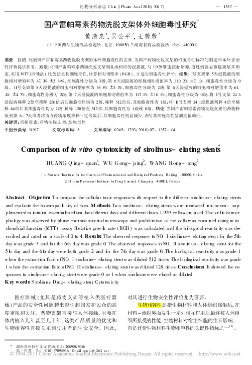
国产雷帕霉素药物洗脱支架体外细胞毒性研究
*
1357
国产雷帕霉素药物洗脱支架体外细胞毒性研究
黄清泉 , 吴公平 , 王蓉蓉
1 2 2
( 1. 中国药品生物制品检定所 , 北京 , 100050; 2 . 湖南省药品检验所 , 长沙 , 410001) 摘要 目的 : 比较国产雷帕霉素药物洗脱支架的体外细胞毒性的差异 , 为国产药物洗脱支架的细胞毒性标准的制定和体外 安全
4 . 7% , 细胞毒性分级为 2级 ; 第 7 天浸提液的细胞相对增值率为 117 . 1%
浸提液稀释 2 倍至稀释 256 倍后其细胞毒性均为 2 级 , 稀释 512 倍后 , 其细胞毒性为 1级 ; 将 II号支架 24 h 浸提液稀释 4 倍至稀 释 64 倍后其细胞毒性均为 2 级 , 稀释 128 倍至 512 倍 , 其细胞毒性为 1 级或 0 级。结论 : 当国产雷帕霉素药物洗脱支架的药物释 放到第 6~ 7天或者将所含药物浓度稀释一定倍数后 , 其细胞毒性明显减少 , 表明其细胞毒性呈剂量依赖性。 关键词 : 雷帕霉素 ; 药物洗脱支 架 ; 细胞毒性 中图分类号 : R 917 文献标识码 : A 文章编号 : 0245- 1793( 2010) 07- 1357- 04
表 1 I 号药物支架浸提不同时间细胞相对 增殖率及细胞毒性评级
产品名称 药物支架 ( I号 ) 药物支架 ( I号 ) 药物支架 ( I号 ) 药物支架 ( I号 ) 药物支架 ( I号 ) 药物支架 ( I号 ) 药物支架 ( I号 ) 浸提天数 /天 细胞相对增殖率 /% 细胞毒性评级 1 2 3 4 5 6 7 7 . 5 2. 7 15 . 1 1. 4 15 . 8 1. 6 19 . 9 1. 2 47 . 3 2. 84 119 . 2 7. 1 179 . 1 19. 6 4 4 4 4 3 0 0
碧云天活性氧检测试剂盒 S0033S S0033M 说明书

碧云天生物技术/Beyotime Biotechnology 订货热线:400-168-3301或800-8283301 订货e-mail :******************技术咨询:*****************网址:碧云天网站 微信公众号活性氧检测试剂盒产品编号 产品名称包装 S0033S 活性氧检测试剂盒 >100次 S0033M活性氧检测试剂盒>500次产品简介:活性氧检测试剂盒(Reactive Oxygen Species Assay Kit ,也称ROS Assay Kit)是一种利用荧光探针DCFH-DA 进行活性氧检测的试剂盒。
DCFH-DA 本身没有荧光,可以自由穿过细胞膜,进入细胞内后,可以被细胞内的酯酶水解生成DCFH 。
而DCFH 不能通透细胞膜,从而使探针很容易被装载到细胞内。
细胞内的活性氧可以氧化无荧光的DCFH 生成有荧光的DCF 。
检测DCF 的荧光就可以知道细胞内活性氧的水平。
本试剂盒提供了活性氧阳性对照试剂Rosup ,以便于活性氧的检测。
Rosup 是一种混合物(compound mixture),浓度为50mg/ml 。
本试剂盒本底低,灵敏度高,线性范围宽,使用方便。
本试剂盒S0033S 包装可以测定100-500个样品,S0033M 包装可以测定500-2500个样品。
包装清单:产品编号 产品名称 包装 S0033S-1 DCFH-DA (10mM)0.1ml S0033S-2 活性氧阳性对照(Rosup, 50mg/ml)1ml —说明书1份产品编号 产品名称 包装 S0033M-1 DCFH-DA (10mM)0.5ml S0033M-2活性氧阳性对照(Rosup, 50mg/ml)5ml —说明书1份保存条件:-20ºC 保存,一年有效。
注意事项:探针装载后,一定要洗净残余的未进入细胞内的探针,否则会导致背景较高。
纳米材料体外细胞毒性研究现状与展望
纳米材料体外细胞毒性研究现状与展望汪保林;邱慧【摘要】Nanoscience emerged in the last 1980 s and is developed as one of the most promising new science and technology in the 21st century.With the increasing widespread application of nanomaterials,their health risk has been greatly increased and researches on its biological safety are imperatively needed.In this paper,the toxic influential factors,the cytotoxicity mechanism of nanomaterials and the evaluation methods on cytotoxicity of nanomaterials in vitro were elucidated indetail.Simultaneously,the latest developments on the toxicity of nanomaterials and the security assessment of nano technologies were also systematically discussed.%纳米科学是上个世纪80年代末发展起来的新兴学科,是21世纪最有前途的新科学技术之一.随着纳米材料应用的日益广泛,其所带来的健康风险也越来越大,对其生物安全性的研究也刻不容缓.文章就纳米材料的毒性影响因素,对细胞造成的毒性效应机制及其体外细胞毒性的评价方法进行详细阐述,并综述了近几年来关于纳米材料毒性研究的最新进展及对纳米技术安全性评估进行了系统的讨论.【期刊名称】《世界中医药》【年(卷),期】2017(012)002【总页数】6页(P446-451)【关键词】纳米材料;细胞毒性;影响因素;评价方法【作者】汪保林;邱慧【作者单位】南昌市食品药品检验所,南昌,330038;南昌市洪都中医院制剂中心,南昌,330000【正文语种】中文【中图分类】R-331;R319从“纳米牙膏”到“纳米防晒霜”,全球目前已有300多种运用纳米技术上市的产品。
马比木植物内生真菌Trichoderma sp.抗癌活性产物的液体发酵工艺优化

马比木植物内生真菌Trichoderma sp.抗癌活性产物的液体发酵工艺优化陈旭;雷帮星;文庭池;曾茜【摘要】从马比木植物中分离获得一株木霉属内生真菌Trichoderma sp.编号为GX-8,其代谢产物中含有薯蓣皂苷类化合物,体外抗癌实验表明该物质对人肝癌细胞BEL-7402、人非小细胞肺癌细胞A549和人前列腺癌细胞PC3均具有中等的细胞毒活力。
本实验以上述化合物的含量为主要指标,通过单因素和正交试验的研究分析,优化确定适宜于菌株GX-8积累目标化合物的液体发酵工艺。
研究结果表明:GX-8发酵生产薯蓣皂苷类化合物的最优培养基为可溶性淀粉40 g/L、蛋白胨10 g/L、色氨酸40 mg/L、磷酸氢二钾1 g/L和硫酸镁2 g/L,pH 6.0,接种量2%( v/v),于28℃下摇瓶发酵16 d,该物质的产量可以达到87.38±5.86μg/mL,相比优化前的含量39.66±3.27μg/mL提高了120.32%。
%The compound extracted from the metabolite produced from a strain named GX-8 of the endophyt-ic fungus Trichoderma sp. was identified as Diosgenin. In vitro experiments, diosgenin shows moderate cyto-toxicity to the human hepatoma cell BEL-7402 , lung cancer cell A549 and prostate cancer cell PC3 . The objective of this research is to use the content of diosgenin as the main index, through the analysis of single factor and orthogonal tests, to optimize the liquid fermentation process suitable for strain GX-8 to accumu-late the compound diosgenin. The results showed that the best medium and fermentation process are:soluble starch 40g/L, peptone 10g/L, tryptophan 40mg/L, K2 HPO4 1g/L, MgSO4 2g/L, initial pH 6. 0, inocula-tion volume 2% ( v/v ) , at 28℃ in fermentation for 16 days. The yield ofdiosgenin could reach up to (87. 38 ± 5. 86) μg/mL, which is an increase of 120. 32% compared to the pre-optimized yield of (39. 66 ± 3. 27) μg/mL.【期刊名称】《山地农业生物学报》【年(卷),期】2015(000)005【总页数】5页(P33-37)【关键词】内生真菌;代谢产物;薯蓣皂苷类;抗癌活性;发酵工艺优化【作者】陈旭;雷帮星;文庭池;曾茜【作者单位】贵州省农业资源与环境研究所,贵州贵阳 550025; 贵州大学西南药用生物资源教育部工程研究中心,贵州贵阳 550025;贵州大学西南药用生物资源教育部工程研究中心,贵州贵阳 550025;贵州大学西南药用生物资源教育部工程研究中心,贵州贵阳 550025;贵阳中医学院基础部微生物研究室,贵州贵阳550025【正文语种】中文【中图分类】S646.2植物内生真菌(Plant Endophytic Fungi)是指存活于宿主植物的全部或几乎全部的生理周期中,而寄主不会表现任何病症的一类真菌,它们是生活在植物组织内微生物群体中的主要成员,是植物微生态系统的重要构成部分[1]。
病毒学术语中英文对照
A single- generation study 单项包括两代(生殖毒性)的研究 Acentric fragment 无着丝点片段Acridine orange 吖啶橙 Active metabolite 活性代谢产物 Additional test 附加试验 Adduct 加合物 ADME 吸引、分布、代谢、排泄 Administration period 给药期 Advers effect 不良反应 Against humanized proteins serum antibodies 抗人源蛋白血清抗体 Aginal smear 阴道涂片 Air righting reflex 空中翻正反射 Alkylating electrophilic cernter 浣化亲电子中心Allele 基因突变产生的遗传因子 Allergic reactions 过敏性反应(变应性反应) Altenative validated test 有效替代试验 Altered growth 生长改变 Ammoniun sulphide staining of the uterus 子宫硫化胺染色 Analogue 类似物(同系物) Analogue series of substance 同系物Analytical method 分析方法 Anaphase 分裂后期 Aneuploidy 非整倍体 Aneuploidy inducer 非整倍体诱导剂 Antigenic specificity 抗原特异性Art and ethical standards 技术和伦理标准Assessment of genotoxicity 遗传毒性评价 AUC 曲线下面积Auditory startle reflex 惊愕反射(听觉惊跳反射) Autoimmune 自身免疫 Autoradiographic assessment 放射自显影评价Autoradiography 放射自显影 Bacterial mutagenicity test 细菌致突变试验 Bacterial reverse mutation test 细菌回复突变试验 Bacterial strains 菌株 Bacterial test organisms 微生物试验菌 Base pairs 碱基对Base set of strains 基本菌株 Base substitution 碱基置换 Bioanalytical method 生物学分析方法 Bioavailability 生物利用度 Biological method 生物学意义Biotechnological products 生物技术产品 Biotechnoloty-derived pharmaceuticals 生物技术药物 Body burden 机体负担 Bone marrow cell 骨髓细胞 Bouin's fixation 包氏液固定Breakage of chromatid 染色单体断裂 Brealage of chromosome 染色体断裂 Bridging character 桥梁作用 C(time) 一定剂量、某一时间的浓度 Carcinogen 致癌物质Carcinogenesis 致癌性 Carcinogenic hazard 致癌性危害 Carcinogenicity bioassay 致癌性生物检测 Carcinogenicity potential of chemical 化合物的潜在致癌性 Carcinoginicity (oncogenicity) 致癌(致瘤) Cardiovascular 心血管 Case-by-case 个例 Cell proliferation 细胞增殖 Cell cultures 细胞培养 Cell line 细胞系 Cell membrane lipid 细胞膜脂质层 Cell replication system 细胞复制系统 Cell suspension 细胞悬液 Cell-mediated immunity 细胞介导的免疫 Cellular therapy 细胞治疗 Central nervous systems 中枢神经系统 Cerebral spinal fluid 脑脊液 Chemical nature 化学性质 Chinese hamster V79 cell 中国仓鼠V79细胞Chromatide 染色单体 Chromosomal aberration 染色体畸变 Chromosomal damage 染色体损伤 Chromosomal integrity 染色体完整性 Chronic toxicity testing 慢性毒性试验 Classfical biotransformation studies 经典的生物转化试验 Clastogen 染色体断裂剂 Clastogenic 致染色体断裂的 Clinical indication 临床适应证 Cloning efficiency 克隆形成率Closure of the hard palate 硬腭闭合 Cmax 峰浓度 Colony sizing 集落大小 Comparative trial 对比试验Complement binding 补体结合 Completely novel compound 全新化合物 Compound bearing structural alerts 结构可疑化合物 Concentration threshold 阈浓度 Concomitant toxicokinetics 相伴毒代动力学 Continuous treatment 连续接触 Corpora lutea 黄体 Corpora lutea count 黄体数 Cross-linking agent 交联剂 Culture condition 培养条件 Culture confluency 培养克隆率 Culture confluenty 培养融合 Culture medium 培养基 Cytogenetic change 细胞遗传学改变 Cytogenetic evaluation 细胞遗传学评价 Cytokines 细胞因子 Cytotoxicity 细胞毒Degradation 降解 Deletion 缺失 Descriptive statistics 描述性统计 Detection of bacterial mutagen 细菌诱变剂检测 Detection of clastogen 染色体断裂剂检测 Determination of metabolites 测定代谢产物 Developmental toxicity 发育毒性Direct genetic damage 直接遗传损伤 Distribution 分布DNA adduct DNA加合物DNA damage DNA损伤DNA repair DNA 修复DNA strand breaks DNA链断裂 Dose escalation 剂量递增 Dose dependence 剂量依赖关系 Dose level 剂量水平 Dose-limiting toxicity 剂量限制性毒性 Dose-raging studies 剂量范围研究 Dose-relatived mutagenicity 剂量相关性诱变性 Dose-related 剂量相关Dose-relatived cytotoxicity 剂量相关性细胞毒性 Dose-relatived genotoxic activity 剂量相关性遗传毒性 Dose-response curve 剂量-反应曲线 Dosing route 给药途径Embryo-fetal toxicity 胚胎-胎仔毒性 Endogenous components 内源性物质 Endogenous gene 内源性基因Endonuclease 核酸内切酶 Emdpmiclease release from lysosomes 溶酶体释放核酸内切酶End-point 终点 Epitope 抗原决定部位 Error prone repair 易错性修复 Escalation 递增Escherichia coli strain 大肠杆菌菌株 Escherichia coli 大肠杆菌Evaluation of test result 试验结果评价 Exaggerated pharmacological response 超常增强的药理作用 Exposure assessment 接触剂量评价 Exposure period 接解期 External metabolizing system 体外代谢系统F1-animals 子一代动物 False positive result 假阳性结果 Fecundity 多产 Fertility studies 生育力研究 Fetal abnormalities 胎仔异常 Fetal and neonatal parameters 胎仔和仔鼠的生长发育参数 Fetal development and growth 肿仔发育和生长 Fetal period 胎仔期 Fetotoxicity 胎仔毒性First pass testing 一期试验Fluorescence in situ hybridization(FISH) 原位荧光分子杂交 Foetuses 胎仔 Formulation 制剂 Frameshift mutation 移码突变 Frameshite point mutation 移码点突变 Free-standing 独立Fresh dissection technique 新鲜切片技术 Funtional deficits 切能缺陷 Functional test 功能试验 Functional indices 功能性指标 Fusion proteins融合蛋白 Gametes 配子 Gender of animals 动物性别 Gender-specific drug 性别专一性药物Gene knockout 基因剔除 Gene therapy 基因治疗 Gene mutation 基因突变 Genetic 遗传Genetic change 遗传学改变 Genetic damage 遗传学损伤 Genetic endpoint 遗传终点Genetic toxicity 遗传毒性 Genotoxic activity 遗传毒性作用 Genotoxic carcinogen 遗传毒性致癌剂 Genotoxic effect 遗传毒性效应 Genotoxic hazard 遗传毒性危害 Genotoxic potential 潜在遗传毒性 Genotoxic rodent carcinogen 啮齿类动物遗传毒性致癌剂 Genotoxicity 遗传毒性 Genotoxicity test 遗传毒性试验 Genotoxicity test battery 毒性试验组合 Genotoxycity evaluation 遗传毒性评价 Germ cell mutagen 生殖细胞诱变剂 Germ line mutation 生殖系统突变 GLP 临床前研究质量管理规范 Gross chromosomal damage 染色体大损伤 Gross evaluation of placenta 胎盘的大体评价 Growth factors 生长因子 Haemotoxylin staining 苏木素染色 Half-life 半衰期 Hematopoietic cells 造血细胞 Heptachlor 七氯化合物 Heritable 遗传 Heritable defect 遗传缺陷 Heritable disease 遗传性疾病 Heritable effect 遗传效应High concentration 高浓度Histologic appearance of reproductive organ 生殖器官的组织学表现 Histopathological chang 组织病理学改变 Homologous proteins 同系蛋白 Homologous series 同系 Host cell 宿主细胞 Human subjects 人体 Human carcinogen 人类致癌剂Human lymphoblastoid TH6cell 人成淋巴TK6细胞 Human mutagen 人类致突变剂 Humoral immunity 体液免疫 Immature erythrocyte 未成熟红细胞Immediate and latent effect 速发和迟发效应 Immunogenicity 免疫原性 Immunopathological effects 免疫病理反应immunotoxicity 免疫毒性 Implantation 着床 Implantation sites 着床部位 In vitro 体外 In vitro test 体外试验 In vivo 体内 In vivo test 体风试验Incidence of polyploidy cell 多倍体细胞发生率 Incisor eruption 门齿萌发 Independent test 独立试验 Individual fetal body weight 单个胎仔体重 Induced and spontaneous models of disease 诱发或自发的疾病模型Inducer of micronuclei 微核诱导剂 Inhalation 吸入 Inhibitor of DNA metabolism DNA代谢抑制剂 Intact animals 完整动物(整体动物) Internal control 内对照 Interphase nuclei 分裂间期细胞核 Intra-and inter-individual 个体与个体间 Isolated organs 离休器官Juvenile animal studies 未成年动物研究 Kinetic profile 动力学特点 Kinetics 动力学 Lactation 授乳、哺乳Large deletion event 大缺失事件 Late embryo loss 后期胚胎丢失 Level of safety 安全水平Libido 性欲 Life threatering 危及生命 Lipophilic compound 亲脂性化合物 Litter size 每窝胎仔数目 Live and deal conceptuese 活胎和死胎 Live offspring at birth 出生时存活的子代Local tolerance studies 局部耐受性研究 Local toxicity 局部毒性 Locu 位点 Long-termcarcinogenicity study 长期致癌性研究Loss of the tk gene tk基因缺失Major organ formation 主要器官形成 Male fertility 雄性生育力 Male fertility assessment 雄性生育力评价Mammalian sells 哺乳动物细胞 Mammalian species 哺乳类动物 Mammalian sell mutation test 哺乳动物细胞致突变试验 Marketing approval 上市许可 Maternal animal 亲代动物Mating behavior 交配行为 Mating period 交配期 Mating ratio 交配比例 Matrices 基质Maximum tolerated dose(MTD) 最大耐受剂量 Mechanism of genotoxicity 遗传毒性机制Mechanistic investigation 机制研究 Metabolic activation 代谢活化 Metabolic activation pathway 代谢活化途径 Metabolic activation system 代谢活化系统 Metabolism 代谢Metabolites profile 代谢物的概况 Metaphase 中期 Metaphase analysis 分裂中期相分析Metaphase cell 分裂中期相细胞 Micronucleus 微核 Micronucleus formation 微核形成Microtitre 微滴定 Mictotitre method 微滴定法 mimicking 模拟 Mitotic index 有丝分裂指数Molecular characterization 分子特性 Molecular technique 分子技术 Monitor 监测Monoclonal antibodies 单克隆抗体 Non-toxic compound 无毒化合物 Mouse lymphoma L5178Y cell 小鼠淋巴瘤L5178Y细胞 Mouse lymphoma tk assay 小鼠淋巴溜tk检测Mutagen 诱变原 Mutagenic carcinogen 诱变性致癌剂 Mutagenic potential of chemical 化合物的潜在致突变性 Mutant colony 突变体集落 Mutation 突变 Mutation induction in transgenes 转基因诱导突变Necropsy(macroscopic examination) 解剖(大体检查) Negative control 阴性对照 Negative result 阴性结果 Newcleated 有核 Non rodent 非啮齿类Non-clinical 非临床 Non-genotoxic carcinogen 非遗传毒性致癌剂 Non-genotoxic mechanism 非遗传毒性机制 Non-human primate 非人灵长类 Non-linear 非线性 No-toxic-effect dose level 无毒性反应剂量水平 Nucleated bone marrow cell 有核骨髓细胞 Nucleoside analogue 核苷酸同系物 Number of live and dead implantation 宫内活胎和死胎数 Numerical chromosomal aberration 染色体数目畸变 Numerical chromosome changes 染色体数目改变Oligonucleotide grugs 寡核苷酸药物 One ,twe,three generation studies 一、二、三子代研究Paraffine embedding 石蜡包埋 Parameter 参数 Parent compound 母体化合物 Parenteral 非肠道 Particulate material 颗粒物 Peripheral blood erythrocyte 外周血红细胞Pharmacodynamic effects 药效作用 Pharmacodynamics 药效学(药效动力学) Pharmacokinetic 药代动力学 Phenylene diamine 苯二胺 Physical development 身体发育 Physiological stress 生理应激 Pilot studies 前期研究 Pinna unfolding 耳廓张开 Plasmid 质粒 Plasminogen activators 纤维蛋白溶解酶原激活因子 Ploidy 整倍体 Point mutation 点突变 Polychromaticerythrocyte 嗜多染色红细胞 Polycyclic hydrocarbon 多环芳烃 Polymer 聚合物 Polyploidy cell 多倍体细胞 Polyploidy 多倍体 Polyploidy induction 多倍体诱导 Poorly soluble compound 难溶化合物 Positive control 阳性对照 Positive result 阳性结果 Post meiotic stages 减数分裂后期 Post-approval 批准后 Postcoital time frame 交配后日期Postimplantation deaths 着床后死亡 Postnatal deaths 出生后死亡 Postweaning development and growth 断奶后发育和生长 Potential 潜在性 Potential immunogenecity 潜在免疫原性Potential target organs for toxicity 潜在毒性靶器官Pre-and post-natal development study 围产期的发育研究 Pre-and postweaning survival and growth 断奶前后的存少和生长 Precipitate 沉淀期 Precision 精密度 Preclinical safety evaluation 临床前安全性评价 Predetermined criteria 预定标准 Prediction of carcinogenicity 致癌性预测Pregnant and lactation animals 怀孕与哺乳期动物 Preimplantation stages of the embryo 胚胎着床前期 Preliminary studies 预试验 Pre-screening 预筛选 Prevalence of abnormalities 异常情况的普遍程度 Primary active entity 主要活性实体 Priority selection 优先选择 Pro-drug 前体药物 Protocol modification 试验方案修改 Quantification of mutant 突变体定量 Racemate 消旋体 Radiolabeled proteins 放射性同位素标记蛋白 Radiolabelled compounds 放性性同位素标记化合物 Range-finding test 范围确定试验 Rate of preimplantation deaths 着床关死亡率 Rational study design 合理的试验设计 Receptor properties 受体性质 Recombinant DNA proteins DNA重组蛋白Recombinant DNA technology DNA重组技术 Recombination 重组 Recombinant plasma factors 重组血浆因子 Reduction in the number of revertants 回复突变数的减少 Relative plating efficiency 相对接种效率 Relative suspension growth 相对悬浮生长率 Relative total growth 相对总生长率 Relevant animal species 相关动物种属 Relevant dose 相关剂量Relevant factor 相关因素 Repeated-dose toxicity studies 重复剂量毒性研究 Reproductive toxicity 生殖毒性 Reproductive/developmental toxicity 生殖/发育毒性 Reverse mutation 回复突变 Reversibility 可恢复性(可逆性) Risk assessment 危险度评价 Rodent hematopoietic cell 啮齿类动物造血细胞 Route of administration 给药途径 Routine testing 常规试验S9-mix constituent S9混合液成分 Safeguards 安全监测 Safety pharmacology 安全药理学Safety margin 安全范围 Salmonella typhimurium 鼠伤寒沙门菌 Sampling time 采样时间Satellite groups 卫星组 Saturation of absorption 吸收饱和 Sensory functions and reflexes 感觉功能和反射Short term toxicity 短期毒性Short or medium-term carcinogenicity study 短或中期致癌性研究 Short treatment 短期处理 Sighting studies 预试验 Singledose(acute)toxicity 单剂量(急性)毒性 Single study design 单一研究设计 Site-specific targeted delivery 定位靶向释放 Small colony 小集落 Small colony mutant 小集落突变体Soft agar method 软琼脂法 Soluble genotoxic impurity 可溶性遗传毒性杂质 Solvent control 溶剂对照 Somatic cell 体细胞 Somatic cell test 体细胞试验 Species 种属 Specificity 特异性 Species specificity 种属特异性 Spindle apparatus 纺缍体 Stages of reproduction 生殖阶段Standard battery of test 标准试验组合Standard 3-test battery 标准三项试验组合 Standard battery 标准组合 Standard battery system 标准组合系统 Standard procedure 标准规程Standard protocol 标准试验方案Standard set of strains 标准菌株组Standard set of tests 标准试验组 Standard test battery 标准试验组合 Statistical evaluation 统计学评价 Steady-state levels 稳态浓度 Step-by-step 逐步 Stepwise process 阶梯式程序 Strain 品系 Structural changes 结构改变 Structural chromosomal aberration 染色体结构畸变 Subgroups 亚组Supravital staining 体外活动染色 Surface righting reflex 平面翻正反射 Survival 存活率suspension 悬浮物 Systemic exposure 全面接触 Target organs 靶器官 Target cell 靶细胞Target histidine genes 组氨酸目的基因 Target tissue 靶组织Target tissue exposure 靶组织接触 Teratogenic response 致畸胎反应 Terminal sacrifice 终末期处死 Test of carcinogenicity 致癌试验 Test approach 试验方法Test battery approach 试验组合方法 Test compound 受试物 Test model 试验模型 Test strategy 试验策略 Test systems 试验系统 Tester strain 试验菌株 Therapeutic 治疗 Therapeutic confirmatory 疗效确定 Therapeutic exploratory 疗效探索Therapeutic indication 治疗适应证 Time course 时程 Timing conventions 分段计时方法Tissue cross-reactivity 组织交叉反应 Tissue distribution 组织分布 Tissue exposure 组织接触Tissue uptake 组织吸收 Tk locus tk位点 Top concentration 最高浓度 Topical 局部的Topoisomerase inhibitor 拓朴异构酶抑制剂 Total erythrocyte 总红细胞Total litter loss 整窝丢失 Toxicity to reproduction 生殖毒性 Toxicokinetics 毒代动力学(毒物代谢动力学) Transgene 转基因 Transgenic animals 转基因动物 Transgenic plants 转基因植物Translocation 移位 Treatment regimen 实施方案 Tubal transport 输卵管运输 Tumor induction 肿瘤诱导 Tumor response 肿瘤反应 Tumor-related gene 肿瘤相关基因 Two or three phase approach 分段(二段或三段)研究 Two study design 分段(两段)研究设计Ovulation rate 排卵率 Unbound concentration 未结合浓度 Unexpected finding 非预期结果Unscheduled DNA synthesis(UDS) 程序外DNA合成 Unstable epoxide 不稳定过氧代物Whole blood 全血。
ICH-安全性领域常用专业术语中英文对照表()

ICH 安全性领域常用专业术语中英文对照表S9-mix constituent S9混合液成分Safeguards 安全监测Safety pharmacology 安全药理学Safety margin 安全范围Salmonella typhimurium 鼠伤寒沙门菌Sampling time 采样时间Satellite groups 卫星组Saturation of absorption 吸收饱和Secondary testing 二期试验Secretion in milk 乳汁分泌Sensitive periods 敏感期Sensitivity 敏感性Sensory functions and reflexes 感觉功能和反射Sexual maturity 性成熟Short term toxicity 短期毒性Short or medium-term carcinogenicity study 短或中期致癌性研究Short treatment 短期处理Sighting studies 预试验Single dose(acute)toxicity 单剂量(急性)毒性Single study design 单一研究设计Site—specific targeted delivery 定位靶向释放Small colony 小集落Small colony mutant 小集落突变体Soft agar method 软琼脂法Soluble genotoxic impurity 可溶性遗传毒性杂质Solvent control 溶剂对照Somatic cell 体细胞Somatic cell test 体细胞试验Species 种属Specificity 特异性Species specificity 种属特异性Sperm analysis 精子分析Sperm count 精子计数Sperm maturation 精子成熟Sperm morphology 精子形态学Sperm motility 精子活动度Sperm viability 精子活力Spermatogenesis 精子形成Spindle apparatus 纺缍体Stages of reproduction 生殖阶段Standard battery of test 标准试验组合Standard 3—test battery 标准三项试验组合Standard battery 标准组合Standard battery system 标准组合系统Standard procedure 标准规程Standard protocol 标准试验方案Standard set of strains 标准菌株组Standard set of tests 标准试验组Standard test battery 标准试验组合Statistical evaluation 统计学评价Steady-state levels 稳态浓度Step—by-step 逐步Stepwise process 阶梯式程序Strain 品系Structural changes 结构改变Structural chromosomal aberration 染色体结构畸变Subgroups 亚组Supravital staining 体外活动染色Surface righting reflex 平面翻正反射Survival 存活率suspension 悬浮物Systemic exposure 全面接触Target organs 靶器官Target cell 靶细胞Target histidine genes 组氨酸目的基因Target tissue 靶组织Target tissue exposure 靶组织接触Teratogenic response 致畸胎反应Terminal sacrifice 终末期处死Test of carcinogenicity 致癌试验Test approach 试验方法Test battery approach 试验组合方法Test compound 受试物Test model 试验模型Test strategy 试验策略Test systems 试验系统Tester strain 试验菌株Therapeutic 治疗Therapeutic confirmatory 疗效确定Therapeutic exploratory 疗效探索Therapeutic indication 治疗适应证Time course 时程Timing conventions 分段计时方法Tissue cross-reactivity 组织交叉反应Tissue distribution 组织分布Tissue exposure 组织接触Tissue uptake 组织吸收Tk locus tk位点Top concentration 最高浓度Topical 局部的Topoisomerase inhibitor 拓朴异构酶抑制剂Total erythrocyte 总红细胞Total litter loss 整窝丢失Toxicity to reproduction 生殖毒性Toxicokinetics 毒代动力学(毒物代谢动力学) Transgene 转基因Transgenic animals 转基因动物Transgenic plants 转基因植物Translocation 移位Treatment regimen 实施方案Tubal transport 输卵管运输Tumor induction 肿瘤诱导Tumor response 肿瘤反应Tumor-related gene 肿瘤相关基因Two or three phase approach 分段(二段或三段)研究Two study design 分段(两段)研究设计Ovulation rate 排卵率Unbound concentration 未结合浓度Unexpected finding 非预期结果Unscheduled DNA synthesis(UDS)程序外DNA合成Unstable epoxide 不稳定过氧代物Vaginal opening 阴道张开Vaginal plug 阴栓Whole blood 全血Dead offspring at birth 出生时死亡的子代Degradation 降解Delay of parturition 分娩延迟Deletion 缺失Descriptive statistics 描述性统计Detection of bacterial mutagen 细菌诱变剂检测Detection of clastogen 染色体断裂剂检测Determination of metabolites 测定代谢产物Development of the offspring 子代发育Developmental toxicity 发育毒性Diminution of the background lawn 背景减少Direct genetic damage 直接遗传损伤Distribution 分布DNA adduct DNA加合物DNA damage DNA损伤DNA repair DNA修复DNA strand breaks DNA链断裂Dose escalation 剂量递增Dose dependence 剂量依赖关系Dose level 剂量水平Dose-limiting toxicity 剂量限制性毒性Dose—raging studies 剂量范围研究Dose-relatived mutagenicity 剂量相关性诱变性Dose—related 剂量相关Dose-relatived cytotoxicity 剂量相关性细胞毒性Dose—relatived genotoxic activity 剂量相关性遗传毒性Dose-response curve 剂量—反应曲线Dosing route 给药途径Duration 周期Duration of pregnancy 妊娠周期Eaning 断奶Earlier physical malformation 早期躯体畸形Early embryonic development 早期胚胎发育Early embryonic development to implantation 着床早期的胚胎发育Electro ejaculation 电射精Elimination 清除Embryofetal deaths 胚胎和胎仔死亡Embryo—fetal development 胚胎—胎仔发育Embryo-fetal toxicity 胚胎—胎仔毒性Embryonic death 胚胎死亡Embryonic development 胚胎发育Embryonic period 胚胎期Embryos 胚胎Embryotoxicity 胚胎毒性Enantiomer 对映异构体End of pregnancy 怀孕终止Endocytic 内吞噬(胞饮)Endocytic activity 内吞噬活性Endogenous proteins 内源性蛋白Endogenous components 内源性物质Endogenous gene 内源性基因Endonuclease 核酸内切酶Emdpmiclease release from lysosomes 溶酶体释放核酸内切酶End-point 终点Epididymal sperm maturation 附睾精子成熟性Epitope 抗原决定部位Error prone repair 易错性修复Escalation 递增Escherichia coli strain 大肠杆菌菌株Escherichia coli 大肠杆菌Evaluation of test result 试验结果评价Exaggerated pharmacological response 超常增强的药理作用Excretion 排泄(清除)Exposure assessment 接触剂量评价Exposure period 接解期External metabolizing system 体外代谢系统F1-animals 子一代动物False positive result 假阳性结果Fecundity 多产Feed-back 反馈Fertilisation 受精Fertility 生育力Fertility studies 生育力研究Fetal abnormalities 胎仔异常Fetal and neonatal parameters 胎仔和仔鼠的生长发育参数Fetal development and growth 肿仔发育和生长Fetal period 胎仔期Fetotoxicity 胎仔毒性False negative result 假阴性结果First pass testing 一期试验Fluorescence in situ hybridization(FISH) 原位荧光分子杂交Foetuses 胎仔Formulation 制剂Frameshift mutation 移码突变Frameshite point mutation 移码点突变Free-standing 独立Fresh dissection technique 新鲜切片技术Funtional deficits 切能缺陷Functional test 功能试验Functional indices 功能性指标Fusion proteins 融合蛋白Gametes 配子Gender of animals 动物性别Gender—specific drug 性别专一性药物Gene knockout 基因剔除Gene therapy 基因治疗Gene mutation 基因突变Genetic 遗传Genetic change 遗传学改变Genetic damage 遗传学损伤Genetic endpoint 遗传终点Genetic toxicity 遗传毒性Genotoxic activity 遗传毒性作用Genotoxic carcinogen 遗传毒性致癌剂Genotoxic effect 遗传毒性效应Genotoxic hazard 遗传毒性危害Genotoxic potential 潜在遗传毒性Genotoxic rodent carcinogen 啮齿类动物遗传毒性致癌剂Genotoxicity 遗传毒性Genotoxicity test 遗传毒性试验Genotoxicity test battery 毒性试验组合Genotoxycity evaluation 遗传毒性评价Germ cell mutagen 生殖细胞诱变剂Germ line mutation 生殖系统突变GLP 临床前研究质量管理规范Gross chromosomal damage 染色体大损伤Gross evaluation of placenta 胎盘的大体评价Growth factors 生长因子Haemotoxylin staining 苏木素染色Half—life 半衰期Hematopoietic cells 造血细胞Heptachlor 七氯化合物Heritable 遗传Heritable defect 遗传缺陷Heritable disease 遗传性疾病Heritable effect 遗传效应High concentration 高浓度Histologic appearance of reproductive organ 生殖器官的组织学表现Histopathological chang 组织病理学改变Homologous proteins 同系蛋白Homologous series 同系Host cell 宿主细胞Human subjects 人体Human carcinogen 人类致癌剂Human lymphoblastoid TH6cell 人成淋巴TK6细胞Human mutagen 人类致突变剂Humoral immunity 体液免疫Immature erythrocyte 未成熟红细胞Immediate and latent effect 速发和迟发效应Immunogenicity 免疫原性Immunopathological effects 免疫病理反应immunotoxicity 免疫毒性Implantation 着床Implantation sites 着床部位In vitro 体外In vitro test 体外试验In vivo 体内In vivo test 体风试验Incidence of polyploidy cell 多倍体细胞发生率Incisor eruption 门齿萌发Independent test 独立试验Individual fetal body weight 单个胎仔体重Induced and spontaneous models of disease 诱发或自发的疾病模型Inducer of micronuclei 微核诱导剂Inhalation 吸入Inhibitor of DNA metabolism DNA代谢抑制剂Intact animals 完整动物(整体动物)Internal control 内对照Interphase nuclei 分裂间期细胞核Intra—and inter-individual 个体与个体间Isolated organs 离休器官Juvenile animal studies 未成年动物研究Kinetic profile 动力学特点Kinetics 动力学Lactation 授乳、哺乳Large deletion event 大缺失事件Late embryo loss 后期胚胎丢失Level of safety 安全水平Libido 性欲Life threatering 危及生命Lipophilic compound 亲脂性化合物Litter size 每窝胎仔数目Live and deal conceptuese 活胎和死胎Live offspring at birth 出生时存活的子代Local tolerance studies 局部耐受性研究Local toxicity 局部毒性Locu 位点Long-term carcinogenicity study 长期致癌性研究Loss of the tk gene tk基因缺失Major organ formation 主要器官形成Male fertility 雄性生育力Male fertility assessment 雄性生育力评价Mammalian sells 哺乳动物细胞Mammalian species 哺乳类动物Mammalian sell mutation test 哺乳动物细胞致突变试验Marketing approval 上市许可Maternal animal 亲代动物Mating behavior 交配行为Mating period 交配期Mating ratio 交配比例Matrices 基质Maximum tolerated dose(MTD) 最大耐受剂量Mechanism of genotoxicity 遗传毒性机制Mechanistic investigation 机制研究Metabolic activation 代谢活化Metabolic activation pathway 代谢活化途径Metabolic activation system 代谢活化系统Metabolism 代谢Metabolites profile 代谢物的概况Metaphase 中期Metaphase analysis 分裂中期相分析Metaphase cell 分裂中期相细胞Micronucleus 微核Micronucleus formation 微核形成Microtitre 微滴定Mictotitre method 微滴定法mimicking 模拟Mitotic index 有丝分裂指数Molecular characterization 分子特性Molecular technique 分子技术Monitor 监测Monoclonal antibodies 单克隆抗体Non—toxic compound 无毒化合物Mouse lymphoma L5178Y cell 小鼠淋巴瘤L5178Y细胞Mouse lymphoma tk assay 小鼠淋巴溜tk检测Mutagen 诱变原Mutagenic carcinogen 诱变性致癌剂Mutagenic potential of chemical 化合物的潜在致突变性Mutant colony 突变体集落Mutation 突变Mutation induction in transgenes 转基因诱导突变Naked eye 肉眼Necropsy(macroscopic examination)解剖(大体检查)Negative control 阴性对照Negative result 阴性结果Neonate adaptation to extrauterine life 新生仔宫外生活的适应性Newborn 新生仔Newcleated 有核Non rodent 非啮齿类Non-clinical 非临床Non-genotoxic carcinogen 非遗传毒性致癌剂Non—genotoxic mechanism 非遗传毒性机制Non-human primate 非人灵长类Non—linear 非线性No-toxic—effect dose level 无毒性反应剂量水平Nucleated bone marrow cell 有核骨髓细胞Nucleoside analogue 核苷酸同系物Number of live and dead implantation 宫内活胎和死胎数Numerical chromosomal aberration 染色体数目畸变Numerical chromosome changes 染色体数目改变Oestrous cycle 动情周期Oligonucleotide grugs 寡核苷酸药物One ,twe,three generation studies 一、二、三子代研究Organ development 器官发育Paraffine embedding 石蜡包埋Parameter 参数Parent compound 母体化合物Parenteral 非肠道Particulate material 颗粒物Parturition 分娩Pediatric populations 小儿人群Peproductive competence 生殖能力Peripheral blood erythrocyte 外周血红细胞Pharmacodynamic effects 药效作用Pharmacodynamics 药效学(药效动力学)Pharmacokinetic 药代动力学Phenylene diamine 苯二胺Physical development 身体发育Physiological stress 生理应激Pilot studies 前期研究Pinna unfolding 耳廓张开Plasmid 质粒Plasminogen activators 纤维蛋白溶解酶原激活因子Ploidy 整倍体Point mutation 点突变Polychromatic erythrocyte 嗜多染色红细胞Polycyclic hydrocarbon 多环芳烃Polymer 聚合物Polyploidy cell 多倍体细胞Polyploidy 多倍体Polyploidy induction 多倍体诱导Poorly soluble compound 难溶化合物Positive control 阳性对照Positive result 阳性结果Post meiotic stages 减数分裂后期Post—approval 批准后Postcoital time frame 交配后日期Postimplantation deaths 着床后死亡Postnatal deaths 出生后死亡Postweaning development and growth 断奶后发育和生长Potential 潜在性Potential immunogenecity 潜在免疫原性Potential target organs for toxicity 潜在毒性靶器官Pre—and post-natal development study 围产期的发育研究Pre-and postweaning survival and growth 断奶前后的存少和生长Precipitate 沉淀期Precision 精密度Preclinical safety evaluation 临床前安全性评价Predetermined criteria 预定标准Prediction of carcinogenicity 致癌性预测Pregnant 怀孕Pregnant and lactation animals 怀孕与哺乳期动物Preimplantation stages of the embryo 胚胎着床前期Preimplantation development 着床前发育Preliminary studies 预试验Premating 交配前Premating treatment 交配前给药Pre-screening 预筛选Prevalence of abnormalities 异常情况的普遍程度Preweaning 断奶前Primary active entity 主要活性实体Priority selection 优先选择Pro-drug 前体药物Prolongation of parturition 产程延长Protein binding 蛋白结合率Protocol modification 试验方案修改Quantification of mutant 突变体定量Racemate 消旋体Radiolabeled proteins 放射性同位素标记蛋白Radiolabelled compounds 放性性同位素标记化合物Range-finding test 范围确定试验Rate of preimplantation deaths 着床关死亡率Rational study design 合理的试验设计Receptor properties 受体性质Recombinant DNA proteins DNA重组蛋白Recombinant DNA technology DNA重组技术Recombination 重组Recombinant plasma factors 重组血浆因子Reduction in the number of revertants 回复突变数的减少Relative plating efficiency 相对接种效率Relative suspension growth 相对悬浮生长率Relative total growth 相对总生长率Relevant animal species 相关动物种属Relevant dose 相关剂量Relevant factor 相关因素Repeated—dose toxicity studies 重复剂量毒性研究Reproductive function 生殖功能Reproductive toxicity 生殖毒性Reproductive/developmental toxicity 生殖/发育毒性Reverse mutation 回复突变Reversibility 可恢复性(可逆性)Risk assessment 危险度评价Rodent 啮齿类动物Rodent hematopoietic cell 啮齿类动物造血细胞Route of administration 给药途径Routine testing 常规试验S9—mix constituent S9混合液成分Safeguards 安全监测Safety pharmacology 安全药理学Safety margin 安全范围Salmonella typhimurium 鼠伤寒沙门菌Sampling time 采样时间Satellite groups 卫星组Saturation of absorption 吸收饱和Secondary testing 二期试验Secretion in milk 乳汁分泌Sensitive periods 敏感期Sensitivity 敏感性Sensory functions and reflexes 感觉功能和反射Sexual maturity 性成熟Short term toxicity 短期毒性Short or medium-term carcinogenicity study 短或中期致癌性研究Short treatment 短期处理Sighting studies 预试验Single dose(acute)toxicity 单剂量(急性)毒性Single study design 单一研究设计Site-specific targeted delivery 定位靶向释放Small colony 小集落Small colony mutant 小集落突变体Soft agar method 软琼脂法Soluble genotoxic impurity 可溶性遗传毒性杂质Solvent control 溶剂对照Somatic cell 体细胞Somatic cell test 体细胞试验Species 种属Specificity 特异性Species specificity 种属特异性Sperm analysis 精子分析Sperm count 精子计数Sperm maturation 精子成熟Sperm morphology 精子形态学Sperm motility 精子活动度Sperm viability 精子活力Spermatogenesis 精子形成Spindle apparatus 纺缍体Stages of reproduction 生殖阶段Standard battery of test 标准试验组合Standard 3—test battery 标准三项试验组合Standard battery 标准组合Standard battery system 标准组合系统Standard procedure 标准规程Standard protocol 标准试验方案Standard set of strains 标准菌株组Standard set of tests 标准试验组Standard test battery 标准试验组合Statistical evaluation 统计学评价Steady-state levels 稳态浓度Step-by-step 逐步Stepwise process 阶梯式程序Strain 品系Structural changes 结构改变Structural chromosomal aberration 染色体结构畸变Subgroups 亚组Supravital staining 体外活动染色Surface righting reflex 平面翻正反射Survival 存活率suspension 悬浮物Systemic exposure 全面接触Target organs 靶器官Target cell 靶细胞Target histidine genes 组氨酸目的基因Target tissue 靶组织Target tissue exposure 靶组织接触Teratogenic response 致畸胎反应Terminal sacrifice 终末期处死Test of carcinogenicity 致癌试验Test approach 试验方法Test battery approach 试验组合方法Test compound 受试物Test model 试验模型Test strategy 试验策略Test systems 试验系统Tester strain 试验菌株Therapeutic 治疗Therapeutic confirmatory 疗效确定Therapeutic exploratory 疗效探索Therapeutic indication 治疗适应证Time course 时程Timing conventions 分段计时方法Tissue cross-reactivity 组织交叉反应Tissue distribution 组织分布Tissue exposure 组织接触Tissue uptake 组织吸收Tk locus tk位点Top concentration 最高浓度Topical 局部的Topoisomerase inhibitor 拓朴异构酶抑制剂Total erythrocyte 总红细胞Total litter loss 整窝丢失Toxicity to reproduction 生殖毒性Toxicokinetics 毒代动力学(毒物代谢动力学) Transgene 转基因Transgenic animals 转基因动物Transgenic plants 转基因植物Translocation 移位Treatment regimen 实施方案Tubal transport 输卵管运输Tumor induction 肿瘤诱导Tumor response 肿瘤反应Tumor-related gene 肿瘤相关基因Two or three phase approach 分段(二段或三段)研究Two study design 分段(两段)研究设计Ovulation rate 排卵率Unbound concentration 未结合浓度Unexpected finding 非预期结果Unscheduled DNA synthesis(UDS)程序外DNA合成Unstable epoxide 不稳定过氧代物Vaginal opening 阴道张开Vaginal plug 阴栓Whole blood 全血。
四种牙科陶瓷材料细胞毒性研究

四种牙科陶瓷材料细胞毒性研究目的:初步评价牙科陶瓷材料的生物相容性。
方法:根据ISO标准采用体外细胞毒性试验(MTT法)测试不同材料和浸提时间的浸提液对L929小鼠结缔组织成纤维细胞的影响,从而对全瓷材料的生物相容性进行初筛。
结果:各组A490 nm 值均与阴性对照无显著性差异(P>0.05),材料毒性级别除培养48 h后金属烤瓷粉浸提液组的细胞毒性为1级外,其他各浸提液组均为0级。
结论:四种材料体外细胞毒性实验阴性,初步认为材料具有较好的生物相容性。
[Abstract] Objective: To preliminarily evaluate the biocompatibility of four dental ceramics materials. Methods: According to ISO standards, in vitro cytotoxicity-test(MTT method) on L929 cells were carried on. Results: The A-value of each test group were similar to the negative control and the cytotoxicity of low-temperature ceramic group after 48 hours culture was in grade 1 and those of the other groups were all in grade 0. Conclusion: It is preliminarily estimated that four dental ceramics are safe for dental clinical application.[Key words] Dental ceramics materials; Biocompatibility; In vitro cytotoxicity-test (MTT method)目前临床普遍应用的金瓷修复体存在色彩及生物相容性等方面的不足,随着修复技术及材料学的发展,非贵金属烤瓷冠的不足之处亦表现出来[1],全瓷冠具有与牙釉质相似的折光性、颜色与邻牙协调等优点,临床效果良好[2]。
精氨酸水溶液复溶的多西他赛聚合物胶束的体外药效学及体内分布

精氨酸水溶液复溶的多西他赛聚合物胶束的体外药效学及体内分布张佳琳;熊晔蓉;涂家生【期刊名称】《药学与临床研究》【年(卷),期】2013(21)3【摘要】目的:考察精氨酸复溶的多西他赛胶束的体外药效及体内分布情况.方法:用CCK-8法考察多西他赛胶束和多西他赛注射液对肿瘤细胞的增殖抑制作用.以荧光染料DIR标记多西他赛聚合物胶束,通过活体成像系统比较精氨酸水溶液复溶组,生理盐水复溶组和多西他赛注射液组的荧光分布.结果:多西他赛胶束组IC50值明显比多西他赛注射液组高.精氨酸复溶的多西他赛胶束组肿瘤部位荧光强度比生理盐水复溶组和多西他赛注射液组都强.结论:精氨酸复溶的多西他赛胶束肿瘤靶向性更强且在肿瘤部位的停留时间更长,但其体外抗肿瘤活性有待提高.%Objective:To investigate the in vitro cytotoxicity and in vivo distribution of the argininedispersed docetaxel polymeric micelles (DTX-PMs).Methods:The CCK-8 method was used to compare the tumor cell growth inhibition of docetaxel micelles and docetaxel injection.The labeled micelles were pre pared by a rotary evaporation method with the fluorescent dye DIR incorporated,and an in vivo imaging system was used to detect the in vivo distribution of arginine-dispersed micells.Results:The fluorescene intensity of arginine-dispersed DTX-PMs at the tumor site was stronger than those of both docetaxel injec tion and saline-dispersed micelles.However,theIC50 value of DTX-PMs was two times higher than that of docetaxelinjection.Conclusion:The arginine-dispersed DTX-PMs have a much better tumor-targeting ef fect and a longer time at tumor site,but the in vitro antitumor effect of DTX-PMs needs to be improved.【总页数】3页(P224-226)【作者】张佳琳;熊晔蓉;涂家生【作者单位】中国药科大学药剂学教研室,南京210009;中国药科大学药剂学教研室,南京210009;中国药科大学药剂学教研室,南京210009【正文语种】中文【中图分类】R965【相关文献】1.两种双亲性聚合物自组装胶束复合水溶液的乳化性能 [J], 王婷立;易成林;江金强;刘晓亚2.添加精氨酸的多西他赛聚合物胶束冻干粉的制备 [J], 郦江平;吴仁荣;徐晓枫;涂家生3.多西他赛Pluronic F127聚合物胶束的制备与表征 [J], 赵丽娟;刘东华;刘志红;刘海霞;张娜4.灯盏花素TPGS聚合物胶束在大鼠体内药动力学及药效学评价 [J], 李云贵;李天平;邹柳5.包载多西他赛的聚合物胶束抑制小鼠乳腺癌转移作用研究 [J], 王爱婷; 鄢丹; 齐宪荣因版权原因,仅展示原文概要,查看原文内容请购买。
药物化学专业英语词汇
medicinal药品,药物, 药的,药用的be split into 分成,分为pharmaceutical 药学的,制药的,药品 starting materialsynthetic, 合成的,人造的,;synthetics化学合成品, synthesis合成(法),semisynthetic, synthesize,synthesesalkaloid 生物硷lead structure 先导结构intermediate中间体isolation 分离natural source 天然来源enzyme 酶 heart glycoside 强心苷steroid 甾体precursor 前体organ/target organ 靶器官 peptidehormone 激素 insulinpancreas vaccinepolysaccharide serumcholesterol 胆固醇amino acidgelatine hydrolysis水解/hydrolysate水解产物/hydrolyze水解hydroxylation antibiotic 抗生素,抗菌的antibody interferon 干扰素fermentation 发酵 dextran 葡聚糖ーlactam natural producttherapy/therapeutic治疗的/therapeutic margincaffeine咖啡因 yeastmicrobiological mutantmicroorganism geneticmould high performancebacterial proteinmucous membranemetabolism新陈代谢metabolite代谢物plasma 血浆 molecule /molecular weightfood additive organiclactic acid乳酸citric acid 柠檬酸penicillin penicilliumtetracyclinederivative衍生物contamination污染 sterile无菌的aerobic oxygencarbon dioxide carbohydratestarch saccharide/ polysaccharideglucose葡萄糖nitrogenureaPhosphate optimalammonium sulfate ammoniaseparate filtrateabsorption extractionrecombinant encodepurification chromatographic procedurecalciumregiospecific reaction区域专一性反应stereospecific reaction 立体专一性反应isomerization/isomeric fructosecountless test diagnosediagnosticanalysis/ analyst/ analytical/ analyze protease Ingredient in combination withDigestion enymatic cleavageBy means of fumaric acidBindimmobilize racemate /racemicacetyl heterogeneouscatalysis mediumester synthetic routeregistration compoundOrganometallic pyridinearomatic toluenexylene phenolrecrystallization/crystalmethanol/ethanolacetone ethyl acetatebenzene/ chlorobenzenediethyl ethersodium hydroxide hydrochloric acidsulfuric acid nitric acidacetic acid potassium carbonatechlorine/ chloride iodine/iodidefluorine/ fluoride bromine/bromideimpurity quality certificateGMP in large amountfacilityInspectionanalogousHygienicbe subjected toadminister/administration biologic response biologicmembrane to a large extentpenetration spatial arrangementpharmacologic stereochemistrythree-dimensional structure lipidstructure-activity relationship stericcorrelation parameterpartition coefficient distribution fuction conformation extractionoptical isomerism/opticalisomer enantiomorphic/ enantiomorphby no means tartaric acidManually magnificationdrug designpolarized lightdextrorotatory levorotatoryClokwise countclockwiseAntipode nonsuperimposable mirror image Coincide with glyceraldehydeAbsolute literatureconfiguration crystallographyasymmetric center accessisomeric enantiomerdiastereoisomeratomic numberPrioritymagnificationsolubilityspatial sequencein vivo/in vitroreceptorintravenous injection静脉注射be susceptible to敏感的With respect to contractSubstrate epoxidationcarcinogen oxidation /oxidasepreparationpredominantspeciescomplexdehydrase/dehydrogenase/decarboxylase/hydrolytic enzymes/isomerase/permease Choline one out of every ten 十分之一clinical 临床的interactionexcrete/excretioninversionCoordination DelayEfficacy in place相称的,合适的entity drug developmentattrition toxicity/toxic/ toxicology/Anti-infectives Healthcarerepro-toxicology/genotoxicity drug candidate indicationpharmacokineticsadverse profileformulary/formulation/formulor onsetdose/once a day dosing dosage/dosage form/overdosageregulatory interdependentsubacute亚急性的/chronic clinical/preclinicalvital optimum/ optimizeimpurity pilot plantcritical path criteriaupdate in paralleladequate stabilitypotency dermalcardiovascular 心血管的respiratory nervousconcurrently labelsynergies 协同作用antagonizereversible/irreversible permissiblelifespandiseasetumourinhalercapsule rodentfoetal teratologyexposurepatchset-up hazardOn a large scale shelf-lifetannin caffeineIn common vacuum fitrationhomogeneous gallic acidhydroxyl group esterifyphenolic precipitatenon-hydrolyzable carboxyl groupacidic calcium carbonatechloroform flavonoiddistillation sublimationsalicylic acid three neck round bottom flask separatory funnel steam bathdistillation flask beakerrinse ozoneice water bath condenserheparin digestionAside from fall intoProvide for as withCation compendialBatch –to batchcoagulation clotdecolorize anticoagulantprecipitation methodologyextraneous intestinalmucosa casingnitrate proteolyticdegrade/ degradation peroxideantithrombin thrombinplatelet aggregationintratracheal parenteraltopical comatoserelegate tabletsyrup suspensionemulsion versusbreakage leakagechip cracktaste masking expirationEven partially portableAdsorbent be free of / be free from Preference 偏爱 otherwise ad. 另外,别样Burden 负担,负重, on standing 搁置microbiologic preservationdispense bioavailabilitysystemic effects self-administration of medication motion sickness medical emergencysterile ophthalmicirrigate mucousabradeViable 能生长发育的,生存的 Mucous menbraneBody compartment 体室,体腔 Body cavityCircumvent 围绕,包围,智胜,防止…发生,迂回 Exceptionally 特殊地,异常地Wound受伤 Vessel 管,脉管Specialized 专业的,专业性的 By far 非常,更加Monograph专题文章,专题论文 Stringent 严格的,严厉的Inclusive 范围广的 Gravimetric 重量分析法的Electrolytic 电介质的,电解的 Conductivity 电导率Conductance 电导,电导性 Immerse 将…浸入Electrode 电极 Specific 比的Resistance Withstand 经受得住Stress 恶劣的 Redictable 可预报的Reproducible 可重现的 Necessary 必然的Solubilizers 加溶剂 Chelate 螯合Excipient 赋型剂 Ingredient 配料Medicinal agent Dispense 使分散,使疏开,配方(药)Ingenuity 独创性,精明 FormulatorMeager 贫乏的 Continuance 持续pellet vehiclegravimetric instantaneousosmosis dissociatepyrogen antioxidantbuffer tonicityantifungal inhibitorantifoaming colligativeextemporaneous specificationpreparation optimizeaccumulation availabilitydelivery/ deliver peroralrelease sustaingastrointestinal predefinecavity marginionic/ion simulatedistinctly efficacypaddle intestinalinterval a steady-state blood or tissue levelelimination blood vesselelectrode/electrolyticconductivity/conductanceresistanceexcipientthermalviabledisintegrationresidence time accomplishmaximum/maximizepotentiateprescribeuniformitycompliancespecificationphysiologicagitationIn the face of 面临 Fluctuation 波动Deliberate 深思熟虑的 Peroral 经口的Depot 仓库 Repository 仓库Sustained release, Sustained action,prolonged action, controlled release,extended action, timed release,repository dosage forms Implicit固有的peak 峰 dumpmaintenance dose maintenance periodmethane, ethane, Propane, butane/tetrane, pentaneethylene, Propylene/propene, butylene, 1-pentenemethanol,ethanol/ethyl alcohol, Propanol/ propyl alcohol, Butanol/Butyl alcohol, 1-pentanolcalibrateasepticstoichiometry replenishmenttubularproduct yieldscirculate atomizediscrete reactantmaterial transfer regenerationreactant conversion deviate fromviscosityexothermic endothermicshort-circuiting 短路laminar flowadiabatic radialproduct yields well-stirred batch reactorreactor configuration semibatch reactorcontinous-flow stirred-tank reactorback-mixing返混cross-sectionpressue drop countercurrentpacked-column rate-limiting stepfluidized or fluid bed tubular reactortubular plug-flow reactor batch operationturbulent trickle bedmultiplicity in series 逐次的,串联的feed Cross-flow 错流,横向流Panel-bed 板式床reaction driving froces 反应驱动力Chain-terminating Hydraulic 水力学的mechanical seal 机械密封 viscous 粘滞的Be prone to 倾向于, 易于中药traditional Chinese drug生药crude drug草药medicinal herb民族药ethnic drug地产药材native drug道地药材famous-region drug中成药Chinese patent medicine海洋生药学marine pharmacognosy药用植物学medicinal botany植物化学phytochemistry植物化学分类学plant chemotaxonomy生药拉丁名Latin name of crude drug学名scientific name来源source混淆品adulterant类同品allied drug伪品counterfeit drug代用品substitute掺伪adulteration天然产物natural product化学成分chemical constituent有效成分effective constituent主成分main constituent活性成分active constituent莽草酸途径shikimic acid pathway乙酸一丙二酸途径acetate-malonate pathway乙酸- 甲瓦龙酸途径acetate-mevalonate pathway单糖monosaccharide戊糖pentose己精hexose庚糖heptose辛糖octose脱氧糖deoxysaccharide, deoxysugar呋喃糖furanose吡喃糖pyranose寡糖oligosaccharide二糖disaccharide三糖trisaccharide四糖tetrasaccharide五糖pentosacc haride多糖polysaccharide淀粉starch树胶gum果胶pectin半纤维素hemicellulose纤维素cellulose甲壳质chitin肝素heparin硫酸软骨素chondroitin sulfate玻璃酸hyaluronic acid直链淀粉amylose支链淀粉amylopectin糖原glycogen费林试验Fehling test苷glycoside糖杂体heteroside苷元aglycone苦杏仁酶emulsin氰苷cyanogenic glycoside, cyanogenetic glycoside酚苷phenolic glycoside多酚polyphenol醛苷aldehyde glycoside醇苷alcoholic glycoside吲哚苷indole glycoside树脂醇苷resinol glycoside硫苷thioglycoside呫吨酮xanthone呫吨酮苷xanthonoid glycoside蒽醌anthraquinone蒽醌苷anthraquinone glycoside蒽酚anthranol氧化蒽酚oxanthranol蒽酮anthrone二蒽酮dianthrone羟基蒽醌hydroxyanthraquinone博恩特雷格反应Borntrager reaction 黄酮类flavonoid黄酮苷flavonoid glycoside黄酮flavone黄烷flavane黄酮醇flavonol黄烷酮flavanone黄烷酮醇flavanonol异黄酮isoflavone异黄烷酮isoflavanone新黄酮类neoflavonoid裂环烯醚萜苷secoiridoid glycoside 木脂体lignan木脂内酯lignanolide新木脂体neolignan木素lignin萜terpene萜类terpenoid半萜hemiterpene单萜monoterpene倍半萜sesquiterpene二萜diterpene三萜triterpene四萜tetraterpene多萜polyterpene齐墩果烷oleanane挥发油volatile oil精油essential oil鞣质tannin鞣酸tannic acid可水解鞣质hydrolysable tannin缩合鞣质condensed tannin鞣酐phlobaphene鞣花鞣质ellagitannin没食子鞣质gallotannin双缩脲反应biuret reaction 脂肪fat脂肪油fatty oil去油de-fatting蜡wax环烯醚萜苷iridoid glycoside环烯醚萜iridoid裂环烯醚苷secoiridoid皂化saponification酸败rancidity饱和脂肪酸saturated fatty acid不饱和脂肪酸unsaturated fatty acid有机酸organic acid树脂resin油树脂oleoresin树胶树脂gum resin香树脂balsam香脂酸balsamic acid苷树脂glycosidal resin苦味素bitter principle色素pigment微量元素trace element生物碱alkaloid吖啶生物碱acridine alkaloid阿朴啡类生物碱aporphine alkaloid苄基异喹啉生物碱benzylisoquinoline alkaloid双苄基异喹啉生物碱bisbenzylisoquinoline alkaloid双吲哚生物碱bisindole alkaloid咪唑生物碱imidazole alkaloid吲哚生物碱indole alkaloid吲哚联啶生物碱indolizidine alkaloid吲哚烷胺生物碱indolylalkylamine alkaloid异喹啉生物碱isoquinoline alkaloid大环生物碱macrocyclic alkaloid吗啡烷生物碱morphinane alkaloid羟吲哚生物碱oxindole alkaloid菲啶生物碱phenanthridine alkaloid苯烷胺生物碱phenylalkylamine alkaloid哌啶生物碱piperidine alkaloid嘌呤生物碱purine alkaloid吡啶生物碱pyridine alkaloid吡咯生物碱pyrrolidine alkaloid吡咯联啶生物碱pyrrolizidine alkaloid喹唑啉生物碱quinazoline alkaloid喹啉生物碱quinoline alkaloid喹啉联啶生物碱quinolizidine alkaloid甾体生物碱steroid alkaloid萜类生物碱terpenoid alkaloid四氢异喹啉生物碱tetrahydroisoquinoline alkaloid碘化汞钾试剂Mayer's reagent碘化铋钾试剂Dragendorff's reagent碘化钾碘试剂Wagner's reagent硅钨酸试剂Bertrand's reagent, silicotungstic acid reagent磷钼酸试剂Sonnenschein's reagent, phospho-molybdic acid reagent苦味酸试剂Hager's reagent, picric acid reagent矾酸铵-浓硫酸试液Mandelin test solution 钼酸铵-浓硫酸试液Frohde test solution甲醛-浓硫酸试液Marquis test solution莨菪烷tropane莨菪烷生物碱tropane alkaloid除虫菊素类pyrethroid-acetal 醛缩醇acetal- 乙酰acid 酸-al 醛alcohol 醇-aldehyde 醛alkali- 碱allyl 丙烯基alkoxy- 烷氧基-amide 酰胺amino- 氨基的-amidine 脒-amine 胺-ane 烷anhydride 酐anilino- 苯胺基aquo- 含水的-ase 酶-ate 含氧酸的盐、酯-atriyne 三炔azo- 偶氮benzene 苯bi- 在盐类前表示酸式盐bis- 双-borane 硼烷bromo- 溴butyl 丁基-carbinol 甲醇carbonyl 羰基-caboxylic acid 羧酸centi- 10-2chloro- 氯代cis- 顺式condensed 缩合的、冷凝的cyclo- 环deca- 十deci 10-1-dine 啶dodeca- 十二-ene 烯epi- 表epoxy- 环氧-ester 酯-ether 醚ethoxy- 乙氧基ethyl 乙基fluoro- 氟代-form 仿-glycol 二醇hemi- 半hendeca- 十一hepta- 七heptadeca- 十七hexa- 六hexadeca- 十六-hydrin 醇hydro- 氢或水hydroxyl 羟基hypo- 低级的,次-ic 酸的,高价金属-ide 无氧酸的盐,酰替…胺,酐-il 偶酰-imine 亚胺iodo- 碘代iso- 异,等,同-ite 亚酸盐keto- 酮ketone 酮-lactone 内酯mega- 106meta- 间,偏methoxy- 甲氧基methyl 甲基micro- 10-6milli- 10-3mono- ( mon-) 一,单nano- 10-9nitro- 硝基nitroso- 亚硝基nona- 九nonadeca- 十octa- 八octadeca- 十八-oic 酸的-ol 醇-one 酮ortho- 邻,正,原-ous 亚酸的,低价金属oxa- 氧杂-oxide 氧化合物-oxime 肟oxo- 酮oxy- 氧化-oyl 酰para- 对位,仲penta- 五pentadeca- 十五per- 高,过petro- 石油phenol 苯酚phenyl 苯基pico- 10-12poly- 聚,多quadri- 四quinque- 五semi- 半septi- 七sesqui 一个半sexi- 六sulfa- 磺胺sym- 对称syn- 顺式,同,共ter- 三tetra- 四tetradeca- 十四tetrakis- 四个thio- 硫代trans- 反式,超,跨-yl 基-ylene 撑(二价基,价在不同原子上)-yne 炔。
三种方法检测和评价美洲商陆叶和果的急性毒性

三种方法检测和评价美洲商陆叶和果的急性毒性张鹏1,李煜1 ,尤育洲1 ,冯颖1 ,滕仁明1 ,马蕊1 ,高珊1 ,宁钧宇1,2 ,敬海明1 ,李国君1,2 ,谭壮生1 ,马玲1,2( 1.北京市预防医学研究中心北京市食物中毒诊断溯源技术重点实验室,北京100013;2.首都医科大学公共卫生学院,北京100069)摘要: 目的分别使用经典的小鼠急性经口毒性试验、体外细胞毒性试验及线虫毒性试验三种方法对美洲商陆叶子和果实进行急性毒性检测和评估。
方法小鼠急性经口毒性试验中,美洲商陆叶使用一次最大限量法,美洲商陆果使用霍恩氏法; 体外细胞毒性试验使用CHL( 中国仓鼠肺细胞) 中性红染色法; 线虫毒性试验采用96 孔板对同步化的秀丽隐杆线虫L4 期幼虫进行24 h 染毒。
结果小鼠急性经口毒性试验表明美洲商陆叶的小鼠经口最大耐受剂量( MTD) ≥20. 00 g/k g BW,为无毒级,而果的LD50 >10. 00 g/k g B W,为实际无毒,果的小鼠急性毒性大于叶的毒性; 在细胞毒性试验中,叶和果的I C50分别为7. 4 和5. 6 μg/m l; 在线虫毒性试验中,经215. 0 m g/m l 的美洲商陆叶染毒24 h 后,仍未出现死亡,而果的LC50 = 16. 5 m g/ ml。
结论经典小鼠和线虫染毒模型均显示果的毒性大于叶的毒性; 在细胞毒性试验中,虽然果的细胞毒性略大于叶的毒性,但差别比较微弱。
提示线虫模型比细胞模型更具有毒性预筛的潜在应用价值。
关键词: 美洲商陆; 急性毒性; 体外细胞毒性; 秀丽隐杆线虫; 细胞毒理学; 毒理学试验; 毒性中图分类号: R155; Q7; Q291文献标志码: A文章编号: 1004-8456(2014) 04-0332-05DO I: 10. 13590/j. cj fh.2014. 04. 007Using three methods to test and evaluate the fruit and l eaf's toxicity of Phytol acc a A mer i c ana L.ZHANG P eng,L I Yu,YOU Y u-zhou,F E NG Y i ng,T E NGRen-m i ng,M A Rui,G A O S han,N I NG J un-y u,J I NG H ai-m i ng,L I G uo-j un,TA N Zhuang-sheng,M A L i ng( Beijing Center of Prevent i ve Medicine Researc h,B ei ji ng Key L aborat or y of D i agnost i c andTraceability Technologies for F ood P oisoni ng,Beijing 100013,Chi na)Abstract: O b j ect iv e The fruit and leaf's t o x i c i ty of P hyt o la cc a Am e r i c a n a L.w ere tested and eva l uated by ac ute o ra lt o x i c i ty test in m o us e,i n v i t r o cyt o t o x i c i ty t est and C.e l e g a ns t o x i c i ty test.Methods I n the exper i m ent of acute o ra lt o x i c i ty test in mouse,the max i m um lim it method w as used to test the leaf's t o x i c i ty and H o rn's method to test the fru i t'st o x i c i ty.I n v i t r o cyt o t o x i c i ty test was carr i ed out on CHL cells us i n g neutra l red uptake m eth o d. C.e l e g a ns t o x i c i ty test w as carr i ed out after 24 h expos ure us i n g96-we ll p l ates.Res u l t s By acut e o ra l t o x i c i ty test in m o us e,i t was proved that the leaf was n o-t o x i c w i th the max i m um t o l erated dose no less than 20. 00 g/k g BW and the fruit w as act ually n o-t o x i c w i t h LD50 greater than10. 00 g/k g B W. The f ru i t's t o x i c i ty was greater than the l eaf's.I n the in v i tr o cyt o t o x i c i ty test,the l eaf and thefru i t's I C50were 7. 4 and 5. 6 μg/m l respect i ve l y.I n C.e l e g a ns ac ute t o x i c i ty test,the death of the nematodes w as n o tdetected at the dose of 215. 0 m g/m l after 24 h exposure to l eaf,whereas the I C50of fruit w as detected as 16. 5 μg/m l.Conclusion I t was i nd i cated that the P hyt o la cc a Am e r i c a n a L.f ru i t's t o x i c i ty wasgreater than the leaf's us i n g classic m o useand C.e l e g a ns expos ure m o de l.I n the in v i t r o cyt o t o x i c i ty m o de l,the fru i t's t o x i c i ty was a litt le h i g her than the leaf's w i thm i n o r d i ff erence.I t w as suggested that C.e l e g a ns m o de l had more practic al p o tent i a l than in v i tr o cyt o t o x i c i ty m o de l o nt o x i c i ty sc reen i n g.K e y w o r ds: P hyt o la cc a Am e r i c a n a L.;acute t o x i c i ty; in v i tr o cyt o t o x i c i ty; C.e l e g a ns; cell t o x i c o l og y; t o x i c o l og i ca l exper i ment; t o x i c i ty收稿日期: 2014-05-14基金项目: 国家自然科学基金( 81273108) ; 首都卫生发展科研专项( 首发2011-1013-03) ; 北京市卫生系统高层次卫生技术人才培养项目( 2011) ; 北京市优秀人才培养项目( 2013)作者简介: 张鹏男主管实验师研究方向为安全性毒理学评价E-ma il: zhangpeng@ bj cd c.org通讯作者: 谭壮生男副高级实验师研究方向为细胞毒理学和安全性毒理学评价E-ma il: tzs000@ a li yun.com马玲女高级实验师研究方向为毒理学和安全性毒理学评价E-ma il: ma li ng609@163.com三种方法检测和评价美洲商陆叶和果的急性毒性———张鹏,等 —333—美洲商 陆 ( P hyt o l a cc a Am e r i c a n a L . ) 是 一 种 入侵植物,原产北美 洲。
人源化基因修饰猪红细胞与人血清免疫相容性的体外研究

· 论著·人源化基因修饰猪红细胞与人血清免疫相容性的体外研究陈蕾佳 崔梦一 宋翔宇 王恺 贾志博 杨鎏璞 董阳辉 左浩辰 杜嘉祥 潘登科 许文静 任洪波 赵亚群 彭江【摘要】 目的 探讨野生型(WT )、四基因修饰(TKO/hCD55)和六基因修饰(TKO/hCD55/hCD46/hTBM )猪红细胞与人血清的免疫相容性和免疫差异。
方法 收集20名不同血型志愿者的血液,将WT 、TKO/hCD55和TKO/hCD55/hCD46/hTBM 猪红细胞、ABO-相容(ABO-C )及ABO-不相容(ABO-I )人红细胞分别暴露于不同血型人血清中,检测血凝集、抗原抗体结合(IgG 、IgM )水平和补体依赖细胞毒性,评估2种基因修饰猪红细胞与人血清的免疫相容性。
结果 ABO-C 组未出现明显凝集;WT 组和ABO-I 组凝集水平高于TKO/hCD55组和TKO/hCD55/hCD46/hTBM 组(均为P <0.001)。
WT 组猪红细胞裂解水平高于ABO-C 组、TKO/hCD55组和TKO/hCD55/hCD46/hTBM 组;ABO-I 组猪红细胞裂解水平高于TKO/hCD55组、TKO/hCD55/hCD46/hTBM 组(均为P <0.01)。
TKO/hCD55组猪红细胞IgM 和IgG 结合水平均低于WT 组和ABO-I 组;DOI: 10.3969/j.issn.1674-7445.2023226基金项目:国家重点研发计划(2019YFA0110704);解放军总医院青年自主创新科学基金项目(22QNFC014)作者单位: 075132 河北张家口,河北北方学院研究生院(陈蕾佳、宋翔宇、贾志博、杨鎏璞、董阳辉);中国人民解放军总医院第一医学中心神经外科医学部(陈蕾佳、宋翔宇、赵亚群);中国人民解放军总医院第四医学中心骨科医学部研究所(陈蕾佳、崔梦一、宋翔宇、王恺、贾志博、杨鎏璞、董阳辉、左浩辰、许文静、彭江);邯郸市中心医院神经外科(任洪波);成都中科奥格生物科技有限公司(杜嘉祥、潘登科)作者简介:陈蕾佳(ORCID 0000-0002-3574-4590),硕士研究生,医师,研究方向为神经外科学、异种输血等,Email :158****************;崔梦一(ORCID 0009-0006-1822-416X ),硕士,助理研究员,研究方向为免疫学和基因技术在异种移植和输血方面的应用研究,Email :135****************(陈蕾佳、崔梦一为共同第一作者)通信作者:赵亚群(ORCID 0000-0001-8318-2660),医学博士,主任医师,研究方向为神经外科学、颅脑创伤等,Email :*******************;彭江(ORCID 0000-0003-4662-9288),医学博士,研究员,研究方向为骨科学、组织工程技术修复骨与神经再生等,Email :********************第 15 卷 第 3 期器官移植Vol. 15 No.3 2024 年 5 月Organ Transplantation May 2024 TKO/hCD55/hCD46/hTBM组猪红细胞IgG和IgM结合水平低于WT组,IgG低于ABO-I组(均为P<0.05)。
- 1、下载文档前请自行甄别文档内容的完整性,平台不提供额外的编辑、内容补充、找答案等附加服务。
- 2、"仅部分预览"的文档,不可在线预览部分如存在完整性等问题,可反馈申请退款(可完整预览的文档不适用该条件!)。
- 3、如文档侵犯您的权益,请联系客服反馈,我们会尽快为您处理(人工客服工作时间:9:00-18:30)。
ORIGINAL ARTICLEIn vitro cytotoxicity of metallic ions released from dental alloysAna Milheiro •Kosuke Nozaki •Cornelis J.Kleverlaan •Joris Muris •Hiroyuki Miura •Albert J.FeilzerReceived:16May 2014/Accepted:15December 2014ÓThe Society of The Nippon Dental University 2014Abstract The cytotoxicity of a dental alloy depends on,but is not limited to,the extent of its corrosion behavior.Individual ions may have effects on cell viability that are different from metals interacting within the alloy structure.We aimed to investigate the cytotoxicity of individual metal ions in concentrations similar to those reported to be released from Pd-based dental alloys on mouse fibroblast cells.Metal salts were used to prepare seven solutions (concentration range 100ppm–1ppb)of the transition metals,such as Ni(II),Pd(II),Cu(II),and Ag(I),and the metals,such as Ga(III),In(III),and Sn(II).Cytotoxicity on mouse fibroblasts L929was evaluated using the MTT assay.Ni,Cu,and Ag are cytotoxic at 10ppm,Pd and Ga at 100ppm.Sn and In were not able to induce cytotoxicity at the tested concentrations.Transition metals were able to induce cytotoxic effects in concentrations similar to those reported to be released from Pd-based dental alloys.Ni,Cu,and Ag were the most cytotoxic followed by Pd and Ga;Sn and In were not cytotoxic.Cytotoxic reactions might be considered in the etiopathogenesis of clinically observed local adverse reactions.Keywords Cytotoxicity ÁMTT ÁMetal salts ÁDental alloys ÁSodium tetrachloropalladate ÁNickel ÁPalladiumIntroductionPalladium (Pd)-based alloys used for dental applications can be subdivided based on their most important alloying metals,such as gold (Au),copper (Cu),and silver (Ag),referred to as Pd–Au,Pd–Cu,and Pd–Ag alloys,respec-tively.To increase the mechanical or handling properties of the alloys,other metal elements are also included.Indium (In),gallium (Ga),and tin (Sn)are added to the cast alloys to increase its strength and hardness,and they also con-tribute to porcelain-bonding by metal-oxide formation [1].Zinc is generally added to prevent porosity by acting as an oxygen scavenger.Ruthenium,iridium,and rhenium are included as grain refiners,improving the characteristics of the alloys [2,3].Corrosion with subsequent metallic ion release is a complex phenomenon that affects all alloys.In case of dental alloys,patients are exposed orally to these constit-uents,which may lead to adverse reactions,such as gin-gival swelling,pain,erythema,burning mouth,lichenoid reactions,and chronic urticaria [4,5].Also,non-plaque related gingivitis due to long-term contact of the metallic restorations with sulcular tissues has been reported [6].These adverse reactions may result from local cytotoxic effects and/or immunologic responses.Interestingly,Pd,like nickel (Ni),is able to activate the innate immune system via Toll-Like receptor 4(TLR-4)indicating that next to allergic reactions,basic (innate)immune responses may be responsible for adverse reactions [7].Furthermore,it has been shown that palladium allergy prevalence might be underestimated due to the low sensitivity of theheiro (&)ÁC.J.Kleverlaan ÁJ.Muris ÁA.J.Feilzer Department of Dental Materials Science,ACTA,University of Amsterdam and VU University,Gustav Mahlerlaan 3004,1081LA Amsterdam,The Netherlands e-mail:heiro@acta.nlK.NozakiDepartment of Material Biofunctions,Institute of Biomaterials and Bioengineering,Tokyo Medical and Dental University,2-3-10Kanda-Surugadai,Chiyoda-ku,Tokyo 101-0062,Japan H.MiuraDepartment of Fixed Prosthodontics,Tokyo Medical and Dental University,1-5-45Yushima,Bunkyo-ku,Tokyo 113-8510,JapanOdontologyDOI 10.1007/s10266-014-0192-zcommonly used test allergen applied for allergy diagnostics [8].Sodium tetrachloropalladate(Na2PdCl4)was found to be more sensitive compared with the commonly used pal-ladium dichloride(PdCl2).The difference in sensitivity was explained by the poor solubility of PdCl2polymer in water compared with that of Na2PdCl4,which is a mono-mer and well soluble in water.This may also imply that adverse reactions to palladium-based alloys may be underestimated[9].Besides immune responses,cytotoxicity may also be responsible for the development of local reactions.Previ-ous cytotoxicity studies analyzed the effects of metal ions on the gingival tissues using various cell lines and testing methods with eluates of submersed dental alloys or aque-ous solutions of metal salts[10–20].In general,the cyto-toxicity of palladium is claimed to be low,but one should realize these experiments were carried out with,nearly insoluble,PdCl2salt.In this study,we focused on the cytotoxic effects of metal ions in concentrations that are reported to be released from the palladium-based alloys,such as Pd–Cu and Pd–Ag[15,21–32].Ni-containing base-metals were investi-gated for comparison.Furthermore,we attempted to rationalize whether these concentrations are clinically relevant.Materials and methodsWe investigated the cytotoxic effects of the transition metals such as Ni(II),Pd(II),Cu(II),and Ag(I),and the metals such as Ga(III),In(III),and Sn(II)in a concentration range from100ppm to1ppb,on mousefibroblast cells. These concentrations were chosen to simulate the range of concentrations expected to be released from dental alloys. Pd–Cu alloys contain approximately80wt%Pd,10wt%Cu and10wt%Ga,and Pd–Ag alloys contain approximately 50wt%Pd,40wt%Ag,and10wt%Sn;trace elements of In and Au are also included.Au was excluded because of its limited solubility,and Ni was investigated for comparison.Preparation of the salts solutionStock solutions(2000ppm)of seven different metal salts were made from Ni2?[NiSO4Á6H2O](Wako Pure Chemical Industries,Ltd.Osaka,Japan),Ga3?[Ga(NO3)3ÁnH2O],Cu2? [CuCl2],Sn2?[SnCl2],In3?[InCl3],Ag?[AgNO3],and Pd2? [Na2PdCl4](Sigma Aldrich Co.LLC,Missouri,USA).The metal salts were diluted with distilled water(stock solutions, 2000ppm),purified by afilter membrane(MillexÒGS Filter unit0.22l m,MF-Millipore MCE Membrane,Carrigtwohill, Co.Cork,Ireland)and further diluted with culture medium to make working solutions of six different concentrations:100, 10,1,0.1,0.01,and0.001ppm.These are hereafter referred as metal salt solutions.Cytotoxicity testA colorimetric assay for assessing cell viability,the MTT assay,was used to determine the cytotoxicity of the metal ion solutions.This test is a basic vital staining technique (tetrazolium dye,MTT)used to rapidly monitor changes in metabolic activity of cells when in contact with an agent. Eight replicas and one control(no metal salt)for each concentration per salt were carried out.The whole exper-iment was carried out in duplicate according to the ISO standard for cytotoxicity testing-research,ISO10995-5, and the results were averaged[33].The cells used in this study were mousefibroblasts cell line929(CCL-1,ATCC Lot58928277),according to ISO10995-5.Previously to the experiment the cells were cultured in E-MEM culture medium with1%P/S and10%horse serum.The cells were harvested and counted under phase contrast micro-scope(ELWD0.3,Diaphot,Nikon,Japan,1009)with the help of an hemocytometer(Burker-Turk deep1/10mm, Erma Tokyo5843),after which they were resuspended in cell culture medium and diluted to a cell density of50000 cells/ml with medium.100l l of this suspension were added to each well(14700cells/cm2)of a sterile96-well cell culture plate(Cellstar,cat-no.655180,Greiner bio-one)and the plates were incubated at37°C in5%CO2, for24h in a humidified incubator(air jacket type incu-bator,Astec).Hereafter,the culture medium was discarded and100l l of each metal salt solution was added to the incubated cells.The metal salts solutions were centrifuged and resuspended to eliminate precipitation of the metals. The cells were then incubated for another24h.Finally, MTT stock solution(5mg/ml in PBS(-)(3-[4,5-dimethyl-2thiazolyl]-2,5-diphenyl-2H-tetrazolium bromide,C18H16-BrN5S,Dotite MTT,Dojindo,Japan.Lot no.BK061)was diluted10times with E-MEM with10%horse serum and 1%P/S.100l l of this MTT solution was added to each well,and the cells were incubated at37°C in5%CO2for 3h.After discard of the cell culture medium,the cells were washed with100l l PBS(-)after which50l l DMSO were added and the spectrophotometric absorbance at570nm was read in a microplate reader(Biorad,model680).The mean absorbance values were calculated as percentages of control,according to the formula:Relative cell viability(%)=1009(A/B)in which A are the viable cells in the experimental well and B are the viable cells in the control.OdontologyStatistical analysisThe data were analyzed with one-way ANOVA and Tukey post hoc test with a significance set at P\0.05using SPSS (IBM SPSS Statistics21).ResultsThe effect of solution concentration for each ion on cell viability is shown in Figs.1and2.Considering that a viability of less than70%of the blank expresses the potential cytotoxic effect of an agent[34],Ni,Cu,and Ag are cytotoxic at10,whereas Pd at a concentration of 100ppm.The metals of Ga,In,and Sn behaved completely different;Sn and In did not have any negative effect on cell viability in the concentrations studied.Interestingly,Ga and In showed increased viability around0.1–1ppm,a decrease in cell viability in1–10ppb region,and a cyto-toxic effect at100ppm for Ga.The exact cell viability at different concentrations and the statistical analysis are summarized in Table1.Table2shows the reported ion release from Pd-based dental alloys and Ni-containing alloys[15,21–32].The cytotoxic concentrations in mousefibroblasts found in the present study are also presented for each metal.These concentrations are within the range of reported release for each considered metal.DiscussionThe present study aimed at investigating the cytotoxicity of metal ions in concentrations that are reported to be released from palladium-based alloys(Pd–Cu and Pd–Ag)and Ni-containing base-metal dental alloys.Several studies report the ion release from dental alloys with different results[15, 21–32].This is due to the different testing method,pH,and composition of the analyzing solutions,but also the com-position,structure,and surface area of the sample of the alloy itself.As a result,comparison among the studies is difficult and no reference values can be considered.For this reason,we attempted to summarize the reported ion release from dental alloys having Pd,Ag,Ga,Cu,and Ni inits Fig.1Cell viability(%)induced by the different concentrations of the transition metals testedOdontologyFig.2Cell viability(%)induced by the different concentrations of the metals testedTable1Cell viability in% [mean(SD)]of different metal ions depending on concentration in ppmValues in bold are considered cytotoxic and values quoted with the same letter are not significantly different(P\0.05)0.0010.010.1110100Ni114(34)c96(28)b,c98(31)b,c116(43)c68(13)a,b40(9)a Pd84(10)b104(11)c101(18)c99(17)b,c90(14)b,c41(7)a Cu91(13)d,e97(15)e81(8)c,d75(13)c54(16)b32(4)a Ag114(25)b112(20)b105(21)b98(15)b35(5)a33(6)a Ga62(8)a70(26)a147(50)b149(40)b124(22)b62(8)a In78(15)a97(11)a,b109(10)b137(30)c84(14)a83(17)a Sn88(15)c85(8)b,c94(11)b,c101(8)a,b110(14)a109(13)aTable2Average range of the reported metal ion release from dental alloys(ppm/day) Please note that the original data were directly converted to ppm/ day.The corresponding cytotoxic concentration in our study is also presentedReported release(ppm/day)Cytotoxic concentration in thepresent study(ppm)Ni0.006–5410Pd0.00006–59100Cu0.045–9810Ag0.003–1910Ga3–27100In0.00008–50–Sn0.00008–8875–Odontologymain constituents,and base the concentration range of the metal salts solutions used on those values.The reports were considered when the authors provided the surface area, time of immersion and ion release.The original data reported were further converted into ppm/day and are summarized in Table2.The transition metals,Cu,Ag,Pd,and Ni,were able to affect cell viability in a toxic way in concentrations as low as10ppm for Ni,Cu,and Ag,and100ppm for Pd.Sn and In were unable to cause cytotoxic effects,and Ga had a non-straightforward behavior with cytotoxic potential at 100ppm.The cytotoxic concentrations of the transition metals are within the range of reported concentrations of ion release due to corrosion of dental alloys and therefore, it may be expected that they potentially can cause cytotoxic effects(Table2),while Sn,In,and Ga are most probably not cytotoxic in dental applications.In our previous studies with Pd–Cu,Pd–Ag(results submitted for publication),and Ni-containing orthodontic retention wires[35],the release of Pd,Ga,and Ni ranged from0.1ppm to1ppb under similar circumstances.Based on these results it is unlikely that these metals are able to induce direct cytotoxicity.The adverse reactions to Pd-based alloys can in principle result from local cytotoxicity or an immunologic reaction that may be part of both the innate immune system via a TLR-4induction[7]and the adaptive immune system in terms of an allergic reaction[36–38].Cytotoxic effects of metal cations are in general dose-dependent and different effects in distinct cell types are observed[11].The testing method,the cell line used,and the number of target cells influence the evaluation of cytotoxicity and explain the discrepancy of the results.Notwithstanding,there is con-sensus in ranking Ag has having high potency followed by Pd,Ni,and Ga[11,39].The ranking of In is contradictory, being considered either highly potent cytotoxic[5,11]or the least potent cation[10,12].Schmalz et al.[17]reported Cu to have a dramatic effect on cell viability,and Pd and Sn to lack toxicity.In our study,Ni,Ag,and Cu seemed to have the most cytotoxic potential,followed by Pd and Ga. Sn and In did not show cytotoxic potential.Reports on the cytotoxicity of gallium are also contradictory;studies with Ga-containing alloys or Ga salts support the non-cytotoxic effects found in our study[40,41],whereas other cyto-toxicity assays with Ga alloys showed that they have cytotoxic potential[29,42].Individual ions may have effects on cell viability that are different from metals interacting within the alloy structure.Caution should be taken in the assessment of cytotoxicity using metal salt solutions since it might be a too simplistic approach.Medium extracts of dental alloys were reportedly non-cytotoxic on mousefibroblasts,but the amount of elements found in those extracts were weakly cytotoxic when prepared as salt solutions[15].Thus,ions released from dental alloys might be below the accepted cytotoxic levels,but because of the inherent interaction between metals within the alloy’s composition,the resul-tant alloy may be cytotoxic[15,43].Furthermore,the phase-structure of the dental alloy and electrochemical changes due to rearrangement of the alloy’s microstructure [44–48]during the casting and manufacturing process[49] may influence both the ion release and the cytotoxicity of the alloy.The correlation of ion release and cytotoxicity is not straightforward[50]which accounts for the complexity of the evaluation of cytotoxicity of the dental alloys.In our study,Pd showed considerable cytotoxicity in contrast to other studies where it was considered non-cytotoxic[11,12,51].This difference in cytotoxicity found for Pd is probably best explained by the different Pd salt used in the current study.Here we used Na2PdCl4which is, in contrast to PdCl2,well soluble in water due to its monomeric structure.Furthermore,it is interesting to note that the concentrations used to assess Pd and Ni allergy in the in vitro LTT tests[38]and for the local immunological reaction via a TLR-4[7]is approximately24–75ppm with the same number of monocyte-derived dendrytic cells (MoDC)cells per experiment[7].This shows that the concentrations in the latter tests,which are suboptimal conditions,are relatively high and very close to cytotox-icity,at least forfibroblasts cells in the conditions of the present study.The in vivo situation is difficult to mimic and to eval-uate.Metallic ions from dental alloys are most likely to contact with the mucosal epithelial cells atfirst,and thus the choice of the cell line in our study might not have been the most adequate to mimic the oral situation.However, dental alloys are likely remaining in the mouth for decades, with a continuous low dose of metallic ion release even-tually reaching the deeper layers of the oral mucosa and being clinically able to induce adverse reactions.Also,fibroblasts are the principal cells of the periodontal liga-ment.Considering the gingival sulcus,a privileged microenvironment for accumulation of metal ions,the role of local cytotoxicity induced by metal ions released from dental alloys in the etiopathogenesis of periodontitis might be hypothesized and deserves further studies. Acknowledgments The staff of the department of Fixed Prostho-dontics and the Institute of Biomaterials and Bioengineering(TMDU) is greatly acknowledged for their personal and technical assistance during the period of this investigation.Conflict of interest The authors declare that they have no conflict of interest.OdontologyReferences1.Rosenstiel L,Fujimoto.Contemporaryfixed prosthodontics.4thed.Mosby Elsevier;2006.pp.605–608.2.Anusavice KJ.Philli’s science of dental materials.10th ed.W.B.Saunders Company;1996.pp.434–442.3.Noort Rv.Introduction to dental materials.2nd ed.Mosby;2002.pp.221–227.4.Schmalz G,Garhammer P.Biological interactions of dental castalloys with oral tissues.Dent Mater.2002;18:396–406.5.Manaranche C,Hornberger H.A proposal for the classification ofdental alloys according to their resistance to corrosion.Dent Mater.2007;23:1428–37.6.van Steenberghe D,Quirynen M.Non plaque-related gingivitis.Ned Tijdschr Tandheelkd.2002;109:419–21.7.Rachmawati D,Bontkes HJ,Verstege MI,et al.Transition metalsensing by Toll-like receptor-4:next to nickel,cobalt and palla-dium are potent human dendritic cell stimulators.Contact Der-matitis.2013;68:331–8.8.Muris J,Feilzer AJ,Rustemeyer T,Kleverlaan CJ.Palladiumallergy prevalence is underestimated because of an inadequate test allergen.Contact Dermatitis.2011;65:62.9.Muris J,Kleverlaan CJ,Rustemeyer T,et al.Sodium tetrachlo-ropalladate for diagnosing palladium sensitization.Contact Der-matitis.2012;67:94–100.10.Yamamoto A,Honma R,Sumita M.Cytotoxicity evaluation of43metal salts using murinefibroblasts and osteoblastic cells.J Biomed Mater Res.1998;39:331–40.11.Schedle A,Samorapoompichit P,Rauschfan XH,et al.Responseof L-929fibroblasts,human gingivalfibroblasts,and human tis-sue mast-cells to various metal-cations.J Dent Res.1995;74:1513–20.12.Wataha JC,Hanks CT,Craig RG.The in vitro effects of metalcations on eukaryotic cell metabolism.J Biomed Mater Res.1991;25:1133–49.13.Yamamoto A,Honma R,Tanaka A,Sumita M.Generic tendencyof metal salt cytotoxicity for six cell lines.J Biomed Mater Res.1999;47:396–403.14.Taira M,Toguchi MS,Hamada Y,et al.Studies on cytotoxiceffect of nickel ions on three culturedfibroblasts.J Mater Sci Mater Med.2001;12:373–6.15.Schmalz G,Langer H,Schweikl H.Cytotoxicity of dental alloyextracts and corresponding metal salt solutions.J Dent Res.1998;77:1772–8.16.Craig RG,Hanks CT.Cytotoxicity of experimental casting alloysevaluated by cell-culture tests.J Dent Res.1990;69:1539–42. 17.Schmalz G,Arenholt-Bindslev D,Hiller KA,Schweikl H.Epi-thelium-fibroblast co-culture for assessing mucosal irritancy of metals used in dentistry.Eur J Oral Sci.1997;105:86–91.18.Locci P,Lilli C,Marinucci L,et al.In vitro cytotoxic effects oforthodontic appliances.J Biomed Mater Res.2000;53:560–7. 19.Ermolli M,Menne C,Pozzi G,Serra MA,Clerici LA.Nickel,cobalt and chromium-induced cytotoxicity and intracellular accumulation in human hacat keratinocytes.Toxicology.2001;159:23–31.20.Al-Hiyasat AS,Darmani H,Bashabsheh OM.Cytotoxicity ofdental casting alloys after conditioning in distilled water.Int J Prosthodont.2003;16:597–601.21.Ortiz AJ,Fernandez E,Vicente A,Calvo JL,Ortiz C.Metallicions released from stainless steel,nickel-free,and titanium orthodontic alloys:toxicity and DNA damage.Am J Orthod Dentofacial Orthop.2011;140:e115–22.22.Garhammer P,Schmalz G,Hiller KA,Reitinger T.Metal contentof biopsies adjacent to dental cast alloys.Clin Oral Investig.2003;7:92–7.23.Garhammer P,Hiller KA,Reitinger T,Schmalz G.Metal contentof saliva of patients with and without metal restorations.Clin Oral Investig.2004;8:238–42.24.Tufekci E,Mitchell JC,Olesik JW,et al.Inductively coupledplasma-mass spectroscopy measurements of elemental release from2high-palladium dental casting alloys into a corrosion testing medium.J Prosthet Dent.2002;87:80–5.25.Elshahawy W,Watanabe I,Koike M.Elemental ion release fromfour differentfixed prosthodontic materials.Dent Mater.2009;25:976–81.26.Syverud M,Dahl JE,Hero H,Morisbak E.Corrosion and bio-compatibility testing of palladium alloy castings.Dent Mater.2001;17:7–13.27.Begerow J,Neuendorf J,Turfeld M,Raab W,Dunemann L.Long-term urinary platinum,palladium,and gold excretion of patients after insertion of noble-metal dental alloys.Biomarkers.1999;4:27–36.28.McGinley EL,Moran GP,Fleming GJ.Base-metal dental castingalloy biocompatibility assessment using a human-derived three-dimensional oral mucosal model.Acta Biomater.2012;8:432–8.29.Wataha JC,Nakajima H,Hanks CT,Okabe T.Correlation ofcytotoxicity with elemental release from mercury-based and gal-lium-based dental alloys in-vitro.Dent Mater.1994;10:298–303.30.McGinley EL,Dowling AH,Moran GP,Fleming GJ.Influence ofS.mutans on base-metal dental casting alloy toxicity.J Dent Res.2013;92:92–7.31.Sjogren G,Sletten G,Dahl JE.Cytotoxicity of dental alloys,metals,and ceramics assessed by Milliporefilter,agar overlay, and MTT tests.J Prosthet Dent.2000;84:229–36.32.Al-Hiyasat AS,Darmani H.The effects of recasting on thecytotoxicity of base metal alloys.J Prosthet Dent.2005;93:158–63.33.ISO10933-5:Biological evaluation of medical devices—part5.Geneva:International Standards Organization;1999.34.ISO10933:biological evaluation of medical devices.Geneva:International Standards Organization;1999.heiro A,Kleverlaan C,Muris J,Feilzer A,Pallav P.Nickelrelease from orthodontic retention wires—the action of mechanical loading and pH.Dent Mater.2012.36.Kobayashi H,Kumagai K,Eguchi T,et al.Characterization of Tcell receptors of Th1cells infiltrating inflamed skin of a novel murine model of palladium-induced metal allergy.PLoS One.2013;8:e76385.37.Minang JT,Arestrom I,Troye-Blomberg M,Lundeberg L,Ahl-borg N.Nickel,cobalt,chromium,palladium and gold induce a mixed Th1-and Th2-type cytokine response in vitro in subjects with contact allergy to the respective metals.Clin Exp Immunol.2006;146:417–26.38.Muris J,Feilzer AJ,Kleverlaan CJ,et al.Palladium-induced Th2cytokine responses reflect skin test reactivity.Allergy.2012;67:1605–8.39.Manaranche C.Corrosion and biocompatibility of dental alloys.Eur Cells Mater.2005;9:35–6.40.Xu G,Zhang C.Ning L[Evaluation on the cytotoxicity of Gal-lium alloy by MTT-assay].Zhonghua Kou Qiang Yi Xue Za Zhi.2001;36:189–92.41.Chandler JE,Messer HH,Ellender G.Cytotoxicity of gallium andindium ions compared with mercuric ion.J Dent Res.1994;73:1554–9.42.Bumgardner JD,Johansson BI.Effects of titanium-dentalrestorative alloy galvanic couples on cultured cells.J Biomed Mater Res.1998;43:184–91.43.Wataha JC,Hanks CT,Craig RG.In vitro synergistic,antago-nistic,and duration of exposure effects of metal cations on eukaryotic cells.J Biomed Mater Res.1992;26:1297–309.Odontology44.Endo K,Ohno H,Matsuda K,Asakura S.Electrochemical andsurface studies on the passivity of a dental Pd-based casting alloy in alkaline sulphide solution.Corros Sci.2003;45:1491–504. 45.Wataha JC,Lockwood PE.Release of elements from dentalcasting alloys into cell-culture medium over10months.Dent Mater.1998;14:158–63.46.Mulders C,Darwish M,Holze R.The influence of alloy com-position and casting procedure upon the corrosion behaviour of dental alloys:an in vitro study.J Oral Rehabil.1996;23:825–31.47.Wataha JC,Craig RG,Hanks CT.The release of elements ofdental casting alloys into cell-culture medium.J Dent Res.1991;70:1014–8.48.Wataha JC.Biocompatibility of dental casting alloys:a review.J Prosthet Dent.2000;83:223–34.49.Viennot S,Lissac M,Malquarti G,Francis D,Grosgogeat B.Influence of casting procedures on the corrosion resistance of clinical dental alloys containing palladium.Acta Biomater.2006;2:321–30.50.Wataha JC,Malcolm CT,Hanks CT.Correlation between cyto-toxicity and the elements released by dental casting alloys.Int J Prosthodont.1995;8:9–14.51.Elshahawy WM,Watanabe I,Kramer P.In vitro cytotoxicityevaluation of elemental ions released from different prosth-odontic materials.Dent Mater.2009;25:1551–5.Odontology。
