Purification and characterization of a lipopeptide produced by
摘要-昆明医科大学

姓名:武静出生年月:1983年1月基本情况:昆明医科大学,基础医学院,讲师,博士,硕导社会职务:无招生专业:生物化学与分子生物学研究领域:多肽和蛋白质的结构与功能研究培养研究生情况:无电子邮件:wujing_205@个人简介:受教育经历:2006.9-2012.1 中国科学院昆明动物研究所,动物模型与人类疾病机理重点实验室,多肽和蛋白质的结构与功能研究,硕士-博士;2001.9-2005.7 云南大学,生命科学学院,生物科学,学士。
研究工作经历2012.7-至今,昆明医科大学,基础医学院生物化学与分子生物学系。
主持或参加科研项目及人才计划项目情况:1. 项目类别:国家基金(地区基金);项目批准号:81360253;项目名称:蚋唾液腺中免疫调节多肽的结构和功能研究;起止时间:2014.01-2017.12;资助经费:49万;项目状态:在研。
代表性论著期刊论文:1. Wei L, Che HL, Han Y, Lv J, Mu LX, Lv LC, Wu J*, Yang HL. The first anionic defensin from amphibians. Amino Acids. 2015 Mar 24. (共享通讯作者)2. Wei L, Mu L, Wang Y, Bian H, Li J, Lu Y, Han Y, Liu T, Lv J, Feng C, Wu J*, Yang HL. Purification and characterization of a novel defensin from the salivary glands of the black fly, Simulium bannaense. Parasit Vectors. 8(1):71, 2015. (共享通讯作者)3. Wei L, Huang C, Yang H, Li M, Yang J, Qiao X, Mu L, Xiong F, Wu J*, Xu W.A potent anti-inflammatory peptide from the salivary glands of horsefly. Parasit Vectors. 2015; 8:556.(通讯作者)4. Wu J, Mu L, Zhuang L, Han Y, Liu T, Li J, Yang Y, Yang H, Wei L. A cecropin-like antimicrobial peptide with anti-inflammatory activity from the black fly salivary glands. Parasit Vectors. 2015; 8:561.5. Wei L, Wu J, Liu H, Yang H, Rong M, Li D, Zhang P, Han J, Lai R.A mycobacteriophage- derived trehalose-6,6'-dimycolate-binding peptide containing both antimycobacterial and anti-inflammatory abilities. FASEB J. 27(8):3067-3077, 2013. (共享第一作者)6. Wu J, Liu H, Yang H, Yu H, You D, Ma Y, Ye H, Lai R. Proteomic analysis of skin defensive factors of tree frog Hyla simplex. J Proteome Res.10(9): 4230-4240.(2011)7. Wu J, Wang Yipeng, Liu H, Yang H, Ma D, Li J, Li D, Yu H, Lai R. Two immunoregulatory peptides with antioxidant activity from tick salivary glands. J Biol Chem. 285(22):16606-16613.(2010)Name: Jing WuDate of Birth: January, 1983E-mail: wujing_205@Research Interests:Structure-Function Relation of Bioactive Peptides and Proteins Education and Appointments:2012.7-Current, School of Basic Medical Sciences, Kunming Medical University, lecturer2006.9-2012.1, Kunming Institute of Zoology, Chinese Academy of Sciences, M.S. & Ph.D2001.9-2005.7, College of Life Science and Technology, Yunnan University, B.S. Selected Publications:1、Wei L, Che HL, Han Y, Lv J, Mu LX, Lv LC, Wu J*, Yang HL. The first anionic defensin from amphibians. Amino Acids. 2015 Mar 24. (Co-corresponding author)2、Wei L, Mu L, Wang Y, Bian H, Li J, Lu Y, Han Y, Liu T, Lv J, Feng C, Wu J*, Yang HL. Purification and characterization of a novel defensin from the salivary glands of the black fly, Simulium bannaense. Parasit Vectors. 8(1):71, 2015. (Co- corresponding author)3、Wei L, Wu J, Liu H, Yang H, Rong M, Li D, Zhang P, Han J, Lai R.A mycobacteriophage- derived trehalose-6,6'-dimycolate-binding peptide containing both antimycobacterial and anti-inflammatory abilities. FASEB J. 27(8):3067-3077.(2013)(Co-first author)4、Wu J, Liu H, Yang H, Yu H, You D, Ma Y, Ye H, Lai R. Proteomic analysis of skin defensive factors of tree frog Hyla simplex. J Proteome Res.10(9): 4230-4240.(2011)5、Wu J, Wang Y, Liu H, Yang H, Ma D, Li J, Li D, Yu H, Lai R. Two immunoregulatory peptides with antioxidant activity from tick salivary glands. J Biol Chem. 285(22):16606-16613.(2010)。
茶多酚对新鲜原果汁保鲜效果的影响

630茶多酚对新鲜原果汁保鲜效果的影响钟艳梅,李耀斌(嘉应学院生命科学学院,广东梅州 514015)摘要:将茶多酚添加到苹果汁、橙子汁和雪梨汁这三种常见果汁中,与山梨酸钾、苯甲酸钠比较研究其对新鲜原果汁的保鲜效果。
结果表明,100 mg/kg 的茶多酚与500 mg/kg 的山梨酸钾防腐效果相当,对酵母菌、大肠杆菌和金黄色葡萄球菌均有明显抑制作用,能有效保持果汁原有风味并延长保鲜期7~10 d 。
三种果汁中,茶多酚对苹果汁的保鲜效果最好,其次是橙子汁,对雪梨汁的保鲜效果则不明显。
关键词:茶多酚;果汁;保鲜 文章篇号:1673-9078(2011)6-630-633Preservation Effects of Tea Polyphenols in Fresh Crude JuicesZHONG Y an-mei, LI Y ao-bin(Department of Biology, Jiaying University, Meizhou 514015, China)Abstract: Tea polyphenols (TP) was added to three types of juices of apple, orangeade and pear and its preservative effect on the juice was compared with potassium sorbate and sodium benzoate. Results showed that 100 mg/kg TP had similar presevative effect to 500 mg/kg potassium sorbate. The flavors of the juices could be held and their shelf-life were lengthened to 7~10 days. In addition, TP had obvious inhibition effect on yeast, E. coli and Staphylococci. In those juices, TP showed the highest preservative effects on the apple juices, followed by orangeade juices, andpear juices.Key words: tea polyphenols; juices; preservation茶多酚(Tea Polyphenols )又称茶鞣或茶单宁,是茶叶中多酚类物质的总称,包括黄烷醇类、花色苷类、黄酮类、黄酮醇类和酚酸类等,其中以黄烷醇类物质(儿茶素)最为重要,是形成茶叶色香味的主要成份之一,也是茶叶中有保健功能的主要成份之一。
英文自我介绍

Tianjing polytechnic universityPharmaceutical engineering单克隆抗体技术免疫分析检测食品残留检测常用的技术手段研究领域和兴趣(1)小分子抗原的合成和多克隆/单克隆抗体制备. The syntheses of antigens of small molecules and preparation of corresponding polyclonal and monoclonal antibodies(2)生物免疫技术药物残留检测方法的研究和开发. Research and development of immunotechnical methods to detect drug residues(3)纳米免疫磁珠的合成和在疾病检测和药残检测中的应用. Preparation of nanomagnetic bead and explore its application in diagnoses of diseases and drug residues(4)抗体的分离纯化和性质研究. Purification and characterization of antibodies1.Meng Meng, Rimo Xi*. Establishment of immunoassays to detect harmful residues in foods and food products. Current Analytical Chemistry, 2011 (invited review in preparation)2.Yabin Wang, Fangyang He, Yuping Wan, Meng Meng, Jing Xu, Yuanyang Zhang, Jian Yi, Caiwei Feng, Shanliang Wang, Rimo Xi*. An Indirect Competitive Enzyme-Linked Immuno-Sorbent Assay (ELISA) to Detect Nitroimidazoles in Food Products, Food Additives & Contaminants: Part A, 2011 (in press).3.Meng Meng, Rimo Xi*. Current development in analyzing drug residue in foods and food products. Analytical Letters, 2011 (review article in press)4.Zhaozhen Cao, Shengxin Lu, Jinting Liu, Jinhua Zhan, Meng Meng, Rimo Xi*. Preparation of anti-Lomefloxacin Antibody and Development of an Indirect Competitive Enzyme-Linked Immunosorbent Assay for Detection of Lomefloxacin Residue in Milk. Analytical Letters, 2011 (in press)5.Zhaozhen Cao, Meng Meng, Shengxin Lu, Rimo Xi*. Development of an Indirect Chemiluminescent Competitive ELISA to Detect Danofloxacin Residues in Milk, Analytical Letters, 2011 (in press).6.Zhang, Yuanyang, He, Fangyang, Wan, Yuping , Meng, Mengb, Xu, Jing, Yi, Jian, Wang, Yabin, Feng, Caiwei, Wang, Shanliang, Xi, Rimo, Generation of anti-trenbolone monoclonal antibody and establishment of an indirect competitive enzyme-linked immunosorbent assay for detection of trenbolone in animal tissues, feed and urine, Talanta, 2011, 83, 732-737.7.Jing Xu, Yuanyang Zhang, Jian Yi, Meng Meng, Rimo Xi*. Preparation of anti-Sudan Red monoclonal antibody and development of an indirect competitive enzyme-linked immunosobent assay for detection of Sudan Red in Chili Jam and Chili Oil, Analyst, 2010,135(10), 2566-2572.8.Yin, Weiwei; Liu, Jinting; Li, Weihua; Liu, Wei; Meng, Meng; Wan, Yuping; Feng, Caiwei; Wang, Shanliang; Lu, Xiao; Xi, Rimo. Preparation of Monoclonal Antibody for Melamine and Development of an Indirect Competitive ELISA for Melamine Detection in Raw Milk, Milk Powder and Animal Feeds, J. Agric. Food Chem. 2010, 58, 8152-8157.9.Wei Liu, Meng Meng, Yuping Wan, Caiwei Feng, Shanliang Wang, Rimo Xi*. Preparation of monoclonal antibody and development of an indirect competitive ELISA for the detection of chlorpromazine residue in chicken and swine liver, Journal of the Science of Food and Agriculture, 2010, 90, 1789-1795.10.Yongchao Lai, Weiwei Yin, Jinting Liu, Rimo Xi, Jinhua Zhan, One-Pot Green Synthesis and Bioapplication of L-Arginine-Capped Superparamagnetic Fe3O4 Nanoparticles, Nanoscale Res. Lett., 2010, 5, 302–30711.Meng Meng, Yulan Zhang, Shengxin Lu, Jinting Liu, Jinhua Zhan, Rimo Xi*. Preparation of anti-Salbutamol Antibody Based on a New Designed Immunogen and Development of a Heterologous Indirect cELISA for Detectionof Salbutamol Residue in Swine Liver, Acta Pharmaceutica Sinica, 2010, 45 (4), 442-450.12.Kai Ding, Cuihua Zhao, Zhaozhen Cao, Zhongqiu Liu, Jinting Liu, Jinhua Zhan, Chen Ma, and Rimo Xi*. Chemiluminescent Detection of Gatifloxacin Residue in Milk. Analytical Letters, 2009, 42, 505.13.Zhongqiu Liu, Shengxin Lu, Cuihua Zhao, Zhaozhen Cao, Yanshuai Wang, Yulan Zhang, Chengbiao Zhao, Wei Liu, Jinhua Zhan, Jinting Liu, and Rimo Xi*. Preparation of anti-Danofloxacin Antibody and Development of an Indirect Competitive Enzyme-Linked Immunosorbent Assay for Detection of Danofloxacin Residue in Chicken Liver. Journal of the Science of Food and Agriculture, 2009, 89, 1115.14.Zhao, C.; Liu, W.; Ling, H.; Lu, S.; Zhang, Y.; Liu, J.; Xi, R*. Preparation of anti-Gatifloxacin Antibody and Development of an Indirect Competitive Enzyme-Linked Immunosorbent Assay for Detection of Gatifloxacin Residue in Milk. J. Agric. Food Chem. 2007, 55, 6879. 15.Liu, W.; Zhao, C.; Zhang, Y.; Lu, S.; Liu, J.; Xi, R*. Preparation of Polyclonal Antibodies to a Derivative of 1-Aminohydantoin (AHD) and Development of an Indirect Competitive ELISA for Detection of Nitrofurantoin Residue in Water. J. Agric. Food Chem. 2007, 55, 6829.16.Xu, P.; Qiu, J.; Zhang, Y.; Chen, J.; Wang, G. P.; Yan, B.; Song, J.; Xi, R.; Deng, Z.; Ma, C. Efficient whole-cell biocatalytic synthesis of N-acetyl-D-neuraminic acid, Adv. Synth. Catal. 2007, 349, 1614.17.Zhang, Y. L.; Lu, S. X.; Liu, W.; Zhao, C. B.; Xi, R*. Preparation of anti-Tetracycline Antibodies and Development of an Indirect Heterologous cELISA Assay to Detect Residue of Tetracycline in Milk. J. Agric. Food Chem. 2007, 55, 211.18.Lu, S. X.; Zhang, Y. L.; Liu, J. T.; Zhao, C. B.; Liu, W.; Xi, R*. Preparation of anti-pefloxacin antibody and development of an indirectcompetitive enzyme-linked immunosorbent assay for detection of pefloxacin residue in chicken liver. J. Agric. Food Chem. 2006, 54, 6995.19.Zhang, X.; Xi, R.; Liu, J.; Jiang, J.; Wang, G.; Zeng, Q. Molecular and electronic structures as well as vibrational spectra assignment of biphenyl, 2,20- and 4,40-dichlorobiphenyl from density functional calculations, J. Mol. Struc-Theochem. 2006, 763, 67.20.Xi R, Wang B, Abe M, Ozawa Y, Kinoshita I, and Isobe K. Tetranuclear Mo2Rh2 Complexes Obtained from Reactions between Triple Cubane-Type Oxide Cluster [(RhCp*)4Mo4O16] (Cp* =η5-C5Me5) and Methanethiol:[{Cp*Rh(μ-SMe)3MoO2}2(μ-O)] and [{Cp*Rh(μ-SMe)3 MoO}2 (μ-X)(μ-Y)] (X, Y = O and X=O, Y=S). Synthesis, X-ray Crystal Structures and Dynamic Behavior in Non-Aqueous Media, Bulletin of the Chemical Society of Japan, 1999, 72, 1985.21.Sita LR, Xi R, Yap GPA, Liable-Sands LM, and Rheingold AL. High Yield Synthesis and characterization of Sn6(μ3-O)4(μ3-OSiMe3)4: A Novel Main Group Cluster for the Support of Multiple Transition Metal Centers, J. Am. Chem. Soc., 1997, 119, 756.22.Sita LR, Babcock JR, and Xi R. Facile Metathetical Exchange Between Carbon Dioxide and the Divalent Group 14 Bisamides, M[N(SiMe3)2]2 (M = Ge and Sn), J. Am. Chem. Soc., 1996, 118, 10912.23.Xi R, Babcock JR, and Sita LR. A. Thermal Reductive Elimination Route to Perbutylated Cyclopolystannanes, Organometallics, 1996, 15, 2849. 24.Xi R, Abe M, Suzuki T, Nishioka T, and Isobe K. Synthesis and Characterization of Pentamethylcyclopentadienylrhodium (III) and–iridium(III) Complexes with 1,2-Benzenedi- thiolate:[(Cp*Rh)2(μ(S)-1,2-C6H4S2-S,S’)2], [(Cp*Rh)2(μ(S)-1,2-C6H4S2-S,S’) (μ(S)-1,2- C6H4S(SO)-S,S’) 2] and [(Cp*Ir)(1,2-C6H4S2-S,S’)](Cp*=η5-C5Me5), J. Organomet. Chem., 1997, 549, 117.25.Xi R and Sita LR. Mechanistic Details for Metathetical Exchange between XCO (X = O and RN) and the Tin(II) Dimer,{Sn[N(SiMe3)2](μ-OBut)}2, Inorg. Chim. Acta, 1997, 270, 118.26.Xi R, Wang B, Isobe K, Nishioka T, Toriumi K, and Ozawa Y. Isolation and X-ray Crystal Structure of a New Octamolybdate:[(RhCp*)2(μ-SCH3)3]4[Mo8O26] 2CH3CN (Cp*=η5-C5Me5), Inorg. Chem., 1994, 33, 833.27.Xi R, Wang B, Abe M, Ozawa Y, and Isobe K. Fragmentation of Triple Cubane- Framework in [(RhCp*)4Mo4O16] (Cp*=η5-C5Me5) by MeSH into Tetranuclear Parts in [{Cp*Rh(μ-SMe)3MoO}2(μ-O)2] and[{Cp*Rh(μ-SMe)3MoO}2(μ-O)(μ-S)] Providing a Novel System for Studying Stereodynamics of Thiolate Complexes Chem. Lett., 1994, 1177.28.Xi R, Wang B, Abe M, Ozawa Y, and Isobe K. New Linear-Type Tetranuclear Complexes, [{Cp*Rh(μ-SMe)3MoO2}2(μ-O)] (Cp*=η5-C5Me5), Chem. Lett., 1994, 323.授权或申报的专利1.郗日沫,张太昌,孟萌,薛虎寅,徐静,张元阳(2011)一种叶酸的酶联免疫检测试剂盒,国家发明专利,国家知识产权局,201110004107.X。
腈水解酶催化腈水解的研究进展_刘颖

腈水解酶催化腈水解的研究进展刘颖,苏昕*1(沈阳药科大学 生命科学与生物制药学院,沈阳 110016)摘要:腈水解酶催化具有毒性、致畸性、致癌性的腈水解合成羧酸因其反应条件温和、成本低、环境污染少及高选择性(立体、化学、区域)而备受学者和企业家的青睐,产物羧酸广泛用于精细化工、医药中间体、维生素前体等,具有高增值价值,因此腈水解酶具有良好的工业应用前景和巨大的经济价值。
目前对腈水解酶的来源、作用机制、筛选途径、酶结构、催化特性以及腈水解酶基因克隆、纯化、固定化、修饰等研究均有报道。
参考20余篇文献,本文综述腈水解酶催化腈类化合物水解的研究进展。
关键词:腈水解酶,腈化合物,生物催化Advances in nitrilase hydrolyzing nitrilesLIU Ying, SU Xin*(School of Life Science and Biopharmaceutics, Shenyang Pharmaceutical University, Shenyang 110016, China) Abstract: The nitrilases attract substantial attention from scholars and entrepreneurs because of their mild reaction conditions, low-cost, small environmental pollution and high selectivity (stereo-, chemical-, regional-) when hydrolyzing toxic, mutagenic and carcinogenic nitriles. The production of carboxylic acid with high added value is widely used in fine chemicals, pharmaceutical intermediates and vitamin premises. Therefore, the nitrilase has good application prospect and the great economic value. Articles about the sources of enzyme, mechanism, screening approach, structure, catalytic properties, nitrilase gene cloning, purification of nitrilase, immobilization, modification etc have been reported. Based on more than 20 domestic and foreign literatures, recent progress on nitrilases hydrolyzing nitriles was reviewed.Keywords: Nitrilase, Nitriles, Biocatalysis腈是一类含−CN基团的有机物,是合成酰胺、羧酸、酯等的起始原料。
R-(-)-扁桃酸的生物合成研究进展
[3]Takahashi E,Nakamichi K,Furui M,et a1.R-(-)-Mandelicacid production from Racemic mandelic acids by pseudomonaspolycolor with asymmetric degrading activity[J].Journal ofFermentation and Bioengineering,1995,79(5):439-442.
R-(-)-扁桃酸的生物合成研究进展
扁桃酸又名α-羟基苯乙酸,是极其重要的手性药物中间体,具有消炎和杀菌的双重作用。目前国际市场上扁桃酸需求约以年均10%左右速度增长,尤其是R-(-)-扁桃酸早已成为国内外急需的产品[1,2]。R-(-)-扁桃酸及其衍生物是合成环扁桃酯、羟苄唑、匹莫林等血管扩张剂、杀菌剂、镇痉剂的重要药物中间体,且具有很好的生物分解性,是目前最受瞩目的酸性光学拆分剂,可使多数外消旋体胺类和氨基酸类经非对映体异构盐形成法进行光学拆分,如治咳药甲吗南的中间体八氢异喳琳衍生物可由R型扁桃酸拆分。
3水解酶生物催化合成R-扁桃酸
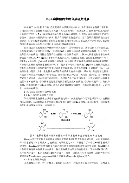
腈水解酶是一类可以将腈转化成相应酸及氨基的酶。当以扁桃腈为底物时,脂肪族水解酶立体选择性地生成R型扁桃酸,最重要的是其理论动力学反应产物收率为100%。具体作用机制如图4所示。
Endo等[22]将消旋体扁桃腈与Rhodococcus微生物反应,酶催化水解扁桃腈得到R-(-)-扁桃酸,R-(-)-扁桃酸的光学纯度达100%。Cesar等[23]采用来自木薯的有选择性的S-氧腈酶和来自荧光假单胞菌EBC191的无选择性的腈水解酶,分两步转化苯甲醛合成S-扁桃酸,产率很大,e.e值最高达98.0%。Yamamoto等[24]利用AlcaligenesfaecalisATCC8750菌株的静息细胞中腈水解酶催化外消旋扁桃腈得R-扁桃酸,产率为91%。Banerjee等[25]从假单胞菌中分离纯化得到的腈水解酶对扁桃腈同样具有立体选择性,可以将扁桃腈水解成R-扁桃酸,具有较高的对映体过量值。Banerjee等[25]报道海藻酸钙固定P.putida MTCC 5110细胞,利用腈水解酶水解扁桃腈,20个循环后固定化细胞仍具有88%的转化活性,e.e为98.8%。
Purification and Characterization

Purification and Characterization of a NovelGalloyltransferase Involved in Catechin Galloylation in the Tea Plant (Camellia sinensis )*Received for publication,July 20,2012,and in revised form,October 31,2012Published,JBC Papers in Press,November 6,2012,DOI 10.1074/jbc.M112.403071Yajun Liu ‡1,Liping Gao ‡1,Li Liu §,Qin Yang ‡,Zhongwei Lu ‡,Zhiyin Nie ‡,Yunsheng Wang ‡,and Tao Xia §2From the ‡School of Life Science and §Key Laboratory of Tea Biochemistry and Biotechnology,Ministry of Education in China,Anhui Agricultural University,130West Changjiang Rd,Hefei,Anhui 230036,ChinaCatechins (flavan-3-ols),the most important secondary met-abolites in the tea plant,have positive effects on human health and are crucial in defense against pathogens of the tea plant.The aim of this study was to elucidate the biosynthetic pathway of galloylated catechins in the tea plant.The results suggested that galloylated catechins were biosynthesized via 1-O -glucose ester-dependent two-step reactions by acyltransferases,which involved two enzymes,UDP-glucose:galloyl-1-O --D -glucosyl-transferase (UGGT)and a newly discovered enzyme,epicat-echin:1-O -galloyl--D -glucose O -galloyltransferase (ECGT).In the first reaction,the galloylated acyl donor -glucogallin was biosynthesized by UGGT from gallic acid and uridine diphos-phate glucose.In the second reaction,galloylated catechins were produced by ECGT catalysis from -glucogallin and 2,3-cis -fla-van-3-ol.2,3-cis -Flavan-3-ol and 1-O -galloyl--D -glucose were appropriate substrates of ECGT rather than 2,3-trans -flavan-3-ol and 1,2,3,4,6-pentagalloylglucose.Purification by more than 1641-fold to apparent homogeneity yielded ECGT with an estimated molecular mass of 241to 121kDa by gel filtration.Enzyme activity and SDS-PAGE analysis indicated that the native ECGT might be a dimer,trimer,or tetramer of 60-and/or 58-kDa monomers,and these monomers represent a het-erodimer consisting of pairs of 36-or 34-of and 28-kDa subunits.MALDI-TOF-TOF MS showed that the protein SCPL1199was identified.Epigallocatechin and epicatechin exhibited higher substrate affinities than -glucogallin.ECGT had an optimum temperature of 30°C and maximal reaction rates between pH 4.0and 6.0.The enzyme reaction was inhib-ited dramatically by phenylmethylsulfonyl fluoride,HgCl 2,and sodium deoxycholate.Flavonoids,a major class of secondary metabolites in plants,have a number of important physiological roles as endogenous auxin transport regulators (1–3),root development (4,5),seed germination (6),allelopathy (7),plant-bacterium interaction (8,9),UV-B protection (10),and plant defense against pathogens and environmental stress (11).Flavonoids can be grouped into several subgroups including chalcone,flavone,flavonol,flavandiol,anthocyanin,proantho-cyanidin (oligomer or polymer of flavan-3-ols and flavan-3,4-diol units)and other specialized forms (12).Flavan-3-ols (cat-echins),which comprise ϳ70–80%of tea polyphenols,are rich in young leaves and shoots of the tea plant (Camellia sinensis (L.)O.Kuntze).Catechins,with a basic 2-phenylchromone structure,are characterized by the di-or tri-hydroxyl group substitution of the B ring,the 2,3-position isomer of the C ring,and presence of a galloyl group at the 3-postion of the C ring (Fig.1).On the basis of the classical definition proposed of galloyl group structural features,catechins are divided into gal-loylated and nongalloylated compounds.Galloylated catechins,including (Ϫ)-epigallocatechin gallate (EGCG)3and (Ϫ)-epi-catechin gallate (ECG),esterified often with gallic acid (GA)in the 3-hydroxyl group of the flavan-3-ol units are major catechin compounds that account for up to 76%of catechins in the tea plant (13,14).Catechins,especially EGCG,possess antioxidant activity,antimutagenic,anticarcinogenic,antidiabetic,antibacterial,and anti-inflammatory potential,antihypertensive and anticar-diovascular disease effects,solar UV protection,body weight control effects,and therapeutic properties for Parkinson dis-ease (15).The health-promoting effects of galloylated catechins are stronger than those of nongalloylated catechins (16,17).Flavonoid biosynthesis has been a major focus of investiga-tion in recent decades (12).As the building blocks of most pro-*Thiswork was supported by the Natural Science Foundation of China (30972401,31170647,and 31170282),the Natural Science Foundation of Anhui Province (11040606M73),the Collegiate Natural Science Founda-tion of Anhui Province (KJ2012A110),the Program for Changjiang Scholars and Innovative Research Team in University (IRT1101),and the Major Pro-ject of Chinese National Programmes for Fundamental Research and Development (2012CB722903).1Both authors contributed equally to this work.2To whom correspondence should be addressed.Tel.:86-551-5786003;Fax:86-551-5785729;E-mail:xiatao62@.3The abbreviations used are:EGCG,(Ϫ)-epigallocatechin gallate;ECG,(Ϫ)-epicatechin gallate;ECGT epicatechin:1-O -galloyl--D -glucose O -gal-loyltransferase;GA,gallic acid;C,(ϩ)-catechin;EC,(Ϫ)-epicatechin;GC,(ϩ)-gallocatechin;EGC,(Ϫ)-epigallocatechin;G,-glucogallin,1-O -gal-loyl--D -glucose;ConA,concanavalin A;GCG,(ϩ)-gallocatechin-3-gallate;SCPL,serine carboxypeptidase-like;UDPG,UDP glucose.THE JOURNAL OF BIOLOGICAL CHEMISTRY VOL.287,NO.53,pp.44406–44417,December 28,2012©2012by The American Society for Biochemistry and Molecular Biology,Inc.Published in the U.S.A.F1Fn3anthocyanidins,the 2,3-trans -flavan-3-ol (ϩ)-catechin and 2,3-cis -flavan-3-ol (Ϫ)-epicatechin biosynthetic pathways have been investigated intensively at the biochemical and genetic levels (18,19).The biosynthetic pathway of nongalloylated cat-echins,which include (ϩ)-catechin (C),(Ϫ)-epicatechin (EC),(ϩ)-gallocatechin (GC),and (Ϫ)-epigallocatechin (EGC)is well documented.Some key genes and enzymes in the pathway include dihydroflavonol 4-reductase (EC 1.1.1.219),leucoan-thocyanidin reductase (EC 1.3.1.77),anthocyanidin synthase (EC 1.3.1.77),and anthocyanidin reductase (EC 1.3.1.77)(20–23).Despite recent progress in improving our understanding of flavan-3-ol synthesis,the mechanism involved in galloylation of catechins remains a mystery (19).In the 1980s,the biosynthesis of galloylated catechins and GA in the tea plant was investi-gated using radioactive tracer techniques.It was found that GA was presumably esterified with epigallocatechin and epicat-echin to form catechin gallates in young tea shoots,and the amount of GA might be a key limiting factor for the formation of EGCG and ECG (24).Understanding the galloylation of flavan-3-ols has been hin-dered by an absence of spontaneous genetic mutants for cate-chin biosynthesis.Niemetz and Gross (25)have done much research on the biogenetic pathways of hydrolyzable tannins.Their research confirmed that -glucogallin (1-O -galloyl--D -glucose (G))exerts a dual role functioning not only as an acyl acceptor but also as an efficient acyl donor.This work indicates G plays the same role in biosynthesis of galloylated catechin (Fig.2).Galloylation of glucose with GA to yield G,the first specific metabolite in the route to hydrolyzable tannins,was catalyzed by enzyme extracts from oak leaves with UDP-glu-cose serving as an activated substrate (26).This result indicated that,in galloylated catechin biosynthesis,G rather than GA might be a precursor of galloylated flavan-3-ols.In this study we sought to identify the enzymatic reactions and purify the key enzyme involved in galloylated catechin biosynthesis.Enzyme assays in vitro were designed to inves-tigate the biosynthesis of galloylated catechins,and the enzymes involved in galloylated catechin biosynthesis werepurified and identified.In addition,the enzyme properties were investigated.EXPERIMENTAL PROCEDURESPlant Materials —Leaves of the tea plant (C.sinensis (L.)O.Kuntze)were plucked from the experimental tea garden of Anhui Agricultural University during early summer.All of the samples were immediately frozen in liquid nitrogen and stored at Ϫ80°before analysis.Enzyme Extraction and Enzyme Assays —Enzyme extraction was performed in accordance with the method of Zhang et al.(27).All enzymes assays were conducted in phosphate buffer.In the multienzyme incorporative reaction system,theUGGT/FIGURE 1.Basic units of typicalcatechins.FIGURE 2.Reaction diagram of the galloylated catechin biosynthetic pathway.In the first reaction (I ),the galloylated acyl donor G was biosyn-thesized by UGGT from the substrates GA and UDPG.In the second reaction (II),galloylated catechins ECG or EGCG were produced by ECGT from the substrates G and nongalloylated catechins EC or EGC.Galloyltransferase Involved in Catechin GalloylationAQ:AAQ:BF2ECGT (UDP glucose:galloyl-1-O --D -glucosyltransferase/epi-catechin:1-O -galloyl--D -glucose O -galloyltransferase)assay solution was incubated at 30°C for 1.5h in a total volume of 1.5ml containing 50m M phosphate buffer (pH 6.0),0.4m M EGC or EC,1.4m M GA,2.3m M UDP glucose (UDPG),4m M ascorbic acid,and crude enzyme extract (0.55mg of total protein).The UGGT reaction solution was incubated at 30°C for 1.5h in a total volume of 1.5ml containing 50m M phosphate buffer (pH 6.0),2.3m M UDPG,1.4m M GA,4m M ascorbic acid,and crude enzyme extract (0.55mg of total protein).The ECGT assay solution was incubated at 30°C for 1h in a total volume of 1.5ml containing 50m M phosphate buffer (pH 6.0),0.4m M EGC or EC,0.96m M G (Advanced Technology and Industrial Company,Hong Kong,China),4m M ascorbic acid,and crude enzyme extract (0.55mg of total protein).The above enzyme reactions were terminated by the addition of ethyl acetate.Each of the reaction products was extracted three times with 3ml of ethyl acetate.The ethyl acetate extract was evaporated and redissolved in 500l of methanol and then used directly for analyses of the enzymatic reaction products.For gel filtration-purified enzyme activity analysis,the ECGT assay solution was incubated at 30°C for 10min in a total vol-ume of 100l containing 50m M phosphate buffer (pH 6.0),0.4m M EGC or EC,0.96m M G,and 5.6g ofenzyme.Enzymatic reactions were terminated by the addition of 10l of 5M HCl to the assay solution,and then the solution was used directly for analysis of the enzymatic reaction products.The protein concentration was determined by the Bradford method (28)using bovine serum albumin as a standard.In the control treatment,crude enzyme extract was heated to 100°C to inactivate enzyme activities.Analysis of UGGT and ECGT Enzyme Reaction Products —Extraction of enzyme reaction products was performed accord-ing to the method of Liu et al.(29).The solution (20l)of reaction products was spotted on a silica GF254TLC sheet (5ϫ20cm;HeFei BoMei Biotechnology Co.,Hefei,China)that was developed in chloroform:methanol:formic acid (28:10:1,v/v)and then sprayed with 1%vanillin-HCl (w/v)reagent.The spots of reaction products in the methanol extract were identified by R f values,and their visual color compared with those of cate-chin standards.The reaction products extract was analyzed by HPLC on a Phenomenex Synergi 4u Fusion-RP80column (5m,250ϫ4.6mm)with detection at 280nm.Ultraviolet spectra were recorded with a Waters 2487UV array detector (Waters Corp.,Milford,MA).For HPLC analysis,the solvent system consisted of 1%(v/v)acetic acid (A)and 100%acetonitrile (B).After injec-tion (5l),a linear gradient at a flow rate of 1.0ml/min was set as follows:B from 10to 13%(v/v)in 20min was initiated,then B from 13to 30%(v/v)between 20and 40min;B from 30to 10%(v/v)between 40and 41min.Peaks were identified by compar-ison of the retention times with those of standards.Analysis of enzyme reaction products by LC-MS followed the method of Miketova et al.(30).Enzymatic products were ana-lyzed by HPLC,and the area of the product peaks were collected and identified by LC-MS.Liquid chromatography electrospray-ionization-MS analyses were performed on a Thermo Finnigan LCQ Advantage instrument using the following conditions:negative ion detection mode,centroiding mode,multiplier at 1600keV,1000atomic mass units/s,source at 4.5kV,sheath gas at 70p.s.i.,auxiliary gas at 25p.s.i.,capillary temperature at 220°C,and UV detection at 220nm.Preparation and Identification of -Glucogallin from Tea Plant —For G analysis,1g of fresh leaves was crushed in liquid nitrogen and extracted with 5ml of methanol by sonication at room temperature for 10min followed by centrifugation at 4000ϫg for 15min,and the residues were re-extracted twice as above.The supernatants were pooled and evaporated and redissolved in 5ml of water.The pooled supernatants were then extracted three times with chloroform.The supernatant water phase was purified further with a SEP-PAK C 18cartridge,and after filtration the supernatant was separated by HPLC,and the G peak was collected and freeze-dried.The powder was used for chemical identification by HPLC,MS,and 1H and 13C NMR spectroscopy.For HPLC analysis,the solvent system consisted of 1%acetic acid in water (A)and acetonitrile (B).After injection (5l),a linear gradient at a flow rate of 1.0ml/min was established as follows:B from 0to 10%(v/v)in 10min was initiated,then B from 10to 30%between 10and 30min.Peaks were identified by comparing the retention times with those of standards.The LC-MS analysis of G employed the same method as that used for analysis of enzyme reaction products described above.1H and 13C NMR spectra were recorded in methanol-d 4on a Bruker Avance 400MHz spectrometer using TMS as an internal standard.Chemical shifts were expressed in ppm (␦).ECGT Isolation and Purification —ECGT purification con-sisted of acetone powder preparation,ammonium sulfate pre-cipitation,hydrophobic interaction chromatography,conca-navalin A (ConA)chromatography,and gel filtration.The first two steps were performed at 4°C,and the last three steps were conducted at room temperature.Gel filtration was performed to estimate the relative molecular mass of the enzyme.Step 1;Ammonium Sulfate Precipitation —The acetone pow-der was prepared by homogenization of 50g of tea leaves in cold acetone (Ϫ20°)with a Waring blender.The finely ground pre-cipitate was collected by vacuum filtration.The precipitate was washed several times with acetone until the washings were col-orless.This precipitate powder was used as a crude material for ECGT preparation.Precipitate powder (20g)was homogenized with 400ml of extraction buffer (50m M phosphate buffer (pH 7.0),4m M -mercaptoethanol,1%(w/v)polyvinylpolypyrroli-done (Sigma))and filtered through cheesecloth.The homoge-nate was centrifuged at 15,000ϫg for 15min at 4°C,and the supernatant was fractionated with 20–40%ammonium sulfate.Step 2;Hydrophobic Interaction Chromatography —The pre-cipitate was dissolved in 20m M phosphate buffer (pH 7.0)con-taining 1M ammonium sulfate and loaded onto a butyl-Sephar-ose column (20cm ϫ2.5-cm inner diameter,Bio-Rad).For hydrophobic interaction chromatography,the solvent system consisted of 20m M phosphate buffer (pH 7.0)including 1M ammonium sulfate (A)and phosphate buffer (pH 7.0)(B).After application of the enzyme solution,the column was washed with five volumes of buffer A and subsequently eluted with a stepped gradient of 38,95,and 100%B at a flow rate 2.5ml/min.Step3;Affinity Chromatography—The active fractions were subjected to ConA-Sepharose4B chromatography(column10 cmϫ1.6cm inner diameter;GE Healthcare).For ConA chro-matography,the solvent system consisted of20m M Tris-HCl (pH7.0)containing0.5M NaCl(A)and20m M Tris-HCl(pH 7.0)containing0.5M␣-D-methylglucoside(B).After applying the enzyme solution,the column was washed with5volumes of buffer A and subsequently eluted with100%B at a flow rate of1 ml/min.Step4;Gel Filtration Chromatography—The active fractions were subjected to gel filtration on a Superdex200column(50 cmϫ1.6cm inner diameter;GE Healthcare)and eluted with20 m M phosphate buffer(pH7.0)containing0.15M NaCl at a flow rate of0.8ml/min.Step5:SDS-PAGE Assay—SDS-PAGE was performed in accordance with the method of Laemmli(31),after which the proteins were visualized with Coomassie Brilliant Blue using the methods of Oakley(32).Protein Identification by MALDI-TOF-TOF MS—Protein spots were cut from gels,destained for20min in50m MNH4HCO3solution containing30%acetonitrile,and washed inMilli-Q water until the gels were destained.The spots wereincubated in0.2M NH4HCO3for20min and then lyophilized.Each spot was digested overnight in12.5ng/ml trypsin in0.1MNH4HCO3.After trypsin digestion,the peptide mixtures wereextracted with8l of extraction solution(50%acetonitrile, 0.5%TFA)at37°for1h.Finally the extracts were dried underthe protection of N2.Samples were reconstituted in3l of50%acetonitrile containing0.1%TFA before MS analysis.A1-l drop of this peptide solution was applied to an Anchorchip target plate.After drying at room temperature,a0.1-l droplet of CHCA matrix was applied to the plate at the same position. Samples were analyzed with ultrafleXtreme(Bruker).All acquired spectra of samples were processed using flexControl-software(Bruker)in the default mode.Parent mass peaks with a mass range of500–3500Da were detected with a minimum S/N filter of10for precursor ion selection.The five most abun-dant MS peaks were selected for MS/MS analysis.The com-bined MS and MS/MS data from the MALDI-TOF-TOF anal-ysis were submitted to Mascot2.3.02for a search against the NCBI C.sinensis protein database(;827 sequences),C.sinensis Genome Database“cam.pep”to con-struct a protein data bank(40,551sequences,data not shown), and C.sinensis Genome Database“tie.pep”to construct a pro-tein data bank(49,413sequences,data not shown).The identi-fication was accepted based on results from three biological replicates.Properties of the ECGT Enzyme—For characterization of ECGT enzyme properties,gel filtration-purified enzyme was used.For determination of the optimum pH for the ECGT, citrate-phosphate buffer(pH4.0–5.5),phosphate buffer(pH 6.0–7.0),and Tris-HCl buffer(pH7.5–8.0)were used.The optimum temperature range for ECGT activity was tested from 0°to70°C at pH6.0.Other assay conditions were identical to those used in the routine assay.To test the effect of inhibitors on ECGT activity,the enzyme was incubated with the inhibitors for5min at30°C before the enzyme assay.Enzymatic activity was measured in the presenceof PMSF,ZnCl2,EDTA,and-mercaptoethanol at a final con-centration of0–50m M,and sodium deoxycholate and HgCl2 were used at a final concentration of0–5m M.Other assay con-ditions were identical to those used in the routine assay.For investigation of the effects of temperature and pH on enzyme stability,ECGT activity was tested after enzyme stor-age atϪ20,0,4,10,20,30,40,or50°for48h and after storage atpH4.0to9.0at4°for48h.In addition,the temporal stability of ECGT was determined after storage at4°C for0,2,7,20,or40 days.RESULTSEvidence for Biosynthetic Enzymes of Galloylated Catechins—Niemetz and Gross(25)confirmed thatG acts not only as anacyl donor but as an acceptor in the biosynthesis of hydrolyz-able tannins.To determine whether catechin galloylation was similar to that of hydrolyzable tannin biosynthesis,a two-step enzyme assay incorporating the substrates GA,EC,or EGC and cosubstrate UDPG was designed,and the enzymatic products were analyzed via TLC and HPLC.The assay showed that UDPG was indispensable in the two-step enzymatic reaction,and a significant amount ofG was detected in the enzymatic products by HPLC analysis(Fig.3).This result suggested a UDPG-dependent glucosyltransferase existed in the tea plant,andG was the enzymatic product.In addition,the galloylated catechins EGCG and ECG(Fig.3),but not(ϩ)-gallocatechin-3-gallate(GCG),were detected by the two-step enzyme assayvia TLC and HPLC(data not shown),which indicated that thecis-catechins EGC and EC were appropriate substrates of a gal-loyltransferase instead of the trans-catechin GC or C.Thesedata further confirmed that EGCG and ECG in the tea plant are biosynthesized via enzymatic galloylation of EGC and EC withG,whereas GCG in green tea beverages is derived from isomerization of EGCG during green tea production(33).To test the above assumptions further,two separate enzyme-reaction assays were performed.The first enzyme assay was designed to detect UDPG-glucosyltransferase activity with the substrates GA and UDPG,and the second assay was to detect galloyltransferase activity with the substratesG(or1,2,3,4,6-pentagalloylglucose)and EGC(or EC and GC).The enzymatic products were identified by TLC,HPLC,and LC-MS.In the UDPG-glucosyltransferase assay,the productG could not be analyzed effectively by TLC for lack of an appro-priate staining reagent.However,HPLC(Fig.4A)and LC-MS confirmed that the product wasG(Fig.5A)and indicated the enzyme UGGT existed in the tea plant.In the galloyltransferase assay,TLC analysis of the enzyme reaction products showedtwo magenta spots with Rfvalues of0.43and0.28correspond-ing to the ECG and EGCG standards that were displayed in TLC sheets by staining with1%(w/v)vanillin-HCl reagent(Fig.6).This conclusion was confirmed by LC-MS.The parent ionswith m/z457and441corresponding to EGCG and ECG stand-ards,respectively,were observed(Fig.5,B and C).EGC,EC,andG were appropriate substrates of the galloyltransferase instead of GC(Fig.6)and1,2,3,4,6-pentagalloylglucose(PGG; Fig.4D).The deduced galloylated catechin biosynthetic path-way is depicted in Fig.2.Galloyltransferase Involved in Catechin GalloylationF3F4F5AQ:CF6Identification of -Glucogallin in Tea Plant —To gain further evidence for the existence of ECGT and UGGT in the tea plant,G was extracted and identified from the leaves.An improved method for extraction and quantification of G was estab-lished.A SEP-PAK C18cartridge was used in sample prepara-tion,and the purity of G in the solvent was enhanced mark-edly.To prevent interference from noisy peaks,the linear gradient of the solvent system was optimized for G analysis based on the method used for HPLC analysis of catechins.A single peak with retention time and spectral information con-sistent with those of the G standard appeared at 11.75min in the chromatogram (Fig.7).The G peak was collected largely via HPLC and identified by MS and NMR (Fig.8).The parent ion of the compound was detected at m /z 331,and majorfrag-FIGURE 3.HPLC analysis of UGGT/ECGT enzyme assay extracts.A and C ,the UGGT/ECGT assay solution was incubated at 30°C for 1.5h in a total volume of 1.5ml containing 50m M phosphate buffer (pH 6.0),0.4m M nongalloylated catechins (EGC or EC ),1.4m M GA,2.3m M UDPG,4m M ascorbic acid,1.5m M salicylic acid,and crude enzyme extract (0.55mg total of protein).The products G and galloylated catechins EGCG or ECG were detected clearly in this two-step reaction enzyme assay.Peaks were identified by comparing the retention times with standards.B and D ,control treatments of the UGGT/ECGT assay extracts with the crude enzyme extract were heated to 100°C to inactivate enzyme activities.There were no enzymatic products in control treatments.AU ,absorbanceunits.FIGURE 4.HPLC analysis of UGGT and ECGT enzyme assay extracts.A ,the UGGT assay solution was incubated at 30°C for 1.5h in a total volume of 1.5ml containing 50m M phosphate buffer (pH 6.0),2.3m M UDPG,1.4m M GA,4m M ascorbic acid,1.5m M salicylic acid,and crude enzyme extract (0.55mg of total protein).The product G was detected clearly in this assay.B and C ,the ECGT assay solution was incubated at 30°C for 1h in a total volume of 1.5ml containing 50m M phosphate buffer (pH 6.0),0.4m M nongalloylated catechins (EGC or EC ),0.96m M G,4m M ascorbic acid,and crude enzyme extract (0.55mg of total protein).The galloylated catechins EGCG or ECG were detected clearly in this assay.D ,the ECGT assay solution was conducted with substrates 0.96m M 1,2,3,4,6-pentagalloylglucose (PGG ),0.4m M nongalloylated catechins (EGC ),and conditions otherwise identical to those of the ECGT assay.No galloylated catechins were produced effectively from the substrates 1,2,3,4,6-pentagalloylglucose and EGC.The product peaks of G in A ,ECG in B ,and EGCG in C were collected and identified by LC-MS (see Fig.5).AU ,absorbance units.Galloyltransferase Involved in Catechin GalloylationF7F8ments were detected at m /z 271and 169,which were consistent with those of the G standard.1H and 13C NMR spectra were recorded in methanol-d 4on a Bruker Avance 400MHz spec-trometer with TMS as an internal standard.Chemical shifts were expressed in ppm (␦).1H NMR ␦:7.086(2H,s,H-2Јand H-6Ј),5.615(1H,d,J ϭ8Hz,H-1),3.813(1H,dd,J ϭ12.0,1.6Hz,H-6a),3.658(1H,dd,J ϭ12.0,4.8Hz,H-6b),3.35to 3.45(4H,m,H-2-H-5).13C NMR ␦:167.06(C ϭO),146.53(C-5Ј),140.34(C-4Ј),120.78(C1Ј),110.54(C-2Ј,C-6Ј),95.97(C-1),78.84(C-5),78.23(C-3),74.15(C-2),71.11(C-4),62.36(C-6).These data were consistent with those of the standard com-pounds,and thus the compound was identified as G.Purification of ECGT —Activity of ECGT was monitored throughout the purification.An ϳ1.64-fold purification was obtained by ammonium sulfate precipitation.The main active parts existed in the 20–60%ammonium sulfate fraction.About 10-fold specific activity was increased by hydrophobic interac-tion chromatography separation (Table 1).Monitoring of ECGT activity showed that ECGT enzyme was eluted from the column with a low ion eluent (Fig.9A ),which suggested ECGT was a highly hydrophobic protein.ConA-affinity chromatogra-phy was the most effective purification step for ECGT (Fig.9B ).Separation by ConA-affinity chromatography yielded an ϳ46-fold increase in purification (Table 1)and indicated that ECGT was a glycoprotein.An ϳ2-fold increase in specific activity was achieved by sep-aration on a Superdex 200column (Table 1).Obvious enzyme activities were detected approximately from 52min (fraction 2Ј)to 74min (fraction 6Ј,Fig.9C )with estimated molecular masses of 241to 121kDa based on a standard curve.To inves-tigate the subunit molecular masses of this enzyme,SDS-PAGE was routinely used for identification.The enzyme activity of lane 2was about double that of lane 6,whereas there were a large number of superfluous bands in lane 6from 40to 80kDa,so we speculated that three bands of estimated molecular masses of 36,34,and 28kDa were the subunits of this enzyme (Fig.9D ).One noteworthy phenomenon was that the SDS-PAGE Coomassie Brilliant Blue-stained bands changed with the degree of protein denaturation.SDS-PAGE analysis of lane 2showed that only two bands of 60and 58kDa remained after the enzyme was denatured in loading buffer containing 1%SDS,and three bands of 36,34,and 28kDa were present when the enzyme was denatured in loading buffer containing 5%SDS (Fig.10A).FIGURE 5.Mass spectra of products in the UGGT,ECGT,reaction assays.A ,shown are mass spectra of peaks corresponding to G in the UGGT enzyme reaction assay (see Fig.4A ).The ions of full MS correspond to the G standard.B and C ,MS assay of products peaks in the ECGT enzyme reaction assay (see Fig.4,B and C )are shown.The ions of 441and 457correspond to galloylated catechins ECG and EGCG standards,respectively.FIGURE 6.TLC assay of the ECGT reaction ne 1,ECG was produced with substrates G and ne 3,EGCG was produced with substrates G and ne 5,no product was produced with substrates G and nes 2,4,and 6are catechin standards.Boxed band a ,ECG;boxed band b ,EGCG.FIGURE 7.-Glucogallin assay in tea leaves by HPLC.For HPLC analysis,the solvent system consisted of 1%acetic acid in water (A )and 100%acetonitrile (B ).After injection (5l),a linear gradient at a flow rate of 1.0ml min Ϫ1was established as follows:B from 0to 10%(v/v)in 10min was initiated,then B from 10to 30%(v/v)between 10min to 30min.Peaks were identified by comparing the retention times with the standard.mAU ,absorbance units.Galloyltransferase Involved in Catechin GalloylationAQ:QAQ:D,T1F9F10。
Appl Microbiol Biotechnol(2006)71833–839
Appl Microbiol Biotechnol(2006)71:833–839DOI10.1007/s00253-005-0207-3BIOTECHNOLOGICALLY RELEVANT ENZYMES AND PROTEINSJingxue Wang.Haijin MouXiaolu Jiang.Huashi GuanCharacterization of a novelβ-agarase from marine Alteromonas sp.SY37–12and its degrading productsReceived:22August2005/Revised:25September2005/Accepted:28September2005/Published online:24November2005 #Springer-Verlag2005Abstract The phenotypic and agarolytic features of anunidentified marine bacteria isolated from the southernocean of China was studied.The strain was gram-negative,aerobic,and polarly flagellated.It was identified as thegenus Alteromonas according to its morphological andphysiological characterization.In solid agar,the isolateproduced a diffusible agarase that caused agar softeningaround the colonies.An extracellular agarase was purifiedby the procedure of ammonium sulfate precipitation,gelfiltration on Sephacryl S-100HR,and ion-exchange chro-matography on diethylaminoethyl-Sepharose.The purifiedprotein exhibited a single band on SDS-PAGE with amolecular mass of39.5kDa.The enzyme hydrolyzed the β-1,4-glycosidic linkages of agar,yielding neoagarote-traose and neoagarohexaose as the main products.Theoptimum reaction temperature of the agarase was35°C,with a narrow range from30to45°C.The enzyme activityreached the maximum at pH7.0and in the presence of2%NaCl.Molecular mass and degrading products showed thatthe agarase from Alteromonas sp.SY37-12was muchdifferent from those previously reported.IntroductionAgar,as an important food additive and bacterial culturemedium,is one of the most well-known marine poly-saccharides.Additionally,various potential physiologicalactivities of polysaccharides and oligosaccharides derivedfrom agar have been reported,such as antivirus(Takemoto1966),antitumor(Fernandez et al.1989),immune en-hancement(Yoshizawa et al.1993),antioxidation(Wang et al.2004;Zhang et al.2003),and elicitor activity on plant (Weinberger et al.2001).Agar has been determined to have a linear chain structure composed of alternating residues[O-3,6-α-anhydro-L-galac-topyranosyl(1!3)O-β-D-galactopyranose]combined byβ-1,4bonds(Hamer et al.1977).It is confirmed that agar oligosaccharide can be attained by many methods,including chemical degradation and enzyme hydrolysis.A special enzyme hydrolyzing the agar,agarase(agarose4-glycanohy-drolase,E.C.3.2.1.81),has been found in certain marine mollusks(Usov and Miroshnikova1975).However,the most was reported from several bacterial genera,including Cytophaga (Van der Meulen et al.1974),Vibrio(Aoki et al.1990), Actinomyces(Stanier1942),Alteromonas(Leon et al. 1992),Pseudoalteromonas(Vera et al.1998),and Pseu-domonas(Ha et al.1997),etc.Most of these bacteria were isolated from marine environments,while a few species isolated from rivers(Agbo and Moss1979),hot spring (Shieh and Jean1998),soil(Sampietro and Vattuone de Sampletro1971),and sewage(van Hofsten and Malmqvist 1975)have also been described.Agarase-producing bacteria can be classified into two groups according to the mode of action on agar:α-agarases cleave theα-1,3linkage of agarose(Young et al.1978)and β-agarases cleave theβ-1,4linkage of agarose(Duckworthand Turvey1969).Some of the agarase have been purified and characterized in the past years.In our laboratory,we have isolated a marine bacterium,Alteromonas sp.strain37-12, which has the high ability to decompose the agar from the southern ocean of China.We describe here the identification of this strain,the characterization of its extracellularβ-agarase,and the composition of its degrading products.Materials and methodsIsolation of strains and cell growthThe agarase-producing strain SY37-12was isolated in this laboratory from the surface of rotted red algae in the South China Sea coast in Sanya,Hainan Island.The screeningJ.Wang.H.Mou(*). X.Jiang.H.GuanOcean University of China, Qingdao,266003,PR Chinae-mail:mousun@ Tel.:+86-532-82032290 Fax:+86-532-82894024was carried out on agar plates in a medium containing2.0% NaCl,0.1%K2HPO4,0.1%NH4Cl,0.05%MgSO4,0.01% CaCl2,and1.5%agar.The plates were incubated at33°C for48h.Colonies that formed pits or clearing zones on agar were picked up and purified further by the same plating method.For liquid culture,0.2%agar was added before sterilization.The agar used here was commercial agar powder extracted from Gracilaria verrucosa(Mingfu Sea-weed Industry Co.,Fujian Province,China).Alteromonas sp.strain37-12is stored now in the China Center for Type Culture Collection(CCTCC),with strain number M204009.Evaluation of agarase activityAgarase activity was determined by measuring the increase in the concentration of reducing sugar as described by von Borel et al.(1952).The isolated colonies were inoculated into a medium of the same composition as that of isolation medium,except that the agar concentration was lowered to 0.2%.The cells were incubated in an orbital shaker at 150rpm and33°C for24h.After centrifuging at7,000×g for15min to remove bacterial cells and gel residues,a1-ml culture supernatant was added to20ml pH7.0phosphate buffered saline solution(PBS)with0.5%agar substrate and incubated at33°C for30min.Then the1-ml reaction solution was mixed with1.5ml3,5-dinitrosalicylic acid (DNS)reagent.After being heated at100°C for5min and then cooled,the mixture was diluted to25ml with deionized water.Optical density was read at520nm,and values for reducing sugars were expressed as D-galactose equivalents.One unit of agarase activity was defined as the amount of enzyme that released1μmol reducing sugar (measured as D-galactose)from agar per minute under the above conditions.Purification of agaraseUnless specified otherwise,all operations were done at 4°C.An overnight culture of strain SY37-12was prepared in the medium described above and transferred to250ml of fresh medium containing0.2%agar.Incubation was carried out in an orbital shaker at150rpm and33°C for24h until the stationary phase,followed by centrifugation at7,000×g for15min.The supernatant was brought to80%saturation with solid ammonium sulfate overnight.The collected enzyme protein was resuspended in20ml of50mM Tris–HCl(pH7.5),sealed in dialysis bags(cut-off value, 12,000–14,000MW),and dialyzed three times against the same buffer at4°C for2days.The dialyzate was loaded onto a diethylaminoethyl(DEAE)-Sepharose column (1.5cm×20cm,Pharmacia)equilibrated with50mM Tris–HCl(pH7.5).The flow rate was adjusted to0.5ml/ min.The protein was eluted from the column with the same buffer containing a linear NaCl gradient(0∼1.0M),and the elute fractions were collected.The eluates were monitored continuously at280nm,and fractions were assayed for activity against agar.Fractions containing agarase activity were pooled and concentrated by polyethylene glycol20,000,then applied for gel filtration chromatography on Sephacryl S-100HR (1.2cm×92cm,Pharmacia)equilibrated with50mM Tris–HCl(pH7.8).The flow rate was adjusted to0.7ml/min. Fractions were assayed for protein and agarase activity. The enzyme was lyophilized and stored at−20°C and was stable for more than2months.The purification of the fractions was assessed by sodium dodecyl sulfate polyacrylamide gel electrophoresis(SDS-PAGE).SDS-PAGE was performed in a0.75-mm slab gel consisting of a stacking gel(5%polyacrylamide)and a separating gel(12.5%polyacrylamide)with25mM Tris–HCl buffer,pH7.8.Proteins were stained with Coomassie brilliant blue R-250.Molecular mass standards were phosphorylase (97.4kDa),bovine serum albumin(66.2kDa),ovalbumin (42.7kDa),carbonic anhydrase(31kDa),soybean trypsin inhibitor(21.5kDa),and lysosyme(14.4kDa).Effects of temperature and pH on enzymeactivity and stabilityThe optimum temperature of agarase was determined at different temperatures(from20to50°C).Temperature stability was determined by measuring the residual ac-tivities after incubation at different temperatures.These studies were carried out in sealed Eppendorf tubes that were completely immersed in a water bath at the required temperatures.Samples(1ml)were removed after incuba-tion at each of the indicated time periods,chilled on ice and then assayed for enzyme activity.The optimum pH was determined with agar as substrate dissolved in KH2PO4–NaOH buffer with different pH values(5.0–9.0).The optimum NaCl concentration in the reaction solution was determined in PBS solution(pH7.0) with different NaCl concentration(0–5.0%).Preparation of agar oligosaccharidesTen grams of agar powder was scattered in500ml deionized water.A50-ml agarase solution(total agarase activity was90U)extracted from strain SY37-12was added to the agar solution.The reaction was carried out at 33°C for12h,and then was stopped by heating the solution in boiling water for10min.Twofold ethanol was added to the reaction mixture to remove the high-molecular-mass polysaccharides.After centrifugation and concentration, the depolymerized end-products were produced by re-peated ethanol fractionation.Structural analysis of agar oligosaccharidesThe molecular mass distribution of the agar oligosaccha-rides was determined by matrix-assisted laser desorption834ionization time-of-flight mass spectrometry(MALDI-TOF-MS)using LDI-1700instrument(Linear Scientific, Inc.,USA).The instrument was fitted with a pulsed nitrogen laser at337nm with3ns pulse duration.2,5-Dihydroxybenzoic acid(DHB)was used as the matrix. Infrared spectroscopy was performed using a Nicolet Nexus470spectrophotometer(Thermo Nicolet,Madison, WI,USA).For13C-NMR spectroscopy analysis,the samples were taken up in2H2O and processed at30°C. Spectra were recorded on a JNM-ECP600SCM spec-trometer(JEOL,Tokyo,Japan).ResultsStrain properties and identificationBacteria isolated from different areas of the China Sea were screened for a stable and effective agar-decomposingenzyme.A total of216strains producing agar-decompos-ing enzyme were isolated from the soil or seaweed surface, including Vibrio,Alteromonas,and Cytophaga.Strain SY37–12,isolated from the red alga Gracilaria verrucosa collected on Hainan Island of China,produced a large quantity of extracellular agarase when incubated in basal salt medium.The strain SY37-12produced soft pits on the agar surface with clear haloes around the bacteria colonies, which was regarded as an obvious mark indicating the high ability of degrading agar.Electron micrograph of the strain showed a polarly flagellated and straight-rod-shaped cell, 0.5–0.6μm×1.2–1.5μm.It was gram-negative and oxidase-and catalase-positive.It requires sodium ion for growth,has an oxidative metabolism,and does not ac-cumulate poly-β-hydroxybutyrate(PHB)as an intracellu-lar reserve product.The preliminary identification results showed that the morphologic and physiological character-istics of this strain were in accordance with Alteromonas, according to Bergey’s Manual of Systematic Bacteriology (Holt et al.1994)(Table1).Agarase produced by SY37-12was shown to be an induced enzyme.In the presence of crude agar,the strain produced extracellular agarase.No detectable agarase activity was found in the culture medium without agar. Furthermore,the addition of carrageenan,alginate,starch, galactose,lactose,and glucose in the absence of agar showed no effects on the production of agarase of SY37-12.When the agar was used as the sole carbon source,theTable1Morphologic and physiological characteristics of Altero-monas sp.SY37–12Characteristics tested Results Cell shape Straightrod Microcysts or endospores-Polar flagellum+ Production of pigments-Growth at4°C-Growth at35°C+ Growth at40°C-Anaerobic growth-Organic growth factors required-Oxidase reaction+ Catalase reaction+Argin1ine dihydrolase-Accumulation of PHB-O/F test O Production of H2S-Requirement of sodium for growth+ Hydrolysis of agar,starch,carrageenan,and gelatin+ Hydrolysis of alginate,chitin,and cellulose-Utilization of D-glucose,D-galactose,D-fructose,sucrose, cellobiose,and D-mannose+ Utilization of L-arabinose,dulcitol,raffinose,andD-ribose-Culture time (h)Agaraseactivity(U/ml)Biomass(OD6)Agarase activityFig.1Growth and agarase activity of Alteromonas sp.SY37–12. Fermentation liquids were monitored at600nm for biomass(□)and assayed for agarase activity(◊)by measuring the increase in the concentration of reducingsugar01836547290108Elution time (min)OD28Agaraseactivity(U/ml)Fig.2Ion-exchange chromatography of agarase on DEAE-Sepha-rose.The Tris buffer containing gradient NaCl rising from0to 1.0M at a flow-rate of0.5ml/min was used to wash out the sample. Fractions were monitored continuously at280nm for protein content(♦)and assayed for agarase activity(□)by measuring the increase in the concentration of reducing sugar835agarase activity of fermentation medium of SY37-12reached a maximum of 1.8U/ml after cultivation in conventional batch culture for 20h,whereas the biomass peaked at 24h (Fig.1).Purification of agaraseAccording to the pattern of ion-exchange chromatography on DEAE-Sepharose,several protein peaks were contained in the agarase sample (Fig.2).Additional purification of the fractions with agarase activity was achieved by gel filtration on Sephacryl S-100(Fig.3).Three protein peaks were shown in the chromatography,which was in ac-cordance with the result of SDS-PAGE (Fig.4).Thepurified enzyme had a molecular mass of 39.5kDa,as determined by a comparison with the mobility of protein standards.The purification steps and recovery rate of the agarase is summarized in the Table 2.Enzyme propertiesThe optimum reaction temperature of the agarase was 35°C,with a narrow range from 30to 45°C.At 20°C,the enzyme activity was only 30%in comparison with the maximum.The agarase was heat-labile,with rapid loss of activity when treated at 50°C for 15min or at 70°C for 1min (Fig.5).It even lost above 80%of its original activity after incubation at 90°C for 20s.The effect of pH on the enzyme activity was determined at 33°C in the pH range 5.0–9.0.The agarase exhibited maximum activity at pH 7.0.Since strain Alteromonas sp.SY37-12camefromElution time (min)O D 2800.20.40.60.811.21.4A g a r a s e a c t i v i t y (U /m l )Fig.3Gel filtration chromatography of agarase on Sephacryl S-100.The Tris buffer at a flow-rate of 0.7ml/min was used to wash out the sample.Fractions were monitored continuously at 280nm for protein content (♦)and assayed for agarase activity (□)by measuring the increase in the concentration of reducingsugarFig.4SDS-PAGE of purified ne 1molecular mass standards,lane 2purified agarase by Sephacryl S-100(only one band with a molecular mass of 39.5kDa),lane 3partly purified agarase by DEAE-Sepharose (three bands exist in the sample)Table 2Purification of agarase from Alteromonas sp.SY37-12StepV olume (ml)Agarase (U)Protein (mg)Specific activity (U/mg)Purification (-fold)Yield (%)Cell-free medium 1,0001,818606.2 3.0110080%(NH 4)2SO 4precipitate 45727157.84.61.540.0DEAE-Sepharose 20064217.137.512.535.3Sephacryl S-100HR2002593.183.527.814.2ab510152025Time (min)106050403020Time (s)R e l a t i v e a c t i v i t y (%)R e l a t i v e a c t i v i t y (%)Fig.5Effect of temperature on stability of agarase836the ocean,high NaCl concentration was necessary for its growth and enzyme production.When enzyme activity was measured in the presence of salt,a significant elevation could be observed.The enzyme activity reached the maximum in the presence of 2%NaCl,and then declined when the NaCl concentration continued to increase.MALDI-TOF-MS of the agarooligosaccharidesAfter sufficient hydrolysis and repeated fractionation by ethanol precipitation,the main depolymerized end-prod-ucts were collected.The MALDI-TOF mass spectrum of the sample was shown in Fig.6.According to the mass spectrum,the sample was an oligosaccharide mixture with polymerization degree from 4to 10.No sulfate group was linked in the sugar ring according to the molecular mass assignment.This speculation could be confirmed further by IR spectrum analysis and chemical determination (data not shown).The main composition was tetrasaccharide (MM-630Da)containing two galactopyranose residues (G-units)and two 3,6-anhydrogalaxtopyranose residues (An-units),hexasaccharide (MM-936Da)containing three G-units and three An-units,and octosaccharide (MM-1242Da)con-taining four G-units and four An-units.Furthermore,several minor peaks could be found in the mass spectrum,which were attributed to pentasaccharide (MM-792Da)containing three G-units and two An-units,heptasaccharide (MM-998Da)containing four G-units and three An-units,and decasaccharide (MM-1548Da)containing five G-units and five An-units.13C-NMR of agarooligosaccharidesMALDI-TOF mass analysis of the hydrolysis products of agar generated by agarase from Alteromonas sp.SY 37-12showed the presence of agarotetraose and agarohexaose as the main products.These products were further analyzed by NMR to determine the specificity of the cleavage.Anomeric carbons usually give downfield signals in 13C-NMR spectrum.The configuration of anomeric carbons can be determined according to the downfield shifts.The 13C-NMR spectrum of the oligosaccharide mixture showed a typical pattern for neoagarooligosaccharide (Fig.7).The neoagarooligosaccharide series is typically produced by the cleavage of β-(1,4)linkages by β-agarase.Resonances at about 97and 93ppm are characteristic for the βand αanomeric forms,respectively,of galactose residues at the reducing end of the neoagarooligosaccharides,indicating that the cleavage occurs at the β-(1,4)linkages.No peaks were present at around 90.4ppm corresponding to hydrolyzed α-(1,3)linkages.Thus,the 13C-NMR spectrum confirmed that agarase from strain SY 37-12is a β-agarase,specifically hydrolyzing the β-(1,4)glycosidic linkage between D -galactose and 3,6-anhydro-L-galactose.Fig.713C-NMR spectrum of the hydrolysis products of agar produced by agarase from Alteromonas sp.SY37–12837DiscussionWe describe here the characterization of a new agarolytic bacterium isolated from the southern ocean of China.This strain was identified as Alteromonas sp.according to its morphologic and physiological characteristics.The purified agarase extracted from Alteromonas sp.SY37-12has a molecular mass of39.5kDa,as indicated by SDS-PAGE. This value is close to those reported forβ-agarase from Pseudoalteromonas sp.N-1(33kDa)(Vera et al.1998), Pseudomonasatlantica(32kDa)(Morrice et al.1983),and Vibrio sp.AP-2(34kDa)(Aoki et al.1990).This enzyme acts as an endoenzyme,which decreased rapidly the viscosity of agar substrate at the initial reaction stage. Agarases(E.C.3.2.1.81)are classified into two groups depending upon their specificity of the degradation on the agar:α-agarases cleave theα-1,3linkage of agar,yielding oligosaccharides with3,6-anhydro-L-galactose at the re-ducing end and D-galactose at the nonreducing end,and β-agarase cleave theβ-1,4linkage of agar,yielding oligo-saccharides with D-galactose at the reducing end and3,6-anhydro-L-galactose at the nonreducing end(Vera et al. 1998).From the experiment result on analyzing the oligosaccharides by13C-NMR,it could be deduced that the agarase from the Alteromonas sp.SY37-12wasβ-agarase,which produced the3,6-anhydro-L-galactose as the nonreducing end and D-galactose as the reducing end. According to the assignment of MALDI-TOF-MS,the main composition of hydrolytic products was neotetrasac-charide(An-G-An-G),neohexasaccharide(An-G-An-G-An-G),and neooctosaccharide(An-G-An-G-An-G-An-G). In addition,pentasaccharide(G-An-G-An-G),heptasac-charide(G-An-G-An-G-An-G),and decasaccharide(An-G-An-G-An-G-An-G-An-G)could also be found in the products.It did not release detectable amounts of agaro-biose and agarotriose.With future experiments,the enzyme was confirmed to be not capable of hydrolyzing agarote-trose,agaropentose,agarohexose,and agaroheptose.It could be speculated that the minimal polymer degree of the agar substrate depolymerized by agarase from Alteromonas sp.SY37-12is eight.The agarase produce mainly agarooligosaccharides with even degree of polymerization, with D-galactose as the reducing end.As an endoenzyme,it cleaves theβ-1,4linkage of agar at random and produces a large quantity of low-molecular-mass fragments during the hydrolysis.Therefore,at the end of the hydrolytic reaction, the oligosaccharides with odd degree of polymerization,Table3Molecular mass and depolymerized products of characterized agarasesGroup Strain Molecular mass(kDa)Products Referenceα-Agarase Alteromonas GJIB360(bipolymer)Agarotetraose Potin et al.1993 Vibrio JI010784(bipolymer)Agaropentaose,agarotriose,agarobiose,3,6-anhydro-galactose and D-galactoseSugano et al.1994 Bacillus MK03320(octamer)Agaropentaose,agarotriose,3,6-anhydro-galactose and D-galactoseSuzuki et al.2002β-agarase Cytophaga flevensis26Neoagarotetraose,neoagarobiose Van der Meulen et al.1974 Pseudoalteromonas N-133Neoagarotetraose,neoagarohexaose Vera et al.1998Pseudomonas-like I a:210Neoagarohexaose,neoagarotetraose,neoagarobioseMalmqvist1978II:63NeoagarotetraosePseudomonas atlantica I:32Neoagarotetraose,neoagarobiose Morrice et al.1983II b:N/A NeoagarobioseVibrio AP-2I:34Neoagarotetraose Aoki et al.1990II:20NeoagarobioseIII:18NeoagarotetraoseVibrio JT0107I:107Neoagarotetraose,neoagarobiose Sugano et al.1993,1995II:72Neoagarotetraose,neoagarobioseVibrio PO-303I:87.5Neoagarohexaose,neoagarotetraose Araki et al.1998II:115NeoagarobioseIII:57Neoagarooctaose,neoagarodecaoseBacillus MK0392Neoagarotetraose Suzuki et al.2003Bacillus cereusASK20290Neoagarobiose Kim et al.1999 Alteromonas E-l82Neoagarobiose Kirimura et al.1999Alteromonas C-152Neoagarotetraose Leon et al.1992Alteromonas SY37-1239.5Neoagarotetraose,neoagarohexaose This worka There are several fractions with agarase activity produced by the strainb Data not available838especially pentasaccharide and heptasaccharide,are un-avoidably formed in the products.Molecular mass and depolymerized products of char-acterized agarases were given in Table3,which showed that the molecular mass of agarase from Alteromonas sp. SY37-12is different from the others,especially that from Alteromonas sp.E-1(Kirimura et al.1999)and Alteromo-nas sp.C-1(Leon et al.1992).The main depolymerized products produced by this enzyme are unique in compar-ison with the reports.Therefore,a conclusion was drawn here that the agarase produced by Alteromonas sp.SY37-12might be a novel enzyme.Further characterization of this enzyme will be discussed in the near future. ReferencesAgbo JAC,Moss MO(1979)The isolation and characterization of agarolytic bacteria from a low land river.J Gen Microbiol 113:355–368Aoki T,Araki T,Kitamikado M(1990)Purification and character-ization of a novelβ-agarase from Vibrio sp.AP-2.Eur J Biochem187:461–465Araki T,Hayakawa H,Lu Z,Karita S,Morishita T(1998) Purification and characterization of agarases from a marine bacterium,Vibrio sp.PO-303.J Mar Biotechnol6:260–265 Duckworth M,Turvey JR(1969)The action of a bacterial agarase on agarose,porphyran and alkali treated porphyran.Biochem J 113:687–692Fernandez LE,Valiente OG,Mainardi V,Bello JL,Velez H,Rosado A(1989)Isolation and characterization of an antitumor active agar-type polysaccharide of Gracilaria dominguensis.Carbo-hydr Res190:77–83Ha J,Kim GT,Kim SK,Oh TK,Yu JH,Kong IS(1997)β-Agarase from Pseudomonas sp.W7:purification of the recombinant enzyme from Escherichia coli and the effects of salt in its activity.Biotechnol Appl Biochem26:1–6Hamer GK,Bhattacharjee SS,Yaphe W(1977)Analysis of the enzymic hydrolysis products of agarose by13C-n.m.r.spec-troscopy.Carbohydr Res54:C7-C10Holt JG,Krieg NR,Sneath PHA,Staley JT,Williams ST(1994) Bergey’s manual of determinative microbiology,9th edn.Williams and Wilkins,BaltimoreKim BJ,Kim HJ,Duck Ha S,Hee Hwang S,Seok Byun D,Ho Lee T,Yul Kong J(1999)Purification and characterization ofβ-agarase from marine bacterium Bacillus cereus ASK202.Biotechnol Lett21:1011–1015Kirimura K,Masuda N,Iwasaki Y,Nakagawa H,Kobayashi R, Usami S(1999)Purification and characterization of a novelβ-agarase from an alkalophilic bacterium,Alteromonas sp.E-1.J Biosci Bioeng87:436–441Leon O,Quintana L,Peruzzo G,Slebe JC(1992)Purification and properties of an extracellular agarase from Alteromonas sp.strain C-1.Appl Environ Microbiol58:4060–4063 Malmqvist M(1978)Purification and characterization of two different agarose-degrading enzymes.Biochim Biophys Acta 537:31–43Morrice LM,McLean MW,Williamson FB,Long WF(1983)β-Agarases I and II from Pseudomonas atlantica.Purifications and some properties.Eur J Biochem135:553–558Potin P,Richard C,Rochas C,Kloareg B(1993)Purification and characterization of the alpha-agarase from Alteromonas agar-lyticus(Cataldi)comb.nov.,strain GJ1B.Eur J Biochem 214:599–607Sampietro AR,Vattuone de Sampletro MA(1971)Characterization of the agarolytic system of agarobacterium pastinator.Biochim Biophys Acta244:65–76Shieh WY,Jean WD(1998)Alterococcus agarolyticu s,gen.nov., sp.nov.,a halophilic thermophilic bacterium capable of agar degradation.Can J Microbiol44:637–645Stanier RY(1942)Agar-decomposing strains of the Actinomyces coelicolor species group.J Bacteriol44:555–570Sugano Y,Terada I,Arita M,Noma M,Matsumoto T(1993) Purification and characterization of a new agarase from a marine bacterium,Vibrio sp.strain JT0107.Appl Environ Microbiol59:1549–1554Sugano Y,Kodama H,Terada I,Yamazaki Y,Noma M(1994) Purification and characterization of a novel enzyme,alpha-neoagarooligosaccharide hydrolase,from a marine bacterium, Vibrio sp.strain JT0107.J Bacteriol176(22):6812–6818 Sugano Y,Nagae H,Inagaki K,Yamamoto T,Terada I,Yamazaki Y (1995)Production and characteristics of some newβ-agarases from a marine bacterium,Vibrio sp.strain JT0107.J Ferment Bioeng79:549–554Suzuki H,Sawai Y,Suzuki T,Kawai K(2002)Purification and characterization of an extracellularα-neoagarooligosaccharide hydrolase from Bacillus sp.MK03.J Biosci Bioeng93:456–463Suzuki H,Sawai Y,Suzuki T,Kawai K(2003)Purification and characterization of an extracellularβ-agarase from Bacillus sp.MK03.J Biosci Bioeng95:328–334Takemoto KK(1966)Plaque mutants of animal viruses.Prog Med Virol8:314–348Usov AI,Miroshnikova LI(1975)Isolation of agarase from Littorinamandshurica by affinity chromatography on BiogelA.Carbohydr Res43(1):204–212Van der Meulen HJ,Harder W,Vedkamp H(1974)Isolation and characterization of Cytophaga flevensis sp.nov.,a new agarolytic flexibacterium.Antonie Van Leeuwenhoek40:329–346van Hofsten B,Malmqvist M(1975)Degradation of agar by a Gram-negative bacterium.J Gen Microbiol87:150–158Vera J,Alvarez R,Murano E,Slebe JC,Leon O(1998)Identifi-cation of a marine agarolytic Pseudoalteromonas and char-acterization of its extracellular agarase.Appl Environ Microbiol 64:4378–4383von Borel E,Hostettler F,Deuel H(1952)Fasciculus I-15.Quantitative zuckerbestimmung mit3,5-dinitrosalicylsaure and phenol.Helv Chim Acta35:115–120Wang JX,Jiang XL,Mou HJ,Guan HS(2004)Anti-oxidation of agar oligosaccharides produced by agarase from a marine bacterium.J Appl Phycol16:333–340Weinberger F,Richard C,Kloareg B,Kashman Y,Hoppe H, Friedlander M(2001)Structure-activity relationships of oligoagar elicitors towards Gracilaria conferta.J Phycol 37:418–426Yoshizawa Y,Enomoto A,Todoh H,Ametani A,Kaminogawa S (1993)Activation of murine macrophages by polysaccharide fractions from marine algae(Porphyra yezoensis).Biosci Biotechnol Biochem57:1862–1866Young KS,Bhattacharjee SS,Yaphe W(1978)Enzymic cleavage of theα-linkages in agarose,to yield agaro-oligosaccharides.Carbohydr Res66:207–211Zhang QB,Li N,Zhou GF,Lu XL,Xu ZH,Li Z(2003)In vivo antioxidant activity of polysaccharide fraction from Porphyra haitanesis(Rhodephyta)in aging mice.Pharmacol Res48(2):151–155839。
苯乳酸生产菌的筛选及鉴定

苯乳酸生产菌的筛选及鉴定黄国昌;顾斌涛;黄朝;金丹凤;熊大维;黄筱萍;刘兰【摘要】苯乳酸是一种具有宽广抑菌谱的新型生物保鲜剂,为筛选得到苯乳酸高产菌株,采用改良的碳酸钙溶解圈法和抑菌圈法,从自然环境中采集的50个样品中分离筛选得到2株高产苯乳酸的乳酸菌,分别命名为BLPC002和SCPC008,并对其进行了初步鉴定.结果显示,BLPC002和SCPC008在以苯丙氨酸为底物的条件下,摇瓶发酵苯乳酸产量分别为358 mg/L和333 mg/L,较改良前不到100 mg/L有明显的提高,说明改良后的筛选方法更加高效、准确.进一步通过对菌株形态和16S rDNA序列分析初步鉴定,均为植物乳杆菌.%Phenyllactic acid is a new biological preservative with broad antibacterial spectrum.In order to obtain high yield lactic acid bacteria,2 lactic acid bacteria strains with high phenyllactic acid yield were isolated from 50 samples in different environment with the modified methods of dissolving calcium ring and bacteriostatic circle,which were named respectively as BLPC002 and SCPC008,and the strains were identified.The results showed that the yield of phenyllactic acid in shake flask fermentation with phenylalanine as substrate was 358 mg/L and 333 mg/L,respectively.The yields of phenyllactic acid were evidently improved compared with less than 100 mg/L before modification.It indicated that the modified method was more efficient and accurate.The strains were identified as Lactobacillus plantarum by morphological analysis and 16S rDNA sequencing.【期刊名称】《食品与发酵工业》【年(卷),期】2018(044)003【总页数】4页(P97-100)【关键词】苯乳酸;乳酸菌;筛选【作者】黄国昌;顾斌涛;黄朝;金丹凤;熊大维;黄筱萍;刘兰【作者单位】江西省科学院微生物研究所,江西南昌,330096;江西省科学院微生物研究所,江西南昌,330096;江西省科学院微生物研究所,江西南昌,330096;江西省科学院微生物研究所,江西南昌,330096;江西省科学院微生物研究所,江西南昌,330096;江西省科学院微生物研究所,江西南昌,330096;江西省科学院微生物研究所,江西南昌,330096【正文语种】中文苯乳酸(phenyllactic acid,PLA,又称3-苯基乳酸)是继乳酸链球菌素(Nisin)和ε-聚赖氨酸(ε-po lylysine,ε-PL)之后人们发现的一种新型生物保鲜剂,相比Nisin和ε-PL,因PLA具有更加宽广的抑菌谱,不仅能抑制多种食品致病菌,还能抑制引起食品腐败的真菌,且其溶解性好、对酸碱和热稳定性强,具有广泛应用于各种食品和果蔬保鲜的潜质,因此,近年来成为了人们研究的热点。
单宁酶发酵生产的研究进展

万方数据 万方数据 万方数据20∞N0.”·4·SerjaINo.188ChinaBrewingFomm衄dS吼蚰q为85.67%、90.48%。
附表单宁酶和没食子酸生产条件的优化AnachedtabIe.0ptimizatbnonp∞ductioncondnb瞧oft鲫naseandga¨icacid霉参数固体发酵范围最优液体发酵范围最优改良的固体发酵范围最优l堵乔周删/h24^962黧端一s石‘【g·(cm2)‘‘】~…””3初始pH值3“.54温度,℃20 ̄405相对湿度,%67埘6床高度/锄O.Ol加.27固液比O.5:l一4:18发酵状态。
培养基选择7④、@、@、(d)·o%船尹7没食子酸~转化率/%7224一母6482.o‰驴loLW·W4.53巧.55.O3220—∞3793O.15l:lO.05:1~1:1O.4:l稳态稳态/搅拌搅拌(c)(c)18.8723.8630.527.52}母672Ⅻ,72203巧.54.520—lo3267q}493O.05 ̄2.31.50067.1 ̄0瞳lO.4:1稳态,搅拌稳态(c)327690.9注:培养基选择(a)含2%(w/∞葡萄糖的察氏培养基:(b)含2%(w/v)单宁酸的改良察氏培养基;(c)含2%“Ⅳ,v)c.digyna粉末的改良察氏培养基;(d)单宁酸培养基。
2.4连续固体发酵法工业上固体发酵主要采用分批发酵的模式,但由于分批发酵存在很多缺点,固体发酵的应用还是有限的。
v锄deLageⅡK斌J等【姗研究了连续固体发酵生产单宁酶的方法,此方法克服了分批发酵接种量大、不能重复利用、生产效率低的缺点。
研究了在生物反应器中采用连续固体发酵模式用含单宁酸的模拟底物生产PeIlicilli啪glab嗍单宁酶,能连续操作固体基质,不用补料接种。
此发酵生物反应器见图2。
发酵容器c是一个倾斜的树脂玻璃管(长1.oom、内直径0.08m),它是由2个半圆柱面组成,3个金属环夹紧这2个半圆柱面,可以打开取样。
α_淀粉酶中某些氨基酸含量与其最适pH的关系

第21卷第6期2002年11月 无锡轻工大学学报Journal of Wuxi U niversity of Light Industry Vol.21 No.6Nov. 2002 文章编号:1009-038X (2002)06-0634-04 收稿日期:2002-04-10; 修订日期:2002-09-04.作者简介:张革新(1965-),男,江苏盐城人,博士研究生,副教授.α2淀粉酶中某些氨基酸含量与其最适p H 的关系张革新1, 宋江宁2, 李炜疆2(1.江南大学化学与材料工程学院,江苏无锡214036;2.江南大学工业生物技术教育部重点实验室,江苏无锡214036)摘 要:针对生产中大多数α2淀粉酶不能适应高酸性和高温条件,需要改进才能适应工业生产的实际,研究了α2淀粉酶的最适p H 与各种氨基酸含量的关系,构建了α2淀粉酶序列库和p H 序列库.通过对α2淀粉酶p H 序列库统计分析,发现α2淀粉酶的最适p H 与某些氨基酸含量有一定的关系,当丙氨酸(A )质量分数大于5.8%并谷氨酸(E )质量分数大于5.0%,苏氨酸(T )质量分数大于6.5%并酪氨酸(Y )质量分数在4.3%~6.3%时,α2淀粉酶的耐酸性能力较好.关键词:α2淀粉酶;蛋白质序列;氨基酸;最适p H ;生物信息学中图分类号:TS 201.2+5文献标识码:AR elation bet w een Percentage of Some Amino Acids and Optimal p Hof Alpha 2AmylasesZHAN G G e 2xin 1, SON G Jiang 2ning 2, L I Wei 2jiang 2(1.School of Chemical and Material Engineering ,S outhern Y angtze University ,Wuxi 214036,China ;2.K ey Labora 2tory of Industrial Biotechnology of Ministry of Education ,S outhern Y angtze University ,Wuxi 214036,China )Abstract :Alpha 2amylase is an enzyme of great significance for industry.As the enzyme used in the process ,most alpha 2amylases are unstable at lower p H and higher temperature.Consequently ,there is a need to develop an enzyme that can operate at industrial conditions.For studying the relation between percentage of kinds amino acids and optimal p H ,we built alpha 2amylase ’s bank and alpha 2amylase ’s p H 2sequence bank.After analysis ,we found that there were 2kinds cases about relation between percent amino acid and optimal p H :as either (1)A (alanine )%>5.0%and E (glutamic acid )%>5.8%,or (2)T (threonine )%>6.5%,and Y (tyrosine )%in arange of 4.3%~6.3%,alpha 2amylase had the higher anti 2acid ability.K ey w ords :alpha 2amylase ;protein sequence ;percent amino acid ;optimal p H ;bioinformatics α2淀粉酶广泛应用于食品、纺织、洗涤剂等轻工行业,在国内外酶制剂市场占有重要地位,已经发现的天然α2淀粉酶大多需要改进才能适应苛刻的工业生产条件或提高生产效率,研究的主要方法就是对已知酶进行优化改造.例如,热稳定性较好的地衣芽孢杆菌α2淀粉酶在低p H 值条件下不稳定,必须通过蛋白质工程改进其性能.α2淀粉酶的优化改造已成为近期的一个研究热点[1~7].酶的优化改造是通过对已知酶分子一级结构的改变来创造新的酶,这一技术实现,依赖于两大技术的发明,即基因工程中的定点诱变技术和生物信息学.为此,从大量的文献中收集序列结构数据,对其整理分析,寻找不同氨基酸含量对其最适p H 的影响,为α2淀粉酶的优化改造提供帮助.1 材料与方法1.1 数据收集与整理首先从SWISS 2PRO T (http ://www.expasy.ch/sprot )收集与α2淀粉酶有关的氨基酸序列数据538条,经过整理,得到了有352条序列的α2淀粉酶序列库.再收集与α2淀粉酶最适p H 有关的文献[1~48],从α2淀粉酶序列库中进行检索,得到既有α2淀粉酶最适p H ,又有序列α2淀粉酶,称为α2淀粉酶p H 序列库.该库共有50条记录,分布为p H 小于5.5的有13条,为耐酸;p H 在5.5~6.5范围的有15条,为弱酸;p H 在6.5~7.5范围的有21条,为中性;p H 大于7.5的有4条,为弱碱.1.2 方法计算α2淀粉酶p H 序列库中每一条序列不同氨基酸的质量分数,得到同一种氨基酸在不同序列(有对应p H 数值)时的含量,用此数据作每一种氨基酸含量与p H 的关系图.该图关系复杂,未能得出规律性的结论.于是,研究了不同氨基酸含量之间对上述4种类型α2淀粉酶分布的影响.2 结果与分析运用上述方法,得到了20种氨基酸两两组合含量之间与α2淀粉酶类型分布的关系图,共190幅,明显联系的有两幅,见图1和图2.纵坐标和横坐标是对应氨基酸的质量分数,氨基酸种类以其对应单字母表示. 从图1中可以看出,当丙氨酸(A )质量分数大于5.8%并且谷氨酸(E )质量分数大于5.0%时,此区域为耐酸α2淀粉酶所在区域.从图2中同样可以看出,存在着一个耐酸α2淀粉酶所在区域,对应范围为苏氨酸(T )质量分数大于6.5%并酪氨酸(Y )质量分数4.3%~6.3%.α2淀粉酶最适p H 与其氨基酸组成、结构应当有着必然地联系,只是影响因素太多,难以寻找.这些统计结果可以运用到α2淀粉酶的优化改造上.如果增加α2淀粉酶的耐酸性,控制丙氨酸、谷氨酸、苏氨酸和酪氨酸含量应是有效的方法.图1 不同类型α2淀粉酶与丙氨酸2谷氨酸对应的关系Fig.1 R elation betw een types of alpha 2amylase and cou 2ples of alanine 2glutamic acid图2 不同类型α2淀粉酶与苏氨酸2酪氨酸对应的关系Fig.2 R elation betw een types of alpha 2amylase and cou 2ples of threonine 2tyrosine 3 结 论通过对α2淀粉酶的最适p H 与序列数据的分析处理,发现存在两个耐酸α2淀粉酶所在区域:(1)丙氨酸(A )质量分数大于5.8%,并且谷氨酸(E )质量分数大于5.0%;(2)苏氨酸(T )质量分数大于6.5%,并酪氨酸(Y )质量分数为4.3%~6.3%.这些统计结果将会对提高α2淀粉酶耐酸性提供有益的帮助.参考文献:[1]HEESE O ,HANSEN G ,HOHN E W E ,et al .A thermostable alpha 2amylase from Thermoactinomyces vulgaris :purification536第6期张革新等:α2淀粉酶中某些氨基酸含量与其最适p H 的关系636 无 锡 轻 工 大 学 学 报 第21卷and characterization[J].Biomed Biochim Acta,1991,50(3):225-32.[2]V IHIN EN M,MAN TSALA P.Characterization of a thermostable Bacillus stearothermo philus alpha2amylase[J].BiotechnolAppl Biochem,1990,12(4):427-435.[3]DEY S,A G ARWAL SO.Characterization of a thermostable al p ha2amylase from a thermophilic Streptomyces megasporus strainSD12[J].Indian J Biochem Biophys,1999,36(3):1502157.[4]MELASN IEMI H.Characterization of al pha2amylase and pullulanase activities of Clostridium thermohydrosulfuricum[J].Biochem J,1987,246(1):193-197.[5]L EE SP,MORIK AWA M,TA K A GI MET Al.Clonin g of the aap T gene and characterization of its product,alpha2amylase2pullulanase(Aap T),from thermophilic and alkaliphilic B acillus sp.Strain XAL601[J].Appl E nviron Microbiol,1994,60(10):3764-3773.[6]SRIVASTAV E RA,MA THUR SN,BARUAH J N.Partial purification and properties of thermistable intracellular amylasesfrom a thermophilic B acillus sp.A K22[J].Acta Microbiol Pol,1984,33(1):57-66.[7]ITOH Y,SUMI M,NA K AMURA K,et al.Processing of an NH22terminally extended thermostable alpha2amylase by B acil2lus subtilis alkaline protease[J].J Biochem(Tokyo),1990,108(6):954-959.[8]STAMFORD TL,STAMFORD NP,COEL HO LC,et al.Production and characterization of a thermostable al pha2amylasefrom Nocardiopsis sp.Endophyte of yam bean[J].Bioresour T echnol,2001,(2):137-141.[9]MAL HO TRA R,NOORWEZ SM,SA T Y ANARA Y ANA T.Production andpartial characterization of thermostable and calci2um2independent alpha2amylase of an extreme thermophile Bacillus thermooleovorans NP54[J].Lett Appl Microbiol,2000,31(5):378-384.[10]CHA KRABORT Y K,BHA TTACHAR YY A B K,SEN SK.Purification and characterization of a thermooleovorans al p ha2amy2lase from Bacillus stearothermophilus[J].Folia Microbiol(Praha),2000,45(3):207-210.[11]K A TO K,SU GIMO TO T,AMEMURA A,et al.A Pseudomonas intracellular amylase with high activity on maltodextrinsand cyclodextrins[J].Biochim Biophys Acta,1975,391(1):96-108.[12]MA TZKE J,SCHWERMANN B,BA KKER EP.Acidostable and acido philic proteins:the example of the alpha2amylase fromAlicyclobacillus acidocaldarius[J].Comp Biochem Physiol A Physiol,1997,118(3):475-479.[13]CL EMEN TI F,ROSSI J.Al pha2amylase and glucoamylase production by Schwanniomyces castellii[J].Antonie V anLeeuw enhoek,1986,52(4):343-352.[14]MOHAMED SA.Alpha2Amylase from developing embryos of the camel tick Hyalomma dromedarii[J].Comp Biochem PhysiolB Biochem Mol Biol,2000,126(1):99-108.[15]MOUL IN G,G AL ZY P.Amylase activity of torulopsis ingeniosa di menna[J].Folia Microbiol,1978,23(6):423-427.[16]TITAREN KO E,CHRISPEEL S MJ.CDNA clonin g,biochemical characterization and inhibition by plant inhibitors of the al2pha2amylases of the western corn rootworm,diabrotica virgifera virgifera[J].I nsect Biochem Mol Biol,2000,30(10):979-990.[17]DERCOVA K,AU GUSTIN J,KRAJ COVA D.Cell growth and alpha2amylase production characteristics of B acillus subtilis[J].Folia Microbiol(Praha),1992,37(1):17-23.[18]L E BERRE2AN TON V,BOMPARD2GILL ES C,PA Y AN F,et al.Characterization and functional properties of the alpha2amylase inhibitor(alpha2AI)from kidney bean(Phaseolus vulgaris)seeds[J].Biochim Biophys Acta,1997,1343(1):31-40.[19]SAN TOS T,NOMBELA C,V ILLANU EVA J R,et al.Characterization and synthesis regulation of penicillium italicum1,62beta2glucanase[J].Arch Microbiol,1979,121(3):265-270.[20]THUDT K,SCHL EIFER KH,G O TZ F.Cloning and expression of the alpha2amylase gene from B acillus stearothermophilusin several staphylococcal species[J].G ene,1985,37(123):163-169.[21]IG ARASHI K,HA TADA Y,HA GIHARA H,et al.Enzymatic properties of a novel liquefying alpha2amylase from an alka2liphilic Bacillus isolate and extire nucleotide and amino acid se quences[J].Appl E nviron Microbiol,1998,64(9):3282-3289.[22]ITO K,L I L F,N ISHIWA KI M,et al.Evidence for conversion of human salivary alpha2amylase family A to family B by anenzyme action[J].J Biochem(Tokyo),1992,112(1):88-92.[23]TULL D,G O TTSCHAL K TE,SV ENDSEN I,et al.Extensive N2glycosylation reduces the thermal stability of a recombi2nant alkalophilic bacillus alpha2amylase produced in pichia pastoris[J].Protein Expr Purif,2001,21(1):13-23.[24]VU KEL IC B,RITONJA A,REN KO M,et al.Extracellular alpha2amylase from streptomyces rimosus[J].Appl MicrobiolBiotechnol,1992,37(2):202-204.[25]EG AS MC,DA COSTA MS,COWAN DA,et al.Extracellular alpha2amylase from thermus filiformis Ork A2:purificationand biochemical characterization[J ].Extremophiles ,1998,2(1):23-32.[26]STRU G ALA G J ,RAUWA A G ,ELBERS R.Intestinal first pass metabolism of amygdalin in the rat in vitro [J ].BiochemPharmacol ,1986,35(13):2123-3128.[27]KOH J I OHDAN ,TA K ASHI KURIKI ,HIROL I TA K A TA ,et al.Introduction of raw starch 2binding domains into bacillussubtilis α2amylase by fusion with the starch 2binding domain of bacillus cyclomaltodextrin glucanotransferase[J ].A Applied and E nvironmental Microbiology ,2000,66(7),3058-3064.[28]BROSNAN MP ,KELL Y CT ,FO G ART Y WM.Investigation of the mechanisms of irreversible thermoinactivation of B acillusstearothermophilus alpha 2amylase[J ].Eur J Biochem ,1992,203(122):225-231.[29]KOSTAREVA IA ,ORL IEVSK AIA OV ,IURCHEN KO VS ,et al.Isolation and study of physico 2chemical properties of al 2pha 2amylase from B acillus subtilis [J ].Prikl Biokhim Mikrobiol ,1981,17(3):430-435.[30]TOMAZIC S J ,K L IBANOV AM.Mechanisms of irreversible thermal inactivation of B acillus alpha 2amylases [J ].J BiolChem ,1988,263(7):3086-3091.[31]BUONOCORE V ,POERIO E ,SILANO V.Ph ysical and catalytic properties of alpha 2amylase from Tenebrio molitor r 2vae[J ].Biochem J ,1976,153(3):621-625.[32]AL I FS ,ABDEL 2MON EIM AA.Physico 2chemical properties of Aspergillus f lavus var.columnaris alpha 2amylase[J ].Z en 2tralbl Mikrobiol ,1989,144(8):615-621.[33]DESSEAUX V ,SEIGN ER C ,PIERRON Y ,et al .Porcine pancreatic alpha 2amylase :a model for structure 2function studies ofhomodepolymerases[J ].Biochimie ,1988,90(9):1163-1170.[34]L IN LL ,CHY AU CC ,HSU WH.Production and properties of a raw 2starch 2degrading amylase from the thermophilic and al 2kaliphilic B acillus sp.TS 223[J ].Biotechnol Appl Biochem ,1998,28(1):61-86.[35]MICHEL ENA VV ,CASTILLO FJ.Production of am ylase by Aspergillus f oetidus on rice flour medium and characterizationof the enzyme[J ].J Appl B acteriol ,1984,56(3):395-407.[36]ARA K ,SAEKI K ,IG ARASHI K ,et al.Purification and characterization of an alkaline amylopullulanase with both alpha 21,4and alpha 21,6hydrolytic activity from alkalophilic Bacillus sp.KSM 21378[J ].Biochim Biophys A cta ,1995,1243(3):315-324.[37]FERNANDEZ 2TARRA G O J ,N ICOLAS G.Purification and characterization of an al p ha 2amylase from the cotyledons of germi 2nating lentils[J ].R ev Esp Fisiol ,1981,37(2):197-204.[38]SHIH NJ ,LABBE R G.Purification and characterization of an extracellular al p ha 2amylase from clostridium perfringens type A[J ].Appl E nviron Microbiol ,1995,61(5):1776-1779.[39]A GU ILAR G ,MORLON 2GU Y O T J ,TRE JO 2A GU ILAR B.Purification and characterization of an extracellular al p ha 2amy 2lase produced by Lactobacillus manihotivorans LM G 18010(T ),an amylolytic lactic acid bacterium[J ].E nzyme microb T ech 2nol ,2000,27(6):406-413.[40]DE MO T R ,V ERACHTERT H.Purification and characterization of extracellular al p ha 2amylase and glucoamylase from theyeast andida Antarctica CBS 6678[J ].Eur J Biochem ,1987,164(3):643-654.[41]FERRER A ,HOEBEKE J ,BOU T D.Purification and characterization of two al p ha 2amylases from Toxoplasma gondii [J ].Exp P arasitol ,1999,92(1):64-72.[42]CHAN G CT ,TAN G MS ,L IN CF.Purification and properties of alpha 2amylase from Aspergillus oryz ae A TCC 76080[J ].Biochem Mol Biol Int ,1995,36(1):185-193.[43]BUONOCORE V ,DEPON TE R ,GRAMENZI F ,et al.Purification and properties of alpha 2amylase from chicken (Gallusgallus L.)pancreas[J ].Mol C ell Biochem ,1977,19,17(1):11-16.[44]ILORI MO ,AMUND OO ,OMIDI J I O.Purification and properties of an alpha 2amylase produced by a cassava 2fermentingstrain of micrococcus luteus[J ].Folia Microbiol (Praha ),1997,42(5):445-449.[45]AWA GUCHI T ,NA G AE H ,MURAO S ,et al.Purification and some properties of a haim 2sensitive alpha 2amylase from new 2ly isolated B acillus sp.No.195[J ].Biosci Biotechnol Biochem ,1992,56(11):1792-1796.[46]COLL INS BS ,KELL Y CT ,FO G ART Y WM ,et al.The high maltose 2producing alpha 2amylase of the thermophilic actino 2mycete ,Thermomonos pora curvata[J ].Appl Microbiol Biotechnol ,1993,39(1):31-35.[47]BOSE K ,DAS D.Thermostable al pha 2amylase production using B acillus lichenif ormis NRRL B14368[J ].Indian J Exp Biol ,1996,34(12):1279-1282.[48]SCHUMANN J ,WRBA A ,JAEN ICKE R ,et al .Topographical and enzymatic characterization of amylases from the extreme 2ly thermophilic eubacterium Thermotoga maritima[J ].FEBS Lett ,1991,22,282(1):122-126.(责任编辑:杨勇)736第6期张革新等:α2淀粉酶中某些氨基酸含量与其最适p H 的关系。
龙须菜,Gracilarialemaneiformis,音标,读音,翻译,英文例句,英语词典

龙须菜,Gracilarialemaneiformis,音标,读音,翻译,英文例句,英语词典龙须菜清汤豌豆龙须菜龙须扒菜心鲜蘑龙须菜蟹肉龙须菜扒龙须菜心说明:双击或选中下面任意单词,将显示该词的音标、读音、翻译等;选中中文或多个词,将显示翻译。
您的位置:首页->词典->龙须菜1)Gracilaria lemaneiformis龙须菜1.Isolation,Purification and Characterization of Agar Polysaccharide from Gracilaria Lemaneiformis;龙须菜琼胶多糖的提取、纯化与性能表征2.Response of Gracilaria lemaneiformis to Dimethyl Phthalate;龙须菜对邻苯二甲酸二甲酯(DMP)毒性的响应3.Determination of Seven Trace Elements in Gracilaria Lemaneiformis by Atomic Absorption Spectrometry;原子吸收光谱法测定龙须菜中7种微量元素含量更多例句>>2)Gracilaria lamaneiformis龙须菜1.The impact of yield of polysaccharides and antioxygenic property extracted with technical conditions from Gracilaria lamaneiformis;提取工艺对龙须菜多糖得率及其抗氧化性能的影响2.Study on the technical conditions of polysaccharide from Gracilaria lamaneiformis by enzymatic extraction;酶法提取龙须菜多糖工艺条件的研究3.Optimization of extraction technology of polysaccharide from Gracilaria lamaneiformis;龙须菜多糖提取工艺优化更多例句>>3)G.lemaneiformis龙须菜1.Se ven strains of algae were analyzed,including four strains of G.所用的7种实验材料包括4株龙须菜(其中3株分别来自中国青岛、委内瑞拉和南非,及1株青岛产龙须菜的高温选育种)和细基江蓠繁枝变型、真江蓠、芋根江蓠。
枯草芽孢杆菌发酵鱼粉产蛋白酶及其酶解效果

枯草芽孢杆菌发酵鱼粉产蛋白酶及其酶解效果卢珍华【摘要】为开发以鱼粉为原料的优质饲料蛋白源以及相关保健食品,开展了用不同鱼粉为氮源发酵枯草芽孢杆菌生产蛋白酶及其酶解效果的研究,比较了不同菌种的发酵效果,确定了最佳发酵时间.通过蛋白酶活性测定和氨基态氮含量测定分析了不同菌种的产酶能力和酶活性大小及其酶解效果.结果表明,用罗非鱼鱼粉和处理过的鳕鱼鱼粉发酵商业化的工业菌种枯草芽孢杆菌产生的酶活性显著高于鲢鱼鱼粉(P 〈0.05),发酵后上清液中的氨基态氮含量也显著高于鲢鱼鱼粉(P〈0.05),即工业化的枯草芽孢杆菌对不同鱼粉的发酵效果不同,罗非鱼鱼粉和处理过的鳕鱼鱼粉发酵效果显著优于鲢鱼鱼粉.%To get higher enzymatic activity of proteases produced by Bacillus subtilis by fermentation,different fish meals were used as nitrogen sources.The effects of fermentation time,as well as the difference between commercial and field strains on enzymatic activity were compared.The content of amino nitrogen in the broth of fermentation was also determined.The results suggested that commercial strain showed a better performance in the fermentation with the fish meal of tilapia and codfish compared to silver carp.During the fermentation of Bacillus subtilis using tilapia and codfish meal as nitrogen sources,higher enzymatic activity and content of amino nitrogen were observed.【期刊名称】《集美大学学报(自然科学版)》【年(卷),期】2012(017)004【总页数】4页(P265-268)【关键词】枯草芽孢杆菌;鱼粉;蛋白酶;氨基态氮【作者】卢珍华【作者单位】集美大学生物工程学院,福建厦门361021【正文语种】中文【中图分类】Q51微生物蛋白酶均为胞外酶,与动植物源蛋白酶相比,它具有下游技术处理相对简单、价格低廉、来源广、菌体易于培养、产量高、高产菌株选育简单快速的特点,具有动植物蛋白酶所具有的全部特性,同时易于实现工业化生产[1].枯草芽孢杆菌(Bacillus subtilis)是生产碱性蛋白酶的传统菌株,具有产蛋白酶量大,耐高温、高碱等特点,因此,对枯草芽孢杆菌碱性蛋白酶的研究成为蛋白酶研究的热点[2].鱼粉是动物饲料中优质的蛋白质来源,同时,随着生物技术的发展,越来越多的以鱼粉为基础的酶解发酵产物如寡肽等被证明不仅发挥营养作用[3],而且具有提高免疫力,改善动物胃肠道菌群结构等功能.并且极有可能作为保健食品的功能因子应用于保健食品,改善人的免疫力或辅助治疗高血压、糖尿病等[4].本文通过比较不同来源枯草芽孢杆菌及其不同发酵条件下不同鱼粉的发酵效果,以探索获得高效蛋白酶及其酶解产物的途径.枯草芽孢杆菌由集美大学生物工程学院发酵实验室提供,有工业菌种和非工业菌种2种.种子培养基(质量分数):牛肉膏1.0%,葡萄糖1.5%,NaCl 0.5%,pH=7.2,121℃蒸气灭菌20 min;发酵培养基 (1000 mL):牛肉膏0.1 g,NaCl 5 g,可溶性淀粉1.0 g,葡萄糖0.5 g,氮源分别选择加工处理过的鳕鱼(Gadoux macrocephalus)、干燥的罗非鱼(Tilapia mossambica)、新鲜未干燥的鲢鱼(Hypophthalmichthysmolitrix)鱼粉各6 g,水,pH=7.2,121℃蒸气灭菌20 min.7200紫外可见分光光度计 (上海第三分析仪器厂);HH.B11型恒温培养箱 (上海跃进医疗器械厂);HQ45B型恒温摇床 (上海科学院武汉科学仪器厂);SP-DJ系列垂直净化工作台 (上海浦东伟普净化设备厂);WH-1漩涡混合器 (上海沪西分析仪器厂);PHS-2C精密酸度计 (上海雷磁仪器厂).从甘油管中取出保存的菌种,以划线形式接种于含氯霉素的固体培养基平板上,在37℃培养箱中培养约24 h.从平板上挑取单菌落接种于种子培养液内,37℃培养6 h.然后以10%接种量转接到含有50 mL培养液的250 mL三角瓶中,37℃培养. 取发酵液于4000 r/min离心20 min,取上清液,采用Folin酚法测定蛋白酶活力[5].酶活力单位定义为:在55℃、pH=10.0条件下,1 min水解干酪素产生1 μg酪氨酸所需酶量定义为1个酶活力单位,以U表示.样品的酶活力/(U·mL-1)=4AKn/10=2AKn/5.式中:A为样品平行实验的平均吸光度值;K为吸光常数,取134;n为稀释倍数.样品液5 mL,加蒸馏水30 mL,用0.1 mol/L的NaOH溶液中和至pH=7.5,加入中性甲醛溶液10 mL摇匀,用0.1 mol/L的NaOH溶液滴定至pH=9.2,根据消耗NaOH溶液毫升数计算氨基态氮含量.利用单因素方差分析法分析不同菌种、不同时间及不同氮源的发酵效果,差异显著则进行多重比较,应用SPSS17.0软件进行分析.以鳕鱼鱼粉为主要氮源,比较工业菌种和非工业菌种发酵后蛋白酶的活力,其结果如图1所示.工业菌种发酵44、52、60 h取样的蛋白酶活力显著高于非工业菌种(P<0.05).以鳕鱼鱼粉为氮源,工业菌种发酵,考察发酵时间与发酵产物蛋白酶活力的关系,其结果如图2所示.由图2可见,酶活力在发酵52~56 h达到最大,显著高于发酵48 h或60 h(P<0.05).工业菌种发酵55 h,考察对加工处理过的鳕鱼鱼粉、干燥的罗非鱼鱼粉及新鲜未干燥的鲢鱼鱼粉的发酵效果,分别测定酶活力及发酵上清液中的氨基态氮.结果表明,枯草芽孢杆菌对罗非鱼鱼粉和处理过的鳕鱼鱼粉发酵中酶活力显著高于鲢鱼鱼粉(P<0.05)(见图3);枯草芽孢杆菌对罗非鱼鱼粉和处理过的鳕鱼鱼粉发酵后上清液中的氨基态氮含量显著高于鲢鱼鱼粉(P<0.05)(见图4).说明,工业化的枯草芽孢杆菌在本实验条件下,对不同鱼粉的发酵效果不同,对罗非鱼鱼粉和处理过的鳕鱼鱼粉的发酵效果显著优于鲢鱼鱼粉.研究表明,工业化的枯草芽孢杆菌产蛋白酶的能力显著优于天然菌株,而工业化的枯草芽孢杆菌对罗非鱼鱼粉和处理过的鳕鱼鱼粉的发酵效果显著优于鲢鱼鱼粉.鱼粉是以蛋白质为主要成分的混合物,除氮源外的其他成分如碳源、添加剂等都对枯草芽孢杆菌的产酶能力有重要影响.发酵效果的主要表征即寡肽和氨基态氮的形成取决于微生物蛋白酶的产生效率及不同蛋白质基质的特性[6-7],其中,在微生物发酵过程中,氮源的主要作用是作为微生物细胞物质和含氮代谢物的氮素来源的营养物质,氮源作为枯草芽孢杆菌发酵培养基的一个主要因素,在芽胞的形成上发挥着重要作用[8].不同的蛋白质基质是影响发酵效果的主要因素[9],不同豆类蛋白质的发酵研究表明,大豆和经油榨工艺后的豆饼豆粕产寡肽和氨基态氮的能力显著高于其他豆类及其副产品[10-11].本研究表明,罗非鱼鱼粉和处理过的鳕鱼鱼粉以及鲢鱼鱼粉作为不同的氮源,经工业化的枯草芽孢杆菌发酵后,罗非鱼鱼粉和处理过的鳕鱼鱼粉相对鲢鱼鱼粉表现较高的酶活力(P<0.05),上清液中的氨基态氮也显著高于鲢鱼鱼粉(P<0.05).本研究还比较了不同菌株的发酵效果,工业化的菌株发酵效果显著优于自然菌株.相关研究报道了几株经基因重组技术改造的工业化菌株表达高活性的蛋白酶,显著提高了蛋白酶的活性和产量,利用该菌种进行的发酵试验表明,寡肽和氨基态氮均显著提高[12-13].本研究明确了枯草芽孢杆菌对不同鱼粉的发酵效果,为以鱼粉为原料的生物技术饲料和功能保健食品的开发奠定了理论基础,后续的研究需要明确发酵产物中寡肽含量及其基本结构,明确发酵产物的主要营养及保健功能.【相关文献】[1] SAEKI K,OZAKI K,KOBAYASHI T,et al.Detergent alkaline proteases:enzymatic properties,genes,and crystal structures [J].J BioSci BioEng,2007,103(6):501-508. [2] JOO H S,CHOI J W.Purification and characterization of a novel alkaline protease from Bacillus horikoshii[J].J Microbiol Biotechnol,2012,22(1):58-68.[3]陈洁,黄佩佩,宋茹.枯草芽孢杆菌发酵鱼蛋白制备抗氧化型发酵液工艺的初步研究[J].浙江海洋学院学报:自然科学版,2011,30(3):254-258.[4] ERBA D,CIAPPELLANO S,TESTOLIN G.Effect of the ratio of casein phosphopeptides to calcium(w/w)on passive calcium transport in the distal small intestine of rats [J].Nutrition,2002,18(9):743-748.[5]王福荣,郭风茹,田栖静,等.QB/T 1803—1993 工业酶制剂通用试验方法[S].天津:轻工业部食品工业司,1993.[6]ÖZDEMIR S,MATPAN F,GÜVEN K,et al.Production and characterization of partially purified extracellular thermostable α-amylase by Bacillus subtilis in submerged fermentation(SmF) [J].Prep Biochem Biotechnol,2011,41(4):365-445.[7] FERNANDEZ-OROZCO R,FRIAS J,MUNOZ R,et al.Fermentation as a bio-process to obtain functional soybean flours[J].J Agric Food Chem,2007,55(22):8972-8980.[8]韦赘,江正强,李里特,等.枯草芽孢杆菌产甘露聚糖酶固体发酵条件的优化[J].微生物学通报,2006,33(1):84-89.[9] SOARES V F,CASTILHO L R,BON E P,et al.High-yield Bacillus subtilis protease production by solid-state fermentation [J].Appl Biochem Biotechnol,2005,34(2):311-319.[10]何勇锦,吴美琼,蔡聪育,等.枯草芽孢杆菌KJ发酵豆粕制备新型蛋白肽饲料的研究[J].云南民族大学学报:自然科学版,2011,20(6):462-465,475.[11] OUOBA L I,RECHINGER K B,BARKHOLT V,et al.Degradation of proteins during the fermentation of African locust bean(Parkia biglobosa)by strains of Bacillus subtilis and Bacillus pumilus for production of Soumbala [J].J Appl Microbiol,2003,94(3):396-402.[12] ISU N R,OFUYA C O.Improvement of the traditional processing and fermentation of African oil bean into a food snack-‘ugba’[J].Int J Food Microbiol,2000,59(3):235-243.[13] ALI A A,ROUSHDY I M.Fermentation of milk permeate by proteolytic bacteria for protease production [J].Appl Biochem Biotechnol,1998,74(2):85-93.。
白腐真菌漆酶英译 -回复

白腐真菌漆酶英译-回复White-rot Fungal Laccase: A Journey through TranslationIntroduction:White-rot fungi are a group of wood-degrading fungi known for their remarkable ability to break down lignin, the complex aromatic polymer that imparts rigidity and hydrophobicity to plant cell walls. One of the key enzymes produced by these fungi is laccase, which plays a crucial role in lignin degradation. Laccase has garnered significant attention due to its potential applications in various fields, including bioremediation, textile dyeing, and biofuel production. In this article, we will explore the translation of scientific information concerning white-rot fungal laccase.Step 1: Understanding White-rot FungiTo begin our journey, it is essential to comprehend the nature of white-rot fungi. These fungi belong to the Basidiomycota phylum and are characterized by their capability to degrade lignin via oxidative mechanisms. Their unique ability arises from a suite of ligninolytic enzymes, chief among them being laccase. Laccase isproduced by numerous white-rot fungal species and is involved in the oxidation of lignin, leading to its depolymerization. This enzyme gains attention not only due to its pivotal role in lignin degradation but also for its potential applications in various biotechnological processes.Step 2: Understanding LaccaseLaccase is a multicopper oxidase enzyme that catalyzes the oxidation of various phenolic and non-phenolic compounds. It belongs to a larger family of enzymes known as the blue copper proteins, owing to their characteristic blue color resulting from the presence of copper ions. The active site of laccase contains multiple copper ions, which enable the enzymatic oxidation of target substrates. The exact mechanism of laccase action is still not fully understood, but it is widely accepted that the enzyme utilizes molecular oxygen as the electron acceptor during catalysis.Step 3: Purification and Characterization of LaccaseTo exploit the potential of laccase in industrial applications, it is crucial to isolate and purify the enzyme. The purification processinvolves various steps such as extraction of fungal biomass, homogenization, and subsequent purification through techniques like ammonium sulfate precipitation, ion exchange chromatography, and gel filtration chromatography. Following purification, the characterization of laccase involves determining its molecular weight, optimal pH, temperature, and stability under different conditions.Step 4: Structural Analysis of LaccaseA deeper understanding of laccase functioning can be gained through structural analysis. X-ray crystallography and nuclear magnetic resonance (NMR) spectroscopy are commonly employed techniques to determine the three-dimensional structure of laccase. These studies provide insights into the architecture of the active site, protein folding, and copper coordination. The obtained structural information aids in elucidating the catalytic mechanism and facilitates enzyme engineering for improved performance.Step 5: Applications of LaccaseThe versatility of laccase has opened up possibilities for itsutilization in various industrial applications. Its ability to oxidize and polymerize phenolic compounds makes it particularly relevant in the fields of environmental biotechnology and bioremediation. Laccase has been employed in the degradation of various xenobiotic pollutants, including dyes, pesticides, and phenols. Additionally, its potential as a biocatalyst in the synthesis ofhigh-value chemicals and biofuels is being explored. Laccase is also valuable in textile industries for eco-friendly dyeing processes, replacing hazardous chemical alternatives.Conclusion:In conclusion, white-rot fungal laccase is a fascinating enzyme with immense potential in different sectors. Its translation from scientific information to practical applications requires a comprehensive understanding of white-rot fungi, the nature of laccase, its purification and characterization, structural analysis, and itswide-ranging applications. Continued research and exploration of laccase will undoubtedly yield further insights and drive innovation in industries seeking sustainable and eco-friendly alternatives.。
植物蛋白质分离纯化的研究进展
植物蛋白质分离纯化的研究进展植物蛋白质在现代人类生活中发挥着日益重要的作用,其分离纯化已成为蛋白质研究的重要课题之一。
本文在概述了植物蛋白质预处理方法的基础上,对植物蛋白质分离纯化技术如膜分离技术、离心分离技术、凝胶层析技术、盐析法、等电沉淀法、电泳、离子交换层析、等进行了综述,并对植物蛋白分离纯化的发展进行了展望。
标签:植物蛋白质;提取;分离纯化Abstract:Plant protein plays an increasingly important role in modern life,and the isolation and purification of target protein from plant has become one of the hot topics in protein research. In this paper,based on introducing pretreatment methods of plant samples,the protein isolation techniques such as membrane separation,centrifugation,gel chromatography,salting out,isoelectric precipitation,electrophoresis,ion exchange chromatography,are reviewed,and the development or the protein purification plant is prospected.Key words:plant protein;extraction;isolation and purification蛋白質是生命活动的物质承担者,存在于所有生物中。
蛋白质参与生物形态结构的建成,基因表达的调节,生物信息传递,生物分子催化、代谢以及学习、防御等多种生命活动过程。
羧基酯脂肪酶在胰腺疾病发病中的作用机制研究进展

化位点,并 参 与 底 物 催 化 71。 C E L 最后一个外显子包含一个 G C 含 量 丰 富 的 可 变 数 量 毕 联 申 : 复 K ( variable number of tandem repeatS,VNTR)a 目前发现的重复区数量有3 ~ 2 3 个 不 等 8),13 ~ 1 7 个 出 现 频 率 最 高 ,1 6 个重复区是所有类型中 最 主 要 的 类 型 W li,有 研 究 将 1 6 个 1 复 定 义 为 N 型 ,< 1 6 个 1 复 区 为 S , > 1 6 个 重 复 区 为 L 121。每 一 个 重 复 区 由 3 3 个 碱基的重复序列组成,可 在 CKI.尾 部 编 码 1 1 个氨基酸序列。 VNTK使 得 CEL呈现高度的多态性。
2 0 1 7 年 Martinez等 [33]在 胰 腺 S0 J> 6 细 胞 中 检 测 到 CEL VNTK区 域 发 生 的 C 碱 基 插 入 突 变 ,该 突 变 使 得 终 止 密 码 子 提 前 出 现 ,从 而 导 致 翻 译 的 蛋 内 质 序 列 变 短 ;在 胰 液 或 胰 腺 组 织 中 ,可 通 过 特 异 性 抗 体 检 测 突 变 后 C E L 的 C-末 端 特 有 氨 基 酸 序 列 ,该 突 变 序 列 或 许 可 作 为 胰 腺 癌 的 诊 断 标 志 物
四 、C EL与 胰 腺 内 外 分 泌 功 能 不 全 胰 腺 的 主 要 功 能 包 括 内 分 泌 和 外 分 泌 功 能 ,胰 腺 内 分 泌 细 胞 主 要 存 在 于 胰 岛 ,可以合成胰岛索•、胰 高 血 糖 素 、生长抑 素 和 胰 多 肽 . 内 分 泌 功 能 障 碍 与 糖 尿 病 相 关 ;膜 腺 外 分 泌 细 胞主要分泌消化酶和碳酸氢盐,其功能障碍可引起消化吸收 障 碍 „ 胰 岛 和 胰 腺 外 分 泌 组 织 共 同 构 成 胰 腺 实 质 ,结 构 上 彼 此 相 邻 ,功 能 上 也 存 在 相 互 依 赖 的 关 系 有 研 究 发 现 ,在 I 型糖尿病和2 型糖尿病中15% ~ 3 0 % 的个体会出现严重的 胰 腺 外 分 泌 功 能 不 全 54 青 少 年 发 病 的 成 人 型 糖 尿 病 (maturity-onset diabetes of iheyoung.MODY)是 一 种 常 染 色 体 ©.性 遗 传 性 糖 尿 病 ,通常 在 2 5 岁 以 前 出 现 M0 DY8 患 者 除 了 搪 尿 病 的 表 现 ,还貝.有 缓慢进展的胰腺外分泌功能损害t Kaeder等 m 在罗威家系 中 发 现 在 CF丄 VNTR区 发 生 的 单 碱 基 缺 失 突 变 可 以 导 致 M0 DY8 和 胰 腺 外 分 泌 功 能 不 全 。在 第 1 个 家 系 中 发 现 的 突 变 发 生 在 V N T R 中 的 第 1 个 重 复 区 (c. I686(W T,p. C563fsX673,命 名 为 D ELI),该 突 变 引 起 移 码 改 变 ,导 致 第 6 7 2 个 氨 基 酸 残 基 后 提 前 出 现 终 止 密 码 子 ,且 与 该 家 系 糖 尿 病 和 粪 弹 力 蛋 白 酶 不 足 (< 2 0 0 mg/ g ) 共 分 离 (lo d 值 分 别 为 4 . 4 7 和 11.6),携 带 这 个 突 变 的 家 庭 成 员 基 本 都 会 发 展 为 糖 尿 病 ,早 期 需 要 检 测 粪 弹 力 蛋 白 以 明 确 其 胰 腺 外 分 泌 功 能 的 状 态 ,因 为 这 种 类 型 的 突 变 携 带 者 最 先 出 现 的 是 胰 腺 外 分 泌 功能受损。第 2 个 家 系 突 变 发 生 在 VNTR的 第 4 个重复区 (c.1785delC, p.C596fsX6 9 5 ) , 改 变 了 自 第 596 个 氨 基 酸 残 基 起 始 的 阅 读 框 ,导 致 第 6 9 4 位 氨 基 酸 残 基 后 提 前 出 现 终 止 密 码 子 ,该 突 变 存 在 于 该 家 庭 有 糖 尿 病 或 粪 弹 力 蛋 内 酶 不 足 的 个 体 中 。在 以 上 2 个 家 系 中 ,携 带 L 述 两 个 突 变 的 个 体 胰 腺 内 分 泌 和 外 分 泌 功 能 表 现 类 似 ,所 有 突 变 携 带 者 都 出 现 了 粪 弹 力 蛋 白 酶 不 足 ,在 未 患 病 的 家 族 成 员 及 无 血 缘 关 系 的 搪顷病志愿者中不存在这两种突变。 有 研 究 ffi示 ,过 表 达 DEL1 的 细 胞 促 进 形 成 蛋 内 聚 集 体 ,进 而 刺 激 产 生 未 折 叠 蛋 内 应 答 ,这 种 现 象 在 未 突 变 的 细 胞 内 不 会 出 现 在 M0 DY8 患 者 中 ,异 常 蛋 白 的 聚 集 、再 摄 取 和 降 解 过 程 可 能 会 导 致 胰 腺 外 分 泌 功 能 障 碍 〜 。有研 究 证 实 ,DEI.1突 变 体 比 正 常 C E L 分 泌 减 少 ,且 大 部 分 蛋 白 保 留 在 细 胞 内 ,在 大 鼠 AR42J 腺 泡 细 胞 和 人 HEK2 9 3 细 胞 中 ,突 变 蛋 白 质 在 细 胞 内 聚 集 引 起 内 质 网 应 激 并 刺 激 产 生 未
Purificationandc...

Purification and characterization of α-glucosidase
in Apis cerana indica
Chanpen Chanchao;Suwisa Pilalam;Polkit Sangvanich
【期刊名称பைடு நூலகம்《中国昆虫科学:英文版》
【总页数】8页(217-224)
【关键词】α-glucosidase,Apis cerana indica,eDNA,chromatography,purification
【作者】Chanpen Chanchao;Suwisa Pilalam;Polkit Sangvanich
【年(卷),期】2008(015)003
【摘要】Apis cerana indica foragers were used for the isolation of a full-length α-glucosidase cDNA,and for purification of the active nascent protein by low salt extraction of bee homogenates,ammonium sulphate precipitation and diethylaminoethyl-cellulose and Superdex 200 chromatographies.The molecular mass of the purified protein was estimated by polyacrylamide gel electrophoresis resolution,and the pH,temperature,incubation,and substrate optima for enzymic activity were determined.Conformation of the purified enzyme as α-glucosidase was performed by BLAST software homology comparisons between matrix assisted laser desorption ionization time of flight mass spectroscopy analysed partial tryptie peptide digests of the purified protein with the predicted amino acid sequences deduced from the α-glucosidase eDNA sequence.
一种简易的尿激酶活性测定方法
Abstract: The study improved the method of plate thrombolytic enzyme screening, established a new method to test the urokinase activity. The method with agarose as carrier, activated fibrinogen into fibrin clots by thrombin, then dissolve it by urokinase, forming a transparent circle, transparent circle size was positively correlated with urokinase activity. compared with the method of bubble method in Pharmacopoeia, plate method has the superiority of low cost, simple operation, good repeatability. Key words: urokinase; activity determination; plate; bubble method doi:10.3969/j.issn.1674-506X.2013.05-023
3 结论 气泡法是我国《药典》推荐使用的尿激酶活性测
定方法,但该方法气泡的形成及深度难于控制,因此 测量难度较大,测量过程中需使用牛纤维蛋白原、牛 凝血酶、牛纤维蛋白溶酶原等蛋白及酶制剂,且用量 较大,因此测量费用较高,但该方法精确度高,适用
于精确测量。而平板法以琼脂糖为载体,纤维蛋白 原经凝血酶激活后转变为纤维蛋白形成凝块,再经 尿激酶作用使其溶解,形成透明圈,透明圈的面积 与尿激酶的活性大小呈正相关。该方法和气泡法一 样直观明了,是一种成本低、操作简单、重复性好尿 激酶活性测定方法,但该方法测量透明圈直径会人 为产生误差。此外平板法的琼脂糖及纤维蛋白原用 量经正交试验分析,更加降低了该方法的尿激酶测 量成本。经过采用两种方法对尿激酶粗品的活性检 测比较发现, 气泡法与平板法测量结果误差稳定, 经过修正即可得到准确结果,因此该方法值得推广 应用。
核心原料:生物源胶原蛋白-RHC
生物源胶原蛋白(关键词:生物源、胶原蛋白、胶原) 〔备选名称:RHC、酵母胶原蛋白、发酵型胶原蛋白〕1. 生物源胶原蛋白1.1 生物源胶原蛋白定义胶原蛋白被称为“骨中之骨,肤中之肤”,是人体最重要的结构蛋白质,约占蛋白总量30%,广泛存在于人体的皮肤、骨骼、肌肉、软骨等组织中,起支撑、修复、保护等重要作用,在肌肤细胞的生长、更新过程中起着重要作用。
20岁时真皮层中胶原蛋白含量达70%以上,随着年龄增长,胶原蛋白不断流失,25岁进入流失高峰,40岁时已不足年轻时一半。
科学合理的补充胶原蛋白能稳固肌底支撑,提供肌肤营养,起到保湿锁水,滋润皮肤,延缓衰老,消除皱纹等成效。
生物源胶原蛋白,又称RHC〔Recombinant Human-source Collagen〕,是根据人胶原蛋白的结构特性,利用先进生物科学技术和国际领先发酵技术,通过微生物〔如活性酵母〕高密度发酵,绿色别离纯化工艺生产的一种高分子的生物源蛋白,与人胶原蛋白高度一致。
1.2 生物源胶原蛋白特点1.2.1 同质性生物源胶原蛋白是根据人胶原蛋白的特点进行结构设计,因此与人胶原蛋白结构高度一致,生物相容性好,亲和性和渗透性好,无排异反应,不易致敏。
1.2.2 无病毒隐患从动物组织提取的胶原蛋白存在携带病毒的隐患,直接限制了其应用领域。
生物源胶原蛋白是利用酵母高密度发酵获得,从根源上防止了病毒隐患,所使用的原料来源可控,成分简单,具有良好溯源性,不存在动物源杂质,弥补了传统胶原蛋白的缺陷,成为更安全的新型高科技胶原蛋白。
1.2.3 纯度高,分子量确切纯度可达99.5%以上,不含其它杂蛋白。
分子量与人胶原蛋白相近,犹如人体自身物质。
1.2.4 水溶性好通过生物发酵技术获得的胶原蛋白为水溶性蛋白,与各种性质的功能原料都能良好的复配,可加工性强,在下游应用和产品开发上更具优势。
1.2.5 无排异反应生物源胶原蛋白是利用生物科学技术,根据人胶原蛋白结构特性进行设计,摒弃了容易引起免疫排异反应的氨基酸残基,不存在动物胶原蛋白序列,其应用于人体时不会发生排异反应。
- 1、下载文档前请自行甄别文档内容的完整性,平台不提供额外的编辑、内容补充、找答案等附加服务。
- 2、"仅部分预览"的文档,不可在线预览部分如存在完整性等问题,可反馈申请退款(可完整预览的文档不适用该条件!)。
- 3、如文档侵犯您的权益,请联系客服反馈,我们会尽快为您处理(人工客服工作时间:9:00-18:30)。
doi:10.1111/j.1365-2672.2004.02356.x
Purification and characterization of a lipopeptide produced by Bacillus thuringiensis CMB26
Biosurfactants have many advantages compared with chemical surfactants: low toxicity, high biodegradability, environmentally friendly characteristics (Muller-Hurtig et al. 1993; Desai and Banat 1997) and low foaming (Zhang and Miller 1992). Furthermore, biosurfactants are produced as biocontrol agents to protect the cells from attacks of other micro-organisms. Cyclic lipopeptide biosurfactants (CLPBS) play an important role in biological activities, such as antibacterial, antifungal or antiviral activity, cytolytic activity, inhibition of fibrin clot formation, and macrophage stimulating activity (Arima et al. 1968; Bernheimer and Avigad 1970; Muhlradt et al. 1997; Vollenbroich et al. 1997). They act on phospholipids and are able to form selective ionic pores in membrane bilayers (Sheppard et al. 1991).
Aims: To isolate an antagonist for use in the biological control of phytopathogenic fungi including Colletotrichum gloeosporioides, then to purify and characterize the biocontrol agent produced by the antagonist. Methods and Results: Bacteria that exhibited antifungal activity against the causative agent pepper anthracnose were isolated from soil, with Bacillus thuringiensis CMB26 showing the strongest activity. A lipopeptide produced by B. thuringiensis CMB26 was precipitated by adjusting the pH 2 with 3 N HCl and extracted using chloroform/ methanol (2 : 1, v/v) and reversed-phase HPLC. The molecular weight was estimated as 1447 Da by MALDITOF mass spectrometry. Scanning electron and optical microscopies showed that the lipopeptide has activity against Escherichia coli O157:ac88, larvae of the cabbage white butterfly (Pieris rapae crucivora) and phytopathogenic fungi. The lipopeptide had cyclic structure and the amino acid composition was L-Glu, D-Orn, L-Tyr, D-allo-Thr, D-Ala, D-Val, L-Pro, and L-Ile in a molar ratio of 3 : 1 : 2 : 1 : 1 : 2 : 1 : 1. The purified lipopeptide showed the same amino acid composition as fengycin, but differed slightly in fatty acid composition, in which the double bond was at carbons 13–14 (m/z 303, 316) and there was no methyl group. Conclusion: A lipopeptide was purified and characterized from B. thuringiensis CMB26 and found to be similar to the lipopeptide fengycin. This lipopeptide can function as a biocontrol agent, and exhibits fungicidal, bactericidal, and insecticidal activity. Significance and Impact of the Study: Compared with surfactin and iturin, the lipopeptide from B. thuringiensis CMB26 showed stronger antifungal activity against phytopathogenic fungi. This lipopeptide is a candidate for the biocontrol of pathogens in agriculture.
*The first two authors contributed equally to this work. Correspondence to: Youn-Tae Chi, School of Biological Sciences and Technology, Chonnam National University, Gwangju 500-757, Korea (e-mail: ytchi@chonnam.chonnam.ac.kr).
P.I. Kim1*, H. Bai2*, D. Bai3, H. Chae3, S. Chung4, Y. Kim5, R. Park2 and Y.-T. Chi3
1Division of Microbiology, National Center for Toxicological Research, Food and Drug Administration, Jefferson, AR, USA, 2Division of Applied Bioscience and Biotechnology, 3School of Biological Sciences and Technology and Biotechnology Research Institute, and 4Division of Applied Plant Science, Chonnam National University, Gwangju, Korea, and 5Proteome Analysis Team, Korea Basic Science Institute, Daejeon, Korea
ª 2004 The Society for Applied Microbiology
C Y C L I C L I P O P E P T I D E O F B A C I L L U S T H U R I N G I E N S I S C M B 943
mycosubtilin (Peypoux et al. 1986), plipastatin (Nishikiori et al. 1986), halobacillin (Trischman et al. 1994) and lichenysins A/B/C/G (Jenny et al. 1993; Lin et al. 1994; Grangemard et al. 1999; Yakimov et al. 1999) are lipopeptide biosurfactants produced by Bacilluffective lipopeptide biosurfactant, surfactin, was first reported for a strain of Bacillus subtilis (Arima et al. 1968). Most species of the genus Bacillus produce a variety of antifungal, antibacterial agents (Katz and Demain 1977; Zuber et al. 1993). Strains of B. subtilis also have been studied as biocontrol agents of phytopathogens (Cook et al. 1995). Only a few antibiotics have been isolated and purified, and their activity as biocontrol agents has been reported (Asaka and Shoda 1996).
