In vivo evidence of IGF-I-estrogen crosstalk in mediating the cortical bone response to me
小鼠氧化损伤性白内障和透明晶状体IGF-1R表达的实验研究
表 达 差 异 。方 法
利 用 离 体 晶 状 体 培 养 技 术 ,采 用 过 氧 化 氢 氧 化 损 伤 法 建 立 实 验 性 白 内障 模 型 ,检 测 各 组 晶状 体 透 明度 。 免
疫 组 化 法 检 测 I F 1 在 不 同 年 龄 小 鼠透 明 晶 状体 和 不 同 程 度 白 内障 晶 状 体 上 皮 细 胞 的 表 达 。 结 果 成 功 建 立 白 内 障模 型 。 G -R IF1 G 一R集 中 表 达 于 晶状 体 上 皮 细 胞浆 中 。 随 白 内 障程 度 加 重 I 一R表 达下 降 ;随小 鼠年 龄 增 大 其 晶 状 体 I F 1 表 达 亦 呈 GF 1 G 一R
去 除 悬 韧 带 及 玻 璃 体 ( 作 均 在 无 菌 条 件 下 ) 将 操 。 所 有 晶 状 体 置 于 装 有 Me im 9 du 1 9培 养 液 + 青 霉 素
2× 1 U/ 链 霉 素 2 5x 0 0 I+ . 1 U/ 的 消 毒 培 养 皿 L
1 1 材料 : ( )实 验 动 物 :健 康 KM 小 鼠 ,雌 雄 . 1
1 福 建 省 立 医 院 ;通 信 作 者 :李 青
状体 按 随 机 分 组 原 则 分 为 空 白 对 照 组 和 实 验 组
福建 医药 杂 志 2 1 0 1年 2月 第 3 卷 第 1 3 期
F j nMe , e ray2 . 1墅 , ui dJF b ur 0 1 a
2 为 1分 , 2 ~ 5 为 2 , 5 ~ 7 为 3 5 6 O 分 5
养组有 8只 晶状 体 。将 各 组 晶状 体 分 别 置 于 2 4孔
培 养 板 内 , 每 孔 1 晶 体 ,各 含 培 养 液 3 ml 各 组 个 。
IGF-1和EPO对新生大鼠海马源性神经干细胞分化的实验研究

组 中。诱 导 分 化 7天 时 , 用 免 疫 细 胞化 学 染 色 、 测 N E、 AP 的表 达 , 察 NS s分 化 状 况 , 作 采 检 S GF 观 C 并
组之 间 NS E阳 性 率 和 GF AP阳性 率 无 统计 学 意 义 ( P> 0 0 ) 结 论 胰 岛 素 样 生 长 因子 一 1和 不 同 .5 。 浓度 的 E O 都 促 进 NS s向神 经元 分 化 , 在 一 定 浓 度 范 围 内 I F 1和 E O 具 有 协 同 效 应 。 P C 且 G 一 P 【 关键 词 】 神 经 干 细胞 胰 岛素 样 生 长 因子 一 1 促 红 细胞 生成 素 分 化
将传 3代 的 神 经干 细 胞 吹 打 为单 细 胞悬 液 , 整 细 调 胞 密度为 1 0/ , ×1 。ml接种 于预 先置有 涂布 多聚赖 氨 酸盖 玻 片 的 6孔 培养 板 中 , 贴壁 培 养 2d后 。撤 除
E F和 b G 。根 据 培 养液 中 的成 分 不 同 分 为 :A G F F 组基 础培 养液 +1 胎 牛 血 清 。B组基 础培 养 液 + O
D ME F 2干 粉培 养 基 , 2 M/ 1 B 7添 加 剂 , 皮 生 长 因 表 子, 碱性成 纤维细 胞生 长因 子 , 岛 素样 生 长 因子 一 胰
1促 红细 胞生成 素 , , 抗大 鼠 B d r u兔抗 大 鼠巢蛋 白单 抗 , 抗 兔 IG, 抗 大 鼠 NS 单 抗 , 抗 大 鼠 羊 g 兔 E 兔
140 :0 。4℃孵 育过夜 , B P S漂 洗 3次 , 滴加 山羊抗兔
igf-1

IGF-1IGF-1(Insulin-like Growth Factor-1,胰岛素样生长因子-1)是一种多肽蛋白质,起源于胰岛素样生长因子家族。
IGF-1在人体内起着重要的生物学作用,特别是对细胞的生长、增殖和分化具有调节作用。
本文将介绍IGF-1的结构、功能和相关的研究进展。
结构IGF-1由70个氨基酸残基组成,与胰岛素结构相似,因此被称为胰岛素样生长因子。
IGF-1的氨基酸序列与胰岛素的A 链和B链相似,但在第3位和第12位氨基酸上有差异。
IGF-1的三维结构显示出一条单链的α螺旋,有两个二硫键连接。
它的C端包含一个亲和性较高的IGF结合蛋白(IGFBP)结构域。
功能IGF-1通过结合细胞膜上的IGF1受体(IGF-1R)发挥生物学功能。
IGF-1激活IGF-1R,导致一系列的细胞信号途径被激活,最终影响细胞生长和分化。
IGF-1的主要功能包括:1.促进细胞生长和增殖:IGF-1通过促进细胞核内DNA的合成和细胞增殖的过程,促进细胞的生长和分裂。
它在胚胎发育、儿童生长和成人组织修复中起到关键作用。
2.调节蛋白合成和细胞代谢:IGF-1通过激活细胞内的蛋白质合成途径,增加细胞内蛋白质的合成和降解,以调节细胞代谢。
它在肌肉生长和组织修复过程中具有重要作用。
3.促进骨骼生长和骨密度的维持:IGF-1促进骨细胞增殖和分化,对骨骼生长和骨密度的维持具有重要作用。
它通过调节骨骼细胞的功能,促进骨骼发育和骨骼修复。
研究进展随着对IGF-1的研究深入,人们发现IGF-1在许多生理和病理状态中起着重要的作用。
以下是一些与IGF-1相关的研究进展:1. IGF-1在肿瘤生长中的作用IGF-1在许多肿瘤类型中被发现具有生长促进作用。
它能够促进肿瘤细胞的增殖和转移,并与肿瘤的恶性程度、预后和治疗反应相关。
因此,IGF-1及其受体已成为肿瘤治疗的重要研究领域。
2. IGF-1在衰老过程中的作用IGF-1对人体的生长和发育起到重要作用,随着年龄的增长,IGF-1的水平逐渐下降。
胰岛素样生长因子-1和阿尔茨海默病的关系研究进展
胰岛素样生长因子-1和阿尔茨海默病的关系研究进展廖联明;杨渐;俞昌喜;许盈【摘要】胰岛素样生长因子-1(IGF-I)是一种与机体组织分化、增殖和成熟有关的重要细胞因子.IGF-1具有保护神经细胞、抑制其凋亡的作用.近年采的研究证明IGF-1和阿尔茨海默病的发生、发展有关,IGF-I有可能作为神经保护剂用于阿尔茨海默病的治疗.【期刊名称】《中国生化药物杂志》【年(卷),期】2010(031)002【总页数】3页(P142-144)【关键词】胰岛素样生长因子-1;阿尔茨海默病【作者】廖联明;杨渐;俞昌喜;许盈【作者单位】福建医科大学,药学院,药理系,福建,福州,350001;福建医科大学,药学院,药理系,福建,福州,350001;福建医科大学,药学院,药理系,福建,福州,350001;福建医科大学,药学院,药理系,福建,福州,350001【正文语种】中文【中图分类】R971;R749阿尔茨海默病(Alzheimer′s disease,AD)是老年人常见的神经系统退行性疾病,以痴呆为主要表现,其典型的病理特征是老年斑和神经纤维缠结及神经元原发变性、坏死。
AD的病因和发病机制尚不完全清楚,目前认为是多种发病因素共同参与的异质性疾病[1]。
近年来,胰岛素样生长因子-1(insulin-like growth factor-1,IGF-1)与AD的关系日益受到重视。
许多研究显示,IGF-1在AD的发病机制中发挥非常重要的作用。
现就这方面的研究进展作一综述。
1 AD的发病机制目前认为β淀粉样蛋白(amyloid beta,Aβ)在脑组织中的沉积可能是AD发生、发展的中心环节。
Aβ是淀粉样前体蛋白的水解产物。
淀粉样前体蛋白是一种大分子跨膜糖蛋白,在神经元和星形胶质细胞中表达丰富。
β-分泌酶和γ-分泌酶对淀粉样前体蛋白进行剪切加工。
根据分泌酶水解位点不同,产生的Aβ可能含有39~43个氨基酸,其中AD患者的神经元中以Aβ 42为主,具有更高成纤维性,更易聚集。
血清NGAL和Cys C联合检测在诊断2型糖尿病早期肾损伤中的价值

文章编号:1673-8640(2021)03-0281-04 中图分类号:R446.1 文献标志码:A DOI:10.3969/j.issn.1673-8640.2021.03.010血清NGAL和Cys C联合检测在诊断2型糖尿病早期肾损伤中的价值朱庆华,王伟伟,邹广慧,董志武(上海市第六人民医院金山分院检验科,上海 201599)摘要:目的探讨血清中性粒细胞明胶酶相关脂质运载蛋白(NGAL)与胱抑素C(Cys C)联合检测在诊断2型糖尿病早期肾损伤中的作用。
方法选取2型糖尿病患者95例,根据24 h尿蛋白排泄率(UAER)分为正常白蛋白尿(NA)组(29例,UAER<30 mg/24 h)、微量白蛋白尿(MA)组(33例,UAER为30~<300 mg/24 h)和临床白蛋白尿(CA)组(33例,UAER>300 mg/24 h)。
以78名体检健康者作为正常对照组。
检测所有对象的血清尿素、肌酐(Cr)、尿酸(UA)、NGAL、Cys C水平及尿α1-微球蛋白(α1-MG)水平,采用受试者工作特征(ROC)曲线评价各项指标诊断2型糖尿病早期肾损伤的价值。
结果 MA组和CA组血清尿素、Cr、UA、NGAL、Cys C水平及尿α1-MG水平均高于NA组和正常对照组(P<0.05)。
MA组与CA组之间、NA组与正常对照组之间各项指标差异均无统计学意义(P>0.05)。
ROC曲线分析结果显示,血清尿素、Cr、UA、NGAL、Cys C及尿α1-MG单项检测诊断2型糖尿病早期肾损伤的曲线下面积(AUC)分别为0.649、0.713、0.632、0.795、0.869和0.660,NGAL与Cys C联合检测诊断2型糖尿病早期肾损伤的AUC为0.881。
NGAL与Cys C联合检测的AUC高于各项指标单项检测的AUC(P<0.000 1)。
结论血清NGAL和CysC在2型糖尿病早期肾损伤的诊断中有重要价值,是理想的筛查指标。
胰岛素样生长因子—I(IGF—I)基因及其产物的表达调节
胰岛素样生长因子—I(IGF—I)基因及其产物的表达调节刘晓辉
【期刊名称】《山西农业大学学报:自然科学版》
【年(卷),期】1993(013)001
【摘要】本文介绍了 IGF—I 的基因及其表达,体内 IGF—I 产生如何受发育、各种激素和营养的调节,以及这些调节途径的性质。
【总页数】6页(P21-26)
【作者】刘晓辉
【作者单位】无
【正文语种】中文
【中图分类】S813.1
【相关文献】
1.衰老情况下大鼠卵巢及子宫组织胰岛素样生长因子(IGFs)系统基因的表达变化[J], 韦敏;汝文文;吕晔
2.猪背最长肌中胰岛素样生长因子(IGFs)系统基因的发育表达模式 [J], 顾以韧;张凯;李明洲;李学伟;朱砺;王金勇;陈磊
3.荷那龙罗非鱼两种胰岛素样生长因子基因(IGF-Ⅰ和IGF-Ⅱ)的克隆、序列分析及组织表达特征 [J], 高风英;卢迈新;黄樟翰;朱华平;可小丽
4.冷诱导下胰岛素样生长因子(IGFs)系统基因的表达变化 [J], 张冬杰;刘娣;汪亮;杨国伟
5.胰岛素样生长因子-1(IGF-1)对缺氧缺血脑损伤新生鼠内源性IGF-1和IGF-1受体基因表达的影响 [J], 王楸;陈超;刘登礼;杨毅;陈莲
因版权原因,仅展示原文概要,查看原文内容请购买。
IGF-1对海马神经元抑制性突触传递的影响
【 关键词 】 胰岛素样生长 因子 1 ; 海 马神经元 ; 一 氨基 丁酸 ; 抑制性突触后 电流 ; 全细胞膜片钳记录 【 中图分 类号】 Q 4 2
Ef fe c t s o f I GF- 1 o n I n h i b i t o r y S y n a p t i c Tr a n s mi s s i o n i n Cu l t u r e d
D i s e se a s , X u a n w u H o s p i t a l ,C a p i t a l Me d i c a l U n i v e r s i t y ; 3 .D e p a t r en m t o fB i o l o g i c a l S c i e n c e s a n d T e c h n o l o g y , N a t i o n a l S t a t e K e y L a b o r a t o r y f o B o i em m b r a n e a d n Me m b r a n e B i o t e c h ol n o g y ,T s i n g h u a U n i v e r s i t y )
I G F - 1 , 终浓度 为 1 0 I  ̄ mo l / L ) 。细胞免疫化学 实验分为正常对照组 、 I G F - 1处理组和 MA P K S转 导通路抑 制剂 ( P D 9 8 0 5 9 ) 预处理组 ( I G F - 1处理 前 1 h加入 P D 9 8 0 5 9, 终浓度为 1 0  ̄ I m o l / L ) 。采用全细胞膜片钳记录方法观察 I G F . 1对抑制性突触后 电流 ( I P S C ) 的
大揭秘:10万一针的干细胞,干细胞疗法可以治疗哪些病

大揭秘:10万一针的干细胞,干细胞疗法可以治疗哪些病大揭秘:10万一针的干细胞,干细胞疗法可以治哪些病?干细胞治疗,作为现代医学的一大突破,为许多难治的病提供了新的希望,干细胞的费用通常由干细胞的来源和患者的病情来决定,一般来说干细胞的费用在1万至10万之间不等。
干细胞疗法能治哪些病?干细胞又被称之为原始细胞,是一类具有自我复制能力的多潜能细胞,在体内或体外特定的诱导条件下,可分化为脂肪、骨、软骨、肌肉、韧带、神经、肝、心肌、内皮等多种组织细胞,连续传代培养和冷冻保存后仍具有多向分化潜能,医学界称之为“万用细胞”。
Stem cells, also known as primitive cells, are a kind of multi-potential cells with self-regeneration ability. Under specific induction conditions in vivo or in vitro, they can differentiate into a variety of tissue cells such as fat, bone, cartilage, muscle, ligament, nerve, liver, myocardium, endothelium, etc. which is called "multipurpose cells" by the medical community.### 干细胞疗法能治哪些病呢?其实,干细胞的作用和功效主要在于神经系统病、自身免疫病、妇科病、眼科病、心血管类、脑衰老、再造皮肤活动力、再造修复消化系统等等。
以神经系统病为例,干细胞可以用于帕森病、阿尔海默、脊髓损伤等。
对于自身免疫病,如多发性硬化症、系统性红斑疮等,干细胞也有效果。
此外,干细胞还可以用于一些妇科病,如子宫内膜异位症、卵巢衰早等。
胰岛素样生长因子-1及其受体在肿瘤中的研究进展
胰岛素样生长因子-1及其受体在肿瘤中的研究进展邹晓岚;叶茸茸;王洋【摘要】肿瘤的发生是多重因素相互作用、逐渐演变的过程,遗传易感、环境因素及生活习惯等均可能导致肿瘤的发生.胰岛素样生长因子-1及其受体(GF-1/IGF-1R)参与了正常细胞增殖、凋亡及器官生长、发育等过程,且在多种恶性肿瘤中的表达均有上调,与肿瘤的增殖、浸润、转移等关系密切.随着对IGF-1/IGF-1R信号通路研究的深入,利用单克隆抗体等特异地干预其传导通路,可能为特定的敏感人群提供更加个体化的治疗方式.本文旨在就GF-1/IGF-1R在肿瘤中的表达情况及其对肿瘤的靶向治疗意义作一综述.【期刊名称】《重庆医学》【年(卷),期】2018(047)018【总页数】4页(P2474-2476,2479)【关键词】胰岛素样生长因子Ⅰ;受体,IGF1型;肿瘤【作者】邹晓岚;叶茸茸;王洋【作者单位】云南省肿瘤医院/昆明医科大学第三附属医院肿瘤胸外科,昆明650118;云南省肿瘤医院/昆明医科大学第三附属医院肿瘤内科,昆明650118;云南省肿瘤医院/昆明医科大学第三附属医院肿瘤教学管理科,昆明650118【正文语种】中文【中图分类】R730.2胰岛素样生长因子(insulin-like growth factor,IGFs)系统由胰岛素样生长因子-1(IGF-1)、胰岛素样生长因子-2(IGF-2)两个配体及其相应的受体IGF-1R、IGF-2R,以及6类胰岛素样生长因子结合蛋白(IGFBP1~6)组成。
多项研究表明,IGFs不仅与人体的正常生长有关,而且是肿瘤细胞的自分泌或旁分泌因子,与肿瘤细胞的增殖、浸润、转移等方面关系密切。
其中,IGF-1与IGF-1R为重要组成部分。
1 IGF-1与IGF-1R的结构与功能IGF-1广泛存在于人体的各个组织中,但主要由肝脏和骨髓合成,由70个氨基酸组成,相对分子质量为7 649,编码基因位于12q,是一类能够促进细胞增殖、分化和血管形成等多种生物活性,具有自分泌特点的单链多肽生长因子。
胰岛素样生长因子-1在儿童性早熟中的诊断价值
胰岛素样生长因子-1在儿童性早熟中的诊断价值林祥泉;杨晓红【摘要】目的:探讨胰岛素样生长因子-1(IGF-1)在儿童性早熟中的诊断价值。
方法收集特发性中枢性性早熟(ICPP)组101例、单纯性乳房早发育(SPT)组96例,同时收集青春发育前健康女童85例为对照组。
用化学发光法检测ICPP组、SPT组及对照组的IGF-1和LH峰值水平。
结果 ICPP组血清IGF-1水平[(361.26±108.55)ng/m1]明显高于SPT组[(232.62±81.14) ng/m1]和对照组[(221.44±88.91)ng/m1],差异有统计学意义(P<0.05);SPT 组IGF-1水平与对照组比较,差异无统计学意义(P>0.05); ICPP组IGF-1水平与LH峰值呈显著正相关(r2=0.6181,P<0.01);IGF-1>298.58ng/ml对诊断ICPP的灵敏度、特异度、阳性预测值分别为94.6%、96.0%、95.1%。
结论 IGF-1水平的测定可作为ICPP诊断的参考指标,对ICPP和SPT有鉴别诊断意义。
%Objective To discuss the diagnostic value of the insulin-like growth factor-1 (IGF-1) in children precocious pu-berty. Methods A total of 101 girls with idiopathic central precocious puberty (ICPP), 96 girls with simple premature thelarche (SPT) were collected as case groups, and 85 healthy girls in prepuberty were collected as the control group. The level of IGF-1 and the peak of luteinizing hormone (LH) in the serum of all the enrolled subjects were detected by using Chemiluminescence. Results The level of IGF-1 in ICPP groups were 361.26±108.55ng/ml, obviously higher than that in SPT groups (232.62±81.14ng/ml) and in controls groups(221.44±88.91ng/ml).The difference above was statistically significant(P<0.05), there was no signifi-cantly difference in the levels of IGF-1between SPT group and controls group (P>0.05); the levels of IGF-1 have a remarkable positive correlation with the peak of LH(r2=0.6181,P<0.01);the IGF-1>298.58ng/ml has the degree of sensitivity(94.6%), specific (96.0%) and positive predictive value (95.1%) for the diagnosis of ICPP. Conclusion The serum concentrate of IGF-1 has a value of reference index in the diagnosis of ICPP and a significance in distinguishing between ICPP and SPT.【期刊名称】《实验与检验医学》【年(卷),期】2015(000)004【总页数】3页(P427-429)【关键词】特发性中枢性性早熟;胰岛素样生长因子-1;促黄体生成素【作者】林祥泉;杨晓红【作者单位】福建省福州儿童医院/福建医科大学教学医院福建福州350005;福建省福州儿童医院/福建医科大学教学医院福建福州350005【正文语种】中文【中图分类】R585;R446.62近年来,儿童性早熟的发生率逐年增高,已成为常见的小儿内分泌系统疾病之一,女孩明显多于男孩,男女比例为1/23~1/3[1]。
In Vivo-Induced Genes in Pseudomonas aeruginosa
I NFECTION AND I MMUNITY, 0019-9567/00/$04.00ϩ0Apr.2000,p.2359–2362Vol.68,No.4Copyright©2000,American Society for Microbiology.All Rights Reserved.In Vivo-Induced Genes in Pseudomonas aeruginosa MARTIN HANDFIELD,1DARIO E.LEHOUX,1FRANC¸OIS SANSCHAGRIN,1MICHAEL J.MAHAN,2DONALD E.WOODS,3AND ROGER C.LEVESQUE1*Microbiologie Mole´culaire et Ge´nie des Prote´ines,Pavillon Charles-Euge`ne Marchand,Faculte´de Me´decine,Universite´Laval,Sainte-Foy,Que´bec,Canada G1K7P41;Department of Microbiology and Infectious Diseases,University of Calgary,Alberta,Canada T2N4M13;and Department of Molecular,Cellular,andDevelopmental Biology,University of California,Santa Barbara,California931062Received23September1999/Returned for modification16November1999/Accepted3January2000In vivo expression technology was used for testing Pseudomonas aeruginosa in the rat lung model of chronic infection and in a mouse model of systemic infection.Three of the eight ivi proteins found showed sequence identity to known virulence factors involved in iron acquisition via an open reading frame(called pvdI) implicated in pyoverdine biosynthesis,membrane biogenesis(FtsY),and adhesion(Hag2).Pseudomonas aeruginosa is an opportunistic pathogen im-portant in cysticfibrosis patients,for whom chronic P.aerugi-nosa infections remain the major cause of acute pneumonia, leading to debilitating lung malfunction and premature death. Although several P.aeruginosa virulence factors have been extensively studied in vitro,less is known about virulence fac-tors during infection.Several approaches have been reported to allow the recovery,identification,and characterization of genes that are expressed in the host(2–4).We have utilized the in vivo expression technology(IVET)purA promoter trap sys-tem(5)to identify P.aeruginosa genes that are specifically induced during mucosal and/or systemic infections.Here,we present evidence that the DNA fragments cloned in the pro-moter trap carry ivi genes in both animal models used. Generation of chromosomal cointegrated P.aeruginosa PAO909library.A library of random genomic DNA fragments from P.aeruginosa were cloned to the promoterless purA-lacZY into pIVET1.Genomic DNA fragments from P.aerugi-nosa strain PAO1from1to4kb were size selected,purified, ligated with pIVET1,and electroporated into Escherichia coli DH5␣pir(strains and plasmids are listed in Table1).Analysis of48recombinant plasmids confirmed that99%had different P.aeruginosa DNA fragments ranging between1and4kb (data not shown).This random pool of plasmids was trans-formed into E.coli SM10pir and transferred by conjugation into the purA mutant P.aeruginosa strain PAO909.The result-ing chromosomal cointegrated library was represented by at least2ϫ105colonies of P.aeruginosa transformants. Selection of P.aeruginosa in vivo-induced genes.The cointe-grated PAO909library was used to infect BALB/c mice weigh-ing18to20g(a septicemia model)intraperitoneally with106 to107bacteria/mouse and to infect Sprague-Dawley rats intra-tracheally with105bacteria enmeshed into agar beads per lung (a chronic lung infection model[1]).After incubation,bacteria recovered from mouse livers and rat lungs were plated on rich selective medium containing the sensitive chromogenic sub-strate,5-bromo-4-chloro-3-indoyl--D-galactopyranoside(X-Gal).A collection of100ivi fusions were recovered from in-fected mouse livers and infected rat lungs(Fig.1).Characterization of ivi genes.Plasmid preparations from in vivo-selected PAO909clones were electroporated into E.coli DH5␣pir to allow plasmid rescue.Next,rescued plasmids which had unique restriction patterns and which gave a LacϪphenotype in vitro were selected for further analysis by DNA sequencing.ivi junctions were sequenced using primers ho-mologous to the5Јregion of the purA gene.Similarity searches with the P.aeruginosa genome were performed at the Na-tional Center for Biotechnology Information using the uncom-pleted P.aeruginosa sequence genome database(http://www ).Bioinformatics analysis was done using GeneMark and software in the University of Wisconsin Ge-netics Computer Group package(version10.0).We identified three ivi genes with homology to known sequences:pvdD,ftsY, and hag2.The remaining six ivi genes were open reading frames (ORFs)found to have no DNA or protein similarity(Table2). Strain131-17,identified by IVET(henceforth IVET131-17),contains an unidentified ORF of15,450nucleotides (named pvdI)coding for a5,150-amino-acid synthetase having 43%identity with PvdD of P.aeruginosa.We refer to this synthetase gene located upstream and in the same orientation as pvdD.The PvdD pyoverdine synthetase is involved in the synthesis of thefluorescent siderophore pyoverdine that is essential for iron uptake(6,7).The independent isolation of IVET131-17from both animal models reflects the relative importance of iron acquisition in the establishment and/or maintenance of P.aeruginosa mucosal and systemic infections. Large-scale isolation of candidate virulence genes of P.aerugi-nosa strain PAK identified the pyochelin receptor(fptA), known to be inducible under iron-deprived conditions,provid-ing further evidence that animal host tissues are deficient in free iron due to the presence of high-affinity iron binding proteins like transferrin(12,13).IVET134-21carries an ORF that encodes a protein sharing 65%identity with E.coli FtsY,a docking protein that interacts with the prokaryotic signal recognition particle-like complex involved in protein targeting and membrane biogenesis(10). Several ivi genes are involved in bacterial membrane modifi-cations,presumably in response to overcoming environmental stresses imposed on the pathogen during infection(4). IVET131-19carries a predicted peptide which has43% identity with hemagglutinin Hag2of Eikenella corrodens,an oral bacterium found in dental plaque(9).Similarly,the ad-herence of P.aeruginosa to the mucosa of the oropharynx is believed to be the initial step in colonization of the lower respiratory tract(14).The ivi of IVET131-19in both infection*Corresponding author.Mailing address:Microbiologie Mole´cu-laire et Ge´nie des Prote´ines,Pavillon Charles-Euge`ne Marchand,Fac-ulte´de Me´decine,Universite´Laval,Sainte-Foy,Que´bec,Canada G1K7P4.Phone:(418)656-3070.Fax:(418)656-7176.E-mail:rclevesq@rsvs.ulaval.ca.2359models suggests a mucosal and systemic requirement for P. aeruginosa adhesins,as is the case for other mucosal and sys-temic infection models(4).Cross talk of virulence factors be-tween different in vivo pathogenesis models has been described previously using plants as hosts to identify P.aeruginosa viru-lence factors(8).The remaining six ivi genes code for proteins having no significant homology to reported proteins found in databases.Induction of fusions is required for in vivo survival.All eight ivi clones showed no or weak-galactosidase activity when in vitro promoter activity was tested as described by Slauch et al.(11)(data not shown).Results shown in Fig.1 indicate that the mutant P.aeruginosa purA strain PAO909 could not be recovered from mouse liver and rat lung tissues, confirming the efficacy of the selection in both animal models. Moreover,the eight ivi fusions showed a103-to105-fold growth advantage in both infection models.Thus,induction of all eight ivi fusions is required for survival in both animal models under conditions of the IVET selection.These eight ivi genes were shown to be required for survival under the conditions of IVET selection in both animal models, suggesting that at least some host signals present during mouse systemic infection are also present in the rat respiratory mu-cosa.The propensity to isolate ivi genes coding for proteins related to the expression of surface proteins such as FtsY, PvdI,and Hag2may suggest that they play a role in virulence by some unknown mechanisms.IVET selects bacterial ivi genes that presumably contribute to the in vivofitness of the pathogen host tissues.Many of the ivi genes that have been recovered from several pathogens infecting a wide variety of animal models are unknown(4).The high possibility of recov-ering ivi genes of unknown function may reflect our limited knowledge of the bacterial functions required to survive during infection.Many of these presumably reflect the unique lifestyle of each individual pathogen during growth in the host and may not be shared by other pathogens.Thus,further studies on both known and unknown P.aeruginosa ivi gene products will contribute to a better understanding of the pathobiology of P. aeruginosa as an opportunistic pathogen.We express our gratitude to Bruce Holloway,Monash University, for strain PAO1;John J.Mekalanos,Harvard University,for plasmid pIVET1and E.coli strains DH5␣pir and SM10pir;Paul Phibbs, Pseudomonas Genetic Stock Center,for strain PAO909;and J. Renaud and G.Cardinal,University of Laval,for excellent assistance in DNA sequencing.TABLE1.Bacterial strains and plasmids used in this studyBacterial strain or plasmid Relevant characteristic(s)Source or reference StrainsE.coliDH5␣pir FϪ80⌬lacZ⌬M15endA1recA1hsdR17(r KϪm Kϩ)supE44thi-1ϪgyrA96relA1⌬(lacZYA-argF)U169;recipient J.J.Mekalanos,Harvard UniversitySM10pir FϪaraD⌬(lac pro)argE(Am)recA56Rif r nalA;recipient J.J.Mekalanos,Harvard University P.aeruginosaPAO1Wild-type P.aeruginosa B.W.Holloway,Monash University PAO909purA pur-67E79tv-2;transduction mutant of PAO910Pseudomonas Genetic Stock Center 100PAO909[purAϩlacZϩYϩ(Amϩ)];LacϪcontrol strain This study101PAO909[purAϩlacZϩYϩ(Amϩ)];LacϪcontrol strain This study102PAO909[purAϩlacZϩYϩ(Amϩ)];Lacϩcontrol strain This study131-8PAO909[purAϩlacZϩYϩ(Amϩ)pIVI131-8]This study131-14PAO909[purAϩlacZϩYϩ(Amϩ)pIVI-131-14]This study131-15PAO909[purAϩlacZϩYϩ(Amϩ)pIVI-131-15]This study131-17PAO909[purAϩlacZϩYϩ(Amϩ)pIVI-131-17]This study131-19PAO909[purAϩlacZϩYϩ(Amϩ)pIVI-131-19]This study134-21PAO909[purAϩlacZϩYϩ(Amϩ)pIVI-134-21]This study152-1PAO909[purAϩlacZϩYϩ(Amϩ)pIVI-152-1]This study153-1PAO909[purAϩlacZϩYϩ(Amϩ)pIVI-153-1]This studyPlasmidspIVET1ЈpurA-lacZY;suicide plasmid pGP704oriR6K Mob bla pir5pIVI-131-8DH5␣pir rescued from131-8;PAO1DNA-purA-lacZY fusion This studypIVI-131-14DH5␣pir rescued from131-14;PAO1DNA-purA-lacZY fusion This studypIVI-131-15DH5␣pir rescued from131-15;PAO1DNA-purA-lacZY fusion This studypIVI-131-17DH5␣pir rescued from131-17;PAO1DNA-purA-lacZY fusion This studypIVI-131-19DH5␣pir rescued from131-19;PAO1DNA-purA-lacZY fusion This studypIVI-134-21DH5␣pir rescued from134-21;PAO1DNA-purA-lacZY fusion This studypIVI-152-1DH5␣pir rescued from152-1;PAO1DNA-purA-lacZY fusion This studypIVI-153-1DH5␣pir rescued from153-1;PAO1DNA-purA-lacZY fusion This studyFIG.1.Induction of ivi genes is required for survival in the animal.The vertical axis represents the number of CFU recovered from the organ of interest after inoculation.The inoculum bar represents the number of CFU of P.aeruginosa injected into each animal.Results are from the BALB/c mouse model of septicemia induced by intraperitoneal injection(104CFU/mouse;3days)(A)and the Sprague-Dawley rat model of chronic lung infection induced via intratracheal instillation of bacterial cells enmeshed in agar beads(5ϫ105CFU/rat;5days)(B).Cells were grown overnight at37°C in rich(adenine-supplemented)laboratory medium.Strains 100and101(LacϪ)and strain102(Lacϩ)were preselected purA-lac fusion strains.PAO909is a P.aeruginosa auxotroph for adenine.Data are presented as averages of two tofive independent assaysϮstandard deviations.2360NOTES I NFECT.I MMUN.V OL.68,2000NOTES2361This work was supported by the Medical Research Council of Canada. Work in b is also funded by the Canadian Cystic Fibrosis Foun-dation and the Canadian Bacterial Diseases Network via the Canadian Centers of Excellence(R.C.L.)and by NIH grant AI36373,ACS Junior Faculty Research Award554,and a Beckman Young Investigator Award (M.J.M.).R.C.Levesque is a Scholar of Exceptional Merit from Le Fonds de la Recherche en Sante´du Que´bec,and M.Handfield obtained a studentship from the Canadian Cystic Fibrosis Foundation.Thefirst two authors(M.H.and D.E.L.)contributed equally to this work and are listed alphabetically.REFERENCES1.Cash,H.A.,D.E.Woods,B.McCullough,W.G.Johanson,Jr.,and J.A.Bass.1979.A rat model of chronic respiratory infection with Pseudomonas aeruginosa.Am.Rev.Respir.Dis.119:453–459.2.Conner,C.P.,D.M.Heithoff,and M.J.Mahan.1997.Bacterial infection:close encounters at the host-pathogen interface,vol.225.In vivo gene ex-pression:contributions to infection,virulence and pathogenesis,p.1–12.Springer-Verlag,New York,N.Y.3.Handfield,M.,and R.C.Levesque.1999.Strategies for isolation of in vivoexpressed genes from bacteria.FEMS Microbiol.Rev.23:69–91.4.Heithoff,D.M.,C.P.Conner,and M.J.Mahan.1997.Dissecting the biologyof a pathogen during infection.Trends Microbiol.5:509–513.5.Mahan,M.J.,J.M.Slauch,and J.J.Mekalanos.1993.Selection of bacterialvirulence genes that are specifically induced in host tissues.Science259:686.6.Merriman,T.R.,and mont.1993.Construction and use of a self-cloning promoter probe vector for gram-negative bacteria.Gene126:17–23.7.Meyer,J.M.,A.Neely,A.Stintzi,C.Georges,and I.A.Holder.1996.Pyoverdin is essential for virulence of Pseudomonas aeruginosa.Infect.Im-mun.64:518–523.8.Rahme,L.G.,M.W.Tan,L.Le,S.M.Wong,R.G.Tompkins,S.B.Calderwood,and e of model plant hosts to identify Pseudomonas aeruginosa virulence A94: 13245–13250.9.Rao,V.K.,J.A.Whitlock,and A.Proguske-Fox.1993.Cloning,character-ization and sequencing of two haemagglutinin genes from Eikenella corro-dens.J.Gen.Microbiol.139:639–650.10.Seluanov,A.,and E.Bibi.1997.FtsY,the prokaryotic signal recognitionparticle receptor homologue,is essential for biogenesis of membrane pro-teins.J.Biol.Chem.272:2053–2055.11.Slauch,J.M.,M.J.Mahan,and J.J.Mekalanos.1994.Measurement oftranscriptional activity in pathogenic bacteria recovered directly from in-fected host tissue.BioTechniques16:641–644.12.Wang,J.,S.Lory,R.Ramphal,and S.Jin.1996.Isolation and character-ization of Pseudomonas aeruginosa genes inducible by respiratory mucus derived from cysticfibrosis patients.Mol.Microbiol.22:1005–1012.13.Wang,J.Y.,A.Mushegian,S.Lory,and rge-scale isolationof candidate virulence genes of Pseudomonas aeruginosa by in vivo selection.A93:10434–10439.14.Woods,D.E.,J.A.Bass,W.G.Johanson,Jr.,and D.C.Straus.1980.Roleof adherence in the pathogenesis of Pseudomonas aeruginosa lung infection in cysticfibrosis patients.Infect.Immun.30:694–699.Editor:J.T.Barbieri TABLE2.P.aeruginosa in vivo-induced genes isolated in both animal models aivi fusion strain Homolog%Identity Possible function Possible role Recovered from both animal models131-17P.aeruginosa(PudD)60Pyoverdine biosynthesis Iron scavenging 134-21H.influenzae(FtsY)66Docking protein Transport-secretion 131-19 E.corrodens(Hag2)43Adhesion Colonization131-15ORF Unknown Unknown153-1ORF Unknown Unknown131-8ORF Unknown Unknown Recovered exclusively from mouse model152-1ORF Unknown Unknown131-14ORF Unknown Unknowna GenBank accession numbers for these nucleotide sequences are AF214673to AF214679.2362NOTES I NFECT.I MMUN.。
胰岛素样生长因子系统及其研究进展
胰岛素样生长因子系统及其研究进展作者:叶帅,瞿明仁,游金明,袁朝晖来源:《湖南饲料》 2010年第2期叶帅1 瞿明仁1 游金明1 袁朝晖2(1. 江西农业大学动物营养教研室南昌 330045;2. 江西省鹰潭市农业局鹰潭 335000)摘要:胰岛素样生长因子系统(insulin-like growth factor system, IGFs)是一类与胰岛素呈高度同源的多肽生长因子。
它在动物生长、中枢神经系统发育、繁殖、脂肪沉积以及肿瘤等关系紧密。
本文就国内外研究现状进行简单阐述。
关键词:IGFs,生长发育,中枢神经系统发育,繁殖,脂肪沉积,肿瘤IGFs从发现到现在已有40多年的历史,它最早被称为硫化因子,1978年被正式命名为胰岛素样生长因子。
胰岛素样生长因子系统(insulin-like growth factor system, IGFs)是一类与胰岛素呈高度同源的多肽生长因子,其主要由肝脏合成。
此外,生殖器官、生殖道、胎盘和胚胎等也能合成IGFs。
IGFs包括两种IGF多肽(IGF-I,IGF-II),两种IGF受体(IGF-IR,IGF-IIR),目前已经确定的6种IGF结合蛋白(IGFBP-1-6),以及一组IGFBP蛋白酶。
IGFs 在动物生长、生殖、免疫及肿瘤方面都具有重要的作用。
近年来有关IGFs的研究日益受到重视,并取得了重要进展,下面就国外研究进展对其进行综述。
1.胰岛素样生长因子系统1.1胰岛素样生长因子胰岛素样生长因子(insulin-like growth factor,IGF)家族由IGF-I和IGF-II两个单链多肽类生长因子组成。
IGF-I是由70个氨基酸组成的碱性多肽,相对分子质量为7649的单链多肽,IGF-II由67个氨基酸组成的碱性多肽,相对分子质量为7471的单链多肽,其分泌与生长激素的依赖性不如IGF-I强。
它们的结构都与胰岛素相似。
IGF-I经自分泌、旁分泌和内分泌等方式与特异性的膜受体-胰岛素样生长因子-I受体(IGF-IR)结合而发挥作用。
雌激素及其受体与卵母细胞成熟的相关性
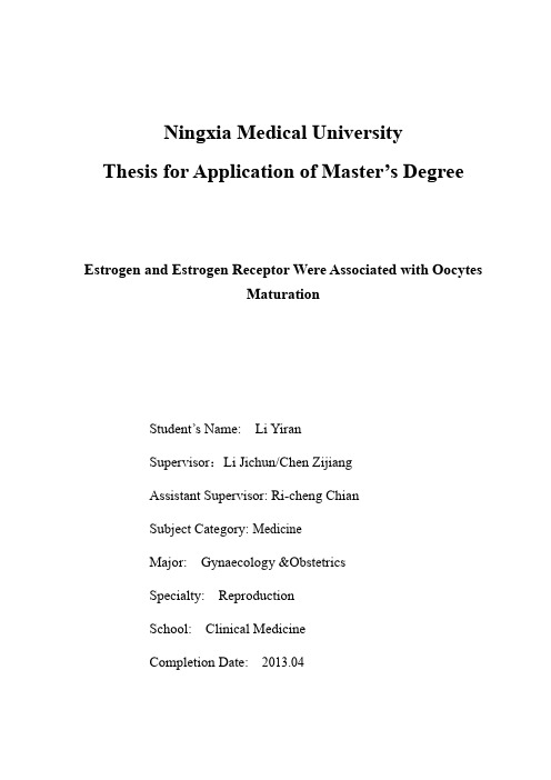
Ningxia Medical UniversityThesis for Application of Master’s DegreeEstrogen and Estrogen Receptor Were Associated with OocytesMaturationStudent’s Name: Li YiranSupervisor:Li Jichun/Chen ZijiangAssistant Supervisor: Ri-cheng ChianSubject Category: MedicineMajor: Gynaecology &ObstetricsSpecialty: ReproductionSchool: Clinical MedicineCompletion Date: 2013.04宁夏医科大学学位论文独创性声明本人郑重声明:所呈交的学位论文,是个人在导师的指导下,独立进行研究工作所取得的成果,无抄袭及编造行为。
除文中已经特别加以注明引用的内容外,本论文不含任何其他个人或集体已经发表或撰写过的作品成果。
对本文的研究做出重要贡献的个人和集体,均已在文中以明确方式标明并致谢。
本人完全意识到本声明的法律结果由本人承担。
论文作者签名_____论文导师签名_____年月日年月日论文使用授权的声明宁夏医科大学有权保留使用本人学位论文,同意学校按规定向国家有关部门机构送交论文的复印件和电子版,允许被查阅和借阅。
本人授权宁夏医科大学可以将本学位论文的全部或部分内容编入有关数据库进行检索,可以采用影印、缩印或其他复印手段保存和汇编本学位论文。
可以公布(包括刊登)论文的全部或部分内容。
(保密论文在解密后应遵守此规定)论文作者签名_____论文导师签名_____年月日年月日雌激素及其受体与卵母细胞成熟的相关性摘要目的通过测定取卵日人血清及卵泡液内雌二醇水平、卵丘细胞雌激素-β受体-β受体mRNA的表达量的关系和卵mRNA的表达,探讨卵泡液雌激素浓度与卵丘细胞E2母细胞成熟与卵丘细胞-β受体mRNA的表达量的相关性;通过观察雌激素G蛋白偶联受体GPR30在小鼠卵母细胞上的表达定位和不同时期的表达量以及体内成熟和体外成熟的卵母细胞表达量的比较,探讨GPR30对小鼠卵母细胞成熟调控作用和对卵母细胞体内、体外成熟调控作用的差异。
胰岛素样生长因子和胰岛素样生长因子结合蛋白-3与胎儿生长受限的关系

胰岛素样生长因子和胰岛素样生长因子结合蛋白-3与胎儿生长受限的关系卢 丹 祝淡抹 单 委 刘艳芝 吴雅娟扬州大学医学院,江苏扬州 225000[摘要] 目的 检测胰岛素样生长因子-1、2(IGF-1、2)和胰岛素样生长因子结合蛋白-3(IGFBP-3)水平,分析IGF-1、2和IGFBP-3的含量变化与胎儿生长受限(FGR)的关系。
方法 选取2011年1月至2013年12月在苏北人民医院妇产科住院,诊断为FGR的孕妇36例及同期住院的正常妊娠妇女30例作为受试对象。
采用酶联免疫吸附法(ELISA法)检测36例FGR孕妇(试验组)和30例正常妊娠女性(对照组)外周血和脐静脉血中IGF-1、2和IGFBP-3的浓度,并比较两组新生儿体重。
结果 试验组孕妇血清及新生儿脐血IGF-1、2和IGFBP-3水平均低于对照组(P<0.05)。
孕妇血清IGF-1、2和IGFBP-3水平均高于新生儿脐血IGF-1、2和IGFBP-3水平(P<0.05)。
试验组的新生儿体重明显小于对照组(P<0.05)。
结论 IGF-1、2和IGFBP-3这三个因子可能是导致FGR发生发展的重要因素之一,可作为判断胎儿生长发育的一项客观指标。
[关键词] 胰岛素样生长因子-1;胰岛素样生长因子-2;胰岛素样生长因子结合蛋白-3;胎儿生长受限[中图分类号] R714 [文献标识码] A [文章编号] 2095-0616(2021)04-0009-04Correlation between insulin-like growth factor and insulin-like growth factor binding protein-3 and fetal growth restrictionLU Dan ZHU Danmo SHAN Wei LIU Yanzhi WU YajuanMedical School of Yangzhou University, Jiangsu,Yangzhou 225000, China[Abstract] Objective To detect the levels of insulin-like growth factors 1, 2 (IGF-1,2) and insulin-like growth factor binding protein-3 (IGFBP-3), and to analyze the correlation between the changes of IGF-1, 2 and IGFBP-3 and fetal growth restriction (FGR). Methods In this experiment, pregnant women diagnosed as FGR and normal pregnant women hospitalized and admitted to the department of obstetrics and gynecology of Northern Jiangsu People's Hospital during the same period from January 2011 to December 2013 were selected as research objects, and they were divided into the experimental group (n=36, pregnant women diagnosed as FGR) and the control group (n=30, normal pregnant women). Enzyme-linked immunosorbent assay (ELISA) was used to detect the concentrations of IGF-1, 2 and IGFBP-3 in peripheral blood and umbilical vein blood in the experimental group and the control group , and compare the neonatal weight between two groups. Results The levels of IGF-1, 2 and IGFBP-3 in pregnant women's serum and neonatal cord blood in the experimental group, and compare thde neonatal weight between two groups were lower than those in the control group (P<0.05). The levels of serum IGF-1, 2 and IGFBP-3 in pregnant women were higher than those in neonatal cord blood (P<0.05). The neonatal weights of the experimental group were significantly lower than those of the control group (P<0.05). Conclusion IGF-1, 2 and IGFBP-3 may be one of the key factors leading to the occurrence and development of FGR. Therefore, it can be used as an objective index to judge fetal growth and development.[Key words] Insulin-like growth factor-1; Insulin-like growth factor-2; Insulin-like growth factor binding protein-3; Fetal growth restriction胎儿生长受限(fetal growth restriction,FGR)指胎儿应有的生长潜力受损,估测的胎儿体重小于同孕龄第十百分位的小于孕龄儿(small for gestation age,SGA)[1]。
哲罗鲑胰岛素样生长因子-I(IGF-I)cDNA 分子克隆、序列分析及组织表达

哲罗鲑胰岛素样生长因子-I(IGF-I)cDNA 分子克隆、序列分析及组织表达王晓玉;纪锋;徐黎明;赵景壮;刘淼;曹永生;尹家胜【摘要】Total RNA was isolated from Hucho taimen liver tissue.The cDNA encoding insulin-like growth factor I (IGF-I) peptide was amplified by reverse transcription polymerase chain reaction (RT-PCR) strategy using isolated total RNA as template.The IGF-I mRNA level was detected in different tissues using real -time quantitative PCR technique.The results displayed that the IGF-I contained an open reading frame (ORF) of 573 bp, and encoded a polypeptide with length of 190 amino acids.The polypeptide was composed of signal peptide , B, C, A, D domain and E peptide and the isoelectric point was 9.21.The amino acid sequence of H.taimen IGF-I was high homologus to that of other Salmonidae.H.taimen shared the highest identity with Salvelinus alpinus and the homology at nucleotide level of the IGF -I gene was 99.2%.The expression analysis of IGF-I gene with real-time quantitative RT-PCR showed that the highest level of IGF -I mRNA was ob-served in liver, followed by in gill and anterior intestine .There was no obvious expression in brain , pronephros, spleen, heart, stomach and muscle.%采用逆转录聚合酶链式反应(RT-PCR)方法,从哲罗鲑(Hucho taimen)肝脏的总 RNA 中扩增出胰岛素样生长因子-I(IGF -I)的 cDNA 开放阅读框(Open reading frame, ORF)序列,运用软件对其进行生物信息学分析,并利用荧光实时定量 PCR 技术检测了哲罗鲑成鱼不同组织中 IGF-I mRNA 的表达情况。
IGF1介导了嗅觉社会学习记忆中僧帽细胞的突出可塑性过程

IGF1介导了嗅觉社会学习记忆中僧帽细胞的突出可塑性过程IGF1-Dependent Synaptic Plasticity of Mitral Cells in Olfactory Memory during Social Learning背景在社会交往传播的食物偏好(STFP)期间,小鼠对食物气味的长期记忆是通过社会交往同伴提供的。
大脑如何将社会环境与气味信号联系起来,以促进记忆编码?方法在SFTP期间,本文采用特定的气味暴露。
在嗅球中,有选择性地将具有突触强度的肾小球特异性长时程增强(LTP)介导于颗粒和僧帽细胞的树突状突触体的GABA能神经元上。
结果突触结合蛋白-10、僧帽细胞的IGF1分泌的钙离子传感器或嗅球IGF1受体的缺失不仅能抑制社会相关的GABA能神经元长时程增强,还能损害STFP后记忆的形成。
相反,通过增强突触后GABA受体反应,将IGF1注入到急性嗅球切片中,就能在僧帽细胞中产生GABA能神经元LTP。
结论因此,我们的研究揭示了一个作用于感觉信息处理最初阶段的社会相关长时记忆的突触底物。
原始文献摘要BACKGROUND: During social transmission of food preference (STFP),mice form long-term memory of food odors presented by a social partner. How does the brain associate a social context with odor signals to promote memory encoding?METHODS: Here we show that odor exposure during STFP, but not unconditioned odor exposure, induces glomerulus-specific long-term potentiation (LTP) of synaptic strength selectively at the GABAergic component of dendrodendriticsynapses of granule and mitral cells in the olfactory bulb.RESULTS: Conditional deletion of synaptotagmin-10, the Ca2+ sensor for IGF1 secretion from mitral cells, or deletion of IGF1 receptor in the olfactory bulb prevented the socially relevant GABAergic LTP and impaired memory formation after STFP. Conversely, the addition of IGF1 to acute olfactory bulb slices elicited the GABAergic LTP in mitral cells by enhancing postsynaptic GABA receptor responses.CONCLUSIONS: Thus, our data reveal a synaptic substrate for a socially conditioned long-term memory that operates at the level of the initial processing of sensory information.罂粟花麻醉学文献进展分享联系我们电话:1331*****13。
18F-FES在乳腺癌患者体内摄取与病理免疫组化的关系

18F-FES在乳腺癌患者体内摄取与病理免疫组化的关系孙艺斐;邵志敏;章英剑;杨忠毅;张勇平;王明伟;姚之丰;薛静;鲍晓;杨文涛;沈镇宙【期刊名称】《中国癌症杂志》【年(卷),期】2014(000)002【摘要】背景与目的:16α-18F-17β-雌二醇(18F-FES)作为雌激素受体(estrogen receptor, ER)特异性显像剂,可在活体内反映ER的表达状况。
本研究主要探讨乳腺癌患者体内18F-FES摄取结果与病理免疫组化的相关性。
方法:自行制备18F-FES,入组26例乳腺癌患者(17例原发性乳腺癌,9例复发转移性乳腺癌),分别进行18F-FES及18F-FDG PET/CT显像,对每例患者进行空心针穿刺或手术治疗,对比相应病灶的免疫组化和18F-FES、18F-FDG摄取结果。
结果:96.15%(25/26)的患者18F-FES结果与ER病理免疫组化一致,以18F-FES SUVmax≥1.5为ER阳性,18F-FES PET/CT显像诊断乳腺癌病灶ER阳性的灵敏度为93.33%,特异度为100%。
ER、PR的免疫组化结果与18F-FES的SUVmax 呈明显的正相关;HER-2/Neu的免疫组化结果与18F-FES的SUVmax呈负相关。
结论:18F-FES有望用于全面反映乳腺癌患者全身病灶的ER表达情况,为临床个体化治疗方案的制定提供帮助。
%Background and purpose:16α-[18F]lfuoroestradiol (18F-FES) is an in vivo speciifc imaging agent for estrogen receptor (ER). We investigated the concordance between tumor ER status as determined by FES-PET and in vitro immunohistochemical assays. Methods: 18F-FES was prepared by ourselves. Twenty-six patients were enrolled (17 primary and 9 metastatic/recurrent). Patients underwent both 18F-FES and 18F-FDG PET/CT. Results:We found good overallagreement (96.15%) between in vitro ER assays and FES-PET. The ER status diagnosis sensitivity of 18F-FES was 93.33%and the speciifcity was100%when using cut-off value of SUVmax≥1.5. There was a positive correlation between in vitro ER, PR assays and the SUVmax of 18F-FES while in vitro HER-2/neu assays correlatived negatively with 18F-FES SUVmax. Conclusion:These results suggested 18F-FES may be useful for studying the ER expression of all malignant lesions in patients with breast cancer and guiding individual therapy.【总页数】7页(P128-134)【作者】孙艺斐;邵志敏;章英剑;杨忠毅;张勇平;王明伟;姚之丰;薛静;鲍晓;杨文涛;沈镇宙【作者单位】复旦大学附属肿瘤医院核医学科,复旦大学上海医学院肿瘤学系,上海200032;复旦大学附属肿瘤医院乳腺外科,复旦大学上海医学院肿瘤学系,上海200032;复旦大学附属肿瘤医院核医学科,复旦大学上海医学院肿瘤学系,上海200032;复旦大学附属肿瘤医院核医学科,复旦大学上海医学院肿瘤学系,上海200032;复旦大学附属肿瘤医院核医学科,复旦大学上海医学院肿瘤学系,上海200032;复旦大学附属肿瘤医院核医学科,复旦大学上海医学院肿瘤学系,上海200032;复旦大学附属肿瘤医院核医学科,复旦大学上海医学院肿瘤学系,上海200032;复旦大学附属肿瘤医院核医学科,复旦大学上海医学院肿瘤学系,上海200032;复旦大学附属肿瘤医院核医学科,复旦大学上海医学院肿瘤学系,上海200032;复旦大学附属肿瘤医院病理科,复旦大学上海医学院肿瘤学系,上海200032;复旦大学附属肿瘤医院乳腺外科,复旦大学上海医学院肿瘤学系,上海200032【正文语种】中文【中图分类】R737.9【相关文献】1.免疫组化三阴乳腺癌患者的临床病理特征及预后分析 [J], 李红英2.动态增强磁共振成像特征与乳腺癌患者病理及免疫组化指标的相关性 [J], 朱默;杨玲;王希明;郝光宇;郝正梅;胡春洪3.青年与中老年乳腺癌患者临床病理特征及免疫组化特点对比研究 [J], 姬荣伟;刘芳;田亚宁;田刘曼;王志斌4.乳腺癌患者分子亚型、病理类型及免疫组化分析 [J], 贾丽丽; 白雪; 张玉清; 邢惠海; 徐书方; 刘立秋5.对比分析青年及中老年乳腺癌患者临床病理特征及免疫组化特点 [J], 宋美英; 王玲; 张财家; 徐艳因版权原因,仅展示原文概要,查看原文内容请购买。
