Targeted-cryosurgical ablation of the prostate with androgen deprivation therapy: quality of li
冷冻消融与外科手术治疗肺癌的现状
[2]
首 先 采 用 液 氮 冷 冻 治 疗 恶 性 肿 瘤;
根据 2018 年 第 5 版 美 国 国 家 综 合 癌 症 网 络
中国肿瘤外科杂志 2019 年 12 月第 11 卷第 6 期 Chin J Surg Oncol, December 2019,Vol.11,No.6
· 471·
( NCCN) 非小细胞肺癌指南推荐,手术治疗主要适
进步,但这一进步的很大一部分在于改善了开放手
期、Ⅱ期以及部分ⅢA 期的肺癌患者。 值得关注的
肺叶切除术与保留肌肉的胸廓切开术相比较似乎
应证为肺癌 TNM 分期中相对早期肺癌患者,如 Ⅰ
是尽管随着肺癌综合治疗方式的不断改进,手术切
除的适应证不断放宽,但中晚期患者手术治疗效果
并不理想。
术技术和围手术期管理。 Kuritzky 等 [10] 比较 VATS
不可忽视的是冷冻消融作为微创治疗方式在肺癌的
微创理念的不断深入,人们对于微创治疗需求不断增Biblioteka 治疗中显示了其独特的优越性。
长,于 20 世 纪 90 年 代 初 期, 以 电 视 胸 腔 镜 手 术
1 外科手术治疗
治疗的常规术式。 冷冻外科治疗最早开始于 1963
1 1 适应证
(VATS)为主的微创胸外科应运而生,现已成为肺癌
· 470·
中国肿瘤外科杂志 2019 年 12 月第 11 卷第 6 期 Chin J Surg Oncol, December 2019,Vol.11,No.6
综述与讲座
冷冻消融与外科手术治疗肺癌的现状
付永强, 刘冰
作者单位: 130117 吉林 长春,长春中医药大学 临床医学院( 付永强) ;130021 吉林 长春,吉林省人民医院
肿瘤靶向治疗英语
肿瘤靶向治疗英语Targeted Therapy for CancerTargeted therapy for cancer is a type of treatment that uses drugs or other substances to identify and attack specific cancer cells without harming healthy cells. Unlike chemotherapy, which can affect both cancerous and healthy cells, targeted therapy is designed to be more precise and less toxic.There are several different types of targeted therapy, including:- Monoclonal antibodies: These drugs are designed to bind to specific proteins on the surface of cancer cells, making it easier for the immune system to recognize and attack those cells.- Small molecule inhibitors: These drugs are designed to block specific pathways or signals that cancer cells use to grow and divide.- Immunotherapy: This type of targeted therapy helps to boost the body's immune response to cancer cells.- Gene therapy: This is a newer form of targeted therapy that involves modifying the genes within cancerous cells to make them more susceptible to treatment.Targeted therapy can be used to treat many different types of cancer, including lung cancer, breast cancer, and melanoma. It can be used alone or in combination with other treatments, such as chemotherapy or radiation therapy.Overall, targeted therapy offers a promising new approach to the treatment of cancer, with fewer side effects and better outcomes than traditional chemotherapy. However, it's important to remember that each patient's case is unique, and that the best treatment approach will depend on a variety of factors, including the type and stage of cancer, the patient's overall health, and their individual preferences and goals for treatment.。
在未来研制出抗癌药物的作文

在未来研制出抗癌药物的作文英文版In the future, the development of anti-cancer drugs is crucial for the advancement of medical science and the improvement of human health. Cancer is a complex and devastating disease that affects millions of people worldwide. It is characterized by the uncontrollable growth of abnormal cells in the body, which can spread to other parts of the body and cause serious harm.The current treatments for cancer, such as chemotherapy and radiation therapy, can be effective in some cases, but they often come with severe side effects and are not always successful in eradicating the disease. That is why the development of new and improved anti-cancer drugs is so important.Scientists and researchers are constantly working to discover and develop new drugs that can target cancer cells more effectively and with fewer side effects. These drugs may work by blocking the growth of cancer cells, stopping the spread of the disease, or triggering the immune system to attack and destroy cancer cells.In recent years, there have been significant advancements in the field of cancer research, leading to the development of targeted therapies and immunotherapies that have shown great promise in treating various types of cancer. These new drugs are more precise in their targeting of cancer cells, leading to better outcomes for patients and fewer side effects.As we look to the future, the development of anti-cancer drugs will continue to be a top priority for the medical community. With ongoing research and innovation, we can hope to see even more effective and targeted treatments for cancer that will improve the lives of millions of people around the world.中文翻译在未来,研制抗癌药物对于医学科学的发展和人类健康的改善至关重要。
美国氩氦刀
美国氩氦刀一、美国氩氦刀的概念,美国氩氦刀的起源,美国氩氦的工作原理CRYOCARE冷冻手术系统中的CRYOCARE是美国ENDOCARE公司初测商标,意为“冷冻治疗”,音译“快尔克”。
1999年10月,广州张积仁教授和赵国江博士将其命名“氩氦刀”,特指美国Endocare公司的Cryocare冷冻治疗产品,同时将美国Endocare公司发明的Targeted Cryoablation Therapy(TCAP)译为“氩氦靶向治疗技术”。
Cryosurgery冷冻治疗(冷冻手术或低温外科),也称为Cryoablation(冷冻消融),Cryotherapy(冷冻疗法),Cryosurgical Ablation(冷冻手术消融)等,是利用超低温选择性原位冷冻和摧毁病变组织的方法。
冻治疗是人类历史上最早使用的肿瘤微创消融技术,起源于19世纪中叶。
1845年,科学家首次在实验室制出低温。
英国医生James Arnott于1845年用低至-24°C的冰盐水治疗溃疡性肿瘤,开创了近代冷冻治疗。
1877年开始,空气、氧气、氢气和氦气被液化。
1907年医生们使用?70°C的干冰(固态CO2)治疗皮肤和浅表肿瘤。
1950年起温度更低的液氮(?196°C)取代干冰成为主要的冷媒,被直接注入各种肿瘤进行治疗。
现代冷冻治疗起源于1960年美国神经外科医生Irving Cooper和工程师Arnold Lee的合作。
他们发明了新型探针状液氮冷冻器,冷冻脑组织。
自此,使用液氮作为冷媒、加热氮气作为热媒的液氮冷冻设备被应用于治疗各种肿瘤。
但液氮设备欠缺准确性、使用不方便和疗效欠佳使冷冻治疗陷于停顿。
合肥凤凰肿瘤医院冷冻治疗的复兴始于上世纪八十年代。
现代冷冻治疗的先驱-美国着名医学家Gary Onik和杰出的低温科学家-加州大学伯克利分校Boris Rubinsky教授(UC Berkeley, CA, USA)合作,于1982年首次利用超声波为冷冻治疗定位和监测,开创了影像引导的冷冻治疗学(image-guided cryosurgery),大大提高冷冻治疗的准确性和可靠性。
[精彩]美国氩氦刀
![[精彩]美国氩氦刀](https://img.taocdn.com/s3/m/a98883fb112de2bd960590c69ec3d5bbfd0ada84.png)
美国氩氦刀一、美国氩氦刀的概念,美国氩氦刀的起源,美国氩氦的工作原理CRYOCARE冷冻手术系统中的CRYOCARE是美国ENDOCARE公司初测商标,意为“冷冻治疗”,音译“快尔克”。
1999年10月,广州张积仁教授和赵国江博士将其命名“氩氦刀”,特指美国Endocare公司的Cryocare冷冻治疗产品,同时将美国Endocare公司发明的Targeted Cryoablation Therapy(TCAP)译为“氩氦靶向治疗技术”。
Cryosurgery冷冻治疗(冷冻手术或低温外科),也称为Cryoablation(冷冻消融),Cryotherapy(冷冻疗法),Cryosurgical Ablation(冷冻手术消融)等,是利用超低温选择性原位冷冻和摧毁病变组织的方法。
冻治疗是人类历史上最早使用的肿瘤微创消融技术,起源于19世纪中叶。
1845年,科学家首次在实验室制出低温。
英国医生James Arnott于1845年用低至-24°C的冰盐水治疗溃疡性肿瘤,开创了近代冷冻治疗。
1877年开始,空气、氧气、氢气和氦气被液化。
1907年医生们使用?70°C的干冰(固态CO2)治疗皮肤和浅表肿瘤。
1950年起温度更低的液氮(?196°C)取代干冰成为主要的冷媒,被直接注入各种肿瘤进行治疗。
现代冷冻治疗起源于1960年美国神经外科医生Irving Cooper和工程师Arnold Lee的合作。
他们发明了新型探针状液氮冷冻器,冷冻脑组织。
自此,使用液氮作为冷媒、加热氮气作为热媒的液氮冷冻设备被应用于治疗各种肿瘤。
但液氮设备欠缺准确性、使用不方便和疗效欠佳使冷冻治疗陷于停顿。
合肥凤凰肿瘤医院冷冻治疗的复兴始于上世纪八十年代。
现代冷冻治疗的先驱-美国着名医学家Gary Onik和杰出的低温科学家-加州大学伯克利分校Boris Rubinsky教授(UC Berkeley, CA, USA)合作,于1982年首次利用超声波为冷冻治疗定位和监测,开创了影像引导的冷冻治疗学(image-guided cryosurgery),大大提高冷冻治疗的准确性和可靠性。
药物涂层球囊扩张术在下肢动脉硬化闭塞患者中的应用价值探讨
DOI:10.19368/ki.2096-1782.2023.22.009药物涂层球囊扩张术在下肢动脉硬化闭塞患者中的应用价值探讨王茂生,王伟,任洪,黄丽琼,晏明鹏云南省曲靖市第二人民医院介入血管外科,云南曲靖655000[摘要]目的探讨药物涂层球囊扩张术在下肢动脉硬化闭塞(arteriosclerosis obliterans, ASO)患者中的应用价值。
方法回顾性分析2020年1月—2022年9月在云南省曲靖市第二人民医院分别进行药物涂层球囊扩张术和支架置入术治疗的68例ASO患者的临床资料,按照手术方式的不同分为球囊组(41例,药物涂层球囊扩张术)和支架组(27例,支架置入术)。
比较两组患者手术前后踝肱指数(ankle brachial index, ABI)、跛行距离、血管狭窄程度,术后1年目标血管再狭窄率、不良事件发生率。
结果术后,两组ABI、跛行距离、血管狭窄程度均显著优于术前,差异有统计学意义(P<0.05)。
术后6个月,球囊组ABI、跛行距离显著高于支架组,差异有统计学意义(P<0.05)。
术后1年,球囊组血管狭窄程度显著低于支架组,差异有统计学意义(P<0.05)。
术后1年,球囊组患者再狭窄率(9.76% vs 44.44%)及不良事件总发生率(7.32% vs 37.04%)均显著低于支架组,差异有统计学意义(χ2=10.887、9.299,P<0.05)。
结论在ASO患者中,药物涂层球囊扩张术与支架置入术的近期疗效相当,远期疗效药物涂层球囊扩张术优于支架置入术,临床可依据患者具体情况建议更适合的手术方案。
[关键词]下肢动脉硬化闭塞症;支架置入术;药物涂层球囊扩张术;再狭窄[中图分类号]R4 [文献标识码]A [文章编号]2096-1782(2023)11(b)-0009-04Exploring the Application Value of Drug-coated Balloon Dilatation in Pa⁃tients with Lower Extremity Arteriosclerosis ObliteransWANG Maosheng, WANG Wei, REN Hong, HUANG Liqiong, YAN MingpengDepartment of Interventional Vascular Surgery, Qujing Second People's Hospital, Qujing, Yunnan Province, 655000 China[Abstract] Objective To explore the application value of drug-coated balloon dilatation in patients with lower extrem⁃ity arteriosclerosis obliterans (ASO). Methods The clinical data of 68 ASO patients who underwent drug-coated bal⁃loon dilatation and stent placement respectively in the Second People's Hospital of Qujing City, Yunnan Province, from January 2020 to September 2022 were retrospectively analyzed and divided into the balloon group (41 cases, drug-coated balloon dilatation) and the stent group (27 cases, stent placement) according to the difference of the surgi⁃cal methods. The ankle brachial index (ABI), claudication distance, degree of stenosis, restenosis rate of the target ves⁃sel at 1 year after the surgery, and the incidence of adverse events were compared between the two groups before and after the surgery. Results After the surgery, ABI, claudication distance, and stenosis degree in both groups were sig⁃nificantly better than before the surgery, and the difference was statistically significant (P<0.05). At 6 months after sur⁃gery, ABI and claudication distance in the balloon group were significantly better than those in the stent group, and the difference was statistically significant (P<0.05). At 1 year after surgery, the degree of stenosis in the balloon group was significantly lower than that in the stent group, and the difference was statistically significant (P<0.05). At 1 year after the surgery, the restenosis rate (9.76% vs 44.44%) and the total incidence rate of adverse events (7.32% vs [作者简介] 王茂生(1981-),男,硕士,副主任医师,研究方向为外周血管疾病的微创治疗。
放射医学专业英语翻译
Unit 4 Section ADNA Strand Breaks and Chromosomal Abberations Inducedby Ionizing Radiation(1)1.What is the principal target for the biologic effects of radiation on the basis of strong circumstantial evidence?基于详细的证据,辐射生物效应的主要靶点是什么?2. What is the structure of a deoxyribonucleic acid molecule?脱氧核糖核酸分子的结构是什么?DNA Strand BreaksDNA链断裂There is strong circumstantial evidence to indicate that deoxyribonucleic acid (DNA) is the principal target for the biologic effects of radiation, including cell killing mutation, and carcinogenesis. A consideration of the biologic effects of radiation therefore must begin logically with a description of the breaks in DNA caused by charged-particle tracks and by the chemical species produced.有足够多且详细的证据表明脱氧核糖核酸是辐射生物效应的主要的靶,包括细胞杀伤性突变和致癌作用。
因此,从带电粒子和化学产物导致的DNA链断裂来考虑辐射生物效应是较合乎逻辑的。
全身运动不安运动阶段质量评估对婴幼儿神经系统疾病预测价值的Meta分析
全身运动不安运动阶段质量评估对婴幼儿神经系统疾病预测价值的Meta分析门光国;王凤敏;崔英波【摘要】目的探讨婴幼儿早期(出生后20周内)全身运动(GMs)不安运动阶段质量评估对婴幼儿神经系统疾病的预测价值.方法利用数据库检索到2015年12月前发表的相关文献,共有16篇文献纳入研究并进行Meta分析.结果 16篇文献QUADAS评分≥10的有8篇,临床特征等信息差异均无统计学意义(P>0.05).GMs 不安运动阶段质量评估对神经系统发育不良结局(包括脑性瘫痪)的预测分析显示,灵敏度、特异度、阳性似然比(PLR)、阴性似然比(NLR)和诊断比值比(DOR)分别为0.78、0.93、11.26、0.24和55.43;SROC曲线表明灵敏度和特异度最佳结合点的Q值为0.852 2,AUC值为0.919 0.GMs不安运动阶段质量评估对脑性瘫痪的预测分析显示,灵敏度、特异度、PLR、NLR和DOR分别为0.91、0.94、12.91、0.12和133.66,SROC曲线表明灵敏度和特异度最佳结合点的Q值为0.918 5,AUC值为0.969 2.结论 GMs不安运动阶段质量评估是预测婴幼儿神经系统疾病的一种有效方法,但不推荐单独使用.【期刊名称】《浙江医学》【年(卷),期】2016(038)014【总页数】5页(P1161-1165)【关键词】全身运动;不安运动阶段;婴幼儿;神经系统疾病;脑性瘫痪;Meta分析【作者】门光国;王凤敏;崔英波【作者单位】315012 宁波市妇女儿童医院新生儿科;315012 宁波市妇女儿童医院新生儿科;315012 宁波市妇女儿童医院新生儿科【正文语种】中文全身运动(general movements,GMs)是一种复杂的动作,包括头部、躯干、手臂和腿的运动,出现于胎儿早期并持续到出生后3~4个月。
近年来,GMs质量评估对婴幼儿脑性瘫痪(CP)等神经系统疾病的预测价值得到越来越多证据支持[1-2]。
胆囊癌临床诊疗的新进展
胆囊癌临床诊疗的新进展中华外科杂志普外空间 2022-08-10 10:00 发表于北京作者:杨自逸,刘诗蕾,蔡晨,吴自友,熊逸晨,李茂岚,吴向嵩,全志伟,龚伟文章来源:中华外科杂志, 2022, 60(8)摘要胆囊癌的恶性程度极高,尚缺乏早期诊断方法和有效治疗手段,亟需高质量研究突破诊疗瓶颈。
本文回顾了2021年国内外发表的胆囊癌研究相关文献,对临床诊疗领域的重要进展进行综述,详细介绍了胆囊癌最新流行病学数据及危险因素、新兴的外周血实验室检查和影像学诊断方法、病理学类型新分类、外科治疗的热点与争议及系统性综合治疗动态。
这些研究结果有助于探索更有效的胆囊癌诊治方法,为改善胆囊癌患者的预后带来希望。
胆囊癌是胆道系统常见的恶性肿瘤,具有症状隐匿、发展迅速、早期转移、预后极差的特点。
我国是胆囊癌的高发地区之一,近年来发病率和病死率呈缓慢上升趋势。
目前仍缺乏特异度和灵敏度均较好的胆囊癌早期诊断手段,临床发现的胆囊癌多为中晚期。
尽管医学科技不断发展,早期诊断和根治性手术切除仍是可能治愈胆囊癌的手段,行之有效的系统性治疗方法依然在不断探索中。
本文展示了2021年胆囊癌临床诊疗领域的研究进展,以探索更好的胆囊癌诊疗方法。
一、流行病学特征(一)发病率与死亡率2020年全球癌症统计数据显示,全球胆囊癌新发115 949例(男性41 062例,女性74 887例),死亡84 695例(男性30 265例,女性54 430例)[1],均居消化系统肿瘤第6位。
胆囊癌全球发病率存在明显的地域差异,全球年龄标准化发病率平均为2.3/10万人,以东亚、南美最高,西欧、北美则发病率较低[2];且近年来男性和年轻群体的胆囊癌发病率呈升高趋势。
我国国家癌症中心数据显示,国内胆囊癌发病率为3.95/10万人(男性3.70/10万人,女性4.21/10万人),死亡率为2.95/10万人(男性1.9/10万人,女性2.1/10万人)[3]。
核酸适体(aptamer)一种具有潜力的肿瘤药物靶向配基
2012年10月第9卷第30期·专家论坛·CHINA MEDICAL HERALD 中国医药导报肿瘤的靶向疗法是利用特异性“靶向配基”的介导,将药物或其他杀伤肿瘤的物质选择性地运送到肿瘤部位、选择性地杀伤肿瘤细胞以提高治疗效果的一种治疗方法。
近年来国内外核酸适体(aptamer )介导的主动靶向给药研究成为热点。
核酸适体(aptamer )是经过一种新的体外筛选技术(systematic evolution of ligands by exponential enrichment ,SELEX ),从随机单链寡聚核苷酸文库中得到的能特异结合蛋白或其他小分子物质的单链寡聚核苷酸,可以是RNA ,也可以是DNA ,长度一般为25~60个核苷酸[1]。
SELEX 技术自Tuerk 等[2]1990年发明以来,在临床诊断、靶向药物研制方面得以广泛应用。
首个核酸适配体药物“Macugen ”[3]由美国FDA 在2005年批准上市,成为核酸适配体领域的一个里程碑。
美国Achemix 、SomaLogic ,德国Noxxon AG 等多个公司正在开发核酸适配体药物和诊断试剂。
肿瘤细胞靶向给药是提高肿瘤治疗效果减少毒副作用的有效途径。
将药物偶联于肿瘤细胞特异性配体上是靶向给药的主要方法。
核酸能特异性结合细胞并且随之内化,是理想的靶向细胞输送剂。
核酸适体“靶向配基”介导或修饰的药物及药物纳米制剂,为主动靶向肿瘤细胞给药系统构建开拓了新方向。
本文简要综述适体作为肿瘤药物“靶向配基”的应用研究。
1核酸适体作为肿瘤药物“靶向配基”的优势具有高特异性与亲和性“靶向配基”的筛选,是制约主动靶向给药系统研究的瓶颈[4-5]。
以往修饰肿瘤药物的“靶向配基”是抗体、多肽和叶酸等分子,但这些配基修饰的靶向药物在实际应用中均存在着难以克服的缺陷。
单克隆抗体潜在的免疫原性和制备的困难大大限制了其临床的应用和产业化的进程[4];叶酸等小分子虽然有诸多优点,但要求病变组织细胞过多表达叶酸受体,靶向作用才得以实现[4-5],而核酸适体却具有以下优点:1.1高亲合力、强特异性随着体外高通量筛选技术SELEX 的发展,筛选出的适体与配体间的亲合力很高。
碱基切除修复抑制剂甲氧胺联合β-榄香烯治疗恶性脑胶质瘤的实验研究
序言β-榄香烯属国家二类非细胞毒性抗肿瘤新药,临床研究证实其对包括脑胶质瘤在内的多种肿瘤疗效确切,且无其他传统化疗药常有的骨髓抑制、肝肾功能损害等毒副作用。
但目前临床应用的榄香烯乳注射液因其存在静脉炎发生率很高、剂型性质不稳定等缺点,其进一步的应用受到了较大的限制。
碱基切除修复抑制剂甲氧胺(Methoxyamine),可通过裂解核酸内切酶破坏DNA碱基切除修复过程,从而抑制肿瘤细胞对损伤作用的修复反应。
据此,可认为抑制DNA 碱基切除修复可能是增强肿瘤细胞化疗敏感性的潜在靶点,目前多项实验报道也已证实了甲氧胺可增强烷化剂和放疗的抗肿瘤效果。
近年来,通过纳米技术构建的纳米脂质体在提高药物溶解度、增加药物稳定性、降低药物副作用、缓控释给药等方面较普通的脂质体有了显著的提高。
研究表明,纳米脂质体对正常细胞和组织无损伤作用,并可长时间吸附于靶细胞周围,因此使药物能充分向靶组织渗透,也可以通过静电吸附效应与细胞膜接触而融合而进入细胞内。
因此将药物包封于纳米脂质体被认为可以改变被包封药物的体内分布,提高药物治疗指数,降低药物毒性。
基于增强β-榄香烯的疗效,减少毒副作用的目的,本课题研究内容分两部分:(一)联合碱基切除修复抑制剂甲氧胺,探讨是否在体内外抗瘤活性上具有协同作用,以期减少榄香烯用量,降低毒副反应,为其在临床的应用提供实验和理论依据。
(二)、利用纳米脂质体技术构建新型的β-榄香烯-纳米脂质体药物传递系统,初步探讨其体外抗瘤活性。
II碱基切除修复抑制剂甲氧胺联合β-榄香烯治疗恶性脑胶质瘤的实验研究中文摘要胶质瘤是成人神经系统最常见的原发性肿瘤,手术全切除率很低,复发率高,当前多种治疗效果仍不理想。
榄香烯属国家二类非细胞毒性抗肿瘤新药,临床研究发现其对多种肿瘤疗效确切,而且还具有提高和改善机体免疫功能,与放化疗协同作用等独特效果。
但是肿瘤细胞具有强大的DNA损伤修复机制,会对化疗药物产生抗性。
因此抑制这种内在的DNA修复过程,如碱基切除修复抑制剂甲氧胺的联合应用有利于提高化疗药物的抗瘤效果。
拉考沙胺与卡马西平治疗成人新诊癫痫的有效性和安全性对比

·药物与临床·拉考沙胺与卡马西平治疗成人新诊癫痫的有效性和安全性对比Δ郭夏青*,李郭飞,孙玉华,郑东琳 #(河南大学淮河医院神经内科一病区,河南 开封 475000)中图分类号 R 742.1;R 969.4 文献标志码 A 文章编号 1001-0408(2024)04-0464-04DOI 10.6039/j.issn.1001-0408.2024.04.15摘要 目的 比较拉考沙胺(LCM )与卡马西平(CAR )单药治疗成人新诊癫痫患者的有效性和安全性。
方法 采用回顾性分析方法,根据用药方案的不同,将2020年9月-2022年6月河南大学淮河医院神经内科收治的新诊癫痫患者(84例)分为对照组(40例,接受CAR 治疗)和观察组(44例,接受LCM 治疗),比较两组患者的总有效率、癫痫发作频率、血脂水平和不良事件(AEs )发生情况。
结果 治疗后第1个月,观察组患者的总有效率(63.64%)与对照组(55.00%)比较,差异无统计学意义(P >0.05);两组患者的癫痫发作频率均较治疗前显著减少(P <0.05),但组间比较差异无统计学意义(P >0.05)。
治疗后第3个月,观察组患者的总有效率(90.91%)显著高于对照组(67.50%,P <0.05);两组患者的癫痫发作频率亦较治疗前显著减少,且观察组显著少于对照组(P <0.05)。
治疗后第3个月,对照组患者的TC 、TG 、LDL-C 水平以及观察组患者的LDL-C 水平均较同组治疗前显著升高,且观察组患者的TC 、TG 、LDL-C 水平均显著低于对照组(P <0.05)。
观察组患者的AEs 发生率(15.91%)与对照组(17.50%)比较,差异无统计学意义(P >0.05)。
结论 LCM 和CAR 在成人新诊癫痫患者的治疗中均具有一定效果,均可降低患者的癫痫发作频率,且安全性相当。
同时,LCM 的长期疗效优于CAR ,对患者血脂水平的影响小于CAR 。
RADIOFREQUENCYCATHETERABLATIONFOR…射频导管消融治疗…

RADIOFREQUENCY CATHETER ABLATION FOR SUPRAVENTRICULAR TACHYCARDIAS: EXPERIENCE AT PESHAWAR Zahid Aslam Awan, Mohammad Irfan*, Bakhtawar Shah, Lubna Noor, Sher Bahadar Khan,Faisal AminDepartment of Cardiology, Postgraduate Medical Institute, Hayatabad Medical Complex, *Lady Reading Hospital, Peshawar, Pakistan Background:Drug therapy is mostly employed in the management of supraventriculartachycardias (SVTs). However, radiofrequency catheter ablation has been found to be highlyeffective and safe in the treatment of SVTs. The current study is aimed at sharing ourexperience of 320 patients who presented with SVTs, and were treated with radiofrequencycatheter ablation. Methods:This descriptive study was carried out in the CardiacElectrophysiology Laboratory of Lady Reading Hospital, Peshawar from October 2006 toDecember 2009. Standard 4-wire electrophysiological study was carried out to identify themechanism of SVT in 320 consecutive patients. Radiofrequency catheter ablation was used tointerrupt the tachycardia circuit. Results:Out of a total 320 patients who underwentelectrophysiologic study, 168 were found to have atrioventricular nodal re-entry as underlyingmechanism; 121 patients were having accessory pathway responsible for re-entry (of these 95presented with orthodromic reciprocating tachycardia and 26 as antidromic reciprocatingtachycardia); 19 patients were having focal atrial tachycardia, 4 atrial fibrillation and 8 atrialflutter as the underlying cause for SVT. Radiofrequency catheter ablation was used with anoverall success of 94% and a complication risk of complete AV block in 0.3% and recurrencerate of 3%. Conclusion: Radiofrequency catheter ablation is safe and highly effective mode oftreatment of SVTs.Keywords: Catheter ablation, Supraventricular tachycardia (SVT), Wolf Parkinson White (WPW)SyndromeINTRODUCTIONIn cardiology, our ability to cure is rare. When cardiac disease is diagnosed, most of our treatments are palliative. Complaints may be diminished and life prolonged, but the disease process will not be stopped. Cure, however, is possible in the patient suffering from a tachycardia in an otherwise normal heart. Supraventricular type of tachyarrhythmias (SVT) are a frequent cause of admissions to emergency room and mostly drug therapy is offered due to limited availability of electrophysiological (EP) services. In the past, occasionally surgery and even catheter based DC shocks have been used for drug refractory SVT.1–4 By carrying out an EP study, we can locate the site of abnormal impulse formation or a critical part of the tachycardia pathway by cardiac activation mapping during the arrhythmia. By means of a catheter, radiofrequency (RF) energy can be applied to that area, resulting in destruction of a few mm of critical tissue and cure of the patient. This technique of EP/RFA has been with us now for over two decades. During this period, RFA has been found to be the first line of therapy for poorly tolerated SVT with hemodynamic intolerance or recurrent symptomatic SVT.3,5–7 The charm of this treatment modality is that most of the patients, once treated with EP/RFA, can have complete cure of their arrhythmia, and do not require any further drug therapy or follow-up. The estimated prevalence of SVT is 3.5%.8 There are different forms of SVT; atrioventricular nodal re-entry (AVNRT) is the most common form accounting for approximately 60% of the cases, while 30% are atrioventricular tachycardia (AVRT), atrial tachycardia and atrial flutter constitute 10% of SVT.9The first interventional catheter fulguration of an accessory pathway was performed by Weber and Schmiz in 1983.10 Since then, there has been lot of development to make ablation safer and now cryo-ablation has been proposed to be the safest for ablation targets close to sensitive structures like compact AN node.11 The success rate of RFA depends upon the type of arrhythmia, however, it is more than 96% in atrioventricular nodal re-entry.12 Although several energy sources have been used for ablation, this article will deal only with ablation of SVT using RF energy.MATERIAL AND METHODSAfter obtaining informed consent, 320 consecutive patients with symptomatic drug resistant SVT were admitted from October 2006 to December 2009 in Hayatabad Medical Complex, Peshawar and underwent EP/RFA at the Cath Lab in Lady Reading Hospital, Peshawar. All antiarrhythmic drugs were discontinued at least three half lives of the respective drugs before the study except amiodarone, which waswithdrawn two months before study. Those with atrial fibrillation and age below 10 years were excluded. The study protocol was approved by the hospital ethical committee and patients were transferred to the lab in a fasting state having been sedated with diazepam or midazolam, while diamorphine or nalbuphin was given as analgesic. An intravenous bolus dose of Regular Heparin 2500 IU for right sided and 100 units/Kg for left sided procedures was routinely administered.Four diagnostic EP catheters were introduced, 2 each through the right and left femoral veins and placed at the following sites; right atrial appendage (quadripolar 6-Fr catheter), right ventricular apex (quadripolar 6-Fr), His bundle region (Octopolar 6-Fr catheter) and Coronary sinus (CS) (decapolar 6-Fr catheter). A 7-Fr 4 mm ablation catheter was introduced through the right femoral vein. Left sided pathways were approached transeptally (via interatrial septal puncture) by using multipurpose 7-Fr sheath.An indifferent patch was applied on back at left scapular area. Bard Pro EP Lab, Bloom stimulator and Cordis EP Shuttle were used to deliver radiofrequency current. Before inducing tachycardia, baseline intervals (PR, QRS, QT, AH, HV) were measured. The following parameters were looked for in all the tachycardias:∙Onset of tachycardia - with or without jump∙VA activation – concentric/eccentric∙VA interval – < or > 70 ms∙Parahisian Pacing response–Nodal or extra nodal ∙Ventricular entrainment – post pacing interval < or > 115 ms∙VAV or VAAV responseThe SVTs were grouped into four on the basis of initiating mechanism:1.Atrioventricular nodal re-entry2.Atrioventricular reciprocating tachycardia(Rightor Left sided)3.Atrial tachycardia (Right or Left sided)4.Atrial flutter (Right or Left sided)The right and left accessory pathways were further grouped into lateral, septal, anterior and posterior, parahisian and middle cardiac vein on the basis of their location.For ablation in atrioventricular nodal re-entry, the RF energy was applied at anterior lip of CS os to modify the slow pathway. Mostly a power of 30 watts and a temperature of 60 ºC energy was delivered for 60 Sec. The accessory pathways were modified by RF energy delivery at AV ring during sinus rhythm in manifest pathways or during tachycardia or ventricular pacing in orthodromic tachycardias. The focal atrial tachycardias were targeted when atrial intracardiac electrogram on ablation catheter was 20–30 ms earlier than surface P wave. Isthmus dependent atrial flutters were managed by ablation line across isthmus from tricuspid valve to inferior vena cava.Ablation was declared successful if no tachycardia could be induced after RFA even with isoproterenol.RESULT STable-1 summarises the frequency of various types of SVTs among the patients who underwent RF ablation at our centre. The mean age of our patients was 38±19 years, with male to female ratio of 1:2.37. Two of our patients had rheumatic heart disease, six had coronary artery disease, and three pregnant patients were considered for RFA after a single documented episode of SVT because of the hemodynamic compromise during SVT. Only one patient (0.3%) with septal atrial tachycardia developed a major complication, i.e., complete heart block for which PPM was implanted; while minor complications like femoral artery aneurysm developed in only one patient (0.3%) and two patients (0.6%) developed right lower limb DVT two weeks post-procedure. Recurrence rate of 3% was observed in our patients.Figure-1 shows how the pre-excitation pattern is lost on the surface ECG as soon as the accessory pathway is ablated. It is simultaneously visible in the intracardiac signals detected by the various catheters placed inside the heart at selective sites; there is fusion of AV signals which is lost after the pathway is ablated, and the AV signals become clearly separated from each other.Figure-2 shows the frequency of the various SVTs we ablated at our centre, while Figure-3 shows their gender-wise distribution as well.Table-1: SVT Catheter Ablation in ourexperience of 320 patients Supraventricular tachycardia Number % AVNRT 168 52.50 AVRT (ORT* =95 , ART**=26) 121 37.81 Right-Sided Pathways 46 14.37Middle cardiac vein 4 1.25Parahisian pathway 2 0.62Mahaim pathway 3 0.93Left-sided pathways 66 20.62 Atrial fibrillation 4 1.25 Atrial flutter 8 2.50 Atrial tachycardia 19 5.93 *orthodromic tachycardia, **antidromic tachycardia1lost in the third beat after RF ablation. Tracings HRA 1, 2 to CS 1, 2 of intracardiac signals show fusion of AVsignals in first two beats, while there is separation of AV signals in the last beat after ablation.that underwent ablation at our centreDISCUSSIONAtrioventricular nodal re-entry is the most common cause of regular narrow complex SVT. In our study, 70% of SVT are due to atrioventricular nodal re-entry. If AVNRT are less frequent and responsive to therapy with beta blocker or Calcium channel blocker, then RFA can be deferred. However in patients withfrequent episodes or hemodynamic intolerance or those who refuse prolonged medication, RFA is a safe and cost effective treatment modality. The success rate is more than 96% and the risk of damaging the compact AV node is less than 1% especially with cryo-ablation and the recurrence rate is also less than 3%.13,14All of the patients we ablated for SVT weresymptomatic despite drug therapy. Our recurrence rate in AVNRT has been very low because it had been part of our protocol to look for slow junctional rhythm during RFA and to reassure with Isoproteronol that tachycardia could no longer be reinduced once RFA had been done. The presence of a junctional rhythm during slow-pathway ablation has been indisputably considered to be the most sensitive but non-specific marker of successful ablation.15 Children under age of 10 years were not considered for the reason that radiofrequency ablation is not very safe in this age and cryo-ablation is a preferable option for AVNRT.16–18The SVTs due to ORT and ART on the rightside were mapped by ablation catheter in LAO 30º view. Those with evidence of ventricular pre-excitation on resting ECG were ablated in sinus rhythm targeting the closest AV site or the site having shortest delta-V wave and/or pathway potentials.19–24 The RF energy was stopped if a pathway was not visualised in a 20 sec break. Also after successful ablation, a confirmatory burn for 60 sec at 50 watts was given in all cases to reduce the risk of recurrence. Amongst our 51 patients with left sided accessory pathways, 48 were approached transeptally in LAO 60º view while in only 3 patients,ablation was done retrogradely via left ventricle; transeptal route was preferred because of the shorter procedure (average 20 min) and lesser radiation exposure. The transeptally approached patients were given Aspirin for 30 days after procedure to avoid any thromboembolic event.The higher failure rate in atrial tachycardia was due to limitations of conventional EP catheters to provide adequate information about the electrical activity at the roof of right atrium.25 3-dimensional or non-contact mapping system is now the preferred approach for atrial tachycardia to have better results.The success for isthmus dependent atrial flutter was 92%; the 8% failure can be attributed to lack of irrigated tip and 8 mm ablation catheter.26 CONCLUSIONSVTs are mostly due to atrioventricular nodal re-entry or accessory pathways. RFA is a very safe and highly effective mode of treatment for SVT and should be considered as first line of therapy if EP services are available. Transeptal approach for left side accessory pathways is also very safe and less time consuming and avoids prolonged exposure to radiation associated with retrograde approach. For atrial tachycardia, the preferred approach is non-contact mapping. However, conventional approach of RFA can be considered for foci on the floor of the right atrium. REFERENCES1.Ward DE, Camm AJ. Treatment of tachycardia associated withthe Wolff-Parkinson-White Syndrome by transvenous electrical ablation of accessory pathways. Br Heart J 1985;53:64-8.2.Bardy GH, Ivey T, Coltorti F. Development, complicationsand limitation of catheter-mediated electrical ablation of posterior accessory atrioventricular pathways. Am J Cardiol 1988;61:309–16.3.Blomström-Lundqvist C, Scheinman MM, Aliot EM, AlpertJS, Calkins H, Camm AJ, et al. ACC/AHA/ESC guidelines for the management of patients with supraventricular arrhythmias executive summary. A report of the American College of Cardiology/American Heart Association Task Force on Practice Guidelines and the European Society of Cardiology Committee for Practice Guidelines (writing committee to develop guidelines for the management of patients with supraventricular arrhythmias). Developed in collaboration with NASPE-Heart Rhythm Society. J Am Coll Cardiol 2003;42:1493–531.4.Kriebel T, Broistedt C, Kroll M, Sigler M, Paul T. Efficacy andsafety of cryoenergy in the ablation of atrioventricular reentrant tachycardia substrates in children and adolescents. J Cardiovasc Electrophysiol 2005;16:960–6.5.Gallager JJ, Pristchett FLC, Sealy WC. The prexcitationsyndromes. Prog Cardovasc Dis 1978;20:285–355.6.Scheinman MM, Morady F, Hess DS. Catheter induced ablationof atrioventricular junction to control refractory arrhythmia.JAMA 1982;248:851–5. 7.Morady F, Scheiman MM. Transvenous catheter ablation of aposteroseptal accessory pathway in a patient with the Wolff-Parkinson-White syndrome. N Engl J Med 1984;310:705–7.8.Orejarema LA, Vidaillet H, Destafano F. Paxoxysmal SVT ingeneral population. JACC 1998;31:150–7.9.Wellens HJ. Twenty-five years of insights into the mechanismsof supraventricular arrhythmias. J Cardiovasc Electrophysiol 2003;14:1020–5.10.Weber H, Schmiz L. Catheter technique for closed chest ablationof an accessory pathway. N Engl J Med 1983;308:654.11.Skanes AC, Dubuc M, Klein GJ, Thibault B, Krahn AD, YeeR, et al. Cryothermal ablation of the slow pathway for the elimination of atrioventricular nodal reentrant tachycardia.Circulation 2000;102:2856–60.12.Lockwood D, Otomo K, Wing Z. Electrophysiologiccharacteristics of atrioventricular nodal reentry tachycardia: Implications for the reentrant circuits. In Zipes DP, Jaliffe J (eds): Cardiac electrophysiology from cell to bedside 4th ed.Philadelphia: Sunders, 2004;537–7.13.Jentzer JH, Goyal R, Williamson BD. Analysis of junctionalectopy during radiofrequency ablation of the slow pathway inpatients with atrioventricular nodal reentrant tachycardia.Circulation 1994;90:2820–6.14.Kimman GP, Theuns DA, Szili-Torok T, Scholten MF, Res JC,Jordaens LJ. CRAVT: a prospective, randomized study comparing transvenous cryothermal and radiofrequency ablation in atrioventricular nodal reentrant tachycardia. Eur Heart J 2004;25:2232–7.15.Hsieh MH, Chen SA, Tai CT, Yu WC, Chen YJ, Chang MS.Absence of Junctional Rhythm During Successful Slow-Pathway Ablation in Patients with Atrioventricular Nodal Reenterant Tachycardia. Circulation 1998;98(21): 2296–300.16.Zrenner B, Dong J, Schreieck J. Transvenous cryoablation versusradiofrequency ablation of the slow pathway for the treatment of atrioventricular nodal re-entrant tachycardia: a prospective randomized pilot study. Eur Heart J 2004;25:2226 –31.17.Skanes AC, Dubuc M, Klein GJ. Cryothermal ablation of theslow pathway for the elimination of atrioventricular nodal reentrant tachycardia. Circulation 2000;102:2856–60.18.Skanes AC, Klein G, Krahn A, Yee R. Cryoablation: potentials andpitfalls. J Cardiovasc Electrophysiol 2004;15(Suppl):S28–S34. 19.Warin JF, Haissaguerre M, Le Metayer Ph. Catheter ablation ofaccessory pathways with a direct approach. Circulation 1988;78:800–15.20.Morady F, Scheinman MM, Kow WH. Long term results ofcatheter ablation of a posteroseptal accessory atrioventricular connection in 48 patients. Circulation 1989;79:1160–70.21.Haissaguerre M, Warin JF. Closed-chest ablation of left lateralatrio-ventricular accessory pathways. Eur Heart J 1989;10:602–10.22.Morady F. Catheter ablation of supraventricular arrhythmias:state-of-the-art. J Cardiovasc Electrophysiol 2004;15:124–39. 23.Haissaguerre M, Gaita F, Marcus Fl. Radiofrequency catheterablation of accessory pathways: A contemporary review. J cardiovasc Electrophysiology 1994;5:532–52.24.Morday F, Strick Berger SA, ManKc. Reasons for prolonged orfailed attempt at RFA of accessory pathways. J Am Cardiol 1996;27:683–9.25.Anguera, Ignasi, Brugada, Jospe, Roba. Outcomes afterradiofrequency catheter ablation of atrial tachycardia. Am J Cardiol 2001;87:886–90.26.Hillock RJ, Melton IC, Crozier IG. Radiofrequency ablation forcommon atrial flutter using an 8-mm tip catheter and up to 150W. EP Europace 2005;7:409–12.Address for Correspondence:Dr. Zahid Aslam Awan, Associate Professor, Department of Cardiology, Postgraduate Medical Institute, Hayatabad Medical Complex, Peshawar, Pakistan. Cell: +92-300-5940698E-mail:***********************。
临床医师看血常规|从今天开始关注红细胞分布宽度

临床医师看血常规|从今天开始关注红细胞分布宽度读完这篇文章,我想您会第一时间打开电子病例系统,查看每一个患者RDW的走势。
如果您发现RDW呈升高的趋势,您或许还会发现患者的病情或者预后会比较严重。
当然,打包票的事情我们从来不干,因为RDW的敏感性不太可能是100%。
红细胞分布宽度(RDW)是反映红细胞体积异质性的参数,用红细胞体积大小的变异系数来表示,正常值12%-15%。
RDW通常用于缺铁性贫血的诊断与疗效观察、小细胞低色素性贫血的鉴别诊断、贫血的分类等。
然而近年来,研究发现RDW与多种疾病的预后存在相关性。
这里,我们首先通过几个创伤或外科重症病危患者RDW的变化趋势给各位朋友建立一个感性认识。
case 1.高位截瘫,病情危重复杂,麻烦接踵而至,随着住院时间的延长RDW呈增加趋势case 2.多发伤伴胰腺挫裂伤,病情危重复杂,随着住院时间的延长RDW呈增加趋势case 3.多发伤有蛛血,病情危重,随着住院时间的延长RDW呈增加趋势case 4.蛛血转出监护室后,病久未愈,感染严重,随着住院时间的延长RDW呈增加趋势下面列举的是RDW在不同疾病预后方面的研究。
您平时查房中有无关注RDW这项指标呢?1、创伤1.1.Paulus etal.Admission red cell distribution width: a novelpredictor of massive transfusion after injury.Am Surg. 2014 Jul;80(7):685-9.1.2.Majercik S etal.Red cell distribution width is predictive of mortality in trauma patients.J Trauma Acute Care Surg. 2013 Apr;74(4):1021-6.1.3.Garbharran etal.Red cell distribution width is an independent predictor of mortality in hip fracture. Age Ageing. 2013 Mar;42(2):258-61.1.4.Lv etal.Red Cell Distribution Width as an Independent Predictor of Long-Term Mortality in Hip Fracture Patients: A Prospective Cohort Study. J Bone Miner Res. 2015 Jul 17.2、中风Lappegård etal.Red cell distri bution width is associated with future risk of incident stroke. The Tromsø Study.Thromb Haemost. 2015 Aug 20;114(6).3、蛛血Chugh etal.Red Blood Cell Distribution Width is Associated with Poor Clinical Outcome After Subarachnoid Hemorrhage: A Pilot Study. Neurocrit Care. 2015 Oct;23(2):217-24.3、肺炎Bello etal.Red blood cell distribution width [RDW] and long-term mortality after community-acquired pneumonia. A comparison with proadrenomedullin. Respir Med. 2015 Sep;109(9):1193-206.4、冠心病3.1.Balta etal. Red Cell Distribution Width in Myocardial Infarction. Med Princ Pract. 2015 Sep 1.3.2.Khaki etal.Relationship Between Red Blood Cell Distribution Width and Mortality of Patients with Acute Myocardial Infarction Referring to Tehran Heart Center.Crit PathwCardiol. 2015 Sep;14(3):112-5.3.3.Li etal. Red Blood Cell Distribution Width is Independently Correlated With Diurnal QTc Variation in Patients With Coronary Heart Disease.Medicine (Baltimore). 2015 Jun;94(23):e822.5、心衰Kojima etal. Usefulness of the Red Blood Cell Distribution Width to Predict Heart Failure in Patients With a Fontan Circulation. Am J Cardiol. 2015 Sep 15;116(6):965-8.Uemura etal. Elevation of red blood cell distribution width during hospitalization predicts mortality in patients with acute decompensated heart failure. J Cardiol. 2015 Jun 30.6、房颤Wan etal.The relationship between elevated red cell distribution width and long-term outcomes among patients with atrial fibrillation. Clin Biochem. 2015 Aug;48(12):762-7.7、静脉血栓危险分层Bucciarelli etal.Association between red cell distribution width and risk of venous thromboembolism. Thromb Res. 2015 Sep;136(3):590-4.发现:individuals with RDW above the 90(th) percentile (>14.6%) had a 2.5-fold increased risk of VTE compared to those with RDW ≤90(th)percentile, independently of age, sex, body mass index, other hematological variables and renal function (adjusted odds ratio: 2.52 [95%CI:1.42-4.47]). The risk was similar for unprovoked and provoked VTE, and slightly higher in patients with pulmonary embolism (adjusted odds ratio 3.19 [95%CI:1.68-6.09]) than in those with deep vein thrombosis alone (2.29 [95%CI:1.22-4.30]).8、贝赛特氏症Aksoy etal.Association between red blood cell distribution width and disease activity in patients with Behçet's disease.J In t Med Res. 2015 Sep 10.9、淋巴瘤Periša etal.Red blood cell distribution width as a simple negative prognostic factor in patients with diffuse large B-cell lymphoma: a retrospective study. Croat Med J. 2015 Aug 31;56(4):334-43.10、急性阑尾炎Bozlu etal. The diagnostic value of red blood cell distribution width in children with acute appendicitis. Pediatr Int. 2015 Aug 14.11、多发性硬化Peng etal. Assessment of the Relationship Between Red Cell Distribution Width and Multiple Sclerosis.Medicine (Baltimore). 2015 Jul;94(29):e1182.12、乙肝Huang etal.Red cell distribution width as a potential index to assess the severity of hepatitis B virus-related liver diseases.Hepatology Research 2014; 44: E464–E470.13、肝癌Smirne etal.Evaluation of the red cell distribution width as a biomarker of early mortality in hepatocellular carcinoma.Dig Liver Dis. 2015 Jun;47(6):488-94.14、子宫内膜癌Kemal etal.The value of red blood cell distribution width in endometrial cancer.Clin Chem Lab Med. 2015 Apr;53(5):823-7.15、结肠肿瘤Ay etal. Is early detection of colon cancer possible with red blood cell distribution width?Asian Pac J Cancer Prev.2015;16(2):753-6.16、类风关Hassan etal.Red cell distribution width: a measure of cardiovascular risk in rheumatoid arthritis patients? Clin Rheumatol. 2015 Jun;34(6):1053-7.17、银屑病Kim etal.Red blood cell distribution width is increased in patients with psoriasis vulgaris: A retrospective study on 261 patients. J Dermatol. 2015 Jun;42(6):567-71.18、亚临床甲减Yu etal. The value of red blood cell distribution width in subclinical hypothyroidism.Arq Bras Endocrinol Metabol. 2014 Feb;58(1):30-6.以下是以病房为单位的相关研究1、内科住院病房Shteinshnaider etal. Prognostic significance of changes in red cell distribution width in an internal medicine ward. Eur J Intern Med. 2015 Aug2、ICU病房2.1、Fujita etal. Red cell distribution width and survival in patients hospitalized on a medical ICU.Clin Biochem. 2015 Jul 10.2.2、purtle etal.The association of red cell distribution width at hospital discharge and out-of-hospital mortality followingcritical illness.Crit Care Med. 2014 Apr;42(4):918-29.2.3、Zhang etal. Red cell distribution width is associated with hospital mortality in unselected critically ill patients.J Thorac Dis. 2013 Dec;5(6):730-6.2.4、Meynaar etal.Red cell distribution width as predictor for mortality in critically ill h J Med. 2013 Nov;71(9):488-93.2.5、Hunziker etal.Red cell distribution width improves the simplified acute physiology score for risk prediction in unselected critically ill patients.Crit Care. 2012 May 18;16(3):R89.2.6、Bazick etal.Red cell distribution width and all-cause mortality in critically ill patients.Crit Care Med. 2011 Aug;39(8):1913-21.3、儿科ICU病房Ramby etal.Red Blood Cell Distribution Width as a Pragmatic Marker for Outcome in Pediatric Critical Illness.PLoS One. 2015 Jun 9;10(6):e0129258.附表.RDW的临床应用本表出自:Gian Luca Salvagno, Fabian Sanchis-Gomar, Alessandra Picanza & Giuseppe Lippi (2015) Red blood cell distribution width: A simple parameter with multiple clinical applications, Critical Reviews in Clinical Laboratory Sciences, 52:2, 86-105其实还有很多疾病的预后被发现与RDW有关,本文不再一一列举。
基因靶点与药物英文
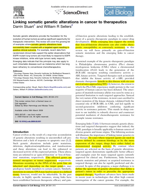
Available online at Linking somatic genetic alterations in cancer to therapeuticsDarrin Stuart 1and William R Sellers 2Somatic genetic alterations provide the foundation for the evolution of human tumors as well as significant opportunity for therapeutic intervention.This review will cover the growing list of examples where somatic genetic alterations havesuccessfully been coupled with a targeted agent resulting in positive clinical outcome.For example,recent data fromrandomized clinical trials support the earlier observations that EGFR mutant lung tumors are most likely to respond to EGFR kinase inhibitors,while wild-type tumors rarely respond.Emerging data indicate that this principle may also apply to such intractable diseases such as melanoma which has long been refractory to conventional chemotherapeutics.Addresses 1Oncology Disease Area,Novartis Institutes for BioMedical Research,4560Horton Street,4/5,Emeryville,CA 94608,United States 2Oncology Disease Area,Novartis Institutes for BioMedical Research,250Massachusetts Avenue,4A/245,Cambridge,MA 02129,United StatesCorresponding author:Stuart,Darrin (Darrin.Stuart@ )and Sellers,William R (William.Sellers@ )Current Opinion in Cell Biology 2009,21:304–310This review comes from a themed issue on Cell regulationEdited by Brian Hemmings and Nikolas Tonks Available online 26th March 20090955-0674/$–see front matter#2009Elsevier Ltd.All rights reserved.DOI 10.1016/j.ceb.2009.02.001IntroductionCancer evolves as the result of a step-wise accumulation of genetic alterations resulting in uncontrolled cell pro-liferation and a lack of response to apoptotic cues [1,2].Such genetic alterations include point mutations,deletions,duplication/amplification,and translocations and these alterations can lead to the enhanced or decreased activity of the expressed protein.These altera-tions are referred to as ‘gain-of-function’or ‘loss-of-func-tion’mutations,respectively.The affected genes are termed oncogenes or tumor suppressors,respectively.Mutations occurring in the DNA of the germ line are inherited and depending on the target gene(s),may predispose family members to certain cancers.Similarly,mutations in somatic DNA may lead to cancer in a defined tissue;however,would not be inheritable.In the past decade,in highly specific instances,strong links have been established between therapeutic efficacy and gain-of-function genetic alterations leading to the establish-ment of a genetic –therapeutic paradigm in cancer drug development.While emerging data support the notion that loss-of-function alterations can also confer thera-peutic susceptibility or conversely resistance,in this review we will focus primarily on gain-of-function somatic mutations and the emerging links to cancer therapy.A seminal example of the genetic –therapeutic paradigm is Philadelphia chromosome positive (Ph+)chronic myelogenous leukemia (CML)where a chromosomal translocation t(9;22)leads to the generation of the BCR-ABL oncogene resulting constitutive activity c-ABL kinase activity.Targeted therapies such as imatinib that inhibit the kinase activity of c-ABL provided dramatic therapeutic benefit in this disease with six-year survival rates approaching 90%[3];however,the extent to which the Ph+CML experience might pertain to the vast majority of human cancers has been debated.The emer-gence of imatinib-resistance alleles was seen by some as a potential limitation to such targeted approaches.Instead the very nature of the resistance,in most cases ascribed to direct mutation of the kinase domain,validated both the essential role of BCR-ABL in CML and led rapidly to second-generation inhibitors conferring significant activity in resistance patients.This stands in sharp con-trast to our inability to robustly discover and counteract potential mediators of chemotherapeutic resistance for example taxane resistance.Emerging links (Table 1)between somatic genetic altera-tions and targeted therapeutics suggest that indeed,the CML paradigm is broadly applicable to human cancers of diverse genetic and tissue origins.The following sections will illustrate the importance of linking tumor genetics to the therapy.In cases where targeted agents are used in tumors defined by common histology or by the mere expression of the target,drugs have either failed or demonstrated marginal activity.By contrast when patients are selected on the basis of genetic alterations,targeted agents demonstrate significantly better efficacy and examples exist for many targeted agents currently approved for clinical use.Furthermore,links between somatic mutation and primary or acquired resistance to targeted therapy can also be rapidly predicted and dis-covered.In spite of this progress,a major challenge is to define the spectrum of genetic alterations that define a patient’s tumor in order to prescribe the appropriate targeted therapy.Significant advances have been made in recent years in technologies that allow high content interrogation of the cancer genome.For example,theability to genotype multiple oncogenic mutations in a given patient’s tumor may facilitate the selection of a targeted therapy[4 ].The ability to transfer these tech-nologies to routine clinical diagnoses and define a tumor based on the underlying genetics may pave the way for therapies based purely on tumor genotype,rather than tumor histology or anatomical site.Targeting the EGF receptor(EGFR)in adenocarcinomas of the lungGefitinib and erlotinib are small-molecule inhibitors of the EGFR tyrosine kinase that have been evaluated in several clinical trials for the treatment of NSCLC.While neither drug has demonstrated a dramatic increase in survival in the overall population of NSCLC patients, there is a subset of patients who experience dramatic and durable responses.The underlying rationale for targeting EGFR was based on its overexpression in lung carcinoma; however,the level of EGFR expression as determined by immunohistochemistry(IHC)does not correlate with response[34].Instead,activating EGFR mutations were identified in patients who responded to gefitinib or erlo-tinib therapy[14 ,15 ,35]and these initial observations were confirmed in larger prospective studies[36,37].Two studies examined the efficacy of gefitinib in chemother-apy-naı¨ve patients whose tumors express activating mutations or deletions in EGFR.In the study by Inoue et al.,the response rate was75%(12/16);while Sequist et al.observed a response rate of55%(17/31),which while the response rate was slightly lower also included tumors with atypical EGFR mutations,including mutations known to confer resistance to gefitinib(T790M)[37]. Results also indicated that progression-free survival and overall survival was approximately twofold longer than typically observed with cytotoxic regimens in unselected NSCLC patients.However,without a cytotoxic therapy comparator arm,a gefitinib-related improvement in sur-vival could not be distinguished from the possibility that EGFR mutation was a favorable prognostic factor for survival.A similar twofold improvement in median sur-vival of patients with EGFR mutations treated with gefitinib has also been observed in a large(n=330) retrospective analysis of Japanese lung adenocarcinoma patients treated before(1999–2001)and after(2002–2004) the approval of gefitinib(13.6months versus27.2months) [16 ].In this study,the prognostic influence of EGFR mutation on disease could be measured and did not account for the positive effect of gefitinib treatment on patients with mutant EGFR.The conclusions drawn from these large clinical studies confirm thefindings of the earlier studies that the patients who derive clinical benefit from gefitinib are those with activating mutations in the kinase domain of EGFR.Just as sensitivity to EGFR tyrosine kinase inhibitors can be linked to somatic genetic mutations,so can the primary or acquired resistance.Patients who initially respond to gefitinib or erlotinib often relapse while on therapy as the result of a second mutation in the EGFR kinase domain [38–40].Intriguingly,a common resistance mutation is the T790M mutation.Threonine790is a so-called gatekeeper residue and is analogous to the BCR-ABL gatekeeper resistance mutation T315I.By analogy it was thought that the T790M mutation probably created a steric block to the binding of gefitinib or erlotinib.Unex-pectedly this appears not to be the case and the resistance Somatic genetic alterations in cancer Stuart and Sellers305Table1Examples of tumors that can be defined by a genetic lesion and for which there is a corresponding efficacious targeted therapy with clinical proof-of-conceptDisease Genetic alteration Targeted therapyEstablished links between genetic alteration and response to therapyAcute promyelocytic leukemia PML–RAR(t15;17)All-trans-retinoic acid[5–7]Breast cancer HER2amplification Trastuzumab,lapatinib[8–11]Chronic myelogenous leukemia(CML)BCR-ABL(t9;22)Imatinib,dasatinib,nilotinib(reviewed in[3]) Gastrointestinal stromal tumors(GIST)KIT mutations Imatinib,sunitinib[12,13]Lung cancer EGFR mutations Erlotinib,gefitinib[14 ,15 ,16 ]Colon cancer EGFR amplification Cetuximab,panitumumab[17 ]GIST PDGFR a mutation Imatinib[18]Dermatofibrosarcoma pertuberans(DFSP)COL1-PDGFB fusion Imatinib[19,20]Hypereosinophilic syndrome(HES)FIP1L1-PDGFA fusion Imatinib[21]Various myeloproliferative disorders PDGFRB translocations Imatinib(reviewed in[22])Future and emerging examplesGastric cancer,NSCLC,endometrial cancer HER2amplification Trastuzumab[23–25]Lung adenocarcinoma HER2mutation Trastuzumab[26,27]Melanoma BRAF mutation MEK and Raf inhibitors in development[28,29 ,30,31]Papillary renal cell carcinoma(PRCC)MET mutation and amplification(somatic and germline)MET kinase inhibitors and MET antibodies in development[32]Thyroid carcinoma RET mutations Multitargeted RET kinase inhibitors indevelopment(reviewed in[33])appears to be the result of a decrease in the K m for ATP. While the common activating oncogenic mutation (L858R)increases the K m for ATP,the T790M mutation decreases the K m making the enzyme less sensitive to inhibition by an ATP-competitive inhibitor[41].This observation supports the notion that higher affinity EGFR inhibitor or potentially those that are irreversible inhibitors may prevent the emergence of the T790M resistance alleles.Acquired resistance to gefitinib has also recently been linked to amplification of MET[42,43]. Thus it appears that both sensitivity and acquired resist-ance to gefitinib can be directly linked to somatic genetic alterations in EGFR or other receptor tyrosine kinases that probably play a compensatory role.Of note,in both mechanisms,the common underlying theme appears to be the activation of PI3K signaling[42,44]. Targeting EGFR in colon cancerGenetic alterations in EGFR also predict sensitivity to EGFR-targeted monoclonal antibodies in colorectal can-cer(CRC).Cetuximab and panitumumab are approved for use in second-line therapy for patients with EGFR-positive tumors as measured by IHC;however,there is clearly a subset of patients who derive significantly greater clinical efficacy.Retrospective analyses from clinical trials in EGFR-positive CRC patients indicate that patients with tumors that amplify EGFR,as measured by comparative genomic hybridization(CGH)orfluorescence in situ hybridization(FISH)have a much higher response rate and improvement in survival[17 ,45–47].In one of the largest retrospective analyses,there was a significant cor-relation between EGFR gene copy number(GCN)as assessed by FISH,and objective response[17 ].Further-more,the authors found a significant improvement in PFS and overall survival in patients with an increase in EGFR GCN.Since this was a large-phase III trial which included a control arm not treated with panitumumab,the authors were able to exclude the possibility that increased EGFR GCN as a positive prognostic factor in CRC[17 ]. Genetic alterations in signal transduction mediators such as KRAS and BRAF which are downstream of EGFR may also be useful markers for insensitivity or primary resist-ance to EGFR antibodies.In a retrospective study of48 patients with metastatic colorectal cancer treated with cetuximab or panitumumab,KRAS or BRAF mutations were negatively associated with response and time to progression[48 ].A larger retrospective study recently confirmed that patients with EGFR amplification and KRAS or BRAF mutation derive less therapeutic benefit from cetuximab than patients expressing wild-type KRAS and BRAF[49].Patients with wild-type KRAS and BRAF had a threefold higher response rate and increased survi-val compared to patients with EGFR amplification and mutant KRAS or BRAF(50%versus16.7%and17.9 months versus9.5months).These data provide strong evidence that stratifying patients according to genetic alterations that positively and negatively impact treatment can significantly increase the efficacy of agents targeting EGFR in lung and colorectal cancer.Additional examples of genetic resistance mechanisms are provided in Table2. Targeting KIT:from gastrointestinal stromal tumors to melanomaIn addition potently inhibiting BCR-ABL,imatinib also inhibits KIT,the tyrosine kinase receptor for stem cell factor(SCF).KIT mutations were discovered in a large proportion of patients with gastrointestinal stromal tumors(GIST)leading to clinical trials and the eventual approval of imatinib for the treatment of GIST[13]. Imatinib trials in GIST have clearly demonstrated higher response rates and improved survival in patients with activating mutations in KIT[12,57].Both SCF and KIT are crucial mediators of melanocyte development and melanoma tumor cells often express the KIT receptor. The convergence of this developmental link with cancer provided the rationale for the evaluation of imatinib in phase II studies.However,in two separate studies,a total of33patients with metastatic melanoma,whose tumors stained positive for KIT expression,were treated with imatinib at doses demonstrated to be efficacious in GIST yet there was no evidence of tumor response or disease stabilization in either study[58,59].Over the past decade,the genetic understanding of melanoma has provided interesting therapeutic leads. Activating mutations in BRAF and NRAS represent two of the most frequently observed genetic alterations in melanoma with a combined frequency of approxi-mately70%[28].In an effort to discover additional306Cell regulationTable2Examples of resistance mechanisms linked to somatic genetic alterationsDisease Target Targeted therapy Mechanism of resistance(primary or acquired)NSCLC EGFR Erlotinib,gefitinib Secondary EGFR mutations,MET amplification(acquired)[38–40,42,43] Colon cancer EGFR Cetuximab,panitumumab Activating mutations in KRAS and BRAF(primary)[48 ,49–51]CML BCR-ABL Imatinib,dasatinib,nilotinib Secondary BCR-ABL mutations(acquired)[52]GIST KIT Imatinib,sunitinib Secondary KIT mutations(acquired)[53,54]Breast cancer HER2Trastuzumab PTEN loss(primary),PIK3CA mutation(primary)[55,56 ]genomic aberrations in melanoma,Bastian and colleagues conducted a survey of primary melanomas from mucosa,acral skin,and skin with and without evidence of chronic sun damage [60 ].CGH and sequencing of DNA isolated from these tumors revealed amplification and mutation of KIT in a large proportion of acral and mucosal melano-mas.These data indicate that there is a clearly defined population of melanoma patients in which to screen for KIT mutations and treat with a KIT inhibitor.Intriguingly in the aforementioned trials of imatinib in melanoma there was only one patient with a well-defined mucosal melanoma and therefore it is unlikely that these studies included melanoma patients with Kit mutations.Based on these observations,Hodi et al.identified a patient with rectal melanoma and determined that the tumor expressed a mutant activated form of KIT (Figure 1a and b)[61 ].In the first three days of treat-ment the patient experienced significant symptomatic improvement and by four weeks experienced a dramatic metabolic tumor response as measured by [18F]-fluoro-deoxyglucose-positron emission tomography (FDG-PET)(Figure 1c –j).A second patient with acral mela-noma,enrolled on a separate phase II study of imatinib,also achieved a near-complete response that lasted for over one year.The study investigators retrospectively identified a KIT mutation in samples from this patient’s tumor [62].The lack of response to standard chemother-apy has led to many touting melanoma as an intrinsically drug-resistant tumor.These two cases,however,show that melanoma can be highly responsive when there is confluence of a ‘driver’genetic alteration and the appro-priate therapeutic.In addition,the lack of efficacy in unselected melanoma patients again demonstrates the failure of ‘expression’-based patient stratification —in particular when the level of expression or detection of is not strictly linked to an underlying genetic alteration.DiscussionThe genetic –therapeutic paradigm has borne fruit in the striking examples cited above and remains to be tested in the context of some of the most common activating oncogenic mutations including those targeting PI3K and RAS.The major hurdle in applying more broadly the lessons illustrated above remain our inability to readily obtain tumor samples from patients and our inability to rapidly and accurately genotype the isolated DNA.Newer methods of circulating tumor cell enrich-ment coupled to accurate genotyping methodologiesSomatic genetic alterations in cancer Stuart and Sellers 307Figure1Imatinib response of a rectal melanoma patient with disseminated disease.Immunohistochemistry confirmed KIT (CD117)expression (a)and denaturing high-performance liquid chromatography genotyping of KIT exon 11revealed an additional peak (b)which was confirmed by directsequencing to be a seven codon duplication (not shown).FDGPET/CT images before (c ,e ,g ,and i)and four weeks after (d ,f ,h ,and j)initiation of imatinib mesylate.Sites of disease are depicted by arrows and normal bladder is depicted by dotted arrow.Reprinted with permission.#2008American Society of Clinical Oncology.All rights reserved.provide some hope for a generic solution to this bottle-neck[63 ].Among the most common genetic alterations in cancer are those that inactivate tumor suppressor genes including the PTEN TSC1/2,LKB1,VHL,RB1,p53,and BRCA1/ 2.Loss-of-function mutations are likely to engender a distinct dependence on residual wild-type proteins and therapies targeting such activated pathways may be able to exploit therapeutic synthetic lethality in the context of specific tumor suppressor mutations.For example, mTOR inhibitors appear to be quite active in patients with tuberous sclerosis harboring angiomyolipomas of the kidney—results that were foreshadowed by the epistasis studies of tsc1/2in Drosophilia.Similarly,preclinical data suggested a PARP dependence elicited by BRCA1/2 deficiency and now emerging clinical data appear to confirm the sensitivity of BRCA1/2null tumors to PARP inhibitors(PC Fong et al.,abstract5510,2008ASCO Annual Meeting,Chicago,IL,May2008).It therefore appears likely that growing links will be established between recessive genetic events and targeted thera-peutics in the future.These early elemental successes,however,should not be confused with curative cancer therapy.Indeed,we know from experience that monotherapy is unlikely to be associated with cures in cancer.On the contrary,the lack of cure and the emergence of resistance should not like-wise be confused with failure.Instead common sense and the lessons of curative cytotoxic chemotherapy continue to apply.Dose-intensive target inhibition remains crucial and incomplete or partial target inhibition will probably lead to a more rapid acquisition of resistance.In this context multitargeted inhibitors may have difficulties attaining specific and robust target inhibition as a result of cumulating plete target inhibition might eventually be understood as complete pathway inhibition.In this light,the role of feedback pathway activation induced by downstream pathway inhibition in both the PI3K pathway and MAP kinase pathway argue for combinations that target more than one node along the same pathway.The confluence of genetic alterations in parallel pathways or the activation of more than one pathway by a single genetic event may require targeting parallel pathways simultaneously.In this regard,it is worth noting that RAS-driven lung adenocarcinomas engineered in mice and activated by viral delivery of Cre-recombinase appear extremely sensitive to the com-bination of a PI3K and MEK inhibitor[64 ].Finally, most attempts at new combinations have focused nearly entirely on currently accepted standard of care agents while at the same time the discovery of truly synergistic and tolerated combinations of targeted therapies remains largely unexplored.Such combinations hold great promise for advancing our initial gains toward the ulti-mate goal of curing patients.References and recommended readingPapers of particular interest published within the period of review have been highlighted as:of special interestof outstanding interest1.Cahill DP,Kinzler KW,Vogelstein B,Lengauer C:Geneticinstability and Darwinian selection in tumours.Trends Cell Biol 1999,9:M57-M60.2.Jones Sn,Chen W-d,Parmigiani G,Diehl F,Beerenwinkel N,Antal T,Traulsen A,Nowak MA,Siegel C,Velculescu VE et al.:Comparative lesion sequencing provides insights into tumorevolution.Proc Natl Acad Sci U S A2008,105:4283-4288.3.Druker BJ:Translation of the Philadelphia chromosome intotherapy for CML.Blood2008,112:4808-4817.RK,Baker AC,DeBiasi RM,Winckler W,LaFramboise T, WM,Wang M,Feng W,Zander T,MacConnaill LE et al.:High-oncogene mutation profiling in human cancer.Nat Genet2007,39:347-351.Established assays to genotype238known mutations in17oncogenes and screened1000tumor specimens,demonstrating the feasibility of scaling the approach to clinical application.Identified previously unrec-ognized mutations in several tumor types and a relatively high number of co-occurring mutations.5.Huang ME,Ye YC,Chen SR,Chai JR,Lu JX,Zhoa L,Gu LJ,Wang ZY:Use of all-trans retinoic acid in the treatment ofacute promyelocytic leukemia.Blood1988,72:567-572.6.de The´H,Lavau C,Marchio A,Chomienne C,Degos L,Dejean A:The PML–RAR[alpha]fusion mRNA generated by the t(15;17) translocation in acute promyelocytic leukemia encodes afunctionally altered RAR.Cell1991,66:675-684.7.Kakizuka A,Miller WH,Umesono K,Warrell RP,Frankel SR,Murty VVVS,Dmitrovsky E,Evans RM:Chromosomaltranslocation t(15;17)in human acute promyelocytic leukemia fuses RAR[alpha]with a novel putative transcription factor,PML.Cell1991,66:663-674.8.Slamon DJ,Clark GM,Wong SG,Levin WJ,Ullrich A,McGuire WL:Human breast cancer:correlation of relapse and survival with amplification of the HER-2/neu oncogene.Science1987,235:177-182.9.Pegram MD,Lipton A,Hayes DF,Weber BL,Baselga JM,Tripathy D,Baly D,Baughman SA,Twaddell T,Glaspy JA et al.: Phase II study of receptor-enhanced chemosensitivity using recombinant humanized anti-p185HER2/neu monoclonalantibody plus cisplatin in patients with HER2/neu-overexpressing metastatic breast cancer refractory tochemotherapy treatment.J Clin Oncol1998,16:2659-2671. 10.Seidman AD,Fornier MN,Esteva FJ,Tan L,Kaptain S,Bach A,Panageas KS,Arroyo C,Valero V,Currie V et al.:Weeklytrastuzumab and paclitaxel therapy for metastatic breastcancer with analysis of efficacy by HER2immunophenotypeand gene amplification.J Clin Oncol2001,19:2587-2595.11.Geyer CE,Forster J,Lindquist D,Chan S,Romieu CG,Pienkowski T,Jagiello-Gruszfeld A,Crown J,Chan A,Kaufman B et al.:Lapatinib plus capecitabine for HER2-positive advanced breast cancer.N Engl J Med2006,355:2733-2743.12.Heinrich MC,Corless CL,Demetri GD,Blanke CD,von Mehren M,Joensuu H,McGreevey LS,Chen C-J,Van den Abbeele AD,Druker BJ et al.:Kinase mutations and imatinib response inpatients with metastatic gastrointestinal stromal tumor.J Clin Oncol2003,21:4342-4349.13.Hirota S,Isozaki K,Moriyama Y,Hashimoto K,Nishida T,Ishiguro S,Kawano K,Hanada M,Kurata A,Takeda M et al.:Gain-of-function mutations of c-kit in human gastrointestinalstromal tumors.Science1998,279:577-580.14.Lynch TJ,Bell DW,Sordella R,Gurubhagavatula S,Okimoto RA, Brannigan BW,Harris PL,Haserlat SM,Supko JG,Haluska FGet al.:Activating mutations in the epidermal growth factorreceptor underlying responsiveness of non-small-cell lungcancer to gefitinib.N Engl J Med2004,350:2129-2139.308Cell regulationIdentified activating EGFR mutations or small in-frame deletions in eight of nine NSCLC patients responding to gefitinib,while zero of seven nonresponders had these mutations.First molecular correlate for response for EGFR inhibitor.15. Paez JG,Janne PA,Lee JC,Tracy S,Greulich H,Gabriel S, Herman P,Kaye FJ,Lindeman N,Boggon TJ et al.:EGFR mutations in lung cancer:correlation with clinical response togefitinib therapy.Science2004,304:1497-1500.Identified activating EGFR mutations or small in-frame deletions inNSCLC tumors that responded to gefitinib.Demonstrated that EGFR mutations are more prevalent in Japanese NSCLC patients than inpatients from the U.S.,providing an explanation for the higher responserates observed in this population.16. Takano T,Fukui T,Ohe Y,Tsuta K,Yamamoto S,Nokihara H, Yamamoto N,Sekine I,Kunitoh H,Furuta K,et al.:EGFR mutations predict survival benefit from gefitinib in patientswith advanced lung adenocarcinoma:a historical comparisonof patients treated before and after gefitinib approval in Japan. J Clin Oncol2008:JCO.2008.2016.7254First demonstration of a statistically significant survival benefit for patients with EGFR mutation treated with gefitinib.Also demonstrated that EGFR mutation may be a positive prognostic factor for survival,independent of treatment with gefitinib.17. Sartore-Bianchi A,Moroni M,Veronese S,Carnaghi C,Bajetta E, Luppi G,Sobrero A,Barone C,Cascinu S,Colucci G et al.: Epidermal growth factor receptor gene copy numberand clinical outcome of metastatic colorectal cancer treated with panitumumab.J Clin Oncol2007,25:3238-3245.A large retrospective analysis demonstrating a correlation between EGFR gene copy number and progression free survival in patients treated with panitumumab.Study included a best supportive care arm allowing the investigators to demonstrate that EGFR gene copy number is not a positive prognostic factor.18.Heinrich MC,Corless CL,Duensing A,McGreevey L,Chen C-J,Joseph N,Singer S,Griffith DJ,Haley A,Town A et al.:PDGFRA activating mutations in gastrointestinal stromal tumors.Science2003,299:708-710.19.Simon M-P,Pedeutour F,Sirvent N,Grosgeorge J,Minoletti F,Coindre J-M,Terrier-Lacombe M-J,Mandahl N,Craver R,Blin N et al.:Deregulation of the platelet-derived growth factor[beta]-chain gene via fusion with collagen gene COL1A1indermatofibrosarcoma protuberans and giant-cellfibroblastoma.Nat Genet1997,15:95-98.20.Rubin BP,Schuetze SM,Eary JF,Norwood TH,Mirza S,Conrad EU,Bruckner JD:Molecular targeting of platelet-derived growth factor B by imatinib mesylate in a patient with metastatic dermatofibrosarcoma protuberans.J Clin Oncol2002,20:3586-3591.21.Cools J,DeAngelo DJ,Gotlib J,Stover EH,Legare RD,Cortes J,Kutok J,Clark J,Galinsky I,Griffin JD et al.:A tyrosine kinasecreated by fusion of the PDGFRA and FIP1L1genes as atherapeutic target of imatinib in idiopathic hypereosinophilic syndrome.N Engl J Med2003,348:1201-1214.22.Pardanani A,Tefferi A:Imatinib targets other than bcr/abl andtheir clinical relevance in myeloid disorders.Blood2004,104:1931-1939.23.Inui T,Asakawa A,Morita Y,Mizuno S,Natori T,Kawaguchi A,Murakami M,Hishikawa Y,Inui A:HER-2overexpression andtargeted treatment by trastuzumab in a very old patient with gastric cancer.J Intern Med2006,260:484-487.24.Jewell E,Secord AA,Brotherton T,Berchuck A:Use oftrastuzumab in the treatment of metastatic endometrialcancer.Int J Gynecol Cancer2006,16:1370-1373.25.Rebischung C,Barnoud R,Stefani L,Faucheron J-L,Mousseau M:The effectiveness of trastuzumab(Herceptin)combined with chemotherapy for gastric carcinoma with overexpression ofthe c-erbB2protein.Gastric Cancer2005,8:249-252.26.Stephens P,Hunter C,Bignell G,Edkins S,Davies H,Teague J,Stevens C,O’Meara S,Smith R,Parker A et al.:Lung cancer:intragenic ERBB2kinase mutations in tumours.Nature2004, 431:525-526.27.Cappuzzo F,Bemis L,Varella-Garcia M:HER2mutation andresponse to trastuzumab therapy in non-small-cell lungcancer.N Engl J Med2006,354:2619-2621.28.Davies H,Bignell GR,Cox C,Stephens P,Edkins S,Clegg S,Teague J,Woffendin H,Garnett MJ,Bottomley W et al.:Mutations of the BRAF gene in human cancer.Nature2002,417:949-954.29.Solit DB,Garraway LA,Pratilas CA,Sawai A,Getz G,Basso A,Ye Q,Lobo JM,She Y,Osman I et al.:BRAF mutation predicts sensitivity to MEK inhibition.Nature2005,439:358-362.Using integrated genetic and pharmacologic approaches,this study demonstrated the exquisite sensitivity of BRAF mutant tumor cells to MEK inhibitors compared to RAS mutant tumor cells or cells wild-type for RAS and BRAF.This study demonstrated that BRAF mutant tumors should be targeted in clinical trials with MEK inhibitors.30.Tsai J,Lee JT,Wang W,Zhang J,Cho H,Mamo S,Bremer R,Gillette S,Kong J,Haass NK et al.:Discovery of a selectiveinhibitor of oncogenic B-Raf kinase with potent antimelanoma activity.Proc Natl Acad Sci U S A2008,105:3041-3046.31.Dummer R,Robert C,Chapman PB,Sosman JA,Middleton M,Bastholt L,Kemsley K,Cantarini MV,Morris C,Kirkwood JM:AZD6244(ARRY-142886)vs Temozolomide(TMZ)in Patients(pts) with Advanced Melanoma:An Open-label,Randomized,Multicenter,Phase II Study Chicago:American Society of Clinical Oncology;2008.32.Olivero M,Valente G,Bardelli A,Longati P,Ferrero N,Cracco C,Terrone C,Rocca-Rossettie S,Comoglio P,Di Renzo M:Novelmutation in the ATP-binding site of the MET oncogenetyrosine kinase in a HPRCC family.Int J Cancer1999,82:640-643.nzi C,Cassinelli G,Nicolini V,Zunino F:Targeting RET forthyroid cancer therapy.Biochem Pharmacol2009,77:297-309.34.Kris MG,Natale RB,Herbst RS,Lynch TJ Jr,Prager D,Belani CP,Schiller JH,Kelly K,Spiridonidis H,Sandler A et al.:Efficacy ofgefitinib,an inhibitor of the epidermal growth factor receptor tyrosine kinase,in symptomatic patients with non-small cell lung cancer:a randomized trial.JAMA2003,290:2149-2158. 35.Pao W,Miller V,Zakowski M,Doherty J,Politi K,Sarkaria I,Singh B,Heelan R,Rusch V,Fulton L et al.:EGF receptor gene mutations are common in lung cancers from never smokers and are associated with sensitivity of tumors to gefitinib and erlotinib.Proc Natl Acad Sci U S A2004,101:13306-13311. 36.Inoue A,Suzuki T,Fukuhara T,Maemondo M,Kimura Y,Morikawa N,Watanabe H,Saijo Y,Nukiwa T:Prospective phase II study of gefitinib for chemotherapy-naive patients withadvanced non-small-cell lung cancer with epidermalgrowth factor receptor gene mutations.J Clin Oncol2006,24:3340-3346.37.Sequist LV,Martins RG,Spigel D,Grunberg SM,Spira A,Janne PA,Joshi VA,McCollum D,Evans TL,Muzikansky A et al.: First-line gefitinib in patients with advanced non-small-celllung cancer harboring somatic EGFR mutations.J Clin Oncol 2008,26:2442-2449.38.Kobayashi S,Boggon TJ,Dayaram T,Janne PA,Kocher O,Meyerson M,Johnson BE,Eck MJ,Tenen DG,Halmos B:EGFR mutation and resistance of non-small-cell lung cancer togefitinib.N Engl J Med2005,352:786-792.39.Pao W,Miller VA,Politi KA,Riely GJ,Somwar R,Zakowski MF,Kris MG,Varmus H:Acquired resistance of lungadenocarcinomas to gefitinib or erlotinib is associated with a second mutation in the EGFR kinase domain.PLoS Med2005, 2:e73.40.Kosaka T,Yatabe Y,Endoh H,Yoshida K,Hida T,Tsuboi M,Tada H,Kuwano H,Mitsudomi T:Analysis of epidermal growth factor receptor gene mutation in patients with non-small cell lung cancer and acquired resistance to gefitinib.Clin Cancer Res2006,12:5764-5769.41.Yun C-H,Mengwasser KE,Toms AV,Woo MS,Greulich H,Wong K-K,Meyerson M,Eck MJ:The T790M mutation in EGFR kinase causes drug resistance by increasing the affinity forATP.Proc Natl Acad Sci U S A2008,105:2070-2075.Somatic genetic alterations in cancer Stuart and Sellers309。
CRYOSURGICAL INSTRUMENT FOR SEPARATING A TISSUE SA
专利名称:CRYOSURGICAL INSTRUMENT FORSEPARATING A TISSUE SAMPLE FROMSURROUNDING TISSUE OF A BIOLOGICALTISSUE THAT IS TO BE TREATED发明人:SIGLE, IRINA,VOIGTLAENDER,MATHIAS,SCHAELLER, DANIEL,SZYRACH,MARA,FISCHER, KLAUS申请号:EP07856570.2申请日:20071211公开号:EP2114276A1公开日:20091111专利内容由知识产权出版社提供摘要:The invention relates to a cryosurgical instrument and to a method for separating a tissue sample from surrounding tissue of a biological tissue that is to be treated. The cryosurgical instrument comprises a probe for guiding a probe head onto a biological tissue to be treated, and gas conduits for delivering coolant gas from a gas source of a cryosurgical apparatus to the probe head and for removing the coolant gas from the latter, wherein the probe head is designed in such a way that, in order to collect a tissue sample, a delimited area of tissue can be cooled by means of the delivered gas and can be separated from surrounding tissue in a state in which it is frozen onto the probe head. The instrument is intended to permit reliable removal of a tissue sample without damaging the tissue and to ensure a high degree of safety for the patient. To this end, the instrument has a support means in which the probe is guided and which can be moved relative to the probe in such a way that the surrounding tissue can be supportedby the support means during separation of the tissue sample. The corresponding method carried out by means of the cryosurgical instrument involves separation of a tissue sample from surrounding tissue of a biological tissue that is to be treated.申请人:ERBE ELEKTROMEDIZIN GMBH地址:Waldhörnlestrasse 17 72072 Tübingen DE国籍:DE代理机构:Bohnenberger, Johannes更多信息请下载全文后查看。
CRYO-ABLATION CATHETER

专利名称:CRYO-ABLATION CATHETER发明人:KEREN, Dvir,LEVANONY, Lior,FELDMAN, Oron申请号:IL2020/050576申请日:20200526公开号:WO2020/240548A1公开日:20201203专利内容由知识产权出版社提供专利附图:摘要:A cooling frame of a cryoablation catheter has tubing defining at least two extents of cooling tubing each extending between a proximal side and a distal tip of the cooling frame, with a tensioning strut also extending between the proximal side and thedistal tip. The tensioning member, in some embodiments, is separately adjustable to press the cooling tubing against a lumenal wall of a body organ targeted for ablation by pressure against an opposite wall. In some embodiments, a loop defined by the cooling tubing is sized to surround all the pulmonary vein ostia of a left atrium, then be chilled by circulation of coolant within the cooling tubing, producing a substantially contiguous loop that electrically isolates the pulmonary vein ostia from the rest of the left atrium.申请人:ARTFIX LTD地址:Ariel University, Upper Campus, Building #10 4070000 Ariel IL国籍:IL代理人:EHRLICH, Gal et al.更多信息请下载全文后查看。
射频消融术与冷冻消融术对凝血系统影响的研究进展

射频消融术与冷冻消融术对凝血系统影响的研究进展脑血管病防治)2007年2月第7卷第1期l9JMintzGS,PichardAD,Kenteta1.TheinflunnceofPreinerven—tionintravascularultrasound~magingsonsubsequenttranscathetertreatmentss吨t晒es[J].Circulation,1993,88(SupplI):1—597.[xo]陈宝霞,刘传木,张明哲,等.血管内超声技术对冠心病介入性诊疗中临床价值的研究[J].中国分子心脏病学杂志,2004;4(I):8一II.[11JJohanssonP,LundanM,EkstromL,eta1.IntensiveuseofIntravas—cularultrasoundduringcoronaryangioplasty[J].ASix—monthcam paignSeamCR13ovasc,J,2001,35(2):75—79.【12]Iakovoul,MintzGS,DangasG,eta1.IncreasedCK—MBreleasein a"trade—off''foroptimalstentimplantation:allintravascularultrasound53?studyJJ.JAmCollCardiol,2003,42(11):1900~1905.【13JKimSW,MintzGS,EscolarE,eta1.Anintravascularultrasound analysisofthemechanismsofrestenosiscomparingdrug—elutingstentstI1brachytherapy[Jj.AmJCardiol,20O6,97(9):1292—1298.114jFujliK,Mintz,CS,Koba'yashiY.eta1.Contributionofstentun—derexpansiontorecurrence'aftersirolimus—duringstentimplan'tatlon forin—stentrestenosis[JJ.Circulation,2004,109(9):1085—1988.[I5]RieberJ,c0ckeIK,KoschvkD,et.Application,feasibility,and efficacyofaeombinedintravascularultrasoundandstentdeliverysys—tem:resultsfromaprospectivemuhicentertrial[JJ.JIntervCardiol,2o05,18(5):367—374.射频消融术与冷冻消融术对凝血系统影响的研究进展任开涵,屈百呜(1.浙江中医药大学,浙江杭州310005;2.浙江省人民医院,浙江杭州310014) [关键词]心律失常;消融术;凝血系统中图分类号:R541.7文献标识码:A文章编号:1009-8l6X(20o7)0l一0053—03 自从射频消融术治疗心律失常问世以来,由于其成功率和安全性高,迅速在世界各地广泛推广.随着技术的进步和临床操作水平的提高,目前射频消融术已成为临床治疗快速性心律失常的一线治疗方法.近年来冷冻消融术也作为一种新的消融能源,开始在国内外应用于临床心律失常的消融治疗,其疗效也得到了临床实践和实验证实.与射频消融相比,在房颤的消融术中,冷冻消融术是一种安全无痛的术式,肺静脉狭窄的风险很低,特别是靠近房室结的部位消融,房室传导阻滞的发生率极低1J,是一种新的心律失常安全消融能源【2,3J.现就射频消融术和冷冻消融术对凝血系统的影响综述如下.1射频消融术和冷冻消融术的血栓栓塞并发症的发生率欧洲多中心资料表明,射频消融术血栓栓塞并发症的发生率:>O.6%,左心手术时发生率为1.8~2.O%,但室速时发生率为2.8%[.国内张劲林等报道了9例射频消融导致的血栓栓塞并发症,发生率为O.2%,均发生在左侧心腔消融治疗时,其中发生在股动脉3例,肺动脉2例,肾动脉1例,脑动脉1例,外周动脉1例,血栓性静脉炎1例_5_.MichelucciA等报道射频消融治疗的血栓栓塞的发生率为O.4%~2%引.还未见冷冻消融血栓栓塞并发症的临床报道,可能因为冷冻消融的临床应用时间较短.但收稿日期:2006-11—7;修回日期:2007—1-6作者简介:任开涵(1981一),男,在读硕士研究生,研究方向心血管病.综述?有认为应用冷冻消融术治疗7J,是降低血栓栓塞风险的方法之一.2射频消融术与冷冻消融术治疗心律失常及影响凝血系统的机制射频消融术是用射频消融仪通过导管头端的电极在心肌局部释放电能,产生热效应,达到一定温度后,使局部心肌细胞脱水,变性,坏死.使局部心肌的自律性和传导特性改变,从而根治心律失常.冷冻消融与之相反是通过降低心脏局部组织的温度,使局部心肌细胞变性,死亡,以改变局部细胞的自律性和传导特性,以根治心律失常.射频消融电流损伤了内皮细胞,使抗凝作用下降,释放组织因子,血管性血友病因子(vWV),血小板黏附,活化,凝血酶生成,以至血栓形成;且射频消融损伤的边界不清,成锯齿状;冷冻消融边界清楚,有致密的纤维组织,坏死组织,损伤的边界清楚,保护了内皮下组织和细胞外结构,有完整的心内膜降低了血栓发生率7J.冷冻消融造成最小的内皮撕裂和血栓形成,没有胶原挛缩,损伤的机制包括:冷冻融化作用,内出血,膨胀和纤维化【.射频消融术比冷冻消融术破坏了更多的血细胞,提高了血小板及凝血系统活性,在血液中和消融导管阴极可见非晶态血凝块,射频消融术可产生肉眼可见的血栓,这些大颗粒物质进入血液中,可以阻塞血管.而冷冻消融术在导管头端周围将血液冻成血液球,可以阻止破坏血细胞,减少激活血小板和凝血系统【10].目前射频消融术和冷冻消融术血栓栓塞的风险尚缺乏循证医学的依据,具体的机制还要更进54?一步的研究.3射频消融术对凝血系统的影响PreventionandTrealrnentofCardio-Cerebral-V ascularDiseaseFeb2O07.V oI7.No1 射频消融术有两个易产生血栓的阶段:在放置导管过程中,损伤穿刺部位急性的止血阶段和延迟的阶段,形成血栓的原因是由于射频消融电流损伤了心内膜"J.BulaveA等报道在心电生理检查后,vWF既显着升高(P<0.001),消融后继续升高,持续24h(P<0.O1)一l2J.射频消融术也激活了血小板活性,李莉等报道血浆vWF浓度,血小板a一颗粒膜蛋白(CD62P)在心内电生理检查之后,消融放电即刻和术后第2天均显着性升高(P<0.05或P<0.01),并于第7天降至术前的水平(P<0.05)-"J.v盯升高提示,血管内皮和心内膜的损伤,导致内膜下组织暴露,启动内源性凝血过程,可以产生血栓.曾秋棠等报道血小板CD62P在电生理后即明显升高(P<0.05),术后2天最高(P<0.O1)-1.血小板co62P升高提示血小板的活化,血小板发生粘附,聚集.但金争鸣等报道术后即刻c2P,血小板溶酶体膜蛋白(CD62P)分别比术前显着升高(t=3.05,P<0.05;t=2.10,P<0.05),47—115小时均恢复正常,但血浆内皮素(El"),vWF术前,术后没有显着差别,作者认为射频消融可促进血小板活化,但没引起明显的内皮损伤,其原因可能是操作技术的成熟15].射频消融同时也激活纤溶系统,出现继发性纤溶.组织型纤溶酶原激活物(t—PA),纤溶酶原激活物抑制物(PAI一1),D一二聚体(DD)消融术后都显着升高16,13J.LeeDS等报道DD在电生理阶段即显着升高(P<0.001),消融后有更高,持续24小时【17].可见射频消融术对凝血系统的作用和机制还不是很清楚,有待继续研究.系统,有高血栓栓塞风险].5药物干预有人建议在植人鞘管时就应用强化的肝素治疗,初始剂量肝素3000U,然后每小时追加1000g;对有下肢静脉曲张者术前3天,术后1天使用低分子肝素皮下注射,可以有效地降低术后发生静脉内血栓及血栓性静脉炎,也可能降低肺栓塞的发生率J.AnsenOG等报道放置血管鞘应用肝素抗凝,术后凝血酶原片段(Fl+2),凝血酶和抗凝血酶复合物(TA T),DD没有升高,相对与电生理后应用肝素有显着差_异.有报道DD水平与肝素应用时间呈正相关,消融前(r=0.83,P<0.01)和消融后(r=0.75,P<0.01)-l6J.作者认为应用肝素可以减少纤维蛋白的生成,可以预防血栓形成.但也有报道认为应用肝素不能降低血栓栓塞发生.应用肝素时,血小板在射频消融术后持续升高,而冷冻消融降到基线水平,作者认为肝素只抑制凝血活性而不是血小板,不能预防血栓事件,建议应用抗血小板药物_1.秦孺子等报道应用氨氯地平组术前,术后血小板CDs2P,血栓烷(TxB2)没有明显变化(P>0.05),而对照组术后明显增加(P<0.05)J.作者认为应用氨氯地平可以阻止血小板的激活,防止血栓形成.有报道认为消融时,应用阿司匹林可以阻止血小板的激活,阻止血栓形成.ManolisAS等报道应用阿司匹林组和应用塞氯匹啶组比两者合用组射频消融术前,电生理后,消融后,48小时后DD高(P<0.05),48小时后两者合用组也升高,作者认为应用阿司匹林或者塞氯匹啶没有减少潜在血栓形成,但见DD水平有降低J.总之目前还没有统一的药物干预方案,缺乏大规模的临床实验,论证如何预防血栓.4冷冻消融术和射频消融术对凝血系统的影响的对比6结语动物实验研究说明,射频消融是血栓形成的强预测因子,射频消融损伤是血栓体积的独立预测因子,消融面积,深度,体积和血栓体积呈阳性相关,但冷冻消融损伤和血栓体积无关J.射频消融术比冷冻消融术产生显着多的血栓,产生的血栓体积也更大(P<0.0001)[18J.TseHF等报道射频消融血小板cDP(82±20%)在消融后比冷冻消融组(22±14%,P=0.02)显着增高,但其他指标没有显着差异.射频消融后可以观察到持续的血小板活化,而冷冻消融没有,因此,射频消融术比冷冻消融术更易产生血栓[19J,提示冷冻消融术更安全.VanOeverenW等在体外模拟血液循环的密闭环境中,研究发现射频消融和冷冻消融术后;血红蛋白,B一血小板球蛋白(B—TG),纤维蛋白单体均比术前升高(P<0.05),但射频消融术比冷冻消融术显着升高(P<0.05);血小板和白细胞在射频消融术后升高5倍,而冷冻消融术后血小板轻度减少,提示射频消融术术后血液呈高凝,破坏了红细胞,激活血小板和释放血小板源性物质,凝血因子和纤维蛋白单体升高.激活凝血射频消融和冷冻消融对凝血系统的影响的机制还不清楚,射频消融和冷冻消融对凝血系统影响的差异尚缺乏循证医学的依据.对于是否应用药物干预,及干预的时间选择.还没有统一的认识,还需要研究.参考文献[1]LoweMD,MearaM,Mason,eta1.Cathetercryoabladonofsupmven- ~culararrhythmias:apainlessalternativetor~tiofrequeneyenergy [J].PacingClinElectmphysiol,2003,26(1Pt2):500—503.[2]HoytRtt.Wood,M,DaoudE.eta1.Troasven~s~~}letercryoobla- tionfortrea~nentofamalfibrillation:resultsofafeasibilitystudy [J].ClinEleetmphysiol,2005,28Suppl1:$78—82.[3]AmarIX),C~ttskalkssonG.Cryooblationforea~tiaeItlThythmiBs [J].Laeknabladid,2005,91(9):665—668.[4]HindricksG.TheMulticentreEuropeanlladiofrequencySurvey(MERFS):complicationsofradiofrequencycatheterablationoflit-~ythmias[JJ.FurHeartJ.1993,14(12):1644—1653.[5]张劲林,王方正,张奎俊,等.九例射频消融术导致的并发症《心脑血管病防治)2o07年2月第7卷第1期[J].中国心脏起搏与电生理杂志,2004,18(4):266.[6]MichelucciA,AntonucciE,ContiAA,eta1.Electropbysiologicpro—ceduresandactivationofthehemostatlcsystem[JJ.AmHeartJ, 1999.138(1Pt1):128—132.[7JPaulKhairy,PatrickChauvetMSe,Johnl_ehraatmMDMPH,eta1.I.owerIncidenceofThrombusFormationWit}1CryoenergyVersusRa—diofrequencyCatheterAblation[JJ.Circulation,2003,107(15):2045—205O.Epub2003Mar31.[8]LustgartenDL,KeaneD,RuskinJ,eta1.Cryothermalablation: mechanismoftissuejnjandcurrentexperienceinthetreat~nentof tachyarrbythmias[J].ProgCardiovascDis,1999,41:481—498.[93SkanesA,KleinG,KrabnA,eta1.Cryoablatlon:potentialsandpit—falls[J].Electrophysiol,2OO4,15(10Supp1):$28—34.[1O]V anOeverenW,Crijns}U,KortelingBJ,eta1.Blooddamage, plateletandclottingactivationduringapplicationofradiofrequencyor cryoablationcatheters:acomparativeinvitrostudy[JJ.JMedEng Teehnol.1999,23(1):20—25.[11]Sas~uloT,HiraoK,Y anoK,eta1.Delayed~enesisfollow—ingradiofrequencycatheterablation[J].CircJ,2OO2,66(7):671—676.[12]BulavaA,SlavikL,FialaM,eta1.Endothelialinjuryandactiva—tionofthecoagulationcascadeduringradiofrequencycatheterablation [J].VntirLek,2OO4,50(4):305—311.[13]李莉,邓晓蕴,尚晓明,等.射频消融对凝血状态影响的观察[J].中华心律失常学杂志,2002,6(5):273.[14]曾秋棠,秦孺子,张桂清,等.射频消融术对凝血系统的影响[J].中华内科学杂志,2001,4O(7):422.[15]金争鸣,陈瑶,郑良荣,等.射频消融对血管内皮及血小板功55?能的影响[J].中华内科学杂志,2003,42(6):400.116JBulavaA,SlavikL,FialaM,eta1.Endothelialdamageandactiva—tlonofthehemostaticsystemduringradiofi'equencycatheterisolationof pulmonaryviens~J].JIntervCardEleetrophysiol,2OO4,10(3):271—279.[17]LeeDS,DorianP,DownarE,eta1.Thrombogenicityofradiofre—quencyablationprocedurcs:whatfactorsinfluencethnanbingeneration [J]?Europace,2001,3(3):195—200.118JZhouL,KeaneD,ReedG,eta1.Thromboliccomplicationsofcar—diacradiofrequencycatheterablation:arenewofthereportedinci—dence,pathogenesisandcurrentresearchdirections[J].Thwn~xeCa~liovascElectrophysiol,1999,10(4):611—620.119JTseHF,KwongYL,LauCP.Transvenouscryoablationreduces plateletactivationduringpulmonaryveinablationc0Il){lredt}1ra—diofrequencyener~inpatientst}1atxialfibrillation[J].Cardiovasc Electrophysiol,2005,16(10):1064—1070.[2o]张建军,杨新春,胡大一,等.强化肝素的应用以及促进静脉血液回流对射频消融术后血栓性静脉炎及肺栓塞的影响[J].中华心律失常学杂志,2002,6(5):270.[21]~lfinsenOG,Gje~alK,Aass,H,eta1.Whenshouldhep~n preferablybeadministeredduringradlofrequencycatheterablation? [J].PacingClinElectrophysiol,2001,24(1):5—12.[22]秦孺子,曾秋棠,张桂清,等.射频消融术对凝血的影响及氨氯地平对其防治效果[J].临床U血管病杂志,2OO0,16(11):506.[23]ManolisAS,MaonnisT,V assilikosV,eta1.Pretream~nttIlan—tidu'oraboticagentsduringradiofrequencycatheterablation:arandom—izedc0s∞ofaspirinvel'~lSticlopidine[J].C_,8rdlovg,~2Electro—physiol.1998,9(11):1144—1151.《中华高血压杂志》影响因子排名公告及征稿征订启事中华高血压杂志(CHll一5540/R,ISSN1673—7245,原《高血压杂志》)是卫生部主管,中华预防医学会,福建医科大学主办,高血压联盟(中国)的学术期刊,是国内目前唯一有关高血压及相关疾病诊疗防治科研的医学专业期刊.以普及高血压防治知识,交流高血压及相关疾病的临床防治经验与科研,介绍国内外最新动态为宗旨,坚持理论与实践,提高与普及相结合,百花齐放,百家争鸣的方针.被多家权威部门收录为核U期刊,有中文核心期刊,中国科技核心期刊,中国科技论文统计源期刊,中国核心期刊(遴选)数据库收录期刊,中国医学核心期刊,中国学术期刊光盘版,万方数据库及有关医学数据库入编期刊,中国医学文摘内科学收录核心期刊.杂志的影响因子逐年提高(生廑Q:12,稳步排在医学类期刊前列(Q生廑堕垫曼!达固王塑鱼昼全国:箜痘堂型笠三2.设有编辑部述评,专家论坛,焦点大家谈,综述,论着(包括临床医学,基础医学,预防医学),讲座,读者来信,读者一作者一编者,病例分析,临床研究快讯,国内外动态,临床经验交流等栏目.欢迎踊跃投稿.本刊为月刊,大16开,铜版纸印刷,国内外发行,每册订价l1元,全年订价132元.全国各地邮局订阅,邮发代表34—54.编辑部亦可办理邮购,一次性订购5O册1,2_k者,享受8折优惠.欢迎订阅!《中华高血压杂志》编辑部地址:350005福州市茶中路2o号.电话:0591—87982785,133****2354;传真:0591—83574968;电子信箱:—edu—.cn,—~@—126.eom@mail.fimu。
