Spectroscopy of Charge Fluctuators Coupled to a Cooper Pair Box
核磁共振中常用的英文缩写和中文名称
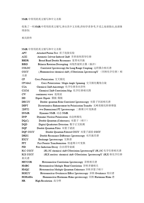
NMR中常用的英文缩写和中文名称收集了一些NMR中常用的英文缩写,译出其中文名称,供初学者参考,不妥之处请指出,也请继续添加.相关附件NMR中常用的英文缩写和中文名称APT Attached Proton Test 质子连接实验ASIS Aromatic Solvent Induced Shift 芳香溶剂诱导位移BBDR Broad Band Double Resonance 宽带双共振BIRD Bilinear Rotation Decoupling 双线性旋转去偶(脉冲)COLOC Correlated Spectroscopy for Long Range Coupling 远程偶合相关谱COSY ( Homonuclear chemical shift ) COrrelation SpectroscopY (同核化学位移)相关谱CP Cross Polarization 交叉极化CP/MAS Cross Polarization / Magic Angle Spinning 交叉极化魔角自旋CSA Chemical Shift Anisotropy 化学位移各向异性CSCM Chemical Shift Correlation Map 化学位移相关图CW continuous wave 连续波DD Dipole-Dipole 偶极-偶极DECSY Double-quantum Echo Correlated Spectroscopy 双量子回波相关谱DEPT Distortionless Enhancement by Polarization Transfer 无畸变极化转移增强2DFTS two Dimensional FT Spectroscopy 二维傅立叶变换谱DNMR Dynamic NMR 动态NMRDNP Dynamic Nuclear Polarization 动态核极化DQ(C) Double Quantum (Coherence) 双量子(相干)DQD Digital Quadrature Detection 数字正交检测DQF Double Quantum Filter 双量子滤波DQF-COSY Double Quantum Filtered COSY 双量子滤波COSYDRDS Double Resonance Difference Spectroscopy 双共振差谱EXSY Exchange Spectroscopy 交换谱FFT Fast Fourier Transformation 快速傅立叶变换FID Free Induction Decay 自由诱导衰减H,C-COSY 1H,13C chemical-shift COrrelation SpectroscopY 1H,13C化学位移相关谱H,X-COSY 1H,X-nucleus chemical-shift COrrelation SpectroscopY 1H,X-核化学位移相关谱HETCOR Heteronuclear Correlation Spectroscopy 异核相关谱HMBC Heteronuclear Multiple-Bond Correlation 异核多键相关HMQC Heteronuclear Multiple Quantum Coherence异核多量子相干HOESY Heteronuclear Overhauser Effect Spectroscopy 异核Overhause效应谱HOHAHA Homonuclear Hartmann-Hahn spectroscopy 同核Hartmann-Hahn谱HR High Resolution 高分辨HSQC Heteronuclear Single Quantum Coherence 异核单量子相干INADEQUATE Incredible Natural Abundance Double Quantum Transfer Experiment 稀核双量子转移实验(简称双量子实验,或双量子谱)INDOR Internuclear Double Resonance 核间双共振INEPT Insensitive Nuclei Enhanced by Polarization 非灵敏核极化转移增强INVERSE H,X correlation via 1H detection 检测1H的H,X核相关IR Inversion-Recovery 反(翻)转回复JRES J-resolved spectroscopy J-分解谱LIS Lanthanide (chemical shift reagent ) Induced Shift 镧系(化学位移试剂)诱导位移LSR Lanthanide Shift Reagent 镧系位移试剂MAS Magic-Angle Spinning 魔角自旋MQ(C) Multiple-Quantum ( Coherence ) 多量子(相干)MQF Multiple-Quantum Filter 多量子滤波MQMAS Multiple-Quantum Magic-Angle Spinning 多量子魔角自旋MQS Multi Quantum Spectroscopy 多量子谱NMR Nuclear Magnetic Resonance 核磁共振NOE Nuclear Overhauser Effect 核Overhauser效应(NOE)NOESY Nuclear Overhauser Effect Spectroscopy 二维NOE谱NQR Nuclear Quadrupole Resonance 核四极共振PFG Pulsed Gradient Field 脉冲梯度场PGSE Pulsed Gradient Spin Echo 脉冲梯度自旋回波PRFT Partially Relaxed Fourier Transform 部分弛豫傅立叶变换PSD Phase-sensitive Detection 相敏检测PW Pulse Width 脉宽RCT Relayed Coherence Transfer 接力相干转移RECSY Multistep Relayed Coherence Spectroscopy 多步接力相干谱REDOR Rotational Echo Double Resonance 旋转回波双共振RELAY Relayed Correlation Spectroscopy 接力相关谱RF Radio Frequency 射频ROESY Rotating Frame Overhauser Effect Spectroscopy 旋转坐标系NOE谱ROTO ROESY-TOCSY Relay ROESY-TOCSY 接力谱SC Scalar Coupling 标量偶合SDDS Spin Decoupling Difference Spectroscopy 自旋去偶差谱SE Spin Echo 自旋回波SECSY Spin-Echo Correlated Spectroscopy自旋回波相关谱SEDOR Spin Echo Double Resonance 自旋回波双共振SEFT Spin-Echo Fourier Transform Spectroscopy (with J modulation) (J-调制)自旋回波傅立叶变换谱SELINCOR Selective Inverse Correlation 选择性反相关SELINQUATE Selective INADEQUA TE 选择性双量子(实验)SFORD Single Frequency Off-Resonance Decoupling 单频偏共振去偶SNR or S/N Signal-to-noise Ratio 信/ 燥比SQF Single-Quantum Filter 单量子滤波SR Saturation-Recovery 饱和恢复TCF Time Correlation Function 时间相关涵数TOCSY Total Correlation Spectroscopy 全(总)相关谱TORO TOCSY-ROESY Relay TOCSY-ROESY接力TQF Triple-Quantum Filter 三量子滤波WALTZ-16 A broadband decoupling sequence 宽带去偶序列WATERGATE Water suppression pulse sequence 水峰压制脉冲序列WEFT Water Eliminated Fourier Transform 水峰消除傅立叶变换ZQ(C) Zero-Quantum (Coherence) 零量子相干ZQF Zero-Quantum Filter 零量子滤波T1 Longitudinal (spin-lattice) relaxation time for MZ 纵向(自旋-晶格)弛豫时间T2 Transverse (spin-spin) relaxation time for Mxy 横向(自旋-自旋)弛豫时间tm mixing time 混合时间τ c rotational correlation time 旋转相关时间。
荧光分光光度法英文

荧光分光光度法英文Fluorescence SpectrophotometryFluorescence spectrophotometry is a widely used analytical technique in the fields of chemistry, biology, and medicine. It is based on the emission of light by certain molecules that have been excited by absorbing light of a specific wavelength. This emitted light is known as fluorescence and can be measured by a spectrophotometer.Principles of Fluorescence SpectrophotometryThe basic principle of fluorescence spectrophotometry is the excitation of a molecule by absorbing light at a specific wavelength. The molecule becomes excited and reaches a higher energy state. This energy is released in the form of emitted light at a longer wavelength. The emitted light is detected by a photodetector, and the signal is amplified and recorded by a computer.Advantages of Fluorescence SpectrophotometryFluorescence spectrophotometry has several advantages over other analytical techniques, such as absorbance spectrophotometry. One major advantage is high sensitivity. Fluorescence spectrophotometry can detect trace amounts of substances, which makes it ideal for research and analysis. Another advantage is selectivity. Fluorescence spectrophotometry can selectively measure certain molecules, which makes it useful in identifying specific compounds in a mixture.Applications of Fluorescence SpectrophotometryFluorescence spectrophotometry has numerous applicationsin various scientific fields. It can be used to study the structure and function of biomolecules, such as proteins and nucleic acids. It is also used in the pharmaceutical industry to develop and analyze drugs. In addition, fluorescence spectrophotometry is used in environmental monitoring, food analysis, and forensic science.ConclusionIn conclusion, fluorescence spectrophotometry is a powerful analytical technique that has revolutionized many scientific fields. Its high sensitivity and selectivity make it an indispensable tool in research and analysis. With the development of new instruments and methods, fluorescence spectrophotometry will continue to play an important role in advancing scientific knowledge.。
光谱法研究药物小分子与蛋白质大分子的相互作用的英文
Spectroscopic Study of the Interaction between Small Molecules and Large Proteins1. IntroductionThe study of drug-protein interactions is of great importance in drug discovery and development. Understanding how small molecules interact with proteins at the molecular level is crucial for the design of new and more effective drugs. Spectroscopic techniques have proven to be valuable tools in the investigation of these interactions, providing det本人led information about the binding affinity, mode of binding, and structural changes that occur upon binding.2. Spectroscopic Techniques2.1. Fluorescence SpectroscopyFluorescence spectroscopy is widely used in the study of drug-protein interactions due to its high sensitivity and selectivity. By monitoring the changes in the fluorescence emission of either the drug or the protein upon binding, valuable information about the binding affinity and the binding site can be obt本人ned. Additionally, fluorescence quenching studies can provide insights into the proximity and accessibility of specific amino acid residues in the protein's binding site.2.2. UV-Visible SpectroscopyUV-Visible spectroscopy is another powerful tool for the investigation of drug-protein interactions. This technique can be used to monitor changes in the absorption spectra of either the drug or the protein upon binding, providing information about the binding affinity and the stoichiometry of the interaction. Moreover, UV-Visible spectroscopy can be used to study the conformational changes that occur in the protein upon binding to the drug.2.3. Circular Dichroism SpectroscopyCircular dichroism spectroscopy is widely used to investigate the secondary structure of proteins and to monitor conformational changes upon ligand binding. By analyzing the changes in the CD spectra of the protein in the presence of the drug, valuable information about the structural changes induced by the binding can be obt本人ned.2.4. Nuclear Magnetic Resonance SpectroscopyNMR spectroscopy is a powerful technique for the investigation of drug-protein interactions at the atomic level. By analyzing the chemical shifts and the NOE signals of the protein in thepresence of the drug, det本人led information about the binding site and the mode of binding can be obt本人ned. Additionally, NMR can provide insights into the dynamics of the protein upon binding to the drug.3. Applications3.1. Drug DiscoverySpectroscopic studies of drug-protein interactions play a crucial role in drug discovery, providing valuable information about the binding affinity, selectivity, and mode of action of potential drug candidates. By understanding how small molecules interact with their target proteins, researchers can design more potent and specific drugs with fewer side effects.3.2. Protein EngineeringSpectroscopic techniques can also be used to study the effects of mutations and modifications on the binding affinity and specificity of proteins. By analyzing the binding of small molecules to wild-type and mutant proteins, valuable insights into the structure-function relationship of proteins can be obt本人ned.3.3. Biophysical StudiesSpectroscopic studies of drug-protein interactions are also valuable for the characterization of protein-ligandplexes, providing insights into the thermodynamics and kinetics of the binding process. Additionally, these studies can be used to investigate the effects of environmental factors, such as pH, temperature, and ionic strength, on the stability and binding affinity of theplexes.4. Challenges and Future DirectionsWhile spectroscopic techniques have greatly contributed to our understanding of drug-protein interactions, there are still challenges that need to be addressed. For instance, the study of membrane proteins and protein-protein interactions using spectroscopic techniques rem本人ns challenging due to theplexity and heterogeneity of these systems. Additionally, the development of new spectroscopic methods and the integration of spectroscopy with other biophysical andputational approaches will further advance our understanding of drug-protein interactions.In conclusion, spectroscopic studies of drug-protein interactions have greatly contributed to our understanding of how small molecules interact with proteins at the molecular level. Byproviding det本人led information about the binding affinity, mode of binding, and structural changes that occur upon binding, spectroscopic techniques have be valuable tools in drug discovery, protein engineering, and biophysical studies. As technology continues to advance, spectroscopy will play an increasingly important role in the study of drug-protein interactions, leading to the development of more effective and targeted therapeutics.。
Spectroscopy and Spectral Analysis

Spectroscopy and Spectral AnalysisSpectroscopy is a branch of science that deals with the study of the interaction between matter and electromagnetic radiation. Spectral analysis is a technique that is used to identify and measure the properties of substances based on the electromagnetic radiation that they emit, absorb, or scatter. The study of spectroscopy and spectral analysis is essential to many fields, including chemistry, physics, environmental science, and biomedical research.Types of SpectroscopyThere are several types of spectroscopy, each based on the type of electromagnetic radiation used. The most common types of spectroscopy include:1. Absorption SpectroscopyAbsorption spectroscopy is a technique that measures the amount of radiation absorbed by a sample. This type of spectroscopy is used to identify the chemical composition and concentration of a substance. Absorption spectroscopy can be used in the ultraviolet, visible, and infrared regions of the electromagnetic spectrum.2. Emission SpectroscopyEmission spectroscopy measures the amount of radiation emitted by a substance. This type of spectroscopy is used to identify the chemical composition of a substance and the temperature and pressure of the environment. Emission spectroscopy can be used in the ultraviolet, visible, and infrared regions of the electromagnetic spectrum.3. Fluorescence SpectroscopyFluorescence spectroscopy is a technique that measures the amount of radiation emitted by a substance when it is excited by light of a particular wavelength. This type of spectroscopy is used to identify the presence of certain substances in a sample, such as proteins and DNA molecules. Fluorescence spectroscopy can be used in the ultraviolet and visible regions of the electromagnetic spectrum.4. Raman SpectroscopyRaman spectroscopy is a technique that measures the scattered radiation produced when a sample is irradiated with a laser beam. This type of spectroscopy is used to identify the chemical composition and structure of a substance. Raman spectroscopy can be used in the visible and near-infrared regions of the electromagnetic spectrum.Applications of Spectroscopy and spectral analysis have a wide range of applications in various fields, including:1. ChemistrySpectroscopy is used extensively in chemistry to identify the chemical composition and properties of substances. Spectroscopy is used to determine the purity of a substance, study chemical reactions, and analyze the structure of molecules.2. PhysicsIn physics, spectroscopy is used to study the properties of materials, such as their electronic and magnetic properties. Spectroscopy is used to study the interactions between atoms and molecules and to investigate the behavior of quantum systems.3. Environmental ScienceSpectroscopy is used in environmental science to study the properties of soil, water, and air. Spectroscopy can be used to identify pollutants in the environment and to monitor the quality of drinking water and industrial wastewater.4. Biomedical ResearchIn biomedical research, spectroscopy is used to study the properties of biological molecules, such as proteins and DNA. Spectroscopy is used to image and diagnose diseases, such as cancer, and to monitor the effectiveness of treatments.ConclusionSpectroscopy and spectral analysis are powerful tools for studying the properties of matter and electromagnetic radiation. There are several types of spectroscopy, each with its own strengths and applications. Spectroscopy and spectral analysis are used in many fields, including chemistry, physics, environmental science, and biomedical research, and have a wide range of applications.。
应用波谱学 英文
应用波谱学英文Applications of spectroscopySpectroscopy has a wide range of applications across various scientific disciplines. Some of the common applications of spectroscopy include:1. Chemistry: Spectroscopy is extensively used in chemistry for the identification and analysis of chemical compounds. It helps in determining the chemical composition, molecular structure, and functional groups present in a sample.2. Pharmaceuticals: Spectroscopic techniques are crucial in the drug discovery and development process. They are used for quality control, impurity analysis, and determining the stability of pharmaceutical products.3. Environmental science: Spectroscopy plays a vital role in environmental monitoring and assessment. It is used to evaluate air quality, analyze water pollutants, and identify harmful substances in soil samples.4. Biochemistry and molecular biology: Spectroscopy is employed in studying the structure, function, and dynamics of biological molecules like proteins, nucleic acids, and carbohydrates. Techniques such as UV-Visible spectroscopy, fluorescence spectroscopy, and circular dichroism spectroscopy are commonly used in this field.5. Material science: Spectroscopy helps in characterizing andstudying various materials and their properties. It is used to analyze the composition, crystal structure, and surface properties of materials such as metals, ceramics, polymers, and semiconductors.6. Astronomy: Spectroscopy is fundamental in studying the properties and composition of celestial objects. Astronomers use spectroscopic techniques to analyze the light emitted or absorbed by stars, galaxies, and other astronomical phenomena to determine their chemical composition, temperature, and motion.7. Forensics: Spectroscopic methods are employed in forensic science for the detection and analysis of trace evidence, such as drugs, explosives, and chemical residues. They are also used in analyzing questioned documents and for the identification of counterfeit or forged materials.8. Food science and agriculture: Spectroscopic techniques are used for analyzing food products, determining their quality, and detecting adulteration. They are also employed in agricultural research for monitoring plant health and analyzing soil fertility. These are just a few examples of the diverse applications of spectroscopy in various fields. Overall, spectroscopy is a powerful analytical tool that enables scientists to study and understand the properties and behavior of substances in a wide range of scientific domains.。
spectroscopy 第四讲
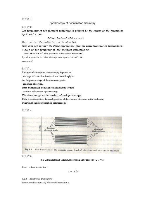
幻灯片1Spectroscopy of Coordination Chemistry幻灯片2The frequency of the absorbed radiation is related to the energy of the transition by Plank’s Law:Efinal-Einitial =E=hν= hc/λWhen exists, the radiation can be absorbed;When does not satisfy the Plank expression, then the radiation will be transmitted.A plot of the frequency of the incident radiation vs.some measure of the percent radiation absorbedby the sample is the absorption spectrum of thecompound.幻灯片3The type of absorption spectroscopy depends onthe type of transition involved and accordingly onthe frequency range of the electromagneticradiation absorbed.If the transition is from one rotation energy level toanother, microwave spectroscopy;Vibrational energy level to another, infrared spectroscopy;If the transition alters the configuration of the valence electrons in the molecule,Ultraviolet-visible absorption spectroscopy幻灯片4幻灯片53.1 Ultraviolet and Visible Absorption Spectroscopy (UV-Vis)Beer’s Law states that:A = εbc3.1.1 Electronic TransitionsThere are three types of electronic transition :1. Transition involving π,σand n electrons;2. charge-transfer electrons3. d and f electrons.幻灯片63.1.2 Absorbing Species Containing π,σand n electronsSince there are superposition of different transition,a continuous absorption band appears.幻灯片7●σσ* Transitions●Transition from a bonding σorbital to the corresponding antibonding orbital (energyis generally large).●CH4 (125 nm) not seen in typical UV-Vis region (200-700 nm).●n σ* Transitions●Saturated compounds containing atoms with lone pairs (non-bonding electrons) arecapable of this type of transitions. In the range of 150-250 nm.●n π* and ππ* Transitions●Most organic compounds have the transitions of n or πelectrons to●the π* excited state and fall in the range of 200-700 nm.●Unsaturated groups providing the πelectrons.●ε= 10 to 100 L·mol-1·cm-1 for n π* transitions.●ε= 1000 to 10000 L·mol-1·cm-1 for ππ* transitions●Solvent effects: blue shift for n π* transitions and red shift for●ππ* transitions with increasing solvent polarity.幻灯片8幻灯片9Charge-Transfer AbsorptionA number of inorganic compounds show charge-transfer absorption.For a complex, if one of its components has electron donating properties and another component can accept electrons.The absorption involves the electron transitions from donor orbital to acceptor orbital. ε> 10000 L·mol-1·cm-1for examples: KMnO4, K2Cr2O7.3.1.3 Electronic Absorption Spectrum of Coordination ComplexThree kinds of electronic transitions: d d transition; MLCT andLMCT(metal-to-ligand charge transfer and ligand-to-metal charge transfer); LC (ligand centered transitions).幻灯片10● d d transitions●According to the selection rules, some transitions are strong (Td●complexes), and others are weak (Oh complexs).●Taking the hydrogen atom as an example:In which the 1s 2p transition is allowed,whereas the 1s 2s transition is “symmetryForbidden”. The reason is that the hydrogen atom possesses a center of inversion.The SALC (Symmetry Adapted Linear Combination)stated that if there is a inversion centre, we require the initial and final states have different parity.Then for a Oh complex, which has an inversion centre, the d d transition is forbidden. Meanwhile, for a Td compound, the d d transition is allowed. Therefore, the d d adsorption intensity of Td complexes is much higher than in Oh complex.We can still observe some d d adsorption in Oh complexes, this is due to the break of Oh symmetry.幻灯片11For example, when a Oh complex vibrate and in some cases the inversion centre does not exist. When this asymmetry is present, a weak absorption is present.This weak relaxation of the Laporte selection rule is known as vibronic(振动) coupling because it arises from the interaction of vibrational modes with the electronic transition modes.This weak absorptions fall in the visible region, which can be used in the explanation of coordination complexes’ colors.(2) MLCT or LMCT transitionThese transitions generally occur in the complexes which involved the metal centered dπground state and ligand π * states and the transitions can be observed in the visible region.For example, for a d6 octahedral metal complex, the molecularorbital diagram is in the following:幻灯片12From the Fig. 3.4, it is clear that the HOMO orbital is predominantlymetal dπorbital based and the LOMO orbital is predominantly ligandπ* orbital based. Normally, π-acceptors ligands will present a low-lying π* orbital and in the same time stabilize the dπorbital centeredon the metal by retro-coordination.Light absorber molecule.幻灯片13The major electronic transition that occur in d6 metal complexeswith unsaturated ligands are ligand based n π*, ππ*, MLCT and LF( ligand field transition) (Fig. 3.5).The intensity of a transition is determined by selection rule.both Laporte and spin rules.(1)Ligand ππ* transition and MLCT are both rules allowed, theεis 103 ~105 L·mol-1·cm-1.(2) LF is spin allowed but Laporte forbidden,εis 102 ~103 L·mol-1·cm-1.幻灯片14The compound [Ru(bpy)3]2+ is a photostable compound(τ=640 nsand emission quantum yield Φ= 0.062). The electronic absorptionspectrum of [Ru(bpy)3]2+ is:幻灯片153.1.4 InstrumentUV-Vis spectrometer:Lamp: a deuterium discharge lamp for UV measurement and a tungsten-halogen lamp for visible and NIR measurements.a normal UV-Vis 190~900 nm. Nitrogen, vacuum detector.幻灯片163.2 Infrared Spectroscopy● 3.2.1 Several types of molecular motion●Translational motion●move through space in some arbitrary direction with a particular velocity.●Rotational motion●rotate about some internal axis.●Vibrational motion●the molecule may vibrate. As shown in Fig. 3.9, a polyatomic molecule has total3N freedom. When abstracting the 3 translational and 3 rotational degrees of freedom, the vibrational freedom is 3N-6.幻灯片17For water molecule, the vibrational freedom is 3*3 –6 = 3.Each of the vibrational motions of a molecule occurs with a certainFrequency, which is characteristic of the molecule and of the particularvibration. The energy involved in particular vibration is characterizedby the amplitude of the vibration, so that the higher the vibrationalEnergy, the larger the amplitude of the motion.Since most vibrational motions in molecules occur at 1014 sec-1. thenLight of wavelength = 3μm will be required to cause transition fromone vibrational energy level to another. This wavelength lies in the so-called infrared region of the spectrum. So, IR spectroscopy deals withvibrational motion of the molecules. Vibrational spectroscopy.幻灯片18● 3.2.2 Application of IR●IR spectrum of organic molecules could be divided into three regions: ●4000 ~ 1300 cm-1 (specific functional groups and bond types);●1300~ 909 cm-1 (the fingerprint region);●909 ~ 605 cm-1( the presence of benzene rings).幻灯片19幻灯片20Here, we consider the IR spectroscopy of inorganic compounds.For example: KNO2, in its lattice, the K+ and NO2- is independently arranged. Therefore, we consider only the NO2- anion (the K+ ionhas no vibrational motion).the nitrite anion has 3 vibrational freedom.One is symmetric stretch at 1335 cm-1,Another is asymmetric stretch at 1250 cm-1,The last one is bending vibration at 830cm-1. and their frequencies are almostSame regardless of the counter ion. So, it can be used for the diagnosis the presence of nitrite ions in a compound.幻灯片21For another salt, NaNO3, it is more complex.For another salt, NaNO3, it is more complex.It should have 3x4-6 = 6 vibrational modes.But its IR spectra exhibit only threeabsorption band centered at 831, 1405 and692 cm-1. it is that the symmetricstretching is not IR active. The reason forthis is that this type of motion gives no rise to the change of thedipole moment of the ion.Among the remaining 5, there are two sets of doubly degeneratevibrations, that is each 2 motions has one band in IR.The IR spectra for some of the more common ions are listed in thefollowing table.幻灯片22幻灯片23The IR absorption bands listed in the above table are for the free ions. When they coordinated with metal ions, the absorption peaks will move. For nitrite anion, it has at least two coordination modes:When coordinated, there willbe an increase of the vibrationalfrequency for the nitrite ionin the order: N-single bondedO (in O-bonded) < NO (in N-bonded) < N-double bond-O(in O-bonded) .幻灯片24In agreement with this, it has been found that in complexes in which NO2- is bonded through oxygen, the two N-O stretching frequencies lie in the ranges 1500~1400 cm-1 for N=O and 1100-1000 cm-1 for N-O.In complexes in which NO2- is bonded through nitrogen, the bands occur at similar frequencies which are intermediate between the range above; namely, 1340~1300 cm-1 and 1430 ~1360 cm-1. Thus it is relatively easy to tell whether a nitrite ion is coordinated through O or N on the basis of IR whether it is coordinated.幻灯片25For nitrate complexes, the nitrate can coordinated with metal atomin the following ways:In the free nitrate ion, thethree oxygen atoms areidentical, but no longeridentical in the coordinatednitrate ion.In all three cases, two of theoxygen atoms are identicaland the third one is unique.we say that the AB3 typeion is to change to an AB2C type species. Then the IR inactive symmetric stretching mode for the free nitrate ion (AB3) type becomes IR active when coordinated to metal atoms(AB2C).幻灯片26Similarly, the doubly degenerated asymmetric stretch in AB3becomes two asymmetric stretch with different energy in AB2C.Free nitrate ion: single band; coordinated nitrate ion: two bands.The more symmetrical a molecules or ion is, the fewer the number of bands that will appearin the IR spectrum.幻灯片27principle of Raman spectroscopyRayleigh散射:弹性碰撞;无能量交换,仅改变方向;Raman散射:非弹性碰撞;方向改变且有能量交换;Rayleigh散射Raman散射E0基态,E1振动激发态;E0 + h0 ,E1 + h0 激发虚态;获得能量后,跃迁到激发虚态.(1928年印度物理学家Raman C V 发现;1960年快速发展)幻灯片28基本原理E0E1 V=1V=0- 激发虚态1. Raman 散射Raman 散射的两种跃迁能量差: E=h(0 - )产生stokes 线;强;基态分子多; E=h(0 + ) 产生反stokes 线;弱; Raman 位移:Raman幻灯片29 2. Raman 位移对不同物质:对同一物质: -转能级的特征物理量;定性与结构分析的依据;Raman 散射的产生:光电场E= E 分子极化率; 幻灯片30E0E1V=1V=0-ANTI-STOKES-RayleighSTOKES诱导偶极矩 = E非极性基团,对称分子;拉曼活性振动—伴随有极化率变化的振动。
Principles of Fluorescence Spectroscopy

For fluorophore
r 0.4,
20040200
P 0.5
I I11,
r P0
XMUPFS01-ITF02
Polarization and anisotropy
荧光偏振与荧光各向异性可通过以下公式相互转换:
3r P 2r
2P r 3 P
当体系中存在多种荧光体时,所测得的荧光各向异性是各 种荧光体荧光各向异性的平均值:
Three dimension spectra
ex / nm
em / nm
引自林竹光 等人的论文
20040200
XMUPFS01-ITF02
1.5.5 Fluorescence lifetime
Definition Lifetime for Single molecule: the time the molecule spends in the excited state prior to return to the ground state. Average lifetime: the average time the molecule spends in the excited state prior to return to the ground state.
20040200
XMUPFS01-ITF02
Definition
polarization z x
激发偏振器
I II I P I II I
y I I
发射偏振器
anisotropy
检测器
I II I r I II 2 I
I 0, r P 1.0
Excitation and emission spectra
红外光谱法

色散型红外光谱仪工作原理图
光源
试样池 参比池
单色器
光楔
记录器
机械装置
检测器 放大器
从光源发出的红外辐射,分成二束,一束进试样池,一束进参比池,先经斩光器周期地 切割进入单色器,即试样光束和参比光束交替进入单色器的色散光栅,若某波数的单色光不被 试样吸收,二束光的强度相等,检测器不产生信号;若有吸收,在检测器上产生一定频率的 信号。经放大器放大,进入带动记录笔和光楔的机械装置。光楔的作用是进行补偿,即减弱 参比光强,使与试样光强相等,致二束光重新处于平衡。试样对各不同波数红外辐射吸收有 多有少,则光楔相应按比例移动补偿。因而光楔改变相当于试样的透射比,以纵坐标被记录 在纸上,单色光的波数连续改变,并与记录纸移动同步,这是横坐标。最后得到透视比对波 数(波长)的红外光谱吸收曲线(光谱图)。
红外光谱的定性分析可分为官能团定性和结构分析两方面。 官能团定性:根据光谱的特征基团频率来检定物质含有哪些基 团,以确定化合物类别。 结构分析:需要由化合物的红外光谱并结合其它资料(如相对 分子质量、物理常数、紫外光谱、核磁共振波谱、质谱等)来推断 具有的化学结构。
定量分析
和其它吸收光谱分析(紫外、可见吸光光度法)一样,红 外光谱定量分析时根据物质组分的吸收峰强度来进行的。它的 依据是朗伯-比尔定律(lambert-Beer’s Law)。各种气体、 液体和固态物质,均可用红外光谱法进行定量分析。
方法名称
原子发射光谱法 原子荧光光谱法 分子荧光光谱法 分子磷光光谱法 化学发光法 X射线荧光分析
激发方式
电弧、火花、等离子炬等 高强度紫外、可见光 紫外、可见光 紫外、可见光 化学能 X射线(0.01—2.5nm)
作用物质或机理
紫外荧光光谱法英语

紫外荧光光谱法英语Ultraviolet Fluorescence Spectroscopy.Ultraviolet fluorescence spectroscopy is an analytical technique that employs the fluorescence emitted by molecules excited by ultraviolet (UV) light to characterize chemical species. This method has found widespread applications in various fields, including chemistry, biochemistry, pharmacology, and environmental science.Principles of UV Fluorescence Spectroscopy.The principle of UV fluorescence spectroscopy lies in the absorption of UV light by molecules, which then emit light at longer wavelengths, known as fluorescence. This emission occurs when the absorbed energy causes electronsin the molecules to transition from a lower energy state to an excited state. As the electrons relax back to the lower energy state, they emit radiation in the form of light. The wavelength and intensity of this emitted light arecharacteristic of the specific molecular structure and can be used for identification and quantification.Instrumentation.UV fluorescence spectroscopy requires specialized instrumentation, primarily a UV-Vis spectrophotometer with a fluorescence detector. These instruments typically consist of a light source, a monochromator to select a specific wavelength of UV light, a sample compartment, and a detector to measure the emitted fluorescence. Modern spectrophotometers often incorporate advanced features such as multi-wavelength excitation and emission scanning, which provide richer spectral information.Applications of UV Fluorescence Spectroscopy.1. Biochemical Analysis: UV fluorescence spectroscopyis widely used in biochemistry to study protein-ligand interactions, protein conformational changes, and nucleic acid structure. Fluorescent probes can be attached to specific sites on proteins or nucleic acids, allowing theirbehavior to be monitored under different conditions.2. Drug Discovery and Pharmacology: This technique is employed in drug discovery to screen potential drugs for their binding affinity to biological targets. By monitoring the changes in fluorescence upon drug binding, researchers can assess the affinity and selectivity of drugs.3. Environmental Science: UV fluorescence spectroscopy has been used to monitor pollutants in water and air. Fluorescent tracers can be used to trace the fate and transport of pollutants, providing insights into environmental contamination and remediation.4. Materials Science: In materials science, UV fluorescence spectroscopy is used to study the optical properties of materials, such as quantum dots and fluorescent dyes. This technique can provide information about the energy levels and electronic states of these materials, which is crucial for their applications in optoelectronic devices.Advantages and Limitations.Advantages:High Sensitivity: UV fluorescence spectroscopy can detect very low concentrations of fluorescent species, making it suitable for trace analysis.Selectivity: By choosing specific excitation and emission wavelengths, UV fluorescence spectroscopy can provide information about specific components in complex mixtures.Non-Destructive: This technique does not require the destruction of samples, allowing multiple measurements to be performed on the same sample.Limitations:Fluorescent Probe Dependence: The application of UV fluorescence spectroscopy often relies on the availability of suitable fluorescent probes or dyes. Not all moleculesexhibit strong fluorescence, limiting the scope of this technique.Interference from Background Fluorescence: The presence of background fluorescence from the sample matrixor solvents can interfere with the measurement, affecting the accuracy and reliability of results.Instrument Cost and Maintenance: Specialized UV-Vis spectrophotometers with fluorescence detection capabilities can be costly, and regular maintenance is required toensure accurate measurements.Conclusion.UV fluorescence spectroscopy is a powerful analytical tool that has found widespread applications in various fields. Its ability to provide sensitive and selective information about molecular structure and interactions has made it a valuable resource for researchers in biochemistry, pharmacology, environmental science, and materials science. Despite its limitations, UV fluorescence spectroscopycontinues to evolve and improve, providing new insights into the behavior and properties of chemical species.。
游离脂肪酸谱

游离脂肪酸谱游离脂肪酸谱(freefattyacidspectra)是指从植物、动物或微生物中分离出来的脂肪酸混合物的光谱。
它是一种实验室中常用的分析技术,用于研究脂肪酸结构和性质,以及它们之间的相互作用。
这一方法主要基于分光光度法(spectrophotometric),需要运用色谱技术(chromatographic),如高效液相色谱(high-performance liquid chromatography)、气相色谱(gas chromatography)、液色谱(liquid chromatography)等。
游离脂肪酸谱对于探究脂肪酸的结构和性质非常重要,因为它们具有结构复杂性,即不同的脂肪酸具有不同的酸值、表示不同的物质性质,且在生物体中具有不同的生理作用。
脂肪酸的性质受它们的分子长度、空间结构、疏水性、能量以及各种活性位点等因素的影响,因此,对脂肪酸结构、性质、以及它们之间的相互作用进行深入的研究就显得尤为重要。
游离脂肪酸谱的实验过程主要分为以下几个步骤:首先,从植物、动物或微生物中分离出来的游离脂肪酸混合物;其次,采用色谱技术,如高效液相色谱、气相色谱等,对其进行分离、浓缩和提纯;第三,运用分光光度测定方法,构建脂肪酸谱;最后,用谱图来分析不同脂肪酸之间的结构和性质,以此来研究它们之间的相互作用。
游离脂肪酸谱技术被广泛应用于基础研究以及特定领域,如食品科学和生物技术等。
它有助于研究脂肪酸营养学以及对于保护身体健康具有重要意义的脂肪酸组成、所谓的“有益脂肪酸”,如有机酸、鱼油、核素等。
此外,游离脂肪酸谱技术还可用于指示和调控抗生素、化肥、农业植物激素等药物疗效的分子机制,对于调控这些药物的结构和性质也有重要的借鉴意义。
综上所述,游离脂肪酸谱技术是研究脂肪酸结构、性质以及它们之间的相互作用的一种重要的实验技术,在食品科学、生物技术、医学等多个领域都有着其重要的应用价值。
它可以帮助我们更好地了解脂肪酸,实现有效的调控和抗生素疗效,从而更好地保护身体健康。
原子吸收光谱的英文
原子吸收光谱的英文原子吸收光谱的英文是"atomic absorption spectroscopy"。
这是一种分析技术,用于测量和确定样品中各种金属元素的浓度。
这种技术基于原子的能级跃迁,当样品中的金属离子吸收特定波长的光时,其能级会从低能级跃迁至高能级,从而生成吸收光谱。
以下是23句中英双语例句:1. Atomic absorption spectroscopy is commonly used in analytical chemistry to determine the concentration of metal ions in samples.原子吸收光谱常用于分析化学中,用于测定样品中金属离子的浓度。
2. The atomic absorption spectrum of each metal elementis unique and can be used for identification purposes.每种金属元素的原子吸收光谱是独特的,可用于鉴定目的。
3. Atomic absorption spectroscopy is a sensitive method for detecting trace amounts of metals in environmental samples.原子吸收光谱是一种敏感的方法,可用于检测环境样品中微量金属。
4. The technique of atomic absorption spectroscopy involves the use of a specific light source, such as ahollow-cathode lamp.原子吸收光谱技术涉及使用特定的光源,例如中空阴极灯。
5. Atomic absorption spectroscopy can be used in various industries, including pharmaceutical, environmental, and food industries.原子吸收光谱可应用于各个行业,包括制药、环境和食品行业。
荧光光谱法英文

荧光光谱法英文Fluorescence SpectroscopyFluorescence spectroscopy is a powerful analytical technique that has found widespread applications in various fields, including chemistry, biology, materials science, and environmental studies. This analytical method is based on the measurement of the emission of light by a substance that has been excited by the absorption of light or other forms of energy. The process of fluorescence involves the absorption of energy by molecules or atoms, followed by the subsequent emission of light at a longer wavelength than the absorbed light.The fundamental principle of fluorescence spectroscopy is that when a molecule or atom is exposed to light, it can absorb the energy of the incoming photons, causing electrons within the molecule or atom to be excited to higher energy levels. This excitation is a temporary state, and the electrons will eventually return to their ground state, releasing the excess energy in the form of a photon. The energy of the emitted photon is typically lower than the energy of the absorbed photon, resulting in a shift in the wavelength of the emitted light compared to the absorbed light. This wavelength shift is known as the Stokes shift, and it is a key characteristic offluorescence.The intensity and wavelength of the emitted light are influenced by various factors, such as the chemical structure of the fluorescent molecule, the solvent environment, temperature, and the presence of other compounds that can interact with the excited molecules. By analyzing the characteristics of the emitted light, researchers can gain valuable insights into the properties and behavior of the sample under investigation.Fluorescence spectroscopy has a wide range of applications in various fields. In chemistry, it is used for the identification and quantification of organic and inorganic compounds, as well as the study of reaction kinetics and molecular interactions. In biology, fluorescence spectroscopy is employed for the investigation of protein structure and dynamics, the detection and quantification of biomolecules, and the study of cellular processes. In materials science, this technique is used to characterize the properties of polymers, semiconductors, and nanomaterials, among others.One of the key advantages of fluorescence spectroscopy is its high sensitivity, which allows for the detection and quantification of analytes at very low concentrations. Additionally, the technique is non-invasive and can be performed in real-time, making it a valuable tool for in-situ and online monitoring applications. Furthermore, thedevelopment of advanced fluorescent probes and labeling techniques has expanded the versatility of fluorescence spectroscopy, enabling the visualization and tracking of specific molecules or cellular components in complex biological systems.Despite its many benefits, fluorescence spectroscopy also faces some limitations. The presence of interfering compounds, quenching effects, and the potential for photobleaching of the fluorescent molecules can challenge the reliability and accuracy of the measurements. Researchers are constantly working to address these challenges through the development of new instrumentation, data analysis methods, and sample preparation techniques.In conclusion, fluorescence spectroscopy is a powerful and versatile analytical tool that has made significant contributions to various scientific disciplines. As technology continues to advance, the applications of this technique are expected to expand further, providing researchers with new opportunities to gain a deeper understanding of the world around us.。
分子光谱学研究 英语
分子光谱学研究英语Molecular spectroscopy is a branch of spectroscopy that deals with the interaction of matter and electromagnetic radiation in the molecular level. It involves the study of the absorption, emission, and transmission of light by molecules, and the information obtained from these interactions can be used to determine the structure, energy states, and dynamics of molecules. Molecular spectroscopy plays a crucial role in various fields such as chemistry, physics, biology, and materials science.The fundamental principle of molecular spectroscopy is the interaction between electromagnetic radiation and matter, which leads to transitions between different energy states of the molecules. These transitions can be electronic, vibrational, or rotational, depending on the type of radiation involved and the nature of the molecular system.Electronic spectroscopy involves transitions betweendifferent electronic states of a molecule, which are typically separated by large energy gaps. This type of spectroscopy is commonly used to study the electronic structure of molecules, including their bonding, electronic configuration, and excited states. UV-visible spectroscopy and infrared spectroscopy are examples of electronic spectroscopy techniques.Vibrational spectroscopy involves transitions between different vibrational states of a molecule, which are typically separated by smaller energy gaps. This type of spectroscopy is useful for studying the vibrational modes of molecules and their interactions with other molecules or their environment. Techniques such as infrared spectroscopy and Raman spectroscopy are commonly used for vibrational spectroscopy.Rotational spectroscopy involves transitions between different rotational states of a molecule, which are separated by even smaller energy gaps. This type of spectroscopy is useful for studying the rotational motion of molecules and their interactions with other molecules ortheir environment. Microwave spectroscopy is a common technique used for rotational spectroscopy.Molecular spectroscopy has a wide range of applications in various fields. In chemistry, it is used to determine the structure and composition of molecules, to identify unknown compounds, and to study the mechanisms of chemical reactions. In physics, molecular spectroscopy is used to understand the quantum mechanical properties of molecules and their interactions with electromagnetic radiation. In biology, it is used to study the structure and function of biological molecules such as proteins and nucleic acids. In materials science, molecular spectroscopy can be used to characterize the properties of materials and to understand their interactions with light.In addition to its fundamental importance in understanding the interaction of matter and electromagnetic radiation, molecular spectroscopy has numerous practical applications. For example, it is used in the development of new materials and technologies, such as solar cells, LEDs, and lasers. It is also used in environmental science tomonitor pollution and to detect harmful chemicals. In medicine, molecular spectroscopy is used in diagnostictests and in the development of new drugs and treatments.In conclusion, molecular spectroscopy is a crucialfield of study that has applications in various disciplines. It provides insights into the structure, dynamics, and interactions of molecules, and has the potential to lead to new discoveries and technological advancements in many fields.。
仪器分析英文缩写1

仪器分析方法英文缩写AAS 原子吸收光谱法Atomic Absorption SpectrometryAES 原子发射光谱法Atomic Emission SpectroscopyAFS 原子荧光光谱法Atomic Fluorescence SpectrometryASV 阳极溶出伏安法anodic stripping voltammetryATR 衰减全反射法Attenuated Total ReflectionAUES 俄歇电子能谱法Auger electron spectroscopyCEP 毛细管电泳法capillary electrophoresisCGC 毛细管气相色谱法capillary gas chromatographyCIMS 化学电离质谱法chemical ionization mass spectrometryCIP 毛细管等速电泳法capillaryisotachor-phoresisCLC 毛细管液相色谱法capillary liquid chromatographyCSFC 毛细管超临界流体色谱法Capillary supercritical fluid chromatography CSFE 毛细管超临界流体萃取法Capillary of supercritical fluid extractionCSV 阴极溶出伏安法cathodic stripping voltammetryCZEP 毛细管区带电泳法capillary zone electrophoresisDDTA 导数差热分析法Derivative differential thermal analysisDIA 注入量焓测定法Determination of enthalpy injection quantity DPASV 差示脉冲阳极溶出伏安法Differential pulse anodic stripping voltammetry DPCSV 差示脉冲阴极溶出伏安法Differential pulse cathodic stripping voltammetry DPP 差示脉冲极谱法differential pulse polarographyDPSV 差示脉冲溶出伏安法Differential pulse stripping voltammetryDPV A 差示脉冲伏安法Differential pulse voltammetryDSC 差示扫描量热法differential scanning calorimetryDTA 差热分析法differential thermal analysisDTG 差热重量分析法differential thermogravimetric analysis EAAS 电热或石墨炉原子吸收光谱法Electric heating or graphite furnace atomic absorptionspectrometryETA 酶免疫测定法enzyme immunoassayEIMS 电子碰撞质谱法The electron impact mass spectrometry ELISA 酶标记免疫吸附测定法Enzyme labeled immunosorbent assay EMAP 电子显微放射自显影法electron microscope autoradiographyEMIT 酶发大免疫测定法Enzyme immune assay hairEPMA 电子探针X射线微量分析法Electron probe X ray trace analysisESCA 化学分析用电子能谱学法Spectral method by electron chemical analysis ESP 萃取分光光度法extraction spectrophotometryFAAS 火焰原子吸收光谱法flame atomic absorption spectrometry FABMS 快速原子轰击质谱法Fast atom bombardment mass spectrometryFAES 火焰原子发射光谱法flame atomic emission spectrometryFDMS 场解析质谱法Field desorption mass spectrometryFIA 流动注射分析法flow injection analysisFIMS 场电离质谱法Field ionization mass spectrometryFNAA 快中心活化分析法Analysis of fast activation centerFT-IR 傅里叶变换红外光谱法Fourier Transform Infrared SpectrometryFT-NMR 傅里叶变换核磁共振谱法fourier transform-nmr spectrometryFT-MS 傅里叶变换质谱法fourier transform mass spectrometryGC 气相色谱法gas chromatographyGC-IR 气相色谱-红外光谱法gas chromatography-infrared spectroscopyGC-MS 气相色谱-质谱法gas chromatography-mass-spectrographyGD-AAS 辉光放电原子吸收光谱法Glow discharge atomic absorption spectrometryGD-AES 辉光放电原子发射光谱法Glow discharge atomic emission spectrometryGD-MS 辉光放电质谱法glow discharge mass spectrometryGFC 凝胶过滤色谱法gel filtration chromatographyGLC 气相色谱法gas chromatographyGLC-MS 气相色谱-质谱法gas chromatography/mass spectrometryHAAS 氢化物发生原子吸收光谱法hydride generation-atomic absorption spectrometryhg-aas HAES 氢化物发生原子发射光谱法Atomic emission spectrometry with Hydride Generation HPLC 高效液相色谱法high performance liquid chromatography HPTLC 高效薄层色谱法Atomic Absorption SpectrometryIBSCA 离子束光谱化学分析法Ion beam spectroscopy for chemical analysis IC 离子色谱法Ion chromatographyICP 电感耦合等离子体Inductively coupled plasmaICP-AAS 电感耦合等离子体原子吸收光谱法Inductively coupled plasma atomic absorptionspectrometryICP-AES 电感耦合等离子体原子发射光谱法Inductively coupled plasma atomic emission spectrometry ICP-MS 电感耦合等离子体质谱法Inductively coupled plasma mass spectrometryIDA 同位素稀释分析法Isotope dilution analysisIDMS 同位素稀释质谱法Isotope dilution mass spectrometryIEC 离子交换色谱法Ion exchange chromatographyINAA 仪器中子活化分析法Instrumental neutron activation analysisIPC 离子对色谱法Ion pair chromatographyIR 红外光谱法Infrared spectroscopyISE 离子选择电极法Ion selective electrode methodISFET 离子选择场效应晶体管Ion selective field effect transistorLAMMA 激光微探针质谱分析法Analysis of laser microprobe mass spectrometry LC 液相色谱法Liquid chromatographyLC-MS 液相色谱-质谱法Liquid chromatography - mass spectrometry MECC 胶束动电毛细管色谱法Micellar electrokinetic capillary chromatographyMEKC 胶束动电色谱法Micellar electrokinetic chromatographyMIP-AES 微波感应等离子体原子发射光谱法Microwave induced plasma atomic emission spectrometry MS 质谱法mass-spectrographyNAA 中子活化法neutron activationNIRS 近红外光谱法near infrared spectroscopyNMR 核磁共振波谱法nuclear magnetic resonance spectroscopyPAS 光声光谱法photoacoustic spectrometryPC 纸色谱法paper chromatographyPCE 纸色谱电泳法paper chromatoelectrophoresisPE 纸电泳法paper electrophoresisPGC 热解气相色谱法pyrolysis gas chromatographyPIGE 粒子激发Gamma射线发射光谱法Particle excitation Gamma ray emission spectrometry PIXE 粒子激发X射线发射光谱法Particle excitation X-ray emission spectrometry RHPLC 反相高效液相色谱法reversed-phase high performance liquid chromatography RHPTLC 反相液相薄层色谱法Reversed phase thin layer chromatography RIA 发射免疫分析法RadioimmunoassayRPLC 反相液相色谱法reversed phase liquid chromatographySEM 扫描电子显微镜法scanning electron microscopySFC 超临界流体色谱法supercritical fluid chromatographySFE 超临界流体萃取法supercritical fluid extractionSIMS 次级离子质谱法Secondary-ion mass spectrometrySIQMS 次级离子四极质谱法Secondary-ion quadrupole mass spectrometry SP 分光光度法spectrophotometrySP(M)E 固相(微)萃取法Solid-phase extraction (micro)STM 扫描隧道电子显微镜法scanning tunneling microscopySTEM 扫描投射电子显微镜法Scanning transmission electron microscopy SV 溶出伏安法stripping voltammetryTEM 投射电子显微镜法Transmission ElectronMicroscopeTGA 热重量分析法Thermal Gravity AnalysisTGC 薄层凝胶色谱法thin layer gel chromatographyTLC 薄层色谱法Thin-Layer ChromatographyUPS 紫外光电子光谱法Ultraviolet Photoelectron SpectroscopyUVF 紫外荧光光谱法Ultraviolet FluorescenceUVS 紫外光谱法Ultraviolet SpectrometerXES X射线发射光谱法X-ray emission spectroscopyXPS X射线光电子光谱法X-ray photoelectron spectroscopXRD X射线衍射光谱法X-Ray DiffractionXRF X 射线荧光光谱法X-ray fluorescence。
国外低能离子与有机体相互作用研究进展课件

质量沉积效应: 只要试验系统中含氮,不管来自注入 离子还是本底元素,都有一定几率形成含氮基团
He+和Ar2+辐射 含N、O、C的 混合冰的红外光 谱
N+辐射仅含 C.H、O的混合 冰的红外光谱
—— G.Strazzulla et al, NIMB 166-167 (2000) 13.
实验室模拟宇宙低能粒子辐射水 及各种简单溶液冰的研究
低能离子与生 物分子相作用
20eV以下能 量的二次电子 引起DNA链断 裂的规律
二次电子
10eV以上能量的二次 电子作用于四种碱基 分子反应截面
keV离子引起体外裸露DNA的链断裂
1keV Ar+辐射PUC18质粒DNA 1: 真空室外对照;2-4: 真空对照;5-6: 辐射4min; 7: 辐射8min; 8: 辐射15min; 9: 辐射21min; 10: 辐射32min
产生阳离子片断所需的Ar+能量阈 值为15-20eV,比产生阴性离子片断 所需能量要低。
脱氧核糖的解离产物最为丰富, 解 离产额较高, 所需的入射离子能量 阈值也较低。
对于100eV Ar+,每个入射离子可引 起6个胸腺嘧啶碱基的解离。
——Z.W.Deng et al, J.Chem.Phys.123 (2005) 144509-9.
——宇航生命支撑: 宇宙高能粒子慢化后击
中并沉积在宇航员身体细胞内,具有潜在的致突致癌
性
✓
—— 分子起源与进化: 宇宙低能粒子轰击各类
星体的冰状表面形成星际分子、近地生物突变
✓ 2000以后,一种普遍接受的观点是:
✓ 高能离子对生物体辐射损伤实际上是一系列低能 事件(甚至低至几到几十eV)的综合结果。
荧光相关光谱法
荧光相关光谱法
荧光相关光谱法(Fluorescence Correlation Spectr oscopy,简称FCS)是一种用于检测和研究荧光标记分子在溶液中的动态过程和相互作用的技术。
FCS通过测量荧光信号的强度和相关性,可以获得分子在特定时间内的浓度、扩散系数、水合动力半径、相互作用亲和力、动态构象及构象转换速率等特性。
荧光相关光谱法的基本原理是,当一个荧光标记分子被激发时,它会发出一个特定波长的荧光信号。
通过测量荧光信号的强度和相关性,可以得到关于分子动态过程和相互作用的详细信息。
FCS的应用非常广泛,包括生物学、医学、化学、材料学、药物研发、光物理学、微纳科学等领域。
在PubMed 中已有超过12900篇应用FCS及其衍生技术产出的学术论文。
荧光相关光谱法的实验过程一般包括以下几个步骤:
1.选择合适的荧光标记分子,通过激发光源激发标记分子,使其发出荧光信号。
2.收集荧光信号,并对其进行强度和相关性分析。
通过分析荧光信号的强度和相关性,可以得到分子在特定时间内的浓度、扩散系数、水合动力半径、相互作用亲和力、动态
构象及构象转换速率等特性。
3.根据实验结果,进一步研究分子之间的相互作用机制,为药物研发、生物传感、纳米技术等领域提供重要的实验依据。
总之,荧光相关光谱法是一种强大的分析工具,可以用于研究分子在溶液中的动态过程和相互作用,为各个领域的研究提供重要的实验依据。
光谱分析幻灯
光谱测定中的两个数据
透光度( ) 透光度(T)
均匀吸收介质的透过光的比例,也叫透 射比。 T=I/Io
吸光度(A) 光密度 光密度OD) 吸光度( )(光密度 )
均匀吸收介质透光度的倒数的对数。 均匀吸收介质透光度的倒数的对数。 =lg( /I) A=lg (1/T) =lg(Io/I)
(三)光学理论依据
(七)紫外可见光分光光度计的结构
光源 单色器 样品室 检测器 显示
1. 光源
在整个紫外光区或可见光谱区可以发射连续光谱, 在整个紫外光区或可见光谱区可以发射连续光谱,具 有足够的辐射强度、较好的稳定性、较长的使用寿命。 有足够的辐射强度、较好的稳定性、较长的使用寿命。 可见光区: 可见光区:钨灯作 为光源, 为光源,其辐射波长范 围在320 320~ nm。 围在320~2500 nm。 紫外区: 紫外区:氢、氘灯, 氘灯, 发射185 185~ nm的连 发射185~400 nm的连 续光谱。 续光谱。
医学实验技术基础
光谱分析
Spectral Analysis
光谱分析的概念
利用各种化学物质(包括原子、基 团、分子及高分子化合物)所具有的 发射、吸收或散射光谱系的特征,来 确定其性质、结构或含量的技术,称 为光谱分析技术。
电磁波谱
区域 γ射线 X射线 远紫外 紫外 可见 红外 远红外 微波 无线电波 波长 10-3-0.1 nm 0.1-10 nm 10-200 nm 200-400 nm 400-760 nm 0.76-50 µm 50-1000 µm 0.1-100 cm 1-1000 m
激发态
n π σ
σ,成键轨道; 成键轨道; π,成键轨道; 成键轨道; n,非成键轨道(电子对未共用) ,非成键轨道(电子对未共用) 反键轨道; σ* ,反键轨道; π*, 反键轨道; , 反键轨道;
- 1、下载文档前请自行甄别文档内容的完整性,平台不提供额外的编辑、内容补充、找答案等附加服务。
- 2、"仅部分预览"的文档,不可在线预览部分如存在完整性等问题,可反馈申请退款(可完整预览的文档不适用该条件!)。
- 3、如文档侵犯您的权益,请联系客服反馈,我们会尽快为您处理(人工客服工作时间:9:00-18:30)。
Spectroscopy of Charge Fluctuators Coupled to a Cooper Pair BoxF. C. Wellstood,1,2 Z. Kim2,3 and B. Palmer31. Joint Quantum Institute and Center for Nanophysics and Advanced Materials2. Department of Physics, University of Maryland, College Park, Maryland 207423. Laboratory for Physical Sciences, College Park, MD 20740AbstractWe analyze the behavior of a Cooper Pair Box (CPB) that is coupled to charge fluctuators that reside in the dielectric barrier layer in the box's ultra-small tunnel junction. We derive the Hamiltonian of the combined system and find the coupling between the CPB and the fluctuators as well as a coupling between the fluctuators that is due to the CPB. We then find the energy levels and transition spectrum numerically for the case of a CPB coupled to a single charge fluctuator, where we treat the fluctuator as a two-level system that tunnels between two sites. The resulting spectra show the usual transition spectra of the CPB plus distinctive transitions due to excitation of the fluctuator; the fluctuator transitions are 2-e periodic and resemble saw-tooth patterns when plotted as a function of the gate voltage applied to the box. The combined CPB-fluctuator spectra show small second-order avoided crossings with a size that depends on the gate voltage. Finally, we discuss how the microscopic parameters of the model, such as the charge times the hopping distance, the tunneling rate between the hopping sites, and the energy difference between the hopping sites, can be extracted from CPB spectra, and why this yields more information than can be found from similar spectra from phase qubits.Two-level fluctuators (TLF) of one kind or another have long been recognized as an underlying cause of resistance fluctuations in metals [1,2], critical current fluctuations in Josephson junctions [3-7], excess flux noise in SQUIDs [7-9], and telegraph noise and excess charge noise in Coulomb blockade devices [10-13]. Not surprisingly, different types of fluctuators lead to different types of fluctuations. For example, observations of excess flux noise in high-T c superconductors have revealed that the two-level fluctuators are just vortices that hop between different pinning sites [14-16]. In other cases, the microscopic nature of the fluctuators is still unclear. For example, the unusual behavior of flux noise at temperatures below 1 K appears to be inconsistent with the hopping of vortices and may be due to fluctuating electron spins [17-18]. The situation for charge and critical current fluctuators is somewhere between these extremes. After more than two decades of research, some basic information is known about the microscopic fluctuators responsible for charge and critical current fluctuations - the fluctuators appear to be moving charges or rotating electric dipoles in the insulating junction barrier or nearby dielectric layers [3] - but the precise microscopic identification of the fluctuators is not settled.There are several reasons why it has been difficult to be certain of the precise microscopic agents causing charge or critical current fluctuations. First, the effects from individual fluctuators are typically very small, especially for the cryogenic or milli-Kelvin temperatures of interest here, and this typically makes the resulting experiments quite challenging. Second, a variety of materials and processes have been used to build devices, and the presence of a fluctuator may well depend on both the materials and the fabrication technique. Third, while measurements of telegraph noise can reveal microscopic information about individual fluctuators, in many cases it is possible only to distinguish the largest one or twofluctuators in a background noise of smaller fluctuations. Compared to the number of atoms in even the smallest device, such observable discrete fluctuators are extremely rare and thus may not be representative of a typical atomic scale defect in the device. Finally, much of the experimental data obtained on fluctuators consists of relatively smooth 1/f noise power spectra. Such noise spectra arise from a distribution of many fluctuators [1,19] and it is often not possible to resolve individual fluctuators. From smooth spectra, it is difficult to determine uniquely such basic microscopic parameters as the hopping distance or the absolute number of fluctuators in a given energy range.Research in quantum computing based on superconducting devices has led to increased interest in understanding two-level fluctuators. In qubit research, fluctuators can be a serious problem because they can lead to decoherence, dissipation, inhomogenous broadening and a decrease in measurement fidelity [20-23]. With this new interest have also come new approaches to the problem. In particular, microwave spectroscopy of Josephson phase qubits and flux qubits has revealed the presence of small un-intended avoided crossings in the transition spectrum due to coupling between the device and individual two-level fluctuators [20-23]. Similar avoided crossings have also been observed recently [23] in the transition spectra of Cooper pair boxes (CPB), allowing the new spectroscopic approach to be applied to charge fluctuators in a system that is a sensitive detector of charge.Here, we examine the behavior of a Cooper pair box [25-29] that is coupled to charged two-level fluctuators and show how microscopic information can be extracted about the fluctuators. We first summarize the basic physics of the Cooper pair box. We then derive the Hamiltonian for a system where charged fluctuators are coupled to a CPB and find the energy levels numerically for the case of a single two-level fluctuator coupled to a CPB. We find thatthe resulting transition spectra show a distinct saw-tooth feature that is characteristic of the fluctuator. In addition, we find that three key model parameters for individual fluctuators (the charge times the hopping distance, the tunneling rate between the hopping sites, and the energy difference between the hopping sites) can be uniquely extracted from the spectra. Finally, we conclude with a summary of our key results and discuss how the CPB-fluctuator spectra provide additional information about the fluctuators that is not found in analogous spectra from phase qubits, suggesting that measurements of CPB-fluctuator spectra may aid in the identification of microscopic charge fluctuators.Charging Energy of the Cooper Pair BoxIn this section we summarize the energy considerations for a Cooper pair box when no charge fluctuators are present. Considering the circuit schematic of the Cooper pair box shown in Fig. 1(a), the charging energy of the capacitors in the CPB without any charge fluctuators is [30]: ()()2221021i g g i j V V C V C U −+−= (1) where C j is the capacitance of the ultra-small junction, C g is the capacitance of the gate electrode, V i is the island potential, and V g is the gate voltage. The island potential V i is determined by V g and the number n of excess Cooper pairs on the island. Each Cooper pair has charge 2e ; note that here e = -1.609x10⁻¹⁹ C is negative, as in ref. [30]. One finds for the island potential [30]:()()g g j g g j g i n n C e C C ne V C C C V −=+++=Σ22 (2) where:g j C C C +=Σ (3) is the total capacitance of the island and n g = -C g V g /e is the gate number. Also, in writing Eq. (2)we have implicitly assumed that there are no quasiparticles present.To properly describe the systems behavior, we need to take into account the work W done by the gate voltage source when the number n of Cooper pairs on the island changes. The appropriate quantity is the charging energy minus the work:W U E −=. (4) To find the work, we note that if n excess pairs have accumulated on the island, a charge of Σ−=C C ne Q gg 2 (5)will be attracted onto the gate electrode (the plate of the gate capacitor C g that is directly attached to the gate voltage source V g ). Since this charge has to be supplied by the gate voltage source at potential V g , the total work the source has to do is Q g V g and thus: ΣΣ=−=C e nn V C C ne W g g g 222. (6)After some algebraic manipulation, one finds that the charging energy minus the work is: ()2212g g j c g c n C C E n n E E ⎟⎟⎠⎞⎜⎜⎝⎛−+−= (7) where Σ=C e E c 2. Since the second term in Eq. (7) does not depend on n , it will not affect the dynamics and can be dropped [30].Analysis of a CPB when Charge Fluctuators are PresentWe now consider the case where two charge fluctuators are coupled to the Cooper pair box. The result for two charges can easily be generalized to more charges and is sufficient to reveal that the CPB will generate a coupling term between any two fluctuators. For this case, we again need to find the charging energy minus the work done by the gate voltage source. We canwrite: ()∑=−−−+=21j j 22W W 2121i g g i j V V C V C E (8)where W j is the work done by the gate voltage source when the j -th fluctuator moves. Here, W is the work done by the gate voltage source when n changes and is given by Eq. (6).To proceed, we assume that the two charge fluctuators are in the ultra-small tunnel junction barrier, i.e. in the capacitor C j , where the device is most sensitive to charge motion [31-32]. The charges, of strength Q 1 and Q 2 respectively, will induce charges Q p1 and Q p2 on the island given by [31-32]: j jpj Q d x Q −= (9)where x j is the distance of the j -th charge from the ground plate of the ultra-small junction and d is the thickness of the junction's oxide barrier [see Fig. 1(b)]. Q p1 and Q p2 are polarization charges that are localized on the island close to the charges Q 1 and Q 2. Since the net charge on the island will be constant for n constant, a compensating charge of -(Q p1+Q p2) must exist on the rest of the island, distributed proportionally over the gate capacitor and junction capacitor. This in turn induces a charge Q g on the gate electrode given by: Σ⎟⎠⎞⎜⎝⎛+−=C C Q d x Q d x Q g g 2211 (10)This charge must be delivered by the gate voltage source, and requires work: g g g j j n Q d x Q d x C e V Q W⎟⎠⎞⎜⎝⎛+==Σ=∑221121 (11) From similar considerations, we find the island potential V i is given by: []⎟⎠⎞⎜⎝⎛+++=ΣΣ2211121Q d x Q d x C ne V C C V g g i (12)From Eqs. (6), (8), (11) and (12), we find: 222112g g j c j j j g c n C C E d x e Q n n E E ⎟⎟⎠⎞⎜⎜⎝⎛−+⎟⎟⎠⎞⎜⎜⎝⎛+−=∑=. (13)Comparing Eqs. (7) and (13), we see that the effect of charge motion is similar to a shift in the gate voltage and, again, the second term in Eq. (13) has no effect on the dynamics and can be dropped.We can now construct the Hamiltonian H for the combined system by taking Eq. (13) and adding the kinetic energy of the charge fluctuator, the Josephson coupling energy of the ultra-small tunnel junction, and the local potential experienced by the charge fluctuators. We can write:122121H H H H H j j CPB j j CPB +++=∑∑=−=(14)The first term in Eq. (14):())cos(22γj g c CPB E n n E H −−= (15) is the Hamiltonian of the Cooper pair box [26] with no charge fluctuators, where E j is the Josephson energy and γ is the gauge invariant phase difference across the junction [31]. The second term in Eq. (14) is a sum over H j , where )(22222j j j j j j j r U d x C Q m p H +⎟⎟⎠⎞⎜⎜⎝⎛+=Σ (16) is the Hamiltonian of the j -th charge, which has charge Q j , position j r , momentum j p , mass m j , and moves in a local potential )(j j r U . The third term in Eq. (14):()⎟⎟⎠⎞⎜⎜⎝⎛−=−d x e Q n n E H j j g c j CPB 22 (17)is the coupling between the CPB and the j -th charge. Finally, 22121211212),(d x x C Q Q x x U H Σ+= (18)describes the interaction between the two charges. The potential U 12 accounts for any direct local interactions between the two charges. If the charge defects are few in number and randomly distributed in the dielectric, then we expect U 12 will typically be small because the junction electrodes will screen electric fields on a length scale given by d /2π ~ 0.2 nm. On the other hand, the term 22121d C x x Q Q Σ in Eq. (18) came from the charging energy, as can be seen by expanding the first term in Eq. (13). This is an indirect electrostatic interaction between two fluctuators due to the capacitor plates and does not directly depend on the separation between the charges.Two-state Model of a Charge FluctuatorTo illustrate the behavior of a CPB coupled to a charge fluctuator, we will further simplify the situation and consider a CPB that is coupled to just one fluctuator that can tunnel between two locations. In this section, we first summarize the properties of such a two-level fluctuator (TLF) when the coupling to the CPB is ignored. From Eq. (16), we can write the Hamiltonian of the fluctuator as: )(2111211r U m p H +=(19) where we have absorbed the quadratic potential term 221212d C x Q Σthat occurs in Eq. (16) intothe arbitrary potential U 1.For the charge fluctuator to behave as a two-level fluctuator, we will assume that thepotential )(11r U has two wells that the fluctuator can tunnel between [13,32], as shown in Fig. 1(c). In the absence of tunneling, we will assume the energy of the fluctuator is E a in well a and E b in well b , and that the corresponding distances from the ground electrode are x a and x b . With these simplifications, H 1 can be written as a 2x2 matrix:⎟⎟⎠⎞⎜⎜⎝⎛=b ab ab a E T T E H 1 (20) where ab T determines the tunneling rate between the two wells. Here we have chosen basis states:⎟⎟⎠⎞⎜⎜⎝⎛=>01 a | ⎟⎟⎠⎞⎜⎜⎝⎛=>10 b | (21) where |a> corresponds to the fluctuator being localized in well a and |b> to the fluctuator being localized in well b.The two eigenstates of the isolated fluctuator can be found from Eq. (20). To simplify the resulting expressions, we set E a = 0. We can then write the ground state as:⎭⎬⎫⎩⎨⎧>⎟⎠⎞⎜⎝⎛+−+>+⎟⎠⎞⎜⎝⎛⎟⎠⎞⎜⎝⎛+−=>b T E E a T T T E E g ab b b ab ab ab b b f |421|4211|222222 (22) and the excited state as: ⎭⎬⎫⎩⎨⎧>+>⎟⎠⎞⎜⎝⎛+−−+⎟⎠⎞⎜⎝⎛⎟⎠⎞⎜⎝⎛+−=>b T a T E E T T E E e ab ab b b ab ab b b f ||4214211|222222 (23) with corresponding energies: ⎟⎠⎞⎜⎝⎛+−=22421ab b b gf T E E E (24)⎟⎠⎞⎜⎝⎛++=22421ab b b f e T E E E . (25) From Eqs. (20)-(25) we can determine all of the properties of the isolated fluctuator. For example, the mean position of the charge when it is in one or the other of the eigenstates is: ⎭⎬⎫⎩⎨⎧⎟⎠⎞⎜⎝⎛+−++⎟⎠⎞⎜⎝⎛⎟⎠⎞⎜⎝⎛+−=><b ab b b a ab ab ab b b f j f x T E E x T T T E E g x g 222222224414211|| (26) ⎭⎬⎫⎩⎨⎧+⎟⎠⎞⎜⎝⎛+−+⎟⎠⎞⎜⎝⎛⎟⎠⎞⎜⎝⎛+−=><b ab a ab b b ab ab b b f f x T x T E E T T E E e x e 2222222214414211|| (27) The corresponding charge shift induced on the island when the fluctuator makes a transition from the ground state to the excited state is:Q d g x g Q d e x e Q f f f f p ><+><−=Δ|||| (28)Substituting for the states we find: 222222224444)(ab ab b b ab ab b b a b p T T E E T T E E d x x Q Q +⎟⎠⎞⎜⎝⎛+−−⎟⎠⎞⎜⎝⎛+−⎟⎠⎞⎜⎝⎛−=Δ. (29)We note that for T ab >>E b , Eq. (29) gives ΔQ p =0. This is what one expects. When the tunneling is very large, the fluctuator will have nearly the same amplitude to be in well a as in well b . This will be true for both the ground state and the excited state, and implies that there will little change in the average position of the ion or corresponding change in the induced charge on the island when the state changes. On the other hand, in the limit T ab <<E b , one finds ΔQ pg =Q Δx/d , which is the full amount expected from a charge Q shifting its position by Δx=x b -x a . This difference in behavior for large and small tunneling implies an interesting disconnectionbetween charge noise and charge-induced microstate splittings. Charge noise requires a change in the induced charge and so will be produced most effectively by fluctuators with relatively smaller T ab /E b . Of course a small T ab /E b would not necessarily prevent fluctuations because movement between the wells could still be driven via thermal activation over the fluctuator barrier. On the other hand, as discussed in the next section, large T ab /E b tends to produce large splittings in the CPB spectra.Energy levels and transition spectrum of a CPB coupled to a charged TLFWe now consider a system that has a CPB and just one charge fluctuator in the tunnel barrier of the CPB. Equation (14) then reduces to:(){}()⎟⎠⎞⎜⎝⎛−+⎪⎭⎪⎬⎫⎪⎩⎪⎨⎧+⎟⎠⎞⎜⎝⎛++−−=ΣΣd x e Q n n C e r U d x C Q m p E n n E H g j g c 11211212112122)(22)cos(2γ (30) The first term in brackets is just H CPB and the second term in brackets is H 1, the Hamiltonian of a charge fluctuator when it is not coupled to the CPB. As described in the previous section we will replace this term with H 1 given by Eq. (20) to produce a two-level fluctuator.We next choose the basis states of the CPB to be the charge eigenstates of the box. Since the resulting expressions are quite cumbersome if more than two levels are used, for simplicity we will restrict ourselves to the subspace spanned by the charge states corresponding to n =0 and 1 excess Cooper pairs on the island. These states are a reasonable choice for a basis for finding the ground state |g b > of the box if E c >> E j and the gate number is in the range of about 0< n g <2. On the other hand, with the restriction to n =0 and 1, we will only achieve a good representation of the first excited state |e b > in the range of about 1/2 <n g < 3/2. We would need to use n = -1, 0, 1 to cover the range 0 <n g < 1, and n = 0, 1, 2 to cover the range 1 <n g < 2. Nevertheless, with thistwo state picture of the CPB, we can then write the uncoupled CPB Hamiltonian in matrix form as [26]:()()⎥⎥⎥⎥⎦⎤⎢⎢⎢⎢⎣⎡−−−−=222220g c jj g c CPB n E E E n E H (31)We now choose the basis states for the combined system (CPB and TLF) as the charge-position states |n,m >, where n = 0, 1 is the charge state of the box and m is a or b , corresponding to the position of the fluctuator. We can write the basis states of the combined system explicitly as:⎟⎟⎟⎟⎟⎠⎞⎜⎜⎜⎜⎜⎝⎛>=0001,0|a ⎟⎟⎟⎟⎟⎠⎞⎜⎜⎜⎜⎜⎝⎛>=0010,1|a ⎟⎟⎟⎟⎟⎠⎞⎜⎜⎜⎜⎜⎝⎛>=0100,0|b ⎟⎟⎟⎟⎟⎠⎞⎜⎜⎜⎜⎜⎝⎛>=1000,1|b (32)For convenience, we will take x a =0 and E a =0. With this choice of basis, the Hamiltonian for the combined system of the CPB and charge fluctuator can be written explicitly in matrix form as:()()()()()()⎟⎟⎟⎟⎟⎟⎟⎟⎟⎠⎞⎜⎜⎜⎜⎜⎜⎜⎜⎜⎝⎛−++−−−−++−−−−−=g r b g c jab j g r b g c ab ab g c j ab j g c n E E n E E T E n E E n E T T n E E T E n E H 222020000220202222 (33) where:c r Ed xe Q E Δ=2 (34) sets the energy scale for the coupling between the fluctuator and the CPB and here Δx=x b -x a =x b since we have taken x a =0.The Hamiltonian given by Eq. (33) can be readily extended to include more charge statesof the CPB so that more accurate levels can be found over a larger range of n g. For example, Fig.2 shows CPB-TLF energy levels as a function of n g found by computing the energy eigenvalues numerically using three levels of the CPB (n=-1, 0,1). In Fig. 2(a) we set E c/h= 12.48 GHz, E b/h = 27.04 GHz, E r/h = 8.32 GHz, T ab= 0, and E j= 0, i.e. the off-diagonal terms are zero. In this case, we see parabolic energy levels, corresponding to the charging energy of states with well-defined charge on the CPB and position of the fluctuator. For example, the levels labeled |1a>, |0a>, and |-1a> correspond to the fluctuator being in well a and the CPB having excess charge of 2e, 0, and -2e on the island, respectively. These levels are just the familiar charging energy of curves of the CPB in the limit E j=0. The levels labeled |1b>, |0b>, and |-1b> are the corresponding levels corresponding to the fluctuator being in well b. They are just copies of the |1a>, |0a>, and |-1a> levels that have been displaced vertically by E b and horizontally by the induced charge from the fluctuator.Figure 2(b) shows the corresponding plot of the CPB-TLF energy levels as a function of n g when T ab and E j are not set to zero. For this plot we used the same values for E c, E b and E r as in Fig. 2(a), but set E j/h = T ab/h = 6.24 GHz. Examination of the plot again reveals similarities to the spectrum of an isolated CPB. Thus, the lowest level is what one would expect for the ground state of the box; this level corresponds predominantly to both the box and the fluctuator being in their ground state and is labeled |g b g f>. With our choice x a = 0, this level differs little from that of an isolated CPB. Similarly, the sections of the curves labeled |e b g f> are very similar to what one would expect for the excited state of the box; this level corresponds predominantly to the box being in its first excited state and the fluctuator being in its ground state. Note in particular the well-known CPB avoided crossing of size E j between |g b g f> and |e b g f> at n g = 1±.The excited level labeled |g b e f> in Fig. 2(b) requires some additional discussion. Thestate |g b e f > that is responsible for this level has a large amplitude for the box to be in its ground state and the fluctuator to be in its excited state. We note that this curve looks similar to the curve for the level for state |g b g f >, except that it has been shifted upward along the energy axis and to the right along the n g axis. The upward shift of the |g b e f > curve is approximately just the difference in energy between the ground state and the excited state of the fluctuator excited state,i.e. from Eq. (24)-(25) this is about 224ab b T E +≈30 GHz, in good agreement with the figure. The shift of the characteristic to the right is caused by charge that is induced on the island when the fluctuator changes from its ground state to its excited state and is just: c r g E E ed 2x Q n =Δ=Δ [35]i.e. the state appears to have an effective gate charge of n g -Δn g . For our parameters, this yields Δn g 33.0 ≈, in good agreement with Figs. 2(a) and 2(b),A similar situation occurs for the sections of curve labeled |e b e f > in Fig 2(b). These sections correspond to both the box being in its first excited state and the fluctuator being in its excited state. The |e b e f > sections appear to be a copy of the |e b g f > curve that has been shifted upward and to the right, and the amount of these vertical and horizontal shifts is the same as for the shift from the |g b g f > to |g b e f > sections. Note in particular that the avoided crossing between |g b g f > and |e b g f > at n g =±1 is replicated between the |g b e f > and |e b e f > curves at n g = -0.67 and n g = 1.33.Microwave spectroscopy allows direct measurement of the allowed transition spectrum corresponding to differences in the energy levels. Figure 3 shows the calculated spectrum for transitions from the ground state |g b g f > to |e b g f > (labeled CPB in Fig. 3) and from |g b g f > to |g b e f > (labeled TLF). Ignoring the small avoided crossings, the section of the curves labeled CPBare essentially just the usual transition spectrum between the ground state and excited state of the CPB. In contrast, the section of the curve labeled TLF in Fig. 3 has a saw-tooth shape which looks quite different from the |g b e f> and |g b g f> curves in Fig. 2(b) from which it was determined by subtraction. In particular, the TLF curve in Fig. 3 shows a nearly linear variation in transition frequency as a function of n g between about n g = -0.5 and n g=1, followed by a rapid reset near n g = ±1. The slope of the linear section is just due to the change in the energy of the charge fluctuator in well b due to the gate voltage, as expressed by Eq. (17), and the slope scales with E r. The tunneling of the fluctuator also has an impact; large T ab/E b causes an effective decrease in QΔx and a shallower slope in the TLF spectrum.The rapid change in the TLF spectrum at n g = ±1 is due to a Cooper pair tunneling onto the island, which resets the island potential V i. This reset happens every Δn g=2, and causes the resulting characteristics of the CPB to be periodic along the n g axis with period 2. Since we assumed the fluctuator is sitting in the ultra-small junction's barrier, between the island and ground, it will be subjected to an electric field that is set by V i, and thus its characteristics will also be periodic in n g with the same period as the CPB. We note that the characteristics would not be strictly periodic if the fluctuator were sitting in the gate capacitor C g, since in that case the fluctuator would experience a field that was set by V g-V i, which is not periodic in n g. The implication is that measurements of the spectrum periodicity could thus reveal some information about the location of a fluctuator.We also note in Fig. 3 the presence of two small avoided crossings in the TLF - CPB spectrum, one near n g=-0.3 and the other near n g=0.5. The avoided crossing at n g = -0.3 is about 1.2 GHz and is due to coupling between the |g b e f> and |e b g f> states which for this value of n g are perturbed from |0b> and |-1a> respectively [see Fig. 2(a)]. The avoided crossing at n g= 0.5 isonly about 0.6 GHz and is also due to coupling between the |g b e f> and |e b g f> states which forthis value of n g are perturbed from |0b> and |1a> respectively. Examination of Fig. 2(b) showsthat these avoided crossings are much smaller in size than the well-known CPB avoided±, which are of size E j~ 8 GHz. Indeed, the TLF-CPB avoided crossings in Fig. crossings at n g=13 are smaller than both of the off-diagonal terms (E j=T ab ~ 6 GHz) as well as the couplingparameter E r ~ 8 Ghz. This is not surprising because <0b|H|1a> = <0b|H|-1a> = 0, asinspection of Eq. (33) reveals so that the coupling between these levels vanishes to first order. Infact, the splitting between the states |g b e f> and |e b g f> only arises when the effect of the off-diagonal terms in H(i.e.E j and T ab) is evaluated to second order in degenerate perturbationtheory, thus explaining the small size of these avoided crossings.The different sizes of the CPB-TLF avoided crossings in Fig. 3 is more complicated tounderstand. The larger size of the splitting near n g=-0.3 is due to the closeness in energy of the|0b> and |-1a> states to the |-1b> state in this region [see Fig. 2(a)]. In second orderperturbation theory, this leads to an amplitude for states |g b e f> and |e b g f> to be in the |-1b> state,which then couples to |0b> and |-1a> with strengths T ab and E j, respectively, and leads to asignificant perturbation and avoided crossing. In contrast, for the avoided crossing between|g b e f> and |e b g f> near n g=0.5, the |1b> state is relatively far in energy from the |0b> and |1a>states. The resulting levels |g b e f> and |e b g f> are much closer to being the pure charge states |0b>and |1a>, and the resulting avoided crossing has a correspondingly smaller splitting.ConclusionsIn summary, we have presented a simple model of charged two-level fluctuators coupled to a Cooper pair box, with the charge fluctuators residing in the dielectric barrier of the CPB'sultra-small junction. We derive a general expression for the Hamiltonian and identify interaction terms that arise between the CPB and each fluctuator and also between a pair of fluctuators. We then considered the specific case of one fluctuator in a CPB and made a further simplifying assumption that the charge fluctuator acted as a two-level system with the charge tunneling between two locations. For this case, we calculated the energy levels and the transition spectrum between the ground state and the two lowest excited states, and identified a distinctive saw-tooth feature in the spectrum that was caused by the fluctuators. We also identified small second-order avoided crossings between the TLF and CPB parts of the spectrum and noted that they are of different sizes.Finally, we note that since n g is adjustable experimentally by applying a gate voltage and E j can be adjusted by applying a magnetic field to the junction, the transition spectrum and splittings predicted by our model can be tested experimentally. In particular, the CPB parameters E j and E c can be determined directly from spectroscopy of the CPB transition from |g b> to |e b> and the fluctuator parameters E b, T ab, and E r can be determined by fitting the predicted TLF spectrum (see Fig. 3 for example) to the measured spectrum. In fact, these three fluctuator parameters are determined rather directly by three features in the TLF spectrum; the slope of the linear section (which is determined mainly by E r and T ab/E b), the average energy needed to excite the TLF (which is determined by E b and T ab), and the magnitude of the splittings (which depends on T ab/E b E r/E c as well as the CPB parameter E j/E c).In contrast, when a charge fluctuator couples to a Josephson phase qubit the resulting TLF spectrum cannot be measured as a function of gate voltage; the Josephson supercurrent acts to short out static potentials from appearing across the tunnel junction capacitance. Instead, one finds a fixed TLF resonance frequency as a function of the current through the junction. The。
