碳纳米管的羧基化
羧基化多壁碳纳米管化学式

羧基化多壁碳纳米管化学式全文共四篇示例,供读者参考第一篇示例:羧基化多壁碳纳米管是一种具有重要应用价值的材料,其化学式为CnHmO2,其中n和m分别代表碳原子和氢原子的数量。
羧基化多壁碳纳米管具有良好的化学稳定性、导电性和光学性能,被广泛应用于催化、储能、传感、药物传输等领域。
羧基化多壁碳纳米管的制备方法有多种,其中最常见的是氧化-还原法和化学修饰法。
氧化-还原法是将碳纳米管在强氧化剂的作用下氧化产生羧基功能团,然后通过还原剂将其还原为羧基化碳纳米管。
化学修饰法是将未处理的多壁碳纳米管与含有羧基官能团的化合物进行反应,将羧基引入碳纳米管表面。
羧基化多壁碳纳米管具有很强的表面活性,可以与金属纳米颗粒、有机分子等发生化学反应,形成复合材料。
这些复合材料在催化反应、传感检测等方面具有重要的应用价值。
将羧基化多壁碳纳米管与贵金属纳米颗粒结合,可以提高催化反应的效率和选择性,广泛应用于化学合成、环境保护等领域。
羧基化多壁碳纳米管还可以作为药物传输载体,将药物通过化学修饰与其结合,实现药物的定向释放和靶向输送,提高药物的生物利用度和抗肿瘤能力。
羧基化多壁碳纳米管本身具有良好的生物相容性,不易被人体免疫系统排斥,可有效提高药物的作用时间和疗效。
第二篇示例:羧基化多壁碳纳米管是一种具有许多潜在应用价值的纳米材料,它通过在多壁碳纳米管表面引入羧基基团而具有改良的化学性质和功能。
本文将介绍羧基化多壁碳纳米管的化学式、制备方法、性质及应用等方面的内容。
我们来看一下羧基化多壁碳纳米管的化学式。
羧基化多壁碳纳米管是指在多壁碳纳米管表面引入羧基(-COOH)基团后得到的产物。
其化学式一般可以表示为C_xH_yO_z,其中x、y、z分别表示碳、氢、氧的原子个数。
羧基化多壁碳纳米管的结构可以在化学式中体现出来,羧基基团的存在会改变多壁碳纳米管的化学性质和表面性质,从而赋予材料新的应用潜力。
羧基化多壁碳纳米管的制备方法一般可以通过化学修饰、酸处理、氧化等方法来实现。
多壁碳纳米管功能化
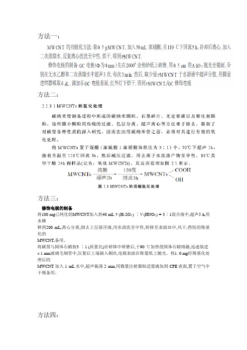
方法一:
方法二:
方法三:
修饰电极的制备
将100 mg已纯化的MWCNT加入到40 mL V (H2 SO4 ) ∶V (HNO3 ) = 3∶1混合液中,超声5 h,用水稀
释到200 mL,离心分离,倒去上层悬浮液,用水清洗至中性,转移至表面皿中,风干,得短的羧基化的
MWCNT,备用。
将碳黑与固体石蜡按3 ∶1 (质量比)在研钵中研磨后,于90 ℃加热使固体石蜡熔融,迅速装进< 1 mm玻璃毛细管中,压紧后上端插入铜丝,电极表面在称量纸上抛光。
将1. 0 mg经羧基化处理后的
MWCNT加入1 mL水中,超声振荡2 min,用微量注射器取适量滴加到CPE表面,置于空气中干燥备用。
方法四:
方法五:
112多壁碳纳米管的酸化处理
将500mg多壁碳纳米管在250mL H2 SO4 /HNO3(体积比为3: 1)的混合酸中水浴加热回流数小时,用5倍的去离子水冲洗,并用直径为0122μm的混合纤维膜和膜过滤器真空过滤,再用去离子水反复洗涤直至PH > 5,将滤出物放在真空干燥箱中烘干待用,将该样品记为MWNTCOOH。
羧基化多壁碳纳米管-Nafion修饰玻碳电极差分脉冲阳极伏安法测定痕量镉
羧基化多壁碳纳米管-Nafion修饰玻碳电极差分脉冲阳极伏安法测定痕量镉马伟光;杨铁金【摘要】Carboxylated multi-wall carbon nano-tubes(MWCNT′s) was dispersed in Nafion solution,and dropped on the surface of glassy carbon electrode(GCE)to prepare MWCNT′s-Nafion modified GCE(MWCNT′s-Nafion/GCE).In the determination of Cd2+-ion,the test solution in a NaOAc-HOAc buffer solution of pH 4.8 was pre-reduced and enriched for 350 s at-1.1 V,Cd2+-ion was then stripped from the electrode by scanning in the range from-1.2 V to-0.4 V and determined by differential pulse anodic stripping voltammetry.Oxidation peak potential of Cd2+-ion was observed at-0.84 V,at where linear relationship between values of peak current of Cd2+-ion and its concentration was obtained in the range of 5-100 μg·L-1,with detection limit(3S/N)of 2.54 μg·L-1.The proposed method has been used in the determination of cadmium in water sample,giving results obtained in consistency with those obtained by ICP-MS.%将羧基化多壁碳纳米管分散在Nafion溶液中并滴涂在玻碳电极表面,制备了羧基化多壁碳纳米管-Nafion修饰电极(MWCNT′s-Nafion/GCE)。
碳纳米管的性能及其在海水淡化中的应用

碳纳米管的性能及其在海水淡化中的应用摘要碳纳米管是近年来国内外广泛关注的一类纳米材料,具有一维特征孔道结构,能够有效促进液体分子的传输速率,是理想的海水淡化膜分离材料。
通过将其引入到常用的海水淡化膜基质中,借以提高膜的分离性能,逐渐成为膜分离领域的一个研究热点。
结了碳纳米管在反渗透、正渗透、膜蒸馏中的应用研究现状并分析了碳纳米管在反渗透、正渗透、膜蒸馏应用中的挑战,探讨了碳纳米管在海水淡化膜分离材料中的应用潜力。
1碳纳米管的结构与功能Kroto和Smalley于1985年首次发现了碳纳米管,直到1991年,由Iijima首次成功制备了碳纳米管。
碳纳米管是一种由单层或多层石墨烯同轴缠绕而成的柱状或层套状的管状物,碳原子以sp2杂化为主并混有sp3杂化。
碳纳米管性能优异,在微电子、生物医药和聚合物复合材料加固等方面应用潜力巨大。
碳纳米管具有独特的本征空腔结构,输水能力超强,水分子在碳纳米管中的传输速度比理论计算的高出几个数量级。
Hummer等采用分子动力学模拟水分子在碳纳米管中的流动行为,并提出了水分子在碳纳米管中的快速输送机理:首先,水分子在碳纳米管内部形成强力、规则的氢键,利于水分子快速通过;其次,碳纳米管内腔疏水、无极性,与水分子之间的相互作用非常弱,水分子能够无摩擦地通过碳纳米管。
Thomas等通过研究水分子在不同直径和长度的碳纳米管内的传输动力学,证明碳纳米管的内径对水分子的传输速度起决定作用。
随着内径的增大,水分子在碳纳米管中的构型逐渐由线性链变为堆叠五边形和六边形,最后成为无规则水流(见图1)。
当碳纳米管内径为0.83nm时,水分子成线性链,流速达到最大。
脱盐效果优异是碳纳米管在膜分离技术应用中的另一个重要性能。
碳纳米管的内径和尺寸排阻效应与毛细管行为的临界尺寸相当,能够在内壁形成能垒,只允许水分子通过,而水合离子则需要克服能垒后通过。
碳纳米管的内径对离子截留率的影响至关重要,当内径由0.66nm增大到0.93nm时,脱盐率由100%降低到95%。
【doc】碳纳米管的分散及表面改性
碳纳米管的分散及表面改性碳纳米管的分散及表面改性高濂刘阳桥(中国科学院上海硅酸盐研究所高性能陶瓷与超微结构国家重点实验室,上海200050)摘要:碳纳米管具有独特的结构和优异的物理化学性能,但碳管间易相互缠绕而发生团聚是限制其应用的主要原因.本文对国内外关于碳纳米管的分散及表面改性的研究进行了综合评述,评述了这些方法的优缺点,并对今后的研究方向做了展望.关键词:碳纳米管;分散;表面改性DispersionandSurfaceModificationofCarbonNanotubes GaoLianLiuYangqiao (StateKeylaboratoryofHighPerformanceCeramicsandSuperfineMierostr uetures,ShanghaiInstituteofCeramics,Shanghai200050)Abstract:Carbonnanotubeshavewidepotentialapplicationsduetotheiruniq uestructuresandexcellentproper-ties.ButtheytendtoagglomerateduetothestrongV anderWaalsinteraction,w hichsignificantlyrestrictstheirappli-cation.Inthispaper,studiesonthedispersionandsurfacemodificationofcarbonnanotubeswerereviewed.Thead- vantagesanddisadvantageswerediscussed.Theresearchdirectionsinthefut urewerealsoproposed.Keywords:carbonnanotube;dispersion;surfacemodification碳纳米管自1991年由Iijimat¨发现以来,以其极高的纵横比和超强的机械性能成为极具应用潜力的一维纳米材料,其应用已涉及到纳米电子器件,催化剂载体,电化学材料,贮氢材料和复合材料增强相等多方面.碳纳米管超强的力学性能可以极大提高复合材料的强度和韧性;独特的导电和光电性能可以改善聚合物材料的电导率和制备新型的光电聚合物复合材料;其独特结构可以制备金属或金属氧化物填充的一维纳米复合材料.这些新兴材料的崛起将对人们的生产和生活产生重大影响.近2年来,随着碳纳米管制备技术的不断发展,大批量,规模化的碳纳米管生产已经成为可能.目前国内已建成有多条碳纳米管的生产线,碳纳米管的产量不断增加,生产成本不断下降,为其走向应用提供了可能.目前制约碳纳米管器件及碳纳米管复合材料应用的主要是其分散以及与基体材料的相容性问题.碳纳米管表面缺陷少,缺乏活性基团,在各种溶剂中的溶解度都很低.另外,碳纳米管之间存在较强的范德华引力加之它巨大的比表面积和很高的长径比,使其形成团聚或缠绕,严重影响了它的应用.而且由于碳纳米管的表面惰性,与基体材料间的界面结合弱,因此,复合材料的性能仍不十分理想.为解决以上2个重要问题,目前人们致力于碳纳米管的分散及表面改性的研究.关于碳纳米管的分散及表面改性,最初是通过对其表面进行共价化学功能化实现的;后来人们开始尝试通过非共价功能化的方法,以期最大限度地保持碳管的结构性能完好;在碳管功能化的基础上,人们将金属,氧化物,氮化物,硫化物等多种纳米粒子包覆在碳管表面对其改性,提高碳管与无机基体的相容性,而且赋予了碳管复合材料更多优异的性能.以下将从上述3方面对碳纳米管的分散及表面改性进行综合评述,并对发展前景进行了展望.作者简介:高濂(1945~),男,本科,研究员,世界陶瓷科学院院士.主要从事高温结构陶瓷和纳米材料的基础研究.现任中国硅酸盐学会特陶分会结构陶瓷专业委员会主任.1141共价功能化碳纳米管的共价化学功能化最初是从氧化剂对碳纳米管的化学切割开始的.1994年Tsang等z1发现,将多壁碳纳米管在强酸中超声可对其进行切割,从而得到开口的碳纳米管.在随后的研究中,Lago等口发现,开口的碳纳米管顶端含有一定数量的活性基团,如羟基,羧基等.1998年,Liu等H 研究了单壁碳纳米管的切割方法,利用强酸和超声波对单壁碳纳米管进行切割,得到了长度介于100~300nm 之间的富勒烯管,接着用体积比为4:1的浓硫酸与30%的过氧化氢氧化,得到端基为羧基的单壁碳纳米管.这些截短的碳纳米管在水中单分散性良好.后来,人们尝试利用其它氧化剂如KCrO,OsO,KMnO等对碳纳米管进行了功能化.活性基团的存在不仅改善了碳纳米管的亲水性,使其更容易溶于水等极性溶剂,而且为碳纳米管与其它物质或基团反应,从而对其表面进行广泛的改性提供了基础.1998年Chen等利用氯化亚砜将强酸氧化单壁碳纳米管(single—walledcarbonnanotubes,简称SWNTs)表面的羧基转换成酰氯,并继续与十八胺反应,得到了SWNTs的十八胺衍生物.这种衍生物可以溶于二硫化碳,氯仿,二氯甲烷等多种有机溶剂,是世界上首次得到的可溶单壁碳纳米管,作者还对其进行了红外光谱,核磁共振氢谱,拉曼光谱,紫外可见光谱等多种表征.SWNTs酰氯与长链醇间的酯化发应也可用于碳纳米管的功能化,而且这种酯化反应是可逆的,在酸或碱的催化下,酯化SWNTs可以水解使单壁碳纳米管得到恢复”.Hamon等还发现,十八胺可以与切割的SWNTs直接发生离子型反应,得到在有机溶剂中呈单分散可溶的碳纳米管.由于离子型反应成本低,操作简单,因此,该法成为适宜大规模功能化碳纳米管的方法之一.Qin等【9还利用碳纳米管羧酸盐与烷基卤(氯,溴,碘)在水介质中的酯化反应,成功地将长烷基链键合在碳纳米管侧壁,实现了碳纳米管在有机介质中的高度分散.并用红外光谱,核磁共振,透射电镜及热分析等方法对功能化的碳纳米管进行了表征.他们的研究发现,反应所需的时间与烷基卤中卤素种类以及碳链长度有关,这种方法简单,高效,而且由于可选择的烷基卤种类很多,是一种十分有前途的功能化方法.酸化碳纳米管表面的羧基与胺类之间的偶合反应也是碳纳米管功能化中常用的反应之一.2000年,Riggs等¨刚利用这类反应首次报道了聚合物共价修饰的可溶性碳纳米管.他们利用线性聚合物(聚丙酰基氮丙啶一氮丙啶)与切割的碳纳米管反应,得到了可溶于有机溶剂和水的SWNTs和多壁碳纳米管(multiwalledcarbonnanotubes,简称MWNTs).他们还发现¨1,这些可溶性的碳纳米管具有光致发光现象,且发光波长覆盖整个可见光谱范围,这表明,可溶性碳纳米管有可能在发光及显示材料中得到应用.同年,刘忠范等¨1利用碳纳米管上的羧基与2一巯基乙胺在二环己基碳二亚胺(DCC)作用下缩合,得到了巯基修饰的碳纳米管,并实现了在金表面上的组装.侧壁氟化的方法最早是由Mickelsont1和Boul等¨1提出的,由于氟化碳管中的氟原子可通过亲核取代反应被其他基团取代,从而实现碳纳米管的进一步功能化,因而成为目前为止较为重要的碳管功能化方法之一.将SWNTs在不同温度下进行氟化反应,得到的氟化碳管在醇溶液中呈亚稳态的单分散.取代氟原子的亲核试剂,包括醇,胺以及烷基锂化合物等【11,取代反应可使碳管侧壁15%的碳原子与功能基团相连接,由于碳管表面与长的烷基链相连,功能化的SWNTs易溶于氯仿,四氢呋喃等多种有机溶剂.Liang等【1引以金属锂和烷基卤化物在液氨中的反应,采用还原烷基化反应实现了单壁碳纳米管的功能化.生成的碳纳米管在常见有机溶剂如氯仿,四氢呋喃及DMF中分散性良好,以单根形式分散.作者认为碳管分散的机理是Li分散在带负电的碳纳米管之间.Liut”1将CVD法制备的单壁碳纳米管在过氧三氟乙酸中进行超声实现了碳纳米管的表面功能化.红外光谱的结果表明:除含氧基团以外,三氟乙酸根基团也与碳管发生了共价键键合.单壁碳纳米管被截断至300nm左右,在极性溶剂如DMF,水,乙醇等介质中分散良好.Sunt等将碳纳米管在苯胺中避光回流3h,利用碳纳米管与苯胺间的质子转移反应,得到了可溶性碳纳米管,其中单壁碳纳米管在苯胺中的溶解度高达8mg/mL,这种苯胺功能化的碳纳米管可溶于多种有机溶剂. 目前为止,尽管有多篇关于碳纳米管共价功能化的文章发表,但这些方法普遍存在的局限是需要大量的溶剂,一般处理1g碳纳米管需要2L左右的溶剂.2003年DykeCA等¨发明了一种无需使用溶剂的功能化方法,为大批l15量功能化碳纳米管开辟了新的途径.方法是将碳纳米管与4一取代苯胺混合,然后缓慢加入异戊基亚硝酸盐,60~C反应完毕获得功能化的碳纳米管,产物在有机溶剂中有比较好的溶解性,在THF中的溶解度达到0.03mg/mL.Sun..和Jiang等¨将碳纳米管在NH,气氛下600~C进行热处理,成功地将胺基等碱性基团通过共价键键合在碳纳米管壁上.作者通过红外光谱对功能化的碳纳米管进行了分析,发现表面存在c—N,N—H等键的伸缩振动峰.一个有趣的现象是,碳管在氨气处理后,大部分开口化,而且通过原位修饰金粒子标识的方法发现,NH处理引入的活性基团主要分布在碳纳米管内壁.这无疑将为碳纳米管管内化学的研究提供了非常有效的基础,将开创化学催化,一维物理及化学等研究的新领域.2非共价功能化虽然碳纳米管的共价功能化在碳纳米管分散及表面改性方面取得了很大的进展,但这类功能化方法是直接与CNT的石墨晶格结构作用,可破坏CNT功能化位点的sp结构,从而可能对CNT的电子特性造成一定程度的破坏.而非共价功能化的方法不会对碳纳米管本身的结构造成破坏,从而可以得到结构保持完好的功能性碳纳米管.碳纳米管的侧壁由片层结构的石墨组成,碳原子的sp杂化形成高度离域化耵电子.这些耵电子可以被用来与含有耵电子的其它化合物通过耵一百非共价键作用相结合,得到功能化的碳纳米管.聚(问一亚苯亚乙烯)衍生物(polyp-phn丫1eneviny1ene—CO一2,5-dioctoxy—m—pheny1eneviny1ene,简称PmPV)是一种共轭发光聚合物,Curran等m】利用多壁碳纳米管与之通过耵一百相互作用形成MWNTs—PmPV 复合材料,这种复合材料在PmPV中形成稳定的悬浮液,用此方法可以分散纯化碳纳米管.Star等1231利用PmPV 对SWNTs进行了功能化研究,结果表明,随着PmPV含量的增大,悬浮液中SWNTs束的平均直径逐渐减小,SWNTs的表面覆盖度逐渐均一.这些结果证实了PmPV通过苯基,乙烯基与SWNTs表面的耵一百相互作用缠绕于碳纳米管上.事实上,本身不含有耵电子的有机化合物也可以与碳纳米管相结合,通过偶极一偶极作用,氢键及范德华力等物理作用与碳纳米管作用,缠绕在碳纳米管表面.O’Connell等成功地将聚乙烯吡咯烷酮(PVP)包裹在SWNTs管壁上,聚合物提高了SWNTs管壁的亲水性,较好地解除了SWNTs 的聚集效应,得到稳定的SWNTs水悬浮液,碳纳米管含量可以达到 1.4g/L.值得一提的是,这种聚合物与碳纳米管间的包裹作用是可逆的,通过改变溶剂体系,聚合物链能从SWNTs管壁上脱落,而且不会影响SWNTs的结构和性质.他们还提出了聚合物链在碳纳米管表面缠绕的3种方式(见图1).赵丽萍等以一种含碱性颜料吸附基团的嵌段结构共聚物聚氨基甲酸乙酯(PAME,BYK—CHEMIEGmbH公司生产)对碳纳米管进行了功能化,分散介质为乙醇.利用共聚物间空间位阻的存在,大大削弱了范德华力的影响,显着改善了碳纳米管在乙醇介质中的分散性,碳纳米管浓度达到10g/L.Bandyopadhyaya等将SWNTs在阿拉伯树胶(GA)水溶液中超声,得到可在数月内保持稳定的悬浮液,这种方法同样适用于MWNTs.他们也认为,GA聚合物链的空间位阻作用是克服碳纳米管间范德华力的主要原因. Islam等1271将单壁碳纳米管在十二烷基苯磺酸钠(NaDDBS)溶液中超声,利用二者间的物理作用,制备了高浓度稳定的SWNTs水悬浮液,原子力显微镜的结果表明,对于浓度高达20mg/mL的SWNTs溶液而言,单根碳纳米管占63%以上.江琳沁等I引系统研究了十二烷基硫酸钠(SDS)对MWNTs 在水中分散性的改善,并采用∈一电位,红外光谱等手段对分散及吸附机理进行了研究.研究表明:SDS的烷基链是通过疏水作用吸附在碳纳米管表面的,SDS上的硫酸根增加了碳纳米管表面的负电量,增加了碳管间的静电排斥力,从而提高悬浮液的稳定性.作者还给出了碳纳米管悬浮液稳定性随碳管和SDS浓度变化的图谱(见图2).116图1聚合物PVP在碳纳米管表面的3种可能的缠绕方式ⅢweigtttofCqqT~,6图2十二烷基硫酸钠(SDS)分散多壁碳纳米管的稳定性图谱汹D--高度分敬∞一少量团聚▲一严重团聚Lil将SWNTs在150g/L的NaOH/乙醇一水(5:1)溶液中超声处理,利用溶液对碳纳米管表面良好的润湿作用,使乙醇钠等离子扩散到碳管束中间,吸附在单根碳管表面,降低管间相互作用力,从而使单根碳管从碳纳米管束上剥离出来.处理后的碳纳米管可容易地溶解在N,N一二甲基甲酰胺(DMF),四氢呋喃(THF),氯仿,四氯乙烯,N一甲基一2一吡咯烷酮(NMP)及环氧树脂等多种有机溶剂中,在NMP中的溶解度达8mg/mL,而且悬浮液可稳定数星期.红外光谱的结果表明,这是一个物理过程,并未有新的共价键生成.3无机纳米颗粒改性碳纳米管碳纳米管经过有机功能化后,表面带上多种活性基团,但为保障其在无机基体介质中良好的分散性,往往需要在功能化的碳纳米管表面包覆或填充某些无机纳米颗粒,改善其与基体的界面结合,从而最大限度地发挥碳纳米管的优异性能.同时,这种无机颗粒改性的碳纳米管本身在非均相催化,太阳能电池,发光材料,传感器等方面也具有重要应用.酸化的碳纳米管由于表面具有一0H,一C00H等活性基团,可以将金属离子或微粒”拴”在碳管上,从而实现无机粒子在碳管表面的包覆.Y u等1成功地将Pt纳米颗粒包裹在酸处理的碳管表面.Huang等¨利用TiC1在碳纳米管硝酸溶液中的水解,原位地将金红石相TiO:颗粒包裹在碳纳米管表面.Liu等在酸化的碳纳米管与Ni¨,Fe,的混合物中滴人NaOH溶液,经水热处理,成功地在碳纳米管表面包覆了NiFeO纳米粒子.利用共价功能化的碳纳米管与有机化合物保护的无机粒子间的化学反应,同样可以实现碳管表面的无机改性.Banerjeel首先对单壁碳纳米管(SWNTs)表面进行酸化处理,在碳纳米管(carbonnanotubes,简称CNTs)表面产生多个羧基基团,利用羧基基团与胺化的TiO颗粒间的反应,得到了TiO颗粒包覆的SWNTs.Ravindran等用含有巯基和端胺基的有机物稳定ZnS包覆的CdSe纳米晶,在乙烯基碳化二亚胺(EDC)脱水剂的作用下,使其胺基与酸处理过的碳管表面的羧基发生偶合反应,以此使半导体纳米晶附着在碳管末端.这种异质连接可作为纳米电子和光电子器件的组建部件等使用.Haremza等通过胺基功能化的CdSe纳米晶与酰卤基团改性的单壁碳纳米管反应的方法实现了CdSe纳米晶在碳管表面的附着.碳纳米管经功能化处理后,表面所带的活性基团在液相介质中常常离解而带有某种电荷,金属离子等受静电引力的作用,会吸附在这些活性基团上,再进行原位合成反应,将生成纳米粒子改性的碳纳米管.图3列举了一些常用表面活性剂及氨气处理后碳管表面垂一电位随pH值的变化曲线.这种方法十分有效,而且选择不同的表面处理手段对碳管表面带电状况进行调控,可实现多种金属,氢氧化物,氧化物,硫化物对碳纳米管的均匀包裹或填充.ZhaoLP_31等以硫化钠为硫源,SDS对碳纳米管进行表面改性,采用原位合成的方法在多壁碳纳米管表面均匀包裹了一层ZnS纳米颗粒,既能保证碳纳米管与ZnS晶粒的有效结合,又能保持碳纳米管霄电子结构的完整性.Jiang等采用2种分散剂SDS和PEI对碳纳米管表面进行非共价键改性,通过原位反应制得CdS包裹的碳纳米管复合材料,以氨气开口的碳纳米管为原料,采用毛细管填充法制备了CdS纳米线填充的碳纳米管复合材料.作者还以SDS改性的MWNTs为原料,利用醋酸锌与氢氧化锂之间的原位反应,在碳纳米管表面均匀包裹了ZnO纳米粉体.这种复合粉体具有极高的光催化活性,对亚甲基蓝降解的光催化活性较单独的ZnO颗粒提高了2倍.作者将其归因于碳纳米管到抑制电子空穴复合的作用.另外,由于碳管独特的结构,还可囤以起到限制原位生成的纳米颗粒长大的作用,在对CdS/MWNTsI371以及ZnS/MWNTs[1的紫外可见光谱分析中,均发现明显的量子尺寸效应.60耋:g墓0瑚_40;一.{毒.一2468lOl2DH经不同处理的碳纳米管考一电位随pH值的变化关系a一原始碳管b一吸附聚乙烯亚胺(PEI)c一氨气处理d一吸附柠檬酸e一吸附PAA卜一吸附SDSg一氨气处理后吸附PEISun等提出一种新颖的方法——反微乳非共价键合法,实现了ZnO,MgO纳米粒子在碳管表面的包覆.在117NaDDBS/环己烷/TritonX一114微乳体系中,利用TritonX一114对二价金属离子的萃取作用,使金属离子聚集在油一水界面,NaDDBS烷基链沿碳管轴向方向水平吸附在碳管表面,疏水链伸入油相.当NHH:O溶液加入时,金属氢氧化物从稳定的微乳液中沉积出来,经过煅烧后,生成表面附着氧化物颗粒的碳纳米管.该方法具有一系列显着优点,如碳纳米管与无机颗粒间是通过非共价键结合的,保证碳纳米管固有的各种物理和化学性质不被破坏;无机纳米颗粒的合成反应被限制在纳米级的微乳液滴中,颗粒尺寸小,团聚程度低;可用于2种或多种无机纳米颗粒在碳纳米管上的包覆等.另外,利用未改性碳纳米管与无机颗粒间较弱的疏水力也可以实现无机颗粒在碳管表面的包覆,但这方面的报道不多,仅Ellis等在2003年报道了疏水作用实现Au颗粒在碳管表面连接的结果,辛硫醇单层保护的金纳米簇连接到未经氧化处理的碳管表面,是通过二者间弱的疏水作用实现的.4结束语目前碳纳米管的合成和应用已经成为材料学界研究的前沿和热点,碳纳米管以其独特的结构和优异的性能,将在纳米制造技术,生物技术,能源,催化,电子材料等方面获得重要应用.如何通过表面处理实现碳纳米管的高度分散并改善其与其它功能和结构材料的相容性,成为推进碳纳米管实用化的关键课题,开展这方面的研究具有重要的意义.对于碳纳米管的分散及表面改性研究,我们认为应着重从以下3方面考虑:(1)选择较为温和的实验条件,最大限度地保持碳纳米管完整的结构和性能;(2)着重研究低成本,大批量简单有效的功能化方法,在这方面气体处理,无溶剂化反应等手段可发挥重要作用.另外,应注重碳纳米管化学与超声化学,微波学,光电化学等多学科的交叉,借助先进的实验手段,实现这一目标;(3)丰富完整碳纳米管的无机改性的研究体系,利用碳管与无机物质间功能特性的协同作用,拓展碳纳米管的应用范围.参考文献1IijimaS.Helicalmicrotubulesofgraphiticcarbon.Nature,1991,354:56—5 82TsangSC.ChenYK,GreenMLH,eta1.Asimplechemicalmethodofopenin gandfillingcarbonnanotubes.Nature,1994,372:159—1623LagoRM,TsangSC,GreenMLH.Fillingcarbonnanotubeswithsmallpalla diummetalcrystallites:theeffectofsurfaceacidgroups. JChemSocChemCommun,1995(13):1355—1356.4LiuJ,RinzlerAG,GaiHJ,eta1.Fullerenepipes.Science,1998,280:1253—1 2565ChenJ,HamonMA,HuH,eta1.Solutionpropertiesofsingle—walledcarbo nnanotubes.Science,1998,282:95—986SunYP,HuangW,LinY,eta1.Solubledendron—functionalizedcarbonnan otubes:preparation,characterizationandproperties.ChemMater,2001,13:2864—28697FuK.HuangW,LinY,eta1.Defunctionalizationoffunctionalizedcarbonna notubes.NanoLett,2001(1):439—4418HamonMA,ChenJ,HaddonRC,eta1.Solutionpropertiesofsingle—walle dcarbonnanotubes(s—SWNTs).JAmChemSoc,2001,123(4):733—7349QinY,ShiJ,WuW,eta1.Conciseroutetofunctionalizedcarbonnanotubes.J PhysChemB,2003,107(47):12899—1290110RiggsJE,GuoZ,CarrollDL,eta1.Strongluminescenceofsolubilizedcarb onnanotubes.JAmChemSoc,2000,122:5879—588011CzerwR,GuoZX,AjayanPM,anizationofpolymersontocarbon nanotubes:aroutetonanoscaleassembly.NanoLett,2001(1):423—42712LiuZF,ShenZY,ZhuT,anizingsingle—walledcarbonnanotubes ongoldusingawetchemicalself-assemblingtechnique?Langrnuir,2000.16(8):3569—357313Mickels onET’HuffmanCB,MargraveJL,eta1.Fluorinationofsingle—w allcarbonnanotubes.ChemPhysLett,1998,296:188一ll8l415l6l7l9202l22232526272829303l323334353637194BoulPJ,LiuJ,SmalleyRE,eta1.Reversiblesidewallfunctionalizationofbuc kytubes.ChemPhysLett,1999,310:367—372KhabasheskuVN,BillupsWE,Margrave.Fluorinationofsingle.wallcarbon nanotubosandsubsequentderivatizationreactions.AccChemRes,2002,35:1087—1095LiangF,SadanaAK,PeeraA,eta1.Aconvenientroutetofunctionalizedcarbo nnanotubes.NanoLett,2004,4(7):1257—1260¨uMH,Y angYL,ZhuT,eta1.Chemicalmodificationofsingle?walledcarbon nanotubeswithperoxytrifluoroaceticacid.Carbon.2005,43:1470—1478SunY,WilsonSR.SchusterDI.Highdissolutionandstronglightemissionofc arbonnanotubesinaromaticaminesolvents.JAmChemSoc.2001.123:5348—5349DykeCA,TourJM.Solvent.freefunctionalizationofcarbonnanotubes.JAmChemSoc,2003,l25:l156一l157SunJ,GaoL,LiW.Colloidalprocessingofcarbonnanotubes/aluminacompo sites.ChemMater,2002,14:5169—5172JiangLQ,GaoL.Modifiedcarbonnanotubes:aneffectivewaytoselectiveatta chmentofgoldnanoparticles.Carbon,2003,41:2923—2929CurranSA,AjayanPM,BlauWJ,eta1.Acompositefrompoly(p-phenylenevi nylene?co?2,5-dioctoxy-m?phenylenevinylene)and carbonnanotubes:anovelmaterialformolecularoptoelectronics.AdvMater, 1998,10(14):109l一1093StarA,StoddartJF,SteuermanD.Preparationandpropertiesofpolymer-wrap pedsingle-walledcarbonnanotubes.AngewChemIntEd.2001,40(9):l72l一1725O’ConnellMJ,BoulP,EricsonLM,eta1.Reversiblewater-solubilizationofsi ngle?walledcarbonnanotubosbypolymerwrapping. ChemPhysLett,2001,342:265—27lZhaoLP,GaoL.Stabilityofmulti—walledcarbonnanotubesdispersionwith copolymerinethano1.ColloidsSurfacesA,2003,224(1~31:127一l34BandyopadhyayaR,Nativ—RothE,RegevO,eta1.Stabilizationofindividu alcarbonnanotubesinaqueoussolutions.NanoLett,2002(2):25—28IslamMF,RoilsE,BergeyDM,eta1.Highweightfractionsurfactantsolubiliz ationofsingle-wallcarbonnanotubesinwater.NanoLett,2003,3(2):269—273JiangLQ,GaoL,SunJ.Productionofaqueouscolloidaldispersionsofcarbon nanotubes.JColloidInterfaceSci,2003.26o:89—94LiQw,KinlochIA,WindleAH.Discretedispersionofsingle?walledcarbonn anotubos.ChemCommu,2005(26):3283—3285Y uRQ,ChenL,LiuQ,eta1.Platinumdepositiononcarbonnanotubesviache micalmodification.ChemMater,1998.10:718—722HuangQ,GaoL.ImmobilizationoffutileTiO2onmuhiwalledcarbonnanotu bos.JMaterChem,2003,13:1517—1519LiuYQ,GaoL_Astudyontheelectricalpropertiesofcarbonnanotubes?NiFe z04nanocomposites:effectofthesurfacetreatmentof carbonnanotubes.Carbon,2005,43(1):47—52BanerjeeS,WongSS.Synthesisandcharacterizationofcarbonnanotube?nan ocrystalheterostructures.NanoLett,2002.2(3):195—200 RavindranS,ChaudharyS,ColbumB,eta1.Covalentcouplingofquantumdo tstomultlwalledcarbonnanotubosforelectronicdeviceapplications.NanoLett,2003(3):447—453HaremzaJM,HahnMA,KraussTD,eta1.AttachmentofsingleCdSenanocry stalstoindividualsingle?walledcarbonnanotubos.NanoLett,2002(2):1253—1258ZhaoLP,GaoL.Coatingofmulti—walledcarbonnanotubeswiththicklayers oftin(IV)oxide.Carbon,2004,42(8—9):1858—1861JiangLQ,GaoL.FabricationandcharacterizationofZnO?coatedmulti?wall edcarbonnanotubeswithenhancedphotocatalyticactivity. MaterChemPhys,2005,91(2?3):313—31638JiangLQ,GaoL.CdS39psulated,CdSnanocrystals?enrobedcarbonnanotuboscompositesandtheir UV?visproperties.ChemLett,2004,33(9):1114—1115SunJ,GaoL,1wasaM.Noncovalentattachmentofoxidenanoparticlesontoc arbonnanotubesbyusingwater-in-oilmicroemulsions. ChemComm,2004(7):832—833EllisA V,VijayamohananK,GoswamiR,eta1.Hydrophobicanchoringofmo nolayer-protectedgoldnanoclusterstocarbonnanotubos.NanoLett,2003,3(3):279—282119。
氯霉素在羧基化单壁碳纳米管修饰电极上的电催化还原
Ab ta t B s gtesa nn l t n m coc p S M)a d t lc o h m c lm e a c e t so y( I )c n t c sr c s c : yu i c n i e cr i so y( E r n h g e o r n ee t c e i p d n es c oc p E S o s u t uf e h e r ai p r r a
化 选 定 的测 试 条件 下 , 原 峰 (一 .0 电 流 与 C P的 浓 度 在 1 5 还 0 6 2V) A .5×1 ~ ~19 0 .7×1 。 m lL范 围 内呈 良好 0 o /
线性关 系 , 检出限达 20 .2×1 ~ m lL 相对标准偏差 ( 0 o . / n=1 ) 2 2 % , 0 为 .4 可用于氯霉素 的含量测定 .
Elc r c a ytc Re c i f Ch o a p n c la o i e asy Ca bo e t o at l i du ton o l r m he i o tM d f d Gl s r n i El c r e b nge- le r o Na t be t r x l e t od y Si l wa l d Ca b n no u s wih Ca bo y
ee to a ay i u cin o e u t n o AP.Ba e n t er s l fe p rme t si fre sa t — lcr n a d t o p oo a s lc r c tl ssf n t n r d ci f o o C s d o h e u t o x ei n si wa e r d a s t n wo ee t n w — rt n t n — o r
单壁碳纳米管和多壁碳纳米管
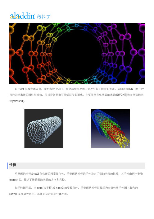
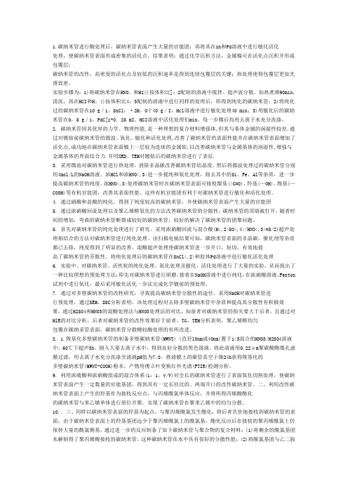
自1991年被发现以来,碳纳米管(CNT)在全球学术界和工业界引起了极大的关注。
碳纳米管(CNT)是一种直径为纳米级的圆柱形结构,可以看做是由石墨烯层卷曲而成。
主要类型有单壁碳纳米管(SWCNT)和多壁碳纳米管(MWCNT)。
性质
单壁碳纳米管是sp2杂化碳的同素异位体。
单壁碳纳米管的手性决定了碳纳米管的性质,其手性由两个整数(n,m)定义,描述了被卷碳纳米管的方向和直径。
如手性图所示,当n=m(扶手椅)或n-m=3的整数倍时,单壁碳纳米管则显示为金属性质手性图上蓝色的SWNT是金属性质的。
其他则显示为半导体性质。
碳纳米管具有优异的强度,很高的导电性或半导体性,热导性,单位质量非常大的表面积,以及独特的光学特性等材料优势。
使其运用于增强碳纤维、增强树脂和弹性体的机械强度;改进锂离子电池和超级电容器的电导性;显示器、太阳能电池和新兴固态照明技术的电极;逻辑器件、非易失性存储元件、传感器和安全标签等领域。
与多壁碳纳米管相比,单壁碳纳米管具有更有明显的优势。
但由于纯度、选择性和分散性也限制了碳纳米管的广泛应用。
由于碳纳米管的柱形结构,使其内部能够填充纳米级的分子和原子。
例如,富勒烯填充的CNT。
此外,碳纳米管还可以填充金属、水,以及分子氧等,填充在碳纳米管中的物质具有与外界相比不同的性质。
单壁碳纳米管
双壁碳纳米管
复壁碳纳米管
碳纳米管分散液
碳纳米管浆料
碳纳米纤维
其他
交叉缝式碳纳米管≥95%,直径:10-20nm,长度:5-15μm 308068-56-6。
碳纳米管的改性
1.碳纳米管进行酸处理后,碳纳米管表面产生大量的官能团;再将其在sn和Pd溶液中进行敏化活化处理,使碳纳米管表面形成密集的活化点。
结果表明:通过化学沉积方法,金属镍可在活化点沉积并形成包覆层;碳纳米管的改性,高密度的活化点及较低的沉积速率是得到连续包覆层的关键;热处理使得包覆层更加光滑致密。
实验步骤为:1)将碳纳米管在HNO。
和Hz()按体积比]:2配制的溶液中搅拌、超声波分散,加热煮沸90min,清洗,再在HCI和H。
()按体积比4:3配制的溶液中进行同样的处理后,即得到纯化的碳纳米管;2)将纯化过的碳纳米管在10 g/i。
SnCl:·2H。
O十40 g/I,Hcl溶液中进行敏化处理40 min;3)用敏化后的碳纳米管在0,5 g/i,PdC[z+0.25 mI。
HCI溶液中活化处理们min。
每一步骤后均用去离子水充分洗涤。
2.碳纳米管因其优异的力学、物理性能,是一种理想的复合材料增强体,但其与基体金属的润湿性较差.通过对镀钴前碳纳米管的微波、氧化、敏化和活化处理,改善了碳纳米管的表面性能并在碳纳米管表面增加了活化点,成功地在碳纳米管表面镀上一层较为连续的金属钴,以改善碳纳米管与金属基体的润湿性,增强与金属基体的界面结合力.并用XRD、TEM对镀钴后的碳纳米管进行了表征.3. 采用微波对碳纳米管进行热处理,消除非晶碳改善碳纳米管结晶度。
然后将微波处理过的碳纳米管分别用4mol/L的NaOH溶液、浓HCl和浓HNO<,3>进一步提纯和氧化处理,除去其中的Si、Fe、Al等杂质,进一步提高碳纳米管的纯度。
浓HNO<,3>处理碳纳米管时在碳纳米管表面可接枝羰基(>C=O)、羟基(—OH)、羧基(—COOH)等有机官能团,改善其表面性能,这些有机官能团有利于对碳纳米管进行敏化和活化处理。
4. 通过硝酸和盐酸的纯化,得到了纯度较高的碳纳米管,并使碳纳米管表面产生大量的官能团5. 通过浓硝酸回流处理以及聚乙烯醇氧化的方法改善碳纳米管的分散性,碳纳米管的顶端被打开,随着时间的增加,弯曲的碳纳米管断裂成较短的碳纳米管,较好的解决了碳纳米管的团聚问题。
碳纳米管的特性及其分析应用
碳纳米管的特性及其分析应用摘要碳纳米管作为一维纳米材料,重量轻,六边形结构连接完美,具有许多异常的力学、电学和化学性能。
近些年随着碳纳米管及纳米材料研究的深入其广阔的应用前景也不断地展现出来。
本文着重介绍碳纳米管的特性及其在仪器分析中的应用。
关键词:碳纳米管;特性;仪器分析I一、引言碳纳米管(CNT,又名巴基管,于1991年被日本电子公司(NEC的饭岛博士发现。
是一种具有特殊结构(径向尺寸为纳米量级,轴向尺寸为微米量级,管子两端基本上都封口)的一维量子材料。
由于其优异的力学、电学和光学特性,碳纳米管受到了越来越多的关注。
随着时间的推移,CNT的制备与表征手段越发完善,由CNT制成的各种产品技术也趋于成熟。
二、碳纳米管的制备方法其主要有三种制备方法:分别为电弧放电,激光蒸发法和碳氢化合物催化分解。
(一)电弧放电电弧放电是指一般情况下由两个电极和它们之间的气体空间所组成电弧能产生高温。
但又不同于一般的燃烧现象,它既没有燃料也没有伴随燃烧过程的化学反应。
电弧放电实质上是一种气体放电现象,在一定条件下使两极之间的气体空间导电,是电能转化为热能和光能的的一种过程。
该方法包括以下具体步骤:对碳纳米管直接施加电压和电流,进行电火花处理,去除碳纳米管表面的附着金属或氧化物催化剂和剥离沉积的非晶碳层,与此同时,切割、定向排列碳纳米管。
本技术所采用的电火花处理可在空气中进行,也可在惰性气氛中进行。
施加电压可为直流也可为交流,电压10〜10 0伏,电流0〜10安培。
本方法的优点在于能完全去除碳管表面用其它方法难以去除的非晶碳和金属杂质,达到纯化碳纳米管的目的;另外,此方法还可切割碳纳米管,获得定向排列的碳纳米管。
(二)激光蒸发法激光蒸发法是制备碳纳米管的一种有效方法•用高能CC2激光或Nd/YAG激光蒸发掺有催化剂的碳靶制备碳纳米管,管径可由激光脉冲来控制。
激光脉冲间隔时间越短,得到的碳纳米管产率越高,而碳纳米管的结构并不受脉冲间隔时间的影响。
碳纳米管修饰电极的制备及其对NADH的电催化氧化
公司) 、KQ.0 B 型 超 声 波 清 洗 器 ( 山 市 超 声 仪 器 有 50 昆 限 公 司 ) Z 9 自动 双 重 纯 水 蒸 馏 器 ( 海 亚 荣 生 化 仪 、S .3 上
器 厂 ) 。
网 印 刷 碳 电 极 ,取 出 后 低 温 干 燥 保 存 。
; 基 化 的 多 壁 碳 纳 米 管 超 声 分修 饰 的丝 网印刷 碳 电极 ,如 图2 示 。 所 4 mm的 圆 盘 为 工 作 电 极 , 银 电 极 作 为 参 比 电
2 羧基 化 多壁碳 纳 米管 修饰 丝 网 印刷 碳 电极 的制 备 、
如 图 1 示 ,先 在 05 所 .mm厚 的 陶 瓷 基 板 上 印刷 图 1 A所 示 银 层 ,放 入 9  ̄ 空 干 燥 箱 中 3 mi;再 印刷 图 1 示 0C真 0 n B所
磷 酸缓 冲溶 液 中 的循环 伏 安 曲线 ,扫描 速 度
由 图 可 知 , 羧 基 化 多 壁 碳 纳 米 管 修 饰 的 丝 网
≯2 绷 o
瞒 幽 目 №
。 。 。
h
兑
n
,
黼图
爱~…
中 , 随 有 电 子 和 质 子 的 转 移 【】 故 溶 液 p 值 对 NADH 伴 m, H
R es ear
蕊 ◇黪
鞭黪鞠
与 开 发
纳 簪 饰戚 韵 螽 制
园国 慰
注 :湖南 省大 学生研 究性 学 习和 刨新 性 买验计划 资助 项 目
的电飕
摘 要 :采用丝 网印刷 技术 ,制 备出羧 基 化多壁 碳纳 米管修 饰 的丝网 E  ̄ 碳 电极 ,并 采用 循环伏 安 3 IU
法 研究 了该 电极对 还原 型烟 酰胺 腺嘌 呤二核 苷酸 ( A H)的 电催 化 氧化 性能 。结果 表明 ,与 未 N D 修 饰丝 网印刷碳 电极 相 比 ,多壁 碳纳 米管修 饰丝 网印刷 碳 电极显 著降低 了 N D 的氧化 峰 电位 , A H 消 除了反 应产 物对 电极的 污染 及其 它电化 学反 应对 测量 的干扰 。将修 饰 电极与 流动 注射 系统结 合 起来 , 建立 了利 用电流 一时间 曲线 测量 NA I 浓度的 方法 。在 l m l  ̄ mm l D- I p o/ 6 o/ 范围 内 , 应 _ L L的 响 电流 与 N D A H的浓 度有 较好 的线性 关系 ,其线 性 回归方程 为 i A =1 14 (mo +0I7 2 ( ) . 1 cp 1 n 5 ) .1 6 ,相 关系 数为 O9 1 ,检测 限为 0 t o/ 。 . 5 9 .t l 7m L 关键 词 :碳 纳米 管 ;还原型 烟酰胺 腺 嘌呤二 核苷酸 ( A H) N D ;丝网 印刷 电极 ;电催 化氧化 中图分 类号 :T 2 2 2 P 1 . 文献标识 码 :A 文章编号 :10 — 8 X ( 0 1 6 0 0 — 5 0 6 8 3 2 1 )0 — 0 6 0
羧基化多壁碳纳米管修饰电极对秦皮中秦皮乙素的灵敏测定

羧基化多壁碳纳米管修饰电极对秦皮中秦皮乙素的灵敏测定郑艳洁;陈伟;刘爱林;王昆;林雅静;林新华【摘要】目的研究秦皮乙素在羧基化多壁碳纳米管(c-MWCNTs)修饰电极上的电化学行为及其测定方法.方法利用交流阻抗法(EIS)和循环伏安法(CV)表征修饰电极,用CV研究秦皮乙素在电极上的电化学行为,以差示脉冲伏安(DPV)对其含量进行测定.结果 c-MWCNTs修饰电极对秦皮乙素有明显的电催化作用,在pH 4.5磷酸缓冲液(PBS)中,氧化峰电流与秦皮乙素浓度在6.0×10⁻²~8.0 μmol/L范围内呈良好的线性关系,检测限为5.0×10-3 μmol/L.结论 c-MWCNTs修饰电极与裸玻碳电极(GCE)相比,提高了秦皮乙素的检测灵敏度,可用于实际样品中秦皮乙素含量的测定.%Objective The electrochemical behavior and determination of esculetine using carbox-ylic multi-wall carbon nanotubes (c-MWCNTs) modified electrode have been studied. Method The modified electrode was characterized by electrochemical impedance spectrum (EIS) and cyclic voltammetry (CV). The electrochemical behavior of esculetine at this electrode was studied by cyclic voltammetry (CV) -while the determination of esculetine -was measured in differential pulse voltammetry (DPV). Results The c-MWCNTs modified electrode shows excellent electrocatalysis characteristics for esculetine. In pH 4. 5 phosphate buffer solution (PBS), the relationship between oxidation peak current and the concentration of esculetine is linear in the range of 6. 0×10-2~8. 0 μmol/L. The detection limit has been estimated as 5. 0×10-3μmol/L. Conclusion The detection sensitivity at c-MWCNTs modified electrode has been greatly enhanced compared -with that of at bare glassy carbon electrode. And this methodcould be used for the determination of esculetine in real sample with satisfactory results.【期刊名称】《福建医科大学学报》【年(卷),期】2012(046)004【总页数】4页(P251-254)【关键词】秦皮;伞形酮类;电极;纳米管,碳【作者】郑艳洁;陈伟;刘爱林;王昆;林雅静;林新华【作者单位】福建医科大学,药学院药物分析系,福州,350004;福建医科大学,药学院药物分析系,福州,350004;福建医科大学,药学院药物分析系,福州,350004;福建医科大学,药学院药物分析系,福州,350004;福建医科大学,药学院药物分析系,福州,350004;福建医科大学,药学院药物分析系,福州,350004【正文语种】中文【中图分类】R282.71;R313;R916.3秦皮乙素(esculetine)是中药秦皮发挥药理作用最重要的化学成分,对机体各系统作用广泛[1],对金黄色葡萄球菌、痢疾杆菌、大肠杆菌、卡他球菌、甲型链球菌均有抑制作用,临床上常用于治疗急性菌痢;有平喘、祛痰作用,可用于治疗慢性气管炎,具有重要的经济价值。
羧基化多壁碳纳米管修饰电极测定依诺沙星

羧基化多壁碳纳米管修饰电极测定依诺沙星孙汉文,邢 涛(河北大学化学与环境科学学院,河北省分析科学技术实验室,河北保定 071002) 摘 要:基于羧基化多壁碳纳米管修饰玻碳电极为工作电极,建立了无汞和高灵敏测定依诺沙星的电化学方法.在0.1mol/L 磷酸盐缓冲溶液(p H 为5.91)中,在0.5V 富集5min ,然后以100mV/s 的扫描速度从0.5V 扫描至1.4V ,1次微分峰电流与依诺沙星在0.02~0.20mg/L 呈线性关系,检出限为0.01mg/L.通过与药典方法比较,证实了方法的可靠性.该方法操作简单,灵敏度高,避免使用汞,可以用于依诺沙星胶囊含量的常规测定.关键词:羧基化多壁碳纳米管;电化学;依诺沙星中图分类号:O 657.7 文献标识码:A 文章编号:1000-1565(2007)03-0257-05Deter mination of Enoxacin Using Electr ode Modif iedby Multi 2w all Car bonnanotubes Functionalized with Car boxylic G r oupSU N Han 2wen ,X I NG Tao(K ey Laboratory of Analyt ical Science and Technology of Hebei Province ,C ollege of Chemistry and Environment al Science ,Hebei University ,Ba oding 071002,China )Abstract :A mercury 2free and high sensitive elect rochemical met hod for t he determi nat ion of enoxacin was establi shed by usi ng a glassy carbon elect rode modified by multi 2wall carbonnanot ubes functionalized wit h car 2boxylic group.Differential voltam mograms of enoxacin was obtai ned in 0.1mol/L phosphate buffer solution of pH 5.91at 100mV/s aft er 5.0mi n accumulati ng at 0.5V.The oxidation current s were linear for enoxacin i n t he range of 0.02~0.20mg/L wit h and t he detect limit i s 0.01mg/L.The reliability of t his mercury 2free met hod was validated by c ompari ng wit h t he pharmac opoeia met hod.The proposed met hod was simple and sen 2sitive ,and can be applied for rout ine determi nat ion of enoxaci n i n capsule wit hout contami nat ion.K ey w or ds :f unctionalized mult i 2wall carbonnanot ubes ;elect r ochemist ry ;enoxacin依诺沙星是第3代喹诺酮类抗菌药,通过作用于细菌DNA 螺旋酶的A 亚单位,抑制DNA 的合成和复制而导致细菌死亡,具有广谱抗菌作用,应用范围广泛,如泌尿生殖系统感染、呼吸道感染、胃肠道感染、伤寒等.但是,有实验表明依诺沙星会使细菌产生抗药性[1],还会引起血糖的降低等副作用[2],因此,依诺沙星的测定成为广大化学和医药工作者研究的一个重要问题.目前对其进行测定的方法有高效液相色谱法[3-6]、紫外分光光度法[7]、荧光光度法[8]、流动注射化学发光法[9]等.电化学方法测定依诺沙星也有报道.Zhang 等采用滴汞电极对依诺沙星进行了测定方法研究[10].钟爱国制备了聚氯乙烯膜依诺沙星选择性电极[11],电极 收稿日期 基金项目教育部博士点学科基金项目(553) 作者简介孙汉文(5),男,河北魏县人,河北大学教授,博士生导师,从事分析化学研究第27卷 第3期2007年 5月河北大学学报(自然科学版)Journal of Hebei U niversit y (Nat ural Science Edition )Vol.27No.3May 2007:2007-01-10:20000700:194-.的Nernst 响应范围为1.0×10⁻²~5.0×10⁻⁵mol/L ,检测限为4.2×10⁻⁶mol/L.碳纳米管自1991年被Sumio I iji ma 发现以来[12],因其独特的力学、电子特性及化学稳定性,成为世界范围内的研究热点之一.碳纳米管是单层或多层石墨片围绕中心轴,按照一定的螺旋角卷曲而成的无缝纳米管.根据制备方法和条件的不同,碳纳米管存在单层纳米管和多层纳米管两种形式.由于碳纳米管的表面效应,即直径小、表面能高、原子配位不足,而使其表面原子活性高,易与周围的其他物质发生电子传递作用,在电催化和电分析化学领域中具有广泛的应用[13-17].为了获得较好的电化学响应,还可以对碳纳米管进行处理.羧基化碳纳米管在电极表面引入了活性基团,可以应用于修饰电极[18-19].为了避免采用汞电极测定过程中对环境的污染,并且获得较高的灵敏度,笔者对依诺沙星在羧基化碳纳米管修饰玻碳电极上的电化学行为进行了研究,并应用于依诺沙星胶囊的测定.1 实验部分1.1 仪器与试剂电化学实验在MEC 212B 型多功能微机电化学分析仪(江苏电分析仪器厂)上进行,玻碳电极(GCE)或羧基化多壁碳纳米管修饰玻碳电极(MWCN T 2COOH )为工作电极,铂丝为对电极,Ag 2AgCl (饱和K Cl )为参比电极.日本岛津LC 210A 高效液相色谱系统,包括:SCL 210A 控制器,SPD 2M10A 检测器,LC 210A T 泵,D G U 212A 在线脱气机,CTO 210A 柱温箱,CLASS 2VP6.1工作站,VP 2ODS (5μm ,150×4.6mm )色谱柱,K Q218超声波清洗器(昆山超声波仪器有限公司),高纯水机2XG J 230(北京永成水净化技术有限公司).甲醇和乙腈为色谱纯,其余试剂为分析纯.不同pH 值的磷酸盐缓冲溶液(PB )由不同比例的0.1mol/L KH 2PO 4和0.1mol/L Na 2HPO 4混合得到.依诺沙星对照品由中国药品生物制品检定所提供,依诺沙星胶囊为山东新华制药股份有限公司生产.多壁碳纳米管(直径:10~20nm ;长度:5~15μm ;纯度≥95%)由深圳纳米港有限公司惠赠.1.2 羧基化多壁碳纳米管电极的制备将多壁碳纳米管加入适量的浓硝酸回流4h ,用纯水洗至中性,然后烘干,得到羧基化多壁碳纳米管.称取3.0mg 羧基化碳纳米管,加入2.0mL 纯水,超声10min ,得到均一、稳定的悬浊液.玻碳电极修饰前分别用0.3μm ,0.05μm α-Al 2O 3依次打磨成镜面.取适量的悬浊液滴到玻碳电极上,室温下自然晾干,得到羧基化碳纳米管修饰的电极.该电极使用前在体积分数为1%HNO 3溶液中从0.5V 至1.4V 循环扫描到稳定,每次测定前在0.1mol/L P B 中循环扫描5次,使其具有良好的重现性.1.3 实验方法在含有依诺沙星的底液中,在某电位富集一段时间,然后以一定的扫描速度进行扫描测定,记录1次微分峰电流.实验分别对底液、pH 值、富集电位、富集时间和扫描速度对峰电流的影响进行了考察.并用该方法测定了胶囊中依诺沙星的含量.2 结果与讨论2.1 羧基化多壁碳纳米管修饰对电极的影响2.1.1 羧基化多壁碳纳米管的循环伏安曲线同未修饰电极(图1a )相比较,羧基化碳纳米管修饰玻碳电极在PB 中出现氧化还原峰(图1b ),氧化还原峰很宽,背景电流很大,可能是由于电极表面电荷增加所致.在羧基化碳纳米管中,电活性物质为羧基,它在电极上得到4个电子被还原为醇基[18].852河北大学学报(自然科学版)2007年底液:0.1mol/L PB (p H 为5.91)a.裸玻碳电极;b.羧基化多壁碳纳米管修饰玻碳电极图1 裸玻碳电极和羧基化多壁碳纳米管修饰玻碳电极的循环伏安图Fig.1 Cyclic volta mmo gr a ms of G CE a nd MWC N T -COOH 底液:0.1mol/L PB (pH 为5.91);依诺沙星质量浓度:1.0mg/L ;0.5V 富集5min ;扫描电位:0.5~1.4V ;扫描速度:100mV/s ;其中a.裸玻碳电极空白,b.样品;c.羧基化碳纳米管修饰玻碳电极空白,d.样品图2 裸玻碳电极和羧基化碳纳米管修饰玻碳电极测定依诺沙星的循环伏安图Fig.2 Cyclic voltammogra ms o f enoxa cin on G CEand MWC N T -COO H2.1.2 羧基化多壁碳纳米管修饰玻碳电极对依诺沙星的响应图2表明在含1.0mg/L 依诺沙星的P B 中,以裸玻碳电极为研究电极时,未观察到氧化峰的出现,而以修饰电极为研究电极时,观察到非常明显的氧化峰.羧基化多壁碳纳米管修饰电极显著地增强了依诺沙星的电化学信号,从而提高了测定的灵敏度,并且在循环扫描过程中,只有氧化峰,表明该反应为不可逆反应.同时研究表明此1次微分可以有效地提高灵敏度,通过比较图3和图4可看出微分后可以获得一个非常明显的峰.图3 0.1mg/L 依诺沙星线形扫描伏安图 Fig.3 Linear volta mmo gr a ms of 0.1mg/Lx x 图4 0.1mg/L 依诺沙星1次微分曲线Fig.4 Differential volta mmo gra ms f L x x 952第3期孙汉文等:羧基化多壁碳纳米管修饰电极测定依诺沙星en o a in o 0.1mg/en o a in2.2 修饰剂用量的选择实验考察了不同修饰量的羧基化多壁碳纳米管对电流的影响,随着修饰量的增加峰电流变大,同时背景电流的影响也变大,修饰液的修饰量以10μL 为宜.2.3 底液种类及p H 值对依诺沙星电化学行为的影响实验还考察了K Cl ,KNO 3,CH 3COO H -CH 3COONa ,KH 2PO 4-Na 2HPO 4,KH 2PO 4-Na 2B 4O 7,C 6H 4(COO H )(COO K )-NaO H 等不同体系对电极反应的影响.实验表明在P B 底液中,依诺沙星有较灵敏的氧化峰.PB 的p H 对氧化峰的影响见图5和图6(0.1mg/L 依诺沙星).峰电位随着p H 值的增加而向负方向移动,说明反应过程中有质子参加;p H 在4~6之间变化时,峰电流随着pH 值得增加而增大,而p H 处于6.7~8.2之间时,峰电流随着p H 的增加而减小,综合考虑选择pH =5.91的PB 作为底液进行测定.图5 p H 对依诺沙星峰电位的影响Fig.5 Effect o f p H on the pea k potential 图6 p H 对1次微分峰电流的影响Fig.6 Effect of p H on the diff er ent i a l pea k cur rent2.4 富集时间和电位的影响由于依诺沙星在电极上的氧化过程受富集时间的影响,在依诺沙星质量浓度为0.1mg/L 、富集电位为0.5V 的条件下,考察了1~10min 的富集时间对峰电流的影响.1~5mi n 时,峰电流随时间延长而增长显著;当富集时间大于5mi n 时,再延长时间,峰电流增长缓慢.兼顾富集效率和分析速度,选择5min 作为富集时间.同时,为了避开羧基化碳纳米管自身氧化还原峰的影响,在0~0.5V 范围内对富集电位进行了考察,发现富集电位没有显著的影响,最后选择0.5V 作为富集电位.2.5 扫描速度的影响实验表明,在10~500mV/s 内进行扫描时,峰电位随着扫描速度的增加而稍向正方向移动,并且,峰电流随着扫描速度的增加而升高,但同时背景电流也变大,因而选择100mV/s 的扫描速度.2.6 线性与检出限在pH 为5.91的PB 缓冲溶液中,于0.5V 富集5min ,然后以100mV/s 的扫描速度从0.5V 单向扫描至1.4V ,对所得的曲线进行一次微分,得到一次微分峰电流.依诺沙星质量浓度在0.02~2.0mg/L 内与一次微分峰电流呈良好的线性关系,其检出限为0.01mg/L.2.7 胶囊的测定取胶囊中粉末适量用0.1mol/L 盐酸溶解,然后用缓冲溶液稀释到适当的浓度,直接用上述方法进行测定,并与2005年药典方法测定结果进行对照,结果见表1,置信度为95%时,F 检验没有显著性差异,验证了方法的可靠性.062河北大学学报(自然科学版)2007年表1 电化学和高效液相色谱测定结果比较T a b.1 Compar ison o f the r esults by electr ochemistr y a nd HPLC 编号标示量/g 电化学法高效液相色谱法w (依诺沙星)/%相对标准偏差3/%w (依诺沙星)/%相对标准偏差/%10.190.7 3.8788.1 3.4520.186.6 1.7888.4 3.1330.187.0 3.3786.0 1.36 3n =113 结论将羧基化碳纳米管修饰到玻碳电极上,在0.1mol/L 的PB 中得到灵敏的1次微分电流,它与依诺沙星的质量浓度在0.02~0.2mg/L 之间呈良好的线性关系,可以应用于依诺沙星胶囊的测定.测定结果同药典方法进行了比较,验证了方法的可靠性.该方法既克服了药典方法仪器成本昂贵和分析费时的弱点,又避免了采用汞电极,对环境不造成污染.本方法简易、灵敏、准确,可用于依诺沙星的常规检测.参 考 文 献:[1]CULL EN M E ,M YKE A W ,K URODA R ,et al.Clonin g and cha racterization of a DNA gyrase a gene from es c herichia colithat confers clinical resistance to 42Quin olones[J ].Antimicrobial Agents and Chemotherapy ,1989,33(6):886-894.[2]HORI SEI J I ,KIZU JUN K O ,KAWAMURA MASAHIRO.E ffect of flu oro quin ol ones on plasma glucose levels in fasted andglucose 2loaded mice[J ].J Infect Chemother ,2006,12:109-111.[3]VIL CHEZJOS ∗LU IS ,ARAUJO L IL IA ,PRI ETO AVISMELSI ,et al.Determination of ci p rofloxacin and enoxacin in humanserum sa m ples by micellar liquid c hromatograp hy[J ].Analytica Chimica Acta ,2004,516:135-140.[4]ZH A I SUOPING ,K ORRAPATI MADHU R ,WEI XIAOZION G,et al.Simu ltaneous deter mination of theo phylline ,enoxacinand ciprofloxacin in human plasma and saliva by high 2performance liquid chromatography [J ].Journal of Chromatography B ,1995,669:372-376.[5]ESPINOSA 2MANS ILLA A ,MUN OZ DE LA P EN A A ,G óM EZ D G ONZ áL EZ,et al.Determination of fluoroquinolones inurine and serum by using high performa nce liquid chromatography and multiemiss ion scan fluorimetric detection [J ].Talanta ,2006,68:1215-1221.[6]ESP INOSA 2MANSILLA A ,MUN OZ DE LA P E N A A ,G óM EZ D G ONZ áL EZ,et al.HPLC deter mination of en oxacin ,ciprofloxacin ,norfloxacin and ofloxacin withp h otoinduced fluorimet ric (PIF)detection and multiemission sca nning Application to urine a nd serum[J ].Journal of Chromatography B ,2005,822:185-193.[7]S ΒSL Β0NCILA Y,TAM ER A Y LA.S pectrophotometric deter mination of enoxacin as ion 2pair s with bromophenol blue andbromocres ol purple in bulk and pharmaceutical d osage form [J ].Journal of Phar maceutical and Biomedical Anal ys is ,2002,29:545-554.[8]代 钢,敖登高娃,乌兰图亚.荧光光度法测定抗生素依诺沙星含量[J ].内蒙古大学学报,2005,36(4):402-404.[9]YI LIN ,ZHAO HU IC HUN ,C HEN SHILV ,et al.Flow 2injection a nalysis of two f luoquinolones by the sensitizing effect of ter 2bium (Ⅲ)on c hemiluminescence of t he potass ium per manganate 2s odium sulfite system[J ].Talanta ,2003,61:403-409.[10]ZHAN G ZHEN G QI ,L I Y ANF EI ,HE XIAOM EI ,et al.E lectroanalytical c haracteristics of en oxacin and their anal ytical ap 2plication[J ].Talanta ,1996,43:635-641.[]钟爱国聚氯乙烯膜依诺沙星选择性电极的研制与应用[]中国医药工业杂志,,33()88[]L I IM SUM IO f []N ,,35()5658(下转第8页)162第3期孙汉文等:羧基化多壁碳纳米管修饰电极测定依诺沙星11.J .20024:10-1 1.12J A .Helical micro tub u les o grap hitic carbo n J .at ure 199147:-.27之一,捕集时间在3mi n 时,灵敏度和线性均较好,所以采用的捕集时间为3min.2.7 测定铅的线性范围,灵敏度和检出限在最佳条件下检测铅的性能参数并进行比较,结果见表4.表4 线性范围,灵敏度、检出限和相对标准偏差T a b.4 Regression equat ions ,s ensitivity ,detect i ng lim it ,and r elat ive stan d a r d devi a tion实验方法线形范围灵敏度/(mg L -1)检出限/(mg L -1)相对标准偏差/%原子吸收法y =-0.00229+0.01031x r =0.9987;0.6430.0356 2.1普通捕集法y =0.0039+0.02097x r =0.99810.0670.0138 2.7罩式捕集法y =0.01657+0.11037xr =0.9983;0.0120.00642.22.8 样品含铅量测定在最佳条件下,对白酒和啤酒中的铅分别进行直接测定,结果为白酒1.227mg/L ,啤酒0.393mg/L ,回收率为94%~106%.参 考 文 献:[1]SUN H ANWEN ,Y AN G LILI ,ZHAN G DEQIAN G,Direct deter minati on of lead in alholic drinks and water s b y flame atomica bs orption s pectrometry using an atom trapping tec hnique[J ].J Anal At S pectrom ,1996,(11):265-269.[2]朱伟军,相 艳.缝管捕集原子吸收光谱法测定镉和银的研究和应用[J ].辽宁化工,2005(1):45-48.[3]冯 辉,张 莹,田 勇,等.导数原子捕集火焰原子吸收法测定粉煤灰中铅[J ].华北电力大学学报,2003(3):97-99.[4]张德强,苑春刚,高 英,等.原子捕集-导数火焰原子吸收光谱法测定中草药中的微量锌[J ].分析试验室,2002(1):13-16.(责任编辑:梁俊红)(上接第261页)[13]蔡称心,陈 静,包建春,等.碳纳米管在分析化学中的应用[J ].分析化学,2004,32(3):381-387.[14]ZH ANG MEININ G,G ONG K UANP ING ,ZH ANG HONGWU ,et yer 2by 2layer assembled carb on na n otubes for selec 2tive determination of dopa mine in the presence of ascorbic acid[J ].Biosensors and Bioelectronics ,2005,20:1270-1276.[15]WU K AN G B ING ,HU SHEN G SHU I ,F EI JUNJ IE ,et al.Mercury 2f ree simu ltaneous deter mination of cadmium and lead at ag lassy carbon electrode modified with multi 2wall carbon nanot ubes[J ].Analytica Chimica Acta ,2003,489:215-221.[16]WU K AN G B IN G,F EI JUNJ IE ,SHE N GSHUI HU.Simulta neous determination of do pamine and serotonin on a g lassy car b onelect rode coated with a film of car b on nanot ubes[J ].Analytical Biochemistry ,2003,318:100-106.[17]HUAN G WENSHENG,Y AN G C HUNH A I ,ZH AN G SHEN GHUI.Simultaneous determination of 22nitrophenol and 42nitro 2p henol based on the multi 2wall carbon nanotubes Naf i on 2modified electrode[J ].Anal Bioanal Chem ,2003,375:703-707.[18]罗红霞,施祖进,李南强,等.羧基化单层碳纳米管修饰电极的电化学表征及其电催化作用[J ].高等学校化学学报,2000,21(9):1372-1374.[19]林 丽,张华杰,仇佩虹.羧基化多壁碳纳米管修饰电极伏安法测定多巴胺[J ].温州医学院学报,2003,33(2):73-75.(责任编辑:梁俊红)872河北大学学报(自然科学版)2007年。
碳纳米管杂化方式 -回复

碳纳米管杂化方式-回复问题中提到的碳纳米管杂化方式,是指将碳纳米管与其他物质进行结合形成复合材料的方法。
碳纳米管具有优异的力学、电学、热学等性质,因此与其他材料的结合能够扩展其应用领域并改善其性能。
本文将详细介绍碳纳米管杂化的几种主要方式,包括物理混合、化学修饰、共价连接、物理吸附和层状组装等。
一、物理混合物理混合是一种简单的碳纳米管杂化方式,其基本原理是将碳纳米管与其他物质直接搅拌或成浆液形式混合。
这种方法适用于一些无法通过化学反应或共价键形成连接的材料。
物理混合的优点是操作简单、易于扩展和适用性广泛。
二、化学修饰化学修饰是指通过对碳纳米管表面进行化学修饰,引入其他材料的官能团与碳纳米管进行化学反应,从而达到杂化的目的。
这种方法可以使碳纳米管与其他物质之间形成稳定的共价键连接,提高复合材料的界面相容性和相互作用。
常用的化学修饰方法包括酸碱处理、氧化、磷酸化、双键修饰等。
例如,通过碳纳米管表面的氧化处理,引入羟基或羧基等官能团,可以与氨基、硫醇等反应,形成稳定的化学键连接。
三、共价连接共价连接是一种更加牢固的碳纳米管杂化方式,其基本原理是通过化学反应,在碳纳米管表面引入与其他材料相匹配的官能团,再通过化学键形成共价连接。
这种连接方式可以提高杂化材料的结合强度和稳定性。
常用的共价连接方法包括通过碳纳米管末端官能团进行反应,或通过一些交联剂连接不同的碳纳米管。
例如,通过碳纳米管表面引入羧基官能团,与胺官能团进行缩合反应,形成酰胺键。
四、物理吸附物理吸附是指通过碳纳米管表面的范德华力或静电引力与其他物质进行相互作用,实现杂化的方式。
这种方法适用于一些疏水性材料或无法通过共价键连接的材料,其优点是操作简单、不需要进行化学修饰。
常见的物理吸附方法包括溶剂插入、静电吸附、π-π堆积等。
例如,通过选择具有亲疏水性的溶剂,可以使碳纳米管与有机小分子形成物理吸附。
五、层状组装层状组装是指通过将碳纳米管与其他材料层层叠加,形成复合薄膜或复合纳米结构的方式。
碳纳米管的羧基化
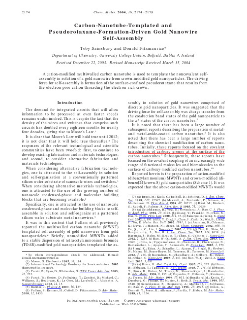
Carbon-Nanotube-Templated andPseudorotaxane-Formation-Driven Gold NanowireSelf-AssemblyToby Sainsbury and Donald Fitzmaurice*Department of Chemistry,University College Dublin,Belfield,Dublin4,Ireland Received December22,2003.Revised Manuscript Received March15,2004A cation-modified multiwalled carbon nanotube is used to template the noncovalent self-assembly in solution of a gold nanowire from crown-modified gold nanoparticles.The driving force for self-assembly is formation of the surface-confined pseudorotaxane that results from the electron-poor cation threading the electron-rich crown.IntroductionThe demand for integrated circuits that will allow information to be processed at even faster speeds remains undiminished.This is despite the fact that the density of the wires and switches that comprise such circuits has doubled every eighteen months for nearly four decades,giving rise to Moore’s Law.1It is clear that Moore’s Law will hold true until2012; it is not clear that it will hold true thereafter.2The responses of the relevant technological and scientific communities have been two-fold:first,to continue to develop existing fabrication and materials technologies; and second,to consider alternative fabrication and materials technologies.When considering alternative fabrication technolo-gies,one is attracted to the self-assembly in solution and self-organization at a conventionally patterned silicon wafer substrate of nanoscale wires and switches.3 When considering alternative materials technologies, one is attracted to the use of the growing number of nanoscale condensed-phase and molecular building blocks that are becoming available.4Specifically,one is attracted to the use of nanoscale condensed-phase and molecular building blocks to self-assemble in solution and self-organize at a patterned silicon wafer substrate metal nanowires.5It was in this context that Fullam et al.previously reported the multiwalled carbon nanotube(MWNT) templated self-assembly of gold nanowires from gold nanoparticles.6Briefly,unmodified MWNTs added to a stable dispersion of tetraoctylammonium bromide (TOAB)-modified gold nanoparticles templated the as-sembly in solution of gold nanowires comprised of discrete gold nanoparticles.It was suggested that the driving force for self-assembly was charge transfer from the conduction band states of the gold nanoparticle to theπ*states of the carbon nanotubes.7It is noted that there has been a large number of subsequent reports describing the preparation of metal-and metal-oxide-coated carbon nanotubes.8It is also noted that there has been a large number of reports describing the chemical modification of carbon nano-tubes.Initially,these reports focused on the covalent introduction of carboxy groups at the surface of the carbon nanotubes.9Subsequently,these reports have focused on the covalent coupling of an increasingly wide range of functional molecules and biomolecules to the surface of carboxy-modified carbon nanotubes.10 Reported herein is the preparation of cation-modified (dibenzylammonium)MWNTs and crown-modified(di-benzo[24]crown-8)gold nanoparticles(Scheme1).It was expected that the above cation-modified MWNTs would*To whom correspondence should be addressed.E-mail: donald.fitzmaurice@ucd.ie.(1)Moore,G.Electronics1965,38,114.(2)International Technology Roadmap for Semiconductors,2002 (/).(3)Parviz,B.;Ryan,D.;Whitesides,G.IEEE Trans.Adv.Pac.2003, 26,233.(4)Parak,W.;Gerion,D.;Pellegrino,T.;Zanchet,D.;Micheel,C.; Williams,S.;Boudreau,R.;Le Gros,M.;Larabell,C.;Alivisatos,A. Nanotechnology2003,14,15.(5)Richter,J.Physica E2003,16,157.(6)Fullam,S.;Rensmo,H.;Cottell,D.;Fitzmaurice,D.Adv.Mater. 2000,12,1430.(7)(a)Brust,M.;Kiely,C.;Bethell,D.;Schiffrin,D.J.Am.Chem. Soc.1998,120,12367.(b)Maxwell,A.;Bruhwiler,P.;Nilsson,A.; Martensson,N.Phys.Rev.B1994,49,10717.(c)Hunt,M.;Modesti, S.;Rudolf,P.;Palmer,R.Phys.Rev.B1995,51,10039.(8)(a)Satishkumar,B.;Vogl,E.;Govindaraj,A.;Rao,C.J.Phys. D:Appl.Phys.1996,29,3173.(b)Zhang,Y.;Franklin,N.;Chen,R.; Dai,H.Chem.Phys.Lett.2000,331,35.(c)Banerjee,S.;Wong,S.Nano Lett.2002,2,195.(d)Li,Y.;Ding,J.;Chen,J.;Cailu,X.;Wei,B.;Liang, J.;Wu,D.Mater.Res.Bull.2002,1847,1.(e)Azamian,B.;Coleman, K.;Davis,J.;Hanson,N.;Green,mun.2002,366.(f) Fu,Q.;Lu,C.;Liu,J.Nano Lett.2002,2,329.(g)Choi,H.;Shim,M.; Bangsaruntip,S.;Dai,H.J.Am.Chem.Soc.2002,124,9058.(h) Haremza,J.;Hahn,M.;Krauss,T.;Chen,S.;Calcines,J.Nano Lett. 2002,2,1253.(i)Han,W.-Q.;Zettl,A.J.Am.Chem.Soc.2003,125, 2062.(j)Ellis,A.;Vijayamohanan,K.;Goswami,R.;Chakrapani,N.; Ramanathan,L.;Ajayan,P.;Ramanath,G.Nano Lett.2003,3,279. (k)Jiang,K.;Eitan,A.;Schadler,L.;Ajayan,P.;Siegel,R.;Grobert, N.;Mayne,M.;Reyes-Reyes,M.;Terrones,H.;Terrones,M.Nano Lett. 2003,3,275.(l)Ravindran,S.;Chaudhary,S.;Colburn,B.;Ozkan, M.;Ozkan,C.Nano Lett.2003,3,447.(m)Han,W.-Q.;Zettl,A.Nano Lett.2003,3,681.(9)(a)Hiura,H.Mol.Cryst.Liq.Cryst.1995,267,267.(b)Hiura,H.;Ebbesen,T.;Tanigaki,K.Adv.Mater.1995,7,275.(c)Ebbesen, T.;Hiura,H.;Bisher,M.;Tracey,M.;Shreeve-Keyer,J.;Haushalter, R.Adv.Mater.1996,8,155.(d)Dujardin,E.;Ebbesen,T.;Krishnan, A.;Treacy,M.Adv.Mater.1998,10,611.(e)Burghard,M.;Krstic,V.; Duesberg,G.;Philipp,G.;Muster,J.;Roth,S.Synth.Met.1999,103, 2540.(f)Satishkumar,B.;Govindaraj,A.;Mofokeng,J.;Subbanna, G.;Rao,C.J.Phys.B:At.Mol.Opt.1996,29,4925.(g)Dillon,A.; Gennett,T.;Jones,K.;Alleman,J.;Parilla,P.;Heben,M.Adv.Mater. 1999,11,1354.2174Chem.Mater.2004,16,2174-217910.1021/cm035368k CCC:$27.50©2004American Chemical SocietyPublished on Web05/01/2004template the noncovalent self-assembly in solution of a gold nanowire from the above crown-modified gold nanoparticles(also Scheme1).This expectation was based on the fact that the electron-poor cation threads the electron-rich crown insolution to form a pseudorotaxane.11This expectation was also based on the fact that cation-modified silica and crown-modified silver nanoparticles self-assemble in solution,and that self-assembly is driven by forma-tion of the corresponding surface-confined pseudorotax-ane.12Experimental MethodsAll solvents and compounds were used as supplied by the Sigma-Aldrich Chemical Co.Ltd.,unless otherwise stated.Visible absorption spectra were recorded using a HP-8452A spectrophotometer with LabView software written to acquire the data.All visible absorption spectra were recorded against appropriate solvent or solution backgrounds.All TEMs were obtained using a JEOL2000FX TEMscan (at an accelerating voltage of80kV)for samples deposited on both uncoated(2000mesh)and carbon-coated(400mesh) copper grids.The preparation of samples for TEM analysis involved the deposition of a drop of the relevant sample onto the above grids.The excess sample was removed by wicking and the grid was allowed to dry in air.Preparation of Unmodified and Crown-Modified Gold Nanoparticles.A stable dispersion of dodecanethiol-modified gold nanoparticles was prepared in chloroform using a method similar to that described by Brust et al.13These nanoparticles were size-selectively precipitated to isolate a relatively size-monodisperse fraction(7.7nm diameter,polydispersity1.13). The near size-monodisperse fraction was subsequently modi-fied by exchange of the adsorbed thiol for a thiol incorporating a crown.The above exchange was promoted using a method similar to that described by Ahern et al.14Purification of Unmodified MWNTs.As-received MWNTs (MER Corporation,3×10-3g)were suspended by sonication (15min)in chloroform(20mL).The resulting suspension was centrifuged(5000rpm,15min)and the supernatant fraction (approximately75vol%)was retained.This procedure was repeated a further three times,with the supernatant fraction being retained in each case.Preparation of Carboxy-Modified MWNTs.MWNTs were treated as described following the method reported by Burghard et al.to introduce carboxy groups at the surface of the nanotubes.15This was achieved by refluxing the as-received MWNTs in nitric acid to produce hydroxyl,carbonyl, and carboxy groups at defects in the nanotube carbon lattice. The above hydroxyl and carbonyl groups were further oxidized to carboxy groups by treatment of the oxidized MWNTs with potassium permanganate and perchloric acid as described below.Specifically,MWNTs(3.2×10⁻³g)were added to nitric acid (3mL,69wt%)and sonicated to ensure they were dispersed. The resulting suspension of MWNTs was refluxed under magnetic stirring(4h,130°C)to oxidize the suspended MWNTs.The oxidized suspension of MWNTs was cooled and filtered using a polycarbonate membrane(Whatman-UK,0.2-µm pore diameter)and washed with deionized water(200mL). The oxidized MWNTs retained on the membrane were dis-persed in deionized water(10mL)by sonication.An acidic solution of potassium permanganate(9×10⁻³g,5.7×10-5 mol)and perchloric acid(3mL,50wt%)was then added to the stirring solution.The potassium permanganate was quenched after10min by the addition of a solution of citric acid(0.133g,6.3×10-4mol).The suspension was filtered using a polycarbonate membrane(Whatman-UK,0.2-µm pore diameter)and washed with deionized water(200mL).The resulting carboxy-modified MWNTs(3.0×10⁻³g)were dispersed in deionized water(10mL)by sonication.TEMs of the MWNTs were obtained before and after the above oxidative process.A drop of one of the following suspensions was deposited onto a carbon-coated copper grid: unmodified MWNTs(3×10⁻³g)in chloroform(10mL),or carboxy-modified MWNTs(3×10⁻³g)in water(10mL).Preparation of Cation-Modified MWNTs.The required cation-precursor,N-(4-carboxydibenzylamine)carbamate6-(10)(a)Huang,W.;Lin,Y.;Taylor,S.;Gaillard,J.;Rao,A.;Sun, Y.-P.Nano Lett.2002,2,231.(b)Huang,W.;Taylor,S.;Fu,K.;Lin, Y.;Zhang,D.;Hanks,T.;Rao,A.;Sun,Y.-P.Nano Lett.2002,2,311.(c)Pompeo,F.;Resasco,D.Nano Lett.2002,2,369.(d)Shim,M.;Wong Shi Kam,N.;Chen,R.;Li,Y.;Dai,H.Nano Lett.2002,2,285.(e)Kahn, M.;Banerjee,S.;Wong,S.Nano Lett.2002,2,1215.(f)Frehill,F.; Vos,J.;Benrezzak,S.;Koos,A.;Konya,Z.;Ruther,M.;Blau,W.; Fonseca,A.;Nagy,J.;Biro,L.;Minett,A.;in het Panhuis,M.J.Am. Chem.Soc.2002,124,13694.(g)Star,A.;Liu,Y.;Grant,K.;Ridvan, L.;Stoddart, F.;Steuerman, D.;Diehl,M.;Boukai, A.;Heath,J.Macromol.2003,36,553.(h)Stevens,J.;Huang,A.;Peng,H.;Chiang, I.;Khabashesku,V.;Margrave,J.Nano Lett.2003,3,331.(i) Chambers,G.;Carroll,C.;Farrell,G.;Dalton,A.;McNamara,M.;in het Panhuis,M.;Byrne,H.Nano Lett.2003,3,843.(j)Besteman,K.; Lee,J.-O.;Wiertz,F.;Heering,H.;Dekker,C.Nano Lett.2003,3,727.(11)(a)Ashton,P.;Campbell,P.;Chrystal,E.;Glink,P.;Menzer, S.;Philp,D.;Spencer,N.;Stoddart,J.;Tasker,P.;Williams,D.Angew. Chem.Intl.Ed.Engl.1995,17,1865.(b)Ashton,P.;Fyfe,M.; Hickingbottom,S.;Menzer,S.;Stoddart,J.;White,A.;Williams,D. Chem.Eur.J.1998,4,577.(12)(a)Fitzmaurice,D.;Rao,S.;Preece,J.;Stoddart,J.;Wenger, S.;Zaccheroni,N.Angew.Chem.Intl.Ed.1999,38,1147.(b)Ryan, D.;Rao,S.;Rensmo,H.;Fitzmaurice, D.;Preece,J.;Wenger,S.; Stoddart,J.;Zaccheroni,N.J.Am.Chem.Soc.2000,122,6252.(13)Brust,M.;Walker,M.;Bethell,D.;Schiffrin,D.;Whyman,R. mun.1994,801.(14)Ahern,D.;Rao,S.;Fitzmaurice,D.J.Phys.Chem.B.1999, 103,1821.(15)Burghard,M.;Krstic,V.;Duesberg,G.;Philipp,G.;Muster, J.;Roth,S.Synth.Met.1999,103,2540.Scheme1.A Cation-Modified Multiwalled CarbonNanotube Templates the NoncovalentSelf-Assembly in Solution of a Gold Nanowire fromCrown-Modified Gold NanoparticlesSelf-Assembly of Gold Nanowire Using MWNTs Chem.Mater.,Vol.16,No.11,20042175nitroveratryloxy chloroformate,was prepared as described in detail elsewhere.16Modification of the MWNTs required two amide-coupling reactions:the first was between carboxy-modified MWNTs and ethylenediamine,and the second was between the re-maining amine of the coupled ethylenediamine and the cation-precursor,as described below.Specifically,ethylenediamine (5.6×10⁻⁶mL,8.3×10-4mol)was added,followed by EDAC (1.91×10⁻¹g,1×10-3mol)and DMAP (12×10-3g,1×10-4mol),to a suspension of carboxy-modified MWNTs (3×10⁻³g)in water (10mL),and the resulting suspension was stirred for 12h.The sus-pension was filtered on a polycarbonate membrane (Whatman-UK,0.2-µm pore diameter)and washed with deionized water (200mL).The above membrane was sonicated in deionized water (10mL)to recover the modified MWNTs.The water was removed using a rotary evaporator,which was followed by drying on a vacuum line (3h).The modified MWNTs were dispersed in anhydrous di-chloromethane (10mL)by sonication.The cation-precursor (10.6×10⁻³g,2.3×10-5mol),followed 30s later by EDAC (20×10-3g,1.0×10-4mol)and DMAP (1.30×10⁻³g,1.0×10-5mol),was added to the stirred suspension of the above modified MWNTs.The suspension was stirred for 16h in a sealed flask in the absence of light.The material was filtered using an Anodisc filter membrane (Whatman-UK,0.02-µm pore diameter),and washed with anhydrous dichloromethane (100mL)followed by ethanol (100mL).The above membrane was sonicated in ethanol (10mL)to recover the cation-precursor-modified MWNTs.The output from a UV-H 253BL Ultra Violet 250-W UV lamp was used as the light source for photoactivation.Hexa-fluorophosphoric acid was added to a suspension of cation-precursor-modified-MWNTs (3×10⁻³g)in ethanol (10mL)until the pH was 2.The suspension was irradiated using the UV lamp for 2h,filtered on a polycarbonate membrane (Whatman-UK,0.2-µm pore diameter),and washed with ethanol (100mL).This procedure results in the photoactivated deprotection of the dibenzylamine in acidic conditions in the presence of a counterion,PF 6-.The resultant salt of thedibenzylammonium cation (cation)is formed.The cation-modified MWNTs were dispersed by sonication in ethanol (10mL).The ethanol was removed by rotary evaporation followed by drying on a vacuum line (3h).The dry cation-modified MWNTs were dispersed by sonication in chloroform (10mL).Templated Self-Assembly of Gold Nanowires.The templated self-assembly of gold nanowires from these compo-nents is described in detail in the Results and Discussion section of this paper and therefore is not described in this section.Results and DiscussionPreparation and Characterization of Cation-Modified MWNTs.A TEM of the as-received MWNTs reveals that there is a significant amount of amorphous carbon present (Figure 1a).To remove this impurity,the above MWNTs were sonicated in chloroform,cen-trifuged,and filtered.A TEM of the purified MWNTs shows a well-dispersed sample with an average nano-tube diameter of 24nm and an average nanotube length of 2µm (Figure 1b).The oxidation of MWNTs under strongly acidic condi-tions results in the introduction of carboxylic,hydroxyl,and carbonyl groups at defect sites on the surface of the MWNTs in the approximate ratio 4:2:1(Scheme 2).9a -d To maximize the number of functional groups suitable(16)(a)Ryan,D.;Nagle,L.;Rensmo,H.;Fitzmaurice,D.J.Phys.Chem.B .2002,106,5371.(b)Ryan,D.;Nagle,L.;Fitzmaurice,D.Nano Lett.,published online Mar.18,2004,/10.1021/nl0351340.Figure 1.As-received multiwalled carbon nanotubes (a)prior to purification,(b)following purification,and (c and d)following carboxy-modification.Scheme 2.Introduction of Carboxyl,Hydroxy,and Carbonyl Groups at the Defect Sites Present at the Surface of the As-Received Multiwalled Carbon Nanotubes Followed by Conversion of Carbonyland Hydroxy Groups to Carboxy Groups2176Chem.Mater.,Vol.16,No.11,2004Sainsbury and Fitzmauricefor covalent modification,the above hydroxy and car-bonyl groups were subsequently oxidized to carboxy groups (Scheme 2).9e It is estimated that the density of carboxy groups present at the surface of the above carboxy-modified MWNT is 4.2per 100Å2.This esti-mate is based on findings reported by Hiura and co-workers,concerning the number of carbon atoms at the surface of a known mass of MWNTs,9a and by Rao et al.,concerning the number of carboxy groups present following acid oxidation of a known mass of MWNTs.9f A TEM of the carboxy-modified MWNTs shows a well-dispersed material with an average diameter of 24nm and an average length of 2µm (Figure 1c and d).The cation-precursor (N -(4-carboxydibenzylamine)-carbamate-6-nitroveratryloxy chloroformate)was co-valently coupled to the surface of the carboxy-modified MWNTs.Because the carboxy-modified MWNTs and cation precursor both have carboxy moieties,ethylene-diamine was deemed a suitable linker.Coupling was achieved by EDAC-catalyzed amide formation between the carboxy-modified MWNTs and ethylenediamine,and between ethylenediamine and the carboxy group of the cation precursor (Scheme 3).To minimize the possibility of cross-linking of MWNTs,the amount of ethylenedi-amine used was in excess of the number of carboxy groups present at the surface of the MWNTs.No aggregation of the MWNTs was observed before or after the above reaction.The surface density of attached molecules in the resulting cation-precursor-modified MWNTs was estimated to be 2.7per 100Å2,assuming a yield of 80%for each amide formation step.The cation-modified MWNT-templated self-assembly of crown-modified gold nanoparticles requires the priordeprotection of the cation precursor under acidic condi-tions to form the cation.This is achieved by photolysis of the cation precursor in the presence of hexafluoro-phosphoric acid.Preparation and Characterization of Modified Gold Nanoparticles.Briefly,thiol-modified (do-decanethiol)gold nanoparticles were prepared using a method similar to that reported by Brust et al .13These nanoparticles were selectively precipitated to isolate a relatively size-monodisperse fraction.These nanopar-ticles were further modified by exchange of the initially adsorbed thiol molecules for crown-thiol molecules (dodecane thiol incorporating a crown moiety in the terminal position).14A TEM analysis of 200nanopar-ticles yields an average diameter of 7.7nm and a polydispersity of 1.13(Figure 2).Visible absorption spectroscopy confirmed the exist-ence of the characteristic absorbance maximum at 520nm,which was assigned to the surface plasmon reso-nance of the gold nanoparticles.Templated Self-Assembly of Gold Nanowires.To initiate the templated self-assembly of a gold nanowire in solution,a dispersion of crown-modified gold nano-particles (0.2mL,4×1012nanoparticles mL -1)in chloroform was added to a freshly sonicated suspension of cation-modified MWNTs (0.5mL,15×10-5g mL -1)also in chloroform.Following equilibration of this suspension for 24h a black precipitate was obtained.TEM analysis revealed the formation of gold nanowires (Figure 3a).A series of related control experiments was performed under similar conditions.Specifically,a dispersion of crown-modified gold nanoparticles (0.2mL,4×1012nanoparticles mL -1)in chloroform was added to a freshly sonicated suspension of cation-precursor-modified MWNTs (0.5mL,15×10-5g mL -1)also in chloroform.Following equilibration of this suspension for 24h no black precipitate was observed.TEM analy-sis revealed no gold nanowire formation (Figure 3b).A dispersion of crown-modified gold nanoparticles (0.2mL,4×1012nanoparticles mL -1)in chloroform was added to a freshly sonicated suspension of unmodified MWNTs (0.66mL,11×10-5g mL -1)also in chloroform.Following equilibration of this suspension for 24h no black precipitate was observed.TEM analysis revealed no gold nanowire formation (Figure 3c).A dispersion of thiol-modified gold nanoparticles (0.2mL,4×1012nanoparticles mL -1)in chloroform was added to a freshly sonicated suspension of cation-modified MWNTs (0.5mL,5×10-5g mL -1)also in chloroform.Following equilibration of this suspensionScheme 3.Covalent Coupling of One End of the Ethylenediamine Linker to a Carbonyl Group Present at the Surface of a Carboxy-Modified Multiwalled Nanotube Followed by Coupling of the Other End of the Linker to the CationPrecursorFigure 2.Crown-modified gold nanoparticles.Self-Assembly of Gold Nanowire Using MWNTs Chem.Mater.,Vol.16,No.11,20042177for 24h no black precipitate was observed.TEM analysis revealed no gold nanowire formation (Figure 3d).A further series of control experiments was also performed under similar conditions.A dispersion of crown-modified gold nanoparticles (0.2mL,4×1012nanoparticles mL -1)in chloroform was added to a freshly sonicated suspension of cation-modified MWNTs (0.66mL,11×10-5g mL -1)also in chloroform,in the presence of an excess of free cation (dibenzylammonium hexafluorophosphoric acid,3.3×10-3g,9.5×10-6mol).Following equilibration of this suspension for 24h no black precipitate was observed.TEM analysis revealed no gold nanowire formation (Figure 3e).Also,a dispersion of crown-modified gold nano-particles (0.2mL,4×1012nanoparticles mL -1)in chloroform was added to a freshly sonicated suspension of cation-modified MWNTs (0.66mL,11×10-5g mL -1)also in chloroform,in the presence of an excess of free crown (dibenzo24-crown-8,4.3×10-3g,9.5×10-6mol).Following equilibration of this suspension for 24h no black precipitate was observed.TEM analysis revealed no gold nanowire formation (Figure 3f).It is clear from these findings that MWNT-templated self-assembly of a gold nanowire is observed only when uncomplexed cation is present on the surface of the MWNTs and uncomplexed crown is present at the surface of the gold nanoparticle.On this basis it may be concluded that the templated self-assembly of nano-wires is driven by formation of the surface-confined pseudorotaxane shown in Scheme 1.The fact that coverage of the MWNT by the adsorbed gold nanoparticles is complete would suggest that the coverage of cation at the surface of the cation-modified MWNTs is extensive.It is estimated that each molecule adsorbed at the surface of a cation-modified MWNT occupies an area of 6.1×6.1Å.9On the basis that the projected area of a crown-modified nanoparticle within 15Å(the length of an adsorbed cation)of the MWNT is 1461Å2,it is further estimated that each particle is in contact with up to 40cations,consistent with the high degree of coverage observed.The possibility that the driving force for the templated nanowire assembly is charge transfer from the crown-modified nanoparticle to the cation-modified MWNT can be excluded.This assertion is supported by the findingsFigure 3.Transmission electron micrographs of (a)crown-modified gold nanoparticles adsorbed at the surface of a cation-modified multiwalled carbon nanotube.The same gold nanoparticles are not adsorbed at the surface of (b)a cation-precursor-modified multiwalled carbon nanotube or (c)an unmodified multiwalled carbon nanotube.Similarly,(d)thiol-modified gold nanoparticles (prior to exchange of thiol incorporating)are not adsorbed at a cation-modified multiwalled carbon nanotube.Crown-modified gold nanoparticles are not adsorbed at the surface of cation-modified multiwalled carbon nanotubes in the presence of (e)free cation or (f)free crown in solution.2178Chem.Mater.,Vol.16,No.11,2004Sainsbury andFitzmauricethat there is no adsorption of crown-modified gold nanoparticles at cation-precursor-modified MWNTs or cation-modified MWNTs in the presence of added crown, both of which would be expected to exhibit similar degrees of steric hindrance to cation-modified MWNTs.ConclusionsThis paper describes the use of a dibenzylammonium-cation-modified multiwalled carbon nanotube to tem-plate the noncovalent self-assembly of a gold nanowire in solution from dibenzo[24]crown-8-modified gold nano-particles.The driving force for self-assembly is forma-tion of a surface-confined pseudorotaxane.By exploiting these capabilities and insights,allied to those recently reported in relation to the patterning of nanoparticles14,17and those that will be reported in relation to the carbon-nanotube-templated self-assembly of other nanoparticles,18it ought be possible to pattern nanotubes by the adsorption of a range of nanoparticles. This,in turn,will offer the prospect of diverse nanoscale components for which many applications can be foreseen. CM035368K(17)(a)Nagle,L.;Ryan,D.;Cobbe,S.;Fitzmaurice,D.Nano Lett.2003,3,51.(b)Nagle,L.;Fitzmaurice,D.Adv.Mater.2003,15,933.(c)Ryan,D.;Nagle,L.;Cobbe,S.;Fitzmaurice,D.International PCTApplication,2002,application PCT/IE02/00120.(18)Sainsbury,T.;Fitzmaurice,D.unpublished findings.Self-Assembly of Gold Nanowire Using MWNTs Chem.Mater.,Vol.16,No.11,20042179。
碳纳米管的性质与应用
碳纳米管的性质与应用【摘要】本文主要介绍了碳纳米管的结构特点,制备方法,特殊性质,由于碳纳米管独特性质而产生的广泛应用,并对其前景进行展望。
【关键词】碳纳米管场发射复合材料优良性能【前言】自日本NEC科学家Lijima发现碳纳米管以来,碳纳米管研究一直是国际新材料领域研究的热点。
由于碳纳米管具有特殊的导电性能、力学性质及物理化学性质等,故其在许多领域具有其广阔的应用前景,自问世以来即引起广泛关注。
目前,国内外有许多科学家对碳纳米管进行研究,科研成果颇丰,尤其是碳纳米管在复合材料、储氢及催化等领域的应用。
【正文】一、碳纳米管的结构碳纳米管中碳原子以sp2杂化为主,同时六角型网格结构存在一定程度的弯曲,形成空间拓扑结构,其中可形成一定的sp3杂化键,即形成的化学键同时具有sp2和sp3混合杂化状态,而这些p 轨道彼此交叠在碳纳米管石墨烯片层外形成高度离域化的大π键,碳纳米管外表面的大π键是碳纳米管与一些具有共轭性能的大分子以非共价键复合的化学基础[1]。
对多壁碳纳米管的光电子能谱研究结果表明,不论单壁碳纳米管还是多壁碳纳米管,其表面都结合有一定的官能基团,而且不同制备方法获得的碳纳米管由于制备方法各异,后处理过程不同而具有不同的表面结构。
一般来讲,单壁碳纳米管具有较高的化学惰性,其表面要纯净一些,而多壁碳纳米管表面要活泼得多,结合有大量的表面基团,如羧基等。
以变角X 光电子能谱对碳纳米管的表面检测结果表明,单壁碳纳米管表面具有化学惰性,化学结构比较简单,而且随着碳纳米管管壁层数的增加,缺陷和化学反应性增强,表面化学结构趋向复杂化。
内层碳原子的化学结构比较单一,外层碳原子的化学组成比较复杂,而且外层碳原子上往往沉积有大量的无定形碳。
由于具有物理结构和化学结构的不均匀性,碳纳米管中大量的表面碳原子具有不同的表面微环境,因此也具有能量的不均一性[2]。
碳纳米管不总是笔直的,而是局部区域出现凸凹现象,这是由于在六边形编制过程中出现了五边形和七边形。
碳纳米管的改性研究进展

碳纳米管的改性研究进展摘要:碳纳米管因其独特的结构与优异的性能,在许多领域具有巨大的应用潜力而引起了广泛的关注。
由于碳纳米管不溶于水和有机溶剂,极大地制约了其性能的应用,因此碳纳米管的功能化改性就成为目前研究的热点。
本文简要介绍了碳纳米管及其性质作,详细阐述了碳纳米管的改性研究进展,并对今后的研究方向进行了展望。
关键词:碳纳米管;结构与性能;功能化;共价改性;非共价改性1. 碳纳米管及其性能简介1.1碳纳米管的结构碳纳米管(Carbon Nanotubes,CNTs)是1991年由日本筑波NEC公司基础研究实验室的Iijima在高分辨透射电子显微镜下检验石墨电弧设备中产生的球状碳分子时意外发现的一种具有一维管状结构的碳纳米材料。
因其独特的准一维管状分子结构、优异的力学、电学和化学性质及其在高科技领域中潜在的应用价值,引起了世界各国科学家们的广泛关注,由此引发了碳纳米管的研究热潮和十多年来纳米科学和技术的飞速发展。
碳纳米管是单层或多层石墨片围绕中心轴按一定的螺旋角卷曲而成的无缝、中空的微管,每层纳米管是一个由碳原子通过SP2杂化与周围3个碳原子完全键合后所构成的六边形平面组成的圆柱面。
根据构成管壁碳原子层数的不同,CNTs可以分为:单壁碳纳米管(single-walled carbon nanotube,SWNT)和多壁碳纳米管(multi-walled carbon nanotube, MWNT)两种形式。
MWNTs的层间接近ABAB堆垛,其层数从2~50不等,层间距为0.34±0.01nm,与石墨层间距(0.34nm)相当。
MWNTs的典型直径和长度分别为2~30nm和0.1~50μm;SWNTs典型的直径和长度分别为0.75~3nm和1~50μm。
与MWNTs 比,SWNTs是由单层圆柱型石墨层构成,其直径的分布范围小,缺陷少,具有更高的均匀一致性。
无论是MWNTs还是SWNTs都具有很大的长径比,一般为100~1000,最大可达到1000~10000,可以认为是一维分子。
- 1、下载文档前请自行甄别文档内容的完整性,平台不提供额外的编辑、内容补充、找答案等附加服务。
- 2、"仅部分预览"的文档,不可在线预览部分如存在完整性等问题,可反馈申请退款(可完整预览的文档不适用该条件!)。
- 3、如文档侵犯您的权益,请联系客服反馈,我们会尽快为您处理(人工客服工作时间:9:00-18:30)。
Carbon-Nanotube-Templated andPseudorotaxane-Formation-Driven Gold NanowireSelf-AssemblyToby Sainsbury and Donald Fitzmaurice*Department of Chemistry,University College Dublin,Belfield,Dublin4,Ireland Received December22,2003.Revised Manuscript Received March15,2004A cation-modified multiwalled carbon nanotube is used to template the noncovalent self-assembly in solution of a gold nanowire from crown-modified gold nanoparticles.The driving force for self-assembly is formation of the surface-confined pseudorotaxane that results from the electron-poor cation threading the electron-rich crown.IntroductionThe demand for integrated circuits that will allow information to be processed at even faster speeds remains undiminished.This is despite the fact that the density of the wires and switches that comprise such circuits has doubled every eighteen months for nearly four decades,giving rise to Moore’s Law.1It is clear that Moore’s Law will hold true until2012; it is not clear that it will hold true thereafter.2The responses of the relevant technological and scientific communities have been two-fold:first,to continue to develop existing fabrication and materials technologies; and second,to consider alternative fabrication and materials technologies.When considering alternative fabrication technolo-gies,one is attracted to the self-assembly in solution and self-organization at a conventionally patterned silicon wafer substrate of nanoscale wires and switches.3 When considering alternative materials technologies, one is attracted to the use of the growing number of nanoscale condensed-phase and molecular building blocks that are becoming available.4Specifically,one is attracted to the use of nanoscale condensed-phase and molecular building blocks to self-assemble in solution and self-organize at a patterned silicon wafer substrate metal nanowires.5It was in this context that Fullam et al.previously reported the multiwalled carbon nanotube(MWNT) templated self-assembly of gold nanowires from gold nanoparticles.6Briefly,unmodified MWNTs added to a stable dispersion of tetraoctylammonium bromide (TOAB)-modified gold nanoparticles templated the as-sembly in solution of gold nanowires comprised of discrete gold nanoparticles.It was suggested that the driving force for self-assembly was charge transfer from the conduction band states of the gold nanoparticle to theπ*states of the carbon nanotubes.7It is noted that there has been a large number of subsequent reports describing the preparation of metal-and metal-oxide-coated carbon nanotubes.8It is also noted that there has been a large number of reports describing the chemical modification of carbon nano-tubes.Initially,these reports focused on the covalent introduction of carboxy groups at the surface of the carbon nanotubes.9Subsequently,these reports have focused on the covalent coupling of an increasingly wide range of functional molecules and biomolecules to the surface of carboxy-modified carbon nanotubes.10 Reported herein is the preparation of cation-modified (dibenzylammonium)MWNTs and crown-modified(di-benzo[24]crown-8)gold nanoparticles(Scheme1).It was expected that the above cation-modified MWNTs would*To whom correspondence should be addressed.E-mail: donald.fitzmaurice@ucd.ie.(1)Moore,G.Electronics1965,38,114.(2)International Technology Roadmap for Semiconductors,2002 (/).(3)Parviz,B.;Ryan,D.;Whitesides,G.IEEE Trans.Adv.Pac.2003, 26,233.(4)Parak,W.;Gerion,D.;Pellegrino,T.;Zanchet,D.;Micheel,C.; Williams,S.;Boudreau,R.;Le Gros,M.;Larabell,C.;Alivisatos,A. Nanotechnology2003,14,15.(5)Richter,J.Physica E2003,16,157.(6)Fullam,S.;Rensmo,H.;Cottell,D.;Fitzmaurice,D.Adv.Mater. 2000,12,1430.(7)(a)Brust,M.;Kiely,C.;Bethell,D.;Schiffrin,D.J.Am.Chem. Soc.1998,120,12367.(b)Maxwell,A.;Bruhwiler,P.;Nilsson,A.; Martensson,N.Phys.Rev.B1994,49,10717.(c)Hunt,M.;Modesti, S.;Rudolf,P.;Palmer,R.Phys.Rev.B1995,51,10039.(8)(a)Satishkumar,B.;Vogl,E.;Govindaraj,A.;Rao,C.J.Phys. D:Appl.Phys.1996,29,3173.(b)Zhang,Y.;Franklin,N.;Chen,R.; Dai,H.Chem.Phys.Lett.2000,331,35.(c)Banerjee,S.;Wong,S.Nano Lett.2002,2,195.(d)Li,Y.;Ding,J.;Chen,J.;Cailu,X.;Wei,B.;Liang, J.;Wu,D.Mater.Res.Bull.2002,1847,1.(e)Azamian,B.;Coleman, K.;Davis,J.;Hanson,N.;Green,mun.2002,366.(f) Fu,Q.;Lu,C.;Liu,J.Nano Lett.2002,2,329.(g)Choi,H.;Shim,M.; Bangsaruntip,S.;Dai,H.J.Am.Chem.Soc.2002,124,9058.(h) Haremza,J.;Hahn,M.;Krauss,T.;Chen,S.;Calcines,J.Nano Lett. 2002,2,1253.(i)Han,W.-Q.;Zettl,A.J.Am.Chem.Soc.2003,125, 2062.(j)Ellis,A.;Vijayamohanan,K.;Goswami,R.;Chakrapani,N.; Ramanathan,L.;Ajayan,P.;Ramanath,G.Nano Lett.2003,3,279. (k)Jiang,K.;Eitan,A.;Schadler,L.;Ajayan,P.;Siegel,R.;Grobert, N.;Mayne,M.;Reyes-Reyes,M.;Terrones,H.;Terrones,M.Nano Lett. 2003,3,275.(l)Ravindran,S.;Chaudhary,S.;Colburn,B.;Ozkan, M.;Ozkan,C.Nano Lett.2003,3,447.(m)Han,W.-Q.;Zettl,A.Nano Lett.2003,3,681.(9)(a)Hiura,H.Mol.Cryst.Liq.Cryst.1995,267,267.(b)Hiura,H.;Ebbesen,T.;Tanigaki,K.Adv.Mater.1995,7,275.(c)Ebbesen, T.;Hiura,H.;Bisher,M.;Tracey,M.;Shreeve-Keyer,J.;Haushalter, R.Adv.Mater.1996,8,155.(d)Dujardin,E.;Ebbesen,T.;Krishnan, A.;Treacy,M.Adv.Mater.1998,10,611.(e)Burghard,M.;Krstic,V.; Duesberg,G.;Philipp,G.;Muster,J.;Roth,S.Synth.Met.1999,103, 2540.(f)Satishkumar,B.;Govindaraj,A.;Mofokeng,J.;Subbanna, G.;Rao,C.J.Phys.B:At.Mol.Opt.1996,29,4925.(g)Dillon,A.; Gennett,T.;Jones,K.;Alleman,J.;Parilla,P.;Heben,M.Adv.Mater. 1999,11,1354.2174Chem.Mater.2004,16,2174-217910.1021/cm035368k CCC:$27.50©2004American Chemical SocietyPublished on Web05/01/2004template the noncovalent self-assembly in solution of a gold nanowire from the above crown-modified gold nanoparticles(also Scheme1).This expectation was based on the fact that the electron-poor cation threads the electron-rich crown insolution to form a pseudorotaxane.11This expectation was also based on the fact that cation-modified silica and crown-modified silver nanoparticles self-assemble in solution,and that self-assembly is driven by forma-tion of the corresponding surface-confined pseudorotax-ane.12Experimental MethodsAll solvents and compounds were used as supplied by the Sigma-Aldrich Chemical Co.Ltd.,unless otherwise stated.Visible absorption spectra were recorded using a HP-8452A spectrophotometer with LabView software written to acquire the data.All visible absorption spectra were recorded against appropriate solvent or solution backgrounds.All TEMs were obtained using a JEOL2000FX TEMscan (at an accelerating voltage of80kV)for samples deposited on both uncoated(2000mesh)and carbon-coated(400mesh) copper grids.The preparation of samples for TEM analysis involved the deposition of a drop of the relevant sample onto the above grids.The excess sample was removed by wicking and the grid was allowed to dry in air.Preparation of Unmodified and Crown-Modified Gold Nanoparticles.A stable dispersion of dodecanethiol-modified gold nanoparticles was prepared in chloroform using a method similar to that described by Brust et al.13These nanoparticles were size-selectively precipitated to isolate a relatively size-monodisperse fraction(7.7nm diameter,polydispersity1.13). The near size-monodisperse fraction was subsequently modi-fied by exchange of the adsorbed thiol for a thiol incorporating a crown.The above exchange was promoted using a method similar to that described by Ahern et al.14Purification of Unmodified MWNTs.As-received MWNTs (MER Corporation,3×10-3g)were suspended by sonication (15min)in chloroform(20mL).The resulting suspension was centrifuged(5000rpm,15min)and the supernatant fraction (approximately75vol%)was retained.This procedure was repeated a further three times,with the supernatant fraction being retained in each case.Preparation of Carboxy-Modified MWNTs.MWNTs were treated as described following the method reported by Burghard et al.to introduce carboxy groups at the surface of the nanotubes.15This was achieved by refluxing the as-received MWNTs in nitric acid to produce hydroxyl,carbonyl, and carboxy groups at defects in the nanotube carbon lattice. The above hydroxyl and carbonyl groups were further oxidized to carboxy groups by treatment of the oxidized MWNTs with potassium permanganate and perchloric acid as described below.Specifically,MWNTs(3.2×10⁻³g)were added to nitric acid (3mL,69wt%)and sonicated to ensure they were dispersed. The resulting suspension of MWNTs was refluxed under magnetic stirring(4h,130°C)to oxidize the suspended MWNTs.The oxidized suspension of MWNTs was cooled and filtered using a polycarbonate membrane(Whatman-UK,0.2-µm pore diameter)and washed with deionized water(200mL). The oxidized MWNTs retained on the membrane were dis-persed in deionized water(10mL)by sonication.An acidic solution of potassium permanganate(9×10⁻³g,5.7×10-5 mol)and perchloric acid(3mL,50wt%)was then added to the stirring solution.The potassium permanganate was quenched after10min by the addition of a solution of citric acid(0.133g,6.3×10-4mol).The suspension was filtered using a polycarbonate membrane(Whatman-UK,0.2-µm pore diameter)and washed with deionized water(200mL).The resulting carboxy-modified MWNTs(3.0×10⁻³g)were dispersed in deionized water(10mL)by sonication.TEMs of the MWNTs were obtained before and after the above oxidative process.A drop of one of the following suspensions was deposited onto a carbon-coated copper grid: unmodified MWNTs(3×10⁻³g)in chloroform(10mL),or carboxy-modified MWNTs(3×10⁻³g)in water(10mL).Preparation of Cation-Modified MWNTs.The required cation-precursor,N-(4-carboxydibenzylamine)carbamate6-(10)(a)Huang,W.;Lin,Y.;Taylor,S.;Gaillard,J.;Rao,A.;Sun, Y.-P.Nano Lett.2002,2,231.(b)Huang,W.;Taylor,S.;Fu,K.;Lin, Y.;Zhang,D.;Hanks,T.;Rao,A.;Sun,Y.-P.Nano Lett.2002,2,311.(c)Pompeo,F.;Resasco,D.Nano Lett.2002,2,369.(d)Shim,M.;Wong Shi Kam,N.;Chen,R.;Li,Y.;Dai,H.Nano Lett.2002,2,285.(e)Kahn, M.;Banerjee,S.;Wong,S.Nano Lett.2002,2,1215.(f)Frehill,F.; Vos,J.;Benrezzak,S.;Koos,A.;Konya,Z.;Ruther,M.;Blau,W.; Fonseca,A.;Nagy,J.;Biro,L.;Minett,A.;in het Panhuis,M.J.Am. Chem.Soc.2002,124,13694.(g)Star,A.;Liu,Y.;Grant,K.;Ridvan, L.;Stoddart, F.;Steuerman, D.;Diehl,M.;Boukai, A.;Heath,J.Macromol.2003,36,553.(h)Stevens,J.;Huang,A.;Peng,H.;Chiang, I.;Khabashesku,V.;Margrave,J.Nano Lett.2003,3,331.(i) Chambers,G.;Carroll,C.;Farrell,G.;Dalton,A.;McNamara,M.;in het Panhuis,M.;Byrne,H.Nano Lett.2003,3,843.(j)Besteman,K.; Lee,J.-O.;Wiertz,F.;Heering,H.;Dekker,C.Nano Lett.2003,3,727.(11)(a)Ashton,P.;Campbell,P.;Chrystal,E.;Glink,P.;Menzer, S.;Philp,D.;Spencer,N.;Stoddart,J.;Tasker,P.;Williams,D.Angew. Chem.Intl.Ed.Engl.1995,17,1865.(b)Ashton,P.;Fyfe,M.; Hickingbottom,S.;Menzer,S.;Stoddart,J.;White,A.;Williams,D. Chem.Eur.J.1998,4,577.(12)(a)Fitzmaurice,D.;Rao,S.;Preece,J.;Stoddart,J.;Wenger, S.;Zaccheroni,N.Angew.Chem.Intl.Ed.1999,38,1147.(b)Ryan, D.;Rao,S.;Rensmo,H.;Fitzmaurice, D.;Preece,J.;Wenger,S.; Stoddart,J.;Zaccheroni,N.J.Am.Chem.Soc.2000,122,6252.(13)Brust,M.;Walker,M.;Bethell,D.;Schiffrin,D.;Whyman,R. mun.1994,801.(14)Ahern,D.;Rao,S.;Fitzmaurice,D.J.Phys.Chem.B.1999, 103,1821.(15)Burghard,M.;Krstic,V.;Duesberg,G.;Philipp,G.;Muster, J.;Roth,S.Synth.Met.1999,103,2540.Scheme1.A Cation-Modified Multiwalled CarbonNanotube Templates the NoncovalentSelf-Assembly in Solution of a Gold Nanowire fromCrown-Modified Gold NanoparticlesSelf-Assembly of Gold Nanowire Using MWNTs Chem.Mater.,Vol.16,No.11,20042175nitroveratryloxy chloroformate,was prepared as described in detail elsewhere.16Modification of the MWNTs required two amide-coupling reactions:the first was between carboxy-modified MWNTs and ethylenediamine,and the second was between the re-maining amine of the coupled ethylenediamine and the cation-precursor,as described below.Specifically,ethylenediamine (5.6×10⁻⁶mL,8.3×10-4mol)was added,followed by EDAC (1.91×10⁻¹g,1×10-3mol)and DMAP (12×10-3g,1×10-4mol),to a suspension of carboxy-modified MWNTs (3×10⁻³g)in water (10mL),and the resulting suspension was stirred for 12h.The sus-pension was filtered on a polycarbonate membrane (Whatman-UK,0.2-µm pore diameter)and washed with deionized water (200mL).The above membrane was sonicated in deionized water (10mL)to recover the modified MWNTs.The water was removed using a rotary evaporator,which was followed by drying on a vacuum line (3h).The modified MWNTs were dispersed in anhydrous di-chloromethane (10mL)by sonication.The cation-precursor (10.6×10⁻³g,2.3×10-5mol),followed 30s later by EDAC (20×10-3g,1.0×10-4mol)and DMAP (1.30×10⁻³g,1.0×10-5mol),was added to the stirred suspension of the above modified MWNTs.The suspension was stirred for 16h in a sealed flask in the absence of light.The material was filtered using an Anodisc filter membrane (Whatman-UK,0.02-µm pore diameter),and washed with anhydrous dichloromethane (100mL)followed by ethanol (100mL).The above membrane was sonicated in ethanol (10mL)to recover the cation-precursor-modified MWNTs.The output from a UV-H 253BL Ultra Violet 250-W UV lamp was used as the light source for photoactivation.Hexa-fluorophosphoric acid was added to a suspension of cation-precursor-modified-MWNTs (3×10⁻³g)in ethanol (10mL)until the pH was 2.The suspension was irradiated using the UV lamp for 2h,filtered on a polycarbonate membrane (Whatman-UK,0.2-µm pore diameter),and washed with ethanol (100mL).This procedure results in the photoactivated deprotection of the dibenzylamine in acidic conditions in the presence of a counterion,PF 6-.The resultant salt of thedibenzylammonium cation (cation)is formed.The cation-modified MWNTs were dispersed by sonication in ethanol (10mL).The ethanol was removed by rotary evaporation followed by drying on a vacuum line (3h).The dry cation-modified MWNTs were dispersed by sonication in chloroform (10mL).Templated Self-Assembly of Gold Nanowires.The templated self-assembly of gold nanowires from these compo-nents is described in detail in the Results and Discussion section of this paper and therefore is not described in this section.Results and DiscussionPreparation and Characterization of Cation-Modified MWNTs.A TEM of the as-received MWNTs reveals that there is a significant amount of amorphous carbon present (Figure 1a).To remove this impurity,the above MWNTs were sonicated in chloroform,cen-trifuged,and filtered.A TEM of the purified MWNTs shows a well-dispersed sample with an average nano-tube diameter of 24nm and an average nanotube length of 2µm (Figure 1b).The oxidation of MWNTs under strongly acidic condi-tions results in the introduction of carboxylic,hydroxyl,and carbonyl groups at defect sites on the surface of the MWNTs in the approximate ratio 4:2:1(Scheme 2).9a -d To maximize the number of functional groups suitable(16)(a)Ryan,D.;Nagle,L.;Rensmo,H.;Fitzmaurice,D.J.Phys.Chem.B .2002,106,5371.(b)Ryan,D.;Nagle,L.;Fitzmaurice,D.Nano Lett.,published online Mar.18,2004,/10.1021/nl0351340.Figure 1.As-received multiwalled carbon nanotubes (a)prior to purification,(b)following purification,and (c and d)following carboxy-modification.Scheme 2.Introduction of Carboxyl,Hydroxy,and Carbonyl Groups at the Defect Sites Present at the Surface of the As-Received Multiwalled Carbon Nanotubes Followed by Conversion of Carbonyland Hydroxy Groups to Carboxy Groups2176Chem.Mater.,Vol.16,No.11,2004Sainsbury and Fitzmauricefor covalent modification,the above hydroxy and car-bonyl groups were subsequently oxidized to carboxy groups (Scheme 2).9e It is estimated that the density of carboxy groups present at the surface of the above carboxy-modified MWNT is 4.2per 100Å2.This esti-mate is based on findings reported by Hiura and co-workers,concerning the number of carbon atoms at the surface of a known mass of MWNTs,9a and by Rao et al.,concerning the number of carboxy groups present following acid oxidation of a known mass of MWNTs.9f A TEM of the carboxy-modified MWNTs shows a well-dispersed material with an average diameter of 24nm and an average length of 2µm (Figure 1c and d).The cation-precursor (N -(4-carboxydibenzylamine)-carbamate-6-nitroveratryloxy chloroformate)was co-valently coupled to the surface of the carboxy-modified MWNTs.Because the carboxy-modified MWNTs and cation precursor both have carboxy moieties,ethylene-diamine was deemed a suitable linker.Coupling was achieved by EDAC-catalyzed amide formation between the carboxy-modified MWNTs and ethylenediamine,and between ethylenediamine and the carboxy group of the cation precursor (Scheme 3).To minimize the possibility of cross-linking of MWNTs,the amount of ethylenedi-amine used was in excess of the number of carboxy groups present at the surface of the MWNTs.No aggregation of the MWNTs was observed before or after the above reaction.The surface density of attached molecules in the resulting cation-precursor-modified MWNTs was estimated to be 2.7per 100Å2,assuming a yield of 80%for each amide formation step.The cation-modified MWNT-templated self-assembly of crown-modified gold nanoparticles requires the priordeprotection of the cation precursor under acidic condi-tions to form the cation.This is achieved by photolysis of the cation precursor in the presence of hexafluoro-phosphoric acid.Preparation and Characterization of Modified Gold Nanoparticles.Briefly,thiol-modified (do-decanethiol)gold nanoparticles were prepared using a method similar to that reported by Brust et al .13These nanoparticles were selectively precipitated to isolate a relatively size-monodisperse fraction.These nanopar-ticles were further modified by exchange of the initially adsorbed thiol molecules for crown-thiol molecules (dodecane thiol incorporating a crown moiety in the terminal position).14A TEM analysis of 200nanopar-ticles yields an average diameter of 7.7nm and a polydispersity of 1.13(Figure 2).Visible absorption spectroscopy confirmed the exist-ence of the characteristic absorbance maximum at 520nm,which was assigned to the surface plasmon reso-nance of the gold nanoparticles.Templated Self-Assembly of Gold Nanowires.To initiate the templated self-assembly of a gold nanowire in solution,a dispersion of crown-modified gold nano-particles (0.2mL,4×1012nanoparticles mL -1)in chloroform was added to a freshly sonicated suspension of cation-modified MWNTs (0.5mL,15×10-5g mL -1)also in chloroform.Following equilibration of this suspension for 24h a black precipitate was obtained.TEM analysis revealed the formation of gold nanowires (Figure 3a).A series of related control experiments was performed under similar conditions.Specifically,a dispersion of crown-modified gold nanoparticles (0.2mL,4×1012nanoparticles mL -1)in chloroform was added to a freshly sonicated suspension of cation-precursor-modified MWNTs (0.5mL,15×10-5g mL -1)also in chloroform.Following equilibration of this suspension for 24h no black precipitate was observed.TEM analy-sis revealed no gold nanowire formation (Figure 3b).A dispersion of crown-modified gold nanoparticles (0.2mL,4×1012nanoparticles mL -1)in chloroform was added to a freshly sonicated suspension of unmodified MWNTs (0.66mL,11×10-5g mL -1)also in chloroform.Following equilibration of this suspension for 24h no black precipitate was observed.TEM analysis revealed no gold nanowire formation (Figure 3c).A dispersion of thiol-modified gold nanoparticles (0.2mL,4×1012nanoparticles mL -1)in chloroform was added to a freshly sonicated suspension of cation-modified MWNTs (0.5mL,5×10-5g mL -1)also in chloroform.Following equilibration of this suspensionScheme 3.Covalent Coupling of One End of the Ethylenediamine Linker to a Carbonyl Group Present at the Surface of a Carboxy-Modified Multiwalled Nanotube Followed by Coupling of the Other End of the Linker to the CationPrecursorFigure 2.Crown-modified gold nanoparticles.Self-Assembly of Gold Nanowire Using MWNTs Chem.Mater.,Vol.16,No.11,20042177for 24h no black precipitate was observed.TEM analysis revealed no gold nanowire formation (Figure 3d).A further series of control experiments was also performed under similar conditions.A dispersion of crown-modified gold nanoparticles (0.2mL,4×1012nanoparticles mL -1)in chloroform was added to a freshly sonicated suspension of cation-modified MWNTs (0.66mL,11×10-5g mL -1)also in chloroform,in the presence of an excess of free cation (dibenzylammonium hexafluorophosphoric acid,3.3×10-3g,9.5×10-6mol).Following equilibration of this suspension for 24h no black precipitate was observed.TEM analysis revealed no gold nanowire formation (Figure 3e).Also,a dispersion of crown-modified gold nano-particles (0.2mL,4×1012nanoparticles mL -1)in chloroform was added to a freshly sonicated suspension of cation-modified MWNTs (0.66mL,11×10-5g mL -1)also in chloroform,in the presence of an excess of free crown (dibenzo24-crown-8,4.3×10-3g,9.5×10-6mol).Following equilibration of this suspension for 24h no black precipitate was observed.TEM analysis revealed no gold nanowire formation (Figure 3f).It is clear from these findings that MWNT-templated self-assembly of a gold nanowire is observed only when uncomplexed cation is present on the surface of the MWNTs and uncomplexed crown is present at the surface of the gold nanoparticle.On this basis it may be concluded that the templated self-assembly of nano-wires is driven by formation of the surface-confined pseudorotaxane shown in Scheme 1.The fact that coverage of the MWNT by the adsorbed gold nanoparticles is complete would suggest that the coverage of cation at the surface of the cation-modified MWNTs is extensive.It is estimated that each molecule adsorbed at the surface of a cation-modified MWNT occupies an area of 6.1×6.1Å.9On the basis that the projected area of a crown-modified nanoparticle within 15Å(the length of an adsorbed cation)of the MWNT is 1461Å2,it is further estimated that each particle is in contact with up to 40cations,consistent with the high degree of coverage observed.The possibility that the driving force for the templated nanowire assembly is charge transfer from the crown-modified nanoparticle to the cation-modified MWNT can be excluded.This assertion is supported by the findingsFigure 3.Transmission electron micrographs of (a)crown-modified gold nanoparticles adsorbed at the surface of a cation-modified multiwalled carbon nanotube.The same gold nanoparticles are not adsorbed at the surface of (b)a cation-precursor-modified multiwalled carbon nanotube or (c)an unmodified multiwalled carbon nanotube.Similarly,(d)thiol-modified gold nanoparticles (prior to exchange of thiol incorporating)are not adsorbed at a cation-modified multiwalled carbon nanotube.Crown-modified gold nanoparticles are not adsorbed at the surface of cation-modified multiwalled carbon nanotubes in the presence of (e)free cation or (f)free crown in solution.2178Chem.Mater.,Vol.16,No.11,2004Sainsbury andFitzmauricethat there is no adsorption of crown-modified gold nanoparticles at cation-precursor-modified MWNTs or cation-modified MWNTs in the presence of added crown, both of which would be expected to exhibit similar degrees of steric hindrance to cation-modified MWNTs.ConclusionsThis paper describes the use of a dibenzylammonium-cation-modified multiwalled carbon nanotube to tem-plate the noncovalent self-assembly of a gold nanowire in solution from dibenzo[24]crown-8-modified gold nano-particles.The driving force for self-assembly is forma-tion of a surface-confined pseudorotaxane.By exploiting these capabilities and insights,allied to those recently reported in relation to the patterning of nanoparticles14,17and those that will be reported in relation to the carbon-nanotube-templated self-assembly of other nanoparticles,18it ought be possible to pattern nanotubes by the adsorption of a range of nanoparticles. This,in turn,will offer the prospect of diverse nanoscale components for which many applications can be foreseen. CM035368K(17)(a)Nagle,L.;Ryan,D.;Cobbe,S.;Fitzmaurice,D.Nano Lett.2003,3,51.(b)Nagle,L.;Fitzmaurice,D.Adv.Mater.2003,15,933.(c)Ryan,D.;Nagle,L.;Cobbe,S.;Fitzmaurice,D.International PCTApplication,2002,application PCT/IE02/00120.(18)Sainsbury,T.;Fitzmaurice,D.unpublished findings.Self-Assembly of Gold Nanowire Using MWNTs Chem.Mater.,Vol.16,No.11,20042179。