Cytokine pathways and joint inflammation in rheumatoid arthritis.
雄性生殖系统中瘦素表达及功能的研究进展

金明昊,黄文一,张梦旖,张一苇,刘悦,丁之德△【摘要】男性生育力呈下降趋势,阐明其发生机制有助于男性不育症的精准医疗。
瘦素是一种主要由脂肪组织产生的激素,在调节机体能量代谢、参与炎性反应、促进生殖系统发育及维持其正常功能等方面具有重要的作用。
瘦素及其受体在哺乳动物下丘脑-垂体-性腺(HPG)轴的内分泌器官、睾丸、生殖道、附属生殖腺以及精子中均有广泛表达,其表达异常与雄性生殖系统发育迟缓或功能障碍相关。
阐明瘦素在雄性生殖系统中的表达及作用机制可为临床上治疗男性不育等相关疾病提供重要的理论基础。
【关键词】瘦素;受体,瘦素;泌尿生殖系统;生育力;精子发生;下丘脑-垂体-性腺轴Leptin and Male Reproduction JIN Ming-hao,HUANG Wen-yi,ZHANG Meng-yi,ZHANG Yi-wei,LIUYue,DING Zhi-de.Department of Clinical Medicine,Grade2018,8Years Program(JIN Ming-hao,HUANGWen-yi,ZHANG Meng-yi,ZHANG Yi-wei),Department of Histology,Embryology,Genetics and DevelopmentalBiology(LIU Yue,DING Zhi-de),School of Medicine,Shanghai Jiao Tong University,Shanghai200025,ChinaCorresponding author:LIU Yue,E-mail:****************.cn;DING Zhi-de,E-mail:***************.cn 【Abstract】The downward trend in male fertility has become increasingly obvious in the world.To clarifythe etiology and pathogenesis of this trend is very helpful for the precision treatment of male infertility.Leptin,ahormone produced mainly by adipose tissue,plays an important role in regulating energy metabolism,responding to inflammation,promoting the development of reproductive system and maintaining reproductive function.Leptin andits receptor can widely express in mammals′HPG axis,testicular tissue,genital tract,accessory glands and sperm.The abnormal expression of leptin is related to the delayed development or dysfunction of the male reproductivesystem.To verify the leptin expression in male reproductive system and to elucidate its molecular mechanism mayprovide critical theoretical evidences for the clinical treatment of reproductive diseases such as male infertility.【Keywords】Leptin;Receptors,leptin;Urogenital system;Fertility;Spermatogenesis;Hypothalamus-pituitary-gonad axis(JIntReprodHealth蛐FamPlan,2021,40:38-43)·综述·基金项目:上海交通大学医学院第十四期大学生创新训练计划(1420Y014),上海交通大学基础医学院第十四期RBL项目(2020001)作者单位:200025上海交通大学医学院临床医学系2018级临床八年制(金明昊,黄文一,张梦旖,张一苇),组织胚胎学与遗传发育学系(刘悦,丁之德)通信作者:刘悦,E-mail:****************.cn;丁之德,E-mail:***************.cn△审校者瘦素(leptin)是一种蛋白质类激素,主要由脂肪组织分泌,也可由脑垂体前叶、精子等非脂肪组织或细胞分泌。
kyani抗癌原理

胡苏玺扬
新乐思(阿拉斯加野生蓝莓)
紫檀芪,阻断癌细胞的营养供应通道。 抗击癌细胞增殖 。含量是灵芝的20倍。
尼多乐(诺丽精华素)
*一氧化氮,氧元素,对癌细胞有杀灭作用。
1.癌细胞属于厌氧细胞, 2.奥图瓦尔堡博士,高氧环境下,细菌,病毒,癌 细胞是无法生存的。 3.切片化验 良性or恶性
尼多乐(诺免疫系统,加强人体的自愈力。 2.中和代谢人体内的毒性物质。 3.缓解晚期癌症患者的疼痛。
新舒康(三文鱼油+胭脂树种子)
奥米加3不饱和脂肪酸和生育三烯醇(顶级 维生素E)结合起来,消除体内所有炎症,
而98%癌症都是由炎症引起的。
三者完美的结合,构成了世界顶级 的金三角组合:细胞基因食品。
*能够使我们的体液快速由酸性变为弱碱性。 *能够增强我们的DNA防御,减少机会患上 癌症。
细胞因子及其受体

3
二.细胞因子的分类
根据来源分类: 1.淋巴因子 (lymphokine)
6
细胞因子发挥作用的三种方式
自分泌 autocrine
作用于分泌 细胞自身
旁分泌 paracrine
作用于比 邻细胞
血液循环
内分泌 endocrine
远距离作用
7
四.细胞因子的作用特点
细胞因子通常以“非特异”方式发挥作用. 细胞因子与相应受体结合具有很高的亲和力,极 少量就能产生明显生物学反应.
2. 干扰素(IFN):机体在病毒感染时合成释放的,能 干扰病毒DNA或RNA的复制.
3. 肿瘤坏死因子(TNF):能使肿瘤发生出血和坏死.
4. 集落刺激因子(CSF):能够刺激多能造血干细胞 和不同发育阶段的造血干细胞的增殖分化.
5. 生长因子(GF):刺激细胞生长、增殖.
6. 趋化因子(chemokine):对粒细胞、单核巨噬细胞 和淋巴细胞有趋化和激活作用.
•免疫调节作用
•大多数细胞因子具有上调免疫功能作用.eg: IL-1 •有些细胞因子具有免疫抑制作用. eg: TGF-β、 IL-10
15
细胞因子的主要生物学作用(2)
•刺激造血细胞增殖分化
eg: GM-CSF
•参与和调节炎症反应
eg: TNF-α
16
六.主要的细胞因子
1. 白细胞介素(白介素,IL):由白细胞分泌并作用 于白细胞的细胞因子.目前统一命名的有三十多种.
莫西沙星与异烟肼治疗老年肺结核的效果对比
and discomfort after cystoscopy: a single-center prospective randomized pilot study[J]. Medicina (Kaunas, Lithuania),2023,59(6):1165.[11]李和,邹筱萌,郑勤云,等.地佐辛复合丙泊酚在老年硬性膀胱镜检查中的麻醉剂量及效果探讨[J].浙江实用医学,2018,23(1):16-18.[12]俞欣欣,张磊,范皓.基于分子对接技术解析地佐辛与阿片受体相互作用[J].浙江中西医结合杂志,2020,30(3):250-252,268.[13] GU Z F,XIN L,WANG H X,et al. Doxapram alleviates lowSpO 2 induced by the combination of Propofol and Fentanyl during painless gastrointestinal endoscopy[J]. BMC Anesthesiology,2019,19(1):216.[14]李军.异丙酚在小儿麻醉中的应用[J].国外医学:麻醉学与复苏分册,1996(5):276-280.[15]徐峰,陈鑫,李金玉.成年患者麻醉诱导期低血压多因素回顾分析[J].中华急诊医学杂志,2015,24(3):332-334.[16]袁军.异丙酚配伍阿托品用于结肠镜检查的观察[J].长江大学学报(自然科学版)医学卷,2010,7(1):145-147.(收稿日期:2024-01-08)①徐州市传染病医院 江苏 徐州 221000通信作者:吴晓华莫西沙星与异烟肼治疗老年肺结核的效果对比沈琛琛① 吴晓华①【摘要】 目的:对比莫西沙星与异烟肼治疗老年肺结核的效果。
方法:选取2020年1月—2023年1月102例徐州市传染病医院收治102例老年肺结核患者作为研究对象,按照随机数表法将其分为A 组(n =51)和B 组(n =51)。
肠-脑-皮肤轴与特应性皮炎

肠-脑-皮肤轴与特应性皮炎王晓萌;张玉环;张理涛【摘要】特应性皮炎作为世界范围内常见的皮肤病,病因不明,临床上具有慢性、复发性等特点,严重影响患者的生活质量.目前发现慢性皮肤病和心理疾病共病率逐年上升,且跨学科研究表明肠道、肠道微生物异常以及心理疾病与皮肤疾病之间存在着关联性的通信轴,例如既往已经证实的肠-脑轴、脑-皮肤轴.因此肠道功能的完整性和肠道茵群的平衡状态可能在皮肤炎症和情绪行为中起到中介作用,即存在肠-脑-皮肤轴.本文探讨了肠道茵群和心理因素对特应性皮炎发病及病情发展的影响,以及肠-脑-皮肤轴对特应性皮炎的作用机制,并以中医理论进行阐述,拟利用16SrRNA 测序法从“健脾”的方面观察中医药通过作用该通信轴治疗特应性皮炎远期疗效.【期刊名称】《中国中西医结合皮肤性病学杂志》【年(卷),期】2018(017)001【总页数】4页(P83-86)【关键词】特应性皮炎;肠道菌群;心理疾病;肠-脑-皮肤轴【作者】王晓萌;张玉环;张理涛【作者单位】天津中医药大学,天津300100;天津市中医药研究院附属医院,天津300120;天津市中医药研究院附属医院,天津300120【正文语种】中文【中图分类】R758.3特应性皮炎是一种病程漫长且易复发的常见皮肤病,在高度工业化社会及在社会经济上占优势的阶层中患病率更高。
以湿疹为主要表现,一般有明显的家族史,患者常出现皮肤干燥,以及特应性体质如出现哮喘、过敏性鼻炎等,并且可能出现IgE 介导的系统表现。
特应性皮炎病因众多,包括遗传、食物等,在过去几年中发现日益严重的环境问题对其也有很大影响。
因此治疗难度大,变应原众多难以避免,一般的药物治疗只能够缓解其症状。
在全球范围内影响2%~7%的成年人,近十年内在美国学生中患病率为10%~20%。
其主要患病人群为学生,以瘙痒为主要症状,且常夜间加重从而影响睡眠,严重影响了患者的学习状况和生活质量[1]。
2009 年国外学者[2]提出肠-脑-皮肤轴(Gutbrain-skin axis)的假说。
不仅仅是保健品,特殊结构的胶原蛋白或可成为抗癌靶点

不仅仅是保健品,特殊结构的胶原蛋白或可成为抗癌靶点原创药明康德药明康德2022-08-01 07:30发表于美国说起胶原蛋白,或许人们最先想到的是超市货架上琳琅满目的胶原蛋白保健品,事实上I型胶原蛋白是人体内含量最为丰富的蛋白质之一,广泛存在于骨骼、肌腱和皮肤组织中。
关于胶原蛋白补充剂在改善皮肤和关节健康中的作用仍旧充满争议,现在,科学家发现比起作为功效不明的保健品,胶原蛋白可能有一个更大的用处:抗击癌症。
最近发表在Cancer Cell的一篇研究发现癌细胞通过产生特异性结构的胶原蛋白,保护其免于机体免疫反应的伤害,而靶向破坏这种特定结构的胶原蛋白可以减少癌细胞增殖、提高免疫治疗的效力。
胶原蛋白作为细胞外基质的一部分,一般情况下它由两条α1链和一条α2链组装形成三螺旋蛋白结构。
然而,在研究人类胰腺癌细胞系时,研究人员发现这些癌细胞仅表达了编码α1链的基因(COL1a1),而相比之下胶原蛋白的“生产大户”成纤维细胞则可以同时表达这两种基因。
进一步的分析发现,癌细胞通过表观遗传调控手段,使编码α2链的基因(COL1a2)发生超甲基化来实现基因沉默,以此产生由三个α1链组成的癌症特异性胶原蛋白“同源三聚体”结构。
为探明这一特殊的蛋白结构对于癌细胞生存和增殖的意义,研究人员构建了COL1a1特异性敲除的胰腺癌小鼠模型,这种小鼠体内癌细胞的COL1a1基因处于沉默状态,故而癌细胞无法产生具有α1同源三聚体结构的胶原蛋白。
这种癌症特异性α1同源三聚体结构的缺失显著减少了癌细胞增殖并引发了癌症微生物组的重编程。
这些变化削弱了肿瘤免疫抑制作用,并伴随着T细胞浸润增加以及癌细胞数量的减少。
不仅如此,COL1a1敲除小鼠对于抗PD-1疗法的应答明显增强,这些发现提示,由三个α1链组成的胶原蛋白同源三聚体结构对于维持癌细胞增殖和免疫抑制作用至关重要,靶向破坏这种“同源三聚体”结构胶原蛋白可以显著提升免疫疗法的抗癌效力。
核酸协同姜黄素靶向富集技术及在光动力防脱生发中的应用
核酸协同姜黄素靶向富集技术及在光动力防脱生发中的应用哎呀,今天我们来聊聊一个有趣又神奇的话题:核酸和姜黄素的搭配,听起来是不是很高大上?它的背后可是有不少故事和惊喜的。
你们知道吗,脱发问题真的是让很多人感到头疼,尤其是那些还年轻的朋友们,心里那种感觉就像是夏天没空调一样难受。
可是,别担心,咱们今天要讨论的这项技术,有可能让你的头发重新焕发生机。
咱们得说说“核酸”。
别看这个名字听起来像是化学课上的公式,实际上,它在咱们身体里可是个重要角色。
想象一下,核酸就像是身体里的小工匠,忙忙碌碌地修补和维护咱们的细胞。
头发的生长也和它有密切关系,缺了它,头发就可能“罢工”。
而姜黄素呢,哎呀,大家应该都听说过,那个黄色的粉末,不仅是厨房里的好帮手,还有着很强的抗氧化和抗炎作用,简直是个小英雄。
好,咱们现在把这两位角色结合起来。
核酸协同姜黄素靶向富集技术,这可不是简单的“你加我,我加你”。
它们结合后,就像是爱情故事里的完美搭档,互相补充,实力倍增。
姜黄素能把核酸送到需要的地方,帮助头发更好地吸收营养,这样一来,头发不仅长得快,还健康得多。
试想一下,满头秀发在阳光下闪闪发光的样子,谁不想要呢?现在,让我们来看看这项技术在光动力防脱生发中的应用。
听起来是不是很酷?这就是借助光线的力量,促进咱们头发的生长。
想象一下,咱们坐在那儿,享受着温暖的阳光,心里美滋滋的,头发也在悄悄地长长。
通过这种技术,光能激发咱们头皮里的活性,让那些懒洋洋的毛囊重新振作,开始工作。
就像是冬眠的熊被叫醒了,哗啦啦,头发开始茁壮成长。
光动力的过程也不乏乐趣哦。
咱们可以把它当成一种养生的方式,轻松又愉快。
你想啊,躺在舒适的沙发上,听着轻音乐,享受着光的滋养,真是一种享受。
配合着姜黄素和核酸的神奇组合,效果更是事半功倍。
头发不仅变得浓密,而且还会变得更加光滑,根本不需要再担心那些毛躁的问题。
咱们也不能忽视日常的护理。
毕竟,万事开头难,光靠技术不行,日常护理也得跟上。
cytokines

500
300
P H A s ti m u l a te d T
400
250
##
ce l l s T ce l l s o n l y
300
##
*
200 150
200
100
*
M o n o cy te s o n l y
100
50
0
0
Control
IL-18
Control
IL-18
Compared with the control group, *p< 0.05, **p< 0.01. Compared with the unstimulated T cell group, #p< 0.05, ##p< 0.01
McInnes et al. (Nature Medicine, 1997):
Synovial T cells induce the TNF- synthesis by direct contact with synovial-derived macrophages or fibroblasts, with no requirement for other stimulatory
IL-18 在RA 的炎症和关节破坏中是如何起作用的?
IL-18对致炎细胞因子产生的调节作用
T 细胞通过与单核/巨噬细胞直接接触发 挥病理作用
T cell
Regulatory factor?
IL-18
Synovial macrophage or Synovial fibroblast
TNF- production
MCP-1/CCL2 concentration ( pg/ml )
cytokine
细胞因子科技名词定义中文名称:细胞因子英文名称:cytokine定义1:一组由多种细胞所分泌的可溶性蛋白与多肽的总称。
在nmol/L或pmol/L水平即显示生物学作用,可广泛调控机体免疫应答和造血功能,并参与炎症损伤等病理过程。
所属学科:免疫学(一级学科);免疫系统(二级学科);免疫分子(三级学科)定义2:由免疫系统细胞以及其他类型细胞主动分泌的一类小分子量的可溶性蛋白质。
包括淋巴因子、干扰素、白介素、肿瘤坏死因子、趋势化因子和集落刺激因子等。
是免疫系统细胞间,以及免疫系统细胞与其他类型细胞间联络的核心,能改变分泌细胞自身或其他细胞的行为或性质,通过与细胞特异的膜受体而起作用。
所属学科:生物化学与分子生物学(一级学科);激素与维生素(二级学科)定义3:细胞释放的可影响其他细胞行为的蛋白质。
常指在免疫反应中起细胞间介导物作用的分子。
所属学科:细胞生物学(一级学科);细胞免疫(二级学科)本内容由全国科学技术名词审定委员会审定公布百科名片细胞因子(cytokine,CK)是一类能在细胞间传递信息、具有免疫调节和效应功能的蛋白质或小分子多肽。
[编辑本段]简介细胞因子是多种细胞所分泌的能调节细胞生长分化、调节免疫功能、参与炎症发生和创伤愈合等小分子多肽的统称。
免疫球蛋白、补体不包括在细胞因子之列。
[编辑本段]分类(一)根据产生细胞因子的细胞种类不同分类细胞因子1.淋巴因子(lymphokine)于命名,主要由淋巴细胞产生,包括T淋巴细胞、B淋巴细胞和NK细胞等。
重要的淋巴因子有IL-2、IL-3、IL-4、IL-5、IL-6、IL-9、IL-10、IL-12、IL-13、IL-14、IFN-γ、TNF-β、GM-CSF和神经白细胞素等。
2.单核因子(monokine)主要由单核细胞或巨噬细胞产生,如IL-1、IL-6、IL -8、TNF-α、G-CSF和M-CSF等。
3.非淋巴细胞、非单核-巨噬细胞产生的细胞因子主要由骨髓和胸腺中的基质细胞、血管内皮细胞、成纤维细胞等细胞产生,如EPO、IL-7、IL-11、SCF、内皮细胞源性IL-8和IFN-β等。
浙江大学研究人员综述:干细胞衍生的外泌体用于治疗年龄相关的肌肉骨骼疾病

浙江大学研究人员综述:干细胞衍生的外泌体用于治疗年龄相关的肌肉骨骼疾病外泌体学习班终于来啦,详情猛戳上图年龄相关的肌肉骨骼疾病一直被主流的生物制药研究人员所忽视。
而事实上干细胞赋予肌肉骨骼系统固有的治愈能力,其中外泌体通过介导细胞之间的相互作用从而增强这种治愈再生能力。
目前的审查集中在干细胞(干细胞)衍生的外来体作为治疗肌肉骨骼疾病的再生疗法。
来自浙江大学爱丁堡大学联合学院欧阳宏伟课题组的研究人员发表综述,对干细胞衍生的外泌体作为治疗年龄相关的肌肉骨骼疾病的再生疗法的可行性进行了描述。
内容主要涉及外泌体介导的RNA转移的机制、如何在体外和体内证明这些机制影响靶细胞中的信号转导途径、用于干细胞培养以及外泌体的分离和表征的标准化流程以及GMP 兼容的干细胞衍生外泌体大规模生产等。
该综述发表于Biomaterials 杂志上(IF=10.273)。
年龄相关的肌肉骨骼疾病(Age-related musculoskeletal disorders,MSD)的特征是肌肉力量丧失、骨骼脆弱性增加、软骨弹性丧失、韧带弹性降低和脂肪重新分布。
传统药物在初级治疗中效果较好,但长期使用可能会产生其他不良影响。
例如,睾丸激素治疗与年龄有关的肌肉减少症会增加罹患心血管疾病和前列腺癌的风险,而用于治疗骨质疏松症的双膦酸盐则会增加颌骨坏死的发生率。
此外,目前的骨关节炎(osteoarthritis,OA)治疗(如对乙酰氨基酚和可的松注射液)不足以阻止关节损伤。
值得注意的是,传统药物也无法恢复受损的骨骼肌肉系统的正常结构和功能。
干细胞(Stem cells,SCs)治疗可能是一种有前途的替代方法。
在过去的十年中,干细胞技术在几乎所有领域都取得了巨大进步。
这其中包括有效的生物制造技术,范围涵盖了优化的制药级培养技术以及全自动、封闭和集成的生物反应器技术。
我们还已经实现在控制微环境方面的巨大进步,可将干细胞分化为具有精确身份和细胞功能的所需细胞类型。
萎缩性胃炎白细胞介素_1_水平和幽门螺杆菌感染的意义
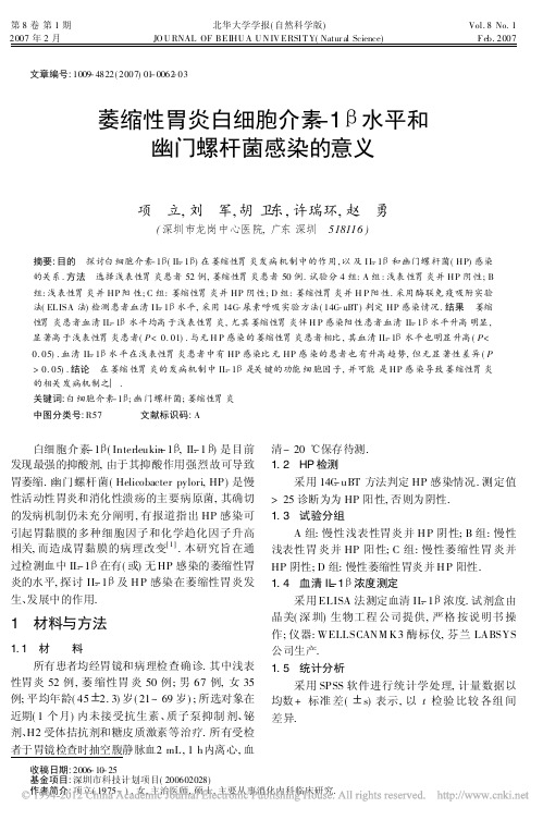
第8卷第1期北华大学学报(自然科学版)Vol.8No.12007年2月JO U RNAL OF BEIHU A U N IV ERSI T Y(Natur al Science)F eb.2007文章编号:1009-4822(2007)01-0062-03萎缩性胃炎白细胞介素-1B 水平和幽门螺杆菌感染的意义项 立,刘 军,胡卫东,许瑞环,赵 勇(深圳市龙岗中心医院,广东深圳 518116)摘要:目的 探讨白细胞介素-1B (IL -1B )在萎缩性胃炎发病机制中的作用,以及I L -1B 和幽门螺杆菌(HP)感染的关系.方法 选择浅表性胃炎患者52例,萎缩性胃炎患者50例.试验分4组:A 组:浅表性胃炎并HP 阴性;B 组:浅表性胃炎并HP 阳性;C 组:萎缩性胃炎并HP 阴性;D 组:萎缩性胃炎并H P 阳性.采用酶联免疫吸附实验法(EL ISA 法)检测患者血清I L -1B 水平,采用14C -尿素呼吸实验方法(14C -uBT )判定HP 感染情况.结果 萎缩性胃炎患者血清IL -1B 水平均高于浅表性胃炎,尤其萎缩性胃炎伴H P 感染阳性患者血清IL -1B 水平升高明显,显著高于浅表性胃炎患者(P <0.01).与无H P 感染的萎缩性胃炎患者相比,其血清IL -1B 水平也明显升高(P <0.05).血清IL -1B 水平在浅表性胃炎患者中有HP 感染比无HP 感染的患者也有升高趋势,但无显著性差异(P >0.05).结论 在萎缩性胃炎的发病机制中IL -1B 是关键的功能细胞因子,并可能是HP 感染导致萎缩性胃炎的相关发病机制之一.关键词:白细胞介素-1B ;幽门螺杆菌;萎缩性胃炎中图分类号:R57 文献标识码:A收稿日期:2006-10-25基金项目:深圳市科技计划项目(200602028)作者简介:项立(1975-),女,主治医师,硕士,主要从事消化内科临床研究.白细胞介素-1B (Interleukin -1B ,IL -1B )是目前发现最强的抑酸剂,由于其抑酸作用强烈故可导致胃萎缩.幽门螺杆菌(Helicobacter pylori,HP)是慢性活动性胃炎和消化性溃疡的主要病原菌,其确切的发病机制仍未充分阐明,有报道指出HP 感染可引起胃黏膜的多种细胞因子和化学趋化因子升高相关,而造成胃黏膜的病理改变[1].本研究旨在通过检测血中IL -1B 在有(或)无HP 感染的萎缩性胃炎的水平,探讨IL -1B 及H P 感染在萎缩性胃炎发生、发展中的作用.1 材料与方法1.1 材 料所有患者均经胃镜和病理检查确诊.其中浅表性胃炎52例,萎缩性胃炎50例;男67例,女35例;平均年龄(45?2.3)岁(21~69岁);所选对象在近期(1个月)内未接受抗生素、质子泵抑制剂、铋剂、H2受体拮抗剂和糖皮质激素等治疗.所有受检者于胃镜检查时抽空腹静脉血2mL,1h 内离心,血清-20e 保存待测.1.2 HP 检测采用14C -uBT 方法判定HP 感染情况.测定值>25诊断为为HP 阳性,否则为阴性.1.3 试验分组A 组:慢性浅表性胃炎并H P 阴性;B 组:慢性浅表性胃炎并HP 阳性;C 组:慢性萎缩性胃炎并HP 阴性;D 组:慢性萎缩性胃炎并H P 阳性.1.4 血清IL -1B 浓度测定采用ELISA 法测定血清IL -1B 浓度.试剂盒由晶美(深圳)生物工程公司提供,严格按说明书操作;仪器:WELLSCANM K3酶标仪,芬兰LABSYS 公司生产.1.5 统计分析采用SPSS 软件进行统计学处理,计量数据以均数+标准差(?s)表示,以t 检验比较各组间差异.2结果2.1H P情况及一般情况的比较102例患者中HP阳性有47例,其中萎缩性胃炎33例,浅表性胃炎24例;H P阴性55例,其中萎缩性胃炎17例,浅表性胃炎28例.各试验组之间在性别方面无差异,萎缩性胃炎患者年龄稍大于浅表性胃炎患者.2.2试验各组血清IL-1B水平比较各组血清IL-1B水平见表1,萎缩性胃炎患者血清IL-1B水平均高于浅表性胃炎,尤其萎缩性胃炎伴H P感染阳性患者(D组)血清IL-1B水平升高明显,显著高于浅表性胃炎患者(A,B组)(P< 0.01).与无H P感染的萎缩性胃炎患者(C组)相比,其血清IL-1B水平也明显升高(P<0.05).血清IL-1B水平在浅表性胃炎患者中有HP感染比无HP感染的患者也有升高趋势,但无显著性差异(P>0.05).表14组IL-1B水平检测结果(x?s)Tab.1Test results of IL-1B level of four groups组别n IL-1BA2843.25?12.39B2467.27?28.17C1789.85?29.73*D33127.05?37.28v#v与A,B组比较,P<0.01;#与C组比较,P<0.05; *与A组比较,P<0.01.3讨论细胞因子IL-1是一种重要的炎症介质,主要包含IL-A,IL-B,IL-C3个成员.IL-1B是一种炎性活细胞素,主要由单核细胞、巨噬细胞等在摄取抗原抗体复合物或抗原递呈过程中产生,在免疫应答中具有十分重要的作用.研究表明,IL-1B是目前发现的最强的抑酸剂,在生理条件下,IL-1B可以调节全部类型的胃黏膜上皮细胞的功能,抑制胃酸分泌,由于其抑制作用强烈故可导致胃萎缩,甚至发生胃癌[2],故认为IL-1是决定胃炎模式和胃癌发展的关键因素.最新的研究表明,IL-1B基因多态性不仅增加胃癌发病风险,且关联持续存在于癌前病变各阶段,与胃酸过少症及萎缩性胃炎的发生有关[3].IL-1B的抑酸作用的确切机制并不明确,但有研究表明它是通过与肠嗜铬样细胞(ECL细胞)和壁细胞上的IL-1受体结合发挥作用,而且证实两者细胞表面均有IL-1受体表达,MAHR等发现IL-1B与ECL细胞结合并诱导它的凋亡,这个过程是经NT-KB,诱导性一氧化氮合酶以及促凋亡BAX 蛋白所介导[4].本研究结果显示两组萎缩性胃炎患者中血IL-1B水平均明显升高,说明IL-1B的强抑酸作用与胃黏膜萎缩密切相关.慢性胃炎从浅表到萎缩是一个渐进的病理过程,研究表明,H P感染可能在这一过程中发挥重要作用.HP感染的浅表性胃炎如不得到有效治疗可能发展为萎缩性胃炎[5],且H P感染程度越重,萎缩程度亦越重[6],且HP感染者发生胃黏膜萎缩的年龄提早,HP感染引起胃黏膜广泛破坏,可加快从浅表性胃炎到萎缩性胃炎的转化过程.HP引起胃酸减少的确切机制尚不明确,可能与其感染后引起某些细胞因子变化有关.Takashima[7]等经口灌注的方法使蒙古沙土鼠感染幽门螺杆菌,12个星期后,鼠血清胃泌素水平,IL-1B mRNA水平增高而胃酸分泌却显著减少,注射重组人IL-1受体拮抗剂后,胃酸及胃泌素的分泌趋于正常.因此,HP感染引起的胃酸减少,可以使通过IL-1B所介导.进一步的研究表明,IL-1基因多态性引起胃酸过少也有赖于幽门螺旋杆菌(HP)的感染,当HP感染时IL-1基因多态性影响着黏膜细胞因子的表达,胃炎和癌前病变的长期发展[3].本研究结果表明,HP感染的萎缩性胃炎患者血清IL-1B明显高于无H P感染的萎缩性胃炎患者,提示H P感染可使血中IL-1B水平升高,从而引起胃黏膜萎缩.同样在浅表性胃炎HP感染的患者中,血中IL-1B水平也有升高趋势,因此若不及时根除H P,则可能因血中IL-1B水平的升高而促进其转化为萎缩性胃炎.综上所述,在萎缩性胃炎的发病机制中IL-1B 是关键的功能细胞因子,并可能是HP感染导致萎缩性胃炎的相关发病机制之一.我们可以通过根除HP感染或阻断其感染后引起IL-1B升高的作用,有利于萎缩性胃炎在临床上的转归.参考文献:[1]Y amaoka Y,Kita M,K odama T,et al.HelicobacterP ylori CagA Gene and Ex pression of Cy tokine M essenger RN A in Gastric M ucosa[J].Gastroenterolog y,1996,110: 1744-1752.[2]EL-Omar M.T he Importance of Interleukin1B inHelicobacter Pylori Associated Disease[J].Gut,2001,48: 743-747.[3]R Rad,A Dossumbekova,B N eu,et al.Cytokine GenePolymor phisms Influence M ucosal Cytokine Ex pressio n Gastric Inflammatio n and Host Specific Colo nizat ion during63第1期项立,等:萎缩性胃炎白细胞介素-1B水平和幽门螺杆菌感染的意义Helicobacter Pylori I nfection[J].Gut,2004,53:1082-1089.[4]M ahr S,Neumayer N,Gerhard M,et al.I L-1beta-Induced Apoptosis in R at Gastric Enterochro maffin-like Cells is Mediated by iNO S,N F-kappaB,and Bax Protein [J].Gastroenterolog y,2000,118:515-524.[5]Hansinp RL,D.Amico H,Levy M,et al.Prediction ofHelicobacter Pylori in Gastr ic Specimens by Inflammatory and M orphological Histological Evaluation[J].Am J-Gastroenterol,1992,87(9):1125-1131.[6]吴玫玲,蓝宇,丁宜,等.慢性萎缩性胃炎患者幽门螺杆菌感染情况的调查[J].世界华人消化杂志,2005,13(11):1362-1364.[7]T akashima M,F uruta T,Hanai H,et al.Effects ofHelicobacter Pylori I nfectio n on Gastr ic Acid Secretion and Serum Gastrin L ev els in M ongolian Gerbils[J].Gut, 2001,48:765-773.Serum Interleukin-1B and HP Infection in Atrophic GastritisXIANG Li,LIU Jun,HU We-i dong,XU Ru-i huan,ZHAO Yong(L onggang Center H osp ital in Shenz hen City,Shenz hen518116,China)Abstract:Objective To observe the effect of interleukin-1B(IL-1B)in atrophic gastritis and to identify the relationship betw een IL-1B level and helicobacter pylori(H P)infection.Methods52cases of superficial g astritis and50cases of atrophic gastritis were investig ated.T he trail has four g roups:group A:superficial g astritis with H P neg ative subjects;group B:superficial gastritis w ith HP positive subjects;g roup C: atrophic gastritis with HP negative subjects;groups D:atrophic gastritis w ith H P positive subjects.Serum IL-1B level w as ex amined by enzyme-linked immunosorbent assay.H P infection w as determined by14C-urea breath test(14C-uBT).Results Serum IL-1B levels in atrophic gastritis w ere higher than those in superficial g astritis.Especially serum IL-1B in atrophic gastritis w ith H P positive subjects(P<0.01)and atrophic g astritis with HP neg ative subjects(P<0.05).In superficial gastritis w ith H P infection serum IL-1B were higher than H P negative subjects,but there w as no significance(P>0.05).Conclusion IL-1B m ay contribute to the pathogenesis of atrophic gastritis.Inducible generation of IL-1B may one of the mechanisms to explain the link betw een H P infection and atrophic gastritis.Key words:Atrophic gastritis;Interleukin-1B;H elicobacter pylori=责任编辑:陈丽华> 64北华大学学报(自然科学版)第8卷。
细胞生物学名词解释大全
细胞生物学名词及其释义α-actinin 辅肌动蛋白一种使肌动蛋白成束的蛋白,有两个相距较远的肌动蛋白结合位点,故形成的肌动蛋白纤维束较为松散。
A kinase (PKA)A激酶因细胞内cAMP浓度升高而被激活催化靶蛋白磷酸化的酶。
accessory cell 辅佐细胞在免疫应答过程中,能摄取、加工、处理并将抗原信息提呈给淋巴细胞的免疫细胞,又称抗原提呈细胞。
actin 肌动蛋白真核细胞中含量丰富,构成肌动蛋白丝的一种蛋白质。
单体称球形肌动蛋白(G-actin);聚合物称丝状肌动蛋白(F-actin)。
actin-binding protein 肌动蛋白结合蛋白在细胞中与肌动蛋白单体或肌动蛋白纤维结合的、能改变其特性的蛋白质。
actinin 辅肌动蛋白一种肌动蛋白结合蛋白,集中分布在Z线和与质膜结合的应力纤维点状黏附端。
actin-related protein(ARP)肌动蛋白相关蛋白促进肌动蛋白丝集结的蛋白质复合物。
active transport 主动运输溶质通过细胞膜逆浓度梯度运输的现象,是一个耗能的生理过程。
actomere 肌动蛋白粒由未聚合的抑丝蛋白-肌动蛋白复合物和一小段肌动蛋白丝束组成的结构。
一旦抑丝蛋白-肌动蛋白复合物发生解离,则引起肌动蛋白聚合成丝。
actomyosin 肌动球蛋白肌肉收缩时肌动蛋白与肌球蛋白瞬时接触形成的复合物。
adaptin 衔接蛋白参与成笼蛋白衣被形成的一类蛋白质,能同时与跨膜受体以及成笼蛋白结合,在两者间起衔接作用。
adaptor protein 衔接器蛋白在细胞内信号传递途径中,凡是在不同蛋白质间起连接作用的蛋白质的通称。
adducin 聚拢蛋白质膜骨架蛋白,为异二聚体。
在钙离子浓度为mmolar级时,加速血影蛋白到血影蛋白-肌动蛋白复合物的装配。
adherens junction 黏合连接在质膜的胞质面附着有肌动蛋白纤维的细胞连接,包括连接相邻的上皮细胞的黏着带和体外培养的成纤维细胞底面的黏着斑(focal contact)。
炎症反应促进肿瘤的侵袭和转移的研究进展
炎症反应促进肿瘤的侵袭和转移的研究进展摘要:恶性肿瘤严重威胁人类健康,其侵袭和转移是肿瘤患者死亡的重要原因。
大量研究表明,肿瘤微环境对肿瘤细胞的侵袭和转移有着重要的作用。
肿瘤细胞在肿瘤微环境中会受到多种因素的影响,其中炎症反应产生的多种炎症细胞、细胞因子等会为肿瘤细胞的恶性转化提供有利条件。
关键词:炎症反应;肿瘤侵袭转移;炎症细胞;细胞因子中图分类号:(77)文献标识码:A 文章编号:1007-7847(2015)02-0160-05Recent Progresses on Inflammation-mediated Promotion of Tumor Invasion and MetastasisTANG Ya-ni,SUN Yang,YE Mao”(College of Biology,Hunan University,Changsha 410082,Hunan,China)Abstract:Malignant tumor is a great threat to human health. Invasion and metastasis of tumor cell are the major cause of death for cancer patients. Recent studies show that tumor microenvironment is very important factor in the regulation of tumor invasion and metastasis. The progress of tumor malignant transformation can be promoted by inflammation via the secretion of inflammatory cells and cytokines.Key words:in flammation;tumor invasion and metastasis;inflammatory cells;cytokines (Life Science Research,2015,19(2):160?164)肿瘤的侵袭转移是肿瘤的恶性特征之一,是大多数癌症病人死亡的主要原因。
肠道菌群与炎症性肠病合并脊柱关节炎的研究进展

肠道菌群与炎症性肠病合并脊柱关节炎的研究进展*雷张妮肖莫凡陈羽玲李海晨厉英超#西安交通大学第一附属医院消化内科(710061)摘要炎症性肠病(IBD )是一种慢性非特异性肠道炎性疾病。
脊柱关节炎(SpA )是一种发生于中轴骨骼和周围关节的慢性炎性疾病,是IBD 最常见的肠外表现之一。
IBD 合并SpA 的主要病理生理机制涉及“肠‑关节轴”假说,菌群失调在解释IBD 合并SpA 患者“肠‑关节轴”的病理联系时起到了关键性作用。
本文就肠道菌群与IBD 合并SpA 的研究进展作一综述。
关键词肠道菌群;炎症性肠病;脊柱关节炎;肠‑关节轴Progress of Research on Gut Flora and Inflammatory Bowel Disease Accompanied With Spondyloarthritis LEIZhangni,XIAO Mofan,CHEN Yuling,LI Haichen,LI Yingchao.Department of Gastroenterology,the First Affiliated Hospitalof Xi ′an Jiaotong University,Xi ′an (710061)Correspondence to:LI Yingchao,Email:*********************.cnAbstractInflammatory bowel disease (IBD)is a chronic non ‑specific intestinal inflammatory disease.Spondyloarthritis (SpA)is a chronic inflammatory disease that occurs in the axial skeleton and surrounding joints,and is one of the most common extraintestinal manifestations of IBD.The main pathophysiologic mechanism of IBD accompanied with SpA involves the "gut‑joint axis"hypothesis,and dysbiosis of gut flora plays a key role in explaining the pathologic connection of the "gut‑joint axis"in patients with IBD accompanied with SpA.This article reviewed the progress of researchon gut flora and IBD accompanied with SpA.Key wordsGut Flora;Inflammatory Bowel Disease;Spondyloarthritis;Gut‑Joint AxisDOI :10.3969/j.issn.1008‑7125.2023.04.007*基金项目:陕西省重点研发计划项目(2022SF‑135)#本文通信作者,Email:*********************.cn炎症性肠病(inflammatory bowel disease,IBD )是一种慢性非特异性肠道炎性疾病,主要包括溃疡性结肠炎(ulcerative colitis,UC )和克罗恩病(Crohn′s disease,CD )。
细胞因子和细胞因子受体

2.集落刺激因子 (Colony-Stimulating Factor, CSF)
•
能够刺激多能造血干细胞和不同发育阶段的造血祖细胞增殖
分化,并在半固体培养基中形成相应细胞集落的细胞因子。
• 根据刺激不同造血细胞系或不同分化阶段的细胞有不同的命名
名称 SCF(干细胞因子)
主要产生细胞
• IL-2受体:CD25(α);CDl22(β);CDl32(γc)
IL-2的生物学活性
• 作用具有沿种系谱向上有约束性,向下无约束性:
1、促进活化的T细胞进入S期,维持细胞的增殖,刺激产生: MHCII,IFN-γ,IL-4、5、6,LT-α,CSF。
2、促进杀伤细胞分化和效应:CTL,NK,LAK; 诱导杀伤细胞: IFN-γ,TNF-α产生,增强CTL穿孔素表达
EPO(红细胞生成素)
TPO(血小板生成素)
肾脏细胞
肾,肝
红细胞
血小板
G-CSF(粒细胞集落刺激因子)
• 1983年命名,1986年克隆成功. • 人G-CSF基因位于17号染色体, • 人类有两种不同的G-CSF DNA,分别编码含207和204
个氨基酸的前体蛋白,均有30个氨基酸的先导序列,除了 在35位插入了3个氨基酸外,其余序列一样. • 有5个半胱氨酸36=42,64=74,17游离. • 来源 • 多种细胞可产生 • 内毒素、TNF-α、IL-1、IFN-γ活化单核/巨噬细胞可 产生, • 刺激成纤维、内皮细胞也可产生, • 某些肿瘤细胞可产生
传统生物学策略
生物活性
别离纯化
蛋白质
cDNA克隆
基因
检测表型
细胞
构建
动物模型
临床相关
虾青素 改善认知
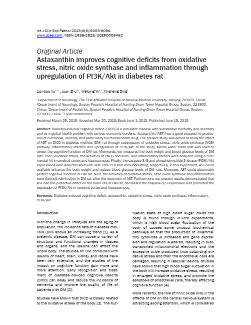
Int J Clin Exp Pathol 2015;8(6):6083-6094 /ISSN:1936-2625/IJCEP0008482Original ArticleAstaxanthin improves cognitive deficits from oxidative stress, nitric oxide synthase and inflammation through upregulation of PI3K/Akt in diabetes ratLianbao Xu1,2*, Juan Zhu3*, Weibing Yin1, Xinsheng Ding11Department of Neurology, The First Affiliated Hospital of Nanjing Medical University, Nanjing 210029, China;2Department of Neurology, Suqian People’s Hospital of Nanjing Drum Tower Hospital Group, Suqian, 223800, China; 3Department of Pediatrics, Suqian People’s Hospital of Nanjing Drum Tower Hospital Group, Suqian, 223800,China. *Equal contributors.Received March 26, 2015; Accepted May 20, 2015; Epub June 1, 2015; Published June 15, 2015Abstract: Diabetes-induced cognitive deficit (DICD) is a prevalent disease with substantial morbidity and mortality and as a global health problem with serious economic burdens. Astaxanthin (AST) has a good prospect in produc-tion of nutritional, medical, and particularly functional health drug. The present study was aimed to study the effect of AST on DICD in diabetes mellitus (DM) rat through suppression of oxidative stress, nitric oxide synthase (NOS) pathway, inflammatory reaction and upregulation of PI3K/Akt. In the study, Morris water maze teat was used to detect the cognitive function of DM rat. Afterwards, we measured the body weight and blood glucose levels of DM rats. Then, oxidative stress, the activities of eNOS and iNOS, and inflammatory factors were analyzed using a com-mercial kit in cerebral cortex and hippocampus. Finally, the caspase-3/9 and phosphoinositide 3-kinase (PI3K)/Akt expressions were also checkout with Real Time PCR and immunoblotting, respectively. In this experiment, AST could availably enhance the body weight and reduce blood glucose levels of DM rats. Moreover, AST could observably perfect cognitive function of DM rat. Next, the activities of oxidative stress, nitric oxide synthase and inflammation were distinctly diminution in DM rat, after the treatment of AST. Furthermore, our present results demonstrated that AST had the protective effect on the brain cell of DM rat, decreased the caspase-3/9 expression and promoted the expression of PI3K/Akt in cerebral cortex and hippocampus.Keywords: Diabetes-induced cognitive deficit, astaxanthin, oxidative stress, nitric oxide synthase, inflammatory, PI3K/AktIntroductionWith the change in lifestyles and the aging of population, the incidence rate of diabetes mel-litus (DM) shows an increasing trend [1]. As a systemic disease, DM can cause a variety of structural and functional changes in tissues and organs, and the lesions can affect the whole body. The studies on DM combined with lesions of heart, brain, kidney and retina have been very extensive, and the studies of the impact on cognitive function gain more and more attention. Early recognition and treat-ment of diabetes-induced cognitive deficits (DICD) can delay and reduce the incidence of dementia and improve the quality of life of patients with DM [2].Studies have shown that DICD is closely related to the oxidative stress of the body [3]. The fluc-tuation state of high blood sugar inside the body is found through in-vitro experiments, which is high blood sugar fluctuation of the body of causes some unusual biochemical pathways so that the production of inflamma-tory cytokines is increased and gene expres-sion and regulation is altered, resulting in over-transported mitochondrial electrons and the excessive oxide produced, thus catalyzing oxi-dative stress and then the endothelial cells are damaged, resulting in vascular lesions. Studies have shown that high blood sugar fluctuation in the body will increase oxidative stress, resulting in enlarged oxidative stress, and promote the apoptosis of endothelial cells, thereby affecting cognitive function [4].More recently, the role of nitric oxide (NO) in the effects of DM on the central nervous system is attracting adding attention, which is consideredto be a connection between diabetes neuropa-thy vascular doctrine and the theory of metabo-lize. The regulations of the body on the different functions of NO are mainly realized by the fine regulation of different nitric oxide synthase (NOS), in order to make NO achieves different accumulation degrees in different media inside and outside of cells [5]. Next, endothelial nitric oxide synthase (eNOS) present in fewer neu-rons is primarily implicated in the regulation of vascular function, however inducible nitric oxide synthase (iNOS) is involved in pathologi-cal conditions and unspecific immune response of nervous tissue [6].DM is an autoimmune and chronic inflammato-ry disease. The levels NF-κB p65, tumor necro-sis factor alpha (TNF-α), interleukin-1β (IL-1β) and interleukin-6 (IL-6) are closely associated with abnormally elevated insulin resistance (IR), which is one of the most important rea-sons for DM microangiopathy and neuropathy [7]. Nervous system and immune system con-stitute inflammation reflex loop via the vagus nerve, regulating systemic or local inflammato-ry reactions: activating the vagus nerve and promoting the increased secretion of the neu-rotransmitter acetylcholine so that can antago-nize or inhibit the synthesis and the release of proinflammatory cytokinein tissue [8]. Mao et al. concluded that HupA ameliorated diabetes-associated cognitive decline through oxidative stress and inflammation (NF-κB p65 unit, TNF-α, IL-1β, IL-6) [9]. Meanwhile, Wang et al. report-ed that chronic treatment with Oxymatrine alle-viated diabetes-associated cognitive decline through the NF-κB p65 unit, TNF-α, IL-1β levels in rats [10].Phosphoinositide 3-kinase (PI3K)/Akt is an important signal transduction molecule in cell. The activated Akt is further activated by phos-phorylation or inhibits its downstream target protein, thus further playing a role in regulating cell proliferation, differentiation, glucose metabolism and migration. There has been part of the trail about the apoptosis inhibition effect and mechanism of PI3K/Akt. Li et al. reported that chitosan coating markedly ame-liorates diabetes-induced impaired bio-perfor-mance of titanium alloy implant through reacti-vation of PI3K/AKT pathway [11]. Francis et al. showed intranasal insulin prevents cognitive decline through upregulation of PI3K/Akt in murine type I diabetic encephalopathy [12]. Astaxanthin (AST), oxygen-containing derivative of carotenoids, can effectively quench reactive oxygen, having high nutritional and medicinal value. AST has been separated from the shells of shrimp and crab since 1930s, of which the physiological function gained widespread attention till 1980s [13]. Since then many scholars have proved through animal and clini-cal trials that AST can inhibit tumorigenesis, enhance immune function and prevent cardio-vascular disease with a wide range of physio-logical functions, having broad application prospects [14]. Therefore, the current study aims to investigate the effect of AST to amelio-rate DICD in rats. Meanwhile, we designed the present study to explore the potential effect of AST on DICD rats and also to elucidate the underline mechanism.Materials and methodsDrugs and chemicalsAST (purity ≥ 97%) were purchased from Sigma (St. Louis, MO, USA) and the chemical structure of it was represented in Figure 1. Malondi- aldehyde (MDA), superoxide dismutase (SOD) and glutathione (GSH) ELISA kits were provided from KeyGen Biotech (Nanjing, China). A com-mercial NOS radioimmunoassay (RIA) kit was provided from Jiancheng Bioengineering Institute (Nanjing, China). NF-κB p65, TNF-α, IL-1β and IL-6 ELISA immunoassay kits were provided from Sangon Biotech (Shanghai, China). RNeasy Plus Mini kit and cDNA was transcribed using the RevertAid H Minus Reverse Transcriptase were provided from Beyotime (Nanjing, China). Power SYBR Green was provided from (TAKARA, Japan). Bicin-choninic Acid (BCA) protein assay kit was pro-vided from Bio-Rad, (Hercules, CA, USA). AnimalsMale Wistar rats (300 ± 10 g) were acquired from the Shanghai Experimental Animal Center (Shanghai, China). The present study was con-ducted according to the National Institutes of Health guide for the care and use of laboratory animals. All rats were housed in a laboratory animal room for at least 1 week before the start of the experiments and maintained at 25°C ± 1°C with 65% ± 5% humidity on a 12-h light/ dark cycle (lights on: 07:30-19:30). The experi-ment rats were freely given food and water ad libitum.Induction and measurement of diabetes After 1 week of acclimatization, a single dose of 65 mg/kg streptozotocin was injected intraper -itoneally (i.p.) to induce diabetes except in nor-mal healthy controls [15]. Fasting blood glu -cose levels was confirmed using an enzymatic glucose oxidase peroxidase diagnostic kit by the elevated glucose level in plasma, deter-mined at 48 h after injection. Fasting blood glu -cose levels > 250 mg/dL were considered dia-betic and used for further.Experimental designAnimal were randomly divided into 5 groups of eight rats each: (1) control group (Con) (n = 8): normal rats were injected with physiological saline (0.1 mL/100 g, i.p.); DM group (n = 8): the diabetic rats were received with physiologi-cal saline (0.1 mL/100 g, i.p.); (3-5) DM + AST group (DM + AST (10), DM + AST (20) and DM + AST (40)) (n = 8): the diabetic rats were treated with AST at doses of 10, 20 and 40 mg/kg, respectively [16]. All rats were anesthesia using chloral hydrate (300 mg/kg, i.p.) in the study. Immediately, the rats were sacrificed, the brains were removed and blood samples were collected under anesthesia. Meanwhile, the samples were stored at -80°C until used for fur-ther experimentation measurements.Morris water maze teatAfter the treatment of AST for 5 days, Morris water maze was evaluated by slightly modifying the protocol adopted in previous studies [17, 18]. All rats were received the non-visible plat-form trial twice per day (every morning and afternoon) for the first 5 days, a probe trial on the 6th day, and a visible platform trial on the 7th day. The apparatus consisted of a circular water tank (diameter, 120 cm; height, 50 cm), filled with water (25 ± 5°C) made opaque withmilk power. Each time, the mouse was put into the pool from different quadrants for training for 120 seconds. The latency and swimming distance in finding the platform were recorded. If the mouse did not find the platform within 120 seconds, the latency was recorded as 120 seconds. Then, the mouse was replaced on the platform for 20 seconds, and the next training was performed after 120 seconds of rest. On day 5 of training, the platform was removed, and the number of crossings of the platform location within 120 seconds (crossing number) was recorded.Measurement of oxidative stressAfter the treatment of AST for 5 days, a portion of the brain tissue was homogenized in physio -logical saline (0.1 mL/100 g) and then centri-fuged at 15,000 g for 15 minutes. The clear upper supernatants were collected for analysis of oxidative stress (MDA, SOD and GSH). In according to the manufacturer’s instructions (KeyGen Biotech, Nanjing, China), the MDA, SOD and GSH concentrations were measured by MDA, SOD and GSH ELISA kits, respectively.Measurement of eNOS and iNOS activities After the treatment of AST for 5 days, a portion of the brain tissue was homogenized in physio -logical saline (0.1 mL/100 g) and then centri-fuged at 15,000 g for 15 minutes. The clear upper supernatants were collected for analysis of NOS (eNOS and iNOS ) activity assay. In according to the manufacturer’s instructions (Jiancheng Bioengineering Institute), the activi -ties of eNOS and iNOS were detected using a commercial NOS radioimmunoassay (RIA) kit.Measurement of inflammationThe p65 subunit has a positive correlation with activated NF-κB signaling. After the treatmentFigure 1.The chemical structure of AST.of AST for 5 days, a portion of the brain tissue was homogenized in physiological saline (0.1 mL/100 g) and then centrifuged at 15,000 g for 15 minutes. The clear upper supernatants were collected for analysis of inflammatory cytokines (NF-κB p65, TNF-α, IL-1β and IL-6). In according to the manufacturer’s instructions (Sangon Biotech, Shanghai, China), the activi-ties of NF-κB p65, TNF-α, IL-1β and IL-6 were measured using ELISA immunoassay kits.Real Time PCRAfter the treatment of AST for 5 days, total RNA was isolated from the brain tissue using an RNeasy Plus Minikit (Beyotime, Nanjing, China). cDNA was transcribed using the RevertAid H Minus Reverse Transcriptase (Beyotime, Nanjing, China). Real time PCR reactions were performed using Power SYBR Green (TAKARA, Japan) and Applied Biosystems 7500 In-strument. The typical thermal profile: 95°C for 5 min, followed by 35 cycles of 95°C for 30 s and 58°C for 30 s. The primer sets used were rat caspase-3: 5’-GCATGATCCGCGACGTGG- AA-3’, 5’-AGATCCATGCCGTTGGCCAG-3’, respec-tively; rat caspase-9: 5’-ATGCAGGTCCCTGT- CATG-3’, 5’-GCTTGAGGTGGTTGTGGA-3’; and β-actin: 5’-AGAGGGAAATCGTGCGTGAC-3’, 5’-CAATAGTGATGACCTGGCCGT-3’. All primers were compound and purchased form (Sangon Biotech, Shanghai, China).ImmunoblottingThe brain tissues were homogenized with lysis buffer and centrifuged at 1000 × g for 3000 min at 4°C. Protein content was measured using with the Bicinchoninic Acid (BCA) protein assay kit (Bio-Rad, Hercules, CA, USA). An equal amount of protein were separated by SDS-PAGE, loaded onto 12% polyacrylamide gels and then transferred to PVDF membrane (Millipore, Bedford, MA). Membranes were blocked at room temperature for 2 h in blocking buffer containing 5% (v/v) nonfat milk for 2 h in TBST buffer. The membranes were then incu-bated with anti-PI3K (1:2000, Santa Cruz Biotechnology, Inc, Calif, USA), anti-Akt (1:1500, Santa Cruz Biotechnology, Inc, Calif, USA) and anti-β-actin (Boster Biological, Wuhan, China). The membranes were washed 3 times with TBST for 30 min. Then membranes were detect-ed by incubating with anti-mouse IgG (1:1000, Santa Cruz Biotechnology, Inc, Calif, USA) con-jugated with horseradish peroxidase for 2 h. The relative band intensity was visualized using the ECL Western Blotting Detection kit (Bio-Rad, USA) and X-ray films (Bio-Rad, USA). Statistical analysisStatistical analyses were done using SPSS 19.0. Data were presented as means ± stan-dard deviation (S.D). One-way analysis of vari-ance was used for the determination of differ-ences in measurements between the groups. A p-value of less than 0.05 was considered the statistically significant.ResultsEffect of AST on body weight and blood glu-cose levelsTo verify the establishment of the diabetic model, we first evaluated the body weight and blood glucose levels of all rats at the onset andAstaxanthin and cognitive deficitsat the end of the experiment. Our results indi-cated that the body weight of the DM group was observably decrease form 302.30 ± 5.31 to 241.70 ± 4.29 g, compared with the control group (Table 1). However, after the treatment of AST (10, 20 and 40 mg/kg) for 5 days, the body weight of diabetic rats were significantly reversed from 299.80 ± 5.68 to 268.60 ±6.12 g, 301.80 ± 6.68 to 273.70 ± 6.28 g, and 304.20 ± 5.26 to 286.40 ± 6.88 g, respective-ly, compared with the DM group (Table 1). Meanwhile, our results reveal that the blood glucose of the DM group was distinctly increased from 116.40 ± 1.36 to 612.70 ± 3.73 g, compared with the control group (Table 1). However, after the treatment of AST (10, 20 and 40 mg/kg) for 5 days, the body weight of diabetic rats were significantly reversed from 115.80 ± 1.27 to 321.10 ± 3.21 g, 111.20 ± 2.31 to 304.70 ± 3.18 g, and 109.80 ± 2.41 to 298.50 ± 3.82 g, respectively, compared with the DM group (Table 1).Effect of AST on diabetes-induced cognitive deficitTo test whether AST would affect cognitive function of DM rat, we performed Morris water maze tests. The effect of AST on DICD, there was a significant reduction of the escape laten-cy in the DM rats from second day to 4th day training. After treatment with AST (10, 20 and 40 mg/kg) for 5 days, shorter escape latency was markedly reduce from second day to 4th day training (Figure 2A). Meanwhile, the mean path length of DM rats was observably reduced at 5th day training, compared with that of con-trol rats (Figure 2B). The mean path length of DM/ AST rats was significantly lower than that of DM rats at 5th day training (Figure 2B). In the probe trials, when compared to that of control rats, DM rats spent less time in the target quadrant, and these differences were statisti-cally significant (Figure 2C).Furthermore, the number of times the animals crossed the for-mer platform location of DM rats was also markedly reduced, compared with that of con-trol group (Figure 2D). After treatment of AST (10, 20 and 40 mg/kg), spent more time of DM/AST rats could significantly mitigated these changes, compared to those of DM rats (Figure 2D). However, no difference in the swimming speed was observed in the each group (Figure 2E).Effect of AST on diabetes-induced changes in oxidative stressTo explore the mechanism underlying the effect of AST (10, 20 and 40 mg/kg) on restraining oxidative stress of DM rat brains, we detected the MDA, SOD and GSH concentrations after the treatment of AST for 5 days. This result indi-cates that the MDA level in cerebral cortex and hippocampus of DM rats were markedly increased compared with the control group (Figure 3A). However, the expression of MDA level in cerebral cortex and hippocampus of DM/AST rats were effectually reduce compared with that in the DM rat group (Figure 3A). Oppositely, the SOD and GSH concentrations in cerebral cortex and hippocampus of DM rats were dramatically decreased compared with the control group (Figure 3B, 3C). Certainly, the SOD and GSH concentrations in cerebral cortex and hippocampus of DM/AST rats were aug-mented compared with that in the DM rat group (Figure 3B, 3C).Effect of AST on diabetes-induced changes in the activities of eNOS and iNOSTo test whether AST would affect the activity NO of DM rat brains, we performed the activi-ties of eNOS and iNOS using NOS radioimmu-noassay (RIA) kit after the treatment of AST (10, 20 and 40 mg/kg) for 5 days. We detected the activities of eNOS and iNOS in cerebral cortex and hippocampus of DM rats were promoted compared with the control group (Figure 4A, 4B). Interestingly, we found that AST could sig-nificantly reversed the tendency and decrease the activities of eNOS and iNOS in cerebral cor-tex and hippocampus of DM/AST rats com-pared with that in the DM rat group (Figure 4A, 4B).Effect of AST on diabetes-induced changes in the activities of inflammationTo check the inflammation activities of DM rat brains, the activities of NF-κB p65, TNF-α, IL-1β and IL-6 were analyzed using ELISA immunoas-say kits. The result showed that the activities of NF-κB p65, TNF-α, IL-1β and IL-6 were signifi-cantly increased in cerebral cortex and hippo-campus of DM rats compared with the control group (Figure 5A, 5D). However, these inflam-matory factors of cerebral cortex and hippo-campus of DM/AST rats were markedly reducedAstaxanthin and cognitive deficitscompared with that in the DM rat group (Figure 5A, 5D).Effect of AST on the expression of caspase-3 and caspase-9To test if AST prevented apoptosis of DM rat brains, the activities of caspase-3 and cas-pase-9 were detected using real time PCR after the treatment of AST (10, 20 and 40 mg/kg) for 5 days. In this study we therefore found the activities of caspase-3 and caspase-9 in cere-bral cortex and hippocampus of DM rats were increased compared with the control group (Figure 6A, 6B). Furthermore, the activities of caspase-3 and caspase-9 in cerebral cortex and hippocampus of the DM/AST rats were sig-nificantly reduced compared with that in the DM rat group (Figure 6A, 6B).Effect of AST on diabetes-induced changes in the expression of PI3K/AktWe next sought to study that the effect of AST on diabetes-induced changes in the expression of PI3K/Akt. We found that AST remarkably pro-moted the expressions of PI3K and Akt protein in cerebral cortex and hippocampus of the DM/ AST rats compared with that in the DM rat group (Figure 7A-D).DiscussionDICD is the result of multiple elements and multiple factors, of which the pathogenesis is related to hyperglycemia and hypoglycemia, as well as the lack of insulin action, insulin resis-tance, vascular factor, etc. [19]. Early DICD may be mainly due to the direct damage on the cen-tral nervous cells from high blood sugar; with the further development of the disease, if the blood sugar cannot be effectively controlled, more and more pathogenic factors are involved, including high blood pressure, high cholesterol and DM complications, which can increase cog-nitive impairment, even lead to dementia [20]. Therefore, early detection and treatment is especially important. Our most significant find-ing was that AST could observably augment the body weight and decrease blood glucose levels of DM rats. Zhao et al. reported AST could ame-liorate diabetic endothelial dysfunction through down-regulation the ox-LDLLOX-1-eNOS path-way [21]. Moreover, our data revealed, AST could effectively improve cognitive function of DM rat. Previous studies suggested that AST improves cognitive function in the healthy aged individuals [22].SOD and GSH are important antioxidant enzymes in vivo, and play an important role in protecting the body from oxidative stress, removing superoxide anion radicals and pro-tecting cells from damage. Experimental results show that the SOD and GSH activities decrease in rat brain model, and MDA value is significant-ly increased, indicating oxidative damage of free radicals has been occurred in rat brain to some extent. Researchers have found that increasing SOD and GSH activity and decreas-ing MDA content can protect the brain nerve from oxidative stress damage. In the present study, the effect of AST could availably changes oxidative stress of DM rat. In cerebral cortex and hippocampus, the MDA level was markedly increased and the SOD and GSH concentra-tions were dramatically decreased in DM/AST –treatment rats. Previous studies also indicat-ed that AST could significantly increased plas-ma and kidney MDA levels for the rats [23]. Leite et al. showed that ASTA caused a reduc-tion in SOD and GSH activities in dental pulp tissue [24]. Meanwhile, AST could significantly decrease the activities of eNOS and iNOS in cerebral cortex and hippocampus of diabetic rats of DM rats. Ohgami et al. reported that AST also decreased production of NO, activity of iNOS in RAW264.7 cells [25].TNF-α and IL-6 levels in the serum of DICD patient are high, correlated to the degree of cognitive impairment. For the patient of TNF-α, both the Wechsler Adult Intelligence Scale and Mini-Mental State Examination scores are low. Overexpression of IL-6 can accelerate neural apoptosis, and decrease learning ability [26]. Moreover, AST also reduced NF-κB p65 unit, TNF-α, IL-1β and IL-6 levels in the cerebral cor-tex and hippocampus of DM rats. Park et al. reported that AST decreased oxidative stressFigure 5. Effect of AST on diabetes-induced changes in the activities of inflammation. Effect of AST on diabetes-induced changes in the activities of NF-κB p65 (A), TNF-α (B), IL-1β (C) and IL-6 (D). **P<0.01 compared with Con group; ##P<0.01 compared with DM group. Con, control; DM, diabetes; DM + AST (10), AST (10 mg/kg)-treated group; DM + AST (20), AST (20 mg/kg)-treated group; DM + AST (40), AST (40 mg/kg)-treated group.Astaxanthin and cognitive deficitsand inflammation (TNF-α and IL-6) of humans [27]. Kim et al. suggest that AST regulated IL-6 production through NF-κB p65-dependent pathway in activated microglial cells. Nagend- raprabhu et al. indicated that AST inhibited tumor invasion via modulating the expressions of NF-κB-p65 [28].PI3K-Akt signaling pathway is necessary for the regulation of cell proliferation and apoptosis. PI3K/Akt signaling pathway is involved in the differentiation process from endothelial pro-genitor cells into endothelial cells. High-density lipoprotein can activate PI3K/Akt signaling pathway to promote progenitor cells differentia-tion into endothelial-like cells; HMG-CoA reduc-tase inhibitors and vascular endothelial growth factor can promote the differentiation of pro-genitor cells via PI3K/Akt pathway. These stud-ies indicate that PI3K/Akt an important way to promote progenitor cell proliferation and differ-entiation. Our present results demonstrated that AST could effectually reduce the activities of caspase-3 and caspase-9 in cerebral cortex and hippocampus. Taken together, these results explained that AST have protected and reduced apoptosis of the heart cells. Song et al. suggested that AST inhibits apoptosis, the acti-vation of caspase-9, caspase-3 in alveolar epi-thelial cells and SH-SY5Y cells [29, 30]. Meanwhile, we found AST remarkably activated the expressions of PI3K and Akt protein in cere-bral cortex and hippocampus of the DM rats. Li et al. reported that AST protected ARPE-19 cells from oxidative stress through upregulation of activation of PI3K/Akt [31]. Zhang et al. provid-ed the evidence that AST alleviated early brain injury following subarachnoid hemorrhage through involvement of Akt/bad signaling in rats [32].Numerous studies have demonstrated natural AST has the potential effect of physiological regulation in the human body, so that it has a good prospect in production of nutritional, medical, and particularly functional health drug [33]. According to the strong antioxidant effect and the potential role in the human body of AST, it can be inferred that supplementing AST is expected to be effective in regulating body functions and maintaining human health [34]. In summary, the present study provides an evi-dence of the potential protective effect of AST on DICD through suppression of oxidative stress, activity of NOS, inflammatory reaction and up-regulation of PI3K/Akt in diabetes rat. Future studies are needed to complete the pic-ture of the cellular mechanisms of effect on neurological function damage.Disclosure of conflict of interestNone.Address correspondence to: Dr. Xinsheng Ding, Department of Neurology, The First Affiliated Hospital of Nanjing Medical University, Guangzhou Road 300, Nanjing, 210029, China. E-mail: xin- shengdingding@References[1] Wang YB, Wang S, Bai R, Du JL, Xing Q, Ba Y,Yang Y, Zhang XY, Shi CH and Yao JJ. Efficacy ofswitching from premixed insulin to insulinglargine regimen in Type 2 diabetes mellituspatients with different islet functions. Mol MedRep 2014; 10: 1096-1102.[2] Datusalia AK and Sharma SS. Amelioration ofDiabetes-induced Cognitive Deficits by GSK-3beta Inhibition is Attributed to Modulation ofNeurotransmitters and Neuroinflammation.Mol Neurobiol 2014; 50: 390-405.[3] Mozaffari-Khosravi H, Ahadi Z and Fallah TaftiM. The Effect of Green Tea versus Sour Tea onInsulin Resistance, Lipids Profiles andOxidative Stress in Patients with Type 2Diabetes Mellitus: A Randomized Clinical Trial.Iran J Med Sci 2014; 39: 424-432.[4] Tahara A, Kurosaki E, Yokono M, Yamajuku D,Kihara R, Hayashizaki Y, Takasu T, Imamura M,Li Q, Tomiyama H, Kobayashi Y, Noda A,Sasamata M and Shibasaki M. Effects ofSGLT2 selective inhibitor ipragliflozin on hyper-glycemia, hyperlipidemia, hepatic steatosis,oxidative stress, inflammation, and obesity intype 2 diabetic mice. Eur J Pharmacol 2013;715: 246-255.[5] Savu O, Iosif L, Bradescu OM, Serafinceanu C,Papacocea R and Stoian I. L-arginine catabo-lism is driven mainly towards nitric oxide syn-thesis in the erythrocytes of patients with typeFigure 7. Effect of AST on diabetes-induced changes in the expression of PI3K/Akt. Indicated representative western blotting analysis of PI3K and Akt protein levels (A and C) and statistical analysis of PI3K and Akt protein level (B and D).##P<0.01 compared with DM group. Con, control; DM, diabetes; DM + AST (10), AST (10 mg/kg)-treated group; DM + AST (20), AST (20 mg/kg)-treated group; DM + AST (40), AST (40 mg/kg)-treated group.。
癌症细胞代谢通路和令关键酶的生物学机制详解
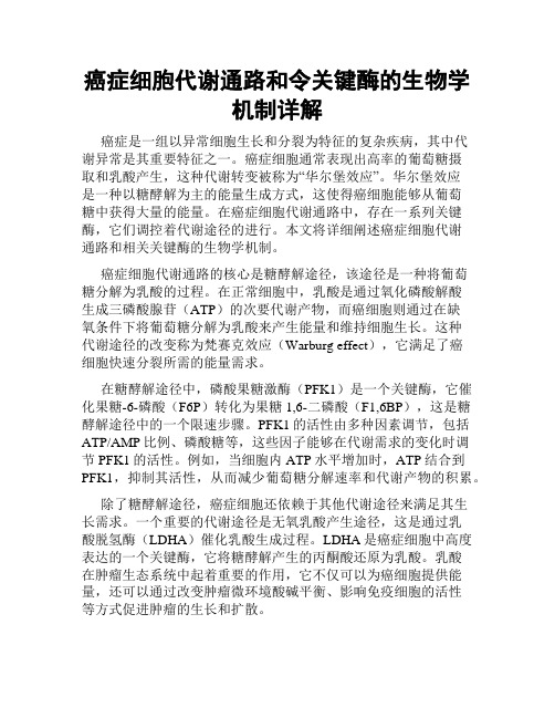
癌症细胞代谢通路和令关键酶的生物学机制详解癌症是一组以异常细胞生长和分裂为特征的复杂疾病,其中代谢异常是其重要特征之一。
癌症细胞通常表现出高率的葡萄糖摄取和乳酸产生,这种代谢转变被称为“华尔堡效应”。
华尔堡效应是一种以糖酵解为主的能量生成方式,这使得癌细胞能够从葡萄糖中获得大量的能量。
在癌症细胞代谢通路中,存在一系列关键酶,它们调控着代谢途径的进行。
本文将详细阐述癌症细胞代谢通路和相关关键酶的生物学机制。
癌症细胞代谢通路的核心是糖酵解途径,该途径是一种将葡萄糖分解为乳酸的过程。
在正常细胞中,乳酸是通过氧化磷酸解酸生成三磷酸腺苷(ATP)的次要代谢产物,而癌细胞则通过在缺氧条件下将葡萄糖分解为乳酸来产生能量和维持细胞生长。
这种代谢途径的改变称为梵赛克效应(Warburg effect),它满足了癌细胞快速分裂所需的能量需求。
在糖酵解途径中,磷酸果糖激酶(PFK1)是一个关键酶,它催化果糖-6-磷酸(F6P)转化为果糖1,6-二磷酸(F1,6BP),这是糖酵解途径中的一个限速步骤。
PFK1的活性由多种因素调节,包括ATP/AMP比例、磷酸糖等,这些因子能够在代谢需求的变化时调节PFK1的活性。
例如,当细胞内ATP水平增加时,ATP结合到PFK1,抑制其活性,从而减少葡萄糖分解速率和代谢产物的积累。
除了糖酵解途径,癌症细胞还依赖于其他代谢途径来满足其生长需求。
一个重要的代谢途径是无氧乳酸产生途径,这是通过乳酸脱氢酶(LDHA)催化乳酸生成过程。
LDHA是癌症细胞中高度表达的一个关键酶,它将糖酵解产生的丙酮酸还原为乳酸。
乳酸在肿瘤生态系统中起着重要的作用,它不仅可以为癌细胞提供能量,还可以通过改变肿瘤微环境酸碱平衡、影响免疫细胞的活性等方式促进肿瘤的生长和扩散。
另一个重要的代谢途径是异物酶途径,该途径与葡萄糖酵解和无氧乳酸产生紧密相关。
异物酶是一种在缺氧条件下将丙酮酸和氢离子转化为乙醇和二氧化碳的酶。
在癌症细胞中,异物酶参与乳酸生成途径的分支,调节着能量代谢和代谢产物的积累。
巨噬细胞介导应激经由十二指肠炎症引发消化不良症状的机制研究
observen ancl compared between FD patients with psychoCpical factors ancl asymptomatic healthy codrols. WAS moose
moOei with 7 h stress claila for 17 eys was used te evaluate the duoCenai inVammatioc at different time points te descVee its
材料与方法
、 研究对象源自因消化不良症状于2211年1月 1日-2222年1月1日在上海交通大学医学院附属 仁济医院消化内镜中心接受上消化道内镜检查者。 纳入标准:①符合罗马W FD诊断标准;②经询问存 在焦虑(如失眠、经常为一件事情感到忧愁)和(或) 抑郁(如早醒、开心不起来)表现,并通过相应精神
ecoPenai inUammatioc. Furthermore, t。iVentifU whether macrophaae is ievelven in the mechanisms of stress-relaten
ecoPenai inVammatiop by usina water
stress ( WAS) anima: moOei. Methods: DcoCenai inVammatiop was
FD paOents with psychoUcical factors developeP severe duoPenai inVammation in comparisop with the healthy cootrols (immimocytes/HPF: 138. 97 ± 7. 13 ee. 87. 44 ± 23. 68, P < 0. 000 7 ) . At the same Onie,the expressioos of
大学精品课件:专业外语Cytokines

CytokinesCytokines are proteins secreted by the cells of innate and adaptive immunity that mediate many of the functions of these cells. Cytokines are produced in response to microbes and other antigens, and different cytokines stimulate diverse responses of cells involved in immunity and inflammation. In the activation phase of adaptive immune responses, cytokines stimulate the growth and differentiation of lymphocytes, and in the effector phases of innate and adaptive immunity, they activate different effector cells to eliminate microbes and other antigens. Cytokines also stimulate the development of hematopoietic cells. In clinical medicine, cytokines are important as therapeutic agents and as targets for specific antagonists in numerous immune and inflammatory diseases.The nomenclature of cytokines is often based on their cellular sources. Cytokine that are produced by mononuclear phageocytes were called monokines, and those produced by lymphocytes were called lymphokines. With the development of anticytokine antibodies and molecular probes, it became clear that the same protein may be synthesized by lymphocytes, monocytes, and a variety of tissue cells, including endothelial cells and some epithelial cells,. Therefore, the generic term cytokines is the preferred name for this class of mediators.General Properties of CytokinesCytokines are polypeptides produced in response to microbes and other antigens that mediate and regulate immune and inflammatory reactions. Although cytokines are structurally diverse, they share several properties.Cytokine secretion is a brief, self-limited event. Cytokines are not usually stored as preformed molecules, and their synthesis is initiated by new gene transcription as a result of cellular activation. Such transcriptional activation is transient, and the messenger RNAs encoding most cytokines are unstable, so cytokine synthesis is also transient. The production of some cytokines may additionally be controlled by RNA processing and by post-transcriptional mechanisms, such as proteolytic release of an active product from an inactive precursor. Once synthesized, cytokines are rapidly secreted, resulting in a burst of release as needed.The actions of cytokines are often pleiotropic and redundant.Pleiotropism refers to the ability of one cytokine to act on different cell types. This property allows a cytokine to mediate diverse biologic effects, but it greatly limits the therapeutic use of cytokines becauseadministration of a cytokine for a desired clinical effect may result in numerous unwanted side effects. Redundancy refers to the property of multiple cytokines having the same functional effects. Because of this redundancy, antagonists against a single cytokine or mutation of one cytokine gene may not have functional consequences, as other cytokines may compensate.Cytokines often influence the synthesis and actions of other cytokines. The ability of one cytokine to simulate production of others leads to cascades in which a second or third cytokine may mediate th e biologic effects of the first. Two cytokines may antagonize each other’s action, produce additive effects, or, in some cases, produce greater than anticipated, or synergistic, effects.Cytokine actions may be local and systemic.Most cytokines act close to where they are produced either on the same cell that secretes the cytokine (autocrine action) or on a nearby cell (paracrine action). T cells often secrete cytokines at the site of contact with antigen-presenting cells, the so-called immune synapse. This may be one reason that cytokines often act on cells in contact with the cytokine producers. When produced in large amounts, cytokines may enter the circulation and act at a distance from the site of production (endocrine action).Cytokines initiate their actions by binding to specific membrane receptors on target cells. Receptors for cytokines often bind their ligands with high affinities, with dissociation constants (K d values) in the range of 10-10 to 10-12M. (For comparison, recall that antibodies typically bind antigens with a K d of 10-7 to 10-11M and that major histocompatibility complex (MHC) molecules bind peptides with a K d of only about 10-6M.) As a consequence, only small quantities of a cytokine are needed to occupy receptors and elicit biologic effects. Most cells express low levels of cytokine receptors (on the order of 100 to 1000 receptors per cell), but this is adequate for inducing responses.External signals regulate the expression of cytokine receptors and thus the responsiveness of cells to cytokines. For instance, stimulation of T or B lymphocytes by antigens leads to increased expression of cytokine receptors. For this reason, during an immune response, the antigen-specific lymphocytes are the preferential responders to secreted cytokines. This is one mechanism for maintaining the specificity of immune responses, even though cytokines themselves are not antigen specific. Receptor expression is also regulated by cytokines themselves, including the same cytokine that binds to the receptor, permitting positive amplification or negative feedback.The cellular responses to most cytokines consist of changes in gene expression in target cells, resulting in the expression of new functions and sometimes in the proliferation of the target cells. Many of the changes in gene expression induced by cytokines result in differentiation of T and B lymphocytes and activation of effector cells such as macrophages. For instance, cytokines stimulate switching of antibody isotypes in B cells, differentiation of helper T cells into T H1 and T H2 subsets, and activation of microbicidal mechanisms in phagocytes. Exceptions to the rule that cytokines work by changing gene expression patterns are chemokines, which elicit rapid cell migration, and a cytokine called tumor necrosis factor (TNF), which induces apoptosis by activating cellular enzymes, without new gene transcription or protein synthesis.Functional Categories of Cytokinescytokines were classified into three main functional categories based on their principal biologic actions.1.Mediators and regulators of innate immunity are produced mainly by mononuclearphagocytes in response to infectious agents. Bacterial products, such aslipopolysaccharide (LPS), and viral products, such as double-stranded RNA, directlystimulate macrophages to secrete these cytokines as part of innate immunity. Thesame cytokines may also be secreted by macrophages that are activated byantigen-stimulated T cells (i.e., as part of adaptive cell-mediated immunity). Mostmembers of this group of cytokines act on endothelial cells and leukocytes tostimulate the early inflammatory reactions to microbes, and some function to controlthese responses. NK (natural killer) cells also produce cytokines during innateimmune reactions.2.Mediators and regulators of adaptive immunity are produced mainly by Tlymphocytes in response to specific recognition of foreign antigens. Some T cellcytokines function primarily to regulate the growth and differentiation of variouslymphocyte populations and thus play important roles in the activation phase of Tcell-dependent immune responses. Other T cell-derived cytokines recruit, activate,and regulate specialized effector cells, such as mononuclear phagocytes, neutrophils,and eosinophils, to eliminate antigens in the effector phase of adaptive immuneresponses.3.Stimulators of hematopoiesis are produced by bone marrow stromal cells,leukocytes, and other cells, and stimulate the growth and differentiation of immatureleukocytes.In general, the cytokines of innate and adaptive immunity are produced by different cell populations and act on different target cells. However, these distinctions are not absolute because the same cytokine may be produced during innate and adaptive immune reactions, and different cytokines produced during such reactions may have overlapping actions.Cytokine Receptors and SignalingAll cytokine receptors consist of one or more transmembrane proteins whose extracellular portions are responsible for cytokine binding and whose cytoplasmic portions are responsible for initiating intracellular signaling pathways. These signaling pathways are typically activated by ligand-induced receptor clustering, bringing together the cytoplasmic portions of two or morereceptor molecules in a process analogous to signaling by T and B cell receptors for antigens.The most widely used classification of cytokine receptors is based on structural homologies among the extracellular cytokine-binding domains. According to this classification, cytokine receptors are divided into five families.Type I cytokine receptors, also called hemopoietim receptors, contain one or more copies of a domain with two conserved pairs of cysteine residues and membrane proximal sequence of tryptophan serine X tryptophan serine (WSXWS), where X is and amino acid. These receptors typically bind cytokine that fold into four α-helical strands. The conserved features of the receptors presumable form structure that bind four α-helical cytokines, but the specific for individual cytokines is determined by amino acid residues that vary from one receptor to another. These receptors consist of unique ligand-binding chains and one or more signal-transducing chain which are often shared by receptors for different cytokines.Type Ⅱcytokine receptors are similar to type receptors by virtue of two extracellular domains which conserved cysteines, but type Ⅱreceptors do not contain the WSXWS motif. These receptors consist of one ligand-binding polypeptide chain and signal-transducing chain.Some cytokine receptors contain extracellular immunoglobulin (Ig) domains and are therefore classified as members of the Ig superfamily. This group of receptors binds diverse cytokines that signal by different mechanisms.TNF receptors belong to a family of receptors (some of which are not cytokine receptors) with conserved cysteine-rich extracellular domains. On ligand binding, these receptors activate associated intracellular proteins that induce apoptosis or stimulate gene expression, or both.Seven-transmembrane α-helical receptors are also called serpentine receptors, because their transmembrane domains appear to “snake” back and forth through the membrane, and G protein-coupled receptors, because their signaling pathways involve GTP-binding (G) proteins. The mammalian genome encodes many such receptors involved in sensing environmental stimuli. In the immune system, members of this receptor class mediate rapid and transient responses to a family of cytokines called chemokines.Cytokine receptors can also be grouped according to signal transduction pathway they activate. Such a grouping will correspond to structural homologies in the cytoplasmic regions of the signaling chains of the receptors. In many cases, members of a family defined by extracellulardomains engage similar signal transduction pathways.Roles of cytokines in innate immunity and inflammationDifferent cytokines play key roles in innate immunity to different classes of microbes. In infections by pyogenic extracellular bacteria, macrophages respond to bacterial endotoxins and perhaps to other bacterial products by producing TNF, IL-1, and chemokines. TNF and IL-1 act on vascular endothelium at the site of the infection to induce the expression of adhesion molecules that promote stable attachment of blood neutrophils and monocytes to the endothelium at this site.C hemokines produced by the macrophages and by endothelial cells stimulate the extravasation of the leukocytes to the infection, where the innate immune reaction is mounted to eliminate the infectious microbes.Macrophages also respond to many microbes, including intracellular bacteria and LPS-producing bacteria, by secreting IL-12, which induces the local production of IFN-r from NK cells and T-lymphocytes. IFN-r then activates the macrophages to destroy phageocytosed microbes. IL-12 also stimulates the subsequent adaptive immune response and directs it forward TH1 cells, which are the mediators of IL-12 are complemented by IL-12. Cytokine-mediated leukocyte recruitment and activation are responsible for the injury to normal tissues that often accompanies innate immune reactions to infections. These macrophage-derived cytokines, especially TNF, IL-1, and IL-12, are also responsible for the systemic manifestations of infection.In viral infection, type I IFNs are secreted by infected cells and macrophages and function to inhibit viral replication and infection. IL-15 stimulates the expansion of NK cells, and IL-12 enhances the cytolytic activity of NK cells. NK cell-mediated killing of virus-infected cells eliminates the reservoir of infection.The dominant cytokines produced in response to different microbes account for the nature of the innate immune reactions to these microbes. For instance, the early response to pyogenic bacteria consists mainly of neutrophils, the response to intracellular bacteria is dominated by activated macrophages, and the response to viruses consists of NK cells in addition to other inflammatory cells. There may be considerable overlap, however, and there varied cellular reactions may be seen to different degrees in many infections.Roles of T cell cytokines in specialized adaptive immune responsesThe cytokines of adaptive immunity are critical for the development of immune responsesand for the activation of effector cells that serves to eliminate microbes and other antigens. Much of specialization of adaptive immunity is due to the actions of cytokines, which may be produced by subpopulations of helper T cells. Different types of microbes stimulate naïve CD4+ T cells to differentiate into effector cells that produce distinct sets of cytokines and perform distinct functions. The best defined of these subsets are the TH1 and TH2 cells. Many intracellular microbes (bacteria and viruses) induce the development of TH1 cells, which produce IFN-r, the cytokine that activates phageocytes to destroy intracellular microbes and stimulates the production of opsonizing antibodies that promote more phageocytosis. Hwlminthic parasites, in contrast, stimulate the development of TH2 cells, which produce IL-4 and IL-5. IL-4 enhances production of helminth-specific IgE antibodies, which coat the parasites, and IL-5 activates eosinphils, which bind to the IgE-coated parasites and destroy them. Thus, cytokines are essential for the development and effectiveness of adaptive immune responses.Summary●Cytokines are a family of proteins that mediate many of the responses of innate and adaptiveimmunity. The same cytokines may be produced by many cell types, and individual cytokines often act on diverse cell types. Cytokines are synthesized in response to inflammatory or antigenic stimuli and usually act locally, in an autocrine or paracrine fashion, by binding to high-affinity receptors on target cells. Certain cytokines may be produced in sufficient quantity to circulate and exert endocrine actions. For many cell types, cytokines serve as growth factors.●Cytokines mediate their actions by binding with high affinity to receptors belonging to alimited number of structural families. Different cytokines use specialized signaling pathways, such as the JAK/STAT pathway.●The cytokines that mediate innate immunity are produced mainly by activated macrophagesand include the following: TNF and IL-1 are mediators of acute inflammatory reactions to microbes; chemokines recruit leukocytes to sites of inflammation; IL-12 stimulates production of the macrophage-activating cytokine IFN-r; type I IFNs are antiviral cytokines;and IL-10 is an inhibitor of macrophages. These cytokines function in innate immune responses to different classes of microbes.●The cytokines that mediate and regulate adaptive immune responses are produced mainly byantigen-stimulated T lymphocytes, and they include the following: IL-2 is the principle T cell growth factor; IL-4 stimulates IgE production and the development of TH2 cells from naïve helper T cells; IL-5 activates eosinophils; IFN-r is an activator of macrophages; and TGF-b inhibits the proliferation of T lymphocytes and the activation of leukocytes.●The colony-stimulating factors (CSFs) consist of cytokines produced by bone marrowstromal cells, T lymphocytes, and other cells that stimulate the growth of bone marrow progenitors, thereby providing a source of additional inflammatory leukocytes. Several of these (e.g., stem cell factor and IL-7) play important roles in lymphopoiesis.●Cytokines serve many functions that are critical to host defense against pathogens andprovide links between innate and adaptive immunity. Cytokines contribute to the specialization of immune responses by activating different types of effector cells. Cytokines also regulate the magnitude and nature of immune responses by influencing the growth and differentiation of lymphocytes. Finally, cytokines provide important amplification mechanisms that enable small numbers of lymphocytes specific for any one antigen to activate a variety of effector mechanisms to eliminate the antigen.●Excessive production or actions of cytokines can lead to pathologic consequences. Theadministration of cytokines or their inhibitors is a potential approach for modifying biologic responses associated with immune and inflammatory diseases.Glossary:cytokine[ ♦♋♓♦☜◆♋♓⏹]n.细胞因子hematopoietic[ ♒♓❍☜♦☜◆☐♓♏♦♓]adj. 生血的,生血药,造血药therapeutic [ ♏❒☜☐◆♦♓] adj. 治疗的,治疗学的target [ ♦♈♓♦] n.目标,靶标antagonist [✌⏹♦✌♈☜⏹♓♦♦] n. 对抗物,拮抗剂nomenclature [⏹☜◆❍♏⏹●☜♦☞☜] n. 命名法,名称,术语monokine [ ❍⏹☜◆♋♓⏹] n.单核因子(由单核细胞或巨噬细胞产生的)lymphokine [♊●✋❍♐☜☺♋✋⏹] n.淋巴素, 淋巴因子, 淋巴endothelial [ ♏☐♓♓●☜●] adj. 内皮的epithelial [ ♏☐♓♓●♓☜● ] adj.上皮的proteolytic [☐❒☜◆♦♓☜●♓♦♓] adj.蛋白水解的pleiotropism [☐●♋♓♦❒☜☐♓☜❍] n. (基因)多效性redundancy [❒✋♎✈⏹♎☜⏹♦✋] n.重复,多余,过多,冗余synergistic [ ♦♓⏹☜♎✞♓♦♦♓] adj.协同的,协同作用的autocrine [ ♦☜◆❒♓⏹] n.自分泌paracrine [ ☐✌❒☜❒♓⏹] n.旁分泌, 副分泌synapse [♦♓⏹✌☐♦] n.突触,神经原的神经线连接, 神经键endocrine [ ♏⏹♎☜◆❒♋♓⏹] adj.内分泌的,激素的,内分泌物的; n. 内分泌物ligand [ ●♓♈☜⏹♎ ●♋♓♈☜⏹♎] n.配位子,配位体,配基microbicidal [❍♋♓❒☜◆♌♓♦♋♓♎●] adj.杀微生物的dissociation constant 解离常数,离解常数,电离常数feedback [ ♐♓♎♌✌] n. 反馈tumor necrosis factor 肿瘤坏死因子lipopolysaccharide [♊●✋☐☜☺☐●✋♊♦✌☜❒♋✋♎♊●♋✋] n. 脂多糖natural killer 自然杀伤neutrophil [♊⏹✞♦❒☜♐✋●] n. 中性白细胞,中性粒细胞,嗜中性的eosinophil [ ♓☜◆♦♓⏹☜♐♓●] n. 嗜曙红细胞;stromal cell 基质细胞transmembrane [ ♦❒✌⏹♦♊❍♏❍♌❒♏✋⏹♦❒✌⏹♦❒⏹] n.经膜,跨膜,横跨膜,贯穿细胞膜cytoplasmic [ ♦♋♓♦☜◆●✌❍♓] adj. 细胞质的homology [♒❍●☜♎✞♓] n.同源性hemopoietim [ ♒♓❍☜◆☐♓♓♦♓⏹] n.生血素, 促红细胞生proximal [ ☐❒♦♓❍☜●] adj. 接近的,邻近的motif [❍☜◆♦♓♐] n. 基序stimuli [ ♦♦♓❍◆●♋♓] n. 刺激,刺激素,刺激物pyogenic [ ☐♋✋☜✍♊♎✞♏⏹✋] adj.化脓的vascular [ ✌♦◆●☜] adj.脉管的,血管的,维管的endothelium [ ♏⏹♎☜◆♓●♓☜❍] n. 内皮adhesion [☜♎♒♓✞☜⏹] n.粘连,接合,吸附,附着[力],粘着[力]extravasation [♏♦♦❒✌☜♦♏♓☞☜⏹] n.外渗,外渗物reservoir [ ❒♏☜♦] n. 贮器, 容器, 贮存宿主, 病(原体)库opsonizing [ ☐♦☜⏹♋♓] n. 受调理素作用lymphopoiesis [ ●♓❍♐☜☐♓♓♦♓♦] n. 淋巴细胞生成administration [☜♎❍♓⏹♓♦♦❒♏♓☞☜⏹] n. 给予,投药, 给药,药的服法难句分析:1. Pleiotropism refers to the ability of one cytokine to act on different cell types. This property allows a cytokine to mediate diverse biologic effects, but it greatly limits the therapeutic use of cytokines because administration of a cytokine for a desired clinical effect may result in numerous unwanted side effects.基因多效性指的是一个细胞因子可以作用于不同的细胞类型的能力。
- 1、下载文档前请自行甄别文档内容的完整性,平台不提供额外的编辑、内容补充、找答案等附加服务。
- 2、"仅部分预览"的文档,不可在线预览部分如存在完整性等问题,可反馈申请退款(可完整预览的文档不适用该条件!)。
- 3、如文档侵犯您的权益,请联系客服反馈,我们会尽快为您处理(人工客服工作时间:9:00-18:30)。
Mechanisms of DiseaseF R A N K L I N H. E P S T E I N , M.D.,EditorC YTOKINE P ATHWAYS AND J OINT I NFLAMMATION IN R HEUMATOIDA RTHRITISE RNEST H.S. C HOY , M.D.,AND G ABRIEL S. P ANAYI , M.D., S C .D.From the Department of Rheumatology, Guy’s, King’s, and St. Thomas’Hospitals School of Medicine, King’s College, London. Address reprint re-quests to Dr. Choy at the Department of Rheumatology, King’s College Hospital, East Dulwich Grove, London SE22 8PT, United Kingdom.HEUMATOID arthritis is a common chron-ic inflammatory and destructive arthropathy that cannot be cured and that has substantialpersonal, social, and economic costs. The long-term prognosis is poor: 80 percent of affected patients are disabled after 20 years, 1 and life expectancy is reduced by an average of 3 to 18 years. 2 The medical cost of rheumatoid arthritis averages $5,919 per case per year in the United States 3 and approximately £2,600 per case per year in the United Kingdom. 4 Current slow-acting antirheumatic drugs have limited efficacy and many side effects. Moreover, they do not improve the long-term prognosis of rheumatoid arthritis. 1The inflammatory process is usually tightly regulat-ed, involving both mediators that initiate and maintain inflammation and mediators that shut the process down. In states of chronic inflammation, an imbalance between the two mediators leaves inflammation un-checked, resulting in cellular damage. In the case of rheumatoid arthritis, this damage is manifested by the destruction of cartilage and bone.Efforts to develop safer and more effective treat-ments for rheumatoid arthritis that are based on an improved understanding of the role of inflammatory mediators are beginning to bear fruit. Treatments such as etanercept, a soluble tumor necrosis factor a (TNF- a ) type II receptor–IgG1 fusion protein, and infliximab, a chimeric (human and mouse) monoclon-al antibody against TNF- a , have been approved by the Food and Drug Administration and the European Medicine Evaluation Agency for rheumatoid arthritis.These therapies could dramatically change the treat-ment and outcome of the disease.PATHOGENESIS OF RHEUMATOIDARTHRITISThe synovial membrane in patients with rheumatoid arthritis is characterized by hyperplasia, increased vas-cularity, and an infiltrate of inflammatory cells, pri-marily CD4+ T cells, which are the main orchestratorRof cell-mediated immune responses. In genetic studies,rheumatoid arthritis is strongly linked to the major-histocompatibility-complex class II antigens HLA-DRB1*0404 and DRB1*0401. 5 The main function of HLA class II molecules is to present antigenic pep-tides to CD4+ T cells, which strongly suggests that rheumatoid arthritis is caused by an unidentified ar-thritogenic antigen. 6 The antigen could be either an exogenous antigen, such as a viral protein, or an en-dogenous protein. Recently, a number of possible endogenous antigens, including citrullinated protein,human cartilage glycoprotein 39, and heavy-chain–binding protein, have been identified. 7Cellular Mediators of Inflammation and Joint DamageAntigen-activated CD4+ T cells stimulate mono-cytes, macrophages, and synovial fibroblasts to pro-duce the cytokines interleukin-1, interleukin-6, and TNF- a and to secrete matrix metalloproteinases (Fig.1) through cell-surface signaling by means of CD69and CD11 8 as well as through the release of soluble mediators such as interferon- g and interleukin-17. In-terleukin-1, interleukin-6, and TNF- a are the key cy-tokines that drive inflammation in rheumatoid arthri-tis. Activated CD4+ T cells also stimulate B cells (Fig.1), through cell-surface contact and through the bind-ing of a L b 2 integrin, CD154 (CD40 ligand), and CD28, to produce immunoglobulins, including rheu-matoid factor. The precise pathogenic role of rheuma-toid factor is unknown, but it may involve the activa-tion of complement through the formation of immune complexes. Activated CD4+ T cells express osteopro-tegerin ligands that stimulate osteoclastogenesis (Fig.1). Such activated T cells caused joint damage in an animal model of rheumatoid arthritis. 9These activated macrophages, lymphocytes, and fi-broblasts, as well as their products, can also stimulate angiogenesis, which may explain the increased vascu-larity found in the synovium of patients with rheu-matoid arthritis. Endothelial cells in the synovium are activated and express adhesion molecules that promote the recruitment of inflammatory cells into the joint.This process is enhanced by the release of chemokines,such as interleukin-8, by inflammatory cells in the joint. The detailed mechanisms of these complex cel-lular interactions remain elusive.Soluble Mediators of Inflammation and Joint DamageMonocytes, macrophages, fibroblasts, and T cells release numerous cytokines on stimulation. Most of these cytokines, including TNF- a and interleukin-1,can be detected in synovial fluid from patients with rheumatoid arthritis. 10 Both TNF- a and interleukin-1are likely to have primary roles in the pathogenesis of rheumatoid arthritis. The serum and synovial concen-trations of both cytokines are high in patients with ac-tive rheumatoid arthritis. 11,12 Furthermore, TNF- aand interleukin-1 are potent stimulators of mesenchymalcells, such as synovial fibroblasts, osteoclasts, and chon-drocytes, that release tissue-destroying matrix metallo-proteinases. 13 Interleukin-1 and TNF- a also inhibit the production of tissue inhibitors of metalloprotein-ases by synovial fibroblasts. 13 These dual actions are thought to lead to joint damage. Perhaps by inducing the production of interleukin-11, TNF- a stimulatesthe development of osteoclasts, which are responsible for bone degradation. 14TNF-aTNF- a is a potent cytokine that exerts diverse ef-fects by stimulating a variety of cells. It is a soluble 17-kd protein composed of three identical subunits.Figure 1. Cytokine Signaling Pathways Involved in Inflammatory Arthritis.The major cell types and cytokine pathways believed to be involved in joint destruction mediated by TNF- aand interleukin-1 are shown. Th2 denotes type 2 helper T cell, Th0 precursor of type 1 and type 2 helper T cells, and OPGL osteoprotegerin ligand.Interferon-gRheumatoid factor and other autoantibodiesSynoviumTNF-a ,interleukin-1,and interleukin-6Production of metalloproteinases andother effector molecules Migration of polymorphonuclear cellsErosion of bone and cartilageB cell Plasma cellMacrophageFibroblastChondrocyteOsteoclastTh0Th2CD4+ T cellInterferon-g Interleukin-12Interleukin-4Interleukin-10Interleukin-4Interleukin-6Interleukin-10OPGLCD11CD69CD11CD69It is produced mainly by monocytes and macrophages, but also by B cells, T cells, and fibroblasts. Newly syn-thesized TNF-a is inserted into the cell membrane and subsequently released through the cleavage of its membrane-anchoring domain by a serine metallopro-teinase.15 Thus, TNF-a secretion might be suppressed by inhibitors of this enzyme.16Perhaps the best-studied aspect of TNF-a is its ability to promote inflammation. TNF-a is an auto-crine stimulator as well as a potent paracrine inducer of other inflammatory cytokines, including interleu-kin-1, interleukin-6, interleukin-8, and granulocyte–monocyte colony-stimulating factor.17-19 TNF-a also promotes inflammation by stimulating fibroblasts to express adhesion molecules, such as intercellular ad-hesion molecule 1.20 These adhesion molecules inter-act with their respective ligands on the surface of leu-kocytes, resulting in increased transport of leukocytes into inflammatory sites, including the joints in patients with rheumatoid arthritis.TNF-a indirectly down-regulates inflammation by stimulating the release of corticotropin from the pi-tuitary.21 This hormone stimulates the adrenal cortex to release cortisol, which inhibits inflammation.As an inflammatory cytokine, TNF-a has an im-portant — perhaps dominant — role in rheumatoid synovitis. In cultures of synovial cells from patients with rheumatoid arthritis, blocking TNF-a with anti-bodies significantly reduced the production of inter-leukin-1, interleukin-6, interleukin-8, and granulo-cyte–monocyte colony-stimulating factor.18Hence, the blockade of TNF-a may have a more global effect on inflammation than the blockade of other cytokines present in high concentrations in synovial fluids, such as interleukin-1.The results of studies in animals provide further evidence of the importance of TNF-a in rheumatoid arthritis. In transgenic mice that expressed a deregu-lated human TNF-a gene, an inflammatory and de-structive polyarthritis similar to rheumatoid arthritis spontaneously developed.22 Pretreatment of these an-imals with a monoclonal antibody against TNF-a pre-vented the development of arthritis. Blocking TNF-a with a soluble TNF-receptor fusion protein or with monoclonal antibodies also ameliorated disease activi-ty in mice with type II collagen-induced arthritis.23,24 Interleukin-1Interleukin-1 is a 17-kd protein that is mostly pro-duced by monocytes and macrophages but is also produced by endothelial cells, B cells, and activated T cells.25 The interleukin-1 signaling system is more complex than the TNF-a system. Interleukin-1 binds to two types of cell-surface receptors.26,27 Only type I receptors have a cytoplasmic tail and are capable of intracellular signaling.28 Type II receptors are decoy receptors: they bind circulating interleukin-1 but do not deliver any intracellular signals.29 The type I re-ceptor is found in low numbers on many cells, where-as the type II receptor is expressed primarily on neu-trophils, monocytes, and B cells.30 Soluble forms of both types of interleukin-1 receptor compete with cell-surface receptors, thereby decreasing interleukin-1–mediated activation of cells. In addition, a naturally occurring antagonist, interleukin-1–receptor antago-nist, binds the type I receptor with high affinity with-out triggering a signal, thus providing another mech-anism for the inhibition of interleukin-1 activity.31 The biologic activity of interleukin-1 is dependent on the precise quantities of many interacting molecules. Studies of arthritis in animals have strongly implicat-ed interleukin-1 in joint damage. Injection of inter-leukin-1 into the knee joints of rabbits results in the degradation of cartilage,32whereas the injection of antibodies against interleukin-1 ameliorates collagen-induced arthritis in mice and decreases the damage to cartilage.33 Macrophages in the synovial tissue of pa-tients with rheumatoid arthritis appear to be an impor-tant source of interleukin-1.34 Like TNF-a, interleu-kin-1 may cause damage by stimulating the release of matrix metalloproteinases from fibroblasts and chon-drocytes.13,35The concentrations of interleukin-1–receptor antagonist are high in the synovial fluid of pa-tients with rheumatoid arthritis, but not high enough to suppress inflammation.36Interleukin-6Interleukin-6 is a pleiotropic inflammatory cytokine produced by T cells, monocytes, macrophages, and synovial fibroblasts.37 Originally identified as a factor that induces the final maturation of B cells into plasma cells, interleukin-6 is involved in diverse biologic proc-esses, such as the activation of T cells, the induction of the acute-phase response, the stimulation of the growth and differentiation of hematopoietic precur-sor cells, and the proliferation of synovial fibroblasts.37 Antiinflammatory CytokinesWhereas some cytokines initiate and maintain the inflammatory process, others dampen it. The two best-studied antiinflammatory cytokines are interleukin-10 and interleukin-4. In vitro, these cytokines cooper-ate to inhibit the production of inflammatory cyto-kines.38,39Interleukin-10Interleukin-10 is produced by monocytes, macro-phages, B cells, and T cells. It inhibits the produc-tion of several cytokines, including interleukin-1 and TNF-a, and the proliferation of T cells in vitro.40 In-terleukin-10 can also reverse the cartilage degradation mediated by antigen-stimulated mononuclear cells from patients with rheumatoid arthritis.38 Although interleukin-10 is found in the synovial fluid of patients with rheumatoid arthritis, the amount may be insuf-ficient to suppress inflammation.41Interleukin-4Interleukin-4 is produced by CD4+ type 2 helper T cells and participates in the differentiation and growth of B cells.40In vitro, interleukin-4 inhibits the activation of type 1 helper T cells, and this, in turn, decreases the production of interleukin-1 and TNF-a and inhibits cartilage damage.42Interleukin-4 also inhibits the production of interleukin-6 and interleu-kin-8.39In cultures of synovium samples from pa-tients with rheumatoid arthritis, interleukin-4 inhib-ited the production of interleukin-1 and increased the expression of interleukin-1–receptor antagonist, both of which actions should decrease inflammation.36 Joint Damage in Rheumatoid ArthritisRheumatoid arthritis is characterized by progres-sive joint damage that is mediated by several mech-anisms (Fig. 1 and 2). Early erosion of cartilage and bone is associated with the formation of a proliferat-ing pannus. The interface between pannus and car-tilage is occupied predominantly by activated macro-phages and synovial fibroblasts that express matrix metalloproteinases and cathepsins.Interleukin-1 and TNF-a stimulate the expression of adhesion molecules on endothelial cells and in-crease the recruitment of neutrophils into the joints. Neutrophils release elastase and proteases, which de-grade proteoglycan in the superficial layer of carti-lage.43 The depletion of proteoglycan enables immune complexes to precipitate in the superficial layer of col-lagens and exposes chondrocytes.44 Chondrocytes and synovial fibroblasts release matrix metalloproteinases when stimulated by interleukin-1, TNF-a, or activat-ed CD4+ T cells. Matrix metalloproteinases, in par-ticular stromelysin and collagenases, are enzymes that degrade connective-tissue matrix and are thought to be the main mediators of joint damage in rheumatoid arthritis. In animals, activated CD4+ T cells stimu-late osteoclastogenesis, and they may cause joint dam-age independently of interleukin-1 and TNF-a in pa-tients with rheumatoid arthritis.SummaryRheumatoid arthritis is initiated by CD4+ T cells, which amplify the immune response by stimulating other mononuclear cells, synovial fibroblasts, chon-drocytes, and osteoclasts. The release of cytokines, es-pecially TNF-a, interleukin-1, and interleukin-6, caus-es synovial inflammation. Joint damage results from the degradation of connective tissue by matrix metal-loproteinases and the stimulation of osteoclastogene-sis by activated CD4+ T cells. Clearly, there are many possible therapeutic targets, but the inhibition of cy-tokines would seem to offer an especially useful ap-proach to suppressing inflammation and preventing joint damage.INHIBITION OF CYTOKINESGiven the complexity of cytokine interactions and the multiplicity of cytokine targets, the effectiveness and toxicity of cytokine-based interventions are dif-ficult to predict. A variety of cytokine-based strategies are being explored for the treatment of inflammatory diseases. These include the neutralization of cytokines (by soluble receptors or monoclonal antibodies), re-ceptor blockade, and the activation of antiinflamma-tory pathways by bioengineered versions of immuno-regulatory cytokines (Fig. 3).Neutralization of CytokinesSoluble receptors have a physiologic role in neutral-izing many cytokines, as exemplified by soluble TNF receptors. TNF-a binds to TNF receptors on the sur-face of many cells, including monocytes, macrophages, T cells, synovial fibroblasts, osteoblasts, and endothe-lial cells. There are two types of TNF receptors, p55 and p75, which are part of a large family of structur-ally related cell-surface receptors.45,46 The cytoplasmic domains of the p55 and p75 receptors are quite dif-ferent,47 suggesting that they may activate different signal-transduction pathways. The p75 receptor is believed to have a primary role in stimulating the proliferation of T cells and in suppressing TNF-a–mediated inflammatory responses, whereas the p55 receptor appears to be critical in triggering host de-fense and inflammatory responses.48,49Soluble forms of both p55 and p75 are part of a feedback loop that can modulate the inflammatory action of TNF-a. The transmembrane domain of both TNF receptors is susceptible to lysis by proteases, in-cluding TNF-a–converting enzyme, leading to the release of a soluble form of the receptor. Hence, both types of receptor are present in body fluids. Soluble TNF receptors are found in high concentrations inFigure 2 (facing page). Pathogenesis of Rheumatoid Arthritis.In the normal knee joint, the synovium consists of a synovial membrane (usually one or two cells thick) and underlying loose con-nective tissue. Synovial-lining cells are designated type A (macrophage-like synoviocytes) or type B (fibroblast-like synoviocytes). In early rheumatoid arthritis, the synovial membrane becomes thickened because of hyperplasia and hypertrophy of the synovial-lining cells. An extensive network of new blood vessels is formed in the synovium. T cells (predominantly CD4+) and B cells (some of which become plasma cells) infiltrate the synovial membrane. These cells are also found in the synovial fluid, along with large numbers of neutrophils. In the early stages of rheumatoid arthritis, the synovial membrane begins to invade the cartilage. In estab-lished rheumatoid arthritis, the synovial membrane becomes transformed into inflammatory tissue, the pannus. This tissue invades and destroys adjacent cartilage and bone. The pannus consists of both type A and type B synoviocytes and plasma cells.Synovial villiExtensive angiogenesisSynovial membraneCapsuleB cellsNeutrophilsFemurTibiaCartilageCartilageTibiaPlasma cellNeutrophilsNormal Knee JointCapsuleFemurSynoviocytesSynovial membraneEarly Rheumatoid ArthritisHypertrophic synoviocytesCapillary formation Hyperplastic synovial membraneEstablished Rheumatoid ArthritisPannusEroded boneT cellsFigure 3. Methods of Blocking the Activity of an Inflammatory Cytokine.Cytokine–Receptor InteractionNeutralization of cytokinesReceptor blockadeActivation of antiinflammatory pathwaysInflammatory cytokineCytokine receptorInflammatory signalsReceptor antagonistMonoclonal antibodySoluble receptorNo signalMonoclonal antibodyAntiinflammatorycytokineSuppression of inflammatory cytokinesNormal interactionNo signalthe synovial fluid and serum of patients with rheu-matoid arthritis.50 Nevertheless, an excess of TNF-a relative to the concentration of soluble TNF recep-tors prolongs joint inflammation.Treating patients with recombinant soluble cyto-kine receptors may help suppress inflammation. How-ever, soluble receptors have short plasma half-lives, and repeated doses would be required to neutralize the effects of inflammatory cytokines. This limitation can be circumvented by conjugating soluble receptors with human Fc (a proteolytic fragment of IgG), which can extend the half-lives of these molecules to approx-imate those of immunoglobulin.51 Another alternative is to polymerize TNF receptor or anti–TNF-a Fab' with polyethylene glycol. This can reduce antigenic-ity and prolong the half-life in circulation. The effi-cacy of these constructs in patients with rheumatoid arthritis is currently being investigated. Antibodies against cytokines are another approach to neutralizing cytokines. The type of antibody ap-pears to be critical to its clinical efficacy. Murine monoclonal antibodies are antigenic and induce the production of antimouse antibodies in recipients.52 Chimeric and humanized monoclonal antibodies are less immunogenic than murine monoclonal antibod-ies, and they are therefore more suitable as therapeu-tic agents.53Receptor AntagonismBlocking the ability of a receptor to bind its cyto-kine is another strategy to interrupt signal transduc-tion. This can be accomplished with either natural receptor antagonists, such as interleukin-1–receptor antagonist, or antibodies against cytokine receptors. For such an approach to be successful, the amount of antagonist must be large enough to bind the ma-jority of receptors for long periods.Activation of Antiinflammatory PathwaysIn addition to natural cytokine antagonists and soluble receptors, immunoregulatory cytokines such as interleukin-10 and interleukin-4 can suppress in-flammation.39,40,42 As is true for most cytokines, how-ever, their effects are pleiotropic and not fully under-stood. For instance, the treatment of synovial-fluid macrophages with exogenous interleukin-10 increased the expression of cell-surface and soluble TNF recep-tors, an effect that could make cells more responsive to TNF-a and its inflammatory effects.54 Furthermore, because cytokines are low-molecular-weight proteins or glycoproteins with short half-lives, maintaining therapeutic serum concentrations of antiinflammato-ry cytokines may be difficult and expensive. A poten-tial solution would be to use gene therapy that would lead to continued synthesis of therapeutic antiinflam-matory cytokines in the joints. In animal models of rheumatoid arthritis, the genes for interleukin-10 and interleukin-4 have been transfected by viral vectors into synovial fibroblasts in vitro. These fibroblasts were subsequently injected back into the joints, where they released the antiinflammatory cytokine, resulting in the suppression of inflammation and destruction of joints.55,56CLINICAL TRIALS OF CYTOKINEINHIBITORSSoluble Human Cytokine-Receptor ProteinsEtanerceptEtanercept is a fusion protein made up of two re-combinant p75 soluble TNF receptors fused with the Fc portion of human IgG1. The dimeric structure of etanercept makes it approximately 1000 times as efficient as the monomeric soluble p75 TNF receptor at neutralizing TNF-a.57In two placebo-controlled trials of 168 and 234 pa-tients with rheumatoid arthritis, twice weekly subcu-taneous injections of 25 mg of etanercept58,59 resulted in significant improvement. The number of swollen joints decreased by approximately 50 percent from base line after six months of treatment.58Treatment with etanercept was well tolerated, and produced only minor reactions at the site of the injection. Synovial bi-opsies showed a statistically significant decrease in the numbers of T cells and plasma cells and in the amount of vascular-cell adhesion molecule 1 and the expres-sion of interleukin-1 after one month of treatment.60 Long-term, open-label studies have indicated that the efficacy of continued treatment with etanercept is sus-tained for at least 33 months, and no major adverse events have occurred.61 Furthermore, the combination of etanercept and methotrexate was significantly more effective than methotrexate alone in a placebo-con-trolled trial of 89 patients with rheumatoid arthritis who had had a partial response to methotrexate.62 Etanercept is also effective in patients with juvenile rheumatoid arthritis.63 In a randomized, placebo-con-trolled trial of 51 patients with juvenile polyarticular rheumatoid arthritis, 0.4 mg of etanercept per kilo-gram of body weight or placebo was injected subcu-taneously twice weekly for four months or until a flare of the disease occurred. The total number of joints with active arthritis decreased by 58 percent from base line, and the range of motion of affected joints was in-creased by 80 percent.In other studies, etanercept was better tolerated and more effective than methotrexate in patients with ear-ly rheumatoid arthritis.64 There was less radiographic evidence of progression of rheumatoid arthritis in pa-tients who were receiving etanercept than in patients who were receiving methotrexate.64Monoclonal Antibodies against CytokinesInfliximabInfliximab is a chimeric IgG1 antibody against TNF-a. In a double-blind, placebo-controlled trialof 73 patients with rheumatoid arthritis, a single in-travenous dose of 10 mg of infliximab per kilogram rapidly reduced the number of swollen joints as well as the serum concentration of C-reactive protein.65 Clinically significant improvement was evident with-in a week after treatment was begun. Synovial-biopsy specimens, obtained before and four weeks after the beginning of treatment, showed significant reductions in the number of T cells and in the tissue content of vascular-cell adhesion molecule 1 and E-selectin.66 In another randomized, placebo-controlled trial of 101 patients with rheumatoid arthritis,67infliximab or placebo was given repeatedly, with or without methotrexate. Antibodies against infliximab developed in many patients after repeated treatment, but the incidence was reduced by concomitant treatment with methotrexate. Furthermore, a dose of 3 mg of inflix-imab per kilogram in combination with methotrexate was as efficacious as a dose of 10 mg per kilogram, with or without methotrexate. This finding was con-firmed in a randomized, placebo-controlled trial of 428 patients with rheumatoid arthritis, in which the infliximab-treated patients had sustained clinical im-provement for at least 30 weeks.68Other Antibodies against TNF-aD2E7 is a human antibody against TNF-a gener-ated by phage-display technology,69 whereas nerelim-omab is a humanized monoclonal antibody against TNF-a that consists of the hypervariable regions of a murine monoclonal antibody against TNF-a grafted onto a human k light chain and an IgG4 heavy chain.70 Both these antibodies were effective in preliminary randomized, placebo-controlled trials in patients with rheumatoid arthritis.69,70Cytokine-Receptor BlockersRecombinant Interleukin-1–Receptor AntagonistIn a randomized, double-blind, placebo-controlled trial of 472 patients with rheumatoid arthritis, treat-ment with recombinant human interleukin-1–recep-tor antagonist71 resulted in moderate clinical improve-ment and decreased the rate of progression of erosions, as assessed by radiography.72 Reactions at the injection site were the most common adverse event. Recombi-nant human interleukin-1–receptor antagonist is cur-rently being tested in combination with methotrexate.A drawback to the therapeutic use of interleukin-1–receptor antagonist is its short (six-hour) half-life in plasma,73 which necessitates frequent daily treatment with high doses to maintain a therapeutic effect. This problem is further compounded by the need for a large (10- to 1000-fold) excess of interleukin-1–recep-tor antagonist to block the effect of interleukin-1 in vitro and in vivo.74 One way to circumvent these prob-lems and achieve high local concentrations of inter-leukin-1–receptor antagonist may be by the use of gene therapy.75 In animals, synovial fibroblasts trans-fected with the gene for human interleukin-1–recep-tor antagonist and then reinjected into joints produced interleukin-1–receptor antagonist in the synovium, with consequent clinical improvement.76 A similar ex vivo gene-transfer strategy was used to introduce the gene for the interleukin-1–receptor antagonist into the joints of three patients with rheumatoid arthritis before they underwent total joint replacement. Sub-sequent removal and analysis of joint tissue indicated that this technique induced the intraarticular expres-sion of the gene for the interleukin-1–receptor antag-onist.75Antibody against Interleukin-6 ReceptorAn antibody against the receptor for interleukin-6 has shown efficacy in mice with collagen-induced ar-thritis.77 A clinical trial of a humanized monoclonal antibody against the interleukin-6 receptor, which is theorized to have the same functional consequenc-es as a monoclonal antibody against interleukin-6, is currently under way.Recombinant Interleukin-10 and Interleukin-4 Recombinant interleukin-1078 and interleukin-479 have been tested in patients with rheumatoid arthritis. The clinical efficacy of the treatments has been disap-pointing; the lack of efficacy may be due to the short half-life of these substances.CONCLUSIONSAlthough the cause of rheumatoid arthritis still eludes us, our improved understanding of the patho-genesis of the disease has opened the door to innova-tive therapies. By targeting molecules that are directly involved in the pathogenesis of rheumatoid arthritis, these therapies may be more efficacious and specific and less toxic in the short and long term than standard therapies. Radiologic evidence suggests that these new therapies, such as anticytokine therapy, may slow dis-ease progression. Finally, the success of anticytokine therapy will also provide valuable insights into the ini-tiation and progression of rheumatoid arthritis. Supported by an Integrated Clinical Arthritis Center grant (PO526) from the Arthritis Research Campaign of Great Britain.REFERENCES1.Scott DL, Symmons DP, Coulton BL, Popert AJ. Long-term outcome of treating rheumatoid arthritis: results after 20 years. Lancet 1987;1:1108-11.2.Pincus T, Callahan LF. Taking mortality in rheumatoid arthritis serious-ly — predictive markers, socioeconomic status and comorbidity. J Rheu-matol 1986;13:841-5.3.Yelin E, Wanke LA. An assessment of the annual and long-term direct costs: the impact of poor function and functional decline. Arthritis Rheum 1999;42:1209-18.4.McIntosh E. The cost of rheumatoid arthritis. Br J Rheumatol 1996; 35:781-90.nchbury JS. The HLA association with rheumatoid arthritis. Clin Exp Rheumatol 1992;10:301-4.。