微生物染色(英文)
专业英语(微生物降解)

分类法 Taxonomy 形态学 Morphology 微生物生理学 Microbial physiology 鉴别染色-格兰氏染色 Differential stains-Gram stain 格兰氏阳性 Gram-positive 格兰氏阴性 Gram-negative
Thank you!
• • • • • • • • • • • • •
4、用什么方法检测、监测 were detected by were analyzed by was assayed polarographically(用副词) The compounds were visualized under UV light at 254 nm. Phenolic compounds were detected by spraying with Gibb’s reagent. The metabolites were analyzed by reversed-phase HPLC. Growth was monitored spectrophotometrically at 600 nm. DDT dioxygenase was assayed polarographically in a Gilson oxygraph with oxygen electrode. 2,3-Dihydroxybiphenyl-1,2-dioxygenase was assayed spectrophotometrically by measuring increase in absorbance at 432 nm. were recorded with/on+仪器厂家、型号 Mass spectra were recorded withJeol, MS-DX303 Mass spectrometer operated at 70 ev. The proton nuclear magnetic resonance (1HNMR) spectra were recorded on a Bruker AMX-300MHz spectrometer with TMS as internal standard.
微生物5 抗酸染色

原理:
由于分歧杆菌的胞壁含大量脂质,主要是分枝菌酸, 它包围在肽聚糖的外面,所以分枝杆菌一般不易着色, 可采用加温、延长着色时间或提高染料浓度等手段使其 着色。但分枝菌酸与染料结合后,就很难被酸性脱色剂 脱色。故分歧杆菌又称为抗酸杆菌,这种染色方法又成 为抗酸染色。
延长时间:5min 提高染料浓度:初染用的酸复红浓度是革兰染色酸复红浓 度的10倍 盐酸酒精:脱色能力远远强于95%酒精
原理:
齐-尼氏抗酸染色法是在加热条件下使分枝菌 酸与石炭酸复红牢固结合成复合物,用盐酸酒 精处理也不脱色。当再加碱性美兰复染后,分 枝杆菌仍然为红色,而其他细菌及背景中的物 质为兰色。
注意:不要使染液沸腾,若蒸发减少,可待玻片 稍冷却后适当补充。
(2)待玻片冷却后,用水缓慢冲洗,用吸水纸吸 干。
(3)脱色:滴加3%盐酸酒精,轻晃玻片,直至 无红色液体流下为止,一般作用1—3min*,水 洗,吸干。
(4)复染:用碱性美兰溶液复染1min,用水冲 洗后吸干。
(5)
*:不同文献报道不同,范围在30s至3min
П、漂浮集菌法。取晨痰2—3ml放入100ml三角瓶内,加1—2倍量 40g/lNaOH溶液,经103.43Pa高压灭菌20—25min(或煮30min)。 冷却后滴加汽油0.3ml,瓶口盖玻璃纸加塞,塞紧瓶口,置振荡器 或手摇振荡10min,再加蒸馏水至满瓶口而又不外溢,静置10— 15min,将已编号的洁净载玻片盖在瓶口上,静置15—20min,取 下载玻片并迅速将载玻片翻转至浸膜向上,或用接种环取瓶口液面 物涂于载玻片上,自然干燥,火焰固定后做抗酸染色或金胺“0”荧 光染色,镜检。
Giemsa染色,X1000
恙虫热立克次体, X1000
常用微生物染色方法

WOIRD格式革兰染色抗酸染色:分支杆菌属,诺卡放线菌属。
石炭酸复红染色,金永染色。
芽孢染色:用于区分细菌的芽孢和营养细胞。
结果芽孢染成绿色,营养细胞为红色。
晶体染色:观察苏云金杆菌不同变种的晶体形状大小。
结果晶体染成紫色,芽孢为透明椭圆形,仅见具有轮廓的折光体。
鞭毛染色:有赖夫生染色法、银盐染色法,西萨-基尔染色法等。
一般以西萨-基尔(Cesares-Gill)染法效果最佳。
异染颗粒染色:用于白喉棒状杆菌染色,直接涂片或改良阿伯特法染色。
墨汁荚膜染色:观察菌体有无荚膜,如新型隐球菌。
碳素墨汁。
镀银染色:螺旋体吉姆萨染色:螺旋体荧光染色魏申染色(wayson)染色:鼠疫耶尔森杆菌。
铬酸P、A、S真菌类染色。
结果:曲霉菌、念球菌属、细胞浆菌属、隐球菌属为红紫色。
曲霉菌丝周边紫红色,弹力纤维紫色,背景绿色。
Mann氏甲基蓝伊红染色法:病毒包涵体染色。
Negri氏小体红色。
Nacchiavello氏染色:病毒包涵体染色。
结果:包涵体、立克次氏体均染红色,其余组织呈蓝色。
Crocott-Gomori氏六胺银法:新型隐球菌碘染色法吉姆萨染色:衣原体乳酸酚棉蓝染色,糖原染色:真菌,前者适用于所有真菌,呈蓝色。
吉姆萨染色,瑞氏染色:鞭毛虫,滴虫,锥虫,疟原虫,弓形虫,隐孢子虫等原虫。
三色染色,碘染色:阿米巴等原虫。
荧光素吖啶橙染色:疟原虫。
金胺-酚染色:隐孢子虫。
甲苯胺蓝,四胺银染色:耶氏肺孢子虫。
专业资料整理。
微生物英文词汇

microorganism微生物;bacterium细菌;Gram stain革兰染色;coccus球菌;bacillus 杆菌;vibro弧菌;peptidoglycan肽聚糖;teichoic acid磷壁酸;lipopolysaccharide(LPS) 脂蛋白;lipid A 脂质A;L formed bacteria L型细菌;mesosome中介体;plasmid质粒formed bacteria L型细菌;mesosome中介体;plasmid质粒;capsule, 荚膜;flagellum,鞭毛;pilus, 菌毛;spore,芽胞Pyrogen,热原质;toxin,毒素;growth curve, 生长曲线Sterilization灭菌;disinfection消毒;autoclave压力蒸汽灭菌器;bacteriophage噬菌体;virulent phage毒性噬菌体;temperate phage 温和噬菌体transposable element转座元件;transposon转座子;mutation突变;transformation 转化;conjugation接合;transduction转导;lysogenic conversion溶原性转换pathogenicity 致病性;virulence毒力;exotoxin外毒素;endotoxin内毒素;normal flora正常菌群;conditioned pathogen机会致病菌;opportunistic infection机会性感染specimens标本;isolation分离;identification鉴定;artificial active immunization 人工主动免疫;artificial passive immunization人工被动免疫;vaccine疫苗;toxoid 类毒素;antitoxin抗毒素Staphlococcus葡萄球菌;Staphlococcus A protein葡萄球菌A蛋白;coagulase血浆凝固酶;enterotoxin肠毒素;Strptococcus链球菌;pyrogenic exotoxin致热外毒素;streptolysin链球菌溶素;hyaluronidase透明质酸酶E.coli大肠埃希菌;ETEC肠产毒性大肠埃希菌;EIEC肠侵袭性大肠埃希菌;EPEC肠致病性大肠埃希菌;EHEC肠出血性大肠埃希菌;LT不耐热肠毒素;Shigella 志贺菌;Salmonella沙门菌Vibrio cholerae 霍乱弧菌;Clostridium tetani 破伤风梭菌;C.perfringens 产气荚膜梭菌;C.botulinum 肉毒梭菌mycobacterium tuberculosis结核分枝杆菌;acid-faststain抗酸染色;BCG卡介苗;OT test结核菌素试验;actinomycetes放线菌mycoplasma支原体;chlamydia衣原体;inclusion body网状体;rickettsia立克次体;spirochete螺旋体;fungus真菌;hypha菌丝;spore孢子capsid衣壳;envelope包膜;nucleocapsid核衣壳;replicative cycle复制周期;adsorption吸附;Penetration)穿入;uncoating脱壳;biosynthesis生物合成;defective viruse缺陷病毒;abortive infection顿挫感染cytopathic effect,CPE细胞病变效应;slow virus infection慢发病毒感染;persistent infection持续性病毒感染;interferon,IFN干扰素influenza virus流感病毒;hemagglutinin,HA血凝素;neuraminidase,NA 神经氨酸;antigenic drift抗原性漂移;antigenic shift抗原性转变;hepatitis B virus乙肝病毒;Dane颗粒;HBsAg乙肝表面抗原;HBIG高效价乙肝免疫球蛋白HIV人类免疫缺陷病毒;AIDS艾滋病;prion朊粒。
Negative staining 阴性(负)染色法

• 實驗注意事項
– 進行細菌的染色時,樣本於玻片上之塗抹務求 均勻,以免影響染色的效果。 – 進行陰性染色法時,染色前皆必須先確定玻片 樣本呈現乾燥之狀態方可進行染色,否則易影 響實驗之最終結果。 – 進行陰性染色法時,染劑固定步驟不宜以熱固 定的方式進行,需以自然風乾的方法操作。
• 實驗結果
– 請畫出以陰性染色法觀察之菌體。
• 材料與儀器
– Bacteria
• 大腸桿菌(Escherichia coli) • 金黃色葡萄球菌(Staphylococcus aureus) • 枯草芽孢桿菌(Bacillus subtilis)
– Staining agent
• Nigrosine
– An acidic stain which can not be stained onto bacterial cell membrane which contain negative-charged particles.
• 染色:將製備完成之玻片樣本置於染色架上,加上一 滴染劑,在加上一滴水,以接種環於塗抹菌體於染色 液中,靜置約3~5分鐘,使菌體上色。 • 可用另一張載玻片,輕放在染液上,將其染液拉開。 • 於無菌操作台內風乾。
• 乾燥:將玻片上多餘的水分輕輕甩乾,並利用濾紙將 其吸乾,接著置於無菌操作台內使其自然風乾(此時 不進行火烤加熱風乾,而以自然風乾為主)後備用。 • 鏡檢:先以低倍數光學顯微鏡找出菌體,然後再逐步 換成高倍數接物鏡最終以油鏡進行菌體的觀察並繪圖 記錄之。
• 討論
– 比較不同菌種間的異同處
– 設備儀器及其他用具
• • • • • • • • • 光學顯微鏡 酒精燈 載玻片 接種環 蒸餾水 染色架 濾紙 75%酒精 乳頭滴管
微生物英文词汇

微生物英文词汇active immunity(主动免疫);active transport(主动运输);Alcohol fermentation(乙醇发酵);aerobe(好氧微生物);aflatoxin(黄曲霉毒素);AIDS(爱滋病);Ames test(艾姆氏实验);anabolism(合成代谢);anaerobe(厌氧微生物);antibiotic(抗生素);antibody(抗体);antigen(抗原);antigenic determinant(抗原决定基);antimetabolite(抗代谢物);antiseptic(防腐剂);antiserum(抗血清);antitoxin(抗毒素);arthrospore(节孢子);ascospore(子囊);asepsis(无菌);autoantibody(自身抗体);autoantigen(自身抗原);autoimmune disease(自身免疫疾病);bacteriophage(噬菌体);bacteriostatic(抑菌);binary fission(二分裂);broad spectrum(广谱);Capsid(衣壳);capsomer(衣壳粒):capsule(荚膜):Catabolism(分解代谢):cell-mediated immune(细胞介导免疫):chemoautotroph(化能自养菌):chemotaxis(趋化性):Chemotherapy(化学治疗剂):chitin(几丁质):complement(补体):Conldia(分生孢子):Conjugation(接合):Colony(菌落):Contaminant(污染物):Culture(培养物):differential medium(鉴别培养基):differential stain(鉴别染色):Disinfection(消毒):ELISA(酶联免疫):endospore(芽孢):endotoxin(内毒素):enriched medium(加富培养基):enveloped virus(包膜病毒):essential nutrient(必须营养):eucaryotic cell(真核细胞):Exotoxin(外毒素):Facultative(兼性的):Fermentation(发酵):Flagellum(鞭毛)Genotype(表型):Glycolysis(糖酵解):Gram stain(革兰氏染色):Granulocyte(粒细胞):growth factor(生长因子)Halophlle(嗜盐菌):H antigen(H-抗原):helper T cell(辅助T-细胞):Heterotroph(异养菌):Immunity(免疫):immunogen(免疫原):immune system(免疫系统):immunoglobulin(免疫球蛋白):Inclusion(内含物):Infection(感染):infectious disease(感染性疾病):Inflammation(发炎):Inoculation(接种):Interferon(干扰素):Isolation(分离):Latency(潜伏):L form(L-型菌):Lipopolysaccharide(脂多糖,LPS):Lysis(溶解):lysosome (溶酶体):病毒学virology噬菌体学bacteriophagology细菌学bacteriology鉴定细菌学determinative bacteriology系统细菌学systematic bacteriology真菌学mycology原生生物学protistology原生动物学protozoology普通微生物学general microbilogy微生物分类学microbial taxonomy微生物生理学microbial physiology微生物生物化学microbial biochemistry微生物遗传学microbial genetics微生物生态学microbial ecology古微生物学paleomicrobiology土壤微生物学soil microbiology水生微生物学aquatic microbiology海洋微生物学marine microbiology悉生生物学gnotobiology医学微生物学medical microbiology兽医微生物学veterinary microbiology农业微生物学agricultural microbiology工业微生物学industrial microbiology石油微生物学petroleum microbiology食品微生物学food microbiology乳品微生物学diary microbiology瘤胃微生物学rumen microbiology诊断微生物学diagnostic microbiology病原学etiology国际微生物学会联合会International Union of Microbiological Societies, IUMS中国微生物学会Chinese Society for Microbiology, CSM世界培养物保藏协会World Federation for Culture Collection, WFCC中国微生物菌种保藏管理委员会China Committee for Culture Collection of Microorganisms,CCCCM美国模式培养物保藏所American Type Culture Collection, A TCC自然发生说,无生源说spontaneous generation, abiogenesis原界urkingdom始祖生物progenote古始生物界archetista古细菌archaebacteria原生生物protista原生动物protozoan原生植物protophyte真核生物eukaryote原核生物prokaryote裂殖植物schizophyte微生物microorganism数值分类法numerical taxonomy模式目type order模式科type family模式属type genus模式种type species模式株type strain真菌fungi捕食真菌predacious fungi虫道真菌ambrosia fungi地下真菌hypogeal fungi虫生真菌entomogenous fungi菌根真菌mycorrhizal fungi木腐菌wood-decay fungi霉菌mold, mould半知菌imperfect fungi子囊菌ascomycetes粘菌slime mold, slime mould壶菌chytrid卵菌oomycetes接合菌zygomycetes担子菌basidiomycetes核菌pyrenomycetes盘菌cup fungi块菌truffles锈菌rust fungi蘑菇mushrooms毒蘑菇poisonous mushroom酵母菌yeast无孢子酵母菌asporogenous yeasts 有孢子酵母菌sporogenous yeasts 黑粉菌smut fungi双态性真菌dimorphic fungi毛外癣菌ectothrix毛内癣菌endothrix完全真菌perfect fungi黑粉病smut disease锈病rust disease菌丝hypha菌髓trama假菌丝体pseudomycelium气生菌丝体aerial mycelium基内菌丝体substrate mycelium球拍状菌丝体racquet mycelium 结节状菌丝nodular mycelium梳状菌丝pectinafe mycelium螺旋菌丝spiral mycelium匍匐菌丝stolon次生菌丝体secondary mycelium有隔菌丝septate hypha无隔菌丝nonseptate hypha生殖菌丝体reproductive mycelium 营养菌丝体vegetative mycelium不育菌丝体sterile mycelium菌丝体mycelium黄癣菌丝favic chandelier mycelium 产囊丝ascogenous hypha产囊体ascogonium原植体thallus粘菌体aethalium合胞体syncytium虫菌体hyphal body盾状体clypeus子实体fruiting body产孢体gleba子实层体hymenophore子实层hymenium子实下层subhymenium菌丝层subiculum菌丝段hyphal fragment菌丝束coremium菌丝索funiculus菌核sclerotium器菌核pycnosclerotium菌环annulus菌裙indusium菌盖pileus顶体apicle藏卵器oogonium雄器antheridium[锈菌]性孢子器pycnium锈子器aecium精子器spermogonium囊状体cystidium粉孢子梗oidiophore小梗sterigma接合孢子柄zygosporophore孢囊柄sporangiophore配囊柄suspensor孢子梗sporophore分生孢子梗conidiophore雄器柄androphore帚状枝penicillus瓶梗phialide梗基metulae芽孔germ pore芽管germ tube芽缝germ slit孢丝capillitium周丝periphysis类周丝periphysoid侧丝paraphysis拟侧丝pseudoparaphysis类侧丝paraphysoid[孢子]外壁exosporium外生菌根ectomycorrhiza内生菌根endomycorrhiza内外生菌根ectendomycorrhiza泡囊丛枝菌根vesicular-arbuscular mycorrhiza 刺突spike弹丝elater刚毛seta微体microbody泡囊vesicle隔膜septum假隔膜pseudoseptum分生孢子盘acervulus分生孢子座sporodochium精子团spermatium囊基膜hypothallus囊层基hypothecium囊层被epithecium囊间丝hamathecium囊托apophysis囊领collarette囊轴columella孔口ostiole菌托volva孢子角cirrus孢子球spore ball孢子印spore print聚簇cluster[菌丝]融合anastomosis[孢子]切落abjunction[孢子]缢断abstriction多态[现象] polymorphism缢缩[作用] constriction粉孢子oidium孢子spore掷孢子ballistospore厚壁孢子chlamydospore环痕孢子annellospore节孢子arthrospore卷旋孢子helicospore腊肠形孢子allantospore孔出孢子porospore星形孢子staurospore线形孢子scolecospore砖格孢子dictyospore侧生孢子aleuriospore芽生孢子blastospore瓶梗孢子phialospore无梗孢子thallospore分生孢子conidium大分生孢子macroconidium小分生孢子microconidium节分生孢子arthroconidium芽分生孢子blastoconidium器孢子pycnidiospore无隔孢子amerospore双胞孢子didymospore多隔孢子phragmospore休眠孢子hypnospore顶生孢子acrospore顶生厚壁孢子fuseau内分生孢子endoconidium担孢子basidiospore双孢担孢子dispore同形孢子isospore柄生孢子stylospore[锈菌]性孢子pycniospore产雄器孢子androspore锈孢子aeciospore夏孢子urediniospore, aeciospore 冬孢子teliospore四分孢子tetraspore粘孢子myxospore多核孢子coenospore孢囊孢子sporangiospore子囊孢子ascospore多核细胞coenocyte分生孢子果conidiocarp分生孢子器pycnidium孢[子]囊sporangium柱孢子囊merosporangium四分孢子囊tetrasporangium原孢子囊prosporangium多核孢子囊coenosporangium 休眠孢子囊hypnosporangium 子囊ascus接合孢子zygospore拟接合孢子azygospore原囊壁子囊prototunicate ascus 单囊壁子囊unitunicate ascus 双囊壁子囊bitunicate ascus子囊果ascocarp子囊壳perithecium闭囊壳cleistothecium闭囊果cleistocarp盘状子囊果discocarp孢囊果sporangiocarp[接]合子zygote单性合子azygote多核合子coenozygote异形合子heterozygote合子核zygotonucleus游动合子planozygote担子basidium半担子hemibasidium隔担子heterobasidium无隔担子holobasidium有隔担子phragmobasidium内生担子endobasidium原担子protobasidium上担子epibasidium下担子hypobasidium同担子homobasidium担子果basidiocarp担子体basidiophore配子gamete原配子progamete雄配子androgamete雄核发育androgenesis同形配子isogamete异形配子heterogamete游动配子zoogamete多核配子coenogamete配子囊gametangium配子母细胞gametocyte同形配子囊isogametangium原配子囊progametangium小孢子囊sporangiole微包囊microcyst足细胞foot cell脚胞foot cell固着器holdfast附着枝hyphopodium吸盘sucker锁状细胞clamp cell锁状联合clamp connection偶核细胞zeugite卵球oosphere卵质ooplasm孢原质sporoplasm卵配子oogamete卵孢子oospore球状胞sphaerocyst子囊腔locule子囊盘apothecium子囊座ascostroma缝裂壳hysterothecium下子座hypostroma包被peridium子座stroma壳心centrum拟包被pseudoperidium无融合生殖apomixis同宗配合homothallism准性生殖parasexuality异宗配合heterothallism同配生殖isogamy异配生殖heterogamy无配生殖apogamy配囊交配gametangial copulation 交配型mating type全型holomorph夏孢子期uredostage冬孢子堆teleutosorus, telium夏孢子堆uredinium子囊孢子形成ascosporulation孢子形成sporulation细菌bacteria薄壁[细]菌类gracilicutes硬壁[细]菌类fermicutes疵壁[细]菌类mendosicutes无壁[细]菌类tenericutes柔膜细菌mollicutes真细菌eubacteria暗细菌scotobacteria无氧光细菌anoxyphotobacteria生氧光细菌oxyphotobacteria放线菌actinomycetes螺[旋]菌spirilla粘细菌slime bacteria鞘细菌sheathed bacteria柄细菌caulobacteria弧菌vibrio根瘤细菌root nodule bacteria硫酸盐还原菌sulfate reducting bacteria硫细菌sulfur bacteria铁细菌iron bacteria紫色无硫细菌purple nonsulfur bacteria产甲烷菌methanogen硝化细菌nitrobacteria反硝化细菌denitrifying bacteria固氮细菌nitrogen fixing bacteria甲基营养菌methylotrophic bacteria产乙酸菌acetogen同型[产]乙酸细菌homoacetogenic bacteria光合作用细菌photosynthetic bacteria产氢产乙酸细菌hydrogen-producing acetogenic bacteria 同型发酵乳酸菌homofermentative lactic bacteria异型发酵乳酸菌heterofermentative lactic bacteria产氢菌hydrogenogens产气菌aerogen不产气菌anaerogen发光细菌luminous bacteria产色细菌chromogenic bacteria化能异养菌chemoheterotrophic bacteria化能自养菌chemoautotrophic bacteria光能异养菌photoheterotrophic bacteria光能自养菌photoautotrophic bacteria化能有机营养菌chemoorganotrophic bacteria 化能无机营养菌chemolithotrophic bacteria 光能有机营养菌photoorganotrophic bacteria 光能无机营养菌photolithotrophic bacteria有机营养菌organotrophic bacteria无机营养菌lithotrophic bacteria贫[营]养细菌oligotrophic bacteria一氧化碳营养菌carboxydotrophic bacteria自养菌autotrophic bacteria异养菌heterotrophic bacteria光养菌phototrophic bacteria需氧菌aerobe微需氧菌microaerobe耐氧菌aerotorelant bacteria厌氧菌anaerobe兼性厌氧菌facultative anaerobe专性厌氧菌obligate anaerobe溶原性细菌lysogenic bacteria腐生菌saprophytic bacteria苛求菌fastidious microorganism极端细菌extreme bacteria嗜压菌barophilic bacteria嗜盐菌halophilic bacteria嗜铁菌siderophilic bacteria嗜高渗细菌osmophilic bacteria微嗜氮菌oligonitrophilic bacteria嗜冷[细]菌psychrophilic bacteria嗜酸菌acidophilic bacteria嗜硫菌thiophilic bacteria中温菌mesophilic bacteria耐热细菌thermophilric bacteria氢营养菌hydrogenotrophic bacteria肠道细菌intestinal bacteria类菌体bacteroid细菌小体bacteriosome微生子gonidium蓝细菌cyanobacteria[蓝细菌]连锁体hormogonium类囊体thylakoid藻胆蛋白体phycobilisome静息孢子akinete滑行gliding异形[囊]胞heterocyst化学型chemotype化学变型chemovar血清型serotype血清变型serovar致病型pathotype致病变型pathovar生物型biotype生物变型biovar形态型morphotype形态变型morphovar革兰氏阳性菌Gram-positive bacteria 革兰氏阴性菌Gram-negative bacteria 球菌coccus双球菌diplococcus四联球菌tetrads八叠球菌sarcina球杆菌coccobacillus杆菌rod双杆菌diplobacillus棒状菌corynebacteria[细菌]毛状体trichome单鞭毛菌monotricha周[鞭]毛菌peritricha丛[鞭]毛菌lophotricha两端单[鞭]毛菌amphitrichate单端丛[鞭]毛菌cephalotricha滑行细菌gliding bacteria细菌L-型L-form of bacterium菌落colony酵母型菌落yeast type colony类酵母型菌落yeast like colony次生菌落secondary colony粗糙型菌落rough colony光滑型菌落smooth colony丝状型菌落filamentous type colony 子菌落daughter colony深层菌落deep colony粘液型菌落mucoid colony巨大菌落giant colony侏儒型菌落dwarf colony菌苔lawn菌胶团zoogloea菌膜pellicle[菌]醭mycoderm, pellicle群游现象swarming菌柄stipe[菌体]附器appendage鞭毛flagellum周质鞭毛periplasmic flagella轴丝axial filament菌毛pilus性丝sex pilus外生孢子exospore内生孢子endospore芽孢spore芽孢形成sporulation终端芽孢terminal spore近端芽孢subterminal spore中生芽孢central spore前芽孢forespore[芽孢]皮层cortex芽孢外膜exitine芽孢内膜intine外壁exine伴胞晶体parasporal crystal菌蜕ghost鞘sheath荚膜capsule粘液层slime layer微荚膜microcapsule壁膜间隙periplasmic space原生质体protoplast原生质球spheroplast气泡gas vacuole甲烷粒体methanochondria间体mesosome载色体chromatophore鞭毛基体flagellar basal body异染质volutin异染粒matachromatic granules致死颗粒killer particle紫膜purple membrane噬菌体bacteriophage无囊盖类inoperculatae超显微微生物ultramicroscopic organism 真菌噬菌体mycophage噬藻体phycophage烈性噬菌体virulent phage温和噬菌体temperate phage前原噬菌体preprophage原噬菌体prophage隐性前噬菌体cryptic prophage营养期噬菌体vegetative phage载体噬菌体carrier phageλ噬菌体lambda particles phage [可]诱导噬菌体inducible phage同源免疫噬菌体homoimmune phage 噬菌体分型bacteriophage typing噬菌体型phagetype噬菌体变型phagevar噬斑plaque[噬菌体]聚合头部polyhead[噬菌体]聚合尾鞘polysheath[噬菌体]伞毛fimbrium[噬菌体]颈须whisker[噬菌体]先导蛋白pilot protein[噬菌体]尾丝抗原fiber antigen[噬菌体]顶体apex[噬菌体]基片插孔base-plate hub [噬菌体]基片丝base-plate fibril [噬菌体]基片楔突base-plate wedge [噬菌体]串联体concatemer[噬菌体]颈部collar[噬菌体]顶部壳粒apical capsomere [噬菌体]尾丝tail fiber[噬菌体]畸形体monster[噬菌体]颈圈connector[噬菌体]髓部core[噬菌体]头部head[噬菌体]尾部tail[噬菌体]尾管tail tube[噬菌体]尾鞘tail sheath类病毒viroid病毒virus真病毒euvirus亚病毒subvirus原病毒provirus拟病毒virusoid卫星病毒satellite virus假型病毒pseudotype virus慢病毒slow virus辅助病毒helper virus过客病毒passenger virus多分体病毒multicomponent virus昆虫痘病毒entomopox virus, EPV颗粒体症病毒granulosis virus, GV多角体病毒polyhedrosis virus核型多角体病毒nuclear polyhedrosis virus, NPV质型多角体病毒cytoplasmic polyhedrosis virus,CPV 多粒包埋型病毒multiple embedded virus单粒包埋型病毒singly embedded virus伴随病毒associated virus浓核病毒densovirus,DNV内源病毒endogenous virus潜伏病毒latent virus肠道病毒enterovirus艾柯病毒ECHO virus虫媒病毒arbovirus腺病毒adenovirus腺伴随病毒adeno associated virus真菌病毒mycovirus肿瘤病毒oncovirus逆[转]录病毒retro virus坏死病毒necrosis virus虹彩病毒irido virus泛嗜性病毒pantropic virus毒株strain原[生小]体elementary body包含体inclusion body顾氏小体Guarnieri's bodies内氏小体Negri's body病毒[粒]体virion裸露病毒[粒]体naked virion假病毒体pseudovirion立体对称cubical symmetry二十面体对称icosahedral symmetry螺旋对称helical symmetry[病毒]五邻体pentomer,pentons[病毒]六邻体hexonmer,hexons复合对称complex symmetry包膜突起peplomerbody包膜envelope, peplos蛋白质包膜protein envelope[病毒]包膜抗原envelope antigen[病毒]壳体capsid[病毒]壳粒capsomer, capsomere二十面[体]壳体icosahedron capsid 核心core核壳nucleocapsid病毒原质体viroplasma病毒束virus bundle多角体polyhedron多角体蛋白polyhedrin颗粒体granule颗粒体蛋白granulin类核nucleoid内含颗粒inclusion granuleX体X-body[病毒]早期蛋白early protein[病毒]晚期蛋白late protein负链negative strand正链positive strand复制子replicon病毒发生基质virogenic stroma衣原体chlamydia[衣原体]始体initial body立克次氏体rickettsia假肽聚糖pseudopeptidoglycan肽聚糖peptidoglycan磷壁酸teichoic acid胞壁酸muramic acid2,6-吡啶二羧酸dipicolinic acid, DPA 脂多糖类lipopolysaccharides多糖包被glycocalyx鞭毛蛋白flagellin菌毛蛋白pilin杀白细胞素leucocidin豆血红蛋白leghaemoglobin藻胆蛋白phycobiliprotein藻青蛋白phycocyanin藻红蛋白phycoerythrin藻青素cyanophycin藻蓝素algocyan, leucocyan藻胆素phycobilin藻红[胆]素phycoerythrobilin藻蓝胆素phycocyanobilin藻青素颗粒cyanophycin granule别藻蓝素allophycocyanin类葫萝卜素carotenoids细菌淀粉粒granulose聚β羟基丁酸盐poly-β-hydroxy butyrate葡萄球菌A蛋白staphylococcal protein A, SPA 纯化蛋白衍生物purified protein derivative, PPD [葡萄球菌]凝固酶staphylocoagulaseβ[细胞]溶素β-lysinα淀粉酶α-amylase通透酶permease胞内酶intracellular enzyme胞外酶extracellular enzyme果胶酶pectinase逆[转]录酶reverse transcriptase凝固酶coagulase受体破坏酶receptor destroying enzyme, RDE透明质酸酶hyaluronidase纤维素酶cellulase链道酶streptodornase,SD链激酶streptokinase,SK神经氨酸酶neuraminidase青霉素酶penicillinase溶菌酶lysozyme[细菌]紫膜质bacteriorhodopsin菌紫素bacteriopurpurin[细]菌[叶]绿素bacteriochlorophyll自溶素autolysin亲菌素bacteriotropin攻击素aggressin抑殖素ablastin粘附素adhesin菌红素bacterioerythrin灵菌毒素prodigiosus toxin细菌素bacteriocin麻风菌素lepromin葡萄球菌素staphylococcin伞菌氨酸agarfitine苏云金菌素thuricin肠球菌素enterococcin布氏菌素brucellin大肠菌素colicin, colicine丁香假单胞菌素syringacin黄色粘球菌素xanthacin链球菌素streptocin流产菌素abortin绿脓[菌]素pyocyanin红假单胞菌素rhodopseudomonacin 绿脓菌荧光素pyofluorescein白喉毒素diphtheria toxin杯伞素clitocybine白细胞溶素leucolysin表皮溶解毒素epidermolytic toxin 产气荚膜梭菌素perfringocin肠毒素enterotoxin毒蝇碱muscarine肺炎球菌毒素pneumotoxin鬼笔[毒]环肽phalloidin根霉蝶呤rhizopterin肺炎[链]球菌溶血素pneumolysin 黑粉菌酸ustilagic acid分枝菌酸mycolic acid齿孔酸eburicoic acid根霉促进素rhizopin蘑菇素agaricin蘑菇酸agaricinic acid红斑毒素erythrogenic toxin黄曲霉毒素aflatoxin菌丝酰胺mycelianamide绿脓杆菌溶血素pyocyanolysin葡萄球菌溶血毒素staphylolysin真菌毒素mycotoxin曲霉毒素aspertoxin赭曲毒素ochratoxin曲酸kojic acid破伤风[菌]痉挛毒素tetanospasmin 溶葡萄球菌素lysostaphin破伤风[菌]溶血素tetanolysin溶纤维蛋白溶酶fibrinolysin溶血素hemolysin鼠疫菌素pesticin神经毒素neurotoxin杀[细]菌素bactericidin外毒素exotoxin内毒素endotoxin细菌毒素bacteriotoxin血凝素hemagglutinin杂色曲霉素A versicolorin A柄曲霉素sterigmatocystin毒植物素phytotoxin真菌醇mykol链球菌溶血素streptolysin剥脱性毒素exfoliative toxin细菌荧光素bacteriofluorescein[放线菌]土臭味素geosmins土壤杆菌素agrobacteriocin产甲烷[作用] methanogenesis生物转化bioconversion生长因子growth factor420 因子factor 420V 因子V factorX 因子X factormixed culture(混合培养):monoclonal antibody(单克隆抗体):Monocyte(单核细胞):Mutagen(诱变剂):Mutation(突变)Mycelium(菌丝体):narrow spectrum(窄谱):negative stain(负染色):nitrogen fixation(固氮):Nucleocapsid(核衣壳):Nucleoid(拟核):Nutrient(营养物质):Obligate(专性的):Parasite(寄生):Pasteurization(巴斯德消毒):Pathogen(病原体):Saprophytes(腐生型)Pathogenidty(致病性):Pathology(病原学):passive transport(被动扩散);Penicillins(青霉素):Peptidoglycan(肽聚糖):Plasmids(质粒)periplasmic space(周质空间):Phage(噬菌体):Phenotype(表型):Photoautotroph(光能自养菌):Pilus(性丝);prophage(前噬菌体):Protoplast(原生质体):Pseudohypha(假菌丝):Psychrophile(嗜冷菌):respiratory chain(呼吸链):reverse transcriptase(逆转录酶):SCP(单细胞蛋白):selective media(选择培养基):Serotyping(血清型):sexual reproduction(有性繁殖)Spheroplast(球形体):spike(刺突):Spirillum(螺菌):Spirochete(螺旋体):Sporangium(孢囊):Sterilization(灭菌):A Strain(菌株):subcellular vaccine(亚单位疫苗):superoxide ion(超氧离子):suppressor T cell(抑制T细胞):temperate phage(温和噬菌体):thermal death point(致死温度):thermal death time( 热致死时间):Therrnophlle(嗜热菌):Toxoid(类毒素):Transduction(转导):Transformation(转化):Transposon(转座):V accine(免疫法):V irold(类病毒):Zygospore(接合孢子)。
临床微生物学检验理论课:02细菌鉴定试验-涂片染色

涂片抗酸染色结果观察
镜检时,油镜下仔细查遍整个涂片或至少100个视野; 抗酸性菌呈红色,其它细菌及细胞呈蓝色; TB为细长,着色不均匀,可2个以上的菌形成V、Y、T
等或平行排列等。
涂片抗酸染色结果报告
结果报告,1984年国内统一暂行规定报告方式: 未找到抗酸菌-; 找到抗酸杆菌1或2个,全视野发现1或2个; 找到抗酸杆菌+,全视野发现3-9个; 找到抗酸杆菌++,全视野发现10-99个; 找到抗酸杆菌+++,每视野发现1-9个: 找到抗酸杆菌++++,每视野发现10个以上。
(二)蛋白质和氨基酸的代谢试验
吲哚(靛基质或蛋白胨水)试验: 阳性:色氨酸酶,分解蛋白胨水中的色氨酸生成吲哚,
加入指示剂形成红色界面; 阴性:加入指示剂不变色; 区分普通变形杆菌(+),奇异变形杆菌(-)。
尿素分解试验: 阳性:尿素分解酶分解尿素产生大量的氨,使培养基
呈碱性,加入指示剂为红色; 阴性:加入指示剂不变色。
4、墨汁染色查隐球菌
新生隐球菌广泛分布于自然界,也可存在于人体体表、 口腔及肠道中;
经呼吸道侵入人体,由肺经血行播散时,可侵犯所有脏 器组织,主要侵犯肺、脑及脑膜;
好发于细胞免疫功能低下者,如AIDS、恶性肿瘤、糖尿 病、器官移植及大剂量使用糖皮质激素者;
临床上怀疑脑膜炎时,常抽取脑脊液检查细菌真菌、抗 酸菌、隐球菌。
3、涂片抗酸染色:检查抗酸杆菌
据世界卫生组织估计,全球每年增加结核病人约800万 1000万,死亡病人约300万;
全人类的三分之一即17亿人曾感染过结核杆菌; 结核病人主要见于发展中国家,尤其经济落后地区; 我国是结核病高发国家,广东地区发病率较高。
医学微生物学英文
A* Absorption 吸附* Acid-fast bacilli 抗酸杆菌* Acquired immunodeficiency syndrome,AIDS 获得性免疫缺陷综合征* Actinomyces 放线菌属* Adenovirus 腺病毒* Anaerobic bacteria 厌氧性细菌* Antisepsis 防腐* Antistreptolysin O (ASO) test, 抗链球菌溶素O试验* Apparent infection 显性感染* Artificial active immunization 人工主动免疫* Artificial passive immunization 人工被动免疫* Asepsis 无菌* Assembly and release 装备与释放* Astrovirus 星状病毒* Attenuated vaccine 减毒疫苗B* Bacillus 杆菌、芽胞杆菌属* Bacillus anthracis 炭疽芽胞杆菌* Bacteremia 菌血症* Bacterial L form 细菌L型* Bacteriocin 细菌素* Bacteriophage 噬菌体* Bacterium 细菌* Bacteriodes fragilis 脆弱类杆菌* Binary fission 二分裂* Bioproduct 生物制品* Biosynthesis 生物合成* Biovar trachoma 沙眼生物亚种* Borrelia burgdoyferi 伯氏疏螺旋体* Botulin 肉毒毒素* Bordetella pertussis 百日咳鲍特菌* Brucella 布鲁菌属C* Candida albicans 白假丝酵母菌* Calicivirus 杯状病毒* Capsid 衣壳* Capsule 荚膜* Carrier 带菌者* Carrier state 带菌状态* C.botulinum 肉毒梭菌* Chlamydia 衣原体—* Chronic infection 慢性感染* Classical biotype 古典生物型* Clostridium 梭菌属* Coagulase 凝固酶* Coccus 球菌* Colony 菌落* Conditional pathogen 条件致病菌* Conjugation 接合* Corynebacterium diphtheriae 白喉棒状杆菌* Coxsackievirus 柯萨奇病毒* Coxiella burnetii 贝纳柯克斯体* C.perfringens 产气荚膜梭菌* C.pneumoniae 肺炎衣原体* Cryptococcus neoformans 新生隐球菌* C.tetani 破伤风梭菌* C.trachomatis 沙眼衣原体* Culture medium 培养基* Cytomegalovirus,CMV 巨细胞病毒* Cytopathic effect,CPE 细胞病变效应D* Darkfeild microscope 暗视野显微镜* Defective virus 缺陷病毒* Dengue virus 登革病毒* Dermatophytes 皮肤癣菌* Diplococcus 双球菌* Disinfectant 消毒剂* Disinfection 消毒* Dysbacteriosis 菌群失调* Dysentery bacterium 痢疾杆菌E* Eclipse period 隐蔽期* Ehrlichia 埃立克体属* El tor biotype Eltor 生物型* Encephalitis B virus 乙型脑炎病毒* Endogenous infection 内源性感染* Endotoxemia 内毒素血症* Endotoxin 内毒素* ECHO , Enteric cytopathogenic human orphan virus ECHO病毒* EAggEC, Enteroaggregative E.coli 肠凝集性大肠埃希菌* EHEC, Enterohemorrhagic E.coli 肠出血性大肠埃希菌* EIEC, Enteroinvasive E.coli 肠侵袭性大肠埃希菌* EPEC, Enteropathogenic E.coli 肠致病性大肠埃希菌* ETEC, Enterotoxigenic E.coli 肠产毒性大肠埃希菌* Enterotoxin 肠毒素* Enterovirus 肠道病毒* Envelope 包膜* Epstein-Barr virus,EBV EB病毒* Escherichia coli 大肠埃希菌* Exogenous infection 外源性感染* Exotoxin 外毒素* Extracellular bacteria 胞外菌F* Facultative anaerobe 兼性厌氧菌* Fertility factor,F factor 致育因子* Filtration 滤过除菌法* Fimbriae 菌毛* Flagellum 鞭毛* Fungus 真菌G* Generalized infection 全身感染* Gene transfer 基因转移* Genus 菌属* Gonococcus 淋球菌* Gram stain 革兰染色* Growth curve 生长曲线* Growth factor 生长因子H* Haemophilus influenzae 流感嗜血杆菌* Hantavirus 汉坦病毒* Helicobacter pylori 幽门螺杆菌* Helical symmetry 螺旋对称* Hemagglutinin ,HA 血凝素* α-hemolytic streptococcus 甲型(α)溶血链球菌* β-hemolytic steptococcus 乙性(β)溶血性链菌* HAV, Hepatitis A virus 甲型肝炎病毒* HBV, Hepatitis B virus 乙型肝炎病毒* HCV, Hepatitis C virus 丙型肝炎病毒* HDV, Hepatitis D virus 丁型肝炎病毒* HEV, hepatitis E virus 戊型肝炎病毒* HGV, hepatitis G virus 庚型肝炎病毒* Horizontal transmission 水平传播* Herpes simplex virus,HSV 单纯疱疹病毒* Human herpes viruses,HHV 人疱疹病毒* Human immunodeficiency viru ,HIV 人类免疫缺陷病毒* Human papillomavirus,HPV 人乳头瘤病毒属* Hyaluronidase 透明质酸酶* Hypha 菌丝I* Icosahedral symmetry 二十面体对称* Inactivated vaccine 灭活疫苗* Inactivation 灭活* Inapparent infection 隐性感染* Inclusion bodies 包涵体* Infection immunity 传染免疫* Influenza virus 流行性感冒病毒* Innate immunity 天然免疫* Integration 整合* Interferon 干扰素* Invasiveness 侵袭L* Latent infection 潜伏感染* Legionella 军团菌属* Leptospira 钩端螺旋体* Lipid A 脂质A* Lipopolysaccharide, LPS 脂多糖* Lyme disease 莱姆病* Lysogenic phage 溶原性噬菌体* Lysogenic bacterium 溶原性细菌* Lysogenic conversion 溶原性转换* Lysozyme 溶菌酶M* Measles virus 麻疹病毒* Medical microbiology 医学微生物学* Medical virology 医学病毒学* Meningococcus 脑膜炎球菌* Metachromatic granule 异染颗粒* Microaerophilic bacterium 微需氧菌* Microbiology 微生物学* Microorganism 微生物* M.leprae 麻风分枝杆菌* Mold 霉菌* M. Pneumoniae 肺炎支原体* M.tuberculosis 结核分枝杆菌* Mumps virus 腮腺炎病毒—* Mycelium 菌丝体* Mycobacterium 分枝杆菌* Mycoplasma 支原体N* Negri body 内基小体* Negative staining 负染* Neisseria 奈瑟菌属* Neuraminidase, NA 神经氨酸酶* N.gonorrhoeae 淋病奈瑟菌* N.meningitidis 膜炎奈瑟菌* Nonpathogenic bacterium,nonpathogen 非病原菌* Nonspecific immunity 非特异性免疫* Normal flora 正常菌群* Nosocomial infction 医院内感染* Nuclear material 核质* Nuclocapsid 核衣壳O* Obligate aerobe 专性需氧菌* Obligate anaerobe 专性厌氧菌* Opportunistic pathogenesis 机会致病性* Orthomyxoviridae 正粘病毒科* Outer membrane protein,OMP 外膜蛋白P* Parvovirus 细小病毒* Pasteurization 巴氏消毒法* Pathogenic bacterium,pathogen 病原菌* Pathogenicity 致病性* Peptostreptococcus 消化链球菌属* Peptidoglycan 肽聚糖* Persistent viral infection 持续性病毒感染* Phagocytosis 吞噬作用* Picornaviridae 小RNA病毒科* Pilus 菌毛* Pinocytosis 吞饮作用* Plasmid 质粒* Pneumocystis carinii 卡氏肺孢菌* Pneumococcus 肺炎球菌* Poliovirus 脊髓灰质炎病毒* Prion ( prion protine,PrP ) 朊粒* Prokaryotae 原核生物界* Prophage 前噬菌体—* Pyemia 脓毒血症* Pyogenic coccus 化脓性球菌*Pyrogen 热原质R* Rabies virus 狂犬病病毒* Respiratory syscytial virus, RSV 呼吸道合胞病毒* Retroviridae 逆转录病毒科* Rickettsia 立克次体* Rotavirus 轮状病毒* Rubella virus 风疹病毒S* Salmonella 沙门菌属* S.boydii 鲍氏志贺菌* S.dysenteriae 痢疾志贺菌* Septicemia 败血症* Serological diagnosis 血清学诊断* S.flexneri 福氏志贺菌* Shigella 志贺菌属* Slow virus infection 慢发病毒感染* S.paratyphi A 甲型副伤寒沙门菌* Specific polysaccharide 特异多糖* Spike 刺突* Spiral bacterium 螺形菌* Spirochete 螺旋体* S.pneumoniae 肺炎链球菌* Spore 芽胞*S.sonnei 宋内氏志贺菌* Staphylococcus 葡萄球菌属* Sterilization 灭菌* Stormy fermentation 汹涌发酵* Strain 菌株* Streptococcus 链球菌属* γ-streptococcus 丙型(γ)链球菌* StreptolysinO, SLO 链球菌溶素O* S.typhi 伤寒沙门菌* Subclinical infection 亚临床感染* Subvirus 亚病毒* Sulfur granule 硫磺样颗粒T* Teichoic acid 磷壁酸* Temperate phage 温和噬菌体—* Tetanospasmin 破伤风痉挛毒素* Tetanus antitoxin,TAT 破伤风抗毒素* Titer 效价或滴度* Toxemia 毒血症* Toxin 毒素* Toxoid 类毒素* Treponema pallidium 苍白密螺旋体* Transduction 转导* Transformation 转化* Type 型* Typhoid fever,or enteric fever 肠热症U* Uncoating 脱壳* Ureaplasma.Urealyticum 溶脲脲原体V* Vaccine 疫苗* Varicella-zoster virus VZV 水痘-带状疱疹病毒* Vertical transmission 垂直传播* Vibrio cholerae 霍乱弧菌* Virion 病毒体* Virology 病毒学* Virulence 毒力* Virulent phage 毒性噬菌体* Virus 病毒* V.parahemolyticus 副溶血性弧菌W* Weil-Felix reaction 外斐反应* Widal reaction 肥达反应Y* Yersina 耶尔森菌Z* Zoonosis 动物源性疾病。
微生物专业术语(英语)

杂色曲霉素 A versicolorin A[孢子]切落abjunction抑殖素ablastin流产菌素abortin[孢子]缢断abstriction分生孢子盘acervulus醋化作用acetification产乙酸菌acetogen消色差透镜achromatic lens抗酸细菌染色法acid-fast staining 酸化作用acidification嗜酸菌acidophilic bacteria拉霉素aclacinomycin顶生孢子acrospore放线酮actidione, cycloheximide 放线菌actinomycetes放线菌素actinomycin活性污泥activated sludge活性干酵母active dry yeast腺伴随病毒adeno associated virus 腺病毒adenovirus粘附素adhesin阿霉素adriamycin 锈孢子aeciospore锈子器aecium气生菌丝体aerial mycelium需氧菌aerobe需氧培养aerobic cultivation需氧呼吸aerobic respiration需氧生活aerobiosis产气菌aerogen趋氧性aerotaxis耐氧菌aerotorelant bacteria粘菌体aethalium黄曲霉毒素aflatoxin琼脂agar琼脂平板agar plate琼脂斜面agar slant伞菌氨酸agarfitine蘑菇素agaricin蘑菇酸agaricinic acid攻击素aggressin攻击力aggressivity陈酿化aging农业微生物学agricultural microbiology土壤杆菌素agrobacteriocin静息孢子akinete阿氏染色Albert‘s staining侧生孢子aleuriospore藻蓝素algocyan, leucocyan腊肠形孢子allantospore异株克生物质allelopathic substances 异种克生[现象] allelopathy别藻蓝素allophycocyanin虫道真菌ambrosia fungi氨丁苷菌素ambutyrosin偏害共栖amensalism美国模式培养物保藏所American Type Culture Collection, ATCC无隔孢子amerospore埃姆斯试验Ames test氨化作用ammonification两端单[鞭]毛菌amphitrichate兼性营养amphitrophy两性霉素amphotericin阿明露法amylo process厌氧菌anaerobe厌氧培养室anaerobic chamber 厌氧培养anaerobic cultivation 厌氧消化anaerobic digestion厌氧呼吸anaerobic respiration 厌氧生活anaerobiosis不产气菌anaerogen无性型anamorph[菌丝]融合anastomosis雄配子androgamete雄核发育androgenesis雄器柄androphore产雄器孢子androspore环痕孢子annellospore菌环annulus无氧光细菌anoxyphotobacteria 安丝菌素ansamitocin拮抗作用antagonism抗蠕霉素anthelmycin雄器antheridium抗菌剂antibacterial agent抗生素antibiotic抗生谱antibiotic spectrum抗白喉血清antidiphtheric serum 消泡剂antifoam agent抗真菌剂antifungal agent抗原性漂移antigenic drift抗原性转变antigenic shift抗溶[菌]素antilysin抗代谢物antimetabolites抗微生物剂antimicrobial agent抗霉素antimycin防腐antisepsis防腐剂antiseptic抗血清antiserum抗毒素antitoxin[噬菌体]顶体apex[噬菌体]顶部壳粒apical capsomere 顶体apicle除疟霉素aplasmomycin全消色差透镜apochromatic lens 无融合生殖apomixis囊托apophysis子囊盘apothecium[菌体]附器appendage阿泊拉霉素apramycin水生微生物学aquatic microbiology虫媒病毒arbovirus古细菌archaebacteria古始生物界archetista流通蒸汽灭菌器Arnold steam sterilizor 节分生孢子arthroconidium节孢子arthrospore子囊果ascocarp产囊丝ascogenous hypha子囊菌ascomycetes子囊孢子ascospore子囊孢子形成ascosporulation子囊座ascostroma子囊ascus无菌操作aseptic technique曲霉菌素aspergillin曲霉毒素aspertoxin无孢子酵母菌asporogenous yeasts[病毒]装配assembly同化作用assimilation伴随病毒associated virus非共生固氮作用asymbiotic nitrogen fixation减毒株attenuated strain减毒作用attenuation高压灭菌器autoclave高压灭菌autoclaving自身接种autoinoculation 自溶素autolysin自溶[现象] autolysis自动气锁automatic airlock自养菌autotrophic bacteria自养演替autotrophic succession 自养autotrophy自身疫苗autovaccine自身菌苗autovaccine生长谱auxanogram生长谱测定[法] auxanography营养缺陷型auxotroph除虫菌素avermectin纯性培养axenic cultivation纯性培养物axenic culture轴丝axial filament固氮菌剂azotogen拟接合孢子azygospore单性合子azygote葡萄球菌A蛋白staphylococcal protein A, SPA杆菌肽bacitracin菌血症bacteremia细菌bacteria菌落计数器bacterial colony counter菌苗bacterial vaccine细菌毒力bacterial virulence杀细菌剂bactericide杀[细]菌素bactericidin[细]菌[叶]绿素bacteriochlorophyll细菌素bacteriocin菌红素bacterioerythrin细菌荧光素bacteriofluorescein细菌学bacteriology溶菌作用bacteriolysis溶[细]菌反应bacteriolytic reaction 噬菌体bacteriophage噬菌体分型bacteriophage typing 噬菌体学bacteriophagology菌紫素bacteriopurpurin[细菌]紫膜质bacteriorhodopsin 细菌小体bacteriosome抑菌作用bacteriostasis抑菌剂bacteriostat细菌毒素bacteriotoxin亲菌素bacteriotropin类菌体bacteroid掷孢子ballistospore嗜压菌barophilic bacteria[噬菌体]基片丝base-plate fibril [噬菌体]基片插孔base-plate hub[噬菌体]基片楔突base-plate wedge 基础培养基basic medium担子果basidiocarp担子菌basidiomycetes担子体basidiophore担孢子basidiospore担子basidium分批培养batch cultivation[罐]批发酵batch fermentation卡介苗BCG vaccine豆芽汁培养基bean sprouts medium 牛肉汁beef broth牛肉膏beef extract双环霉素bicyclomycin二分分裂binary fission双目显微镜binocular microscope 生物测定bioassay生化培养基biochemical medium 生物转化bioconversion沼气biogas生物防治biological control生物制品biological products生物质biomass生物量biomass生物圈biosphere生物型biotype生物变型biovar双相培养biphasic cultivation两极染色bipolar staining双囊壁子囊bitunicate ascus杀稻瘟素blasticidin芽分生孢子blastoconidium芽生孢子blastospore盲传blind passage水华blooms下面发酵bottom fermentation下面酵母bottom yeast麸曲bran koji酿造brewing酿造技术brewing technique明视野显微镜bright field microscope 肉汤培养broth cultivation肉汤培养物broth culture布氏菌素brucellin芽殖budding[噬菌体]裂解量burst size丁苷菌素butirosin, butyrosin杀假丝菌素candicidin 烛罐candle jar孢丝capillitium卷曲霉素capreomycin, capromycin[病毒]壳体capsid[病毒]壳粒capsomer, capsomere荚膜capsule石炭酸品红carbol fuchsin碳平衡carbon balance一氧化碳营养菌carboxydotrophic bacteria洋红霉素carminomycin类葫萝卜素carotenoids带菌者carrier带[病]毒者carrier载体噬菌体carrier phage[水解]酪蛋白氨基酸casamino acids柄细菌caulobacteria香柏油cedar oil天青菌素celesticetin细胞系cell line细胞株cell strain纤维素酶cellulase中生芽孢central spore壳心centrum头孢菌素cephalosporin单端丛[鞭]毛菌cephalotricha化能自养菌chemoautotrophic bacteria 化能自养chemoautotrophy化能异养菌chemoheterotrophic bacteria 化能无机营养菌chemolithotrophic bacteria化能无机营养chemolithotrophy化能有机营养菌chemoorganotrophic bacteria化能有机营养chemoorganotrophy恒化器chemostat趋化性chemotaxis化学疗法chemotherapy化能营养chemotrophy向化性chemotropism化学型chemotype化学变型chemovar中国微生物菌种保藏管理委员会China Committee for Culture Collection of Microorganisms, CCCCM中国微生物学会Chinese Society for Microbiology, CSM衣原体chlamydia厚壁孢子chlamydospore氯霉素chloromycetin, chloramphenicol金霉素chloroteracycline, aureomycin 载色体chromatophore产色细菌chromogenic bacteria色霉素chromomycin创新霉素chuangxinmycin[流感病毒]朱氏抑制物Chu-inhibitor ,β-inhibitor壶菌chytrid孢子角cirrus锁状细胞clamp cell锁状联合clamp connection棒酸clavulanic acid闭囊果cleistocarp闭囊壳cleistothecium顶极群落climax community氯林肯霉素clindamycin 杯伞素clitocybine克隆clone封闭生长系统closed growth system 聚簇cluster盾状体clypeus凝固酶coagulase凝固酶因子coagulase factor球杆菌coccobacillus球菌coccus半透明型突变噬斑cococarde协同培养co-cultivation多核细胞coenocyte多核配子coenogamete多核孢子囊coenosporangium多核合子coenozygote共固定化作用co-immobilization大肠菌素colicin, colicine大肠杆菌素生成因子colicinogenic factor 大肠菌类coliform group大肠[杆]菌指数coli-index粘菌素colistin大肠[杆]菌值colititre噬菌体]颈部collar囊领collarette菌落colony菌落形成单位colony forming units, CFU囊轴columella联合疫苗combined vaccine偏利共栖commensalism伴生种companions相容性compatibility 感受态competence兢争[共栖] competition互补complementation完全培养基complete medium复合对称complex symmetry[噬菌体]串联体concatemer凹玻片concave slide条件致死conditional lethal条件突变株conditional mutant条件致病菌conditioned pathogen分生孢子果conidiocarp分生孢子梗conidiophore分生孢子conidium[噬菌体]接合分裂conjugate division 接合[作用] conjugation[噬菌体]颈圈connector聚生体consortium缢缩[作用] constriction污染物contaminant污染contamination连续培养continuous cultivation连续发酵continuous fermentation 连续灭菌continuous sterilization 庖肉培养基cooked meat medium鬼伞菌素coprinin索状因子cord factor蛹虫草菌素cordycepin[噬菌体]髓部core核心core菌丝束coremium[芽孢]皮层cortex香菇菌素cortinellin棒状菌corynebacteria粘粒cosmid对染[法] counterstaining盖玻片cover glass临界杀菌浓度critical killing dilution 交叉复活cross reactivation深低温保藏[法] cryopreservation隐性前噬菌体cryptic prophage隐生现象cryptobiosis立体对称cubical symmetry培养cultivation培养物culture 培养瓶culture flask移种culture transfering盘菌cup fungi[噬菌体]自愈curing蓝细菌cyanobacteria藻青素cyanophycin藻青素颗粒cyanophycin granule环丝氨酸cycloserine环孢菌素cyclosporin杯碟法cylinder plate method囊状体cystidium松胞菌素cytochalasin胞霉素cytomycin致细胞病变[效应] cytopathic effect, CPE质型多角体病毒cytoplasmic polyhedrosis virus,CPV大曲Daqu (massive raw stater for alcholic liquor)暗视野dark-field暗视野显微镜dark-field microscope暗视野显微术dark-field microscopy子菌落daughter colony道诺霉素daunomycin衰亡期decline phase分解者decomposer深层菌落deep colony干扰缺损颗粒defective interfering particle[病毒]缺损突变株defective mutant缺损病毒defective virus确定成分培养基defined medium干燥培养基dehydrated medium反硝化作用denitrification 反硝化细菌denitrifying bacteria浓核病毒densovirus,DNV皮肤坏死毒素dermatonecrotoxin越霉素destomycin生物致劣deterioration鉴定细菌学determinative bacteriology 诊断微生物学diagnostic microbiology 透析培养dialysis cultivation乳品微生物学diary microbiology双峰生长曲线diauxie growth curve砖格孢子dictyospore双胞孢子didymospore[分辨]干涉差显微镜Differential interference contrast microscope, DICM鉴别培养基differential medium鉴别染色法differential staining稀释法dilution method稀释平板分离法dilution-plate method 双态性真菌dimorphic fungi双态现象dimorphism白喉毒素diphtheria toxin2,6-吡啶二羧酸dipicolinic acid, DPA 双杆菌diplobacillus双球菌diplococcus盘状子囊果discocarp消毒剂disinfectant消毒disinfection双孢担孢子dispore异化作用dissimilation偏端霉素distamycin 供体donor休眠dormancy倍增时间doubling time抗药基因drug resistant gene抗药性菌株drug resistant strain药物敏感性drug susceptibility抗药性drug-resistance干热灭菌dry heat sterilization杜氏发酵管Durham‘s fermentation tube侏儒型菌落dwarf colony菌群失调dysbacteriosis[病毒]早期基因early genes[病毒]早期蛋白early protein齿孔酸eburicoic acid艾柯病毒ECHO virus隐蔽期eclipse period内外生菌根ectendomycorrhiza外生菌根ectomycorrhiza毛外癣菌ectothrix成斑效率efficiency of plating弹丝elater电[子显微]镜electron microscope 原[生小]体elementary body伊红甲烯蓝琼脂EMB agar远藤培养基Endo‘s medium内生担子endobasidium内分生孢子endoconidium内源变量endogenenous variable 内源病毒endogenous virus内生菌根endomycorrhiza内生孢子endospore内共生endosymbiosis毛内癣菌endothrix内毒素endotoxin 滋养培养基enriched medium富集培养基enrichment medium 肠球菌素enterococcin肠毒素enterotoxin肠道病毒enterovirus虫生真菌entomogenous fungi昆虫病原微生物entomopathogen 昆虫痘病毒entomopox virus, EPV [病毒]包膜抗原envelope antigen [细菌]包被抗原envelope antigen 包膜envelope, peplos上担子epibasidium表皮溶解毒素epidermolytic toxin 囊层被epithecium红斑毒素erythrogenic toxin红霉素erythromycin抑酯酶素esterastin病原学etiology真细菌eubacteria真核生物eukaryote广温微生物eurythermal microorganism真病毒euvirus扁枝衣霉素everninomicin剥脱性毒素exfoliative toxin外壁exine芽孢外膜exitine外生孢子exospore[孢子]外壁exosporium外毒素exotoxin指数生长exponential growth胞外酶extracellular enzyme极端细菌extreme bacteria乙型肝炎e抗原HBeAg420 因子factor 420兼性厌氧菌facultative anaerobe 苛求菌fastidious microorganism黄癣菌丝favic chandelier mycelium 反馈抑制feed back inhibition可发酵性fermentability发酵fermentation发酵[能]力fermentation capacity发酵罐fermenter硬壁[细]菌类fermicutes冯巴赫瓶Fernbach flask致育因子fertility factor富尔根染色Feulgen staining[噬菌体]尾丝抗原fiber antigen溶纤维蛋白溶酶fibrinolysin丝状型菌落filamentous type colony 过滤除菌filtration sterilization[噬菌体]伞毛fimbrium[狂犬病]固定毒fixed virus鞭毛抗原flagellar antigen鞭毛基体flagellar basal body鞭毛蛋白flagellin鞭毛flagellum平罐酸败菌flat sour bacteria平[罐]酸败flat-sour spoilage亚麻浸渍flax retting菌群flora区系flora荧光显微镜fluorescence microscope 食品微生物学food microbiology食物中毒food poisoning足细胞foot cell脚胞foot cell前芽孢forespore间型霉素formycin 健霉素fortimicin磷霉素fosfomycin, phosphonomycin [蜜蜂]幼虫腐臭病foulbrood分部培养[法] fractional cultivation断裂fragmentation弗氏[细胞]压碎器French cell press子实体fruiting body真菌fungi抑真菌剂fungistat菌丝索funiculus镰孢菌酸fusarinic acid顶生厚壁孢子fuseau配囊交配gametangial copulation配子囊gametangium配子gamete配子母细胞gametocyte气泡gas vacuoleGC值GC values明胶液化gelatine liquefication基因簇gene cluster基因转移gene transfer普通微生物学general microbilogy 遗传转化genetic transformation 基因型genotype艮他霉素gentamicin[放线菌]土臭味素geosmins芽孔germ pore芽缝germ slit芽管germ tube无菌动物germ-free animal无菌植物germ-free plant杀菌剂germicides菌蜕ghost巨大菌落giant colony产孢体gleba滑行gliding 滑行细菌gliding bacteria胶霉毒素gliotoxin葡萄糖效应glucose effect多糖包被glycocalyx悉生生物学gnotobiology悉生生物gnotobiote微生子gonidium谷氏菌素gougerotin薄壁[细]菌类gracilicutes梯度平板gradient plate革兰氏染色Gram staining短杆菌肽gramicidin革兰氏阴性菌Gram-negative bacteria 革兰氏阳性菌Gram-positive bacteria 颗粒体granule颗粒体蛋白granulin细菌淀粉粒granulose颗粒体症病毒granulosis virus, GV灰霉素grisein灰黄霉素griseofulvin生长因子growth factor顾氏小体Guarnieri‘s bodies溶血[作用] haemolysis嗜盐菌halophilic bacteria囊间丝hamathecium悬滴法hanging drop method乙型肝炎核心抗原HBcAg乙型肝炎表面抗原HBsAg[噬菌体]头部head热固定heat fixation螺旋对称helical symmetry卷旋孢子helicospore辅助病毒helper virus血细胞吸附hemadsorption, HD血凝抑制hemagglutination inhibition 血细胞凝集hemagglutination, HA 血凝素hemagglutinin半担子hemibasidium溶血素hemolysin溶血试验hemolytic test除莠菌素herbicidin除莠霉素herbimycin隔担子heterobasidium异形[囊]胞heterocyst异型发酵heterofermentation异型发酵乳酸菌heterofermentative lactic bacteria异形配子heterogamete异配生殖heterogamy异源性heterology异宗配合heterothallism异养菌heterotrophic bacteria异养潜能heterotrophic potential 异养演替heterotrophic succession 异养heterotrophy杂合[现象] heterozygosis异形合子heterozygote[病毒]六邻体hexonmer,hexons高频重组high frequency of recombination, Hfr高倍物镜high power objective变异H-O variation H-O固着器holdfast无隔担子holobasidium全型holomorph全霉素holomycin 同型[产]乙酸细菌homoacetogenic bacteria同担子homobasidium同型发酵homofermentation同型发酵乳酸菌homofermentative lactic bacteria同源免疫噬菌体homoimmune phage同源性homology同宗配合homothallism[蓝细菌]连锁体hormogonium宿主范围突变株host range mutant透明质酸酶hyaluronidase硫化氢试验hydrogen sulfide production test产氢菌hydrogenogens氢营养菌hydrogenotrophic bacteria产氢产乙酸细菌hydrogen-producing acetogenic bacteria潮霉素hygromycin子实层hymenium子实层体hymenophore菌丝hypha虫菌体hyphal body菌丝段hyphal fragment溶菌丝作用hypholytic action附着枝hyphopodium休眠孢子囊hypnosporangium休眠孢子hypnospore下担子hypobasidium地下真菌hypogeal fungi下子座hypostroma囊基膜hypothallus囊层基hypothecium缝裂壳hyste二十面体对称icosahedral symmetry二十面[体]壳体icosahedron capsid 鉴定identification 特需营养要求型idiotroph岛霉素ilamycin固定化细胞immobilized cell免疫粘连血凝测定immune adherence hemagglutination assay, IAHA半知菌imperfect fungi原位培养in situ cultivation体外in vitro体内in vivo烧灼灭菌incineration rothecium包含体inclusion body内含颗粒inclusion granule培养箱incubator墨汁荚膜染色India-ink capsule staining指示菌indicator土著区系indigenous flora土著菌群indigenous flora吲哚试验indole test诱发变异induced variation[可]诱导噬菌体inducible phage菌裙indusium工业微生物学industrial microbiology传染infection感染infection侵染infection感染中心infectious center侵染中心infectious center半感染剂量infectious dose 50%, ID 50 侵染剂量infective dose, ID 感染剂量infective dose, ID侵染性infectivity感染性infectivity[衣原体]始体initial body接种环inoculating loop接种针inoculating needle接种箱inoculation hood接种inoculation, inoculating, inoculate接种物inoculum无囊盖类inoperculatae干扰[现象] interference干扰素interferon国际微生物学会联合会International Union of Microbiological Societies, IUMS种间分子氢转移interspecies H transfer肠道细菌intestinal bacteria 芽孢内膜intine胞内酶intracellular enzyme 侵袭力invasiveness倒置显微镜invert microscope 衰老型involution form虹彩病毒irido virus铁细菌iron bacteria同形配子囊isogametangium 同形配子isogamete同配生殖isogamy分离isolation分离培养基isolation medium 同形孢子isospore 天神霉素istamycin双氢除虫菌素ivermectin交沙霉素josamycin卡那霉素kanamycin春日霉素kasugamycin致死颗粒killer particle柯斯特染料Koester‘s stain日本酒曲koji曲酸kojic acid克氏瓶Kolle flask科氏试验Kolmer test柯氏氧化酶试剂Kovac‘s oxidase reagent柯氏试剂Kovac‘s reagent延滞期lag phaseλ噬菌体lambda particles phage蓝氏链球菌分群试验Lancefield‘sstreptococcal grouping test兔化法lapinization兔化毒lapinized virus[病毒]晚期基因late genes[病毒]晚期蛋白late protein潜伏病毒latent virus菌苔lawn渗漏突变体leaky mutant豆血红蛋白leghaemoglobin赖夫松鞭毛染色Leifson‘s flagella staining透镜lens麻风菌素lepromin杀白细胞素leucocidin白细胞溶素leucolysin 柱晶白霉素leucomycin林可霉素lincomycin脂多糖类lipopolysaccharides液体曲liquid koji液体培养基liquid medium液氮保藏[法] liquid nitrogen cryopreservation液态发酵liquid state fermentation无机营养菌lithotrophic bacteria石蕊牛奶litmus milk活菌苗live bacterial vaccine活疫苗live vaccine子囊腔locule吕氏甲烯蓝Lo effler‘s methylene blue吕氏血清培养基Loeffler‘s serum medium对数期log phase丛[鞭]毛菌lophotricha低倍物镜low power objective弱毒株low virulent strain鲁戈氏碘液Lugol‘s iodine solution 发光细菌luminous bacteria冻干[保藏]法lyophilization[噬菌体]裂解阻抑lysis inhibition溶原现象lysogenesis溶原性细菌lysogenic bacteria溶原性转换lysogenic conversion 溶原化lysogenization溶原性lysogeny溶葡萄球菌素lysostaphin溶菌酶lysozyme 裂解周期lytic cycle细菌L-型L-form of bacterium辅酶M coenzyme M麦氏[立克次氏体]染色Macchiavello‘s staining麦氏培养基MacConkey medium大分生孢子macroconidium放大率magnification主发酵main fermentation孔雀石绿溶液malachite green solution[马]鼻疽菌素试验mallein test麦芽汁malt wort曼德勒尔滤器Mandler filter手动气锁manual airlock海洋微生物学marine microbiology醪液mash异染粒matachromatic granules交配型mating type美登素maytenin肉膏meat extract医学微生物学medical microbiology 培养基medium薄膜滤器membrane filter疵壁[细]菌类mendosicutes柱孢子囊merosporangium中温菌mesophilic bacteria间体mesosome半自养mesotrophy代谢共栖metabiosis甲烷粒体methanochondria 产甲烷菌methanogen产甲烷[作用] methanogenesis甲基红试验methyl red test甲烯蓝还原试验methylene blue reduction test甲基营养菌methylotrophic bacteria甲基营养methylotrophy酒霉素methymycin梗基metulae微需氧菌microaerobe微生物生物化学microbial biochemistry 微生物生态学microbial ecology微生物生态系统microbial ecosystem微生物遗传学microbial genetics微生物浸矿microbial leaching微生物杀虫剂microbial pesticide微生物生理学microbial physiology 微生物传感器microbial sensor微生物分类学microbial taxonomy微体microbody微荚膜microcapsule小分生孢子microconidium微包囊microcyst微生物区系microflora显微操作micromanipulation显微操作器micromanipulator微量营养micronutrient微生物microorganism显微镜microscope显微镜集光器microscope condenser 镜检microscopic examination显微肥达氏凝集试验microscopic Widal agglutination test微孔滤器millipore filter最小感染量minimal infecting dose, MID最小抑菌浓度minimal inhibitory concentration, MIC最小致死量minimal lethal dose, MLD 光神霉素mithramycin丝裂霉素mitomycin混合培养mixed cultivation混合培养物mixed culture霉菌mold, mould柔膜细菌mollicutes莫能菌素monensin单形现象monomorphism单鞭毛菌monotricha[噬菌体]畸形体monster形态型morphotype形态变型morphovar最大机率法most probable number method, MPNM最大机率数most probable number, MPN粘液型菌落mucoid colony多分体病毒multicomponent virus多粒包埋型病毒multiple embedded virus复分裂multiple fission胞壁酸muramic acid毒蝇碱muscarine蘑菇mushrooms 诱变剂mutagen诱变mutagenesis突变体mutant突变mutation互利共栖mutualism菌丝酰胺mycelianamide菌丝体mycelium菌丝球mycelium pellet真菌中毒mycetism含菌细胞mycetosome杀真菌剂mycocide[菌]醭mycoderm, pellicle 分枝菌酸mycolic acid真菌学mycology菌霉素mycomycin真菌噬菌体mycophage霉酚酸mycophenolic acid菌根mycorrhiza菌根真菌mycorrhizal fungi真菌毒素mycotoxin真菌病毒mycovirus麦迪霉素mydecamycin, midecamycin 真菌醇mykol粘孢子myxospore裸露病毒[粒]体naked virion天然培养基natural medium暗霉素nebramycin致坏死寄生物necroparsite坏死病毒necrosis virus负染[色法] negative staining 负链negative strand内氏小体Negri‘s body新霉素neomycin纺锤菌素netropsin神经氨酸酶neuraminidase神经毒性neurotoxicity神经毒素neurotoxin神经毒力neurovirulence无关共栖neutralism中和试验neutralization test根瘤菌剂nitragin硝酸盐还原试验nitrate reduction test 硝化作用nitrification硝化细菌nitrobacteria氮平衡nitrogen balance固氮作用nitrogen fixation固氮细菌nitrogen fixing bacteria诺卡菌素nocardin结节状菌丝nodular mycelium无隔菌丝nonseptate hypha正常菌群normal flora新生霉素novobiocin核型多角体病毒nuclear polyhedrosis virus, NPV核壳nucleocapsid类核nucleoid数值分类法numerical taxonomy营养琼脂nutrient agar营养肉汤nutrient broth制霉菌素nystatin 菌体抗原O antigen物镜objective专性厌氧菌obligate anaerobe赭曲毒素ochratoxin目镜ocular目镜测微计ocular micrometer[流感病毒] O-D相变异O-D variation 粉孢子梗oidiophore粉孢子oidium油浸物镜oil immersion objective老培养物old culture旧结核菌素old tuberculin, OT竹桃霉素oleandomycin微[量]动[力]作用oligodynamic action 微嗜氮菌oligonitrophilic bacteria贫[营]养细菌oligotrophic bacteria 肿瘤病毒oncovirus一步生长[曲线] one step growth卵配子oogamete藏卵器oogonium卵菌oomycetes卵质ooplasm卵球oosphere卵孢子oospore敞口发酵open fermentation开放生长系统open growth system 操纵子operon蛇孢菌素ophiobollin机会致病菌opportunistic pathogen 有机营养菌organotrophic bacteria 嗜高渗细菌osmophilic bacteria 孔口ostiole牛津杯Oxford cup氧化酶试验oxidase test需氧量oxygen requirement生氧光细菌oxyphotobacteria向氧性oxytropism古微生物学paleomicrobiology 泛嗜性病毒pantropic virus侧丝paraphysis类侧丝paraphysoid准性生殖parasexuality寄生parasitism伴胞晶体parasporal crystal巴龙霉素paromomycin过客病毒passenger virus巴氏效应Pasteur effect巴氏消毒法pasteurization病原体pathogen致病性pathogenicity致病型pathotype致病变型pathovar展青霉素patulin梳状菌丝pectinafe mycelium 果胶酶pectinase菌膜pellicle[病毒]侵入penetration青霉酸penicillic acid青霉素penicillin青霉素酶penicillinase 帚状枝penicillus[病毒]五邻体pentomer,pentons包膜突起peplomerbody肽聚糖peptidoglycan[蛋白]胨peptone[蛋白]胨酵母膏葡萄糖培养基peptone-yeast extract-glucose medium完全真菌perfect fungi产气荚膜梭菌素perfringocin包被peridium周丝periphysis类周丝periphysoid周质鞭毛periplasmic flagella壁膜间隙periplasmic space子囊壳perithecium周[鞭]毛菌peritricha通透酶permease鼠疫菌素pesticin培养皿Petri dish石油微生物学petroleum microbiology 费氏[溶菌]现象Pfeiffer‘s phenomenon 噬菌体型phagetype噬菌体变型phagevar鬼笔[毒]环肽phalloidin相差显微镜phase contrast microscope 噬粒phasmid石炭酸系数phenol coefficient表型phenotype表型混杂phenotypic mixing瓶梗phialide 瓶梗孢子phialospore磷平衡phosphorus balance光同化作用photo-assimilation光能自养菌photoautotrophic bacteria 光能自养photoautotrophy光能异养菌photoheterotrophic bacteria光能无机营养菌photolithotrophic bacteria光能无机营养photolithotrophy光能有机营养菌photoorganotrophic bacteria光能有机营养photoorganotrophy光复活作用photoreactivation光合作用细菌photosynthetic bacteria 趋光性phototaxis光养菌phototrophic bacteria 光营养phototrophy向光性phototropism有隔担子phragmobasidium 多隔孢子phragmospore藻胆素phycobilin藻胆蛋白phycobiliprotein藻胆蛋白体phycobilisome藻青蛋白phycocyanin藻蓝胆素phycocyanobilin藻红蛋白phycoerythrin藻红[胆]素phycoerythrobilin 噬藻体phycophage叶际微生物phyllospheric microorganism 植物杀菌素phytocidin毒植物素phytotoxin菌盖pileus菌毛蛋白pilin[噬菌体]先导蛋白pilot protein菌毛pilus游动合子planozygote蚀斑plaque噬斑plaque蚀斑形成单位plaque forming unit,PFU 噬斑突变株plaque mutants质粒plasmid质粒不相容性plasmid incompatibility 平板plate平板培养plate cultivation平板培养物plate culture平板划线plate streaking平板接种plating肺炎[链]球菌溶血素pneumolysin 肺炎球菌毒素pneumotoxin痘疱试验pock assay毒蘑菇poisonous mushroom多重营养缺陷型polyauxotroph [噬菌体]聚合头部polyhead多角体蛋白polyhedrin多角体polyhedron多角体病毒polyhedrosis virus 多形现象polymorphism多态[现象] polymorphism多粘菌素polymyxin 多氧菌素polyoxin[噬菌体]聚合尾鞘polysheath聚硅酸盐平板polysilicate plate多价菌苗polyvalent bacterial vaccine 多价疫苗polyvalent vaccine聚β羟基丁酸盐poly-β-hydroxy butyrate混合分型血清pooled typing serum孔出孢子porospore正链positive strand马铃薯葡萄糖琼脂potato dextrose agar, PDA倾注培养[法] pour plate method捕食真菌predacious fungi捕食作用predation前原噬菌体preprophage原始培养物primary culture前发酵primary fermentation 原发性感染原primary invader 原生演替primary succession 灵菌红素prodigiosin灵菌毒素prodigiosus toxin[噬菌体]生产性感染productive infection原配子囊progametangium原配子progamete始祖生物progenote原核生物prokaryote启动子promotor原噬菌体prophage原孢子囊prosporangium 蛋白质包膜protein envelope原生生物protista原生生物学protistology原担子protobasidium初级合作protocooperation原生植物protophyte原生质体protoplast原生质体融合protoplast fusion原生质体再生protoplast regeneration 原养型prototroph原养菌prototrophic bacteria原囊壁子囊prototunicate ascus原生动物protozoan原生动物学protozoology原病毒provirus假菌丝体pseudomycelium拟侧丝pseudoparaphysis假肽聚糖pseudopeptidoglycan拟包被pseudoperidium假隔膜pseudoseptum假型病毒pseudotype virus假病毒体pseudovirion嗜冷[细]菌psychrophilic bacteria纯培养pure cultivation纯培养物pure culture纯化purification纯化蛋白衍生物purified protein derivative, PPD紫膜purple membrane紫色无硫细菌purple nonsulfur bacteria 腐败putrefaction器孢子pycnidiospore分生孢子器pycnidium[锈菌]性孢子pycniospore[锈菌]性孢子器pycnium器菌核pycnosclerotium绿脓[菌]素pyocyanin绿脓杆菌溶血素pyocyanolysin 绿脓菌荧光素pyofluorescein核菌pyrenomycetes致热原pyrogen曲Qu(raw starter)荚膜膨胀试验quellung test球拍状菌丝体racquet mycelium受体破坏酶receptor destroying enzyme, RDE受体recipient重组体recombinant重组recombination重组子recon重分配redistribution再接种reinoculation影印培养法replica plating复制replication病毒复制replication of virus复制周期replicative cycle复制子replicon生殖菌丝体reproductive mycelium耐药[性决定]因子resistance determining factor抗药因子resistance factor 耐药性转移因子resistance transfer factor, RTF分辨率resolving power静息细胞resting cell局限转导restricted transduction逆[转]录病毒retro virus沤麻retting疫苗再接种revaccination逆[转]录酶reverse transcriptase 制反转录酶素revistin根霉促进素rhizopin根霉蝶呤rhizopterin根际微生物rhizospheric microorganism红假单胞菌素rhodopseudomonacin 核糖霉素ribostamycin, vistamycin立克次氏体rickettsia利福霉素rifamycin龟裂杀菌素rimocidin环状染色体ring chromosome瑞斯托菌素ristocetin杆菌rod滚管培养法rolling tube cultivation 滚管技术rolling tube technique根瘤root nodule根瘤细菌root nodule bacteria蔷薇霉素rosamicin粗糙型菌落rough colony粗糙型rough type红比霉素rubidomycin瘤胃微生物学rumen microbiology 锈病rust disease锈菌rust fungi沙氏葡萄糖琼脂Sabouraud‘s dextrose agar糖化剂sacchariferous agents糖化作用saccharification相模霉素sagamicin盐霉素salinomycin沙门-志贺氏琼脂Salmonella-Shigella agar, S.S.agar腐生菌saprophytic bacteria腐生营养saprophytic nutrition腐生现象saprophytism八叠球菌sarcina肉瘤霉素sarkomycin卫星病毒satellite virus卫星现象satellitism扫描电[子显微]镜scanning electron microscope, SEM裂殖schizogenesis裂殖植物schizophyte菌核sclerotium线形孢子scolecospore暗细菌scotobacteria次生菌落secondary colony后发酵secondary fermentation继发性感染原secondary invader次生菌丝体secondary mycelium次生演替secondary succession扇形突变secter mutation种子罐seeding tank 酒母seeding yeast赛氏[细菌]滤器Seitz filter选择性培养基selective medium半连续[型]发酵semicontinuous fermentation半固体培养基semisolid medium半合成培养基semisynthetic medium 敏化细菌sensitized bacteria有隔菌丝septate hypha隔膜septum血清学鉴定serological identification 血清学特异性serological specificity 血清型serotype血清变型serovar刚毛seta性因子sex factor性丝sex pilus摇合培养shake cultivation振荡培养shake cultivation浅盘发酵shallow tray fermentation 鞘sheath鞘细菌sheathed bacteria制唾酸酶素siastatin嗜铁菌siderophilic bacteria青贮饲料silage单细胞蛋白single cell protein, SCP单粒包埋型病毒singly embedded virus 紫苏霉素sisomicin斜面培养slant cultivation斜面培养物slant culture载玻片slide 玻片培养slide cultivation玻片培养物slide culture粘细菌slime bacteria粘液层slime layer粘菌slime mold, slime mould慢病毒slow virus涂片smear光滑型菌落smooth colony光滑型smooth type黑粉病smut disease黑粉菌smut fungi土壤微生物学soil microbiology固体培养基solid medium固态发酵solid state fermentation 比生长速率specific growth rate无特定病原动物specific pathogen free animals, SPFA壮观霉素spectinomycin, actinospectacin精子团spermatium精子器spermogonium球状胞sphaerocyst原生质球spheroplast刺突spike螺旋菌丝spiral mycelium螺旋霉素spiramycin螺[旋]菌spirilla酸败spoilage自然发生说,无生源说spontaneous generation, abiogenesis孢囊果sporangiocarp 小孢子囊sporangiole孢囊柄sporangiophore孢囊孢子sporangiospore孢[子]囊sporangium孢子spore芽孢spore孢子球spore ball孢子印spore print杀芽孢剂sporicide分生孢子座sporodochium有孢子酵母菌sporogenous yeasts 孢子梗sporophore孢原质sporoplasm孢子形成sporulation芽孢形成sporulation。
微生物学英文单词材料
Microorganism/Microbe微生物微生物学MicrobiologyPasteur 巴斯德细菌Bacteria古生菌(Archaea)细菌(Bacteria)真核生物(Eukaryotes)真核微生物Eukaryotic microorganisms 病毒(Virus)球菌coccus杆菌bacillus螺旋菌spirilla革兰氏阳性细菌 Gram positive bacteria) 革兰氏阴性细菌 Gram negative bacteria) Actinomycetes(放线菌)Yeast(酵母菌)Molds(霉菌)Culture dish/Petri dish(平皿)Shake Flask (三角瓶)Fermentor(发酵罐)菌落 colony平板plateInoculation (接种)Luise Pasteur(巴斯德)Robert Koch(柯赫)Cell wall(细胞壁)Cytoplasmic membrane细胞质膜Cytoplasm(细胞质)蓝细菌CyanobacteriaNuclear region(核区)Inclusion body(内含物)Glycocalyx(糖被)Flagella (鞭毛)Spore (芽孢)Pili(性毛)Fimbria(菌毛)Gram stain (革兰氏染色)脂多糖 (LPS)球状体(sphaeroplast)原生质体(protoplast)支原体(mycoplasma)Cytoplasmic membrane Cytoplasm贮藏物(Reserve materials)核糖体(Ribosome)质粒(plasmid)芽孢(Spore)鞭毛(Flagella)Fungi(真菌)菌丝体mycelium类病毒(Viroid)朊病毒(prion)噬菌体p h a g e病毒v i r u sNutrition(营养)Nutrient(营养物)Source of carbon (碳源)Source of Nitrogen (氮源)Inorganic salt(无机盐)Growth factor(生长因子)Water(水分)Energy source(能源)Source of carbon (碳源)Source of Nitrogen (氮源)Inorganic salt(无机盐)Growth factor(生长因子)Energy source(能源)Culture medium培养基呼吸respiration无氧呼吸anaerobic respiration发酵fermentation连续培养 continuous culture分批培养batch culture生长曲线growth curve纯培养(Pure culture)灭菌(sterilization)消毒(disinfection)抗生素antibiotics转化transformation转导transduction接合conjugation,mating诱变剂mutagen基因突变 gene mutation营养缺陷型auxotroph原养型prototroph野生型wild type菌种 culture或stock culture菌种保藏 preservation 或conservation或者maintenance疫苗vaccine防腐(antisepsis)化疗(chemotherapy) 艾姆斯试验法Ames test基因工程 Gene Engineering试熟记以下最基本的微生物学名:(1)细菌Bacillus subtilis[枯草芽孢杆菌Bacillus thuringiensis(苏云金芽孢杆菌)E.coli [大肠(埃希氏)杆菌],Rhizobium(根瘤菌)Staphalococcus aureus(金黄色葡萄球菌)(2)放线菌Actinomyces 放线菌Streptomyces griseus(灰色链霉菌)。
常用的微生物染色方法
常用的微生物染色方法微生物染色是微生物学研究中非常重要的方法,通过染色可以使微生物的形态和结构更加清晰,便于观察和研究。
下面将介绍几种常用的微生物染色方法。
1. 革兰氏染色法(Gram染色):革兰氏染色法是最常用的微生物染色方法之一、该方法可以根据细胞壁结构的差异将细菌分为革兰氏阳性菌和革兰氏阴性菌。
革兰氏阳性菌具有较厚的细胞壁,可以保留紫色的革兰氏染料-碘-酒精复合物,呈紫色;而革兰氏阴性菌由于细胞壁较薄,无法保留染料-碘-酒精复合物,经酒精洗涤后以褪色方式显现嫩蓝色。
2. 去瓶球菌染色法(Ziehl-Neelsen染色):该染色方法主要用于诊断结核菌感染。
结核菌具有酸酒杆菌的特征,该染色方法通过热定性进行,即将杆菌加热固定在玻璃片上。
染色液为仲子红与甲苯红的酒精醚溶液,结核菌上的脂质物质可以吸附染色液,呈现红色。
3.金黄色葡萄球菌染色法:金黄色葡萄球菌染色采用的是接种在含有豆粉和盐的琼脂糖平板上的细菌进行。
染色液为碘酸钾和碘甘液的混合物,在细菌细胞内部染出棕色团块。
4. 吉姆萨染色法(Giemsa染色):吉姆萨染色法主要用于染色血液细胞、细菌和寄生虫等。
染色液为吉姆萨染料溶液,通过染色液和蒸馏水的混合来染色。
吉姆萨染色方法对捕获染色成分极为敏感,将蓝色碱性染料用于碱性碳水化合物和核酸材料,将红色和紫红色酸性染料用于酸性成分,如蛋白质和细胞质组分。
5. 格拉姆-韦瑞染色法(Gram-Weigert染色):该染色法用于菌体内基质和胞外多糖的特异染色。
六元蔗糖银试剂与格拉姆染色特殊染料结合,生成黑色的沉淀物,用以观察菌体内多糖地点和部位。
6.碘染色法:碘染色用于染色藻类和真菌。
该染色法主要原理是将菌丝或细胞的内部特异性细胞器染为紫黑色,以便更容易观察和研究。
7.寇歇染色法:寇歇染色法主要应用于真菌的染色,染色方法与碘染色类似,可以观察到真菌菌丝和孢子的形态和结构。
总结起来,微生物染色方法有很多种,每种方法适用于特定的微生物和研究目的。
微生物英译汉

A.abortive infection顿挫感染acquired resistance获得耐药性acid-fast bacilli抗酸杆菌acid-fast stain抗酸染色adenovirus腺病毒adsorption吸附AIDS获得性免疫缺陷综合症anaerobic bacteria 厌氧性细菌antiseptics防腐剂antisepsis防腐antibiotic抗生素antiseptic technique无菌操作apparent infection显性感arbovirus虫媒病毒asepsis无菌attenuated live vaccine减毒活疫苗B. bacterium细菌bacillus杆菌bacteriocin细菌素bacteremia菌血症bacteriophage噬菌体bacteriostasis抑菌bacillus芽孢杆菌属bacilli calmette-guerin(BCG)卡介苗B.anthracis炭疽芽胞杆菌biosynthesis生物合成biomembrane生物膜borrelia疏螺旋体属B.recurrentis回归热疏螺旋体brucella布氏菌属 B.burgdorferi伯氏疏螺旋体C.Candida albicans白假丝酵母菌candidiasis白假丝酵母菌病capsule荚膜capsomere壳粒capsid衣壳C.diphtheriae白喉棒状杆菌chlamydia衣原体clostridium厌氧芽孢梭菌属coagulase凝固酶coccus球菌colony菌落conjugation结合conditional pathogen条件致病菌conditioned infection条件致病性感染chronic infection慢性感染corynebacterium棒状杆菌属coxsackievirus柯萨奇病毒cryptococcosis隐球菌病Cryptococcus neoformans新生隐球菌culture medium培养基cytopathic effect(CPE)细胞病变效应D.defective virus缺陷病毒dermatophytes皮肤癣菌dengue virus登革病毒diphtheria白喉disinfection消毒disinfectan消毒剂diphtherotoxin白喉毒素drug resistance耐药性dysbacteriosis菌群失调dysbiosis生态失调E.ecology生态学ecological adjustment生态调整endotoxin内毒素endotoxemia内毒素血症enterotoxin肠毒素enterobacteriaceae肠杆菌科enterovirus肠道病毒envelope包膜epidemic type B encephalitis virus流行性乙型脑炎病毒escherichia埃希菌属escherichia coli大肠埃希菌eubiosis生态平衡exotoxin外毒素Epstein-Barr virus,EB病毒F.filter滤过器filamentous type colony丝状菌落filtration滤过除菌法flagellum鞭毛forest encephalitis virus森林脑炎病毒fungus真菌G.gonococcus淋球菌gram stain革兰染色H.hantavirus汉坦病毒helper virus辅助病毒hepatitis B virus(HBV)乙型肝炎病毒herpes simplex virus,HSV 单纯疱疹病毒hospital infection医院感染hospital acquired infection医院内获得性感染horizontal transmission水平传播horizontal infection水平感染human cytomegalovirus,HCMV人巨细胞病毒human papillomavirus,HPV人乳头瘤毒病human rotavirus人轮状病毒hypha菌丝I.inapparent infection隐性感染inactivated vaccine灭活疫苗Inclusion body包涵体interference干扰现象interferon干扰素intrinsic resistance固有耐药性invasiveness侵袭力K.koch phenomenon科赫现象L. latent infection潜伏感染leptospira钩端螺旋体属lysogenic conversion溶原性转换lyme diserase莱姆病M.major outer membrane protein 主要外膜蛋白maturation成熟measeles virus麻疹病毒median lethal dose(LD50)半数致死量median infective dose(ID50)半数感染量meningococcus脑膜炎球菌metachromatic granules异染颗粒methicillin-resistant Staphylococcus aureus(MRSA)耐甲氧西林金黄色葡萄球菌microbiology微生物学microdysbiosis微生态失调microeubiosis微生态平衡microorganism微生物microwave微波modified enzyme钝化酶M.tuberculosis结核分枝杆菌M.leprae麻风分枝杆菌multiple resistance多重耐药性mumps virus腮腺炎病毒mutation突变mycelium菌丝体mycobacterium分枝杆菌数mycoses真菌病mycoplasma支原体N. negri body内基小体neisseria奈瑟菌属nonpathogenic bacterium非病原菌nonpathogen非致病菌nontuberculosis mycobacteria非结核分枝杆菌nongonococcal urithritis非淋球菌性尿道炎normal flor正常菌落normal microbiota正常微生物群nosocomial infection医院内感染nucleocapsid核衣壳O. old tuberculin旧结核菌素oppotunistic pathogen机会致病菌opportunistic infection机会性感染P. pasteurization巴氏消毒法pathogen致病菌pathogenic bacterium病原菌pathogenicity致病性penetration穿入persistent infection持续感染pilus菌毛plasmid质粒pneumococcus肺炎球菌poliovirus脊髓灰质炎病毒prion阮粒pseudohypha假菌丝purified protein derivative(ppd)纯蛋白衍生物pyemia脓毒血症pyogenic coccus化脓性球菌pyrogen热原质R. rabies virus狂犬病病毒release释放replication复制resistance plasmid耐药质粒respiratory syncytial virus呼吸道合胞病毒rickerrsia立克次体rubelle virus风疹病毒rhinovirus鼻病毒S.salmonella typhi伤寒沙门菌salmonella沙门菌属Satellites卫星病毒Schick test锡克试验septicemia败血症shigella志贺菌属slow virus infection慢发病毒感染spiral bacterium螺形菌spirochete螺旋体spore芽胞specimens标本Staphylococcus葡萄球菌staphylococcal protein A(SPA)葡萄球菌A蛋白Staphylococcus aureus金黄色葡萄球菌sterilization灭菌Streptococcus链球菌subvirus亚病毒subclinical infection亚临床感染suiface infection表面感染superinfection二重感染T. tinea癣toxin毒素toxemia血毒症transposon转座子transformation转化transduction转导treponema密螺旋体属treponema pallidum梅毒螺旋体U. ultraviolet Ray(UV)紫外线uncoating脱壳ureaplasma脲原体V.vaccine疫苗varicella-zoster virus,VZV水痘-带状疱疹病毒vertical transmission垂直传播vertical infection垂直感染vibrio弧菌属Vibrio cholera霍乱弧菌viremia病毒血症virion病毒体virology病毒学virulence毒力virus病毒W. Well-Felix reaction外斐反应widal test肥达试验Y.yeast type colony酵母型菌落yeastlike type colony类酵母型菌落yersinia耶尔森菌属Y.pestis鼠疫杆菌Z.zoonosis人兽共患病β.β-lactamaseβ-内酰胺酶β-hemolytic streptococcus乙型溶血性链球菌。
微生物英语试题

临床微生物出科英语试卷年级:姓名:分数:一.英汉互译。
1.专性厌氧菌2.peptidoglycan3. 质粒4.metachromatic gramule5. 革兰染色6. 芽胞7.迁徙生长现象8.Weil—Felix9.anaerobic bacteria 10.satellite phenomenon 11.解脲脲原体12.fungus 13.yeast 14.丝状菌15.hypha 16.孢子17.chlamydospore18.Mycoplasma19. 灭活20. prion二.选择题。
1.测定人群对白喉有无免疫力,需要进行哪种试验?A.Dick's test B.Schultz-Charlton's test C.Schick's test D.Elek's test 2 .检验饮水被肠道传染病原污染,常以哪种细菌作为指示?A.Dycentery bacilli B.Salmonella typhi C.Escherichia coli D.Vibrio cholera 3.下列哪种细菌,一般不引起食物中毒?A.Staph aureus B.Salmonella C.Shig ella bacilli D.Clos tridium welchii 4.疱疹病毒不包括:A HSVB VZVC CMVD HBVE EBV三.英文文献翻译。
Thiomonas genus is a group of facutatively autotrophi sulphur-oxidizing bacteria formerly belonging to the genus Thiobacillus. Thiomonas genus was created with the former Thiobacillus species Thiomonas intermedia, Thiomonascuprina, Thiomonas perometabolis and Thiomonas thermosulfatabased on their distinctive feature of growing autotrophically,heterotrophically as well as mixotrophically and their phylogenetic relatedness (Moreira and Amils 1997). Recently, a few new Thiomonas strains, which were able to oxidize iron (Dennison et al. 2001; Coupland et al. 2003) or arsenic (Battaglia-Brunet et al. 2002; Bruneel et al. 2003;Coupland et al. 2003), have been isolated from acid-minedrainage. However, there has been no report so far on the application of Thiomonas species in biological deodorization.In our screening programme to obtain active sulphur oxidizers for biological deodorization, a Thiomonas strain that was able to oxidize H2S and some other reduced sulphur compounds was isolated from an activated sludge sample in Singapore. This paper describes the isolation and characterization of this sulphur-oxidizing bacterium. Furthermore,data from a continuous H2S removal study are also presented to demonstrate the potential application of this bacterium in biological deodorization processes.。
细菌的简单染色(Simple stain)和革兰氏染色(Gram stain)(1
细菌的简单染色(Simple stain)和革兰氏染色(Gram stain)(1虽然各种类型的显微镜能够观察到微生物的各种形态结构,但一般实验室常用的是普通光学显微镜。
由于细菌体积小且透明,在活体细胞内又含有大量的水分,因此,对光线的吸收和反射与水溶液相差不大。
当把细菌悬浮在水滴内,放在显微镜下观察时,由于与周围背景没有显著的明暗差,难于看清它们的形状,更谈不上识别其细微结构。
而经过染色,就可借助颜色的反衬作用比较清楚地看到菌体形态,亦即菌体表面及内部结构着色与背景形成鲜明对比,这样便可在普通光学显微镜下清晰地观察到微生物的形状和结构,而且还可以通过不同的染色反应来鉴别微生物的类型和区分死、活细菌等,因此,微生物染色技术是观察微生物形态结构的重要手段。
本实验通过对细菌的染色观察,使同学们在掌握细菌染色技术的基础上,了解各种其他的染色制片技术;观察微生物的各种形态结构,以巩固课堂知识,增强感性认识。
一、目的要求1.学习微生物涂片、染色的基本技术,掌握细菌的单染色方法及无菌操作技术。
2.巩固显微镜的使用方法。
二、基本原理1.简单染色法:所谓单染色法是利用单一染料对细菌进行染色的一种方法。
此法操作简便,适用于菌体一般形态的观察。
在中性、碱性或弱酸性溶液中,细菌细胞通常带负电荷,所以常用碱性染料进行染色。
碱性染料并不是碱,和其他染料一样是一种盐,电离时染料离子带正电,易与带负电荷的细菌结合而使细菌着色。
例如,美蓝(亚甲蓝)实际上是氯化亚甲蓝盐,它可被电离成正、负离子:带正电荷的染料离子可使细菌细胞染成蓝色。
常用的碱性染料除美蓝外,还有结晶紫(crystal violet)、碱性复红(basicfu-chsin)、番红(又称沙黄,safranine)等。
细菌体积小,较透明,如未经染色常不易识别,而经着色后,与背景形成鲜明的对比,使易于在显微镜下进行观察。
2.革兰氏染色法:是1884年由丹麦病理学家C.Gram所创立的。
环境微生物学实验-Environmental Microbiology Experiment
实验操作
实验报告
综合
接种技术与微生物的分离纯化
3
实验操作
实验报告
综合
土壤中α-淀粉酶产生菌的分离与鉴定
3
实验操作
实验报告
综合
*考核方式
(Grading)
(成绩构成)
实验操作:40%;实验报告:40%;预习:20%。
*教材或参考资料
(Textbooks & Other Materials)
环境微生物学实验讲义,张小凡,上海交通大学教材科
课程教学大纲(course syllabus)
*学习目标(Learning Outcomes)
通过环境微生物学实验,使学生掌握环境微生物学的基本研究方法,为进行有关环境污染的生物处理技术与科学研究打下基础。
*教学内容、进度安排及要求
(Class Schedule
&Requirements)
教学内容
学时
环境微生物实验是环境微生物学教学的一个重要环节,它不仅可以加深学生对所学理论知识的理解和巩固,掌握实验基本操作技能,更重要的是培养学生实际动手能力、独立分析解决问题的能力,以及初步的科研及创新能力。本实验包括;光学显微镜的操作及微生物个体形态的观察,微生物的染色,微生物大小的测量,微生物细胞数的计数,培养基的制备和灭菌,细菌纯种分离、培养和接种技术,以及微生物的生理生化实验。
环境微生物学实验课程教学大纲
课程基本信息(Course Information)
课程代码
(Course Code)
EV214
*学时
(CreditHours)
48
*学分
(Credits)
3
*课程名称
(Course Name)
微生物学术语双语(中英文)对照
Brock Biology of MicroorganismsBilingual Glossary(For Internal Circulation Only)微生物学术语双语(中英文)对照北京林业大学生物科学与技术学院微生物教研室谢响明生物秀-专心做生物www.bbioo.com2007年6月10日Catalogue目录Chapter1 Microorganisms and MicrobiologyChapter 2 An Overview of Microbial LifeChapter 3 MacromoleculesChapter 4 Cell Structure/FunctionChapter5 Nutrition, Laboratory Culture, and Metabolism of MicroorganismsChapter 6 Microbial GrowthChapter 7 Principles of Microbial Molecular Biology Chapter 8 Regulation of Gene ExpressionChapter 9 Essentials of VirologyChapter 10 Bacterial GeneticsChapter 11 Microbial Evolution and Systematics Chapter 15 Microbial GenomicsChapter 18 Methods in Microbial EcologyChapter 19 Microbial Habitats, Nutrients Cycles Chapter 20 Microbial Growth ControlBilingual Glossary for MicrobiologyChapter 1Landmark:里程碑Ramifications:分支non-cellular life :非细胞生命prion:朊病毒microbial diversity and evolution:微生物的多样性和进化pathogens:病原体genetic engineering:基因工程entity:实体macromolecules:大分子Reproduction:繁殖Differentiation:分化Communication:信息沟通coding devices:编码机制attributes:特征,品质coordination.:协调regulation:调节optimally attuned to最适地调和populations:种群habitat.:生境assemblages:集合体microbial communities:微生物群落biofilms:生物被膜hot springs:温泉Aquatic:水生的Terrestrial:陆生的Prokaryotic cells:原核细胞ecosystem :生态系统biomass:生物量nitrogen:氮phosphorus:磷Bubonic Plague:鼠疫Fleas:跳蚤Mortality:死亡率Grotesque:奇异Liquefy:液化Influenza and pneumonia:流感和肺炎Tuberculosis:肺结核spontaneous generation:自然发生学说microbes:微生物Broth:肉汤Flask:烧瓶Guncotton filters:棉花滤器Dissolved:溶解的Ether:醚Particles:微粒flask with swan neck:曲颈瓶sterilization:灭菌vaccines:疫苗anthrax:炭疽热fowl cholera:禽流感rabies:狂犬病Germ theory:病菌说Koch’s postulates:科赫假设(法则) contagious diseases:传染病artificially infected animals:人工感染的动物Solid medium:固体培养基Gelatin:明胶Agar:琼脂Colony formation:菌落形成Differential staining:鉴别染色Pure culture:纯培养isolation:分离, 隔离inoculation:接种Tuberculin:结核菌素Diagnosis:诊断Subdisciplines:(学科的)分支enrichment culture:富集培养aerobic:需氧的N-fixing bacteria:固氮细菌sulfate-reducing:硫酸盐还原sulfur-oxidizing bacteria:硫氧化细菌root nodule:根瘤Lactobacillus:乳酸杆菌tobacco mosaic virus:烟草花叶病毒tenets:原则virology:病毒学nitrifying bacteria:硝化细菌nitrification:硝化作用oxidation of ammonia to nitrate:从氨氧化为硝酸盐hydrogen sulfide:硫化氰chemolithotrophy:无机化能营养型autotrophs:自养生物anaerobe :厌氧生物Clostridium pasteurianum:巴斯德羧菌属Medical microbiology and immunology:医学微生物学和免疫学Aquatic microbiology:水生微生物学Microbial ecology:微生物生态学Microbial systematic:微生物的系统学Microbial physiology:微生物生理学Cytology :细胞学Bacterial genetics:细菌遗传学Chapter 2Evolutionary History:进化史Elements:原理,基础Viral Structure:病毒结构The Tree of Life:生命树Physiological:生理学的Eukaryotic:真核的Cytoplasmic (cell)membrane:细胞质膜Cytoplasm:细胞质Macromolecules:大分子Ribosome:核糖体organic molecules:有机分子inorganic ions:无机离子rod-shaped prokaryote:杆状原核生物organelles:细胞器Archaea:古生菌Nucleus:细胞核(nuclear的复数)Mitochondrion(Mitochondrion复数)线粒体Chloroplast:叶绿体Metazoans:后生生物Cytoplasmic:细胞质的Membrane:膜,隔膜Endoplasmic reticulum:内质网Nucleoid:类核,拟核Nucleolus:核仁Nuclear:核的,细胞核Static:静态的metabolic abilities:代谢能力biosynthetic:生物合成genetic alterations:遗传改造Genomes:基因组Chromosome:染色体Circular:环状copy:拷贝haploid:单倍体extrachromosomal:染色体外的plasmids:质粒conferring:赋予properties:性质,特性Packaged :包裹的Yeast:酵母Folding:折叠的Packing:包装gene expression:基因表达diploid:二倍体cell division:细胞分裂mitosis:有丝分裂mitotic division:有丝分裂a full complement of genes:一整套基因meiosis:减数分裂gametes:配子sexual reproduction:有性繁殖Fusion:融合Zygote:接合子,受精卵Sequenced:测序的Phylogeny:系统发生Phylogenetically:系统发生地deduced from 从推论出comparative sequencing:比较测序Ribosomal RNA:核糖体RNAbarometers :气压计clinic:临床microbiology:微生物学identical copies:相同的拷贝polymerase chain reaction (PCR):聚合酶链式反应aligned :排列的algorithm:运算法则pair-wise comparisons:配对比较Domains:域Lineages:血统,世系Proteobacteria:蛋白细菌Cyanobacteria:蓝细菌Hyperthermophiles:极端嗜热菌Methanogens:产甲烷菌extreme halophiles:极端嗜盐菌slime molds:黏菌flagellates:鞭毛虫giardia:双滴虫(贾弟虫属)Eukarya (eukaryotes):真核生物Morphology:形态学Motility:运动性Mechanism:机制Developmental biology:发育生物学Adaptation:适应environmental extremes:极端环境Organic chemicals:有机化合物Inorganic chemicals:无机化合物Oxidizing:氧化Conserved:保存,保留high-energy compound:高能量化合物Aerobes:需氧生物Anaerobes:厌氧生物Chemoorganotrophs:化能有机营养生物Chemolithotrophs:化能无机营养生物Phototrophic:光和营养的Pigments:色素major nutrient:主要营养Heterotrophic:异养的Autotrophic:自养的Autotrophs;自养生物primary producers:初级生产者extreme environmental habitats:极端环境生境extremophiles:极端环境微生物,嗜极菌hot spring:温泉tolerant :忍耐的pathogenic:致病的Phylum:门Aquifex:产液菌属Thermotoga:栖热孢菌属green nonsulfur bacteria:绿色非硫细菌deinococcus:异常球菌属spirochetes:螺旋体green sulfur bacteria紫硫细菌planctomyces:浮霉状菌属phylogenetic:系统发生的depicted:描述的marine:海洋halobacterium:盐杆菌属natronobacterium:嗜盐碱杆菌属halophilic methanogens:嗜盐产烷生物methanosarcina:甲烷八叠球菌属thermoplasma:热源体属methanobacterium:甲烷杆菌属methanococcus:甲烷球菌属pyrococcus:火球菌属pyrolobus:热叶菌属methanopyrus:嗜热甲烷菌属thermoproteus:热变形菌属desulfurococcus:硫还原球菌属sulfolobus:硫化叶菌属hyperthermophiles:嗜热细菌halophiles:嗜盐菌acidophiles:嗜酸菌filamentous:丝状的Protozoans:原生动物Motile:运动的Lichens:地衣Diplomonads:双滴虫Trichomonads:毛滴虫,微孢子虫Flagellates:鞭毛虫Ciliates:纤毛虫Green algae:绿藻Red algae:红藻Fungi:真菌Diatoms:硅藻属Brown algae:褐藻Apex:顶点Chapter 4Locomotion:运动、行动Inclusions:内含物、包涵物Light Microscopy:光学显微镜Bright-field:明视野Phase contrast:相差Dark-field:暗视野Fluorescence:荧光Magnification:放大率Resolution:分辨率Staining:染色Increasing Contrast for Bright-Field Microscopy:提高明视野显微镜的对比效果positively charged (cationic):带正电的(阳离子,正离子)negatively charged:带负电的polysaccharide:多糖cationic dyes:阳离子染料Methylene blue:亚甲蓝Crystal violet:结晶紫Safranin:番红Differential stains-Gram stain:鉴别染色—格兰氏染色Gram-positive:格兰氏阳性Gram-negative:格兰氏阴性ethanol decolorizing:乙醇脱色refractive index:折射率specimen:样品scatter:分散,散射chlorophyll (autofluorescence):叶绿素(自身荧光)Three-Dimensional Imaging:三维成像Drawbacks:限制,缺点Differential Interference Contrast Microscopy (DIC):相差干涉显微镜Nucleus:核Eukaryotic cells:真核细胞Spores:孢子、芽孢Vacuole:液泡Granule:颗粒internal structure:内部结构Atomic Force Microscopy (AFM):原子力显微镜Fixatives:固定剂Coatings:膜、层、覆盖物Hydrated:水合物Confocal Scanning Laser Microscopy (CSLM):聚焦扫描激光显微镜Couples:连接,伴随Habitat:生境resolving power:分辨率vacuum:真空Transmission Electron Microscope (TEM):透射电子显微镜thin sectioning, 超薄切片EM staining:电子显微镜染色Scanning Electron Microscope (SEM):扫描电子显微镜external features:外部特征prokaryote:原核生物Coccus (cocci):球菌Rod:杆菌Spirilla:螺菌Spirochetes:螺旋体Appendaged bacteria:附属细菌Filamentous bacteria:丝状细菌in general inversely proportional to cell size:从大体上说是于细胞大小成反比的nanobacteria:纳米细菌precipitates:沉淀物biofilms:生物膜surface-to-volume (S/V) ratio表面积与体积比Membrane Transport Systems:膜运输系统Peptidoglycan:肽聚糖outer Membrane:外膜Glycerol backbone:甘油主键fatty acids:脂肪酸phosphate-containing groups-ester linkage:含有磷酸的基团—酯键phospholipid bilayer:磷脂双分子层hydrophobic:疏水的hydrophilic:亲水的phosphate group:磷酸基团embedded:内嵌的hydrophobic external surface spanning the membrane:疏水的外表面跨膜hydrogen bonds:氢键hydrophobic interaction:疏水相互作用transverse:横切fluid mosaic:流动镶嵌Membrane Strengthening Agents: Sterols and Hopanoids:膜巩固剂:固醇和类何帕烷chemical composition:化学组分methanotrophic:嗜甲烷细菌mycoplasmas:支原体exception:例外side chains:侧链isoprene:异戊二烯Glycerol diethers:甘油二醚glycerol tetraethers:甘油四醚monolayers:单分子层hyperthermophilic:极端嗜热Permeability barrier:渗透屏障(透性障)passive leakage:被动泄漏Protein anchor:蛋白锚定Energy conservation:能量贮存Proton motive force:质子动力Aquaporins:水通道蛋白low osmotic conditions:低渗透势条件hypo-osmotic shock:低渗透压休克Simple diffusion:简单扩散Carrier-mediated process:载体介导过程Uptake:摄取、吸收Saturated:饱和的Symporter:同向运输蛋白Uniporter:单向运输蛋白Antiporters:逆向运输蛋白Lac Permease:乳糖透过酶Lactose:乳糖Group translocation:基团转位Phosphotransferase system:磷酸转移酶系统Glucose:葡萄糖Mannose:甘露糖Fructose:果糖Phosphorylation:磷酸化Dephosphorylation:去磷酸化cascading fashion:级联方式phosphoenolpyruvate:磷酸烯醇式丙酮酸Periplasmic:周质的periplasmic -binding protein:周质结合蛋白maltose:麦芽糖disaccharide sugar:双糖a family of related proteins:相关蛋白的家族High affinity for substrate:对底物的高亲和力Hydrolysis:水解drive transport across the membrane:促进跨膜运输translocase:移位酶amylase:淀粉酶cellulase:纤维素酶starch:淀粉cellulose:纤维素toxin:毒素deleterious:有害的solutes:溶质turgor pressure:膨胀压Multilayered structure and complex:多层结构和复合体Periplasm space:周质空间Teichoic acids:磷壁酸ribitol phosphate residues核糖醇磷酸盐残基Lipoteichoic acid:脂磷壁酸Murein:胞壁质Derive:衍生物N-acetylglucosamine (G):N—乙酰葡糖胺N-acetylmuramic acid (M):N—乙酰胞壁酸L-alanine:L—丙氨酸D-glutamic acid:D—谷氨酸L-lysine:L—赖氨酸diaminopimelic acid (DAP):二氨基庚二酸tetrapeptide:四肽glycan:聚糖ß-1,4 linkage:ß-1,4连接interbridge:肽间桥capsular polymers:荚膜Protoplasts:原生质体Spheroplasts:原生质球Mycoplasma:支原体Osmotically:渗透地Thermoplasma:热原体属Lysozyme:溶菌酶Penicillin:青霉素ß-1,4 –glycosidic bonds:ß-1,4糖苷键saliva:唾液autolysin:自溶素transpeptidation:转肽Pseudopeptidoglycan:假肽聚糖N-acetyltalosaminuronic acids:N-乙酰塔罗糖胺糖醛酸Glycosidic bonds:糖苷键Polysaccharide:多糖Glycoprotein:糖蛋白Methanosarcina:甲烷八叠球菌glucuronic acid葡糖醛酸:galactosamine:半乳糖胺acetate:乙酸haiophilic Archaea:嗜盐古生菌Halococcus:盐球菌Paracrystalline surface layer:类结晶表面层hexagonal symmetry:六角对称lipopolysaccharide (LPS layer) :脂多糖Core polysaccharide:核心多糖O-polysaccharide:O—特异侧链LipidA:类脂ADisaccharide:双糖Endotoxin:内毒素Pathogenic:致病的Shigella:志贺氏菌属mol-weight :分子量Hydrolytic enzymes:水解酶Chemoreceptor:化学受体Flagella:鞭毛(复数)Flagellum:鞭毛Peritrichous:周生Polar:极生Lophotrichous:丛生Filament:鞭毛丝Flagellin:鞭毛蛋白Hook:钩motor portion:motor蛋白L-ring:L环P-ring:P环MS-ring:MS环Flexing:摆动Cheetah:猎豹Gliding:滑动Cyanobacteria:蓝细菌Myxococcus xanthus:黄色粘球菌physical or chemical gradients:物理或化学梯度Chemotaxes:趋化性Phototaxes:趋光性Other Taxes:其他趋性Temporal:时间的Spatial:空间的Tumbles:翻滚Attractant:引诱剂Scotophobotaxis (darkness escaping phototaxis):避暗趋光性Aerotaxis:趋氧性Osmotaxis:趋渗透性Elucidated:阐述signal transduction pathway,:信号传导途径Gas Vesicle:气泡Endospore:芽孢Fimbriae:菌毛Pili:性毛Pellicles:菌膜Conjugation:接合Symmetries:对称性Capsules and Slime Layers:荚膜和黏液层The Glycocalyx:多糖包被Matrix:矩阵Deformed:变形Phagocytic:吞噬细胞的Desiccation:干燥作用Carbon Storage Polymers:碳源贮存物PHB, poly-ß-hydroxybutyric acid:聚ß羟丁酸PHA, poly-ß-hydroxyalkanoate:聚ß羟基链烷酸Magnetosomes:磁小体Intracellular crystal particles:细胞内晶体颗粒Magnetotaxis:趋磁性aquatic Bacteria:水生细菌buoyancy:浮力purple and green phototrophic bacteria:紫色和绿色光养细菌watertight:防水的ribs:脊ß-sheet:ß折叠ą-helix:ą螺旋Germination:萌发Halotolerant:耐盐的Permian:休眠Dominican amberGut:消化道Morphology:形态学Refractile:折光的Exosporium:孢子外壁Cortex:皮层Calcium-dipicolinic acid complex:钙—吡啶二羧酸Cytoplasm:细胞质Small acid-soluble spore proteins (SASPs:酸溶小芽孢蛋白Ceases:终止Sporulation:芽孢形成Bacillus subtili:枯草芽孢杆菌s Outgrowth:生长Chapter 51. Oxidation-Reduction 氧化还原反应2. Catabolic Pathways 异化途径3. Proton Motive Force 质子动势4. Macronutrients 大量营养元素5. Cytochromes 细胞色素6. cellular respiration 细胞呼吸7. Siderophores 铁传递蛋白8. hydroxamate, 异羟肟酸9.enterobactin 肠杆菌素10.Acuqchelin儿茶酚11. Micronutrients 微量营养元素12. Trace elements微量元素13. Streptococcus链球菌属14. Lactobacillus乳杆菌属15. Leuconostoc明串球菌属16. distilled water 蒸馏水17. casein 酪蛋白18. soybean 大豆19. Leuconostoc mesenteroides肠膜明串珠菌20. Fastidious 营养复杂的,挑剔的21. Aseptic Technique 无菌技术22. Exergonic 放能的23. Endergonic 吸能的24. Redox 氧化还原作用25. hydrogen atom 氢原子26. reactants 反应物27. Chemoorganotrophy 化能有机营养28. Photophosphorylation 光和磷酸化29. Pyruvate 丙酮酸盐(或酯)30. Glycolysis 糖酵解31. glyceraldehyde 3-phosphate 3-磷酸甘油醛32. Saccharomyces cerevisa e啤酒酵母33. Beverage 饮料34. Yogurt 酸奶酪,酵母乳35. Pickled vegetables 盐渍蔬菜36. anaerobically 厌氧地37. Aerobic oxidation 有氧氧化38. Anaerobic oxidation 无氧氧化39. Dehydrogenase 脱氢酶40. Flavoproteins 黄素蛋白41. Iron-sulfur proteins 铁硫蛋白42. Quinones-non-protein 辅酶Q43. Heme 亚铁血红素44. Chemiosmosis 化学渗透势45. electrochemically potential 电子势46. alkaline 碱性的47. headpiece 帽子,头盔48. cyanide 氰化物49. Dinitrophenol(DNP) 二硝基苯酚50. Dicumarol 血液凝固防止剂51. the Citric Acid Cycle 柠檬酸循环52. aerobic respiration有氧呼吸53. Nitrate 硝酸盐54. ferric iron 铁离子55. sulfate 硫酸盐56. carbonate 碳酸盐57. Chemolithotrophy 矿质(无机)化能营养58. Ammonia 氨,氨水59. Chemolithotrophs 化能自养60. chemoorganotrophs 化能异养61. Phototrophy 光合营养62. Photoautotrophs 光合自养生物63. Phtoheterotrophs 光合异养生物64. Catabolism 分解代谢65. Anabolism 合成代谢Chapter 6Binary Fission二分裂Peptidoglycan肽聚糖Viable Counts活菌计数Turbidity浊度Continuous Culture连续培养Preexisting先前存在的Duplicate复制Monomers单体Coenzymes辅酶Cofactors辅助因子Polymerization reactions聚合反应Polymers聚合体Fts(f ilamentous t emperature s ensitive )Protein :丝状温敏蛋白mitochondria 线粒体chloroplasts叶绿体tubulin微管蛋白Divisome:分裂体Apparatus设备,仪器Cylinder圆柱体polymerize to form an intact ring聚合形成完整环penicillin青霉素MreBhomology 相同actin肌动蛋白cytoskeleton细胞骨架structural integrity结构的完整性autolysins自溶素lysozyme溶解酵素bactoprenol:细菌萜醇N-acetyl glucosamine N-乙酰葡糖胺N-acetylemuramic acid N-乙酰胞壁酸Pentapeptide peptidoglycan precursors五肽肽聚糖前体物Transpeptidation 转肽作用muramic acid residues 胞壁酸残基adjacent glycan chains 邻近多糖链noteworthy 值得注目的periplasm 周质Unsterilized 未灭菌的Detrimental 有害的Slope 斜面Semilogarithmic 半对数的Vessel 导管,器皿Elapsed时间流逝Inoculum 接种体Cryptic 秘密的Counting chambers:计数室plate count 平板计数colony count菌落计数Serial Dilutions 系列稀释Statistically:统计学地colony-forming unit 菌落形成单位Anomaly 不规则Magnitude 数量,量级Photometer 光度计Spectrophotometer 分光光度计The Chemostat 恒化器Cardinal 主要的,最重要的Optimum 最适宜的Pschrophile 嗜冷生物Mesophile 嗜温生物Thermophile 嗜热生物Hyperthermophile 极端嗜热生物Extremophiles 极端生物Psychrotolerant 耐寒的unsaturated fatty acids 不饱和脂肪酸morphological and physiological types形态的和生理的类型a critical amino acid substitution关键的氨基酸替代物counterpart 副本,配对物hydrophobic interiors 疏水的内部saturated fatty acids 饱和脂肪酸isoprene 异戊二烯monolayer 单层melting 熔化annealing退火Acidophile 嗜酸细菌Thiobacillus硫杆菌属Sulfolobus 硫化叶菌属Thermoplasma热源体属Alkaliphile 嗜碱微生物Carotenoids:类胡萝卜素Proteases 蛋白酶Lipases 脂肪酶Detergent 清洁剂Water activity:水活度vapor pressure 蒸汽压Osmophiles 嗜渗透微生物Xerophiles 旱生生物Sucrose 蔗糖Trehalose 海藻糖Glycerol 甘油,丙三醇Glycosides 配糖类Glycine 甘氨酸,氨基乙酸Betaine 甜菜碱Praline 果仁糖Staphylococcus葡萄球菌potassium ions钾离子Aerobes 需氧生物Anaerobes 厌氧生物thioglycolate broth巯基乙酸盐肉汤Anoxic jar 厌氧罐anoxic glove bag 厌氧手套peroxidase enzymes 过氧化物酶Airborn bacteria 空气携带菌Catalase 过氧化氢酶Peroxidase 过氧化物酶Superoxide dismutase 超氧化歧化酶(SOD)Superoxide reductase 超氧化还原酶obligately anaerobic:专性厌氧的Pyrococcus furiousus:激烈热球菌Chapter 7Pneumococcus 肺炎球菌Pathogenic 致病的Pneumonia 肺炎polysaccharide capsule 多糖荚膜pathogenicity 致病性transformation 转化Versatile 通用的,万能的in vivo体内in vitro体外ultracentrifugal, 超离心的diffusive 扩散electrophoretic 电泳的Polypeptide cleaving enzymes 多肽裂解酶Ribonulclease 核糖核酸酶Deoxyribonuclease 脱氧核糖核酸酶Amino acid 氨基酸hydrogen bonds 氢键grooves 凹槽polynucleotide多(聚)核苷酸Supercoiling 超螺旋Twisted 盘旋Predominantly 主要地Nucleosome 核小体Torsion 扭转Gyrase 解旋酶Topoisomerase:拓扑异构酶Nonchromosomal 非染色体的Extracellular 细胞外的Templates 模板deoxyribonucleic acid 脱氧核糖核酸isotope同位素Density gradient equilibrium sedimentation密度梯度平衡沉降Cesium chloride 氯化铯Centrifugation 离心Detection of peaks检测高峰fidelity 忠诚度dilemma 进退两难的局面Okazaki fragment 冈崎片断Polymerase 聚合酶Exonuclease 核酸外切酶Ligase 连接酶Helicase 螺旋酶Initiation 起始Elongation 延伸Termination 终止Triphosphate 三磷酸盐Purine 嘌呤Bacillus subtilis :枯草芽孢杆菌intrinsic terminators 内在终止子Transcription 转录Cotranscribed 共转录Polycistronic 多顺反子性的Operon 操纵子introns 内含子exons 外显子Splicing 拼接Ribonucleoprotein核蛋白(略作RNP)Capping 加帽Polyadenylation 多聚腺苷酸Deciphering 译码Polylysine多聚赖氨酸Polyproline 多聚脯氨酸Proline 脯氨酸Polyphenylalanine多聚苯基丙氨酸Phenylalanine 苯基丙氨酸Trinucleotide 三核苷酸密码子Codons 密码子codon-degeneracy:密码子的简并性Formylmethionine 甲酰甲硫氨酸Methionine 蛋氨酸Methylated 甲基化的Synthetase 合成酶Catalyzing 催化The peptide transferase reaction 转肽反应Streptomycin 链霉素Puromycin 嘌呤霉素Chloramphenicol 氯霉素Cycloheximide 放线菌酮,环己酰亚胺Tetracycline 四环素molecular chaperones 分子伴侣aggregation 聚合Signal recognition particle (SRP) 信号识别颗粒Chapter 8Scarce protein 稀有蛋白Abundant protein 丰度蛋白Posttranslationally 翻译后地Coarser 粗糙的Precursor 前体物(分子),-先导物Degrading 降解Allostery 变构Conformational change 构象变化Isozymes 同工酶Covalent modification 共价修饰Methylation 甲基化Glutamine synthetase 谷氨酸盐合成酶allosteric enzyme 变构酶adenylylation 腺嘌呤形成Global Control全局调控Regulator proteins调节蛋白Histones 组蛋白Nucleosomes 核小体Inducer诱导物Substrate 底物Corepressor 辅阻遏物Polymerase 聚合酶Maltose regulon 麦芽糖调节子consensus sequence 保守序列utilization 利用operons 操纵子Regulon调节子global regulatory system全局调控系统lac Operon 乳糖操纵子simultaneously 同时地Constitutive mutant组成型突变体lactose 乳糖Glucose 葡萄糖glucose effect葡萄糖效应diauxic growth二次生长Catabolite Repression分解代谢物阻遏catabolite activator protein (CAP)激活蛋白(活化蛋白)cAMP-receptor protein (CRP)环腺苷酸受体蛋白hydrolyze 水解galactose 半乳糖Galactosidase半乳糖苷酶Hydrolysis 水解Modulates 调节galactoside permease 半乳糖苷透性酶thiogalactoside transacetylase 硫代半乳糖苷转乙酰酶allolactose 异乳糖diffusible 可扩散的Intact copy完整拷贝positive control正调节negative control副调节Attenuation 衰减,弱化Two-component Regulatory Systems双组分调控系统Sensory proteins (transducers)感受器蛋白MCP –transducers MCP感受器m ethyl-accepting c hemotaxis proteins(MCP)接受甲基趋化性蛋白Methylase甲基化酶Chemotaxis 趋化性Tryptophan Operon 色氨酸操纵子Tandom 串联重复Insufficient 不足Termination 终止Dual Regulation of Trp Operon色氨酸的二重调节Translational attenuation翻译衰减(弱化)Bacillus subtilis 枯草芽孢杆菌trp attenuation protein色氨酸弱化蛋白Polycistronic 多顺反子性的Heat Shock Response 热激反应Global Control Networks全局调控网络Quorum Sensing:最少密度感应degraded by proteases 被蛋白酶降解Heat shock protein热激蛋白chaperones (chaperonin) 分子伴侣acylated homoserine lactone 酰化的高丝氨酸内酯bioluminescence 生物发光Sensor Kinases感受器激酶phosphorylation 磷酸化The phosphoryl group 磷酰基团Demethylated 脱甲基Flagellum 鞭毛Rotation of flagellum鞭毛旋转clockwise 顺时针counterclockwise 逆时针Antisense Nucleic Acid反义核酸Compartmentation分区Polycistronic mRNA多顺反子mRNA Splicing 接合,粘接Chapter 9Essentials本质Viral Replication病毒复制Viral Diversity病毒多样性Chromosome染色体Conferring new properties on the host赋予宿主新的特性Extracellular state-virus particles (virions)胞外状态-病毒颗粒(病毒粒子)Lysis:裂解genomic nucleic acid基因组核酸Intracellular state胞内状态the central dogma of molecular biology分子生物学中心法则Taxonomy分类学Bacteriophage嗜菌体Order目family (and subfamily)科(亚科)genus属species种morphology形态学suffix后缀Hepadnaviruses嗜肝DNA病毒Smallpox 天花病毒Polio virus 脊髄灰质炎病毒Nucleocapsid核壳体naked and enveloped裸露和包被的self-assembly自我装配molecular chaperones分子伴侣Virus Symmetry病毒对称Helical symmetry螺旋状对称Tobacco mosaic virus (TMV)烟草花叶病毒Icosahedral Symmetry二十面体Complex viruses复合体病毒Complex Symmetry复合体对称lipid bilayer脂双层glycoproteins糖蛋白Lysozyme溶菌酶reverse transcriptase逆转录酶retroviruses逆转录病毒Neuraminadase 神经氨酸酶(唾液酸苷酶)Semiliquid半流体Protoplasts原生质体Monolayer单层permanent cell lines:亲本细胞系primary cell lines:原初细胞系virus infection unit病毒侵染单位Plaque嗜菌斑tumor virus肿瘤病毒Plaque assay空斑测定Titer滴度,效价Suspension悬浮液plaque-forming units噬菌斑形成单位Attachment and Penetration吸附和侵入tail fibers尾丝polysaccharide多聚糖tail pins尾针Contraction 收缩tail sheath尾鞘Restriction enzyme限制性酶Invasion入侵methylation of purine and pyrimidine bases嘌呤和嘧啶碱基的甲基化glucosylation糖基化methylation甲基化Virulent Bacteriophage裂性噬菌体Temperate Bacteriophage温和噬菌体Viriods and Prions类病毒和朊病毒Schematic representations示意表示图Linear线性的Encode编码Hydroxymethylcytosine羟甲基胞嘧啶Cytosine胞嘧啶restriction endonucleases限制性内切酶circularly permuted环状变化Terminal repeats 末端重复Concatemer多联体Lysogeny溶原性Synchrony同步Provirus前病毒nitrogen mustards氮芥cohesive粘性的lysis or lysogeny裂解或溶源bidirection双向repressor protein 阻遏蛋白irrevocably不可逆地integrase整合酶PE(promoter establishment)启动子建立Topoisomerase拓扑异构酶Bracket括号Excisionase切除酶Compartmentation分隔Polycistronic多顺反子性的Monocistronic单顺反子性的5’ methylated guanosine triphosphate cap and 3’ poly A tail:5’甲基化三磷酸鸟苷酸帽子和3’多聚腺苷酸尾巴Endocytosis 内吞作用Lytic infection裂解传染Persistent infection持久传染Latent infection潜伏传染Foci焦距benign 良性的malignant 恶性的metastasis转移oncogenes致癌基因intermediate中间的Human immunodefiency virus (HIV)人类免疫缺陷病毒acquired immunodefiency syndrome (AIDS)获得性免疫缺陷综合症internal proteins内部蛋白endonuclease核酸内切酶Protease蛋白酶5’ capped and 3’ polyadenylated: 5’帽化和3’聚腺苷酸化Encapsidation壳体化Budding出芽RNA dependent DNA polymerase(RdDp)依赖于RNA的DNA聚合酶DNA dependent DNA polymerase(DdDp)依赖于DNA的DNA聚合酶Ribonuclease H核糖核酸酶Hcoconut cadang-cadang viroid椰子可可树类病毒citrus exocortis viroid柑橘裂皮类病毒scrape刮伤BSE: bovine spongiform encephalopathy牛绵状脑病pathogenic proteins病原蛋白质insoluble不可溶解的mad cow disease 疯牛病Chapter 10In Vivo活体内Mutants突变体Carcinogenesis致癌作用The Ames Test埃姆斯试验Genotype基因型Phenotype表现型lowercase letters小写字母uppercase letters大写字母italics斜体字superscript上标progeny后代Screening筛选Selection挑选Penicillin青霉素Lysozyme水解酶Spheroplast原生质球Nutritional Mutants营养突变体Imprint印迹Prototroph原养型Auxotroph营养缺陷型histidine auxotrophs组氨酸营养缺陷型微生物Spontaneous自发的Induced诱发的Point mutations点突变Base-Pair Substitutions碱基置换Silent mutation沉默突变Missense mutation错义突变Triplet三联体Temperature-sensitive mutants 温度敏感突变体conditionally lethal mutants条件致死突变体Nonsense mutation 无义突变Back Mutations or Reversions回复突变Compensate 补偿Transposition转位,转座proof-reading校对epidemic流行病Mutagens诱变剂Base analogs碱基类似物Radiation 辐射Transposon转位子,转座子Site-Directed Mutagenesis定点突变Mutagenicity致突变性Protocol 方案salmonella enterica沙门氏肠菌Trp- auxotroph色氨酸营养缺陷型Errorprone错误倾向Plasmids质粒Homologous 同源的redundant pathways冗余的途径Homologous Recombination同源重组SSB protein单链结合蛋白RecA protein RecA蛋白Nick formation缺口形成Strand invasion链侵入Heteroduplex formation 杂合双链形成Transformation转化Transduction转导Conjugation接合Transfection 转染Artificially Induced Competence人工诱导感受态Acinetobacter不动杆菌属Azotobacter固氮菌属Bacillus杆菌属Streptococcus链球菌属Haemophilus嗜血菌属Neisseria奈瑟球菌属Thermus栖热菌属Electroporation电穿孔Generalized transduction普遍性转导Specialized transduction特异性(局限性)转导Recipient受体temperate or virulent温和的或者裂性的lysogenization溶源化作用randomly incorporate随机整合Phage Conversion溶源转变,噬菌体转变Prophage前噬菌体Polysaccharide多糖Nontoxin非毒素Corynebacterium diphtheriae白喉杆菌toxin 毒素Supercoiled configuration超螺旋构型Incompatibility 不相容性,不亲和性Episomes游离体,附加体Curing(噬菌体)治愈Conjugative可接合的Hfr (high frequency of recombination)高频重组Rhizobium根瘤菌属Pseudomonas of octane辛烷假单胞菌camphor and naphthalene樟脑和卫生球Bacteriocins细菌素Resistance Plasmids- R Plasmids抗性质粒Sulfonamides磺胺药物Streptomycin链霉素Spectinomycin壮观霉素fusidic acid梭链孢酸chloramphenicol 氯霉素tetracycline四环素mercury汞Therapies治疗物Diarrhea痢疾CFA-colonization factor antigen 入侵因子抗原hemolysin and enterotoxin溶血素和肠毒素a wider spectrum of activity广谱活性colicins大肠肝菌素subtilisin枯草杆菌蛋白酶NisinA乳酸菌肽Arolling cycle replication滚环复制pilus菌毛IS(insertion sequences)插入序列Interrupted Mating中断杂交Agitation搅拌transposable elements转座因子transposase转位酶inverted terminal repeats末端反向重复序列Conservation保存,保守Replicative重复的Bacteriophages噬菌体Cointegrate共合体cointegrate structure共合体结构Mutagenesis with Transponsable Elements用转座子诱变insertional inactivation插入失活neomycin and kanamycin resistance新霉素和卡那霉素抗性tetracycline 四环素biological mutagen生物诱变剂vector载体Integrons整合子Integrase整合酶Genetic Map基因图谱Operon操纵子Polycistronic mRNA 多顺反子mRNABidirection双向Highly expressed genes高度表达基因Horizontal gene transfer水平基因转移Haemophilus influenzae流感嗜血杆菌Hyperthermophiles极端嗜热微生物Mycoplasma genitalium生殖道支原体Chlorella 绿藻的一种Methanococcus jannaschii詹氏甲烷球菌Pseudomonas aeruginosa铜绿假单胞菌Saccharomyces cerevisiae啤酒酵母Streptomyces coelicolor天蓝色链霉菌Treponema pallidum苍白密螺旋体(梅毒密螺旋体)Thermotoga maritima海栖热孢菌Unidentified reading frame-URF未经确认的阅读框架Genomic mining基因组挖掘Paralogs侧向同源Orthologs直向同源obligate parasitic bacterium严格寄生菌Helicobacter pylori螺旋幽门菌cyanobacterium Synechocytis 蓝细菌introns内含子Plasmodium falciparum疟原虫Malaria疟疾Encephalitozoon cuniculi 家兔脑内原虫病Ustilago maydis玉米黑粉病Phytopathogenic fungus植物病原真菌smut disease黑穗病Chapter 11Stromatolites叠层石Fossiled 化石Filamentous丝状的Systematics系统学Taxonomy分类法Diversification多样化Phylogeny系统发生, 发展史Endosymbiosis内共生Prokaryotes原核生物trapped sediments捕捉的沉淀物phototrophic bacteria光养细菌anoxygenic phototrophic bacteria 厌氧光养细菌lipoprotein vesicles脂蛋白小泡cyanobacterium 蓝细菌entity实体ferrous亚铁的photosynthesis光合作用oxygenation加氧作用,氧气形成oxic有氧的ozone shield臭氧层eukaryotes cell真核细胞chemoorganotrophic化能有机营养mitochondrion线粒体cytoplasm细胞质chloroplast叶绿体endosymbiotic内共生的phototroph光养生物,光能利用菌symbionts共生体metazoan后生生物Evolutionary Chronometers进化时钟Homologous function相同功能Sequence alignment序列排列,序列对比ATPase ATP酶genetic recombination遗传重组Ribosomal核糖体的Phylogenetic系统发生的Cellular细胞的Microbial Community Analysis微生物群体分析phylogenetic tree系统发育树Evolutionary distance ED 进化距离fluorescent i n-s itu h ybridization荧光素原位杂交Horizontal (lateral) gene transfer 水平(侧向)基因转移human genome人类基因组nematode线虫yeast酵母mustard芥菜parasites寄生虫vertebrates脊椎动物Peptidoglycan肽聚糖Cellulose纤维素Chitin几丁质,壳质Archaea古生菌Lipids脂质Ester酯RNA polymerase RNA聚合酶Polypeptides多肽Framework框架Phenotype显型,表现型Phylogenetics系统发生学Classical Taxonomy Morphology传统形态分类学fatty acid methyl ester (FAME) 脂肪酸甲基酯hybridization杂交genus属Molecular fingerprinting分子指纹PCR amplification聚合酶链式反应扩增Speciation物种形成coexist 共存prime ecological niche最初生态位ecotypes生态型Nomenclature命名法Manual手册Chapter 18Biodiversity 生物多样性Quantification 定量microorganisms微生物habitats栖息地Enrichment富集培养Staining染色Radioisotopes放射性同位素Microelectrodes微电极in situ(原位)Pure Culture纯培养Counterselective反选择enrichment vehicle富集培养手段purple紫色phototrophic光养anaerobes厌氧微生物microbial ecosystem 微生物生态系统tubes试管Agar shake琼脂震荡法dilution 稀释molten agar熔化的琼脂serial dilution 系列稀释inoculum接种体most probable numbers (MPN)最大概率数The Laser Tweezers 激光镊子microscopy显微镜术infrared laser 红外线激光器micromanipulation device显微操作装置trapped捕集的laser beam 激光柱contaminating污染Stable Isotope稳定同位素Azotobacter n.固氮(细)菌。
微生物英文题库

Test 1: Development of Microbiology■Multiple Choice (choose one answer)1. The fundamental unit (基本单位)of all living organisms is the: C■.membrane■.cell■.nucleus■.cell wall2. Organisms that do not contain a true nucleus are referred to as:C■.fungi 真菌■.eukaryotic 真核生物■.prokaryotic 原核生物■.nankaryotic3. T he three kingdom classification system (三界分类系统)of organisms was proposed by:D■.Pasteur■.Bacon■.Winogradsky■.Woese4. Fungi differ from bacteria in a number of characteristics. The cell walls in fungi are composedof , while the cell walls of bacteria are composed of peptidoglycan. A■.chitin 壳多糖■.phospholipid 磷脂■.protein 蛋白质■.glucosamine 葡糖胺5. The first microscopes were developed by: C■.Ehrlich■.Metchnikoff■.Leewenhoek■.Lister6. Control of microbial infections can be accomplished by chemical or immune mechanisms. The first report on the production of an antibiotic(抗生素)is credited to:C■.Lister■.Fleming■.Ehrlich■.Koch7. The term "antibiotic" means:C■.a substance produced by the laboratory that kills or inhibits other microorganisms■.a substance produced by microorganisms that kills or inhibits molds(霉菌)■.a substance produced by microorganism that kills or inhibits other microorganisms■.a substance produced by microorganisms that kills or inhibits cancer cells8. The first documented use of a vaccine(疫苗)for smallpox天花was reported by the English physician:D■.Lister■.Florey■.Fleming■.Jenner9. Active immunity (主动免疫)can be distinguished from passive immunity in that the former requires:B■.development of antibodies in one's own body by stimulation with external antibodies■.development of antibodies in one's own body by stimulation with external antigens(抗原)■.Flemingdevelopment of antibodies in a foreign host and transfer to one' s own body■.development of antigens in one's own body by stimulation with external antibodies10. The process of nitrification(硝化作用)by bacteria described by Winogradsky converts:A■.ammonia to nitrate ions 将氨转化成硝酸盐■.nitrate ions to ammonia 将硝酸盐转化成氨■.N2 to ammonia 将氮气转化成氨■.ammonia to urea 将氨转化成尿素11. The transfer of DNA from one organism to another through the use of a viral vector(病毒载体)is referred to as:B■.electroporation 电穿孔■.conjugation 接合生殖■.transformation 转化■.transduction 转导12. The genetic material of a bacteria is located in the molecule:B■.RNA■.DNA■.protein■.lipid■Fill in the Blank1. Organisms that contain a true nucleus are called__eukaryotic_____2. Bacteria do not have a true nucleus and are considered _____prokaryotic__3. Bacteria can be divided into two groups, the ___archeabacteria____and the ____eubacteria(真细菌)___.4.___anaerobes(厌氧菌)___ are organisms that can grow without using molecular oxygen.5. Microorganisms that can synthesize complex organic compounds from CO2:are called ___autotrophs_(自养菌)__.6. _photoautotrophs(光能自养生物)_____ are microorganisms that obtain their energy to synthesize organic compounds from light.7. _heterotroph_(异养菌)___ require organic compounds for growth.8. Organisms that survive only at very high temperatures are referred to as__thermophile_(适温性)___.9. _methanogen_(产甲烷菌)____ are organisms that produce methane(甲烷)from CO2.10. ___halophile_(好盐的)__organisms grow under conditions of high salinity.11. Eubacteria can exhibit a number of morphological shapes. Identify four: a._spherical or cocci_____ b._cylindrical or rod_____ c.__spirals____ d.___irregular__12. Fungi, algae and protozoa can be differentiated from bacteria by the following characteristic:___eukaryotic____ .13. Fungi have cell wall consisting of __chitin(壳多糖)_____.14. Viruses consist of _nucleic acid_____surrounded by a protein coat.15. The scientific method utilizes deductive reasoning(演绎推理)and observations or experiments to prove or disprove a _hypothesis_(假说)___.16. The theory _spontaneous generation______of held that living organisms could arise from nonliving matter.(非生命物质)17. The process used to reduce the number of viable organisms(活菌)by moderate heating is called:_pasteurization__(巴士消毒法)___ .18. The process of tyndallization(间歇灭菌法)uses repeated heating to eliminate or___sterilize (杀菌)____ microorganisms from solutions.19.An _antibiotic(抗生素)______is a substance produced by microorganisms that inhibits or kills other microorganisms.20. The process of stimulating the immune defenses of the body is referred to as__immunization_____.21. White blood cells that engulf(吞食)foreign particles(异物颗粒)are referred to as_phagocytes___(吞噬细胞)__.22. A substance in serum(免疫血清)that can neutralize(中和)foreign material is referred to as __antitoxin_(抗毒素)___or __antibody__(抗体)__.23. Cells infected with a virus produce a substance called __interfewn____ that inhibits viral replication.24. Avery, Colin and MacLeod first demonstrated that transformation of nonpathogenic(非病原的)to pathogenic strains (致病菌株)of bacteria could be carried out by the transfer of ___DNA___.25. Exchange of genetic information by direct contact is referred to as__conjugation____.26. _transformation_(转化)____ is the process in which DNA is transferred from one bacteria to another.■Matchingl. Francis Bacon a. phagocytosis 吞噬作用2. Anton Leeuwenhoek b. antibody 抗体3. Paul Ehrlich c. nitrification 硝化作用4. Hans Gram d. immunization 免疫法5. Louis Pasteur e. three kingdom classification based on rRNA6. Robert Koch f. structure of DNA7. Joseph Lister g. first microscope 第一台显微镜8. Alexander Fleming h. conjugation or transduction 接合和转导作用9. Edward Jenner i. differential stain for bacteria10. Eli Metchnikoff j. interferon 干扰素11. Emil von Behring k. rabies vaccine 狂犬病疫苗12. Alick Isaac 1. penicillin 青霉素13. Sergei Winogradsky m. antiseptic(防腐剂)technique14. Joshua Lederberg n. established that bacteria can cause disease15. Watson and Crick o. magic bullet16. Carl Woese p. scientific method1.p2.g3.o4.i5.k6.n7.m8.l9.d 10.a 11.b 12.j13.c 14.h 15.f 16.eTest 2: Methods for Studying Microorganisms■Multiple Choice (choose one answer)1. Light microscopy (光学显微镜术)is dependent on the interaction of light with on object. The ability of light to pass through an object is referred to as:B■.transported light■.transmitted light 透射光■.reflected light 反射光■.refracted light 折射光2. The resolving power (R)(分辨率)of a microscope is dependent on the wavelength(波长)of light (;~) and the numerical aperture (NA) of the lens. The formula (公式)for R is: B ■.R = 0.5~. xNA■.R = 0.5;~/NA■.R = NA/0.5Jr■.R = Square root of 0.5)./NA?3. The gram stain(革兰氏染色)uses ~ as a mordant(媒染剂)to fix the primary stain:A ■.iodine 碘■.alcohol 乙醇■.acetone 丙酮■.safranin 番红4. The acid-fast stain (抗酸性染色)is useful in the identification of which of the following organisms:C■.Staphylococcus aureus 金黄色葡萄球菌■.Mycoplasma mycoides 霉菌样支原体■.Mycobacteria tuberculosis 结核分枝杆菌■.Moraxella osloensis 奥斯陆摩拉克菌,5. Botulism(肉毒中毒)is a serious disease that can develop from the improper cooking of food containing bacterial spores(孢子). Which of the following genera (属)are capable of producing spores?D■.Salmonella 沙门氏菌属■.Listeria 利斯塔氏菌属■.Escherichia 埃希氏菌属■.Clostridia 梭菌属6. Which of the following types of microscopes utilizes electron beams (电子束)to visualize (使可见)objects?B■.Nomarski■.TEM 投射型电子显微镜■.PCM 脉冲■.Confocal 共焦的7. A mixture of organisms was isolated from a patient suspected of having "Strep Throat." (脓毒性咽喉炎)Which type of media would you use to isolate the suspected pathogen(病原体)? D ■.defined■.enriched■.selective■.differential8. Sterilization(灭菌)of material with an autoclave(高压灭菌锅)utilizes steam to kill microorganisms. The correct procedure for sterilization with an autoclave is:A■.15 min at 121℃at 15 lb/in2■.15 min at 256℃at 15 lb/in2■.15 min at 121℃at 1 lb/in2■.15 rain at 121℃at 30 lb/in29. An antibiotic was added to a culture of bacteria to determine its effect. What method of enumeration would you use to determine the efficacy of the antibiotic? B■.direct count 直接计数■.viable count 活菌数■.turbidimetric count 浊度计数■.absorbance 吸光度10. Identification of microorganisms(微生物)can be accomplished(完成)by a number of techniques. Which of the following requires the growth of the organism?C■.enzyme linked assay(含量测定)■.gene probe 基因探针■.metabolic 代谢■.fluorescent 荧光■Fill in the Blank1. A media (培养基)where all the ingredients(成分)are known is called a _defined_____media.2. __aseptic(无菌的)____technique is used to maintain a pure culture(纯培养物)and avoid contamination.(污染)3. Sterilization instrument(灭菌器械)that utilizes steam under pressure: _autoclave_(高压灭菌锅)_____.4.A___streak___ plate utilizes a loop(接种环)or needle(接种针)to distribute and isolate colonies on a culture plate.(培养皿)5.__serological(血清学的)____ identification utilizes antibodies(抗体)for naming of bacterial species.6. Bacteria can be preserved(保藏)for long periods of time by freeze-drying(冷冻干燥)or__lyophilization____(冻干保藏法).7. The mrbidimetric method of counting bacteria utilizes a _spectrophotometer(分光光度计)_____ to measure the amount of light passing through a solution.8. The viable plate(平板细菌计数)count counts live bacterial colonies(菌落)in the range or____30__ to__300____ .9. A counting chamber(计数板)and a microscope (显微镜)are used in the_direct_____ count of bacteria.10. The _gene probe_(基因探针)____technique utilizes a labeled(示踪的)complementary strand of nucleic acid to identify specific bacteria in a specimen.(样本)■MatchingMatching I:l. Primary stain for gram stain a. Negative stain 负染色2. Stains bacterial cell b. Carbohl fuchsin 品红3. Used to fix stain c. Crystal violet 结晶紫4. Decolorize脱色 d. Malachite green 孔雀绿5. Spore stain e. Safranin 番红6. Acid-fast stain f. Positive stain 正染7. Gram- bacteria take up this counterstain g. Alcohol 乙醇8. Stains background h. Mordant 媒染剂Matching II:1. Media used to inhibit growth of unwanted organisms a. Enrichment 富集2. Media where all components are not known b. Selective 选择性的3. Media used to contrast organisms on same plate c. Differential4. Media used to enhance growth d. ComplexMatching I:1.c2.f.3.h4.g5.d.6. b7.e8.aMatching II:1.b2.d3.c4.aTest 3: Organization and Structure of Microorganisms■Multiple Choice (choose one answer)1. Eukaryotic membranes can be differentiated from prokaryotic membranes because eukaryotic membranes contain____as part of the lipid(脂质)component of the membrane. D ■.phosphates 磷酸盐类■.fatty acids 脂肪酸类■.proteins 蛋白类■.sterols 甾醇类2. The arrangement of proteins and lipids in the membrane is referred to as the:B■.bilayer model 双层膜模型■.fluid mosaic model 流动镶嵌模型■.trilayer model■.permeable(有渗透性的)model3. The movement of water molecules across the membrane in response to a concentration gradient is referred to as: B■.diffusion 扩散■.osmosis 渗透■.translocation 易位■.transport 运输4. The membrane of a cell is able to differentiate molecules that enter or exit the cell and act as a ____ barrier(屏障). C■.semipermanent 半永久性■.semitransparent 半透明的■.semipermeable 半渗透性的■.semidiffuse5. Movement of molecules at an enhanced rate across the membrane is called: A■.facilitated diffusion 易化扩散■.passive diffusion 被动扩散■.osmosis 渗透作用■.permeation6. Which of the following mechanisms transports molecules without chemical alteration? A■.active transport 主动运输■.group translocation基团转位■.facilitated diffusion易化扩散■.binding protein transport 蛋白质转运7. Which of the following transport mechanism occurs only in Gram-negative bacteria?D■.active transport 主动运输■.group translocation 基团转位■.facilitated diffusion 易化扩散■.binding protein transport蛋白质转运8. Which of the following transport mechanisms occurs only in prokaryotes? B■.active transport■.group translocation■.facilitated diffusion■.binding protein transport9. Lysozyme(溶菌酶)and penicillin (青霉素)have activity against the cell wall. Lysozyme breaks this component;penicillin prevents its formation. C■.lipopolysaccharide 脂多糖■.phospholipid 磷脂■.peptidoglycan 肽聚糖■.teichoic acid 磷壁酸10. Partial destruction of the cell wall with lysozyme leaves a cell called a: B■.protoplast 原生质体■.spheroplast 原生质球■.periplast 周质体■.capsule 荚膜11. A capsule(荚膜)can be differentiated from a slime layers(粘液层)since the capsule: D■.is made up of complex carbohydrates(复合糖)and the slime layer contains protein ■.is bound to the cell membrane■.is bound to the cell wall■.is bound to the outer membrane12. The chromatin of eukaryotic cells is composed of DNA and____ A■.histone proteins 组蛋白■.non histone proteins■.RNA■.ribosomes13. DNA transfers information to make proteins in molecules referred to as:B■.iRNA■.mRNA■.rRNA■.tRNA14. Mitochondrial ribosomes (线粒体核糖体)are____in size. C■.40S■.60S■.70S■.80S15. The process whereby ATP is generated by the flow of protons (质子)across a membrane is: B■.substrate level phosphorylation 底物水平磷酸化■.chemiosmosis 化学渗透作用■.protokinesis■.glycolysis 糖酵解16. The endoplasmic reticulum (ER)(内质网)is a membranous structure within eukaryotic cells. It is the site for protein synthesis and for storage and transportation of molecules out of the cell. Which part of the ER is used for protein synthesis? B■.golgi apparatus 高尔基体■.rough ER 粗面内质网■.smooth ER 光面内质网■.microbody 微体17. Flagella of bacteria are composed of protein subunits called flagellin(鞭毛蛋白); eukaryotic flagella are composed of subunits called: D■.flagellin鞭毛蛋白■.cilin■.spectrin 血影蛋白■.tubulin 微管蛋白18. Flagella(鞭毛)are used to propel the cell in response to an environmental signal. Bacterial flagella and eukaryotic flagella can be differentiated since the former moves by:A ■.rotating around its base■.pulling itself once it is attached to a surface or mate■.waving or whipping to move the cell■.twisting and releasing similar to a rubber band19. Endospores(内生孢子)are multilayered structures that provide protection from environmental stress and are composed of: B■.peptidoglycan(肽聚糖)and lipopolysaccharide (脂多糖)■.peptidoglycan and calcium dipicolonate■.peptidoglycan and calcium bicarbonate碳酸脂■.lipopolysaccharide and succinic acid (琥珀酸)20. Gram-positive bacteria can be differentiated from Gram-negative bacteria since the peptidoglycan (肽聚糖)layer of later comprises____% of the cell wall. D■.90■.50■.30■.10■Fill in the Blank1. Most cells use energy in the form of__ATP____ to run the cell.2. Phospholipids(磷脂类)of eubacterial cells are composed of a __phosphate (磷酸盐)____group and a _fatty acid____on a glycerol(甘油)backbone.3. Membrane proteins found on the surface are called __peripheral(次要的)____ proteins.4.The energy source for active transport in eukaryotes is derived from ATP.The energy for active transport in prokaryotes is derived from __protomotive force____.5. The region between the outermembrane in Gram-negative(革兰阴性)bacteria and the cell wall is called the ___periplasmic space_(壁膜间隙)___.6. Extrachromosomal(染色体外的)DNA elements found in bacteria are called____plasmids____.7. Ribosomes are structures composed of ____proteins___ and ____rRNA__.8. The fluid inside a cell is referred to as the ___cytoplasm_____.9.The hereditary organelle (具遗传效应的细胞器)of eukaryotic cells is called the __nucleus____.10. The process by which a cell engulfs(吞食)and internalizes(陷入)particles such as bacteria or other cells is called ____phagocytosis_(吞噬作用)_.■Matchingl. Prokaryotes原核生物 a. hook and basal body2. Eukaryotes真核生物 b. end of cell3. hydrophobic 疏水的 c. microtubles4. Hydrophilic亲水的 d. pill5. Permease通透酶 e. eukaryotes6. eubacteria 真细菌 f. surrounding cell7. Archeobacteria古细菌g. 9 + 2 arrangement8. cellulose 纤维素h. prokaryotes9. chitin 几丁质i. fatty acid10. circular chromosome 环状染色体j. algae11.linear chromosome线状染色体k. transport protein12. 70 S ribosomes 1. lack organelles无细胞器13. 80 S ribosomes m. posses nucleus14. Polar(两极的)flagella n. water loving 亲水性15. Peritrichous(周围的)flagella o. fungi 真菌16.bacterial flagella p. L-amino acids17. eukaryotic flagella q. D-amino acids18. fimbria 菌毛19. cilia 纤毛20. cytoskeleton 细胞骨架1.l2.m,j,o3.n4.i5.k6.p7.q8.j9.o 10.h 11.e,j,o 12.h,j,o13.e 14.b 15.f 16.a17.g 18.d 19.e 20.cTest 4 : Prokaryotes■Genus Match: (Match the Genus with the Appropriate Group)Match the Genus with the Appropriate Group:l. Spirochete 螺旋体 a. Halococcus 噬盐球菌属2.Gm- aerobic(好氧的), motile, vibroid b. Clostridium 梭菌属3.3. Gm- aerobic cocci (球菌) c. VeiUonella4.Gm- facultative (兼性的)rod (杆状) d. Caulobacter 柄杆菌属5.5. Gm- anerobic(厌氧的)rod e. Treponema 密螺旋体6. Gm- anaerobic cocci f. Myxococcus 粘球菌7. Budding(芽殖)/appendaged g. Streptococcus 链球菌8. Fruiting body子实体h. Pyrobaculum 热棒菌属9. Gm+ cocci i. Campylobacter 弯曲杆菌10. Gm+ rods (no spores) j. Methanococcus 产甲烷球菌11. Gm+ rods (endospores内孢子) k. Listeria 李斯特菌属12. Gm+ irregular rod 1. Bacteroides 拟杆菌属13. Halophile 喜盐生物m. Neisseria 奈瑟氏菌14. Thermophile 噬热生物n. Salmonella 沙门氏菌15. Methanogen 产烷生物o. Corynebacteria 棒状杆菌l.e 2.I 3.m4.n5.16.c7.d 8.d 9.g10.k 11.b 12.o13.a 14.h 15.j■Characteristic Match: (Match the Characteristic with the Appropriate Genus or Group)Match the Characteristic with the Appropriate Genus or Group:l. Borrelia 包柔氏螺旋体 a. sulfur reducing 硫降低2. Helicobacter 螺杆菌 b. acid fast 耐酸的3. Shigella 志贺氏杆菌 c. Gm+ rod(杆状), aerobic(需氧),endospores (内孢子)4.Desulfovibrio 硫磷弧菌属 d. gliding 滑动5. Chlamydia 衣原体 e. psedomurein6. Anabaena 鱼腥藻 f. cyanobacteria 蓝藻细菌7. Chemolithotrophic无机化能营g. Helical(螺旋形)rod, no central fibrils (中央纤维)8. Caulobacter 柄杆菌属h. helical rod, central fibrils9. Cytophaga 纤维菌属i. filamentous 丝状的10. Staphylococcus葡萄球菌j. obligate intracellular parasite必须寄生在细胞的寄生虫11. Bacillus 芽孢杆菌k. Gm+ cocci in clusters12. Actimomycetes 1. Enterobacteriacea13. Mycoplasma 支原体m. Nitrobacter 硝化杆菌属14. Mycobacteria 分枝杆菌n. prosthecae 菌柄15. Methanogen 产甲烷菌o. fried egg1.h2.g3.14.a5.j6.f7.m 8.n 9.d10.k 11.c 12.f13.o 14.b 15.eTest 5: Eukaryotes■Multiple Choice (choose one answer)l. Fungi are considered heterotrophic(非自养的)because they obtain nutrition through: C ■. phagocytosis 吞噬作用■. endocytosis 内吞作用■. adsorption 吸附作用■. photosynthesis 光合作用2. The separation between filamentous(丝状的)fungal cells are referred to as:B■. cell walls■. septa 隔膜■. chitin 几丁质■. side walls 侧壁3. Fungi that can appear as a yeast or filamentous are referred to as:D■. Fungi imperfecti 半知菌纲■. Fungi perfecti■. cheterotrophic fungi■. dimorphic fungi4. Thick walled spores(厚壁孢子)formed within fungal cells are called:D■. Arthrospores分节孢子■. sporangiospores 包囊孢子■. blastospores 芽生孢子■. chlamydospores 后垣孢子5. Asexual fungal spores that are formed from fragmented hyphae(支离破碎的菌丝)are called:A■. arthrospores■. sporangiospores■. ascospores■. chlamydospores6. Asexual fungal spores formed within a sac-like structure are called:B■. arthrospores■. sporangiospores■. blastospores■. ascospores7. Sexual fungal spores(孢子)formed within a sac-like structure are called:D■. Chlamydospores厚垣孢子■. sporangiospores 包囊孢子■. blastospores 芽孢子■. ascospores 子囊孢子8. Which of the following classes of fungi cause hypertrophy (肥大)of cells similar to the bacterium A. tumifaciens?C■. Oomycetes■. Ascomycetes■. Chytridiomycetes■. Deuteromycetes9. Which of the following fungi are motile by two flagella(鞭毛)? A■. Oomycetes 卵菌■. Ascomycetes 子囊菌■. Chytridiomycetes 壶菌纲■. Deuteromycetes 半知菌纲10. Common bread mold(发霉)is caused by Rhizopus stolonifer匍枝根霉which is a: D■. Deuteromycete■. Ascomycete■. Basidiomycete■. Zygomycete11. Ascomycetes子囊菌can be differentiated from zygomycetes 接合菌since the ascomycetes have hyphae菌丝.B■. septated 有隔膜■. aseptated 无隔膜12. Which of the following fungi have a sexual reproductive phase? B■. Coccidiodes 球孢子菌■. Histoplasma 组织浆胞菌■. Aspergillus 曲霉■. Alternaria 链格孢属13. Which class of fungi do not have a sexual reproductive phase(有性生殖阶段)? A■. Deuteromycete 半知菌■. Ascomycete 子囊菌■. Basidiomycete 担子菌■. Zygomycete 结合菌14. The cell structures of bracket(多孔菌)fungi are referred to as: A■. Septa隔膜■. basidiocarp 担子果■. anteridium■. Zygomycet15. The toxin (毒素)from which of the following mushrooms inhibits polymerase activity(聚合酶活性)?A■. Agaricus bisporous■. Ischnorderma resinosum■. anteridium■. Zygomycet16. The common mushroom(蘑菇)belongs to which group of fungi? B■. Ascomycetes 子囊菌纲■. Basidiomycetes 担子菌纲■. Chytridiomycetes 壶菌纲■. Deuteromycetes 半知菌纲17. Which of the following Deuteromycetes(半知菌)are often colored green and the conidiospores(分生孢子)are arranged in a brush shape?A■. Penicillium 青霉菌■. Alternaria 链格孢属■. Coccidiodes 球孢子菌■. Geotrichum 地霉菌属18. All of the following algae are green with the exception of D■. Euglena 裸藻■. Volvox 团藻■. Spirogyra 绿藻■. Nemalion19. Which algae contain a red pigmented area known as the eyespot?(眼点) B■. Euglenoids■. Chlorophycophyta■. Rhodophycophyta■. Phaeophycophyta20. The outer layer of Euglena(裸藻)is called: C■. cell wall■. fmstule■. pellicle 菌膜■. blade21. Xanthophyll pigments give algae a color. C■. red■. blue■. yellow■. green22. Which of the following algae are closer phylogenetically(系统发育)to higher plants(高等植物)? C■. brown algae■. yellow-green algae■. red algae■. green algae23. The storage material, paramylon, is made in which of the following groups of algae(藻类)?A■. euglenoid 眼虫藻■. red algae 红藻■. green algae 绿藻■. brown algae 褐藻24. The mouth of a ciliated protozoa(有纤毛的原生动物)is called a: B■. Phagosome吞噬体■. cytosome 胞质体■. lysosome 溶酶体■. porosome25. Sarcodina (肉足纲)are protozoa that are propelled (推进)by:B■. flagella■. cilia■. pseudopodia■. they are technically nonmotile26. Trypanosomes(椎体虫)belong to which group of protozoa: A■. pseudopodia formers■. ciliates■. spore formers27. Plasmodium 疟原虫is grouped as a:D■. flagellates■. pseudopodia formers■. ciliates■. spore formers28. The mature form of spore (孢子)forming protozoa (原生动物)are called: C■. protozoites■. sporozoites■. trophozoites■. cytozoite29. Paramecium (草履虫)are classified as: C■. Flagellates鞭毛虫类■. pseudopodia(伪足)formers■. ciliates 纤毛虫类■. spore formers 芽孢菌30. The resting stage of a protozoa (原生动物)are called:D■. Trophozoites营养体■. sporozoites 孢子体■. saprozoites 腐生动物■. cysts 囊肿■Fill in the Blank1. Unicellular fungi are called __yeasts____.2. Filamentous fungi form branching structures called _hyphae_____.3. The most common form of reproduction in yeasts occurs by __budding____.4.Silica is found in the cell wall of __diatoms____.5. The external structures of mushrooms are referred to as ___fruiting_bodies_.6. The growth of fungi can be expressed by (_measuring the increase in the mass of the fungus____).7. Red tide is caused by a toxin released by the organism, Gonyaulax, which belongs to the __fire algae____ group of fungi.8. Agar is made from this group of algae: __brown algae____.9.Trypanosoma gambiense causes the disease __African sleeping sickness____.10. A flagellate protozoa that can be found in mountain streams and causes diarrhea is __Giardia____.Test 6: Bacterial Growth and Reproduction■Multiple Choice (choose one answer)1.In bacterial cultures, growth can be demonstrated by an increase in: C■.mass■.cell size■.cell number2.DNA replication in bacteria is controlled by: B■.cell size■.cell division 细胞分裂■.cell separation■.cell initiation3.During which phase of bacterial growth is there an increase in cell size but not in cell number? A■.lag 滞后■.log 对数■.stationary 稳定期■.exponential 指数期4. The generation time(寿命)for bacteria is determined by: D■.measuring the time it takes to double the number of bacteria from the time the culture (培养)was initiated until the beginning of stationary phase 稳定期■.measuring the time it takes to double the number of bacteria from lag phase(迟滞期)to death phase衰亡期■.measuring the time it takes to double the number of bacteria from log phase to the end of stationary phase■.measuring the time it takes to double the number of bacteria from log phase to the beginning of stationary phase5. Most pathogenic bacteria(致病菌)are considered: B■.psychrophiles 嗜冷微生物■.mesophiles 嗜温微生物■.thermophiles 嗜熱菌■.merophiles6. Bacteria that grow at low nutrient concentrations(营养浓度)are referred to as:D■.autotrophs 自养生物■.phototrophs 光合自养微生物■.copiotrophs■.oligotrophs7. In times of nutrient deficiencies(营养不足), the bacteria Clostridium(芽孢杆菌)produce____until conditions are permissive for vegetative growth.(营养生长)B ■.prosthecae 菌柄■.spores 芽孢■.stalks 茎杆■.fruiting bodies 子实体8. The temperature of the incubator(恒温箱)was raised from 15~(2 to 35~(2. The cultures(培养物)in the incubator demonstrated a____fold increase in enzymatic(酶活性)activity. B ■.two■.four■.eight■.twenty9. Organisms that grow at or near their optimal(最佳的)growth temperature are called:B■.stenothermal(狭温性的)bacteria■.euthermal bacteria■.cauldoactive bacteria■.mesophilic bacteria 嗜常温菌10. All of the following are toxic oxygen products(有毒氧化产物)except: D■.02■.OH-■.H20■.H20211.Catalase(过氧化氢酶), which is produced by Staphylococci(葡萄球菌), catalyzes(催化)which of the following reactions?C■.202+ 2H+ →2H202 + 02■.2H202→2H20 + 02■.H202 + NADH + H+→2H20 + NAD■.H202 + e- + H+→H2O + OH-12. A saturated solution(饱和溶液)of NaC1 has a water activity index of:C■.1.0■.0.90■.0.80■.0.7013. Organisms that can grow at a water index(指数,标准)at or below that of NaCI are called:A■.xero tolerant 耐旱的■.salt tolerant 耐盐的■.meso tolerant■.salo tolerant14. All of the following organisms will survive an environment of 0.9 Aw(水分活度)except: D■.Lactobacillus 乳酸菌■.Staphylococcus 葡萄球菌■.Saccharomyces 酵母菌■.SpiriUum15. Halophiles (嗜盐微生物)are classified as organisms that require ____for growth. B■.sugar■.salt■.water■.air16. Osmophiles (嗜高渗菌)require a ____Aw水分活度for growth. B■.low■.high17. The pressure exerted on a cell due to high solute concentrations is referred to as:A■.osmotic pressure 渗透压■.hydrostatic pressure 液体静压力■.barometric pressure 气压■.surface tension 表面张力18. A diver encountered a new bacterial isolate while she was diving at 1000 m. The organism will be classified (归类为)as: D■.marine■.barotolerant■.barophilic 适压的■.normal19. Fungi can be differentiated from most bacteria by culturing(培养)at:B■.marine 海洋■.low pH■.neutral pH20. All phototacfic bacteria respond to light by: D■.moving away from the source of light 远离光源■.moving toward the source of light 向光源移动■.increasing the movement of their flagella 增加鞭毛■.creating gas vesicles to rise to the surface 产生气泡浮出水面■Fill in the Blank1. Organisms that grow best above 40oC are called__thermophile____.2. Organisms that grow best below 20oC are called___psychrophile___.3. Organisms that grow best between 20 and 40oC are called _mesophile_____.4.Myxobacteria form unique structures called _fruiting body_____ to cope with nutrient deficiencies.5. Bacteria that grow only at reduced oxygen concentrations are called __obligate anaerobe_____.6. Bacteria that require oxygen for growth are called__obligate aerobe___.7. Bacteria that grow at high nutrient concentrations are called __copiotroph____.8. Caulobacter is an example of a _stalked_____ bacteria.9.At temperatures above the optimum, E. coli and other bacteria induce a change in gene expression called___heat shock response___.10. A change in hydrostatic pressure of 10 atm is experience with an increase in depth of ___100___ m.■MatchingCell Cycle Matching:l. C a. cell enlargement2. M b. condensation of chromosomes 染色质的浓缩3. G1 c. replication of the genome 基因组的复制4. G2 d. separation of chromosomes 染色体的分离5. S e. cell division 细胞分裂1.e2.d3.a4.b5.cTest 7: Control of Microbial Growth■Multiple Choice (choose one answer)1.Chemicals used on the body to control microorganisms are called:A(使用于尸体上用以抑制细菌生长的化学物质被称为)■.antiseptics 防腐剂■.disinfectants 消毒剂■.antibiotics 抗生素。
试剂级别中文-英文-缩写
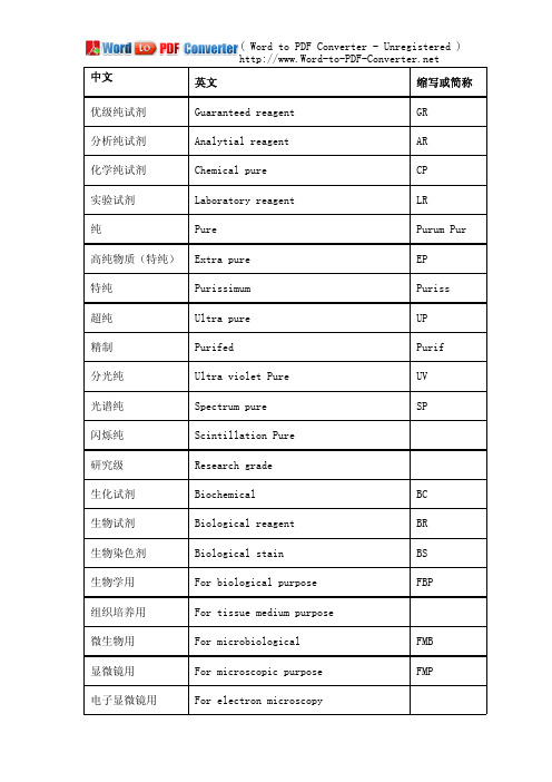
中文英文缩写或简称优级纯试剂Guaranteed reagent GR分析纯试剂Analytial reagent AR化学纯试剂Chemical pure CP实验试剂Laboratory reagent LR纯Pure Purum Pur 高纯物质(特纯)Extra pure EP特纯Purissimum Puriss超纯Ultra pure UP纾制Purifed Purif分光纯Ultra violet Pure UV光踱纯Spectrum pure SP闪烁纯Scintillation Pure研究级Research grade生化试剂Biochemical BC生物试剂Biological reagent BR生物染色剂Biological stain BS生物学用For biological purpose FBP组织培养用For tissue medium purpose微生物用For microbiological FMB显微镜用For microscopic purpose FMP电子显微镜用For electron microscopy涂镜用For lens blooming FLB工业用Technical grade Tech实习用Pratical use Pract分析用Pro analysis PA纾密分析用Super special grade SSG合成用For synthesis FS闪烁用For scintillation Scint电滳用For electrophoresis use测折光率用For refractive index RI显色剂Developer指示剂Indicator Ind配位指示剂Complexon indicator Complex ind 荧光指示剂Fluorescene indicator Fluor ind 渧化还原指示剂Redox indicator Redox ind 吸附指示剂Adsorption indicator Adsorb ind 基准试剂Primary reagent PT光踱标准物质Spectrographic standard substance SSS原子吸收光踱Atomic adsorption spectorm AAS红外吸收光踱Infrared adsorption spectrum IR核磁共振光踱Nuclear magnetic resonance spectrum NMR有机分析试剂Organic analytical reagent OAS微量分析试剂Micro analytical standard MAS微量分析标准Micro analytical standard MAS点滴试剂Spot-test reagent STR渔相色踱Gas chromatography GC液相色踱Liquid chromatography LCHPLC高效液相色踱High performance liquidchromatography渔液色踱Gas liquid chromatography GLC渔固色踱Gas solid chromatography GSC薄幂色踱Thin layer chromatography TLC凝胶渗透色踱Gel permeation chromatography GPC幂析用For chromatography purpose FCP常见质量级别优级纯(GR,绿标签):主成分含量很高、纯度很高,适用于纾确分析和研究工作,有的可作为基准物质。
- 1、下载文档前请自行甄别文档内容的完整性,平台不提供额外的编辑、内容补充、找答案等附加服务。
- 2、"仅部分预览"的文档,不可在线预览部分如存在完整性等问题,可反馈申请退款(可完整预览的文档不适用该条件!)。
- 3、如文档侵犯您的权益,请联系客服反馈,我们会尽快为您处理(人工客服工作时间:9:00-18:30)。
Role of Special Histochemical Stains in Staining MicroorganismsRashmil Saxena, BFA, HT(ASCP)CMDivision of TransplantationDepartment of Surgery, Indiana UniversityIndianapolis, IN, USAM icroorganisms encountered in routine pathology specimens include bacteria, fungi, protozoa and viruses1. Several histochemical stains help to visualize the first three groups of organisms; however, histochemical stains do not offer an advantage over H&E in the visualization of viruses and immunohistochemistry is the preferred method for this purpose. Histochemical stains also help to identify and classify bacteria, fungi and protozoa.The Giemsa and Gram’s stains help to visualize bacteria as well as classify them on their morphological characteristics. Thus bacteria can be classified into cocci or bacilli and cocci can be further classified into diplococci, staphylococci and streptococci based on their appearances on the Gram and Giemsa stains. The Gram stain also classifies bacteria into Gram-positive and Gram-negative organisms depending upon whether they take up the Gram stain or not; this classification is clinically useful and helps in therapeutic decisions. Some bacteria may not be adequately visualized with the Gram’s and Giemsa stains. Of these, the clinically most significant ones are mycobacteria and spirochetes. Mycobacteria stain with carbol fuschin and resist decolorization with acid-alcohol, leading to their designation as “acid-fast bacilli”. Spirochetes can be stained with a variety of silver stains such as the Warthin-Starry, Dieterle and Steiner stains. Finally, due to the large number of gastrointestinal biopsies in routine practice, a large number of stains are available for visualization of the Gram-negative bacillus, Helicobacter pylori. These include Giemsa, Alcian yellow - toludine blue, Diff-Quik, Genta, and Sayeed stains. A large number of laboratories prefer immunohistochemistry for identification of Helicobacter pylori.The Giemsa stain highlights several protozoa such as toxoplasma, leishmania, plasmodium, trichomonas, cryptosporidia and giardia. Ameba can be highlighted by the PAS stain due to their large glycogen content. Histochemical stains for fungi are discussed separately in this publication.Special Stains for Detection of BacteriaGram StainUtility of the Stain: The Gram stain is used to stain both bacillary and coccal forms of bacteria (Fig. 1). The most basic classification of bacteria consists of dividing them into Gram-positive and Gram negative bacteria based on whether they take up the Gram’s stain or not. Although the exact mechanism of staining is not known, bacteria that have large amounts of peptidoglycan in their walls retain the methyl violet stain, i.e., they are gram positive, whereas those that have more lipids and lipopolysaccharides in their cell walls are Gram-negative. The definite diagnosis of a bacterial species requires culture but the Gram stain provides a good initial indication of the nature of infection.1 Some microbiologists also include viruses as microorganisms, but others consider these as non-living. Lwoff (1957). “The concept of virus”. J. Gen. Microbiol. 17 (2): 239–53.T echnicalFigure 1. Photomicrograph of ulcerated skinstained with Gram’s stain. The purple stain represents gram-positive bacteria which are seen as clumps (arrowhead) or as separate clusters of cocci (arrows). Everything other than gram-positive bacteria is stained pink by the carbol fuschin counterstain. The underlying structure of the skin cannot be seen. Figure 2.Giemsa stained section showinga gastric pit containing Helicobacter pylori which appear as delicate, slightly curved rod-shaped purple organisms (arrowheads). The stomach is inflamed and shows many neutrophils (arrows). The background is stained light pink by the eosin counterstain.Principles of Staining:The method consists of initial staining of the bacterial slide with crystal violet or methyl violet which stain everything blue. This is followed by Gram’s or Lugol’s iodine made up of iodine and potassium iodide, which act by allowing the crystal violet to adhere to the walls of gram-positive bacteria. Decolorization with an acetone-alcohol mixture washes away the methyl violet which is not adherent to bacterial cell walls. At this stage, Gram-positive bacteria stain blue while the Gram-negative bacteria are colorless. A carbol fuschin counter-stain is then applied which stains the Gram-negative bacteria pink. Modifications: The Brown-Hopps and Brown-Brenn stains are modifications of the Gram stain and are used for demonstration of gram negative bacteria and rickettsia.Giemsa StainUtility of the Stain:The Giemsa is used to stain a variety of microorganisms including bacteria and several protozoans. Like the Gram stain, the Giemsa stain allows identification of the morphological characteristics of bacteria. However, it does not help further classification into Gram-negative or Gram-positive bacteria. The Giemsa stain is also useful to visualize H. pylori(Fig. 2, 3; See also Fig. 4 for a high resolution H&E stain showing Giardia). The Giemsa also stains atypical bacteria like rickettsia and chlamydiae which do not have the peptidoglycan walls typical of other bacteria and which therefore do not take up the Gram stain. The Giemsa stain is used to visualize several protozonas such as toxoplasma, leishmania, plasmodium, trichomonas, cryptosporidia and giardia.Principles of Staining: The Giemsa stain belongs to the class of polychromatic stains which consist of a mixture of dyes of different hues which provide subtle differences in staining. When methylene blue is prepared at an alkaline pH, it spontaneously forms other dyes, the major components being azure A and B. Although the polychromatic stain was first used by Romanowsky to stain malarial parasites, the property of polychromasia is most useful in staining blood smears and bone marrow specimens to differentiate between the various hemopoeitic elements. Nowadays, the Giemsa stain is made up of weighted amounts of the azures to maintain consistency of staining which cannot be attained if methylene blue is allowed to “mature” naturally.Modifications: The Diff-Quick and Wright’s stains are modifications of the Giemsa stain.Carbol Fuschin Acid-Alcohol StainUtility of the Stain: The carbol fuschin stain helps to identify mycobacteria which are bacilli containing thick waxy cell walls (Latin, myco=wax). Several mycobacteria can cause human disease; the two most significant ones are M. tuberculosis and M.leprae causing tuberculosis and leprosy respectively. Mycobacteria have large amounts of a lipid called mycolic acid in their cell walls which resists both staining as well as decolorization by acid-alcohol once staining has been achieved. The latter property is responsible for the commonly used term “acid-fast bacilli”. Mycobacteria cannot be stained by the Gram stain because it is an aqueous stain that cannot penetrate the lipid-rich mycobacterial cell walls.Principles of Staining: The mycobacteral cell walls are stained by carbol fuschin which is made up of basic fuschin dissolved in alcohol and phenol. Staining is aided by the application of heat. The organisms stain pink with the basic fuschin. Staining is followed by decolorisation in acid-alcohol; mycobacteria retain the carbol fuschin in their cell wall whereas other bacteria do not retain carbol fuschin, which is extracted into the acid-alcohol. Counterstaining is carried out by methylene blue. Mycobacteria stain bright pink with basic fuschin and the background stains a faint blue. Care has to be taken to not over-counter stain as this may mask the acid-fast bacilli.Modifications: The 2 commonly used methods for staining of M. tuberculosis are the Ziehl-Neelsen and Kinyoun’s acid fast stains. The Fite stain is used for staining of M.leprae which has cell walls that are more susceptible to damage in the deparaffinization process. The Fite procedure thus includes peanut oil in the deparaffinization solvent to protect the bacterial cell wall. The acid used for decolorization in the Fite procedure is also weaker (Fig. 5).“ Silver stains are very sensitive for the stainingof bacteria and therefore most useful forbacteria which do not stain or stain weaklywith the Grams and Giemsa stains.”Figure 3.Giemsa stained section of small intestinal mucosa showing clusters of Giardia which stain purple (arrows) in the crypts. The background is stained faint pink by the eosin counterstain.Figure 4.An H&E section of an intestinal crypt showing clusters of Giardia (arrowheads). The oval shape and clustering gives them a “tumbling leaves” appearance. Faint nuclei can be seen in some organisms (arrow).Figure 5. Ziehl-Neelsen stained section oflymph node. The pink color demonstrates clusters of mycobacteria stained with carbol-fuschin (arrows). The stain has resisted decolorisation by acid-alcohol. Other cells in the background are stained light blue by themethylene blue counterstain.Figure 6. Warthin-Starry stain of stomachcontaining Helicobacter pylori which appear as black and slightly curved, rod-like bacteria (arrows). The background is stained light yellow.Table 1. Provides a summary of some special stains used in detecting microorganisms.Silver Stains (Warthin Starry Stain, Dieterle, Steiner Stains)Utility of the Stains: Silver stains are very sensitive for the staining of bacteria and therefore most useful for bacteria which do not stain or stain weakly with the Grams and Giemsa stains. Although they can be used to stain almost any bacteria, they are tricky to perform and are therefore reserved for visualizing spirochetes, legionella, bartonella and H. pylori.Principles of Staining: Spirochetes and other bacteria can bind silver ions from solution but cannot reduce the bound silver. The slide is first incubated in a silver nitrate solution for half an hour and then “developed” with hydroquinone which reduces the bound silver to a visible metallic form. The bacteria stain dark-brown to black while the background is yellow (Fig. 6).Auramine O- Rhodamine B StainThe auramine O-rhodamine B stain is highly specific and sensitive for mycobateria. It also stains dead and dying bacteria not stained by the acid-fast stains. The mycobacteria take up the dye and show a reddish-yellow fluorescence when examined under a fluorescence microscope. SummaryHistochemical stains available for demonstrating microorganisms include Giemsa stain, Grams stain, carbol fuschin acid-alcohol stain and a variety of silver stains such as Warthin-Starry, Dieterle and Steiner stains. The Gram stain allows classification of bacteria into Gram-positive and Gram-negative bacteria. The acid-alcohol stain allows classification of bacilli into acid-fast and non-acid-fast bacilli. These are both clinically useful classifications. The silver stains are very sensitive and help to visualize difficult-to-stain bacteria. Most protozoans are stained by the Giemsa stain. GlossaryBacteria are unicellular organisms that do not contain a nucleus or other membrane-bound organelles. Most bacteria have a rigid cell wall composed of peptidoglycan. Although there are exceptions, bacteria come in 3 basic shapes: round (cocci), rod-like (bacilli) and spiral (spirochetes). Bacteria cause a variety of infections in various organs.Bartonella is a Gram-negative bacillus which causes cat-scratch disease. The bacilli are transmitted to humans by cat-bite or cat-scratch.Chlamydia are Gram-negative bacteria which are unusual because they do not have typical bacterial cell walls. They are obligate intracellular parasites which means that they can only survive within cells. Chlamydia cause sexually transmitted diseases and pneumonia in humans.Helicobacter pylori is a Gram-negative bacteria which causes inflammation (gastritis) and ulcers of the stomach. The name derives from the Greek “helix” for spiral and “pylorus” for the distal end of the stomach.Legionella is a Gram-negative bacillus so named because it caused an outbreak of pneumonia in people attending a 1976 convention of the American Legion in Philadelphia.The organism was unknown till then and was subsequently named Legionella. It causes pneumonia.Mycobacteria are bacilli that have a thick and waxy (Latin, myco = wax) cell wall composed of a lipid called mycolic acid. This cell wall is responsible for the hardiness of this organism as well as for its staining characteristics. The waxy cell wall is hydrophobic and resists staining with aqueous stains like the Gram and Giemsa stains. It also resists decolorisation once stained. Mycobacteria causes tuberculosis, leprosy and infections in patients with AIDS.Protozoa (Greek, proton = first; zoa = animal) are unicellular organisms that have a membrane-bound nucleus and other complex membrane-bound organelles.Rickettsia are Gram-negative bacteria that like Chlamydia lack typical cell walls and are obligate intracellular parasites. The rickettsial diseases are primary diseases of animals (zoonosis) such as the deer which are transmitted to humans by bites of insects like fleas and ticks. Rickettsial diseases include typhus fever and Rocky Mountain Spotty Fever.Spirochetes are long Gram-negative bacilli with tightly-coiled helical shapes. Spirochetes cause syphilis, leptospirosis and Lyme’s disease.。