医学报告 中英翻译版.pdf
[医学英语文章带翻译]医学类英语文章
![[医学英语文章带翻译]医学类英语文章](https://img.taocdn.com/s3/m/a27f7987caaedd3382c4d3da.png)
[医学英语文章带翻译]医学类英语文章医学英语文章带翻译医学英语文章带翻译医学英语文章带翻译 1 椎间盘突出Unit 2 Text A Herniated Disc (Disc Herniation of the Spine) 第二单元主题 A 椎间盘突出症Many patients with back pain, leg pain, or weakness of the lower extremity muscles arediagnosed with a herniated disc. 许多患腰腿疼痛,下肢肌端乏力的病患均为椎间盘突出症。
When a disc herniation occurs, the cushion that sits between the spinal vertebra is pushedoutside its normal position. 椎间盘突出发生时,脊柱间的缓冲带将发生侧突。
A hrniated disc would not be a problem if it weren“t for the spinal nerves that are very close tothe edge of these spinal discs. 如果脊神经不是离椎间盘特别近的话,椎间盘突出就不是什么大问题了。
HOW ARE THE SPINE AND ITS DISCS *****D 脊柱与椎间盘The vertebras are the bony building blocks of the spine. 脊椎是建造脊柱的构件。
Between each of the largest parts (bodies) of the vertebrae are the discs. 各椎骨之间为椎间盘。
Ligaments are situated around the spine and discs. 脊椎和椎间盘周围散布着韧带。
医学英语翻译3

医学英语文献3翻译(缺Unit1、3、7)U3 TCBeneficial bacteria may protect babies from HIV有益菌或能保护婴儿防止感染艾滋病毒毫无疑问,在喂养婴儿时,母乳是最好的选择。
但感染HIV(艾滋病病毒)的母亲面临着一个困境:由于有些病毒会流入母乳,婴儿吃奶时便面临被感染的风险。
目前两组研究队伍正在调查一场细菌之战以治疗这些脆弱的婴儿。
他们将在母乳中加入抑制艾滋病的细菌。
黎巴嫩,达特茅斯医学院HIV专家Ruth Connor 说,这些有益菌并不能保证儿童一定不会被感染,但可以大大降低感染概率。
在八月的“母乳喂养医学”中,她和来自西班牙马德里康普腾斯大学的同事,报告说将可以充分抑制HIV-1增长和感染的某些乳酸菌从健康女性的母乳中隔离出来。
试管研究对代表15个不同种类的38个细菌菌株进行了实验,它们都显示出了对HIV的抑制性。
效力最小的可以抑制细菌6.7%的传染性;效果最好的可以抑制HIV感染细胞能力的55.5%.研究者们试验了隔开的细菌和细菌产生的无细胞粘液。
“我们想分清抑制HIV 的能力是不是由细菌的某些特定组成部分造成的,例如外部细胞壁,或仅仅是组成细菌的可溶性成分,”Connor 解释说。
最终,她说每一项都被证明有抗病毒性,虽然细菌整体抑制性最好。
所有热灭菌都或多或少显示出了对HIV传染的抑制性,有11个可以杀掉41%甚至更多的HIV传染性。
相比之下,只有六种菌株产生的可溶性细胞化合物证实具有抑制作用,而且只有一种可以减少至少41%的传染性。
Connor说小组的发现揭示了她和其他病毒学家以前一直困扰的问题:为什么艾滋病病毒测试呈阳性的女性母乳喂养的婴儿没有被传染。
她说,目前还不知道,到底是最有抑制性的一种或几种细菌(例如乳酸菌/gramininis VM25;酵母VMA和戊糖片球菌VM95)单独提供了本质上防止HIV的保护作用还是共同作为自然的微生物群来起作用。
病历中中英文对照
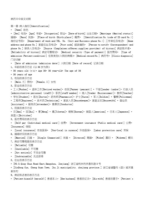
病历中中英文对照第一章病人身份[Identification]• [Name] 姓名• [Sex] 性别• [Age] 年龄• [Occupation] 职业• [Date of birth] 出生日期• [Marriage (Marital status)] 婚姻• [Race] 民族• [Place of birth (Birth place)] 籍贯• [Identification No.(code of ID card No.)] 身份证号码• [Department of work and TEL. No. (Unit and Business phone No.)] 工作单位及电话• [Home address and phone No.] 家庭住址及电话• [Post code] 邮政编码• [Person to notify (Correspondent) and phone No.] 联系人及电话• [Source (Complainer;offerer;supplier;provider) of history] 病史陈术者•[Reliability of history] 病史可靠程试• [Medical security (Type of payment)] 医疗费用• [Type of admission (Patient condition)] 住院类别(入院时病情)• [Medical record No.] 病历号• [Clinic diagnosis] 门诊诊断• [Date of admission (admission date)] 入院日期 [Date of record] 记录日期1、年龄的表示方法(以36岁为例)•36 years old (y/o)•Age 36•36 year-old•The age of 36•36 years of age2、性别的表示方法• [Male,♂] 男性• [Female,♀] 女性3、职业的表示方法•工人[Worker] •退休工作[Retired worker]•农民[Farmer (peasant)] •干部[Leader (cadre)]•行政人员[administrative personnel (staff)]•职员[staff member] •商人[Trader (Businessman)]•教师[Teacher] •学生[Student] •医生[Doctor]•药剂师[Pharmacist]•护士[Nurse] •军人[Soldier] •警察[Policeman]•工程师[Engineer] •技术员[Technician] •家政人员[Housekeeper]•家庭主妇[Housewife] •营业员[Assistant] •服务员[Attendant]•售票员[Conductor]4、民族的表示方法•汉[Han] •回[Hui] •蒙[Meng] •藏[Tibetan]•朝鲜[Korean]•美国人[American] •日本人[Japanese] •英国人[Britisher]5、医疗费用的表示方法• [Self pay (Individual medical care)] 自费• [Government insruance (Public medical care)] 公费•[Insurance] 保险• [Local insurance] 本地医保• [Non-local in surance] 外地医保• [Labor protestion care] 劳保6、婚姻状况的表示方法• [Married] 已婚• [Single (Unmarried)] 未婚• [Diverced] 离婚• [Widow] 寡妇• [Widower] 鳏夫7、病史可靠程度的表示方法• [Reliable] 可靠• [Unreliable] 不可靠• [Not entirely] 不完全可靠• [Unobtainable] 无法获得8、住址的表示方法•[NO.3,Qing Chun Road East,Hangzhou, Zhejiang] 浙江省杭州市庆春东路3号•[XinDong Cun, Cheng Guan Town, Zhu Ji municipality, zhejiang province.] 浙江省诸暨市(县)城关镇新东村9、病史陈述者的表示方法• [Patient himself (herself)] 患者本人• [Her husband] 患者的丈夫• [His wife] 患者的妻子• [Patient`scolleague] 患者的同事• [Patient`s neighbor] 患者的邻居• [Patient`s Kin (Mother; Son; daughter;brother;Sister)] 患者的亲属(父亲、母亲、儿子、女儿、兄弟、姐妹)• [Taximan] 出租车司机• [Traffic police] 交通警察10、日期的表示方法•2002年10月1日[10-1-2002(10/1/2002; Oct.1,2002; Oct.lst,2002)](美国)•2002年10月1日[1-10-2002(1/10/2002; 1 Oct.,2002; 1st of Oct.,2002)] (英国)11、住院类别的表示方法• [Emergent (Emergency call)] 急诊• [Urgent] 危重• [Elective (General)] 一般(普通)12、入院时病情的表示方法• [Stable] 稳定• [Unstable] 不稳定• [Relative stable] 相对稳定• [Critical (Imminent)] 危重• [Fair (General)] 一般第二章主诉[Chief Complaint]1、主诉的表示方法:症状+时间(Symptom+Time)•症状+for+时间如: [Chest pain for 2 hours] 胸痛2小时•症状+of+时间如: [Nausea and vomiting of three days` duration] 恶心呕吐3天•症状+时间+in duration如: [Headache 1 month in duration] 头痛1月•时间+of+症状如: [Two-day history of fever] 发热2天2、常见症状• [Fever] 发热• [Pain] 疼痛• [Edema] 水肿• [Mucocutaneous hemorrhage (bleeding)] 皮肤粘膜出血• [Dyspnea (Difficuly in breathing;Respiratory difficulty;short of breath)] 呼吸困难• [Cough and expectoration (Sputum hlegm)] 咳嗽和咯痰• [Hemoptysis] 咯血• [Cyanosis] 紫绀• [Palpitation] 心悸• [Chest discomfort] 胸闷• [Nausea (Retch;Dry Vomiting)and Vomiting] 恶心和呕吐• [Hematemesis (Vomiting of blood)] 呕血• [Hematochezia (Hemafecia)] 便血• [Diarrhea] 腹泻• [Constipation (Obstipation)] 便秘• [Vertigo (Giddiness; Dizziness)] 眩晕• [Jaundice (Icterus)] 黄疸• [Convulsion] 惊厥• [Disturbance of consciousness] 意识障碍• [Hematuria] 血尿• [Frequent micturition,urgent micturition and dysuria] 尿频,尿急和尿痛• [Incontinence of urine] 尿失禁• [Retention of urine] 尿潴留(1)发热的表示方法• [Infective (Septic)fever] 感染性发热• [Non-infective (Aseptic)fever] 非感染性发热• [Dehydration (Inanition)fever] 脱水热• [Drug fever] 药物热• [Functional hypothermia] 功能性低热• [Absorption fever] 吸收热• [Central fever] 中枢性发热• [Fever type] 热型▲ [Continuous fever] 稽留热▲ [Remittent fever] 驰张热▲ [Intermittent fever] 间歇热▲ [Undulant fever] 波状热▲ [Recurrent fever] 回归热▲ [Periodic fever] 周期热▲ [Irregular fever] 不规则热▲ [Ephemeral fever] 短暂热▲ [Double peaked fever] 双峰热• [Fever of undetermined(unknown) origin, FUO] 不明原因发热• [Rigor (shivering;chill;shaking chill;ague)] 寒战• [Chilly Sensation (Fell chilly;cold fits;coldness)] 畏寒• [Ultra-hyperpyrexia] 超高热• [Hyperthermia (A high fever;hyperpyrexia;ardent fever)] 高热• [Moderate fever] 中度发热• [Hypothermia (Low-grade fever;slight fever;subfebrile temperature)] 低热• [Become feverish (Have a temperature)] 发热• [Crisis] 骤降• [Lysis] 渐降• [Typhoid fever] 伤寒热• [Rheumatic fever] 风湿热• [Cancerous fever] 癌性发热• [Fervescence period] 升热期• [Defervescence period] 退热期• [Persistent febrile period] 持续发热期(2)疼痛的表示方法• [Backache (Back pain)] 背痛• [Lumbago] 腰痛• [Headache] 头痛▲ [Vasomotor headache] 血管舒缩性头痛▲[Post-traumatic headache] 创伤后头痛▲[Migraine headache] 偏头痛▲ [Cluster headache] 丛集性头痛• [Chest pain] 胸痛• [Precardial pain] 心前区痛• [Retrosternal pain] 胸骨后痛• [Abdominal pain (Stomachache)] 腹痛• [Acrodynia (pain in limbs)] 肢体痛• [Arthrodynia (Arthralgia)] 关节痛• [Dull pain] 钝痛• [Sharp pain] 锐痛• [Twinge pain] 刺痛• [Knife-like pain (Piercing pain)] 刀割(刺)样痛• [Aching pain] 酸痛• [Burning pain] 烧灼痛• [Colicky (Griping;cramp) pain] 绞痛• [Colic] 绞痛• [Bursting pain] 胀痛(撕裂痛)• [Hunger pain] 饥饿痛• [Tic pain] 抽搐痛• [Bearing-down pain] 坠痛• [Shock-like pain] 电击样痛• [Jumping pain] 反跳痛• [Tenderness pain] 触痛(压痛)• [Girdle-like pain] 束带样痛• [Wandering pain] 游走性痛• [Throbbing pain] 搏动性痛• [Radiating pain] 放射性痛• [Cramping pain] 痉挛性痛• [Boring pain] 钻痛• [Intense pain] 剧痛• [Writhing pain] 痛得打滚• [Dragging pain] 牵引痛• [Labor pain] 阵痛• [Cancerous pain] 癌性疼痛• [Referred pain] 牵涉痛• [Persistent pain (Unremitting pain)] 持续性痛• [Constant pain] 经常性痛• [Intermittent pain] 间歇性痛(3)水肿的表示方法• [Mucous edema (Myxedema)] 粘液性水肿• [Cardiac (Cardiogenic) edema] 心源性水肿• [Nephrotic (renal) edema] 肾源性水肿• [Hepatic edema] 肝源性水肿• [Alimentary (Nutritional) edema] 营养不良性水肿• [Angioneurotic edema] 血管神经性水肿• [Pitting] 凹陷性• [Nonpitting] 非凹陷性• [Localized (Local) edema] 局限性水肿• [Generalized edema (Anasarca)] 全身性水肿• [Hydrops] 积水• [Elephantiasic crus] 橡皮肿• [Cerebral(Brain) edema] 脑水肿• [Pulmonary edema (Hydropneumonia0] 肺水肿• [Hydrocephalus] 脑积水• [Edema of endoscrinopathy] 内分泌病性水肿• [Invisible (Recessive) edema] 隐性水肿• [Frank edema] 显性水肿• [Inflammatory edema] 炎性水肿• [Idiopathic edema] 特发性水肿• [Cyclical edema] 周期性水肿• [Ascites (Abdominal effusion;hydroperiotoneum)] 腹水• [Pleural effusion (Hydrothorax)] 胸水• [Pericardial effusion (Hydropericardium)] 心包积液• [Bronchoedema] 支气管水肿• [Slight (Mild)] 轻度• [Moderate] 中度• [Serious] 重度• [Transudate] 漏出液• [Exudate] 渗出液(4)呼吸困难的表示方法• [Cardiac dyspnea] 心原性呼吸困难• [Inspiratory] 吸气性• [Expiratory] 呼气性• [Mixed] 混合性• [Obstructive] 梗阻性• [Dyspnea at rest] 静息时呼吸困难• [Dyspnea on exertion] 活动时呼吸困难• [Dyspnea on lying down] 躺下时呼吸困难• [Paroxysmal nocturnal dyspnea,PND] 夜间阵发性呼吸困难• [Orthopnea] 端坐呼吸• [Asthma] 哮喘• [Cardiac asthma] 心源性哮喘• [Bronchial asthma] 支气管性哮喘• [Hyperpnea] 呼吸深快• [Periodic breathing] 周期性呼吸• [Tachypnea (Rapid or fast breathing;accelerated breathing;short of breath)]气促• [Bradypnea (Slow breathing)] 呼吸缓慢• [Irregular breathing] 不规则呼吸(5)皮肤粘膜出血的表示方法• [Bleeding spots in the skin] 皮肤出血点• [Petechia] 瘀点• [Eccymosis] 瘀斑• [Purpura] 紫癜• [Splinter hemorrhage] 片状出血• [Oozing of the blood (Errhysis)] 渗血• [Blood blister (Hemophysallis)] 血疱• [Hemorrhinia (Nasal bleeding)] 鼻衄• [Ecchymoma] 皮下血肿(6)咳嗽与咯痰的表示方法• [Dry cough (Nonproductive cough;hacking cough)] 干咳• [Sharp cough] 剧咳• [Wet cough (Moist cough)] 湿咳• [Productive cough (Loose cough)] 排痰性咳• [Chronic cough] 慢性咳嗽• [Irritable cough] 刺激性咳嗽• [Paroxysmal cough] 发作性(阵发性)咳嗽• [Cough continually] 持续性咳嗽• [Spasmodic cough] 痉挛性咳嗽• [Whooping cough] 百日咳• [Winter cough] 冬季咳• [Wheezing cough] 喘咳• [Short cough] 短咳• [Distressed cough] 难咳• [Shallow cough] 浅咳• [Droplet] 飞沫• [Frothy sputum] 泡沫样痰• [Bloody sputum] 血痰• [Mucous (Mucoid) sputum] 粘液样痰• [Purulent sputum] 脓痰• [Mucopurulent sputum] 粘液脓性痰• [White (Yellow,green) sputum] 白(黄,绿)痰• [Fetid (Foul) sputum] 恶臭痰• [Iron-rust (Rusty) sputum] 铁锈色痰• [Chocolate coloured sputum] 巧克力色痰• [Thick sputum] 浓痰• [Thin sputum] 淡痰• [Viscous sputum] 粘痰• [Transparent sputum] 透明痰• [Much (Large amounts of) sputum] 大量痰• [Moderate amounts of sputum] 中等量痰• [Not much (Small amounts of ) sputum] 少量痰(7)内脏出血的表示方法• [Goldstein’s hemoptysis]戈耳斯坦氏咯血• [Massive hematemesis]大量呕血• [Epistasis (Nosebleed;Nasal bleeding; Hemorrhinia;rhinorrhagia)]鼻衄• [Hematuria] 血尿• [Initial hematuria] 初血尿• [Idiopathic hematuria] 特发性血尿• [Painless hematuria] 无痛性血尿• [Terminal hematuria] 终末性血尿• [Gross (Macroscopic) hematuria] 肉眼血尿• [Microscopic hematuria] 镜下血尿• [Hematuria in the whole process of urination] 全程血尿• [Gingival bleeding (Ulaemorrhagia;gum bleeding)] 牙龈出血• [Hematochezia] 便血• [Bloody stool] 血便• [Black stool (Melena)] 黑便• [Tarry stool] 柏油样便• [Bleeding following trauma] 外伤后出血• [Spontaneous bleeding] 自发性出血• [Bleeding Continuously] 持续出血• [Occult blood,OB] 隐血• [Hematobilia] 胆道出血• [Hemathorax] 血胸• [Hemarthrosis] 关节积血• [Hematocoelia] 腹腔积血• [Hematoma] 血肿• [Hemopericardium] 心包积血• [Cerebral hemorrhage] 脑出血• [Subarachnoid hemorrhage(SAH)] 蛛网膜下腔出血• [Excessive (Heavy) menstrual flow with passage of clots] 月经量多伴血块• [Mild (Moderate) menses] 月经量少(中等)• [Painless Vaginal bleeding] 无痛性阴道出血• [Postcoital bleeding] 性交后出血• [Pulsating bleeding] 搏动性出血• [Post-operation wound hemorrhage] 术后伤口出血• [Excessive bleeding after denal extraction] 拔牙后出血过多(8)紫绀的表示方法• [Congenital cyanosis] 先天性紫绀• [Enterogenous] 肠源性• [Central] 中枢性• [Peripheral] 周围性• [Mixed] 混合性• [Acrocyanosis] 指端紫绀(9)恶心与呕吐的表示方法• [Vomiturition (Retching)] 干呕• [Feel nauseated] 恶心感• [Postprandial nausea] 饭后恶心• [Hiccup] 呃逆• [Sour regurgitation] 返酸• [Fecal (Stercoraceous) vomiting] 吐粪• [undigested food Vomiting] 吐不消化食物• [Bilious Vomiting] 吐胆汁(10)腹泻与便秘的表示方法• [Moning diarrhea] 晨泻• [Watery (Liquid)diarrhea] 水泻• [Mucous diarrhea] 粘液泻• [Fatty diarrhea] 脂肪泻• [Chronic (Acute)] 慢性(急性)• [Mild diarrhea] 轻度腹泻• [Intractable (Uncontrolled)diarrhea] 难治性腹泻• [Protracted diarrhea] 迁延性腹泻• [Bloody stool] 血梗• [Frothy stool] 泡沫样便• [Formless (Formed)stool] 不成形(成形)便• [Loose (Hard) stool] 稀(硬)便• [Rice-water stool] 米泔样便• [Undigested stool] 不消化便• [Dysenteric diarrhea] 痢疾样腹泻• [Inflammatory diarrhea] 炎症性腹泻• [Osmotic] 渗透性• [Secretory] 分泌性• [Malabsorption] 吸收不良性• [Lienteric] 消化不良性• [Pancreatic diarrhea] 胰性腹泻• [Tenesmus] 里急后重• [Pass a stool (Have a passage; open or relax the bowel)] 解大便• [Have a call of nature] 便意• [Fecal incontinence (Copracrasia)] 大便失禁• [Functional constipation] 功能性便秘• [Organic constipation] 器质性便秘• [Habitual constipation] 习惯性便秘• [Have a tendency to be constipated] 便秘倾向(11)黄疸的表示方法• [Latent (occult) jaundice] 隐性黄疸• [Clinical jaundice] 显性黄疸• [Nuclear icterus] 核黄疸• [Physiologic icterus] 生理性黄疸• [Icterus simplex] 传染性黄疸• [Toxemic icterus] 中毒性黄疸• [Hemolytic] 溶血性• [Hepatocellular] 肝细胞性• [Obstructive] 阻塞性• [Congenital] 先天性• [Familial] 家族性• [Cholestatic] 胆汁淤积性• [Hematogenous] 血源性• [Malignant] 恶性• [Painless] 无痛性(12)意识障碍的表示方法• [Somnolence] 嗜睡• [Confusion] 意识模糊• [Stupor] 昏睡• [Coma] 昏迷• [Delirium] 谵妄• [Syncope (swoon; faint)] 晕厥• [Drowsiness] 倦睡(13)排尿的表示方法• [Enuresis (Bed-wetting)] 遗尿• [Anuria] 无尿• [Emiction interruption] 排尿中断• [Interruption of urinary stream] 尿线中断• [Nocturia] 夜尿• [Oliguria] 少尿• [Polyuria] 多尿• [Pass water (Make water; urinate; micturition)] 排尿• [Frequent micturition (Frequency of micturition; fruquent urination;Pollakiuria)] 尿频• [Urgent micturition (Urgency of urination or micturition)] 尿急• [Urodynia (Pain on micturition; painful micturition; alginuresis; micturition pain)] 尿痛• [Dysuria (Difficulty in micturition; disturbance of micturition)] 排尿困难• [Small urinary stream] 尿线细小• [Void with a good stream] 排尿通畅• [Guttate emiction (Dribbling following urination;terminal dribbling)] 滴尿• [Bifurcation of urination] 尿流分叉• [Residual urine] 残余尿• [Extravasation of urine] 尿外渗• [Stress incontinence] 压力性尿失禁• [Overflow incontinence] 溢出性尿失禁• [Paradoxical in continence] 反常性尿失禁3.少见症状• [Weekness( Debility; asthenia; debilitating)] 虚弱(无力)• [Fatigue (Tire; lassitude)] 疲乏• [Discomfort (Indisposition; malaise)] 不适• [Wasting (thin; underweight; emaciation; lean)] 消瘦• [Night sweating] 盗汗• [Sweat (Perspiration)] 出汗• [Cold sweat] 冷汗• [Pruritus (Iching)] 搔痒• [Asthma] 气喘• [Squeezing (Tightness; choking; pressing) sensation of the chest] 胸部紧缩(压榨)感• [Intermittent claudication] 间歇性跛行• [Difficulty in swallowing( Dysphagia; difficult swallowing; acataposis)] 吞咽困难• [Epigastric (Upper abdominal) discomfort] 上腹部不适• [Anorexia (Sitophobia)] 厌食• [Poor appetite (Loss of appetite)] 纳差• [Heart-burn( Pyrosis)] 胃灼热• [Stomachache( Pain in stomach)] 胃部痛• [Periumbilial pain] 脐周痛• [Belching (Eructation)] 嗳气• [Sour regurgitation] 返酸• [Abdominal distention(bloating)] 腹胀• [Pass gas( Break wink)] 肛门排气• [Small(Large) stool] 大便少(多)• [Expel(Pass) worms] 排虫• [Pain over the liver] 肝区痛• [Lumbago] 腰痛• [Pica(Parorexia; allotriophagy)] 异食癖• [Dysmenorrhea] 痛经• [Menoxenia (Irregular menstruation)] 月经不调• [Polymenorrhea (Epimenorrhea)] 月经过频• [Oligomenorrhea] 月经过少• [Excessive menstruation (Menorrhagia; menometrorrhagia; hypermenorrhea)] 经量过多• [Hypomenorrhea (Scantymenstruation)] 经量过少• [Menopause (Menostasia; menostasis)] 绝经• [Amenorrhea (Menoschesis)] 闭经• [Leukorrhagia] 白带过多• [Asexuality (lack of libido)] 无性欲• [Hyposexuality] 性欲低下• [Hypersexuality] 性欲亢进• [Prospermia (Ejaculatio praecox)] 早泄• [Impotency (impotence)] 阳萎• [Nocturnal emission (Spermatorrhea)] 遗精• [Lack of potency] 无性交能力• [Hair loss] 脱发• [Joint pain (Arthralgia; arthrodynia)] 关节痛• [Polydipsia (Excessive thirst)] 多饮(烦渴)• [Polyphagia (Excessive appetite; hyperorexia; bulimia)] 多食• [Cold (Heat) intolerance] 怕冷(热)• [Dwarfism (Excessive height)] 身材矮小(高大)• [Excessive sweating] 多汗• [Hands tremble] 手抖• [Obesity (Fatty)] 肥胖• [Agitation (Anxiety;nervous irritability)] 焦虑(忧虑)• [Mania] 躁狂• [Hallucination] 幻觉• [Aphasia (Logopathy)] 失语• [Amnesia (Poor memorization;memory deterioration)] 记忆力下降• [Hemianesthesia] 偏身麻木• [Formication] 蚁走感• [Tingling] 麻刺感• [Hyperpathia] 痛觉过敏• [Hypalgesia] 痛觉减退• [Illusion] 错觉• [Hemiplegia] 半身不遂• [Insomnia (Poor sleepness;sleeplessness)] 失眠• [Nightmare] 多梦• [Numbness] 麻木• [Pain in limbs (Acrodynia)] 肢体痛• [Limitation of motion] 活动受限• [Tetany] 手足抽搐• [Discharge of pus] 流脓• [Blurred vision(Hazy vision;blurring of vision; dimness of vision)]视物模糊• [Burning (Dry) sensation] 烧灼(干燥)感• [Tearing (Dacryorrhea;Lacrimation)] 流泪• [Double vision (Diplopia)] 复视• [Strabismus] 斜视• [Hemianopia] 偏盲• [Tired eyes (Eyestrain)] 眼疲劳• [Foreign body sensation] 异物感• [Lose the sight (Lose of vision)] 失明• [Diminution of vision] 视力减退• [Nictition] 眨眼• [Ophthalmodynia (Eye-ache;ocular pain)] 眼痛• [Photophobia] 畏光• [Spots before the eyes] 眼前黑点• [Deafness(Anacusia)] 耳聋• [Auditory dysesthesia] 听力减退• [Otalgia (Otodynia;pain in the ear ;ear-ache)] 耳痛• [Stuffy feeling in the ear] 耳闭气• [Tinnitus] 耳鸣• [Outophony] 自声过强• [Nasal obstruction (blockage)] 鼻塞• [Dryness of the nose] 鼻干燥• [Rhinorrhea (Snivel;Nasal discharge)] 流鼻涕• [Sneezing] 打喷嚏• [Snoring] 打鼾• [Hyposmia (Reduction of the sense of smell)] 嗅觉减退• [Anosmia (Complete loss of sense of smell)] 嗅觉丧失• [Dysphonia] 发音困难• [Hoarseness] 声嘶• [Pain on swallowing] 吞咽痛• [Saliva dribblies from the mouth] 流涎• [Troaty voice] 声音沙哑• [Stridor] 喘鸣• [Red and swollen] 红肿• [Scurf] 头皮屑• [Show] 见红• [Amniotic fluid escaped] 破水• [Uterine contraction] 宫缩• [Acalculia] 计算不能• [Apathy] 情感淡漠• [Delusion] 妄想第三章现病史[History of present illness (HPI/PI)]现病史书写的重点包括:一、主诉中症状的详细描述;二、疾病的发展过程;三、诊疗经过;四、目前的一般情况。
(医学影像学)中英文对照学生翻译版
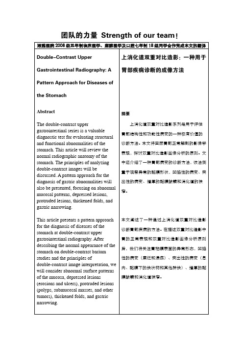
团队的力量 Strength of our team!湘雅医院2008级五年制临床医学、麻醉医学及口腔七年制18组同学合作完成本文的翻译Double-Contrast Upper Gastrointestinal Radiography: A Pattern Approach for Diseases of the StomachAbstractThe double-contrast upper gastrointestinal series is a valuable diagnostic test for evaluating structural and functional abnormalities of the stomach. This article will review the normal radiographic anatomy of the stomach. The principles of analyzing double-contrast images will be discussed. A pattern approach for the diagnosis of gastric abnormalities will also be presented, focusing on abnormal mucosal patterns, depressed lesions, protruded lesions, thickened folds, and gastric narrowing.This article presents a pattern approach for the diagnosis of diseases of the stomach at double-contrast upper gastrointestinal radiography. After describing the normal appearance of the stomach on double-contrast barium studies and the principles ofdouble-contrast image interpretation, we will consider abnormal surface patterns of the mucosa, depressed lesions (erosions and ulcers), protruded lesions (polyps, submucosal masses, and other tumors), thickened folds, and gastric narrowing. 上消化道双重对比造影:一种用于胃部疾病诊断的成像方法摘要上消化道双重对比造影系列是用于评估胃部结构性和功能性病变的一种极有价值的诊断方法。
医学英语课文翻译
Unit OneText A: Hippocratic Oath, The Medical Ideal或许在医学史上最持久的,被引用最多次的誓言就是”希波克拉底誓言”.这个以古希腊著名医师希波克拉底命名的誓言,被作为医师道德伦理的指导纲领.虽然随着时代的变迁,准确的文字已不可考,但誓言的主旨却始终如一——尊敬那些将毕生知识奉献于医学科学的人,尊重病人,尊重医师尽己所能治愈病人的承诺。
作为被大家公认的”医学之父”,我们对希波克拉底知之甚少.他生活于约公元前460-380年,作为一名职业医师,与苏格拉底是同代人.在他的时代,他被推举为当时最著名的医师和医学教育者.收录了超过60篇论文的专著——希波克拉底文集,被归于他的名下;但是其中有些论文的内容主旨相冲突,并成文于公元前510-300年,所以不可能都是出自他之手.这个宣言是以希波克拉底命名的,虽然它的作者依然存在疑问。
根据医学历史权威的看法,这个宣言的内容是在公元前四世纪起草的,这使希波克拉底自己起草这个宣言成为可能。
无论如何,不管是否是希波克拉底自己起草的(希波克拉底宣言),这个宣言的内容都反映了他在医学伦理上的看法。
作为代表当时希腊观点的唯一一小部分,希波克拉底誓言首次被写时并没有受到很好的欢迎。
然而,在那远古时代结束时,医生们开始遵循誓言的条款。
当科学医学在罗马帝国衰亡后遭受一显而易见的衰退时,这个誓言,连同希波克拉底医学的指示命令,在西方都几乎被遗忘是有可能的。
正是通过东方坚持不懈的探索精神,使得希波克拉底医学信念和希波克拉底宣言得以在这一恶化的时期幸存下来,尤其是通过阿拉伯当局在医学上的著作。
希腊医学知识而后在西方基督教复活是通过了阿拉伯文论著和原始希腊文的拉丁文翻译。
到17世纪后期,专业行为标准已经在西方世界建立。
被专业组织通过的第一部医学伦理学的法典是由英国内科医生托马斯·珀西瓦尔(1740 - 1804)1794年编写的, 并在1846年被改编和通过了美国医学协会(AMA)。
医学检验报告单中英文翻译解读表

国际标准化值
CRP
C-反应蛋白
AAT
ai.抗陆蛋白酶
医学检验报告单中英文翻译解读表
英文名称
中文名称
英文名称
中文名称
ALT
丙氨酸氨基转移酶
TC
总胆固醇
AST
天门冬氨酸氨基转移酶
TG
甘油三酯
ALP
碱性磷酸酶
HDL
高密度脂蛋白
GGT
谷氨酸转肽酶
LDL
低密度脂蛋白
TB-V
总胆红素
APOAT
载脂蛋白A
DB-V
直接胆红素
APOBT
载脂蛋白B
BIDL
间接胆红素
LPA
脂蛋白a
TP
总蛋白
HCYS
同性半胱氨酸
ALB
白蛋白(清蛋白)
GLU
葡萄糖
GLB
球蛋白
Hb-ALC
糖化血红蛋白
CHE
胆碱酯酶
K
钾
TBA
总胆汁酸
Na
钠
LPIC
脂肪酶
CI
氯
UREA
尿素
Ca
钙
UA
尿酸
Mg
镁
CREA-J
肌酥
p
磷
CYs-C
挠抑素-C
Fe
铁
a-HBDL
a-疑丁酸脱氢酶
CK
肌酸激酶
LDH
乳酸脱氢酶
CK-MB
肌酸激酶同工
ASO
抗链球菌溶血素
TT
凝血酶原
RF
类风湿因子
PT
凝血酶原时间
ESR
血沉
APTT
活化部分凝血酶原时
医学报告中英翻译版

101 of 121Laboratory General Checklist实验室常规检查表supervisory review of work, reassignment of duties, or otheractions deemed appropriate by the biorepository director.工作的监督检查,分配职责,或采取其他行动,似乎是生物研究者们最欣赏的Evidence of Compliance合规的依据:✓Records of corrective action to include evidence ofretraining and reassessment of competency✓纠正措施包括培训和评估能力证据记录,记录关于培训和评估的胜任的积极行为PHYSICAL FACILITIES物质设施;Inspector Instructions:检查员指示:●Floor plan and equipment locations地面计划及设备位置●Overview of Building Automation System (BAS), ifavailable如果有可能,可以建筑自动化系统(BAS)●Physical facility (adequate space, acceptabletemperature/humidity, areas clean, adequatestorage areas, adequate emergency power)物理设施(适当的空间,可以接受的温度/湿度,清洁,充足的存储区域,足够的应急电源)●Perimeter security and access security to specificspecimen collections●确保周边安全性和特定的样本集的访问安全●Is the work area sufficient for you to perform yourduties safely and accurately?你的工作是否足够安全、准确地履行你的职责?GLOBALEUROPEANATMNETWORK泛欧ATM网;Restricted Access 限制访问Phase I段落一Access to the biorepository is restricted to authorizedindividuals.、进入生物是被禁止的授权人。
成人血常规报告中英文

成人血常规报告中英文Adult Blood Routine Examination ReportName: XXX Sex: Male Age: 30 Date: XX/XX/XXXXSample Type: Peripheral BloodTest Indicators:1. White Blood Cell (WBC) CountNormal Range: 4.0-10.0 × 10^9/LResult: XX.X × 10^9/L (Within normal range)2. Red Blood Cell (RBC) CountNormal Range: 4.5-5.5 × 10^12/LResult: X.X × 10^12/L (Within normal range)3. Hemoglobin (Hb)Normal Range: 130-175 g/LResult: XXX g/L (Within normal range)4. Hematocrit (Hct)Normal Range: 38-50%Result: XX% (Within normal range)5. Mean Corpuscular Volume (MCV)Normal Range: 80-100 fLResult: XX fL (Within normal range)6. Mean Corpuscular Hemoglobin (MCH)Normal Range: 27-33 pgResult: XX pg (Within normal range)7. Mean Corpuscular Hemoglobin Concentration (MCHC)Normal Range: 32-36 g/dLResult: XX g/dL (Within normal range)8. Platelet (PLT) CountNormal Range: 125-350 × 10^9/LResult: X.X × 10^9/L (Within normal range)9. Mean Platelet Volume (MPV)Normal Range: 7.4-10.4 fLResult: X.X fL (Within normal range)10. Red Blood Cell Distribution Width (RDW)Normal Range: 11.6-14.6%Result: X.X% (Within normal range)Discussion:This blood routine examination report indicates that the patient's blood parameters fall within the normal ranges for all indicators tested. The results suggest that the patient does not have any significant abnormalities in their blood composition.White blood cell count (WBC) is an important indicator of immune system function. The patient's WBC count is within the normal range, indicating that the patient does not have a high or low whiteblood cell count, which can be indicative of certain infections or diseases.Red blood cell count (RBC) is a measure of the number of red blood cells in the blood. The patient's RBC count is within the normal range, suggesting that the patient does not have issues with anemia or problems with red blood cell production.Hemoglobin (Hb) is a protein in red blood cells that carries oxygen. The patient's hemoglobin level falls within the normal range, indicating that the patient has an adequate amount of hemoglobinin their blood.Hematocrit (Hct) is the proportion of red blood cells in the blood. The patient's hematocrit level is within the normal range, indicating a normal volume of red blood cells.Mean corpuscular volume (MCV) is a measure of the average volume of red blood cells. The patient's MCV falls within the normal range, suggesting that the red blood cells are of a normal size.Mean corpuscular hemoglobin (MCH) is a measure of the average amount of hemoglobin in each red blood cell. The patient's MCH level falls within the normal range, indicating that the amount of hemoglobin in their red blood cells is normal.Mean corpuscular hemoglobin concentration (MCHC) is a measure of the concentration of hemoglobin in each red blood cell. The patient's MCHC level is within the normal range, suggesting anormal concentration of hemoglobin.Platelet count (PLT) measures the number of platelets in the blood. The patient's platelet count falls within the normal range, indicating no abnormalities with their platelet levels.Mean platelet volume (MPV) measures the average size of platelets in the blood. The patient's MPV falls within the normal range, suggesting normal platelet size.Red blood cell distribution width (RDW) measures the variation in the size of red blood cells. The patient's RDW is within the normal range, indicating that the size of their red blood cells is relatively consistent.Conclusion:The results of this blood routine examination suggest that the patient's blood parameters are within the normal range, indicating a healthy blood composition. However, please note that additional tests may be required to fully evaluate the patient's overall health. It is recommended that these results be interpreted by a qualified healthcare professional to provide a comprehensive assessment and make appropriate medical decisions.。
X线诊断报告中英文对照

1. 头颅骨质未见异常The shape and the size of the skull are normal。
The inner and outer tables,and the diploe of the cranial vault are unremarkable,on the lateral view ,the sizes,the shape and the density of the sella turcica are nothing remarkabl e。
Impression:plain films of the head are normal.2.头颅正常AP and lateral views of the skull are submitted. The cavarium has n ormal configuration and appearance. There is no evidence of fracture. The soft tissues are normal.Impression: Normal skull.3.鼻旁窦炎症There is generalized haziness of the frontal, ethmoid and bilateral m axillary sinuses. Findings are consistent with sinusitis.Impression: Frontal,(额窦)ethmoid(筛)and maxillary (上颌)sinusitis.4. 右膝关节正常The bones and joints of the right knee are normal. There is no evi dence of fracture or subluxation. The soft tissues are normal. There is no joint effusion.Impression: Normal right knee.5.右膝退变The bones and joints of the right knee are normal. Mild degenerative changes of the knee joint is present. There is no evidence of fra cture or subluxation. The soft tissues are normal. There is no joint effusion.Impression: Mild DJD of knee joint。
中英文X光报告

中英文X光报告
摘要
本报告是对患者进行的X光检查的结果进行分析和描述的文档。
通过X光图像的观察和解读,我们提供了对患者体部的病变和异常情况的详细描述。
此报告包含患者的基本信息、检查所用仪器、医
生的观察结果和初步诊断建议。
患者信息
- 姓名:XXX
- 年龄:XX
- 性别:X
- 就诊日期:XXXX年XX月XX日
检查结果
头部 X光检查
- 医生观察结果:患者头部X光显示正常,未发现异常结构或病变。
- 初步诊断建议:患者头部X光结果未显示明显的异常,建议针对其他症状进行进一步检查。
胸部 X光检查
- 医生观察结果:患者胸部X光显示右上叶阴影存在,可能与感染或结节有关。
- 初步诊断建议:建议进行进一步检查,如胸部CT扫描或其他相关检查,以确定阴影的性质和起因。
腹部 X光检查
- 医生观察结果:患者腹部X光显示胃和肠道正常,未发现明显异常。
- 初步诊断建议:腹部X光结果在观察范围内未显示明显的异常,建议根据其他症状和检查结果进行进一步诊断。
结论
根据患者的X光检查结果,需要进一步评估和诊断,以确定任何潜在的异常情况或疾病。
进一步的检查,如CT扫描或其他相关检查,将有助于明确诊断和制定适当的治疗方案。
此报告仅为初步分析和诊断建议,具体的诊断和治疗方案应由专业医生根据进一步检查和患者具体情况来确定。
请咨询专业医生以获得个体化的诊断和治疗建议。
医学文献翻译(中英对照)

Current usage of three-dimensional computed tomography angiography for the diagnosis and treatment of ruptured cerebral aneurysmsKenichi Amagasaki MD, Nobuyasu Takeuchi MD, Takashi Sato MD, Toshiyuki Kakizawa MD, Tsuneo Shimizu MD Kanto Neurosurgical Hospital, Kumagaya, Saitama, JapanSummary Our previous study suggested that 3D-CT angiography could replace digital subtraction (DS) angiography in most cases of ruptured cerebral aneurysms, especially in the anterior circulation. This study reviewed our further experience. One hundred and fifty patients with ruptured cerebral aneurysms were treated between November 1998 and March 2002. Only 3D-CT angiography was used for the preoperative work-up study in patients with anterior circulation aneurysms, unless the attending neurosurgeons agreed that DS angiography was required.Both 3D-CT angiography and DS angiography were performed in patients with posterior circulation aneurysms, except for recent cases that were possibly treated with 3D-CT angiography alone. One hundred sixteen (84%) of 138 patients with ruptured anterior circulation aneurysms underwent surgical treatment, but additional DS angiography was required in 22 cases (16%).Only two recent patients were treated surgically with 3D-CT angiography alone in 12 patients with posterior circulation aneurysms. Most patients with ruptured anterior circulation aneurysms could be treated successfully after 3D-CT angiography alone. However, additional DS angiography is still necessary in atypical cases. 3D-CT angiography may be limited to complementary use in patients with ruptured posterior circulation aneurysms.a 2003 Elsevier Ltd. All rights reserved.Keywords: 3D-CT angiography, cerebral aneurysm, subarachnoid haemorrhage, surgeryINTRODUCTIONRecently, three-dimensional computed tomography (3D-CT) angiography has become one of the major tools for the identification of cerebral aneurysms because it is faster, less invasive, and more convenient than cerebral angiography.1–7 Patients with ruptured aneurysms could be treated under diagnoses based on only 3D-CT angiography.5;6 3D-CT angiography has some limitations for the preoperative work-up for ruptured cerebral aneurysms, so additional digital subtraction (DS) angiography is still necessary, especially for aneurysms in the posterior circulation.8 Our previous studysuggested that 3D-CT angiography could replace DS angiography in most patients with ruptured cerebral aneurysms in the anterior circulation.1 This study reviewed our experience of treating ruptured cerebral aneurysms in the anterior and posterior circulations based on 3D-CT angiography in 150 consecutive patients to assess the current usage of 3D-CT angiography.METHODS AND MATERIALPatient populationWe treated 150 patients, 60 men and 90 women aged from 23 to 80 years (mean 57.5 years), with ruptured cerebral aneurysm identified by 3D-CT angiography between November 1998 and March 2002.Managementof casesThe presence of nontraumatic subarachnoid haemorrhage (SAH) was confirmed by CT or lumbar puncture findings of xanthochromic cerebrospinal fluid. 3D-CT angiography was performed routinely in all patients. DS angiography was performed in patients with anterior circulation aneurysms only if additional information was considered necessary following a consensus interpretation of the initial CT and 3D-CT angiography by four neurosurgeons. Patients with rupturedaneurysms in the posterior circulation underwent both 3D-CT angiography and DS angiography except for two recent patients with typical vertebral arteryposterior inferior cerebellar artery (VA-PICA) aneurysm.Typical saccular aneurysms were treated by clipping surgery. Fusiform and dissecting aneurysms were treated by proximal occlusion by either surgery or endovascular treatment with or without bypass surgery. Regrowth of bleeding aneurysms was treated by either surgery or endovascular treatment. Postoperatively, all patients were managed with aggressive prevention and treatment of vasospasm including intra-arterial infusion of papaverine or transluminal angioplasty.3D-CT angiography acquisition and postprocessing CT angiography was performed with a spiral CT scanner (CT-W 3000 AD; Hitachi, Ibaraki, Japan). Acquisition used a standard technique starting at the foramen magnum, with injection of 130 ml of nonionic contrast material (Omnipaque; Daiichi Pharmaceutical,Tokyo, Japan). The source images of each scan were transferred to an off-line computer workstation (VIP station; Teijin System Technology, Japan). Bothvolume-rendered images and maximum intensity projection images of the cerebral arteries were constructed. The anteriorcirculation and posterior circulation were evaluated separately on the volume-rendered images, after a general superior view was obtained. The anterior circulation was evaluated by first observing the anterior communicating artery (ACoA) by rotating the view, and then each side of the carotid system by rotating the image with editing out of the contralateral carotid artery. The posterior circulation was also evaluated by rotating the image but without editing out of any vessel. Once a possible rupture site was found, the view was zoomed and closely rotated with the other vessels edited out. Theaneurysm size was measured on 3D-CT angiography as the larger of the length of the dome or the width of the neck. Manipulation was performed by the scanner technician, with a neurosurgeon to provide editing assistance.DS angiography acquisitionStandard selective three- or four-vessel DS angiograms with frontal, lateral, and oblique projections were obtained. The 3D-CT angiogram was always available as a guide for possible additional DS angiography projections. Aneurysm size was measured with DS angiography when the quality of 3D-CT angiography was inadequate. All patients except elderly patients or patients in severe condition underwent DSangiography postoperatively.Grading of patientsThe clinical conditions of the patients at admission were classified according to the Hunt and Kosnik grade.9 Clinical outcome was determined at 3 months according to the Glasgow OutcomeScale.10RESULTSThe aneurysm locations and sizes are shown in Table 1. One hundred sixteen (84%) of 138 cases of aneurysms in the anterior circulation were treated after only 3D-CT angiography, and 22 cases (16%) required additional DS angiography. Ten of 12 cases of aneurysms in the posterior circulation required both 3D-CT angiography and DS angiography, but two recent cases of typical VA-PICA aneurysm were clipped after only 3D-CT angiography (Fig.1). The first 10 of the 22 cases in the anterior circulation, which required additional DS angiography were described previously, 1 so the most recent 12 patients are listed in Table 2. These recent cases included some atypical aneurysms. Cases 6 and 8 had a fusiform aneurysm of the internal carotid artery (ICA). Additional DS angiography was performed to obtain haemodynamic information. ICA trapping with superficialtemporal artery-middle cerebral artery anastomosis was performed in Case 6 because the atherosclerotic arteries failed to demonstrate the balloon occlusion test (Fig. 2). ICA occlusion by endovascular treatment was performed in Case 8 because the patient could tolerate the balloon occlusion test. Cases 4, 9, and 10 suffered regrowth of bleeding aneurysms after clipping surgery. Clip artifacts prevented evaluation of the ruptured site as well as identification of de novo aneurysms in these cases (Fig. 3). Surgical clipping was performed in Cases 4 and 10 and endovascular treatment in Case 9. Case 11 had an ACoA aneurysm associated with an arteriovenous malformation (AVM) (Fig. 4). DS angiography was performed to evaluate the AVM. Case 12 had a large ICA-posterior communicating artery (PCoA) aneurysm, and additional DS angiography was performed because the PCoA could not be detected by 3D-CT angiography (Fig. 5). Cases 1, 2, 3, 5, and 7 presented with small aneurysms, and DS angiography was performed to exclude other lesions as well as to obtain information about the proximal ICA for patients with supraclinoid type aneurysms.Table 1 Distribution and size of cerebral aneurysms in 150 consecutive patientsSite No. of patientsAnterior circulation 138ICA (supraclinoid) 3ICA bifurcation 1ICA-OphA 3ICA-PCoA 39 (1) ICA fusiform 2ACoA 50Distal ACA 4MCA 36 (1) Posterior circulation 12PCA 1BA tip 3BA-SCA 1BA trunk 1 (1) VA-PICA 3VA dissecting 3 (1) Size (mm)<5 42P5 to <12 99P12 9Number in parentheses indicates patients who underwent endovascular treatment.OphA, ophthalmic artery; ACA, anterior cerebral artery; MCA, middle cerebral artery; PCA, posterior cerebral artery; BA, basilar artery; SCA, superior cerebellar artery.Table 2 Twelve patients with ruptured anterior circulation aneurysms whounderwent additional DS angiographyCase No. Location Size (mm)1 lt. ICA-PCoA 3.12 ACoA 2.23 lt. ICA supraclinoid 1.64 lt. ICA-PCoA 7.85 lt. ICA supraclinoid 2.46 lt. ICA (fusiform) 11.87 lt. ICA-PCoA 3.28 rt. ICA (fusiform) 18.89 lt. MCA 9.610 lt. ICA-PCoA 10.511 ACoA 10.112 lt. ICA-PCoA 18.2The surgical findings correlated well with the 3D-CT angiography or DS angiography. Table 3 shows the condition on admission and outcome at 3 months after surgery. Some patients with good grades on admission died of severe spasm, acute brain swelling, or poor general condition, but these outcomes were not related to the preoperative radiological information. DISCUSSIONThe present study of ruptured aneurysms in both anterior and posterior circulations found that the indications for additional DS angiography in the anterior circulation are similar to that found previously, but we experienced some new atypical cases. Treatment of fusiform aneurysms depends on the haemodynamic information, which could only be obtained by DS angiography. ACoA aneurysm associated with AVM, although the initial CT indicated that the aneurysm had bled, required accurate evaluation of the AVM prior to surgery. Clip artifacts affected 3D-CT angiography in cases of recurrent SAH after clipping surgery, so 3DCT angiography is not indicated for such cases.3D-CT angiography was only of complementary use in most of the 12 cases of posterior circulation aneurysms. Only two cases oftypical VA-PICA aneurysms were treated based on only 3D-CT angiography. Typical basilar artery-superior cerebellar artery and VA-PICA aneurysms can be treated surgically after only 3D-CT angiography. DS angiography should always be performed for basilar tip aneurysms to evaluate the perforating arteries nearby as well as assess the vessel tortuosity for the possibility of endovascular treatment. Treatment of VA dissecting aneurysms needs information about the true and false lumens of the VA which requires DS angiography. The small population of posterior circulation aneurysms in this study indicates that the variation of aneurysms as well as the treatment choices in the posterior circulation require DS angiography in most cases.In our series, most aneurysms measured 5–12 mm, and typical saccular aneurysms of that size could be treated after 3D-CT angiography. However, there were problems with some large aneurysms. DS angiography was not necessary if the neck and nearby arteries of a large aneurysm were clearly detected. DS angiography was necessary in two cases of large aneurysms. A case of large ophthalmic artery aneurysm was located close to the anterior clinoid process.1 Small PCoA aneurysms may not be detected by 3D-CT angiography, but the artery would not bedifficult to observe during the operation. In our case of a large PCoA aneurysm, DS angiography was performed because the large neck would prevent intraoperative observation of the PCoA.Although not experienced in our series, treatment including bypass surgery for some large or giant aneurysms will require the haemodynamic information provided by DS angiography. Some small aneurysms (less than 4 mm) required additional DS angiography. 3D-CT angiography may be better for detecting small aneurysm than DS angiography.11;12 However, we suggest DS angiography is still necessary in the following cases. Firstly, compatibility of the initial CT scan and aneurysm location by 3DCT angiography is important. Patients with ruptured aneurysm and asymmetrical SAH with laterality compatible with the rupture site present no problem. However, we cannot always depend on the initial CT scans if the SAH is diffuse or symmetrical, especially if ACoA aneurysm or basilar tip aneurysm is not found the responsible lesion. DS angiography is more useful to exclude other lesions because of the smooth opacification of the vessels.Secondly, cases with small aneurysm located on the supraclinoid portion require proximal ICA control during the operation. DSangiography is necessary to provide information about the haemodynamics including the cross circulation.Magnetic resonance (MR) angiography is potentially the only modality required for preoperative assessment of ruptured cerebral aneurysms.13 However, MR imaging is time-consuming and access to MR scanners may be restricted. Patients could be in an unstable condition in the very early period of SAH, so that the emergent condition of the patients could be much easier to manage in the CT facility. On the other hand, MR angiography does reduce the use of contrast medium, so is a safe diagnostic tool.MR angiography may be the best modality for diagnosis in patients with good grade presenting several days after the onset, because the risk of rerupture falls with time.3D-CT angiography has been used to analyze the anatomical structures for surgery.14;15 Information about the venous and arterial structures near the aneurysm are preferable, but do not always reflect the findings of DS angiography. Normal anatomical structures, such as perforating arteries and veins, are likely to be encountered during surgery although not detected clearly by 3D-CT angiography.This study of the overall management of ruptured cerebralaneurysms with 3D-CT angiography and additional DS angiography indicates that more patients with anterior circulation aneurysms will be treated after only 3D-CT angiography except for the following cases requiring additional DS angiography: Aneurysms close to bone structures, such as an ICA-ophthalmic artery aneurysm; fusiform aneurysms, and large or giant aneurysms requiring accurate neck information and haemodynamic information for bypass surgery; patients with discrepancies between the distribution of SAH on CT and the location of the aneurysm, especially small aneurysms, to exclude other lesions; small aneurysms located on the supraclinoid portion of ICA, which require information about haemodynamics and proximal ICA control; regrowth of aneurysms that leads clip artifacts; and aneurysms associated with AVM in related locations. A clear conclusion about patients with posterior circulation aneurysms cannot be reached because of the small population. Typical basilar artery-superior cerebellar artery and VA-PICA aneurysms can be treated surgically after only 3D-CT angiography, but 3D-CT angiography may be limited to complementary use for basilar tip aneurysms and other posterior circulation aneurysms because of the need for close observation of nearby perforating arteries and the possibility ofendovascular treatment. Dissecting aneurysm, which is often observed in the VA, requires DS angiography to detect true and false lumens.REFERENCES1. Amagasaki K, Sato T, Kakizawa T, Shimizu T. Treatment of ruptured anterior circulation aneurysm based on computerized tomography angiography: surgical results and indications for additional digital subtraction angiography. J Clin Neurosci 2002; 9: 22–29.2. Anderson GB, Steinke DE, Petruk KC, Ashforth R, Findlay JM. Computed tomographic angiography versus digital subtraction angiography for the diagnosis and early treatment of ruptured intracranial aneurysms. Neurosurgery 1999; 45: 1315–1322.3. Hsiang JN, Liang EY, Lam JM, Zhu XL, Poon WS. The role of computed tomographic angiography in the diagnosis of intracranial aneurysms and emergent aneurysm clipping. Neurosurgery 1996; 38: 481–487.4. Lenhart M, Bretschneider T, Gmeinwieser J, Ullrich OW, Schlaier J, Feuerbach S. Cerebral CT angiography in the diagnosis of acute subarachnoid hemorrhage. Acta Radiol 1997; 38: 791–796.5. Matsumoto M, Sato M, Nakano M et al. Three-dimensionalcomputerized tomography angiography-guided surgery of acutely ruptured cerebral aneurysms. J Neurosurg 2001; 94: 718–727.6. Velthuis BK, Van Leeuwen MS, Witkamp TD, Ramos LM, Van Der Sprenkel JW, Rinkel GJ. Computerized tomography angiography in patients with subarachnoid hemorrhage: from aneurysm detection to treatment without conventional angiography. J Neurosurg 1999; 91: 761–767.7. Zouaoui A, Sahel M, Marro B et al. Three-dimensional computed tomographic angiography in detection of cerebral aneurysms in acute subarachnoid hemorrhage. Neurosurgery 1997; 41: 125–130.8. Carvi y Nievas MN, Haas E, Hollerhage HG, Drathen C. Complementary use of computed tomographic angiography in treatment planning for posterior fossa subarachnoid hemorrhage. Neurosurgery 2002; 50: 1283–1289.9. Hunt WE, Kosnik EJ. Timing and perioperative care in intracranial aneurysm surgery. Clin Neurosurg 1974; 21: 78–79.10. Jennett B, Bond M. Assessment of outcome after severe brain damage. Lancet 1975; 1: 480–484.11. Hashimoto H, Iida J, Hironaka Y, Okada M, Sakaki T. Use of spiral computerized tomography angiography in patients withsubarachnoid hemorrhage in whom subtraction angiography did not reveal cerebral aneurysms. J Neurosurg 2000; 92: 278–283.12. Takabatake Y, Uno E, Wakamatsu K et al. Thethree-dimensional CT angiography findings of ruptured aneurysms hardly detectable by repeated cerebral angiography. No Shinkei Geka 2000; 28: 237–243 (Jpn).13. Watanabe Z, Kikuchi Y, Izaki K, Watanabe K et al. The usefulness of 3D MR angiography in surgery for ruptured cerebral aneurysms. Surg Neurol 2001; 55: 359–364.14. Kaminogo M, Hayashi H, Ishimaru Het al. Depicting cerebral veins by three-dimensional CT angiography before surgical clipping of aneurysms. AJNR Am J Neuroradiol 2002; 23: 85–91.15. Velthuis BK, van Leeuwen MS, Witkamp TD, Ramos LM, van der Sprenkel JW, Rinkel GJ. Surgical anatomy of the cerebral arteries in patients with subarachnoid hemorrhage: comparison of computerized tomography angiography and digital subtraction angiography. J Neurosurg 2001; 95: 206–212.三维CT血管造影对破裂脑动脉瘤的诊断和治疗的当前应用Kenichi Amagasaki MD, Nobuyasu Takeuchi MD, Takashi Sato MD, Toshiyuki Kakizawa MD, Tsuneo Shimizu MD Kanto Neurosurgical Hospital, Kumagaya, Saitama, Japan摘要我们以往的研究表明,3D-CT血管造影破裂脑动脉瘤大多数情况下,可以取代(DS)的数字减影造影,尤其是前循环的动脉瘤。
医学文献翻译(中英对照)
The clinical and cost-effectiveness of Pharmalgen® for the treatment of bee and wasp venom allergy1 TITLE OF PROJECTThe clinical and cost effectiveness of Pharmalgen® for the treatment of bee and wasp venom allergy2 TAR TEAMLiverpool Reviews and Implementation Group (LR i G), University of Liverpool Correspondence to:Rumona Dickson, MsDirector, LR i GUniversity of LiverpoolRoom 2.12Whelan BuildingThe QuadrangleBrownlow HillLiverpoolL69 3GBTel: +44 (0) 151 794 5682Fax: +44 (0)151 794 5585Email:****************.ukFor details of expertise within the TAR team, see section 7.3 PLAIN ENGLISH SUMMARYAllergic reactions to bee and wasp venom may occur in venom-sensitive patients immediately following a sting, and can vary in severity, with initially mild symptoms sometimes progressing to critical conditions within seconds. The most severe systemic allergic reactions (generalised reactions) are known as anaphylaxis, a reaction characterised by abnormally low blood pressure, fainting or collapse, and in extreme reactions these symptoms can cause death.Each year in the UK there are between two and nine deaths from anaphylaxis caused by bee and wasp venom. The immediate treatment for severe allergic reactions to bee and wasp venom cons ists of emergency treatment with drugs to decrease the patient’s response to the venom and support breathing, if required.To avoid further reactions, the use of sensitisation to bee and wasp venom, through a process known as venom immunotherapy (VIT), has been investigated. Venom immunotherapy consists of subcutaneous injections of increasing amounts of venom into patients with a history of anaphylaxis to bee and wasp venom. Pharmalgen® has had UK marketing authorisation for the diagnosis and treatment (using VIT) of allergy to bee venom (using Pharmalgen® Bee Venom) and wasp venom (using Pharmalgen® Wasp Venom) since March 1995, and it is used by more than 40 centres across the UK. This review aims to assess whether using Pharmalgen® in VIT is clinically useful when treating people with a history of severe reaction to bee and wasp stings. The review will compare preventative treatment with Pharmalgen® to other treatment options, including high dose antihistamines, advice on theavoidance of bee and wasp stings and adrenaline auto-injector prescription and training. If suitable data are available, the review will also consider the cost effectiveness of using Pharmalgen® for VIT and other subgroups including children and people at high risk of future stings or severe allergic reactions to future stings.4 DECISION PROBLEM4.1 Clarification of research question and scopePharmalgen® is used for the diagnosis and treatment of immunoglobin E (IgE)-mediated allergy to bee and wasp venom. The aim of this report is to assess whether the use of Pharmalgen® is of clinical value when providing VIT to individuals with a history of severe reaction to bee and wasp venom and whether doing so would be considered cost effective compared with alternative treatment options available in the NHS.4.2 BackgroundBees and wasps form part of the order Hymenoptera (which also includes ants), and within this order the species that cause the most frequent allergic reactions are the Vespidae (wasps, yellow jackets and hornets), and the Apinae (honeybees).1Bee and wasp stings contain allergenic proteins. In wasps, these are predominantly phospholipase A1,2 hyaluronidase2 and antigen 5,3 and in bees are phospholipase A2 and hyaluronidase.4 Following an initial sting, a type 1 hypersensitivity reaction may occur in some individuals which produces the IgE antibody. This sensitises cells to the allergen, and any subsequent exposure to the allergen may cause the allergen to bind to the IgE molecules, which results in an allergic reaction.These allergens typically produce an intense, burning pain followed by erythema (redness) and a small area of oedema (swelling) at the site of the sting. The symptoms produced following a sting can be classified into non-allergic reactions, such as local reactions, and allergic reactions, such as extensive local reactions, anaphylactic systemic reactions and delayed systemic reactions.5-6 Systemic allergic reactions may occur in venom-sensitive patients immediately following a sting,7 and can vary in severity, with initially mild symptoms sometimes progressing to critical conditions within seconds.1The most severe systemic allergic reaction is known as anaphylaxis. Anaphylactic reactions are of rapid onset (typically up to 15 minutes post sting) and can manifest in different ways. Initial symptoms are usually cutaneous followed by hypotension, with light-headedness, fainting or collapse. Some people develop respiratory symptoms due to an asthma-like response or laryngeal oedema. In severe reactions, hypotension, circulatory disturbances, and breathing difficulty can progress to fatal cardio-respiratory arrest.Anaphylaxis occurs more commonly in males and in people under 20 years of age and can be severe and potentially fatal.84.3 EpidemiologyIt is estimated that the prevalence of wasp and bee sting allergy is between 0.4% and 3.3%.9 The incidence of systemic reactions to wasp and bee venom is not reliably known, but estimates range from 0.15-3.3%,10-11 Systemic allergic reactions are reported by up to 3% of adults, and almost 1% of children have a medical history of severe sting reactions.9, 12 After a large local reaction, 5–15% of people will go on to develop a systemic reaction when next stung.13 In people with a mild systemic reaction, the risk of subsequent systemic reactions is thought to be about 18%.13 Hymenoptera venom are one of the three main causes of fatalanaphylaxis in the USA and UK.14-15 Insect stings are the second most frequent cause of anaphylaxis outside of medical settings.16 Between two and nine people in the UK die each year as a result of anaphylaxis due to reactions to wasp and bee stings.17 Once an individual has experienced an anaphylactic reaction, the risk of having a recurrent episode has been estimated to be between 60% and 79%.13In 2000, the register of fatal anaphylactic reactions in the UK from 1992 onwards was reported by Pumphrey to determine the frequency at which classic manifestations of fatal anaphylaxis are present.18 Of the 56 post-mortems carried out, 19 deaths were recorded as reactions to Hymenoptera venom (33.9%). A retrospective study in 2004 examined all deaths from anaphylaxis in the UK between 1992 and 2001, and estimated 22.19% to be reactions to Hymenoptera venom (47/212). This further breaks down into 29/212 (13.68%) as reactions to wasp stings, and 4/212 (1.89%) as reactions to bee stings. The remaining 14/212 were unidentified Hymenoptera stings (6.62%).194.4 Current diagnostic optionsCurrently, individuals can be tested to determine if they are at risk of systemic reactions to bee and wasp venom. The primary diagnostic method for systemic reactions to bee and/or wasp stings is venom skin testing.Skin testing involves intradermal injection with the five Hymenoptera venom protein extracts, with v enom concentrations in the range of 0.001 to 1.0 μg/ml. This establishes the minimum concentration giving a positive result (a reaction occurring in the individual). As venom tests show unexplained variability over time,20 and as negative skin tests can occur following recent anaphylaxis, it is recommended that tests be repeated after 1 to 6 months.21Other methods of diagnosis in patients following an anaphylactic reaction include radioallergosorbent test (RAST), which detects allergen-specific IgE antibodies in serum. This test is less sensitive than skin testing but is useful when skin tests cannot be done, for example in patients with skin conditions.22-234.5 Current treatment optionsPreventative treatments include education on how to avoid bee and wasp venom, and prescription of high dose antihistamines. Patients with a history of moderate local reactions should be provided with an emergency kit,24 containing a H1-blocking antihistamine and a topical corticosteroid for immediate use following a sting. Patients with a history of anaphylaxis should be provided with an emergency kit containing a rapid-acting H1-blocking antihistamine, an oral corticosteroid and an auto-injector for self administration, containing epinephrine.Injected epinephrine (a sympathomimetic drug which acts on both alpha and beta receptors) is regarded as the emergency treatment of choice for cases of acute anaphylaxis as a result of Hymenoptera stings.25 For adults, the recommended dose is between 0.30 mg/ml and 0.50mg/ml I.M, and 0.01 ml/kg I.M. for children. Individuals with a history of anaphylactic reactions are recommended to carry auto injectors containing epinephrine (commonly known as EpiPen®, Adrenaclick®, Anapen® or Twinject®). These are intended for immediateself-administration by individuals with a history of hypersensitivity to Hymenoptera stings and other allergens.Preventive measures following successful treatment of a systemic allergic reaction to Hymenoptera venom consists of either allergen avoidance or specific allergen immunotherapy,known as VIT. Venom immunotherapy is considered to be a safe and effective treatment.26 Currently, VIT can be used with several regimes, including Pharmalgen® (manufactured by ALK Abello, and licensed in the UK), Aquagen® and Alutard SQ® (both manufactured by ALK Abello and unlicensed in the UK but licensed in some parts of Europe), VENOMENHAL® (HAL Allergy, Leiden, Netherlands, unlicensed in the UK), Alyostal® (Stallergenes, Antony Cedex, France, unlicensed in the UK), and Venomil® (Hollister-Stier Laboratories LLC, unlicensed in the UK). Venom immunotherapy is recommended to prevent future systemic reactions. It is recommended that VIT is considered ‘when positive test results for specific IgE antibodies correlate with suspected triggers and patient exposure’.27 Venom immunotherapy consists of subcutaneous injections of increasing amounts of venom, and treatment is divided into two periods: the build up phase and maintenance phase. Venom immunotherapy is now the standard therapy for Hymenoptera sting allergy,28 and is a model for allergen-specific therapy,29-30 with success rates (patients who will remain anaphylaxis free) being reported as more than 98% in some studies.4, 31 There are now 44 centres across the UK which provide VIT to people for bee and wasp sting allergy. Venom immunotherapy is normally discontinued after 3 to 5 years, but modifications may be necessary when treating people with intense allergen exposure (such as beekeepers) or those with individual risk factors for severe reactions. There is no method of assessing which patients will be at risk of further anaphylactic reactions following administration of VIT and those who will remain anaphylaxis free in the long term following VIT.27Local or systemic adverse reactions may occur as a result of VIT. They normally develop within 30 minutes of the injection. Each patient is monitored closely following each injection to check for adverse reactions. Progression to an increased dose only occurs if the previous dose is fully tolerated.4.6 The technologyPharmalgen® is produced by ALK Abello, and has had UK marketing authorisation for the diagnosis (using skin testing/intracutaneous testing) and treatment (using VIT) ofIgE-mediated allergy to bee venom (Pharmalgen® Bee Venom) and wasp venom (Pharmalgen® Wasp Venom) since March 1995 (marketing authorisation number PL10085/0004). The active ingredient is partially purified freeze dried Vespula spp. venom in Pharmalgen® Wasp Venom and freeze dried Apis mellifera venom in Pharmalgen® Bee Venom, each provided in powder form for solution for injection.Before treatment is considered, allergy to bee or wasp venom must be confirmed by case history and diagnosis. Treatment with Pharmalgen® Bee or Wasp Venom is performed by subcutaneous injections. The treatment is carried out in two phases: the initial phase and the maintenance phase.In the build up phase, the dose is increased stepwise until the maintenance dose (the maximum tolerable dose before an allergic reaction) is achieved. ALK Abello recommends the following dosage proposals: conventional, modified rush (clustered) and rush updosing. In conventional updosing, the patient receives one injection every 3-7 days. In modified rush (clustered) updosing, the patient receives 2-4 injections once a week. If necessary this interval may be extended up to two weeks. The 2-4 injections are given with an interval of 30 minutes. In rush updosing, while being hospitalised the patient receives injections with a 2-hour interval. A maximum of four injections per day may be given in the initial phase.The build up phase ends when the individual maintenance dose has been attained and the interval between the injections is increased to 2, 3 and 4 weeks. This is called the maintenance phase, and the maintenance dose is then given every 4 weeks for at least 3 years. Contra-indications to VIT treatment are immunological diseases (e. g. immune complex diseases and immune deficiencies); chronic heart/lung diseases; treatment with β-blockers; severe eczema. Side effects include superficial wheal and flare due to shallow injection; local swelling (which may be immediate or delayed up to 48 hours); mild general reactions such as urticaria, erythema, rhinitis or mild asthma; moderate or severe general reactions such as more severe asthma, angioedema or an anaphylactic reaction with hypotension and respiratory embarrassment; anaphylaxis (often starting with erythema and pruritus, followed by urticaria, angioedema, nasal or pharyngial congestion, wheezing, dyspnoea, nausea, hypotension, syncope, tachycardia or diarrhoea). 324.7 Objectives of the HTA projectThe aim of this review is to assess the clinical and cost effectiveness of Pharmalgen® in providing immunotherapy to individuals with a history of type 1 IgE-mediated systemic allergic reaction to bee and wasp venom. The review will consider the effectiveness of Pharmalgen® when compared to alternative treatment options available in the NHS, including advice on the avoidance of bee and wasp stings, high dose antihistamines and adrenaline auto-injector prescription and training. The review will also examine the existing health economic evidence and identify the key economic issues related to the use of Pharmalgen® in UK clinical practice. If suitable data are available, an economic model will be developed and populated to evaluate if the use of Pharmalgen® for the treatment of bee and wasp venom allergy, within its licensed indication, would be a cost effective use of NHS resources.5 METHODS FOR SYNTHESISING CLINICAL EFFECTIVENESS EVIDENCE5.1 Search strategyThe major electronic databases including Medline, Embase and The Cochrane Library will be searched for relevant published literature. Information on studies in progress, unpublished research or research reported in the grey literature will be sought by searching a range of relevant databases including National Research Register and Controlled Clinical Trials. A sample of the search strategy to be used for MEDLINE is presented inAppendix 1.Bibliographies of previous systematic reviews, retrieved articles and the submissions provided by manufacturers will be searched for further studies.A database of published and unpublished literature will be assembled from systematic searches of electronic sources, hand searching, contacting manufacturers and consultation with experts in the field. The database will be held in the Endnote X4 software package.5.1.1 Inclusion criteriaThe inclusion criteria specified in Table 1 will be applied to all studies after screening. The inclusion criteria were selected to reflect the criteria described in the final scope issued by NICE for the review. However, as there is likely to be a limited amount of RCT data, the inclusion criteria of study design may be expanded to include comparative studies and descriptive cohorts. The clinical and cost effectiveness of Pharmalgen®for the treatment of bee and wasp venom allergy Page 11 of 21Table 1: Inclusion criteria Intervention(s) Pharmalgen® for the treatment of bee and waspvenom allergy,Population(s) People with a history of type 1 IgE-mediatedsystemic allergic reactions to:wasp venom and/or bee venomComparators Alternative treatment options available in theNHS, without venom immunotherapy including:advice on the avoidance of bee and waspvenom,high-dose antihistamines,adrenaline auto-injector prescription andtrainingStudy design Randomised controlled trialsSystematic reviewsOutcomes Outcome measures to be considered include:number and severity of type 1 IgE-mediatedsystemic allergic reactionsmortalityanxiety related to the possibility of future allergicreactionsadverse effects of treatmenthealth-related quality of lifeOther considerations If the evidence allows, considerations will begiven to subgroups of people, according totheir:risk of future stings (as determined, forexample, by occupational exposure)risk of severe allergic reactions to future stings(as determined by such factors as baselinetryptase levels and co-morbidities)If the evidence allows, the appraisal willconsider separately people who have acontraindication to adrenaline.If the evidence allows, the appraisal willconsider children separately.Two reviewers will independently screen all titles and abstracts of papers identified in the initial search. Discrepancies will be resolved by consensus and where necessary a third reviewer will be consulted. Studies deemed to be relevant will be obtained and assessed for inclusion. Where studies do not meet the inclusion criteria they will be excluded.5.1.2 Data extraction strategyData relating to study design, findings and quality will be extracted by one reviewer andindependently checked for accuracy by a second reviewer. Study details will be extracted using a standardised data extraction form. If time permits, attempts will be made to contact authors for missing data. Data from studies presented in multiple publications will be extracted and reported as a single study with all relevant other publications listed.5.1.3 Quality assessment strategyThe quality of the clinical-effectiveness studies will be assessed according to criteria based on the CRD’s guidance for undertaking reviews in healthcare.33-34 The quality of the individual clinical-effectiveness studies will be assessed by one reviewer, and independently checked for agreement by a second. Disagreements will be resolved through consensus and if necessary a third reviewer will be consulted.5.1.4 Methods of analysis/synthesisThe results of the data extraction and quality assessment for each study will be presented in structured tables and as a narrative summary. The possible effects of study quality on the effectiveness data and review findings will be discussed. All summary statistics will be extracted for each outcome and where possible, data will be pooled using a standard meta-analysis.35 Heterogeneity between the studies will be assessed using the I2 test.34 Both fixed and random effects results will be presented as forest plots.6 METHODS FOR SYNTHESISING COST EFFECTIVENESS EVIDENCE The economic section of the report will be presented in two parts. The first will include a standard review of relevant published economic evaluations. If appropriate and data are available, the second will include the development of an economic model. The model will be designed to estimate the cost effectiveness of Pharmalgen® for VIT in individuals with a history of anaphylaxis to bee and wasp venom. This section of the report will also consider budget impact and will take account of available information on current and anticipated patient numbers and service configuration for the treatment of this condition in the NHS.6.1 Systematic review of published economic literatureThe literature review of economic evidence will identify any relevant published cost-minimisation, cost-effectiveness, cost-utility and/or cost-benefit analyses. Economic evaluations/models included in the manufacturer submission(s) will be included in the review and critiqued as appropriate.6.1.1 Search strategyThe search strategies detailed in section 5 will be adapted accordingly to identify studies examining the cost effectiveness of using Pharmalgen® for VIT in patients with a history of allergic reactions to bee or wasp venom. Other searching activities, including electronic searching of online health economic journals and contacting experts in the field will also be undertaken. Full details of the search process will be presented in the final report. The search strategy will be designed to meet the primary objective of identifying economic evaluations for inclusion in the cost-effectiveness literature review. At the same time, the search strategy will be used to identify economic evaluations and other information sources which may include data that can be used to populate a de novo economic model where appropriate. Searching will be undertaken in MEDLINE and EMBASE as well as in the Cochrane Library, which includes the NHS Economic Evaluation Database (NHS EED).6.1.2 Inclusion and exclusionIn addition to the inclusion criteria outlined in Table 1, specific criteria required for thecost-effectiveness review are described in Table 2. In particular, only full economic evaluations that compare two or more options and consider both costs and consequences will be included in the review of published literature. Any economic evaluations/models included in the manufacturer submission(s) will be included as appropriate. Studies that do not meet all of the criteria will be excluded and their bibliographic details listed with reasons for exclusion.Table 2: Additional inclusion criteria (cost effectiveness) Study design Full economic evaluations that consider both costs and consequences(cost-effectiveness analysis, cost-utility analysis, cost-minimisation analysis and cost benefit analysis)Outcomes Incremental cost per life year gainedIncremental cost per quality adjusted lifeyear gained6.1.3 Data extraction strategyData relating to both study design and quality will be extracted by one reviewer and independently checked for accuracy by a second reviewer. Disagreement will be resolved through consensus and, if necessary, a third reviewer will be consulted. If time constraints allow, attempts will be made to contact authors for missing data. Data from multiple publications will be extracted and reported as a single study.6.1.4 Quality assessment strategyThe quality of the cost-effectiveness studies/models will be assessed according to a checklist updated from that developed by Drummond et al.36 This checklist will reflect the criteria for economic evaluation detailed in the methodological guidance developed by NICE.37 The quality of the individual cost-effectiveness studies/models will be assessed by one reviewer, and independently checked for agreement by a second. Disagreements will be resolved through consensus and, if necessary, a third reviewer will be consulted. The information will be tabulated and summarised within the text of the report.6.2 Methods of analysis/synthesis6.2.1 Cost effectiveness review of published literatureIndividual study data and quality assessment will be summarised in structured tables and as a narrative description. Potential effects of study quality will be discussed.To supplement findings from the economic literature review, additional cost and benefit information from other sources, including the manufacturer submission(s) to NICE, will be collated and presented as appropriate.6.2.2 Development of a de novo economic model by the AGa. Cost dataThe primary perspective for the analysis of cost information will be the NHS. Cost data will therefore focus on the marginal direct health service costs associated with the intervention. Quantities of resources used will be identified from consultation with experts, primary data from relevant sources and the reviewed literature. Where possible, unit cost data will be extracted from the literature or obtained from other relevant sources (drug price lists, NHS reference costs and Chartered Institute of Public Finance and Accounting cost databases).Where appropriate costs will be discounted at 3.5% per annum, the rate recommended in NICE guidance to manufacturers and sponsors of submissions. 37b. Assessmentof benefitsA balance sheet will be constructed to list benefits and costs arising from alternative treatment options. LRiG anticipates that the main measures of benefit will be increased QALYs. Where appropriate, effectiveness and other measures of benefit will be discounted at 3.5%, the rate recommended in NICE guidance to manufacturers and sponsors of submissions. 37 b. ModellingThe ability of LRiG to construct an economic model will depend on the data available. Where modelling is appropriate, a summary description of the model and a critical appraisal of key structures, assumptions, resources, data and sensitivity analysis (see Section d) will be presented. In addition, LRiG will provide an assessment of the model’s strengths and weaknesses and discuss the implications of using different assumptions in the model. Reasons for any major discrepancies between the results obtained from assessment group model and the manufacturer model(s) will be explored.The time horizon will be a patient’s lifetime in order to reflect the chronic natu re of the disease.A formal combination of costs and benefits will also be performed, although the type of economic evaluation will only be chosen in light of the variations in outcome identified from the clinical- effectiveness review evidence.If data are available, the results will be presented as incremental cost per QALY ratios for each alternative considered. If sufficient data are not available to construct these measures with reasonable precision, incremental cost-effectiveness analysis or cost-minimisation analysis will be undertaken. Any failure to meet the reference case will be clearly specified and justified, and the likely implications will, as far as possible, be quantified.d. Sensitivity analysisIf appropriate, sensitivity analysis will be applied to LRiG’s model in order to assess the robustness of the results to realistic variations in the levels of the underlying parameter values and key assumptions. Where the overall results are sensitive to a particular variable, the sensitivity analysis will explore the exact nature of the impact of variations.Imprecision inthe principal model cost-effectiveness results with respect to key parameter values will be assessed by use of techniques compatible with the modelling methodology deemed appropriate to the research question and to the potential impact on decision making for specific comparisons (e.g. multi-way sensitivity analysis, cost-effectiveness acceptability curves etc).7 HANDLING THE MANUFACTURER SUBMISSION(S)All data submitted by the drug manufacturers arriving before 22nd March 2011 and meeting the set inclusion criteria will be considered for inclusion in the review. Data arriving after this date will only be considered if time constraints allow. Any economic evaluations included in the manufacturer submission(s) will be assessed. This will include a detailed analysis of the appropriateness of the parametric and structural assumptions involved in any models in the submission and an assessment of how robust the models are to changes in key assumptions. Clarification on specific aspects of the model may be sought from the relevant manufacturer. Any 'commercial in confidence' data taken from a manufacturer submission will be clearly。
新冠肺炎核酸检测报告英文版翻译模板

2019-NCOV
Fluorescence PCRຫໍສະໝຸດ Negative Negative
(Seal: Special Seal for Report of the Laboratory Department of xxx Hospital)
2.The report is valid for the specimen delivered and tested only.
Application Physician: xxx Report Time: xxxx Tested by: xxxx Reviewed by: xxxx
(voluntary)
Sample No.: xxx Name: xx Medical Record No.: xxxx Specimen: Throat swab Time of Application: xxxxx
Sex: Male Department: Physical Examination
文档下载所有分类新冠肺炎核酸检测报告英文版翻译模板
新冠肺炎核酸检测报告英文版翻译模板
新冠肺炎核酸检测报告英文版翻译模板
Genetic Diagnosis Testing Report of XXX Hospital Page 1 of 1 Test item: COVID-19 Nucleic Acid Test
Department Expense Category: Outpatient
service in cash
Sampling Time: xxxx
医学报告中英翻译版.pdf

医学报告中英翻译版.pdf101 of 121 Laboratory General Checklist 实验室常规检查表supervisory review of work, reassignment of duties, or other actions deemed appropriate by the biorepository director.⼯作的监督检查,分配职责,或采取其他⾏动,似乎是⽣物研究者们最欣赏的Evidence of Compliance合规的依据:Records of corrective action to include evidence of retrainingand reassessment of competency纠正措施包括培训和评估能⼒证据记录,记录关于培训和评估的胜任的积极⾏为PHYSICAL FACILITIES物质设施;Inspector Instructions:检查员指⽰:●Floor plan and equipment locations地⾯计划及设备位置●Overview of Building Automation System (BAS), if available如果有可能,可以建筑⾃动化系统(BAS)●Physical facility (adequate space, acceptabletemperature/humidity, areas clean, adequate storage areas,adequate emergency power)物理设施(适当的空间,可以接受的温度/湿度,清洁,充⾜的存储区域,⾜够的应急电源)●Perimeter security and access security to specific specimencollections●确保周边安全性和特定的样本集的访问安全●Is the work area sufficient for you to perform your duties safely and accurately你的⼯作是否⾜够安全、准确地履⾏你的职责GLOBALEUROPEANATMNETWORK泛欧ATM⽹;Restricted Access 限制访问Phase I段落⼀Access to the biorepository is restricted to authorized individuals.、进⼊⽣物是被禁⽌的授权⼈。
实验报告英文医学模板
Abstract:This study aimed to evaluate the efficacy and safety of a newantipyretic drug in children with acute febrile illness. A total of 100 children aged 6 months to 12 years with fever were randomly assigned to either the treatment group (n=50) receiving the new antipyretic drug or the control group (n=50) receiving a standard antipyretic medication. The primary outcome measure was the time to achieve a reduction in body temperature to normal levels. Secondary outcomes included the duration of fever, adverse events, and parental satisfaction. The results showed that the new antipyretic drug was significantly more effective than the standard medication in reducing body temperature and缩短发热持续时间,with no significant difference in adverse events between the two groups. The study concludes that the new antipyretic drug is a safe andeffective treatment option for children with acute febrile illness.Keywords: Antipyretic drug, children, fever, efficacy, safety, acute febrile illnessIntroduction:Acute febrile illness is a common condition in children, often caused by infections such as viral or bacterial infections. Fever is a common symptom of these illnesses and can be distressing for both children and their parents. Effective antipyretic therapy is crucial for managing fever and reducing the discomfort associated with acute febrile illness. The current standard of care for fever management in children is the use of nonsteroidal anti-inflammatory drugs (NSAIDs) such as ibuprofen or acetaminophen. However, there is a need for new antipyretic drugs with improved efficacy and safety profiles. This study aimed to evaluate the efficacy and safety of a new antipyretic drug in children with acute febrile illness.Materials and Methods:Study Design:This was a randomized, double-blind, parallel-group clinical trial.Participants:A total of 100 children aged 6 months to 12 years with a diagnosis of acute febrile illness were enrolled in the study. Inclusion criteria were: age between 6 months and 12 years, body t emperature ≥38.0°C (100.4°F), and presence of fever for less than 48 hours. Exclusion criteria were: known contraindications to the study medications, chronic diseases that could cause fever, and a history of severe adverse reactions to antipyretic drugs.Randomization:Participants were randomly assigned to either the treatment group (n=50) or the control group (n=50) using a computer-generated randomizationlist. The study medications were identical in appearance and packaging, and the researchers were blinded to the group assignment.Interventions:The treatment group received the new antipyretic drug (10 mg/kg orally every 6 hours as needed), while the control group received a standard antipyretic medication (10 mg/kg ibuprofen suspension orally every 6 hours as needed).Outcome Measures:The primary outcome measure was the time to achieve a reduction in body temperature to normal levels (≤37.5°C or 99.5°F). Secondary outcomes included the duration of fever, incidence of adverse events, andparental satisfaction assessed using a standardized questionnaire.Statistical Analysis:Data were analyzed using the Statistical Package for the Social Sciences (SPSS) software. The primary analysis was an intention-to-treat analysis. The efficacy of the new antipyretic drug was assessed using the time to achieve normal body temperature. The duration of fever was compared between groups using the Mann-Whitney U test. Incidence of adverseevents was analyzed using Fisher's exact test. Parental satisfaction was compared using the Chi-square test.Results:A total of 100 children were enrolled in the study, with 50 in the treatment group and 50 in the control group. The mean age of the participants was 7.5 years (range 6-12 years). The mean time to achieve normal body temperature was significantly shorter in the treatment group (3.5 hours) compared to the control group (5.2 hours) (p<0.05). The duration of fever was also significantly shorter in the treatment group (24 hours) compared to the control group (36 hours) (p<0.05). There were no significant differences in the incidence of adverse events between the two groups (p>0.05). Parental satisfaction with the new antipyretic drug was higher than with the standard medication (p<0.05).Discussion:This study demonstrated that the new antipyretic drug was significantly more effective than the standard medication in reducing body temperature and缩短发热持续时间 in children with acute febrile illness. The new drug was well tolerated, with no significant differences in adverse events between the treatment and control groups. These findings suggest that the new antipyretic drug could be a valuable addition to the treatment options available for children with acute febrile illness.Conclusion:The new antipyretic drug is a safe and effective treatment option for children with acute febrile illness, offering a faster reduction in body temperature and shorter duration of fever compared to standard antipyretic medications. Further research is needed to evaluate thelong-term effects and cost-effectiveness of this new drug.References:- American Academy of Pediatrics. (2011). Febrile seizures. Pediatrics, 127(3), e726-e735.- Thacker SB, Cherry JD, Poehling KA, et al. (2008). The burden of childhood febrile seizures in the United States. Pediatrics, 121(6),e1487-e1494.- Blumer JL, Flanagan EP, Towner K, et al. (2012). Ibuprofen vs acetaminophen for the treatment of fever in children with acute infections: a randomized controlled trial. JAMA, 308(14), 1453-1461.Appendices:- Informed Consent Form- Participant Demographics- Study Medication Dosing Schedule- Adverse Event Reporting Form- Parental Satisfaction Questionnaire。
- 1、下载文档前请自行甄别文档内容的完整性,平台不提供额外的编辑、内容补充、找答案等附加服务。
- 2、"仅部分预览"的文档,不可在线预览部分如存在完整性等问题,可反馈申请退款(可完整预览的文档不适用该条件!)。
- 3、如文档侵犯您的权益,请联系客服反馈,我们会尽快为您处理(人工客服工作时间:9:00-18:30)。
101 of 121Laboratory General Checklist 07.28.2015实验室常规检查表supervisory review of work, reassignment of duties, or other actionsdeemed appropriate by the biorepository director.工作的监督检查,分配职责,或采取其他行动,似乎是生物研究者们最欣赏的Evidence of Compliance合规的依据:✓Records of corrective action to include evidence ofretraining and reassessment of competency✓纠正措施包括培训和评估能力证据记录,记录关于培训和评估的胜任的积极行为PHYSICAL FACILITIES物质设施;Inspector Instructions:检查员指示:●Floor plan and equipment locations地面计划及设备位置●Overview of Building Automation System (BAS), if available如果有可能,可以建筑自动化系统(BAS)●Physical facility (adequate space, acceptabletemperature/humidity, areas clean, adequate storageareas, adequate emergency power)物理设施(适当的空间,可以接受的温度/湿度,清洁,充足的存储区域,足够的应急电源)●Perimeter security and access security to specific specimencollections●确保周边安全性和特定的样本集的访问安全●Is the work area sufficient for you to perform your duties safelyand accurately?你的工作是否足够安全、准确地履行你的职责?GEN.84000GENGLOBALEUROPEANATMNETWORK泛欧ATM网;Restricted Access 限制访问Phase I段落一Access to the biorepository is restricted to authorizedindividuals.、进入生物是被禁止的授权人。
NOTE: This may be accomplished through the use of access codes(security codes, user codes) that limit individuals' access to thoseareas they are authorized to enter or use. Authorization is requiredfor access to the注:这可能是通过使用访问代码(安全代码,用户代码),限制个人进入这些领域,他们被授权进入或使用。
需要授权为获得:1.Biorepository生物2.Specimens, aliquots and any extracts thereof标本,样品和任何其提取物3.Participant/client and study records参与者/客户和学习记录Access codes/user codes must be maintained and current (e.g.inactivated when employment of an authorized individual'semployment ends)接入码/用户代码必须保持和当前(如授权个人的就业结束时,灭活)。
SPACE空间Deficiencies in space should be recorded so there is incentive to improve. Deficiencies in space are regarded as minor unless they are so severe as to interfere with the quality of work or quality control activities and safety, in which case they become a Phase II deficiency. As biorepository operations expand over time, Phase I space deficiencies may become Phase II deficiencies by the time of the next inspection.在空间上的不足之处应记录,所以有激励改善。
在空间上的不足被认为是次要的,除非他们是如此严重,以干扰质量的工作或质量控制控制活动和安全,在这种情况下,他们成为一个II期不足。
作为生物行动扩大随着时间的推移,我相空间不足可能成为二期不足,TH的时间下一次检查GEN.84100Adequate Space 充足的空间Phase II102 of 121Laboratory General Checklist 07.28.2015 The general biorepository has adequate, convenientlylocated space so the quality of work, safety of personnel,and patient care services are not compromised.一般的生物有足够的空间,方便工作人员的安全,质量,和病人护理服务不妥协。
REFERENCESR工具书类1)Mortland KK, Reddick JH. Laboratory design for today'stechnologies and b Med. 1997;28:332-336 莫特兰公司,雷迪克JH。
实验室设计为今天的技术和市场。
199728:332-336;Clinical and Laboratory Standards Institute (CLSI). Laboratory Design; Approved Guideline - Second Edition. CLSI Document QMS04-A2. (ISBN 1-56238-631-X). Clinical and Laboratory Standards Institute, 940 West Valley Road, Suite 2500, Wayne, PA 19087-1898, USA, 2007.临床和实验室标准协会(CLSI)。
实验室设计;认可指南-第二版。
qms04-a2 CLSI文件。
(书号1-56238-631-x)。
临床和实验室标准研究所,940西峪道,套房2500,韦恩,PA 19087-1898,美国,2007。
GEN.84200Adequate SpacePhase IAll of the following areas have sufficient space and are locatedso there is no hindrance to the work.所有以下的区域有足够的空间和位置,所以没有任何阻碍的工作。
1.Biorepository director生物董事2.Staff pathologists and researchers 病理学家和研究人员3.Biorepository technician生物技术人员4.Clerical staff文书工作人员5.Chief technologist/biorepository manager首席技师/生物经理6.Section supervisors第监事7.Freezer storage area冷库储存区8.Ambient temperature storage环境温度存储vatories厕所10.Library, conference and meeting room图书馆,会议和会议室11.Personnel lounge and lockers人员休息室、储物柜ENVIRONMENT环境Ambient or room temperature and humidity must be controlled to minimize evaporation of specimens and reagents, to provide proper growth conditionsfor room temperature incubation of cultures, and not to interfere with the performance of electronic instruments.环境或室内温度和湿度必须控制,以尽量减少试样和试剂的蒸发量,以提供适当的生长条件,室温培养的文化,而不是电子乐器的性能。
GEN.84300Climate Control控制气候Phase IThe room temperature and humidity are adequately controlled inall seasons.在所有季节里,房间的温度和湿度都得到了充分的控制。
Evidence of Compliance遵守的依据✓Temperature and humidity records, if specific ranges are required for instrument and/or reagent use温度和湿度记录,如果特定的范围是必需的仪器和/或试剂使用✓GEN.84400 HVACPhas e IHVAC units, if present, are properly serviced andfunctioning to maintain appropriate compressor activity.暖通空调机组,如果存在,正确的维修和运作,以保持适当的压缩机活动。
Evidence of Compliance:遵守的依据✓Records of maintennce✓保养记录GEN.845 00 Hallway Obstructions走廊障碍物Phase II Passageways are unobstructed.通道通畅。
