胎儿的循环系统(英文版)
孕期胎儿循环系统发育知识

孕期胎儿循环系统发育知识孕期是一个重要的发育阶段,胎儿的各个系统都在迅速成长和形成。
其中,胎儿循环系统的发育尤为重要,决定了胎儿的血液循环和供氧供养能力。
在本篇文章中,我们将探讨孕期胎儿循环系统的发育知识。
因此,文章将遵循一个专业信息传达的形式来进行叙述。
孕期胎儿循环系统发育知识孕期是胎儿发育最快的阶段之一,而循环系统的发育又是孕期中的关键部分。
胎儿循环系统的发育与孕早期及胚胎期的器官形成密切相关,它对胎儿的生长和发育有着重要的影响。
一、胎儿心脏的形成胎儿心脏的形成始于受精卵着床后第三周左右。
最初,胎儿的心脏只是一个简单的管状结构,逐渐发展为一个四腔心脏。
在胚胎的第四周,出现了心室和心房的分离。
在接下来的几周内,心脏结构进一步成熟,包括二尖瓣、三尖瓣和主动脉瓣的形成。
最终形成四腔心脏后,胎儿的心脏开始跳动,并通过血管向全身输送氧气和营养物质。
二、胎儿循环系统的形成随着胎儿心脏的形成,胎儿循环系统逐渐形成。
胎儿循环系统由胎盘、脐带和血液组成。
胎盘在胚胎期初期中起着重要作用,它连接胎儿和母体,提供供氧和营养物质的输送,并排出代谢产物。
脐带是连接胎盘和胎儿心脏的管状结构,其中包含动脉和静脉。
动脉将含有二氧化碳和废物的血液从胎儿心脏运送到胎盘,而静脉则将富含氧气和营养物质的血液从胎盘输送回胎儿心脏。
三、胎儿循环系统的功能胎儿循环系统的功能主要是保证胎儿的氧气和营养供应。
在胎儿循环系统中,静脉血和动脉血通过胎儿心脏的不同腔室进行混合。
这样的机制使得胎儿能够获取相对较高的氧气水平,并在胎盘中进行气体和物质交换。
胎盘通过母体的血液从中获得氧气和养分,然后将其传递给胎儿。
与此同时,胎盘还帮助胎儿排出二氧化碳和代谢产物,保持胎儿环境的稳定。
四、孕期胎儿循环系统的发育问题孕期胎儿循环系统的发育过程复杂而微妙,各个因素都可能对其发育产生影响。
饮食不良、疾病感染、药物使用等均可能导致胎儿循环系统的异常发育。
出现发育异常的胎儿循环系统可能导致胎儿发育不良、死胎、早产等严重后果。
胎儿血液循环图课件

脐
动
脉
降主动脉
动脉导管
上腔静脉
现在学习的是第29页,共35页
胎儿血液循环箭头图
肝圆韧带
脐
静 脉
静
导
脉
管
下 腔 静 脉
右 心 房
卵 园 孔
右心室
脐外侧韧带
腹、盆部,下肢
肺
肺
动 脉
肺
静 脉
脐
动
脉
降主动脉
动脉导管
左心房 左心室 主动脉弓
头,颈,上肢 上腔静脉
现在学习的是第30页,共35页
胎儿血液循环箭头图
右 心 房
卵 园 孔
右心室
左心房 左心室
主动脉弓
腹、盆部,下肢
肺
肺
动 脉
肺
静
头,颈,上肢
脉
脐
动
脉
降主动脉
动脉导管
上腔静脉
现在学习的是第28页,共35页
胎儿血液循环箭头图
肝圆韧带
脐 静 脉
静 脉 导
管
下 腔 静 脉
右 心 房
卵 园 孔
右心室
左心房 左心室
主动脉弓
腹、盆部,下肢
肺
肺
动 脉
肺
静 脉
头,颈,上肢
静 脉 导 管
下 腔 静 脉
右 心 房
卵 园 孔
右心室
腹、盆部,下肢
肺
肺
动 脉
肺
静 脉
降主动脉
动脉导管
现在学习的是第25页,共35页
左心房 左心室
主动脉弓
头,颈,上肢
上腔静脉
返回
出生后血液循环的变化
胎儿出生以后:
妊娠生理pregnancyphysiology

二、 妇科检查
阴道粘膜、宫颈充血呈 紫蓝色。孕 5~6周时子宫增大呈球形;孕8周为非孕 时的2倍,孕12周为非孕时的3倍。
黑加征(Hegar sing) 孕6~7周,子宫 峡部极软,感觉宫颈与宫体似不相连。
三 、 辅助检查
1. 超声检查 2. 妊娠试验 3. 宫颈粘液检查 4. 基础体温测定
第二节 中、晚期妊娠的诊断 一、 病史与症状 二、 检查与体征
三、 胎儿
㈠ 胎儿大小 1. 胎头径线
双顶径 平均9.3cm 枕额径 平均11.3cm 枕下前囟径 9.5cm 枕颏径 12.5cm
㈡胎位 ㈢胎儿畸形
四、 精神心理因素
第三节 枕先露的分娩机制
● 分娩机制:是指胎儿先露部随着骨 盆各平面的不同形态,被动进行一连串 适应性转动,以其最小径线通过产道的 全过程。
4. 羊水量、性状及成分:
38w约1000ml,足月时约800ml 比重1.007~1.025, PH约7.20 含水分98-99%,1-2%为无机盐和有机物 质,含大量激素和酶。
早期:无色澄清液体 足月:略混浊,不透明
5. 羊水的功能:
⑴ 保护胎儿 ⑵ 保护母体
第三节 妊娠期母体变化
一、 生殖系统的变化 1. 子宫 ⑴ 宫体
㈡ 心排出量 孕10周开始增加,妊娠32w达高峰,
非孕时增加30%, 每次心排出量平均约 80ml。临产后第二产 程时心排出量↑↑ ㈢ 血压 ㈣ 静脉压
四 、 血液的改变
1 血容量于6-8周开始增加,孕32-34 周达高峰,约增加40%-45%,平均增 加1450ml
血浆增加约1000ml 血容量
未衔接、胎位异常、有剖宫产史、宫缩强 估计1小时 内将分娩、严重心脏病。
胎儿循环图解PPT课件

第1页/共35页
第2页/共35页
第3页/共35页
第4页/共35页
第5页/共35页
第6页/共35页
第7页/共35页
第8页/共35页
第9页/共35页
第10页/共35页
第11页/共35页
第12页/共35页
第13页/共35页
第14页/共35页
第15页/共35页
第31页/共35页
胎儿血液循环箭头图
肝圆韧带 静脉韧带
脐 静 脉
静 脉 导 管
下 腔 静 脉
脐外侧韧带
腹、盆部,下肢
脐
动
脉
降主动脉
关闭
右
卵
心
园
左心房
房
孔
右心室
左心室
主动脉弓
肺
肺
动 脉
肺
静 脉
头,颈,上肢
动脉导管
上腔静脉
第32页/共35页
胎儿血液循环箭头图
肝圆韧带 静脉韧带
脐 静 脉
静 脉 导 管
第20页/共35页
• 从头、颈部及上肢回流的静脉血经上腔静脉进入右心房经右心室进入肺动脉, 由于胎儿肺处于不张状态,故肺动脉血仅少量入肺,大部分经动脉导管进入 降主动脉。降主动脉的血液除供应躯干、腹腔、盆腔器官及下肢外,还经脐 动脉流入胎盘,与母体血液进行气体和物质交换后,再由脐静脉送往胎儿体 内。
脐
动
脉
降主动脉
右心室
肺
肺
动
静
脉
肺
脉
动脉导管
左心房 左心室 主动脉弓 头,颈,上肢 上腔静脉
第30页/共35页
胎儿血液循环箭头图
肝圆韧带 静脉韧带
胎儿的循环系统生理特点

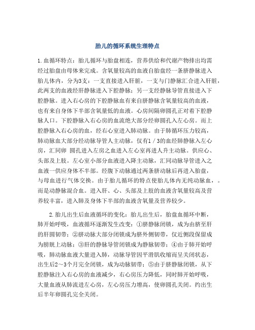
胎儿的循环系统生理特点1.血循环特点:胎儿循环与胎盘相连,营养供给和代谢产物排出均需经过胎盘由母体来完成。
含氧量较高的血液自胎盘经一条脐静脉进入胎儿体内,分为3支:一支直接进入肝脏,一支与门静脉汇合进入肝脏,此两支的血液经肝静脉进入下腔静脉;另一支经静脉导管直接进入下腔静脉。
进入右心房的下腔静脉血有来自脐静脉含氧量较高的血液,也有来自身体下半部含氧量低的血液。
心房间隔卵圆孔正对着下腔静脉人口。
下腔静脉入右心房的血流绝大部分经卵圆孔入左心房。
而上腔静脉入右心房的血,经右心室进入肺动脉。
由于肺循环压力较高,肺动脉血大部分经动脉导管人主动脉,仅有l/3的血经肺静脉入左心房,汇同卵圆孔进入左房之血进入左心室再进人升主动脉。
供应心、头部及上肢。
左心室小部分血液进入降主动脉,汇同动脉导管进入之血液一供应身体不半部。
经腹下动脉通过两条脐动脉后再进入胎盘,与母血进行气体交换。
由于胎儿循环的特点使胎儿体内无纯动脉血,。
而是动静脉混合血。
进入肝、心、头部及上肢的血液含氧量较高及营养较丰富,进入肺及身体下半部的血液含氧量及营养较少。
2.胎儿出生后血液循环的变化:胎儿出生后,胎盘血循环中断,肺开始呼吸,血液循环逐渐发生改变:①脐静脉闭锁,成为由脐至肝的肝圆韧带;②脐动脉大部分闭锁成为脐外侧韧带,仅近侧段保留成为膀胱上动脉;③肝的静脉导管闭锁成为静脉韧带;④由于肺开始呼吸,肺动脉血液大量进入肺,动脉导管因平滑肌收缩而呈关闭状态,出生后2~3个月完全闭锁,成为动脉韧带;⑤由于脐静脉闭锁,从下腔静脉注入右心房的血液减少,右心房压力降低,同时肺开始呼吸,大量血液从肺流进左心房,左心房压力增高,使卵圆孔关闭。
约出生后半年卵圆孔完全关闭。
胎儿血液循环和出生後血液循环的变化

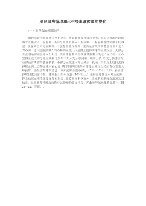
胎兒血液循環和出生後血液循環的變化(一)胎兒血液循環途徑臍靜脈從胎盤經臍帶至胎兒肝。
臍靜脈血富含氧和營養,大部分血液經靜脈導管直接注入下腔靜脈,小部分經肝血竇入下腔靜脈。
下腔靜脈還收集由下肢和盆、腹腔器官來的靜脈血,下腔靜脈將混合血(主要是含氧高和豐富的血)送入右心房。
從下腔靜脈導入右心房的血液,少量與上腔靜脈來的血液混合,大部分血液通過卵圓孔進入左心房,與由肺靜脈來的少量血液混合後進入左心室。
左心室的血液大部分經主動脈弓及其三大分支分布到頭、頸和上肢,以充分供應胎兒頭部發育所需的營養和氧;小部分血液流入降主動脈。
從頭、頸部及上肢回流的靜脈血經上腔靜脈進入右心房,與下腔靜脈來的小部分血液混合後經右心室進入肺動脈。
胎兒肺無呼吸功能,故肺動脈血僅小部分(5%~10%)入肺,再由肺靜脈回流到左心房。
肺動脈大部分血液(90%以上)經動脈導管注入降主動脈。
降主動脈血液除經分支分布到盆、腹腔器官和下肢外,還經臍動脈將血液運送到胎盤,在胎盤與母體血液進行氣體和物質交換後,再由臍靜脈送往胎兒體內(圖24-12,彩圖)。
圖23-12 胎兒血液循環經路(二)胎兒出生後血液循環的變化胎兒出生後,胎盤血循環中斷。
新生兒肺開始呼吸活動。
動脈導管、靜脈導管和臍血管均廢用,血液循環遂發生一系列改變。
主要變化如下:①臍靜脈(腹腔內的部分)閉鎖,成為由臍部至肝的肝圓韌帶;②臍動脈大部分閉鎖成為臍外側韌帶,僅近側段保留成為膀胱上動脈;③肝的靜脈導管閉鎖成為靜脈韌帶,從門靜脈的左支經肝到下腔靜脈;④出生後臍靜脈閉鎖,從下腔靜脈注入右心房的血液減少,右心房壓力降低,同時肺開始呼吸,大量血液由肺靜脈回流進入左心房,左心房壓力增高,于是卵圓孔瓣緊貼于繼發隔,使卵圓孔關閉。
出生後約一年左右,卵圓孔瓣方與繼發隔完全融合,達到解剖關閉,但約有25%的人卵圓孔未達到完全的解剖關閉;⑤動脈導管閉鎖成為動脈韌帶,出生後3個月左右成為解剖關閉。
胎儿出生前的心血管系统分布及其血液流通途径。
血循环系统 - 英文版

Circulatory System∙ Organ system that passes nutrients (such as amino acids, electrolytes and lymph), gases,hormones blood cells, etc. to and from cells in the body to help fight diseases, stabilize body temperature and pH, and maintain homeostasis. ∙ Anything and everything about circulation the body ∙ Composition:o Cardiovascular system: distributes blood: Circulation that happens in the heart and blood vessels ∙ Lymphatic system: returns excess filtered blood plasma from interstitial fluid (between cells as lymph): protection∙ Principal functions of vertebrate circulatory systemof all the substances essential for cellular metabolism▪Respiratory –transports O 2to the tissue cells through the RBC ▪ Nutritive – carries absorbed nutrients through the liver and to the cells ▪ Excretory – removes metabolic wastes , excess water and other substances▪Hormones – part of endocrine systemo Regulation – transport regulatory hormones & temperature regulation▪ Hormone transport – blood carries hormones and carry them to distant targetorgans they regulate▪ Temperature regulation – maintenance of a constant body temperature inwarm-blooded animals.o Protection – protection against injury and foreign microbes▪ Blood clotting – protects against blood loss when vessels are damaged▪ Immune defense – WBCs (leukocytes) provide immunity against many disease-causing agents∙ Basic Typeso Gastrovascular cavity∙ Hydras have 1 opening; water entersand circulates∙ Oxygen/ nutrients are absorbed byChoanocytes∙ Consists of a epidermis andChoanocyteso Open Circulatory System▪Arthropods and some mollusks▪Basic components ofcardiovascular systems∙Hemolympho Limits sizeo Blood + lymph; goesBack into the heart∙Blood vessels- terminate in aopening∙One or more hearts∙Hearts are just musculartubes ▪There is no distinction between the circulating fluid (blood) & the extracellular fluid of the body tissues (interstitial fluid or lymph)▪Fluid in vessels and interstitial fluid mingle in 1 compartment as hemolymph▪Nutrients & waste exchanged by diffusion between hemolymph and body cells ▪Energetically inexpensive▪Limitation: hemolymph can’t be selectively delive red to different tissueso Closed Circulatory System▪Blood and interstitial fluid arephysically separated▪Larger, more active animals need ahigher pressure to pump blood to allbody cells (more efficient bloodpumping)▪Found in earthworm, cephalopods,and all vertebrates▪Advantages:∙Animals can grow larger withmore efficient supply∙Blood flow can be selectivelycontrolled▪TYPES∙Single Circulationo Fishes∙Double Circulationo Crocodiles,birds,Mammals∙Amphibians andmost Reptileshave systems withfeatures of bothCLOSED CIRCULATORY SYSTEMSingle Circulation System – fish (most primitive)∙ Blood passes through the heart only once in a full circuit∙ Heart → Gills (have thin membrane that’s why arteries have lowpressure) → Body → Heart∙ Single atrium collects blood from tissues∙ Single ventricle pumps blood out of the heart ∙ Arteries carry blood away from the heart∙ Blood picks up O 2 and drops CO 2 and goes on through arteries toother body tissues∙ Limitation: pressure lowers; big fishes have to breathe continuouslyDouble Circulation System∙ Blood passes through the heart twice in one full circuit ∙ Heart →Lungs →Heart → Body → Heart∙ Amphibianso Unique in that they can breathe through theirlungs and skino Heart Pumps blood to either▪ Pulmocutaneous circulation – carriesdeoxygenated blood to both the lung and skin▪ Systemic circulation – body tissueso Heart has▪ 3 chambers▪ 2 atria to collect bloodo Right atrium – blood from the body (NOTLUNGS) and is low in O 2 (except oxygenated blood from skin)o Left atrium - blood from lungs (O 2 rich whenair is breathing)o Single ventricle – mixture of oxygenated anddeoxygenated bloodo Internal recesses – separates oxygenatedand deoxygenated (not perfect)o Both atria dump into 1 ventricleo Internal structure keeps 2 O 2 oxygenated and deoxygenated blood mostly separated o Some mixing does occur reducing efficiencyo Noncrocodalian reptiles also have 2 atria and 1 ventricleo Ventricle is partially divide – higher effieciencyo Both must use low moderated pressure systems to minimize pressure flowing through lung tissue∙Crocodiles, birds and mammalso Reptiles have transitional heartso Oxygenated and deoxygenated blood separates into 2 distinct circuitso Systemic circulation – to the bodyo Pulmonary circulation – to the lungso 2 atria and 2 ventriclesThe Human Heart∙Hollow, cone-shaped muscle located between the lungs and behind the sternum (breastbone), tilted at to the left∙About the size of a human fist∙2/3 is located to the left of the midline of the body and 1/3 to the right∙The apex (pointed end) points down and to the left∙Ave. weight between 250-350 grams∙ 4 chambers:o 2 superior atria; the receiving chamberso 2 inferior ventricles; the discharging chambers∙(interatrioventicular) septum – separated the left atrium and ventricle from the right atrium and ventricle, dividing the heart into 2 functionally separate and anatomically distinct units.∙Layerso Endocarium– smooth, inside lining of the heart▪In contact with the blood that the heart pumps▪Protects the cavityo Merges with the inner lining (endothelium)of blood vessels and cover heart valveso Myocardium - middle layer of heart muscle▪Layer that contractso Epicardium or visceral pericardium– outer layer of the heart▪ A fluid sac that surrounds the heart∙Valves:o Atrioventricular (AV) valves - found between the atria and ventricles.▪tricuspid valve, or right atrioventricular valve▪ Between the right atrium and the right ventricle.▪Usually has three papillary muscles.▪Prevents blood from the right ventricle to go to the right atriumo Mitral valve or bicuspid valve or left atrioventricular valve▪ A dual-flap valve that lies between the left atrium and the left ventricle.▪Prevents blood from the left ventricle to go to the left atriumo Semilunar valves - separate the left and right ventricle from the pulmonary artery and the aorta, respectively▪aortic valve∙found between the left ventricle and aorta▪pulmonary valve∙lies between the right ventricle and the pulmonary artery ∙Blood Vessels∙Aortao largest artery in the bodyo arises from the left ventricle of the heart, goes up(ascends) a little ways, bends over (arches), thengoes down (descends) through the chest andthrough the abdomen to where ends by dividinginto 2 common iliac arteries that go to the legs.o Anatomically, it is divided into the ascending aorta,the aortic arch, and the descending aorta.o Can accommodate the greatest pressureo It serves to supply oxygenated blood to the majororgans of the body.o the central conduit from the heart to the body.∙Superior Vena Cava:o A large vein that receivesblood from the head, neck,upper extremities, and thoraxand delivers it to the rightatrium∙Inferior Vena Cava:o A large vein that receivesblood from the lowerextremities, pelvis andabdomen and delivers it tothe right atrium of the heart ∙Pulmonary Artery:o begins at the base of the right ventricle.o it delivers deoxygenatedblood to the lung.∙Pulmonary Vein:o large blood vessels that carry oxygenated blood from thelungs to the left atrium of theheart.o In humans there are fourpulmonary veins, two fromeach lung.∙Myogenic Hearto Electrically excitable, generates own action potentialo Contains auto-rhythmic fibers- can initiate periodic action potential w/o neural activation▪SA Node – most important group of auto-rhythmic cellso Nervous input can increase or decrease rate∙Sino-Atrial Nodeo Found at the upper part of the right atrium of the hearto Acts as the hearts natural pacemakero Triggers a sequence of electrical events in the heart that control the regular muscle contractions (every 0.6 seconds or 100/min) that pump blood out of the heart with a rhythm of about 60-70 beats/min (resting heart)o Depolarization is transmitted through 2 pathways▪Cardiac muscles of the left atrium▪Cardiac muscles of the right atrium and AV Nodeo Depolarization spread quickly among the muscles of the left and right atriasimultaneously▪ Possible because of gap junctions in intercalated diskso AV node provides the only pathway for conduction of depolarization from atria toventricleso Delays ventricular contraction by 0.1 sec (part where atrium transfers blood to ventricles) o Permits atria to finish contraction and emptying of contentso Wave of depolarization is conducted to ventricles by AV bundle or bundle of His▪ Relays depolarization to Purkinje Fibers∙ Stimulates contraction of myocardial cells of the L and R ventricle almostsimultaneouslyo Contraction of the heart is controlled by Ca and troponin/tropomyosin system similar toskeletal muscleso Pattern of voltage change produced by SA node can be measured with electrodes onthe skin▪ Voltage measurements on the skin of the chest are called electrocardiogram(ECG)∙ Record of electrical impulses generated during the cardiac cycle ∙ Monitor electrical activity produced by SA node ∙ Examine fornormal frequency, strength,duration and direction of signals∙ P wave - begins when SA node fires; coincides with depolarization of the atria and, therefore, associatedwith atrial systole ∙ QRS Complex – 3 waves – AV node excites ventricle∙ T wave- repolarization of ventricles back to resting state FLOW OF BLOOD∙ Functioning:o Flow of blood through the heart: one direction▪ from the atria to theventricles, and out of the arteries.▪ Blood is prevented fromflowing backwards by the valves.∙From the left atrium the blood moves to the left ventricle which pumps it out to the body (via the aorta).∙From the R atrium blood moves to the R ventricle which pumps the blood to the lungs∙On both sides, the lower ventricles are thicker and stronger than the upper atria.∙The muscle wall surrounding the left ventricle is thicker than the wall surrounding the right ventricle due to the higher force needed to pump the blood through the systemiccirculation.∙IT ONLY TAKES ABOUT 20 SECONDS TO PUMP BLOOD TO EVERY CELL OF OUR BODYCARDIAC CYCLE (heart beat)∙Filling of atrium, pumping of ventricle∙the sequence of events that occurs when the heart beats.∙ two phases:o diastole phase, the heart ventricles are relaxed and the heart fills with blood.o systole phase, the ventricles contract and pump blood to the arteries.∙One cardiac cycle is completed when the heart fills with blood and the blood is pumped out of the heart.∙Diastole Phase – R sideo Atria and ventricles are relaxed and AV valves are openo Deoxygenated blood from the superior and inferior vena cavae flows into the right atriumo Open AV valves allow blood to pass through to the ventricleso SA node contracts triggering the atria to contracto R atrium empties its contents into R ventricleo Tricuspid Valve closes ∙Systole Phase – R sideo R ventricle receives impulses from the Purkinje Fibers and contracts o SL (pulmonary) valve openso Deoxygenated blood is pumped in to the pulmonary artery. SLvalve closeso Pulmonary artery carries blood to lugs to pick up oxygeno Blood is returned to the L atrium by pulmonary veins∙Diastole Phase – L sideo Blood from pulmonary veins fill left atrium (blood from venacava is also filling the R Atrium) o SA node contracts triggering left atrium to contracto AV (mitral) valves openso L atrium empties its content to L ventricle ∙Systole Phase – L sideo L ventricle receives impulses from the Purkinje fibers and contracts o SL (aortic) valve openso Oxygenated blood is pumped into the aorta. Aorta providesoxygenated blood to the body o Oxygen depleted blood isreturned to the heart via venacavae∙CARDIAC OUTPUT∙Amount of blood the heart pumpsper unit time in L/min∙Depends in the size of the heart and how often it beats∙Stroke volume - amount of blood a heart ejects at each beat∙Higher heart rates of smaller animals gives them a greater cardiac outputthat would be predicted based onthe size of their hearts (must meethigh metabolic demands) HEART SOUNDS∙Lub-dubo the 1st heart sound (lub) is causedby the vibration of the heart atthe time of the closure of thetricuspid and mitral valves.o the 2nd heart sound (dub) iscaused by the vibrations at thetime of closure of the pulmonicand aortic valvesHEART BEAT∙Regular Heart Beat: 70-80 beats a minute∙When you run around a lot your heart pumps more blood into yout body—maybe up to 200 times a minute∙As people grow older their heart rates changeo A newborn baby has a heart rate of about 130o 3 year-old: about 100o18 year-old: about 90 times a minuteo Adult: 70-80o The older you get the slower your heart beats∙An average heart pumps about 10 mililiters of blood into your body with every beat∙That’s about 5L every minute or about 7200L everydayPULSE∙ Represents the tactile aterial palpitationof the heartbeat through the fingertips ∙ May be palpated in any place wherean artery is compressed against a bone, such as at the neck (carotid artery), at the wrist (radial artery), behind the knee (popliteal artery), on the inside of the elbow (brachial artery), and near the ankle joint (posterior tibial artery)BLOOD∙ Specialized bodily fluid in animals thatdelivers necessary substances such as nutrients and oxygen to the cells and transports metabolic waste products away from those some cells ∙ Amount in adults: 4.5-6 quarts ∙ Functionso Transport of gases, nutrients,waste products, and hormones o Maintenance of bodytemperatureo Protection from substance o Clot formationCOMPONENTS OF BLOOD∙ Erythrocytes (Red Blood Cells)o Life span: 120 dayso Function: to carryoxygen from the lungs to every cell in the bodyo Makeup almost 99.9%of formed elements in bloodo Composition: proteinand iron compound, called HEMOGLOBIN , that captures oxygen molecules as the blood moves through the lungsUnites readily with oxygen to form oxyhemoglobin (found in arteries)∙ Form of oxygen as it is transported in the blood stream ∙ Together with iron, it gives blood a bright red coloro Membrane is flexibleo Able to bend in many directions w/o breaking o Biconcave disc shape o No nuclei o About 4.5M in female and 5M in males per cubic mm of blood o Formed in the reticulo-endothelial tissue of the bone marrow o Discharged into the blood capillaries after losing nuclei o Remain in the blood for 3-5 weeks o Destroyed by spleen and liver∙ Leukocytes(White blood cells)o With nucleo Average from 6 thousand – 10Tcells per cubic mm of blood, or about 1 for every 700 RBC o Primary defense mechanismagainst bacteria, viruses, fungi, and parasiteso Produce antibodies, which arereleased into the circulating blood to target and attach to foreign organismso WBC are classified according to their size, shape of nucleus & reaction to dyes used in staining them∙ Thrombocytes (Platelets)o Small irregularly shaped clearcell fragments (i.e. cells that do not have a nucleus o 2-3 m in diametero Derived from fragmentation ofprecursor megakaryocytes o Average lifespan: 5-9 dayso Natural source of growth factor▪ Play a significant role inthe repair and regeneration of connective tissueso Involved in hemostasis(process that stops the flow ofblood), leading to the formation of blood clots▪ Ability of the system to prevent excessive blood loss upon injuryo Normal platelet count: 150,000-450,000/ of blood▪ If too low, excessive bleeding can occur▪ If too high, blood clots/thrombosiscan form∙ May obstruct blood vesselsand result in stroke, myocardial infarction, pulmonary embolism or the blockage of blood vessels to other parts of the body, such as extremities of the arms or legsBLOOD CLOT FORMATION∙ Platelets (cell fragments) in the bloodstream come into contact w/ damaged blood vesselFirst to defendMacrophages∙Platelets and vessel wall release enzyme thrombokinase∙Conversion of inactive enzyme prothrombin into active thrombin∙Thrombin catalyses the conversion of fibrinogen to insoluble fibrin∙Fibrin forms a over the wound that traps RBCS and seals the wound∙Resulting jelly clot like exposure to air to form a scab∙Calcium, vitamin K, and a variety of enzymes called factors are also necessary for efficient blood clotting∙HEMOPHILIAo Genetic defect in clotting factorso Inherited deficiency of a specific clotting factor causes thiso Most common form is X-linked recessive mutationo Treatment requires transfusions of purified clotting factors from donors or genetically engineered organismso Attempting gene therapyBLOOD TYPE∙The most common blood type classification systemis the ABO system discovered by Karl Landsteiner.o observed two distinct chemical moleculeson the surface of the red blood cells and labeledmolecules "A" and "B."o If the red blood cell had only "A" moleculeson it, that blood was called type A.o If the red blood cell had only "B" moleculeson it, that blood was called type B.BLOOD VESSELS∙Arterieso Conduct blood away from the hearto contain a high percentage of smooth muscle.o The artery walls consist of three layers:▪Tunica Adventitia: strong outer covering of arteries and veins which consists of connective tissues, collagen and elastic fibers.▪Tunica Media: the middle layer and consists of smooth muscle and elastic fibers.This layer is thicker in arteries than veins.▪Tunica Intima: the inner layer which is in direct contact with the blood flowing through the artery. It consists of an elastic membrane and smooth endothelialcells.∙Arterioleso Most highly regulated blood vessels in the bodyo Contribute the most to overall blood pressureo Respond to a wide variety of chemical &electrical messages and are constantly changing size to speed up or slow down blood flow∙Capillarieso Site of gas and nutrient/waste exchangeo Single-celled layer of endothelium on a basement membraneo Smallest and narrowest vessels in the body∙Blood enters capillary on arteriole end under pressure∙Pressure forces some fluid out of the blood (not RBC or large proteins)∙Most of the fluid that leaves will be recaptured by the venule end of the capillary∙Venuleso Small, thin extensions of capillaries∙Veinso Conduct blood back to the hearto Thinner and less muscular than arteries o Need help returning blood to the heart▪Smooth musclecontractions help propelblood▪Valves inside veinssqueezed by skeletalmusclesDISORDERS∙Systemic hypertension (High Blood Pressure)o High BP in the systemic arterieso Usually caused by the constriction of the small arteries (arterioles)o Causes: obesity, smoking, aging, etc.∙Congestive heart failureo Condition in which the heart’s function as a pump is inadequate to meet the body’s needo Leads to a buildup of fluid in the lungs and surrounding body tissueso Results from pulmonary hypertension- blood backs up in the lungs, raising pressure, and forcing fluid out into lung tissue∙Atherosclerosiso Disease of the arteries characterized by the deposition of plaques of fatty material on their inner wallso Usual cause of heart attack, strokes and peripheral vascular diseaseo Coronary artery disease results from plaque forming in the coronary arteries supplying the heart muscle∙Myocardial infarction (MI) /Heart Attacko Occurs when blood flow stops to a part of the heart causing damage to the heart muscleo Localized regions of the heart muscle dieo Cardiac angioplasty can detect narrowing of coronary vesselso Balloon angioplasty can widen the lumen of narrowed vesselso Coronary artery bypasses take a healthy blood vessel and use it to replace a blockedcoronary artery。
循环系统诊断常见症征英文版

Atrial myxoma
Subvalvular ring Pulmonary vein stenosis
Thrombus Neoplasm
Infective vegetation Prosthetic valve disfunction
Mitral stenosis
Cause of MS requiring intervention(n=1050)
dyspnea, orthopnea Palpitation due to arhythmia Miscellaneous
Hemoptysis, blood-tinge sputum, pink frothy sputum, chest pain, mitral facies, Cough, hoarseness, dysphagia
Occasionally diastolic rumbling can be heard at severe MR because of relative MS
No murmur can be heard at mild even moderate MR sometime
Aortic stenosis
Auscultation(2)
Music-like murmur can be heard at severe MR, at the valve prolapse
Midsystolic click with or without murmur presents at the valve prolapse
radiating to the axilla even the back, the base of the heart . Murmur at late systolic can be heard at the valve prolapse or at mild MR
循环系统组织结构(英文版)课件

2. Artery
---large A: aorta, pulmonary trunk
---medium-sized A: all named A, the diameter > 1mm (radial A, ulnar A)
---small A: 300um<D<1mm
1) Medium-ype ---correspond with A except for LV ---three layers
---structural features: a. larger diameter, thinner walls- collapsed b. no internal and external elastic membrane,
• processes –microvilli-like, finger-liked
• vesicle
/60-70nm, constitute about 25-35% of total volume
/transendothelial channel
function: transport large molecules and storage of membrane (for enlarge, enlongated, pore-formed and microvilli)
so the boundaries between three tunica are not very clear c. contains more CT, less smooth M, SM are arranged in bundles d. vein valve:
/infolding of tunica intima /semilunar-liked /prevent back flow of blood
circulatorysystem循环系统英文资料实用PPT

The lymphatic system is a network of vessels that transport lymphatic fluid throughout the body, carrying away waste and helping to fight infections. It is also involved in immune function.
PVD can lead to problems with the circulation of blood to the arms, legs, and feet. Symptoms include pain or cramping in the legs, numbness, and skin changes.
removing carbon dioxide and other waste products.
Blood Vessels
01
Types
Blood vessels come in three types: arteries, veins, and capillaries.
02 03
Function
They transport blood throughout the body, delivering essential nutrients and gases to the cells and removing waste products.
Structure
Arteries and veins are thick-walled and elastic, while capillaries are tiny, thin-walled vessels that allow for the exchange of nutrients, gases, and waste products between the blood and the surrounding tissue.
循环系统1

• Capillary beds 毛细血管网facilitate exchange
Capillary beds separate arteries from veins Highly branched and very tiny
Infiltrate 浸浴 all tissues in the body
• Mammals and birds are NOT monophyletic 单系 起源的 • Four-chambered hearts evolved independently
Chambered heart pumps blood
• Atria receive blood
Soft walled, flexible
Outer layer is elastic connective tissue 弹性 Middle layer is smooth muscle and elastic fibers Inner layer is endothelial tissue 内皮
Fishes have Single circuit
• A fish heart has two main chambers
– One ventricle and one atrium
• Blood pumped from the ventricle
– Travels to the gills, where it picks up O2 and disposes释放 of CO2
Crocodilians 鳄鱼 have a complete septum
• reptiles have two arteries that lead to the systemic circuits体循环
胎儿血液循环图ppt课件

精品ppt
33
胎儿血液循环箭头图
肝圆韧带 静脉韧带
脐 静 脉
静 脉 导 管
下 腔 静 脉
脐外侧韧带
腹、盆部,下肢
脐
动 脉
降主动脉
右 心 房
右心室
关闭
卵
园
左心房
孔
左心室
主动脉弓
肺
肺
动 脉
肺
静 脉
头,颈,上肢
动脉导管
上腔静脉
精品ppt
34
胎儿血液循环箭头图
肝圆韧带 静脉韧带
脐 静 脉
静 脉 导 管
下 腔 静 脉
箭头图
精品ppt
2返6 回
胎儿血液循环箭头图
脐 静 脉
胎 盘
脐 动 脉
静 脉 导 管
下 腔 静 脉
右 心 房
卵 园 孔
右心室
腹、盆部,下肢
肺
肺
动 脉
肺
静 脉
降主动脉
动脉导管
精品ppt
左心房 左心室 主动脉弓 头,颈,上肢 上腔静脉
27
胎儿血液循环箭头图
脐 静 脉
静 脉 导 管
下 腔 静 脉
右 心 房
卵 园 孔
右心室
左心房 左心室
主动脉弓
腹、盆部,下肢
肺
肺
动 脉
肺
静 脉
头,颈,上肢
脐
动 脉
降主动脉
动脉导管
上腔静脉
精品ppt
28
胎儿血液循环箭头图
肝圆韧带
脐 静 脉
静 脉 导 管
下 腔 静 脉
右 心 房
卵 园 孔
右心室
左心房 左心室
- 1、下载文档前请自行甄别文档内容的完整性,平台不提供额外的编辑、内容补充、找答案等附加服务。
- 2、"仅部分预览"的文档,不可在线预览部分如存在完整性等问题,可反馈申请退款(可完整预览的文档不适用该条件!)。
- 3、如文档侵犯您的权益,请联系客服反馈,我们会尽快为您处理(人工客服工作时间:9:00-18:30)。
Interventricular Septum
Left Ventricular Outflow Tract
Right Atrium Right Ventricle
Main Pulmonary Artery
Ao Right Pulmonary Artery Ductus Arteriosus
dAo Sp
ห้องสมุดไป่ตู้
Spine
Left Atrium Right Atrium Left Ventricle Right Ventricle Moderator Band
Sp
Foramen Ovale
Four Chamber
Interventricular Septum
Left Atrium Left Ventricle Right Ventricle Aorta Mitral Valve (closed) Aortic Valve (open)
GOALS
• Review normal cardiac anatomy and its sonographic appearance (four chamber, LVOT, RVOT)
• Explore diagnostic pitfalls • Review the appearance of more common structural cardiac defects
The Four Chamber View
1. Heart fills one third of the chest
The Four Chamber View
2. Apex points to the left (45 degree angle)
The Four Chamber View
3. Size of right chambers approximates left chambers
Left Ventricular Outflow Tract
• Identify: LV, RV, IV septum, aorta (normal caliber), +/LA, +/- RA • Medial wall of the ascending aorta merges with the top of the IV septum (most frequent location for VSD) • Pathology: VSD, tetralogy of Fallot, transposition, truncus arteriosus
The Four Chamber View
1. MV and TV move on real time imaging 4. Ventricular septum symmetric
The Four Chamber View
6. Portion of the atrial septum present (crus)
Fetal Cardiology
Kottler NE, Leopold GR, O’Boyle M, Pretorius D, Sirlin CB
Fetal Cardiology
• Cardiac anomalies are the most frequently overlooked group of abnormalities
Fetal Cardiology
• AIUM / ACR standards in the 2nd and 3rd trimesters include:
Four chamber view Position of fetal heart in the thorax
• LVOT and RVOT not yet part of standards • 4 chamber view alone: 33-63% sensitive • With outflow tracts: 83-85% sensitive [2]
Fetal Cardiology
• Risk Factors for congenital heart disease:
Family history Recurrence risk (hypoplastic left heart as high as 13.5%) Nongestational DM Maternal infection (rubella) Lupus Drugs (anticonvulsants, etoh, amphetamines, ocp, vit A, steroids, etc.)
Ascending Aorta Tricuspid Valve Pulmonic Valve (open)
Right Ventricular Outflow Tract
Descending Aorta
Pitfalls: PseudoVSD
IV septum parallels US beam (echo dropout)
Right Ventricular Outflow Tract
• Identify: branching of the main PA into right PA and ductus arteriosus (to desc Aorta), asc aorta in cross section, desc aorta to left of spine; verify PA crosses anterior to asc aorta • Pathology: transposition, truncus arteriosus
• Congenital heart disease = 0.8% of all pregnancies
4% one sibling affected; 10% two siblings affected 9% father affected 12% mother affected
• Causes > 50% deaths from congenital disease
