蔡司Axio_Scope_A1_使用说明书
偏光显微镜在岩矿鉴定工作中的使用技巧和方法

Vol.35 No.5Mar. 2019第35卷第5期2019年3月甘肃科技Gansu Science and Technology偏光显微镜在岩矿鉴定工作中的使用技巧和方法冯俊环(甘肃省地质矿产勘査开发局第一地质矿产勘査院,甘肃天水741020)摘要:偏光显微镜是光学显微法进行岩矿鉴定的基本工具,也是研究矿物光学性质的重要仪器,比普通生物显微镜更为复杂,最主要的区别是装有两个偏光镜。
本文以CarlZeissAxioScope.Al 型为例介绍了偏光显微镜的使用方法和使用技巧,为同仁们学习交流提供浅显易懂的学习心得。
关键词:偏光显微镜;岩矿鉴定;技巧;方法中图分类号:P5751概况偏光显微镜是光学显微法进行岩矿鉴定的的 基本工具,也是研究矿物光学性质的重要仪器,比 普通生物显微镜更为复杂,最主要的区别是装有两个偏光镜。
其中一个偏光镜在载物台之下,称下偏 光镜(起偏镜);另一个在物镜之上的镜筒中,称上偏光镜(分析镜)。
二者透过偏光的振动面通常是互 相垂直的。
偏光显微镜的品牌、型号很多,但基本的结构构造大致相似。
在世界范围内主要品牌有奥林巴 斯、尼康、蔡司、彳来卡等。
本文以CarlZeissAxioScope.Al 型(如图1所示)为例加以阐述。
该型显微镜是全 新的万能研究级高品质偏光显微镜,遵循蔡司最高质量标准制造,提供优异的价值和性能。
特别是在透射光和反射光的所有标准反差技术和测量技术 中拥有许多独创的特色。
适用于石油地质,矿物,晶体,陶瓷,玻璃,聚合物,纺织品的检验和研究。
本文 仅就透射光观察透明矿物薄片加以讨论,希望对同 仁们的使用有所帮助。
图1卡尔•蔡司多功能型显微镜主机主要优点:根据需求灵活配置方便升级6位物 镜转盘聚焦灵活模块化系统,为需求而定制系统采用模块化设计.AxioScope.Al 偏光显微镜可以根据 用途和个性化需求进行灵活配置。
AxioScope.Al 偏光显微镜能够实现快速物镜转 盘更换,例如:从反射光切换至透射光:非常高效。
zeiss 科研级正置显微镜AXIO scope A1中文介绍

蔡司科研级正置显微镜Axio Scope A1 彩页资料一、灵活性前所未有的模组化Axio Scope——卡尔•蔡司的这款新型多功能显微镜在各方面都是您的不二选择。
您将被这款系统的性能深深震撼,与此同时,您的财务部门也将为其价格优势而叫好。
您只需买您所需,因此各个配置方案都十分经济。
购买之后您可以在任何时间按照您的需要来升级。
1. 高度弹性设计,23种配置,灵活升级,满足您不同需求2. 可以定制5种显微镜上部、3个下部和2个可变柱子,非常灵活I:用于纯粹的透射光。
6个用于明场的物镜孔(6xBF)II:用于标准的荧光应用。
含3xDIC/3xBF,用于HBO50,HBO100和HXP120,Colibri 的标准界面。
III:用于LED荧光应用。
3xDIC/3xBF,可接受4种不同LED模块的综合照明,长寿命,经济。
IV: 用于反射光和荧光。
6xBF/DF,用于HAL100或HBO的标准界面,物镜转盘上的DF插件。
V:用于反射光和荧光。
6xBF/DF,用于HAL100或HBO的标准界面,物镜转盘上的DF插件。
可调的apertA:常规应用。
带LED照明B:标准应用:50W的反射光。
带有视场光栏和孔径光栏的科勒光路、带滤块滑片和6位滤块转轮。
C:用于高强度照明的透射光应用。
100W的卤素灯。
带有视场光栏和孔径光栏的科勒光路、带滤块滑片和6位滤块转轮。
D: 380mm高的Vario柱,用于反射光和荧光应用。
E: 560mm高的Vario柱,用于反射光和荧光应用。
二、方便升级Axio Scope模块界面的概念使得将来升级变得更简单,您不需要等待昂贵的服务支持自己就可以安装组件。
三、多样化的用途Axio Scope在研究所和实验室提供多样的用途,从简单的日常应用到更复杂的研究。
从解剖到细胞学,从透射光应用到多种荧光标记,从最薄的组织切片到380mm厚的样品。
高达560mm大样品空间可选多人共览成像可选;23mm大视野观察;多种光学变倍可选;适合显微镜操作使用透射光照明成像LED透视光可选,无需色温转换滤片以及减光片,无需库勒照明调节,方便使用;明场、暗场、相差、DIC、PlasDIC、偏光等多种观察方式;专利PlasDIC设计,允许塑料器皿DIC成像。
ZEISS Axio Imager Light Manager手册说明书
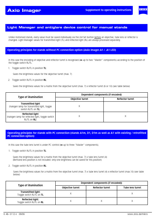
Unlike motorised stands, every value must be saved individually via the LM Set button before an objective, tube lens or reflector ischanged. Light Manager values for transmitted light (TL) and reflected light (RL) are always processed separately.In this case the encoding of objective and reflector turret is recognised (⇒ up to two "Master" components) according to the position ofthe toggle switch RL/TL.1. Toggle switch RL/TL in position TL:Saves the brightness values for the objective turret (max. 7)2. Toggle switch RL/TL in position RL:Saves the brightness values for a matrix from the objective turret (max. 7) x reflector turret (6 or 10) (see table below)Dependent components (if encoded)Type of illuminationObjective turret Reflector turretTransmitted lightX (halogen lamp for transmitted light, toggleswitch RL/TL on TL)Reflected lightX X (halogen lamp for reflected light, toggle switchRL/TL on RL)In this case the tube lens turret is under PC control (⇒ up to three "Master" components).1. Toggle switch RL/TL in position TL:Saves the brightness values for a matrix from the objective turret (max. 7) x tube lens turret (4)(Bertrand lens position is not included: only one brightness can be saved for this position)2. Toggle switch RL/TL in position RL:Saves the brightness values for a matrix from the objective turret (max. 7) x tube lens turret (4) x reflector turret (max.10) (see tablebelow)Dependent components (if encoded))Type of illuminationObjective turret Reflector turret Tube lens turretTransmitted light:X X Toggle switch RL/TL on TLReflected light:X X X Toggle switch RL/TL on RLDefault setting of the stand after switching on:Transmitted light:• Toggle switch RL/TL on TLButton TL on (shutter open or lamp on)Button RL offReflected light:• Toggle switch RL/TL on RLButton TL offButton RL on (shutter open or lamp on)Saving LM value:• To save the current lamp voltage for the current objective turret position press the LM Set button brieflySaving 3200K:This function determines whether the stand is set at 3200K when it is switched on.• To set 3200K to be active on switching on: activate 3200K and press LM Set button.• To set 3200K to be inactive on switching on: deactivate 3200K and press LM Set button.The 3200K setting is saved globally and is independent of other LM values that have already been saved. The normal LM values are available at any time as soon as 3200K is deactivated.Overwriting the LM values:• To save the new value at the relevant position press the LM Set buttonDeleting of the LM values:This is not possible.Activating an LM value:This is done by switching on and changing the position of a "Master" component.To permanently deactivate/activate Light Manager (LM) & antiglare device (AG)• Keep the "RL" button pressed down when you switch on:• One beep signifies deactivation.• Two beeps signify activation.To permanently deactivate/ activate Light Manager only• Keep "3200"button pressed down when you switch on:One beep signifies deactivation. Two beeps signify activation.To permanently deactivate/ activate antiglare device only• Keep “TL” button pressed down when you switch on:One beep signifies deactivation. Two beeps signify activation• If button "RL" is pressed when you switch on and only one of the two functions is activated, that function will be deactivated:Starting condition OutcomeLM AG LM AG⇒0 01 1⇒0 01 0⇒0 00 1⇒ 1 10 0These parameters can also be set via MTB 2004 for motorised stands.Antiglare device:If there is a shutter in the TL optical path, the lamp voltage remains constant when the objective is changed and the shutter takes over thefunction of the antiglare device.If no shutter is present, the lamp is switched off.Safety function:If the reflector turret flap is opened or the reflector turret is completely removed, the safety switch-off device automatically closes thereflected light shutter. In addition, the shutter can no longer be opened by pressing a button as long as the reflected light path is "open".The shutter also closes automatically when the stand is switched off.The brightness of the Light Control LEDs can be adjusted by the user.Manual stands:• Keep SET button pressed down for about 3 seconds until a long beep is heard.All LEDs go on.The brightness of the LEDs can now be adjusted by the brightness control (control knob).However, the brightness cannot be completely extinguished!Activating the control knob in this mode has no effect on the lamp voltage!This mode is exited automatically by releasing the LM Set buttonThe setting is saved permanently!Motorised stands:The adjustment of the LED brightness is linked with the brightness control of the TFT Display.All motorised reflector turrets can be mounted on the stands D1 and D1m. The reflector turrets for the D1 stands have been incorporated into the MTB 2004 in the same way as those for the motorised stands.The motorised reflector turrets can be operated either by the AxioVision Software or by the keyring in Z drive. If a motorised reflector turret is recognised when the microscope is switched, keys are assigned automatically in the following order:Refl.turret to the right (Pos. +), Refl.turret to the left (Pos. -), RL shutter, lamp voltage +, lamp voltage -.Otherwise the default assignment of keys applies:TL shutter, RL shutter, unassigned, lamp voltage +, lamp voltage -.The position indication is shown by the LED Bargraph. As soon as a motorised reflector turret is recognised, the LED Bargraph indicates the reflector turret position, if ”RL on” was set (by pressing button on the Light Control or on the keyring or via Software). If TL is switched on as well (only possible if the toggle switch HAL is on TL), the reflector position will continue to be displayed, overriding the display of the lamp brightness.。
Axio Vert A1-Xcite使用简述_华兰生物-zgh201903——【蔡司精品】

Axio Vert A1显微镜操作简述一、操作说明:一般操作习惯为用透射光找到目标物后,再用荧光观察。
1.开机:打开主机底座左侧的电源开关,底座前方的电源指示灯亮起;2.使用透射光观察2.1滤光器插板切换至空位在光路内,在载物台上放好样品。
滤光器插板2.2 选择合适的物镜2.3 调节聚光镜转盘,选择合适的观察模式。
2.3.1 看有色样本时通常用1号位(明场观察);2.3.2 看无色透明生物样本(细胞等)使用“Ph”(相差)观察时,注意与物镜上的“Ph”相匹配:5x、10x物镜——Ph120x、40x物镜——Ph22.3.2 看无色透明生物样本(细胞等)使用“PlasDIC”时,聚光镜旋至“PlasDIC”位置,使用20x物镜,将插板推至下图红框标记处显示“DIC”位。
112.4调节反光镜转盘,将荧光滤块位置调至空位或BF 位置2.5 调节底座右侧的光源强度调节开关调节合适的光强,通过粗、细焦螺旋聚焦样品; 2.6 调整聚光镜上孔径光阑的大小(切换不同物镜后可能需要重新调整以获得合适对比度)及亮度等,进行观察;2.7 调节清楚后,将下图所示结构拉出,切换到相机,可在ZEN 软件进行拍照3.使用反射光:X-cite 荧光光源3.1 打开X-cite灯箱电源开关(若确定使用荧光观察,开启显微镜时即可打开);3.2 将滤光器插拔推至金属挡片在光路的位置;滤光器插板3.3 选择合适的荧光滤片组,调节X-cite灯箱上转轮选择合适的亮度档位等,进行观察;4. 使用完毕后,将物镜清洁,降至最低位置;5. 关闭X-cite灯箱上的电源上开关,盖上防尘罩。
注:为延长X-cite灯泡寿命,关闭和打开之间请保证间隔在30min以上;短时间内不使用X-cit 荧光光源时,可将X-cite灯箱上的亮度旋钮调至0档位(注意最后不用时要关掉电源)。
二、系统概览1。
Axio Scope A1 简易操作说明( X-Cite 120Q-荧光)

Axio Scope A1 简易操作说明开机:显微镜主机电源——荧光发射器(超高压汞灯)——电脑电源软件关机:电脑电源软件——荧光发射器(超高压汞灯)——显微镜主机电源显微镜主要功能:明场、荧光一、明场1.找合适高度的凳子坐好,打开显微镜主电源开关;2.检查显微镜光路,转动底座下靠后的转盘(图2 13),将光强调至偏高,转动底座下的靠前转盘(图2 17),选择1、2、3、4、5中的任意模式,将底座上的视场光阑(图2 19)顺时针转至最大,可以看到有光从底部透出来;3.检查聚光镜(图2 18),选择聚光镜转盘“BF”模式,将聚光镜下的孔径光阑往左掰,选择低倍镜10X,将主机上部的荧光转盘转至1号位,拉出三目镜筒的拉杆,将光路切换至目镜,观察目镜有无光线透出来,若无,重复以上操作;4.将样品放入载物台(图1 14),将样品移至中间,观察目镜,转动粗准焦螺旋(图2 12),寻找样品焦距,稍微调整细准焦螺旋(图2 11),找到清样品焦距;5.选择需要放大的倍数的物镜(图2 21)(注:不可手扶镜头旋转)、中间变倍体观察,稍微调节细准焦螺旋,看清样品,调节聚光镜下的孔径光阑和底座下的调光转盘,选择合适的景深、对比度和分辨率观察;6.打开软件,拍照。
二、荧光1.重复明场1~4步骤;2.将底座下靠前的转盘调至●模式,转动主机上部的荧光转盘,选择“2”或“3”或“4”位置的荧光模式;3.按下主机上部左边的滚轮,可以看到有光从物镜中透出来;4.调节荧光电源X-Cite 120Q上的滚轮,选择合适的强度的荧光;5.转动物镜转盘,选择合适物镜观察,稍微调节细准焦螺旋,看清样品;6.打开软件,拍照。
显微镜维护:平时注意防尘,室内湿度不要高于70%,温度不要超过35度,清洁时用99.9%的酒精清洁。
注意事项:(1)严格按照荧光显微镜出厂说明书要求进行操作,不要随意改变程序。
(2)应在暗室中进行检查。
进入暗室后,接上电源,点燃超高压汞灯5~15min,待光源发出强光稳定后,眼睛完全适应暗室,再开始观察标本。
蔡司偏光显微镜技术指标

蔡司偏光显微镜技术指标Axio Scope. A 1 A Pol1、基本要求(1)研究级正立式,高稳定度多功能集成式。
(2)采用第二代国际最先进标准的IC2S无限远轴向和横向(即CF)双重色差校正及反差增强的光学系统,从而提高分辨率,彻底消除杂散光等干扰因素。
并使用国际标准齐焦距离45mm。
(3)总放大倍数为50x-1000x。
(4)采用光陷阱技术,防止眩光,提高成像质量。
*(5)反射光万能全消色差照明器具有不小于6位的功能转换器并带有位置编码,具有明场、单偏光、正交偏光(其中检偏器可旋转360°)观察功能和预留功能位置。
透射光照明器具有明场、单偏光、正交偏光(其中检偏器可旋转360°)观察功能和预留功能位置。
功能转换方便,增减功能操作简捷。
(6)记录光口,可安装数码采集、视频输出等多种记录及输出方式。
(7)必须是原装正品,原产地制造,非组装品。
部件和罩壳为全金属(光学部件及灯箱除外),无塑料件。
(8)中文版图象分析,通用软件功能强大,专用软件测量准确、精度高。
操作简便、快捷。
2、镜头(1)目镜:高眼点,带视度补偿的宽场目镜,每个目镜均可单独进行屈光度调整并配置预装十字线10/100测微尺。
30°倾角的双目观察镜筒为铰链式,眼点高低可调。
眼距调整范围50~75mm,调眼距时,保证目镜齐焦50mm。
10x目镜要求其视场数不小于23。
(2)物镜:使用低折射率低色散天然萤石材料及特殊镀膜技术,精湛的手工研磨工艺。
镀膜防霉,不用药剂的防霉。
必须是N或EC物镜,即在传统平场半复消色物镜的基础上进一步校正色差和应变最小化,增强短波长的透过率,并增强反差。
是同时用于明场、偏光等的高反差、高衬度、高分辨率的高性能多功能研究级无应力偏光物镜。
即物镜的数值孔径具体要求如下。
5x数值孔径(NA)≥0.13。
(透反两用)10x数值孔径(NA)≥0.25。
(透反两用)20x数值孔径(NA)≥0.45。
ZEISS Axioscope 物料实验室 upright light 微观显微镜说明书
ZEISS AxioscopeYour Microscope for Research and Routine in the Materials LabProduct Information Version 1.0The Axioscope upright light microscope was designed specifically to meet the most common optical imaging requirements of materials laboratories.Coded and automation features make it particularly well suited to routine tasks that place high demands on data quality and reproducibility. But Axioscope doesn’t stop there. It is also capable of handling advanced optical microscopy for materials science studies.Axioscope is a turnkey solution for metallography and materials science in research and industry – with functions for determining grain size, phases and layer thickness as well as for the classification of graphite particles. Analyze your samples with established contrast techniques. Advanced light management ensures that your samples are always optimally illuminated.With its versatility to handle many daily tasks, Axioscope has a good chance of becoming the preferred instrument of your laboratory staff.Ready to serve both Research and Routine Investigations› In Brief › The Advantages › The Applications › The System› Technology and Details › ServiceSimpler. More Intelligent. More Integrated.Affordable High PerformanceEveryday life in the materials laboratory is charac-terized by both routine tasks and challenging detailed investigations. While microscopes for routine applications quickly reach their limits when high performance imaging and enhanced contrast techniques are required, high-priced research microscopes offer a range of perfor-mance that is rarely fully exploited. Axioscope – with its outstanding usability and advanced auto-mation features – is ideal for demanding routine tasks. And, even at its attractive price, it also offers powerful capabilities commonly associated with more advanced research light microscopes.Digital IntegrationOne of the best reasons to select ZEISS is theircomprehensive integration platform that allowsdata from all ZEISS microscopes to be connected.Combine Axioscope with the ZEISS Axiocamcamera portfolio and ZEISS ZEN 2 core imagingsoftware, and Axioscope now becomes a powerfuldigital documentation system. From device control– to image capture, analysis and documentation –to archiving your valuable analytics, Axioscopedelivers a fully digitized workflow. In addition,Axioscope can be integrated into correlativeworkflows via Shuttle & Find.Reliable ResultsWith coded components and advanced lightmanagement, Axioscope delivers trustworthy,reproducible results. The motorized Axioscope 7gives you the ability to fully automate investigativeworkflows. Perform repetitive tasks with presetparameters, automatically navigate to regions ofinterest on the sample, or capture images withextended depth of field. Axioscope packs a lot ofpower and reliability into its small footprint, so itis quick to become the lab favorite.Axioscope in a connected laboratory environmentMultiphase analysis with ZEISS ZEN 2 coreAxioscope for polarization› In Brief› The Advantages› The Applications› The System› Technology and Details › ServiceMeet Routine Microscopy Demands—without Compromise to Advanced Inspection NeedsZEISS is well known for their expertise in developing light microscope solutions. The Axioscope product familytakes a well-defined position in the ZEISS materials lab solution portfolio: Axioscope is the right choice ifyour routine inspection tasks place high demands on usability, reproducibility and automation – and you alsoneed advanced optical microscopy for materials analysis and metallography. Being a complete materiallaboratory solution, Axioscope is also the first choice from an economic point of view.ZEISS PrimotechCompact manual microscope for material and geoscience educationZEISS AxioscopeEncoded and motorized microscope for highlyproductive materials research and routineZEISS Axio ImagerHigh-end microscope system for advancedmaterials researchZEISS Axio Lab.A1Manual routine microscope for thematerials laboratory with ergonomicoperation› In Brief› The Advantages› The Applications› The System› Technology and Details › ServiceA Turnkey Metallography SolutionAxioscope is performance-ready, with all features working in concert to deliver a complete metallography solution for the materials laboratory: cameras as the most important interface for digitizing your sample data, lenses with application-specific properties, and an imaging software specially developed for materials research and metallography.ZEN 2 core: Imaging Software with Integrated Materials ModulesZEN 2 core is your command center for automated imaging and analysis functions. Modules for the deter-mination of grain sizes, phases and layer thicknesses, as well as for the classification of graphite particles, enable ZEN 2 core to provide all meaningful metallographic applications under a uniform user interface.ZEISS objective lensesSelect the objectives that fit your application, imaging perfor-mance or cost requirements and imaging performance.ZEISS Axiocam camerasChoose from a wide range of microscope cameras to get the resolution, color fidelity and processing speed you need.Cast iron analysis with ZEISS ZEN 2 core50 µm› In Brief› The Advantages › The Applications › The System› Technology and Details › ServiceEasy to Use for Powerful Workflow EfficienciesErgonomic Operating ConceptAxioscope is designed to make everyday opera-tions as comfortable and safe as possible. Impor-tant controls – like focus drive, stage drive, light manager and image capture – are arranged on both sides such that they can be operated without overworking either hand.Axioscope controlsEasy Image AcquisitionUsing the snap button, digital image acquisition is easy. Simply press this ergonomically located button, and you can acquire images while main-taining control over position, magnification or contrast. In this way, the microscopic examination can be fully documented, while you always keep the sample in view.Perfect Control of All Stage AxesThe innovative operating concept of Axioscope 7, the motorized product version, gives you full control over all stage movement, without having to take your hands off the microscope or relying on external controllers. With the simple press of a button, you can switch the focus drives between Z-axis control and XY stage control. With the XY control activated, you can move the stage along the X axis with the right focus drive and along the Y axis with the left focus drive.Axioscope 5: Snap button for image acquisition on both sides Axioscope 7: Snap button (right) and stage control button (left)› In Brief› The Advantages › The Applications › The System› Technology and Details › ServiceCoded Components Assure Reliable and Reproducible ResultsFull Confidence in Your DataThe coded components of the microscope not only make your work easier and more comfortable, but also ensure that erroneous operation and the associated falsification of the examination results can be largely ruled out.Modern Light ManagementThe system detects changes to objectives or contrast techniques, then adjusts dependent parameters – such as light intensity and scaling – automatically. This allows multi-faceted routine workflows to be processed more quickly and easily. Using process parameters that you or others have stored, anyone can reproduce an exact workflow at any time and achieve comparable results, independent of individual users’ operating habits or preferences.Light manager controlAutomatic adjustment of the light intensity after changing the objective (upper right)Automatic adjustment of the light intensity after changing theobjective and contrasting technique (upper right)10× (Brightfield)50× (Brightfield)50× (Darkfield)100 µm 20 µm› In Brief› The Advantages › The Applications › The System› Technology and Details › ServiceMotorization Facilitates AutomationMotorization of the X, Y and Z axesAxioscope 7, the motorized model in the Axioscope product family, enables you to automate much of your work process. Benefit from higher productivity, repeatable processes based on predefined parameters, and better comparability of results. Full motorization of the X, Y, and Z motion axes opens many opportunities for advanced imaging:Extended Depth of Field:• Automatically acquire multiple images at different focus positions (Z-stack) and combine them to create an image with enhanced depth of field.Panorama Images:• Create composite images of larger sample areas in just a few clicks.Tiles & Positions:• Record exact, highly resolved images of multiple field of views by automatically scanning predefined areas.Correlative Microscopy:• Examine samples with different light and electron microscopes. Relocate regions of interest automatically using the Shuttle & Find module of ZEN 2 core.Metal bump, imaged with Extended Depth of FieldTiles & Positions: Overview image of a cam with predefined area(left);. Acquired image of the predefined area (right)› In Brief› The Advantages › The Applications › The System› Technology and Details › ServiceConnect and CorrelateThe Connected LaboratoryZEN 2 core helps you to make your laboratory even more productive. With workflow solutions that connect data from different microscopes, ZEN 2 core delivers more meaningful information. And thanks to its archive and database connectivity features, you keep your valuable data together across instruments, laboratories, and locations.Shuttle & FindShuttle & Find is the ZEISS correlative microscopy interface, designed specifically for use in materials analysis and industrial QA.Shuttle & Find allows you to:• Transfer samples between ZEISS light and electron microscope systems faster than ever • Relocate regions of interest automatically • Improve efficiency and throughput • Collect the maximum relevant information • Make well informed material decisionsConnected laboratory environment with Axioscope (1), ZEISS EVO electron microscope (2) and Smartzoom 5 digital microscope (3). In a multi-modal workflow, the sample to be examined is passed on from microscope to microscope (4). ZEN 2 core (5) ensures consistent data exchange between all involved devices, off-line analysis workstations (6), and remote laboratories (7).› In Brief› The Advantages › The Applications › The System› Technology and Details › ServiceZEISS Axioscope at Work: Contrast TechniquesVersatile Options: The Contrast TechniquesA multitude of contrast options have been implemented in the Axioscope in order to meet the special requirements of materials microscopy. Such variety of reflected- and transmitted-light techniques is unusual in this performance class.Brightfield – contrast method to identify size and shape of different phases Darkfield – contrast method to enhance the visibility of phase boundariesC-DIC (Circular Differential Interference Contrast) – relief-like appearance of the surface shows structures like scratches Polarization Contrast – the colors are connected with chrystallo-graphic orientation of the different phases100 µm Reflected light:• Brightfield• Darkfield• Polarization• DIC• C-DIC• FluorescenceTransmitted light:• Brightfield• Polarization• Darkfield• DIC• PlasDIC• Phase contrast› In Brief› The Advantages› The Applications› The System› Technology and Details › ServiceZEISS Axioscope at Work: MetallographyTypical tasks and applications• Imaging and analysis of microstructure of metal materials• Quantitative microstructure analysis• Evaluation according to international standards • Grain size analysis • Multiphase analysisGet these benefits from ZEISS Axioscope • Reveal microstructural information using different contrast methods.• Use brightfield contrast to get information about the overall number, size and shape of features within a material.• Enhance grain boundaries and particle edges with darkfield contrast to reveal sharper fea-tures and clearer definition of interfaces. • With Circular Differential Interference Contrast(C-DIC) your sample surface appears as a 3D relief. You can easily detect polishing marks. • Encoded components assure¬ that you always get the right light intensity and scaling to provide reproducible results.Cast Iron Analysis – Size and Shape Distribution› In Brief › The Advantages › The Applications › The System› Technology and Details › ServiceZEISS Axioscope at Work: MetallographyGrain Size Analysis – Planimetric Method Grains Size Analysis – Intercept Method Porosity Analysis with Multi-Phase ModuleComparative Diagrams – sample comparison with wall charts Cast Iron Analysis – Segmentation of graphite particlesLayer Thickness Measurement› In Brief › The Advantages › The Applications › The System› Technology and Details › ServiceAxioscope 5Manual microscope with coded components for reproducible and reliable results in the analysis ofmaterial cuts, thin sections, and fracture surfacesThe ZEISS Axioscope FamilyZEISS Axioscope 5ZEISS Axioscope 5 for Polarization ZEISS Axioscope 7The Axioscope product family offers instrument variants for routine tasks and advanced research applications. Each configuration has been optimized for specificapplications with all relevant contrast techniques available to support your microscopic inquiry. Attention to ergonomics assures that all users benefit from comfortableand easy operation.Axioscope 5 for PolarizationManual microscope with coded componentsfor reproducible and reliable results in typicalapplications for polarization microscopy: geology,mineralogy and metallographyAxioscope 7Microscope with coded and motorized compo-nents for material microscopy tasks that requireadvanced imaging capabilities and workflowautomation› In Brief› The Advantages› The Applications› The System› Technology and Details› ServiceThe ZEISS Axioscope FamilyAxioscope VarioThe most flexible material microscope in the Axioscope family, Axioscope Vario is the ideal solution for more unusual specimens. Axioscope Vario is designed for reflected-light and fluores-cence applications, with extended specimen space that accommodates large objects up to 380 mm. An important operating advantage is the crank device at the top of the stand’s column. This crank allows users to continuously adjust the vertical position of the microscope body by hand, without need for special tools. The metal base plate further reduces vibration to provide the stability required for all materials investigations.ZEISS Axioscope Vario› In Brief › The Advantages › The Applications › The System› Technology and Details › ServiceMicroscope • Axioscope 5• Axioscope 5 for Polarization • Axioscope 7 • Axioscope Vario Objectives • EC-EPIPLAN• EC-Epiplan-NEOFLUAR • EC-Epiplan-APOCHROMATYour Flexible Choice of ComponentsIllumination • LED 10W• HAL 100W (Halogen)Cameras • Axiocam 105• Axiocam 305 • Axiocam 503• Axiocam 506• Axiocam 512Software • ZEN 2 core •Matscope› In Brief › The Advantages › The Applications › The System› Technology and Details › ServiceSystem Overview› In Brief› The Advantages› The Applications› The System› Technology and Details› ServiceSystem Overview› In Brief› The Advantages› The Applications› The System› Technology and Details› ServiceProduct Dimensions: Axioscope› In Brief› The Advantages› The Applications› The System› Technology and Details› ServiceTechnical Specifications› The Advantages› The Applications› The System› Technology and Details› ServiceTechnical Specifications› The Advantages› The Applications› The System› Technology and Details› ServiceTechnical Specifications› The Advantages› The Applications› The System› Technology and Details› Service>> /microserviceBecause the ZEISS microscope system is one of your most important tools, we make sure it is always ready to perform. What’s more, we’ll see to it that you are employing all the options that get the best from your microscope. You can choose from a range of service products, each delivered by highly qualified ZEISS specialists who will support you long beyond the purchase of your system. Our aim is to enable you to experience those special moments that inspire your work.Repair. Maintain. Optimize.Attain maximum uptime with your microscope. A ZEISS Protect Service Agreement lets you budget for operating costs, all the while reducing costly downtime and achieving the best results through the improved performance of your system. Choose from service agreements designed to give you a range of options and control levels. We’ll work with you to select the service program that addresses your system needs and usage requirements, in line with your organization’s standard practices.Our service on-demand also brings you distinct advantages. ZEISS service staff will analyze issues at hand and resolve them – whether using remote maintenance software or working on site. Enhance Your Microscope System.Your ZEISS microscope system is designed for a variety of updates: open interfaces allow you to maintain a high technological level at all times. As a result you’ll work more efficiently now, while extending the productive lifetime of your microscope as new update possibilities come on stream.Profit from the optimized performance of your microscope system with services from ZEISS – now and for years to come.Count on Service in the True Sense of the Word› In Brief › The Advantages › The Applications › The System› Technology and Details › ServiceN o t f o r t h e r a p e u t i c , t r e a t m e n t o r m e d i c a l d i a g n o s t i c e v i d e n c e . N o t a l l p r o d u c t s a r e a v a i l a b l e i n e v e r y c o u n t r y . C o n t a c t y o u r l o c a l Z E I S S r e p r e s e n t a t i v e f o r m o r e i n f o r m a t i o n .E N _42_011_255 | C Z 04-2018 | D e s i g n , s c o p e o f d e l i v e r y , a n d t e c h n i c a l p r o g r e s s s u b j e c t t o c h a n g e w i t h o u t n o t i c e . | © C a r l Z e i s s M i c r o s c o p y G m b HCarl Zeiss Microscopy GmbH 07745 Jena, Germany ********************/axioscopemat。
蔡司 Axioscope 显微镜产品资料说明书
产品资料版本1.0蔡司Axioscope材料实验室用于日常工作和研究的显微镜Axioscope 正置式光学显微镜专为材料实验室最常见的光学成像要求而设计。
具备带编码和自动化功能,特别适合于对数据质量和可重复性要求较高的检测工作。
但 Axioscope 的功能并不只有这些。
它还善于进行材料科学研究中的高级光学显微分析。
Axioscope 可以对晶粒尺寸、物相含量以及膜层厚度进行测量,还可对石墨颗粒进行评级,为科研与工业中的金相学和材料科学提供了一套完整的解决方案。
具有多种成熟的观察模式可以分析您的样品。
先进的照明管理可以确保您的样品始终处于优化的照明状态。
Axioscope 功能多样,处理日常工作得心应手,是实验室检测设备的理想之选。
全力服务于研究和日常检测› 概述› 优势› 应用› 系统› 技术与详细介绍› 服务更简单、更智能、更集成经济实惠,性能卓越材料实验室的工作特点在于结合了常规的日常任务和具有挑战性的高级分析任务。
当需要高性能成像和更丰富的观察方式时,适合于常规应用的显微镜会迅速达到性能极限,但另一方面,昂贵的研究级显微镜所提供的丰富功能有时候也经常会被束之高阁。
Axioscope 具有出色的用途多样性和先进的自动化功能,是要求苛刻的日常工作的理想选择。
它的价格极具吸引力,并提供通常只有更先进的研究级光学显微镜才配有的强大功能。
数字集成选择蔡司的理由之一便是其全方位的集成平台,可以连接所有蔡司显微镜的数据。
将 Axioscope 与蔡司 Axiocam 系列相机和蔡司 ZEN 2 core 成像软件相结合,Axioscope 如今能够成为一套功能强大的数字记录系统。
从设备控制到图像拍摄、从分析记录到归档您宝贵的分析数据,Axioscope 提供完全数字化的工作流程。
此外,Axioscope 还可以通过 Shuttle & Find 集成到关联工作流程中,提供与电镜以及其他显微成像设备关联分析同一样品的可能。
ZEISS Axioscope 5智能生物学显微镜商品说明书
ZEISS Axioscope 5Your Smart Microscope for Biomedical Routine and ResearchProduct InformationVersion 1.0In the past, documenting samples with multiple fluorescent labels in your routine lab could be time consuming. To get best image quality, you needed to manually switch filters, adjust illumination intensities and exposure times and to snap each single channel image. For three different channels, this could sum up to 15 steps and clicks. With Smart Microscopy from ZEISS, this is a thing of the past.Your Axioscope 5 with Axiocam 202 mono and Colibri 3 LED illumination takes this workload from you. You don't even need to move your hands from them icroscope stand anymore. All you have to do is focus and press Snap – and you're done! You can now concentrate on the essence of your job and let your Axioscope 5 work for you. You'll work more efficiently, save time and produce high contrast images with best image quality. What's more: this even works without any PC involved.Your Smart Microscope for Biomedical Routine and ResearchClick here to explore all features in an interactive infographic.J› In Brief › The Advantages › The Applications › The System› Technology and Details ›ServiceAnimationSimpler. More Intelligent. More Integrated.Capture Four Fluorescence Channelswith Just One ClickAcquiring fluorescent images has never been so easy. Combine Axioscope 5 with the high perfor-mance LED light source Colibri 3 and the sensitive, standalone microscope camera Axiocam 202 mono to have the perfect setup for easy multi-channel fluorescence d ocumentation. Switche ffortlessly between the channels for UV, blue, green and red excitation. Just select the relevant channels and press Snap. The system then takes over and automatically a djusts the exposure time, acquires the image, switches the channel and starts again. That's it: you get your overlayedm ultichannel fluorescence image including scale bar – even without a PC.Benefit from Smart LED IlluminationAxioscope 5 uses its transmitted white light LEDto provide powerful illumination with high colorfidelity. You will clearly see the subtle d ifferencesin your sample. And experience all the advantagesof LED illumination such as stable color tempera-ture, low energy consumption and long lifetime.Axioscope 5 comes with a light intensity managerthat produces uniform brightness at all magnifica-tions. Adjusting lamp brightness when you changemagnification is a thing of the past. That savesyou time and reduces eye fatigue, too.Smart Microscopy Makes Your DigitalD ocumentation FasterAxioscope 5 makes documenting your specimensvery efficient. The color impression shows up inthe camera image exactly the same as it appearsthrough the eyepieces. The smart Axioscope 5s ystem makes automatic adjustments for bright-ness and white balance to keep digital documen-tation easy. All you have to do is focus on yoursample, press the ergonomic Snap buttonon the microscope, and that's it. Acquiring highquality images with high color fidelity has neverbeen easier – and faster.› In Brief› The Advantages› The Applications› The System› Technology and Details ›ServiceExpand Your PossibilitiesUsed in combination with the microscope cameras Axiocam 202 mono or Axiocam 208 color, you have the full advantage of a smart standalonem icroscope solution.Camera settings such as white balance, contrast and exposure time are done automatically.W ithout needing additional imaging software or even a computer, you can:Stand-alone for Basic Routine Imaging ZEISS Axioscope 5 operates i ndependently of acomputer system.VZEISS Labscope for Advanced Routine Imaging Operating ZEISS Axioscope 5 with ZEISS Labscope imaging software is ideal for c onnected microscopyand standard multichannel fluorescence imaging.VZEISS ZEN for Research ApplicationsUse ZEN imaging software to perform advanced imaging tasks with ZEISS Axioscope 5.VVV• Snap images and record videos directly from your stand• Use mouse (and optionally keyboard) to control your c amera via OSD (on screen display)• Save settings• Store images with all metadata of the m icroscope and camera as well as scaling i nformation • Predefine the name or rename your imageThis is Smart Microscopy – Digital Documentation Made Easy › In Brief› The Advantages › The Applications › The System› Technology and Details › ServiceExpand Your PossibilitiesBoost your Efficiency – with Smart MicroscopyEfficiency and quality are key in your lab, but it can take a lot of time to acquire detail-rich, true-color images. You know the drill: place the sample, focus your region of interest, switch to the computer,a djust settings such as white balance, exposure time and gain, then acquire an image, insert a scale bar, switch back to the microscope … and so on. That's what a typical documentation workflowlooks like. Now, with the Axioscope 5 system, you can stay focused on your sample at all times, thanks to smart microscopy. Digital documentation is inherent in the system design. Just press thee rgonomic Snap button on the microscope and you're done. The procedure integrates perfectly with your established microscopy workflow and boosts your efficiency tremendously.› In Brief› The Advantages › The Applications › The System› Technology and Details › ServiceExpand Your PossibilitiesRat kidney, acquired in transmitted light brightfield,objective: Plan-Apochromat 20× / 0.8Rabbit muscle, acquired in DIC contrast,objective: Plan-Apochromat 63× / 1.4Trout cartilage acquired in phase c ontrast, objective: Plan-Apo-chromat 63× / 1.4Crystal, acquired in polarization c ontrast,objective: Plan-Neofluar 20×Whether unstained cells, histologically staineds ections, or other samples: transmitted light tech-niques continue to be the standard for manye xaminations.With Axioscope 5 you can use a sheer variety ofc ontrasting techniques for your applications:the classical methods of brightfield, darkfield,phase contrast, but also Differential I nterferenceContrast (DIC) and polarization c ontrast.A xioscope 5 can also be equipped with P lasDIC,the cost effective interference contrasting tech-nique.› In Brief› The Advantages› The Applications› The System› Technology and Details› ServiceExpand Your PossibilitiesMink Uterus Endometrium Epithelial Cells, vimentin – red, F-actin – green, nucleus – blue; acquired with ZEISS Axioscope 5, Colibri 3 andAxiocam 202 mono in stand-alone mode, objective: Plan-Apochromat 40× / 0.95ZEISS Colibri 3 LED IlluminationComplement your Axioscope 5 with the optional fluorescence LED illumination Colibri 3, and acquire brilliant fluorescence images with ease. Colibri 3 delivers the right wavelength and intensity to ex -cite fluorescent dyes and proteins in a gentle way.• Save time and money thanks to the long LED lifetime and adjustment-free operation. • Choose up to four configurable wavelengths to fit your needs. Upgrade anytime you need to.• Individually control and switch between c hannels for UV, blue, green and red excitation – or use selected wavelengths simultaneously.• With direct visual status feedback, you area lways sure which FL-LED is in use.• The integrated design saves space and makesfor easy and ergonomic operation.100 µm50 μmIndian muntiac, fibroblasts, F-actin – red, nucleus – green objective: Plan-Apochromat 20× / 0.8Mouse kidney in fluorescence, cryosection, AF 488 – WGA, AF 568 Phalloidin, DAPI, objective: Plan-Apochromat 20× / 0.8› In Brief› The Advantages › The Applications › The System› Technology and Details › ServiceHistological specimen, CDx immunohistological stain;Red: immunoreactive antigens in cytoplasm;Blue: nuclear counterstaining Ziehl-Neelsen-Färbung, objective: EC Plan-Neofluar 63× / 0.95 Korr.Chromosome specimen, Giemsa stain, objective: Plan-Apochromat 63× / 1.4 Renal tissue, Trichrome stain,objective: Plan-Apochromat 40× / 0.95Tailored Precisely to Your Applications› In Brief › The Advantages › The Applications › The System› Technology and Details › Service1235341 Microscope• ZEISS Axioscope 5, transmitted light, LED • ZEISS Axioscope 5, transmitted light, Hal 50• ZEISS Axioscope 5, fluorescence 2 Recommended Objectives • Plan-Apochromat • Plan-Neofluar • N-Achroplan5 Software • Stand-alone• Labscope imaging app • ZEN imaging softwareYour Flexible Choice of Components3 Illumination Transmitted light:• LED 10W, Hal 50, Hal 100Reflected light, fluorescence: • Colibri 3, HXP 120, and other4 Recommended Microscope Cameras • ZEISS Axiocam 202 mono • ZEISS Axiocam 208 color› In Brief › The Advantages › The Applications › The System› Technology and Details › ServiceSystem Overview› The Advantages› The Applications› The System› Technology and Details› ServiceSystem Overview› The Advantages› The Applications› The System› Technology and Details› ServiceTechnical Specifications› In Brief› The Advantages› The Applications› The System› Technology and Details› ServiceTechnical Specifications› The Advantages› The Applications› The System› Technology and Details› ServiceTechnical Specifications› The Advantages› The Applications› The System› Technology and Details› Service>> /microserviceBecause the ZEISS microscope system is one of your most important tools, we make sure it is always ready to perform. What’s more, we’ll see to it that you are employing all the options that get the best from your microscope. You can choose from a range of service products, each delivered by highly qualified ZEISS specialists who will support you long beyond the purchase of your system. Our aim is to enable you to experience those special moments that inspire your work.Repair. Maintain. Optimize.Attain maximum uptime with your microscope. A ZEISS Protect Service Agreement lets you budget for operating costs, all the while reducing costly downtime and achieving the best results through the improved performance of your system. Choose from service agreements designed to give you a range of options and control levels. We’ll work with you to select the service program that addresses your system needs and usage requirements, in line with your organization’s standard practices.Our service on-demand also brings you distinct advantages. ZEISS service staff will analyze issues at hand and resolve them – whether using remote maintenance software or working on site. Enhance Your Microscope System.Your ZEISS microscope system is designed for a variety of updates: open interfaces allow you to maintain a high technological level at all times. As a result you’ll work more efficiently now, while extending the productive lifetime of your microscope as new update possibilities come on stream.Profit from the optimized performance of your microscope system with services from ZEISS – now and for years to come.Count on Service in the True Sense of the Word› In Brief › The Advantages › The Applications › The System› Technology and Details › ServiceN o t a l l p r o d u c t s a r e a v a i l a b l e i n e v e r y c o u n t r y . U s e o f p r o d u c t s f o r m e d i c a l d i a g n o s t i c , t h e r a p e u t i c o r t r e a t m e n t p u r p o s e s m a y b e l i m i t e d b y l o c a l r e g u l a t i o n s .C o n t a c t y o u r l o c a l Z E I S S r e p r e s e n t a t i v e f o r m o r e i n f o r m a t i o n .E N _41_011_205 | C Z 05-2019 | D e s i g n , s c o p e o f d e l i v e r y , a n d t e c h n i c a l p r o g r e s s s u b j e c t t o c h a n g e w i t h o u t n o t i c e . | © C a r l Z e i s s M i c r o s c o p y G m b HCarl Zeiss Microscopy GmbH 07745 Jena, Germany ******************** /axioscope。
蔡司Axio-Scope-A1-使用说明书

研究级正立万能材料显微镜Axio Scope A1ZEISS一百多年的骄人历史从发明世界上首台显微镜开始。
一个世纪后的今天,ZEISS仍致力于为用户研发最具创造力的显微镜系列产品。
通过我们不断改进的显微技术,我们正在为全世界的用户开拓一条探索微观世界的道路。
今天的显微镜与以往相比,它们的成像质量更好、效率更高、机械性能更加稳定,并且更加环保。
总体描述:金相学主要指借助光学(金相)显微镜和体视显微镜等对材料显微组织、低倍组织和断口组织等进行分析研究和表征的材料学科分支,既包含材料显微组织的成像及其定性、定量表征,亦包含必要的样品制备、准备和取样方法。
其主要反映和表征构成材料的相和组织组成物、晶粒(亦包括可能存在的亚晶)、非金属夹杂物乃至某些晶体缺陷(例如位错)的数量、形貌、大小、分布、取向、空间排布状态等。
金相学的兴起给金属材料研究带来了历史性的变革,而蔡司长久以来一直致力于金相显微镜的研发与应用,并将金相学的科研水平推向一个又一个高点。
2010年蔡司最新推出的金相显微镜Axio Scope A1再次为金相学的长足发展了提供最佳检测工具。
蔡司研究级正立万能材料显微镜Axio Scope A1的诞生源于蔡司精湛的光学技艺与客户利益的完美结合,Axio Scope A1能够给用户提供最优秀的成像质量的同时也能够实现用户经济利益的最大化,并且为用户日后的研发水平的提高提供了足够大的升级空间。
这是基于用户利益的设计理念,Axio Scope A1已经成为业内最具竞争力的显微镜产品。
技术参数:光学系统:ICCS光学系统镜体:5种镜体,23种组合,FEM设计,ACR位置编码物镜:5× 10× 20× 50× 100×可选1.25× 2.5× 150×目镜:10×/23观察功能转盘:2、4、6三种模块盒观察功能:反射光:明场、ADF高级暗场、圆偏光、微分干涉、荧光透射光:明场、ADF高级暗场、圆偏光、相衬物镜转盘:6孔最大式样高度:380mm数字化图像分析工作站:计算机、打印机、数字摄像头、软件可配自动扫描台升级空间,可升级为颗粒度分析系统、高温金相系统特点:1、采用世界上最优秀的无限远双重色彩校正及反差增强型(ICCS)光学系统,为用户提供最锐利的图像。
农大Scope软件ZEN Blue Lite简易使用手册
5
单色图像拍摄 2
选择相机模式“B/W” ;“B/W”为单色 模式,“RGB”为彩色模式。
河南德泉兴业商贸有限公司
点击Live按钮,进行图像预览。 点击Set Exposure按钮,自动设定曝光时 间Exposure Time。推荐手动输入。
根据预览窗口中显示的图像,调节微调
焦螺旋,使图像清晰。 根据直方图的显示,输入适当的曝光时间。
点击Set Exposure后,软件自动计算所得的 曝光时间将显示在Camera工具栏下的 Exposure输入框中。也可以在此输入框中根 据需要自定义曝光时间,然后直接点击 Snap按钮拍摄,从而保证各实验组拍摄条 件相同。
如果图像亮度不均匀,可勾选Spot Meter复 选框。将预览窗口中出现的红色虚线方框 移至感兴趣区域,点击Set Exposure按钮, 软件将根据感兴趣区域的亮度自动计算曝 光时间。
• 祝各位用户使用愉快!
河南德泉兴业商贸有限公司
ZEN Blue Lite 简易使用手册
2
目录
• ZEN 主界面 • 单色图像拍摄 • 彩色图像拍摄 • 拍摄实用技巧 • Camera默认参数设置 • 录制样品的动态变化 • 图像的简单处理 • 截取图片中感兴趣的区域
• 添加标注 • 手动测量 • 图片叠加 • 多通道图像的展示 • 生成对比图像 • 图像保存 • 图像输出
• 依前所述,调节好曝光时间和白平衡参数。 • 点击Movie Recorder工具栏中的Start Movie按钮,软件将会按照
设定的曝光时间联系拍摄图片,形成动画。此时,Start Movie 按钮上方会出现Stop按钮。
• 用户可根据需要点击Pause暂停拍摄,或点击Stop按钮停止拍摄。
蔡司正立金相显微镜AxioScopeA1中文使用说明书

每半年对光路系统进行全面检查,清洁透镜、反 射镜和聚光镜等组件。
校准
每年进行一次校准,确保显微镜各项参数准确无 误。
故障维修流程
初步检查
如出现故障,首先进行初步检查,查看 是否有明显的物理损坏或线材松动。
寄送维修
按照厂家要求将显微镜寄送至维修中 心,并保持与维修人员的沟通,确保
维修进程顺利。
蔡司正立金相显微镜 axioscopea1中文
使用说明书
目录
• 设备简介 • 设备安装与调试 • 操作说明 • 常见问题与解决方案 • 设备保养与维护
01
设备简介
设备概述
蔡司正立金相显微镜axioscopea1是 一款高性能、高精度的光学显微镜, 专为材料科学、生物学和医学等领域 的研究和应用而设计。
对于图像出现色差问题,可以 重新选择合适的滤色片或确保 样品厚度均匀。
预防性维护建议
01 定期清洁显微镜表面和各个光学元件,保持 整洁。
02
定期检查调焦环和聚光镜等部件是否松动或 损坏,及时维修或更换。
03
定期校准光源和滤色片,确保成像质量。
04
对于长时间不使用的显微镜,应定期开机运 行,保持性能稳定。
05
设备保养与维护
日常保养
01
02
03
清洁镜头
每次使用后,用镜头纸轻 轻擦拭镜头表面,保持镜 头清洁。
检查光路
确保光源、反射镜、聚光 镜等光路组件无遮挡物, 光路畅通。
清理台面
保持显微镜台面整洁,避 免杂物和灰尘影响观察效 果。
定期维护
清洁物镜
每一个月将物镜拆下,用专用清洁剂清洗,晾干 后重新安装。
多种观察方式
支持明场、暗场、偏光等多种 观察方式,满足不同样品的观 察需求。
zeiss 科研级正置显微镜AXIO scope A1中文介绍

蔡司科研级正置显微镜Axio Scope A1 彩页资料一、灵活性前所未有的模组化Axio Scope——卡尔•蔡司的这款新型多功能显微镜在各方面都是您的不二选择。
您将被这款系统的性能深深震撼,与此同时,您的财务部门也将为其价格优势而叫好。
您只需买您所需,因此各个配置方案都十分经济。
购买之后您可以在任何时间按照您的需要来升级。
1. 高度弹性设计,23种配置,灵活升级,满足您不同需求2. 可以定制5种显微镜上部、3个下部和2个可变柱子,非常灵活I:用于纯粹的透射光。
6个用于明场的物镜孔(6xBF)II:用于标准的荧光应用。
含3xDIC/3xBF,用于HBO50,HBO100和HXP120,Colibri 的标准界面。
III:用于LED荧光应用。
3xDIC/3xBF,可接受4种不同LED模块的综合照明,长寿命,经济。
IV: 用于反射光和荧光。
6xBF/DF,用于HAL100或HBO的标准界面,物镜转盘上的DF插件。
V:用于反射光和荧光。
6xBF/DF,用于HAL100或HBO的标准界面,物镜转盘上的DF插件。
可调的apertA:常规应用。
带LED照明B:标准应用:50W的反射光。
带有视场光栏和孔径光栏的科勒光路、带滤块滑片和6位滤块转轮。
C:用于高强度照明的透射光应用。
100W的卤素灯。
带有视场光栏和孔径光栏的科勒光路、带滤块滑片和6位滤块转轮。
D: 380mm高的Vario柱,用于反射光和荧光应用。
E: 560mm高的Vario柱,用于反射光和荧光应用。
二、方便升级Axio Scope模块界面的概念使得将来升级变得更简单,您不需要等待昂贵的服务支持自己就可以安装组件。
三、多样化的用途Axio Scope在研究所和实验室提供多样的用途,从简单的日常应用到更复杂的研究。
从解剖到细胞学,从透射光应用到多种荧光标记,从最薄的组织切片到380mm厚的样品。
高达560mm大样品空间可选多人共览成像可选;23mm大视野观察;多种光学变倍可选;适合显微镜操作使用透射光照明成像LED透视光可选,无需色温转换滤片以及减光片,无需库勒照明调节,方便使用;明场、暗场、相差、DIC、PlasDIC、偏光等多种观察方式;专利PlasDIC设计,允许塑料器皿DIC成像。
Axio Vert A1-Xcite使用简述_华兰生物-zgh201903——【蔡司精品】

Axio Vert A1显微镜操作简述一、操作说明:一般操作习惯为用透射光找到目标物后,再用荧光观察。
1.开机:打开主机底座左侧的电源开关,底座前方的电源指示灯亮起;2.使用透射光观察2.1滤光器插板切换至空位在光路内,在载物台上放好样品。
滤光器插板2.2 选择合适的物镜2.3 调节聚光镜转盘,选择合适的观察模式。
2.3.1 看有色样本时通常用1号位(明场观察);2.3.2 看无色透明生物样本(细胞等)使用“Ph”(相差)观察时,注意与物镜上的“Ph”相匹配:5x、10x物镜——Ph120x、40x物镜——Ph22.3.2 看无色透明生物样本(细胞等)使用“PlasDIC”时,聚光镜旋至“PlasDIC”位置,使用20x物镜,将插板推至下图红框标记处显示“DIC”位。
112.4调节反光镜转盘,将荧光滤块位置调至空位或BF 位置2.5 调节底座右侧的光源强度调节开关调节合适的光强,通过粗、细焦螺旋聚焦样品; 2.6 调整聚光镜上孔径光阑的大小(切换不同物镜后可能需要重新调整以获得合适对比度)及亮度等,进行观察;2.7 调节清楚后,将下图所示结构拉出,切换到相机,可在ZEN 软件进行拍照3.使用反射光:X-cite 荧光光源3.1 打开X-cite灯箱电源开关(若确定使用荧光观察,开启显微镜时即可打开);3.2 将滤光器插拔推至金属挡片在光路的位置;滤光器插板3.3 选择合适的荧光滤片组,调节X-cite灯箱上转轮选择合适的亮度档位等,进行观察;4. 使用完毕后,将物镜清洁,降至最低位置;5. 关闭X-cite灯箱上的电源上开关,盖上防尘罩。
注:为延长X-cite灯泡寿命,关闭和打开之间请保证间隔在30min以上;短时间内不使用X-cit 荧光光源时,可将X-cite灯箱上的亮度旋钮调至0档位(注意最后不用时要关掉电源)。
二、系统概览11 目镜2 双目镜筒3 透射光光源4 滤光片插板(透射光通路中2孔位)5 聚光镜(带模块转盘)6 荧光光源接口7 机械载物台8 X方向移动载物台旋钮9 Y方向移动载物台旋钮10 反光镜转盘11 聚焦驱动- 精调(右侧)112 聚焦驱动- 粗调(右侧)13 透射光源强度调节及开关- 拧动控制旋钮以升高或降低光强度- 按下控制按钮以打开或关闭透射光14 长期/ ECO开关:- 长期位置: 透射光源被长期开启,同时ECO 省电功能未被激活。
蔡司 Axiolab 5 智能显微镜说明书
蔡司 Axiolab 5智能显微镜,让您的实验室日常工作更高效产品信息 版本 1.0Axiolab 5 适用于实验室中进行的常规显微镜检查工作。
其紧凑且符合人体工程学的设计可节省空间且易于操作。
Axiolab 5 是研究团队中的得力助手。
与 Axiocam 208 color 组合,充分发挥智能显微镜功能的优势——体验全新的显微数码成像方式。
只需聚焦样品并按下一个按钮,便可轻松获得清晰的真彩图像。
数字图像与您从目镜中观察的效果一致,所有细节和细微色差均清晰可辨。
另外,Axiolab 5 还会自动向图像添加正确的比例尺信息。
这一系列操作可单机完成,无需使用计算机或任何其他软件。
使用 Axiolab 5 既节省时间又节约成本,而且还可节约实验室的宝贵空间。
显微数码成像从未如此简单。
智能显微镜,让您的实验室日常工作更高效› 简介› 优势› 应用› 系统› 技术参数›售后服务更简单、 更智能、 更高度整合提升实验室日常工作的效率找到感兴趣区域后,只需按下显微镜主机两侧的拍照按钮,即可获得图像,操作简单便捷。
Axiolab 5 易于操作且符合人体工程学的设计理念,使其成为实验室日常工作的好帮手。
您甚至无需移动手的位置即可控制显微镜及其连接的相机。
智能显微镜系统将自动调节参数,以当时所显示的样品状况进行精确地记录,并获得包含细节信息的真彩图像。
同时也会自动添加正确的比例尺信息。
您无需再额外购买计算机或软件。
智能显微镜可以让您的工作更高效,始终专注于样品。
更经济、更可靠Axiolab 5 对您而言既节省成本又节能。
例如,启用 Eco 模式后,Axiolab 5 将在闲置 15 分钟后自动进入待机模式。
这一操作不仅节能,而且还延长了光源的使用寿命。
与传统照明系统相比,LED 的使用寿命更长。
在透射光下,全新的高性能白光 LED 让您能够观察到原色样品的图像。
即使是细微色差,仍清晰可辨。
在荧光应用中,具有不同波长的内置 LED 相比于传统汞灯使用更方便且更安全。
ZEISS Axioscope 研究与常规实验室微观显微镜说明书

ZEISS AxioscopeYour Microscope for Research and Routine in the Materials LabThe Axioscope upright light microscopewas designed to meet the optical imagingrequirements of materials laboratories.Coded and automation features make itparticularly well suited to routine tasksthat place high demands on data qualityand reproducibility. It is also capable ofhandling advanced optical microscopyfor materials science studies.Axioscope is a turnkey solution for metallo-graphy and materials science in researchand industry – with functions for deter-mining grain size, phases and layer thick-ness as well as for the classification ofgraphite particles.Advanced light management ensures thatyour samples are always optimally illuminated.With its versatility to handle many dailytasks, Axioscope has a good chanceof becoming the preferred instrumentof your laboratory staff.Highlights• Instrument variants for routine tasksand advanced research applications• Ergonomic operating concept and easyimage acquisition• Full control over all stage movement,without having to take hands off themicroscope or relying on externalcontrollers (Axioscope 7)• Coded components to assure reliableand reproducible results• Modern light management to automati-cally adjust light intensity and scaling• Automated advanced imaging thanksto full motorization of the X, Y, and Zmotion axes (Axioscope 7)• A multitude of contrast techniquesto meet the special requirements ofmaterials microscopy (Brightfield,Darkfield, Polarization, DIC, C-DIC,PlasDIC, Phase contrast, Fluorescence)• ZEN 2 core: imaging software speciallydeveloped for materials research andmetallographyZEN 2 core: imaging software for metallographicapplications, such as Multiphase Analysis and LayerThickness MeasurementErgonomic operating concept: Axioscope controls********************/axioscopematN o t f o r t h e r a p e u t i c , t r e a t m e n t o r m e d i c a l d i a g n o s t i c e v i d e n c e . N o t a l l p r o d u c t s a r e a v a i l a b l e i n e v e r y c o u n t r y . C o n t a c t y o u r l o c a l Z E I S S r e p r e s e n t a t i v e f o r m o r e i n f o r m a t i o n .ZEISS Axioscope 5Manual microscope with coded compo-nents for reproducible and reliable results in the analysis of material cuts, thin sec-tions, and fracture surfacesZEISS Axioscope 5 for Polarization Manual microscope with coded compo-nents for reproducible and reliable results in typical applications for polarization microscopy: geology, mineralogy and metallographyZEISS Axioscope 7Microscope with coded and motorized components for material microscopy tasks that require advanced imaging capabilities and workflow automationZEISS Axioscope VarioFlexible material microscope, designed for reflected-light and fluorescence applica-tions, with extended specimen space that accommodates large objects up to 380 mmZEISS AxioscopeYour Microscope for Research and Routine in the Materials LabMicroscope • Axioscope 5• Axioscope 5 for Polarization• Axioscope 7• Axioscope Vario Objectives • EC-EPIPLAN• EC-Epiplan-NEOFLUAR• EC-Epiplan-APOCHROMATIllumination • LED 10W• HAL 100W (Halogen)Cameras • Axiocam 105• Axiocam 305• Axiocam 503• Axiocam 506• Axiocam 512Software • ZEN 2 core • MatscopeMaterial Modules in ZEN 2 core • Grain Size Analysis • Cast Iron Analysis• Multiphase and Porosity Analysis • Layer Thickness Measurement • Comparative Diagrams E N _42_012_262 | C Z 08-2018 | D e s i g n , s c o p e o f d e l i v e r y a n d t e c h n i c a l p r o g r e s s s u b j e c t t o c h a n g e w i t h o u t n o t i c e . | © C a r l Z e i s s M i c r o s c o p y G m b H。
Axio vert.A1技术说明

显微镜及图像分析系统技术文件仪器型号:Axio Vert.A12014年4月21日目录附件一、品牌介绍附件二、设备用途附件三、技术指标附件四、供货范围及报价附件五、计划进度及培训附件六、环境要求附件七、质保及其它服务附件一:聚焦CARL ZEISS世界顶级品牌,可见光及电子光学领导企业-----蔡司是一家致力於应用研究,对於光学、玻璃技术、精密技术以及电子等高品质的产品开发、制造、销售有着突出贡献的德国军工企业。
自1846 年开始,carl zeiss已有160多年的传统与创新。
百年历史缔造了蔡司在光学领域不可撼动的领导地位,至今显微镜的生产标准中的83%是以蔡司厂标为基准。
国际物镜的检测标准是以蔡司物镜为基准。
蔡司显微镜以其不断领先的技术和可靠的质量推动了世界材料科学的发展同时也受到知名科学家和诺贝尔奖得主的青睐!作为显微镜的鼻祖国际标准的缔造者,蔡司公司将以更新的技术延续carl zeiss成功的传奇故事!北京普瑞赛司仪器有限公司BEIJING PRECISE INSTRUMENT CO.,LTD,是一家专业从事理化测试仪器及相关产品的研发、销售、技术咨询的高新技术企业,是德、英、美、瑞士等多家仪器生产商在中国地区的总代理。
2001年德国卡尔.蔡司(Carl.Zeiss )基金会特别授权普瑞赛司公司为蔡司(Zeiss)材料显微镜中国独家代理商;负责蔡司(Zeiss)材料显微镜系列产品、相关设备和附件在中国地区的销售、服务与技术咨询业务。
与分布在世界各地的经销商一样,普瑞赛司在中国建立了完善的研发、销售、及服务一体化的现代化管理体系,为中国用户带来国际品质的尖端产品和专业服务。
为了给中国客户提供一个了解材料显微镜产品,网上技术交流的平台,2006年1月,我们开通了“中国材料显微镜网”(),它强大的产品和专家资源为客户建立了一个完善的技术和商务合作环境.我们将秉承“把世界上最好的仪器介绍到中国”的经营理念,为提高中国材料科学的发展提供先进的,高精度的,高质量的测试工具及手段,并希望与国内有识之士携手在质量控制,科研及前沿技术领域共创辉煌。
- 1、下载文档前请自行甄别文档内容的完整性,平台不提供额外的编辑、内容补充、找答案等附加服务。
- 2、"仅部分预览"的文档,不可在线预览部分如存在完整性等问题,可反馈申请退款(可完整预览的文档不适用该条件!)。
- 3、如文档侵犯您的权益,请联系客服反馈,我们会尽快为您处理(人工客服工作时间:9:00-18:30)。
研究级正立万能材料显微镜Axio Scope A1
ZEISS一百多年的骄人历史从发明世界上首台显微镜开始。
一个世纪后的今天,ZEISS仍致力于为用户研发最具创造力的显微镜系列产品。
通过我们不断改进的显微技术,我们正在为全世界的用户开拓一条探索微观世界的道路。
今天的显微镜与以往相比,它们的成像质量更好、效率更高、机械性能更加稳定,并且更加环保。
总体描述:
金相学主要指借助光学(金相)显微镜和体视显微镜等对材料显微组织、低倍组织和断口组织等进行分析研究和表征的材料学科分支,既包含材料显微组织的成像及其定性、定量表征,亦包含必要的样品制备、准备和取样方法。
其主要反映和表征构成材料的相和组织组成物、晶粒(亦包括可能存在的亚晶)、非金属夹杂物乃至某些晶体缺陷(例如位错)的数量、形貌、大小、分布、取向、空间排布状态等。
金相学的兴起给金属材料研究带来了历史性的变革,而蔡司长久以来一直致力于金相显微镜的研发与应用,并将金相学的科研水平推向一个又一个高点。
2010年蔡司最新推出的金相显微镜Axio Scope A1再次为金相学的长足发展了提供最佳检测工具。
蔡司研究级正立万能材料显微镜Axio Scope A1的诞生源于蔡司精湛的光学技艺与客户利益的完美结合,Axio Scope A1能够给用户提供最优秀的成像质量的同时也能够实现用户经济利益的最大化,并且为用户日后的研发水平的提高提供了足够大的升级空间。
这是基于用户利益的设计理念,Axio Scope A1已经成
为业内最具竞争力的显微镜产品。
技术参数:
光学系统:ICCS光学系统
镜体:5种镜体,23种组合,FEM设计,ACR位置编码
物镜:5× 10× 20× 50× 100×可选1.25× 2.5× 150×
目镜:10×/23
观察功能转盘:2、4、6三种模块盒
观察功能:反射光:明场、ADF高级暗场、圆偏光、微分干涉、荧光
透射光:明场、ADF高级暗场、圆偏光、相衬
物镜转盘:6孔
最大式样高度:380mm
数字化图像分析工作站:计算机、打印机、数字摄像头、软件
可配自动扫描台
升级空间,可升级为颗粒度分析系统、高温金相系统
特点:
1、采用世界上最优秀的无限远双重色彩校正及反差增强型(ICCS)光学系统,为用户提供最锐利的图像。
2、业界最大式样高度可达到380毫米的,给您提供非凡的操作空间
3、贴近用户的灵活性,设备的部件升级无需专业人员,用户可自行操作完成
4、采用5种上部部件和3种下部部件及两个立柱组合方式,可根据您对材料检测的要求和经济成本进行
任意灵活的组合,可实现对透明材料、不透明材料以及荧光材料的分析,同时具有强大的升级空间,保证您未来的检测要求。
应用:
复合材料(涂有塑料的铝)
合区EDF图像,反射光。
