蔡司材料金相显微镜Axio Vert A1
最新AxiIMAGER说明(中英对照)汇总
A x i I M A G E R说明(中英对照)蔡司图像分析系统Carl Zeiss Imaging Analysis Systems北京普瑞赛司仪器有限公司BEIJING PRECISE INSTRUMENT CO., LTDAxio ImagerA1M技术说明Axio ImagerA1M Technical Specifications用户名称:Username:日期:2009-4-30Date: 2009-4-30尊敬的杜工您好!首先感谢贵方的询问及询价!我方根据贵方对材料检验研究的最新要求,推荐ZEISS顶级研究级倒置万能材料显微镜Axio ImagerA1MDear Manager Du,Hello!First of all, thank you for your Inquiries!According to your refreshed requirements on Materials analysis, we recommend Axio Imager A1M to you.品牌介绍:世界顶级品牌,可见光及电子光学领导企业-----蔡司是一家致力於应用研究,对於光学、玻璃技术、精密技术以及电子等高品质的产品开发、制造、销售有着突出贡献的德国军工企业。
自1846 年开始,carl zeiss已有160多年的传统与创新。
百年历史缔造了蔡司在光学领域不可撼动的领导地位,至今显微镜的生产标准中的83%是以蔡司厂标为基准。
国际物镜的检测标准是以蔡司物镜为基准。
蔡司显微镜以其不断领先的技术和可靠的质量推动了世界材料科学的发展同时也受到知名科学家和诺贝尔奖得主的青睐!作为显微镜的鼻祖国际标准的缔造者,蔡司公司将以更新的技术延续carl zeiss成功的传奇故事!Brand Introduction:The world's top brand, the leading enterprises of visible light and electron optical——Carl Zeiss is a German military enterprises with outstanding contributions to the development, manufacturing and sales of optics, glass technology, microtechnic and electronic products. Since 1846, Carl Zeiss has a history of tradition and innovation for 160 years. The nearly I00 years history has established the leadership of Carl Zeiss. Up to now, 83% of the microscope production use Zeiss criterion and the international objective testing standard are based on Carl Zeiss. Zeiss microscopes have also win favor of famous scientists and Nobel Prize winner by its technology and quality. As the originator of the microscope international standards, Carl Zeiss will update its technology constantly and carry on the legend!设备名称:德国蔡司金相显微镜Device Name: ZEISS Metallographic Microscopy (Germany)规格型号: Axio ImagerA1MModel: Axio ImagerA1M用途:对钢铁有色金属等材料的显微组织观察和分析。
ZEISS Axio Observer 逆向金属学微显微镜系统说明书

ZEISS Axio ObserverSu sistema de microscopio invertido para metalografíaCatálogo Version 1.050 µmGrafito esferulítico en hierro fundido nodular observado en contraste C-DIC.Su sistema de microscopio invertido para metalografíaRápido, flexible, económico: aproveche el diseño de estativo invertido de Axio Observer para investigar un gran número de muestras de manera prácticamente inmediata; o para explorar con eficacia muestras pesadas. No es necesario reenfocar, incluso cuando se cambia el aumento o las muestras. Axio Observer combina la calidad probada de la óptica de ZEISS con componentes automatizados para proporcionarle unos resultados fiables y reproducibles. Usando módulos de software específicos puede analizar, por ejemplo, inclusiones no metálicas, tamaños de grano y fases; es totalmente automático. Axio Observer es su plataforma de captura de imágenes de código abierto: invierta sólo en las características que necesita ahora. Cuando cambien sus necesidades, una simple actualización pondrá su sistema a punto para todas las aplicaciones de materiales.› Resumen › Las ventajas › Las aplicaciones › El sistema› Tecnología y detalles ›ServicioAnimationMás sencillo. Más inteligente. Más integrado.Ahorre tiempo en las investigaciones metalográficasComo plataforma de microscopio invertido,Axio Observer hace que el trabajo resulte mucho más gratificante. Tanto si se trata de investigarun gran número de muestras o de muestras pesadas, ahorrará tiempo en su preparación y en la investigación. Además, su diseño invertido facilita la alineación paralela a las lentes del objetivo. Observe más muestras en menos tiempo: simplemente coloque la muestra en la platina, enfoque una vez y mantenga el enfoque para los demás aumentos y muestras.Actualice su sistemaMantenga el control sobre su presupuesto y susmuestras. Con Axio Observer usted sólo invierte enlas características que necesita ahora. Puede actuali-zar su sistema en todo momento de manera sencillay económica. Escoja entre componentes codificadoso motorizados y entre una gama de accesorios:cuente con la posibilidad de poder disponer de lastécnicas de contraste relevantes que requiera suaplicación.Confíe en unos resultados fiables y enunas imágenes brillantesApreciará el valor de las condiciones estables decaptura de imágenes de Axio Observer, especialmen-te cuando trabaje con aumentos altos. La ilumina-ción homogénea en todo el campo de visión permitecapturar imágenes brillantes. Y obtendrá unosresultados fiables y reproducibles siempre graciasa la calidad óptica probada de ZEISS combinada conlos componentes automatizados. Benefíciese delbreve intervalo de tiempo para la realización de laimagen para el análisis de la estructura metalográficacon módulos de software específicos, p. ej., NMI,Granos, Multifase.› Resumen› Las ventajas› Las aplicaciones› El sistema› Tecnología y detalles › ServicioAmplíe sus posibilidadesSeleccione entre tres estativos diferentes• Opere todos los componentes motorizadosAxio Observer 7 materials a través de su pantallatáctil. Reconocimiento automático de componen-tes (ACR, por sus siglas en inglés) significa quesiempre reconocerá los parámetros que usted haseleccionado para los objetivos y los juegos defiltros.• Axio Observer 5 materials – prácticamentetodos los componentes se pueden leer e inclusomotorizar• Axio Observer 3 materials con revólver codificado,gestor de luz, interfaz CAN e USB para permitirla lectura del aumento.Obtenga imágenes nítidas con contrastede polarizaciónExamine sus muestras con contraste de polarizaciónusando analizadores fijos, un analizador medidorcon rotación de 360° y un analizador giratorio conplaca de onda completa giratoria.Ahora, usted también puede usar un polarizadorrotativo para cambiar la dirección de la incidenciade la luz polarizada. Esto también hace visibles labirreflexión y el pleocroísmo sobre las muestrasanisotrópicas. Además, algunas fases mineralesmuestran anisotrofía en la luz polarizada reflejada,lo que provoca un cambio del color según la coloca-ción del polarizador unos grados +/- respecto de laposición marcada.Aprovéchese de las diversidad deinserciones de platinaEscoja entre una gran variedad de inserciones deplatina para adaptar el sistema a sus necesidades.El acero para muelles de alta calidad no cede bajo lascargas, aun cuando se estén examinando muchasmuestras. Esto le permite estar seguro de que semantiene el planto de referencia óptica. Las insercionesde platina con diferentes aperturas laterales paraacomodar diferentes diámetros estándar de muestra,más una apertura de 10 mm para la muestras muypequeñas.› Resumen› Las ventajas› Las aplicaciones› El sistema› Tecnología y detalles› ServicioHecho a la medida de sus aplicaciones› Las ventajas› Las aplicaciones› El sistema› Tecnología y detalles› Servicio50 µm50 µm50 µm50 µmCampo claroCampo oscuroContraste de polarizaciónPolarización con placa lambda adicionalZEISS Axio Observer en funcionamientoGrafito esferulítico en hierro fundido nodular gris, esferulitos con envoltura de ferrita y pasta mineral perlítica, misma posición obtenida en la luz reflectada con diferentes técnicas de contraste, objetivo: EC Epiplan-NEOFLUAR 50×/0.80 HD DIC› Resumen › Las ventajas › Las aplicaciones › El sistema› Tecnología y detalles › Servicio50 µm50 µm50 µm50 µm50 µmAluminio-silicio fundido, luz reflectada,objetivo: EC Epiplan-NEOFLUAR 20×/0,50 HD DICZEISS Axio Observer en funcionamientoAluminio-silicio fundido, luz reflectada, campo oscuro, objetivo: EC Epiplan-NEOFLUAR 20×/0,50 HD DICNiccolite, luz reflectada, contraste de polarización con placa lambda, objetivo: EC Epiplan-NEOFLUAR 20×/0,50 HD DICZinc, luz reflectada, contraste de polarización con placa lambda, objetivo: EC Epiplan-NEOFLUAR 20×/0,50 HD DICNiccolite, luz reflectada, contraste de polarización conpolarizadores ligeramente cruzados, objetivo: EC Epiplan-NEO-FLUAR 20×/0,50 HD DIC› Resumen › Las ventajas › Las aplicaciones › El sistema› Tecnología y detalles › Servicio500 µm500 µm500 µm500 µmAluminio grabado Barker, luz reflectada, contraste de polarización , objetivo: EC Epiplan-NEOFLUAR 5×/0,13 HD DICZEISS Axio Observer en funcionamientoAluminio grabado Barker, luz reflectada, contraste de polimeriza-ción con placa lambda, objetivo: EC Epiplan-NEOFLUAR 5×/0,13 HD DICAluminio grabado Barker, luz reflectada, contraste de polimeriza-ción circular, objetivo: EC Epiplan-NEOFLUAR 5×/0,13 HD DICAluminio grabado Barker, luz reflectada, contraste de interferencia diferencial con luz polarizada circular (C-DIC), objetivo: EC Epiplan-NEOFLUAR 5×/0,13 HD DIC› Resumen › Las ventajas › Las aplicaciones › El sistema› Tecnología y detalles › Servicio1234561 Microscopio• Axio Observer 3 materials (codificado)• Axio Observer 5 materials (codificado, parcialmente motorizado)• Axio Observer 7 materials (motorizado)2 Objetivos • EC Epiplan• EC Epiplan-NEOFLUAR • EC Epiplan-APOCHROMATElija de forma flexible los componentes3 Iluminación Luz reflejada:• microLED • HAL 100• HBO Luz transmitida:• HAL 100• microLED4 Cámaras • Axiocam HRc • Axiocam MRc 5• Axiocam MRc • Axiocam 506 color• Axiocam 503 color • Axiocam ICc 5• Axiocam ICc 1• Axiocam 105 color 5 Software • AxioVision • AxioVision LE • ZEN 2 core • ZEN 2 starter6 Accesorios• Microscopía correlativa • Analizador y polarizadores fijos, y giratorios con medición • Platina de deslizamiento, platinas de barrido› Resumen › Las ventajas › Las aplicaciones › El sistema› Tecnología y detalles › ServicioVista general del sistema› Las ventajas› Las aplicaciones› El sistema› Tecnología y detalles› ServicioVista general del sistema› Las ventajas› Las aplicaciones› El sistema› Tecnología y detalles› ServicioEspecificaciones técnicas› Las ventajas› Las aplicaciones› El sistema› Tecnología y detalles› ServicioEspecificaciones técnicas› Las ventajas› Las aplicaciones› El sistema› Tecnología y detalles› ServicioEspecificaciones técnicas› Las ventajas› Las aplicaciones› El sistema› Tecnología y detalles› ServicioEspecificaciones técnicas› Las ventajas› Las aplicaciones› El sistema› Tecnología y detalles› ServicioEspecificaciones técnicas› Las ventajas› Las aplicaciones› El sistema› Tecnología y detalles› ServicioEspecificaciones técnicas› Las ventajas› Las aplicaciones› El sistema› Tecnología y detalles› ServicioEspecificaciones técnicas› Las ventajas› Las aplicaciones› El sistema› Tecnología y detalles› ServicioEl microscopio ZEISS es una de sus herramientas más importantes. Por eso, nos aseguramos de que esté siempre listo para trabajar. Es más: nos preocuparemos de que usted pueda utilizar todas las opciones a su alcance para poder obtener lo mejor de su microscopio. Puede elegir entre una amplia gama de productos de servicios, cada uno suministrado por especialistas altamente cualificados de ZEISS, que le apoyarán m ucho más allá de la compra de su sistema. Nuestro objetivo es que usted pueda experimentar esos momentos especiales que inspiran su trabajo.Reparar. Mantener. Optimizar.Obtenga el máximo rendimiento de su microscopio. Un Acuerdo de servicio Protect de ZEISS le permite presupuestar los gastos de funcionamiento, a la vez que evita costosos tiempos de inactividad, y conseguir los mejores resultados a través del rendimiento mejorado de su sistema. Elija entre los diversos acuerdos de servicio que se han diseñado para ofrecerle una amplia gama de opciones y niveles de control. Le ayudaremos a seleccionar el Acuerdo de servicio ZEISS Protect que responda a las necesidades de su sistema y requisitos de uso, en línea con las prácticas habituales de su organización.Nuestros servicios bajo demanda también le ofrecen algunas ventajas destacadas. El personal de servicio de ZEISS analizará las incidencias que tenga y las resolverá, ya sea a través de un software de mantenimientor emoto o desplazándose a su lugar de trabajo.Mejore su microscopio.Su microscopio ZEISS está diseñado para poder admitir una gran variedad de actualizaciones: las interfaces abiertas le permiten mantener un alto nivel tecnológico en todo momento. Por este motivo, podrá trabajar a partir de ahora de manera más eficiente, al tiempo que incrementa la vida productiva de su microscopio a través de las nuevas posibilidades de actualización.Optimice el rendimiento de su microscopio con la asistenciatécnica de ZEISS: ahora y en los años venideros.Cuente con el servicio en el verdadero sentido de la palabra› Resumen › Las ventajas › Las aplicaciones › El sistema› Tecnología y detalles › ServicioCarl Zeiss Microscopy GmbH 07745 Jena, Alemania Notfortherapeutic,treatmentormedicaldiagnosticevidence.Notallproductsareavailableineverycountry.ContactyourlocalZEISSrepresentativeformoreinformation.ES_42_11_176|CZ8-216|Diseño,ámbitodeentregayprogresotécnicosujetoacambiossinnotificación.|©CarlZeissMicroscopyGmbH。
不同工艺对3C 产品用Al-Mg 合金力学性
0前言随着信息时代的来临,手机、平板电脑、智能手表等3C电子产品成为人们生活和工作中不可或缺的一部分。
这些消费电子产品的外壳、背板、结构件不仅需要具备较高的强度和硬度以保护核心电子器件,还需要在外观设计、光洁度、色彩等诸多方面满足消费者个性化需求,促使对用于生产这类产品的耐腐蚀低镁Al-Mg合金的需求也不断增加[1-3]。
5×××系铝合金是不经热处理的非强化型铝合金,具有中等力学强度、易于加工成型,以及美观、质轻、安全环保等特点,特别是其优异的耐腐蚀性能,是铝合金中除纯铝外耐腐蚀性能最好的合金[4-7]。
但是大量3C电子产品是阳极氧化产品,需要经强酸或强碱处理,这对合金的耐腐蚀性能有着极高的要求[8-9]。
部分产品因阳极氧化后产生料纹、氧化白点等缺陷而使外观面受损,影响使用。
本文结合实际生产,探讨了不同工艺对Al-Mg 合金的力学性能、耐腐蚀性能的影响,以及材料阳极氧化工艺后氧化白点形成的原因,并确定了阳极制品用铝合金材料的较优工艺要点,以期为后续生产提供参考。
1试验材料和方法1.1试验材料本次试验材料采用的是低镁Al-Mg合金,材料具体的化学成分如表1所示。
表1材料的化学成分(质量分数/%)Si0.023Fe0.060Cu0.083Mn0.003Mg2.62其他元素单个0.05总和0.15Al余量1.2试验方法本试验通过采用两种不同工艺试制,探究了影响材料力学性能和耐腐蚀性能的工艺参数。
方案1对应的是冷加工率为50%的H3n状态材料,方案2为冷加工率为75%的H2n状态材料,其工艺流程如不同工艺对3C产品用Al-Mg合金力学性能和耐腐蚀性能的影响邓松云1,2,闫玮1,2,庞博文1,2,韦克剑1,2,莫灼强1,2(1.广西南南铝加工有限公司,南宁530031;2.广西铝合金材料与加工重点实验室,南宁530031)摘要:采用两种不同工艺制度制备3C产品用Al-Mg合金材料,通过阳极氧化测试、盐雾腐蚀试验及拉伸试验对材料进行检测,研究了不同工艺对Al-Mg合金力学性能和耐腐蚀性能的影响。
ZEISS Axioscope 物料实验室 upright light 微观显微镜说明书
ZEISS AxioscopeYour Microscope for Research and Routine in the Materials LabProduct Information Version 1.0The Axioscope upright light microscope was designed specifically to meet the most common optical imaging requirements of materials laboratories.Coded and automation features make it particularly well suited to routine tasks that place high demands on data quality and reproducibility. But Axioscope doesn’t stop there. It is also capable of handling advanced optical microscopy for materials science studies.Axioscope is a turnkey solution for metallography and materials science in research and industry – with functions for determining grain size, phases and layer thickness as well as for the classification of graphite particles. Analyze your samples with established contrast techniques. Advanced light management ensures that your samples are always optimally illuminated.With its versatility to handle many daily tasks, Axioscope has a good chance of becoming the preferred instrument of your laboratory staff.Ready to serve both Research and Routine Investigations› In Brief › The Advantages › The Applications › The System› Technology and Details › ServiceSimpler. More Intelligent. More Integrated.Affordable High PerformanceEveryday life in the materials laboratory is charac-terized by both routine tasks and challenging detailed investigations. While microscopes for routine applications quickly reach their limits when high performance imaging and enhanced contrast techniques are required, high-priced research microscopes offer a range of perfor-mance that is rarely fully exploited. Axioscope – with its outstanding usability and advanced auto-mation features – is ideal for demanding routine tasks. And, even at its attractive price, it also offers powerful capabilities commonly associated with more advanced research light microscopes.Digital IntegrationOne of the best reasons to select ZEISS is theircomprehensive integration platform that allowsdata from all ZEISS microscopes to be connected.Combine Axioscope with the ZEISS Axiocamcamera portfolio and ZEISS ZEN 2 core imagingsoftware, and Axioscope now becomes a powerfuldigital documentation system. From device control– to image capture, analysis and documentation –to archiving your valuable analytics, Axioscopedelivers a fully digitized workflow. In addition,Axioscope can be integrated into correlativeworkflows via Shuttle & Find.Reliable ResultsWith coded components and advanced lightmanagement, Axioscope delivers trustworthy,reproducible results. The motorized Axioscope 7gives you the ability to fully automate investigativeworkflows. Perform repetitive tasks with presetparameters, automatically navigate to regions ofinterest on the sample, or capture images withextended depth of field. Axioscope packs a lot ofpower and reliability into its small footprint, so itis quick to become the lab favorite.Axioscope in a connected laboratory environmentMultiphase analysis with ZEISS ZEN 2 coreAxioscope for polarization› In Brief› The Advantages› The Applications› The System› Technology and Details › ServiceMeet Routine Microscopy Demands—without Compromise to Advanced Inspection NeedsZEISS is well known for their expertise in developing light microscope solutions. The Axioscope product familytakes a well-defined position in the ZEISS materials lab solution portfolio: Axioscope is the right choice ifyour routine inspection tasks place high demands on usability, reproducibility and automation – and you alsoneed advanced optical microscopy for materials analysis and metallography. Being a complete materiallaboratory solution, Axioscope is also the first choice from an economic point of view.ZEISS PrimotechCompact manual microscope for material and geoscience educationZEISS AxioscopeEncoded and motorized microscope for highlyproductive materials research and routineZEISS Axio ImagerHigh-end microscope system for advancedmaterials researchZEISS Axio Lab.A1Manual routine microscope for thematerials laboratory with ergonomicoperation› In Brief› The Advantages› The Applications› The System› Technology and Details › ServiceA Turnkey Metallography SolutionAxioscope is performance-ready, with all features working in concert to deliver a complete metallography solution for the materials laboratory: cameras as the most important interface for digitizing your sample data, lenses with application-specific properties, and an imaging software specially developed for materials research and metallography.ZEN 2 core: Imaging Software with Integrated Materials ModulesZEN 2 core is your command center for automated imaging and analysis functions. Modules for the deter-mination of grain sizes, phases and layer thicknesses, as well as for the classification of graphite particles, enable ZEN 2 core to provide all meaningful metallographic applications under a uniform user interface.ZEISS objective lensesSelect the objectives that fit your application, imaging perfor-mance or cost requirements and imaging performance.ZEISS Axiocam camerasChoose from a wide range of microscope cameras to get the resolution, color fidelity and processing speed you need.Cast iron analysis with ZEISS ZEN 2 core50 µm› In Brief› The Advantages › The Applications › The System› Technology and Details › ServiceEasy to Use for Powerful Workflow EfficienciesErgonomic Operating ConceptAxioscope is designed to make everyday opera-tions as comfortable and safe as possible. Impor-tant controls – like focus drive, stage drive, light manager and image capture – are arranged on both sides such that they can be operated without overworking either hand.Axioscope controlsEasy Image AcquisitionUsing the snap button, digital image acquisition is easy. Simply press this ergonomically located button, and you can acquire images while main-taining control over position, magnification or contrast. In this way, the microscopic examination can be fully documented, while you always keep the sample in view.Perfect Control of All Stage AxesThe innovative operating concept of Axioscope 7, the motorized product version, gives you full control over all stage movement, without having to take your hands off the microscope or relying on external controllers. With the simple press of a button, you can switch the focus drives between Z-axis control and XY stage control. With the XY control activated, you can move the stage along the X axis with the right focus drive and along the Y axis with the left focus drive.Axioscope 5: Snap button for image acquisition on both sides Axioscope 7: Snap button (right) and stage control button (left)› In Brief› The Advantages › The Applications › The System› Technology and Details › ServiceCoded Components Assure Reliable and Reproducible ResultsFull Confidence in Your DataThe coded components of the microscope not only make your work easier and more comfortable, but also ensure that erroneous operation and the associated falsification of the examination results can be largely ruled out.Modern Light ManagementThe system detects changes to objectives or contrast techniques, then adjusts dependent parameters – such as light intensity and scaling – automatically. This allows multi-faceted routine workflows to be processed more quickly and easily. Using process parameters that you or others have stored, anyone can reproduce an exact workflow at any time and achieve comparable results, independent of individual users’ operating habits or preferences.Light manager controlAutomatic adjustment of the light intensity after changing the objective (upper right)Automatic adjustment of the light intensity after changing theobjective and contrasting technique (upper right)10× (Brightfield)50× (Brightfield)50× (Darkfield)100 µm 20 µm› In Brief› The Advantages › The Applications › The System› Technology and Details › ServiceMotorization Facilitates AutomationMotorization of the X, Y and Z axesAxioscope 7, the motorized model in the Axioscope product family, enables you to automate much of your work process. Benefit from higher productivity, repeatable processes based on predefined parameters, and better comparability of results. Full motorization of the X, Y, and Z motion axes opens many opportunities for advanced imaging:Extended Depth of Field:• Automatically acquire multiple images at different focus positions (Z-stack) and combine them to create an image with enhanced depth of field.Panorama Images:• Create composite images of larger sample areas in just a few clicks.Tiles & Positions:• Record exact, highly resolved images of multiple field of views by automatically scanning predefined areas.Correlative Microscopy:• Examine samples with different light and electron microscopes. Relocate regions of interest automatically using the Shuttle & Find module of ZEN 2 core.Metal bump, imaged with Extended Depth of FieldTiles & Positions: Overview image of a cam with predefined area(left);. Acquired image of the predefined area (right)› In Brief› The Advantages › The Applications › The System› Technology and Details › ServiceConnect and CorrelateThe Connected LaboratoryZEN 2 core helps you to make your laboratory even more productive. With workflow solutions that connect data from different microscopes, ZEN 2 core delivers more meaningful information. And thanks to its archive and database connectivity features, you keep your valuable data together across instruments, laboratories, and locations.Shuttle & FindShuttle & Find is the ZEISS correlative microscopy interface, designed specifically for use in materials analysis and industrial QA.Shuttle & Find allows you to:• Transfer samples between ZEISS light and electron microscope systems faster than ever • Relocate regions of interest automatically • Improve efficiency and throughput • Collect the maximum relevant information • Make well informed material decisionsConnected laboratory environment with Axioscope (1), ZEISS EVO electron microscope (2) and Smartzoom 5 digital microscope (3). In a multi-modal workflow, the sample to be examined is passed on from microscope to microscope (4). ZEN 2 core (5) ensures consistent data exchange between all involved devices, off-line analysis workstations (6), and remote laboratories (7).› In Brief› The Advantages › The Applications › The System› Technology and Details › ServiceZEISS Axioscope at Work: Contrast TechniquesVersatile Options: The Contrast TechniquesA multitude of contrast options have been implemented in the Axioscope in order to meet the special requirements of materials microscopy. Such variety of reflected- and transmitted-light techniques is unusual in this performance class.Brightfield – contrast method to identify size and shape of different phases Darkfield – contrast method to enhance the visibility of phase boundariesC-DIC (Circular Differential Interference Contrast) – relief-like appearance of the surface shows structures like scratches Polarization Contrast – the colors are connected with chrystallo-graphic orientation of the different phases100 µm Reflected light:• Brightfield• Darkfield• Polarization• DIC• C-DIC• FluorescenceTransmitted light:• Brightfield• Polarization• Darkfield• DIC• PlasDIC• Phase contrast› In Brief› The Advantages› The Applications› The System› Technology and Details › ServiceZEISS Axioscope at Work: MetallographyTypical tasks and applications• Imaging and analysis of microstructure of metal materials• Quantitative microstructure analysis• Evaluation according to international standards • Grain size analysis • Multiphase analysisGet these benefits from ZEISS Axioscope • Reveal microstructural information using different contrast methods.• Use brightfield contrast to get information about the overall number, size and shape of features within a material.• Enhance grain boundaries and particle edges with darkfield contrast to reveal sharper fea-tures and clearer definition of interfaces. • With Circular Differential Interference Contrast(C-DIC) your sample surface appears as a 3D relief. You can easily detect polishing marks. • Encoded components assure¬ that you always get the right light intensity and scaling to provide reproducible results.Cast Iron Analysis – Size and Shape Distribution› In Brief › The Advantages › The Applications › The System› Technology and Details › ServiceZEISS Axioscope at Work: MetallographyGrain Size Analysis – Planimetric Method Grains Size Analysis – Intercept Method Porosity Analysis with Multi-Phase ModuleComparative Diagrams – sample comparison with wall charts Cast Iron Analysis – Segmentation of graphite particlesLayer Thickness Measurement› In Brief › The Advantages › The Applications › The System› Technology and Details › ServiceAxioscope 5Manual microscope with coded components for reproducible and reliable results in the analysis ofmaterial cuts, thin sections, and fracture surfacesThe ZEISS Axioscope FamilyZEISS Axioscope 5ZEISS Axioscope 5 for Polarization ZEISS Axioscope 7The Axioscope product family offers instrument variants for routine tasks and advanced research applications. Each configuration has been optimized for specificapplications with all relevant contrast techniques available to support your microscopic inquiry. Attention to ergonomics assures that all users benefit from comfortableand easy operation.Axioscope 5 for PolarizationManual microscope with coded componentsfor reproducible and reliable results in typicalapplications for polarization microscopy: geology,mineralogy and metallographyAxioscope 7Microscope with coded and motorized compo-nents for material microscopy tasks that requireadvanced imaging capabilities and workflowautomation› In Brief› The Advantages› The Applications› The System› Technology and Details› ServiceThe ZEISS Axioscope FamilyAxioscope VarioThe most flexible material microscope in the Axioscope family, Axioscope Vario is the ideal solution for more unusual specimens. Axioscope Vario is designed for reflected-light and fluores-cence applications, with extended specimen space that accommodates large objects up to 380 mm. An important operating advantage is the crank device at the top of the stand’s column. This crank allows users to continuously adjust the vertical position of the microscope body by hand, without need for special tools. The metal base plate further reduces vibration to provide the stability required for all materials investigations.ZEISS Axioscope Vario› In Brief › The Advantages › The Applications › The System› Technology and Details › ServiceMicroscope • Axioscope 5• Axioscope 5 for Polarization • Axioscope 7 • Axioscope Vario Objectives • EC-EPIPLAN• EC-Epiplan-NEOFLUAR • EC-Epiplan-APOCHROMATYour Flexible Choice of ComponentsIllumination • LED 10W• HAL 100W (Halogen)Cameras • Axiocam 105• Axiocam 305 • Axiocam 503• Axiocam 506• Axiocam 512Software • ZEN 2 core •Matscope› In Brief › The Advantages › The Applications › The System› Technology and Details › ServiceSystem Overview› In Brief› The Advantages› The Applications› The System› Technology and Details› ServiceSystem Overview› In Brief› The Advantages› The Applications› The System› Technology and Details› ServiceProduct Dimensions: Axioscope› In Brief› The Advantages› The Applications› The System› Technology and Details› ServiceTechnical Specifications› The Advantages› The Applications› The System› Technology and Details› ServiceTechnical Specifications› The Advantages› The Applications› The System› Technology and Details› ServiceTechnical Specifications› The Advantages› The Applications› The System› Technology and Details› Service>> /microserviceBecause the ZEISS microscope system is one of your most important tools, we make sure it is always ready to perform. What’s more, we’ll see to it that you are employing all the options that get the best from your microscope. You can choose from a range of service products, each delivered by highly qualified ZEISS specialists who will support you long beyond the purchase of your system. Our aim is to enable you to experience those special moments that inspire your work.Repair. Maintain. Optimize.Attain maximum uptime with your microscope. A ZEISS Protect Service Agreement lets you budget for operating costs, all the while reducing costly downtime and achieving the best results through the improved performance of your system. Choose from service agreements designed to give you a range of options and control levels. We’ll work with you to select the service program that addresses your system needs and usage requirements, in line with your organization’s standard practices.Our service on-demand also brings you distinct advantages. ZEISS service staff will analyze issues at hand and resolve them – whether using remote maintenance software or working on site. Enhance Your Microscope System.Your ZEISS microscope system is designed for a variety of updates: open interfaces allow you to maintain a high technological level at all times. As a result you’ll work more efficiently now, while extending the productive lifetime of your microscope as new update possibilities come on stream.Profit from the optimized performance of your microscope system with services from ZEISS – now and for years to come.Count on Service in the True Sense of the Word› In Brief › The Advantages › The Applications › The System› Technology and Details › ServiceN o t f o r t h e r a p e u t i c , t r e a t m e n t o r m e d i c a l d i a g n o s t i c e v i d e n c e . N o t a l l p r o d u c t s a r e a v a i l a b l e i n e v e r y c o u n t r y . C o n t a c t y o u r l o c a l Z E I S S r e p r e s e n t a t i v e f o r m o r e i n f o r m a t i o n .E N _42_011_255 | C Z 04-2018 | D e s i g n , s c o p e o f d e l i v e r y , a n d t e c h n i c a l p r o g r e s s s u b j e c t t o c h a n g e w i t h o u t n o t i c e . | © C a r l Z e i s s M i c r o s c o p y G m b HCarl Zeiss Microscopy GmbH 07745 Jena, Germany ********************/axioscopemat。
蔡司 Axiovert 5 智能倒置细胞培养显微镜说明书

质臻至简蔡司Axiovert 5用于细胞培养和研究的智能显微镜/axiovert20 µm HeLa Kyoto细胞,物镜:LD Plan-Neofluar 63×。
双通道荧光成像:细胞核为蓝色,微管蛋白为红色。
正在为您的实验室寻找一款功能强大的显微镜?想要一款成像时间短、图像质量优的显微镜?这很有必要!拥有一款高质易用的显微镜,对于需要在实验室进行长时间工作的您来说显得十分重要。
智能的倒置细胞培养显微镜蔡司Axiovert 5是您明智的选择:您仅需专注于样品和工作流,按下拍照按钮,即可获得用于数据记录的清晰图像。
该设备将透射光配备的各种观察方式与多通道荧光相结合,用于研究您的细胞或组织培养。
不仅如此,当实验室空间紧张时,您甚至可以将该智能显微镜作为单机使用,将图像保存在U盘上,而无需使用额外的计算机或软件。
用于细胞培养和研究的智能显微镜› 简介› 优势› 应用› 系统› 技术参数›售后服务更简单、更智能、更高度集成使用智能显微技术,让工作更智能蔡司Axiovert 5显微镜十分智能,且成像快速、结果出众。
您只需专注于样品,按下按钮,即可保存细胞或组织培养的清晰图像。
这款智能显微镜还会自动为您调整透射光以及多通道荧光图像的设置及参数。
自动叠加的多通道荧光图像包含标尺信息,该信息将自动保存在图像文件的元数据中。
轻松自如,享受您的日常工作Axiovert 5让您不用再时时刻刻焦急地等待实验结果。
其设计符合人体工程学,功能巧妙,可为您全天候的工作提供支持。
您只需专注于样品本身,使用单手便能完成包括拍照、移动载物台、调焦和控制亮度在内的各种主要操作。
光强管理功能可在所有放大倍率下提供统一的亮度,让您无需在更换物镜时手动调节灯泡亮度。
为了进一步提高细胞分析流程的速度和数据可靠性,您可以选择使用Labscope 中的AI 细胞融合度和AI 细胞计数分析功能,实时获得可重复的结果。
放眼未来,选择一款立足前沿的活细胞 显微镜从常规细胞培养到研究,Axiovert 5可无缝融入您的实验室和工作流。
蔡司 Axioscope 显微镜产品资料说明书

产品资料版本1.0蔡司Axioscope材料实验室用于日常工作和研究的显微镜Axioscope 正置式光学显微镜专为材料实验室最常见的光学成像要求而设计。
具备带编码和自动化功能,特别适合于对数据质量和可重复性要求较高的检测工作。
但 Axioscope 的功能并不只有这些。
它还善于进行材料科学研究中的高级光学显微分析。
Axioscope 可以对晶粒尺寸、物相含量以及膜层厚度进行测量,还可对石墨颗粒进行评级,为科研与工业中的金相学和材料科学提供了一套完整的解决方案。
具有多种成熟的观察模式可以分析您的样品。
先进的照明管理可以确保您的样品始终处于优化的照明状态。
Axioscope 功能多样,处理日常工作得心应手,是实验室检测设备的理想之选。
全力服务于研究和日常检测› 概述› 优势› 应用› 系统› 技术与详细介绍› 服务更简单、更智能、更集成经济实惠,性能卓越材料实验室的工作特点在于结合了常规的日常任务和具有挑战性的高级分析任务。
当需要高性能成像和更丰富的观察方式时,适合于常规应用的显微镜会迅速达到性能极限,但另一方面,昂贵的研究级显微镜所提供的丰富功能有时候也经常会被束之高阁。
Axioscope 具有出色的用途多样性和先进的自动化功能,是要求苛刻的日常工作的理想选择。
它的价格极具吸引力,并提供通常只有更先进的研究级光学显微镜才配有的强大功能。
数字集成选择蔡司的理由之一便是其全方位的集成平台,可以连接所有蔡司显微镜的数据。
将 Axioscope 与蔡司 Axiocam 系列相机和蔡司 ZEN 2 core 成像软件相结合,Axioscope 如今能够成为一套功能强大的数字记录系统。
从设备控制到图像拍摄、从分析记录到归档您宝贵的分析数据,Axioscope 提供完全数字化的工作流程。
此外,Axioscope 还可以通过 Shuttle & Find 集成到关联工作流程中,提供与电镜以及其他显微成像设备关联分析同一样品的可能。
超细铜线的微观组织、结构及物相的检验方法研究
前沿技术L eading-edge technology 超细铜线的微观组织、结构及物相的检验方法研究王丽丽,刘 奇,饶锦武,彭得林,艾圆华(江西省铜及铜产品质量监督检验中心,江西 鹰潭 335000)摘 要:超细铜线(直径小于0.05mm)因尺寸小,使用常规手段难以实现XRD、SEM等检测样品的制备及测试分析,不利于超细铜线生产过程中的组织结构表征。
本文拟采用集束手段,在不改变材料组织性能的前提下,将多根超细铜线集束成一体,以实现样品的制备及检测,为微细尺度材料组织、结构、物相的表征提供新思路。
关键词:超细铜线、集束手段、XRD、SEM中图分类号:TG146.11 文献标识码:A 文章编号:1002-5065(2021)07-0129-3Study on the test method of microstructure, structure and phase of ultra fine copper wireWANG Li-li, LIU Qi, RAO Jin-wu, PENG De-lin, AI Yuan-hua(Jiangxi copper and copper product quality supervision and inspection center, Yingtan 335000)Abstract: Due to the small size of ultra-fine copper wire (diameter less than 0.05 mm), it is difficult to achieve the preparation and test analysis of XRD, SEM and other test samples by conventional means, which is not conducive to the microstructure characterization of ultra-fine copper wire in the production process. In this paper, without changing the structure and properties of the material, we intend to cluster several ultra-fine copper wires to achieve the preparation and detection of samples, and provide a new idea for the characterization of the microstructure, structure and phase of micro scale materials.Keywords: ultra fine copper wire, cluster method, XRD, SEM超细铜线广泛用于集成电路用封装导线、高速宽频传输用缆线、航天航空电机用精细线等,是电子电器、轨道交通、航天航空等领域中的关键耗材。
金相显微镜——精选推荐

金相显微镜科技名词定义中文名称:金相显微镜英文名称:metallurgical microscope定义:用入射照明来观察金属试样表面(金相组织)的显微镜。
百科名片金相显微镜是将光学显微镜技术、光电转换技术、计算机图像处理技术完美地结合在一起而开发研制成的高科技产品,可以在计算机上很方便地观察金相图像,从而对金相图谱进行分析,评级等以及对图片进行输出、打印。
系统简介电脑型金相显微镜系统是将传统的光学显微镜与计算机金相显微镜(数码相机)通过光电转换有机的结合在一起,不仅可以在目镜上作显微观察,还能在计算机(数码相机)显示屏幕上观察实时动态图像,电脑型金相显微镜并能将所需要的图片进行编辑、保存和打印。
技术参数1.目镜类型放大倍数焦距(mm)视场(mm)大视野目镜10X 25 Φ18目镜倍数:10x /16x/20x(可选)2.物镜物镜类型放大倍数数值孔径工作距离(mm)平场消色差物镜(无盖玻片)5X 0.12 18.310X 0.25 8.940X 0.60 3.760X 0.85 0.2680x 0.9 0.183.光学放大倍数:50X 100X 400X 600X1000X4.系统参考放大倍数:50X-2600X5.照明系统落射照明:6V/20W卤素灯,亮度可调;220V(50Hz)透射照明:6V/20W卤素灯,亮度可调;220V(50Hz)6.载物台:大小185mm*142mm移动范围75mm*50mm7.调焦系统:带限位和调节松紧装置的同轴粗微动,微动格值0.001mm8.瞳距调节范围:53-75mm9.滤色片组:转盘式,黄色、蓝色、绿色、磨砂玻璃10.偏光装置:可插入式起偏振片和三目头内置检偏振片11.防霉:特有的防霉系统系统组成电脑型金相显微镜(GSM-C289A):1、金相显微镜2、适配镜3、摄像器(CCD) 4、A/D(图像采集) 5、计算机数码相机型金相显微镜(GSM-C289A):1、金相显微镜2、适配镜3、数码相机仪器的选购件1.金相图像测量软件COMPANY2.金相分析软件SEVENOCEAN用途GSM-C289A系列透反射金相显微镜适宜于微小和不方便倒置的试样,及需要寻找特定范围目标试样的观察研究和分析。
蔡司正立金相显微镜AxioScopeA1中文使用说明书

每半年对光路系统进行全面检查,清洁透镜、反 射镜和聚光镜等组件。
校准
每年进行一次校准,确保显微镜各项参数准确无 误。
故障维修流程
初步检查
如出现故障,首先进行初步检查,查看 是否有明显的物理损坏或线材松动。
寄送维修
按照厂家要求将显微镜寄送至维修中 心,并保持与维修人员的沟通,确保
维修进程顺利。
蔡司正立金相显微镜 axioscopea1中文
使用说明书
目录
• 设备简介 • 设备安装与调试 • 操作说明 • 常见问题与解决方案 • 设备保养与维护
01
设备简介
设备概述
蔡司正立金相显微镜axioscopea1是 一款高性能、高精度的光学显微镜, 专为材料科学、生物学和医学等领域 的研究和应用而设计。
对于图像出现色差问题,可以 重新选择合适的滤色片或确保 样品厚度均匀。
预防性维护建议
01 定期清洁显微镜表面和各个光学元件,保持 整洁。
02
定期检查调焦环和聚光镜等部件是否松动或 损坏,及时维修或更换。
03
定期校准光源和滤色片,确保成像质量。
04
对于长时间不使用的显微镜,应定期开机运 行,保持性能稳定。
05
设备保养与维护
日常保养
01
02
03
清洁镜头
每次使用后,用镜头纸轻 轻擦拭镜头表面,保持镜 头清洁。
检查光路
确保光源、反射镜、聚光 镜等光路组件无遮挡物, 光路畅通。
清理台面
保持显微镜台面整洁,避 免杂物和灰尘影响观察效 果。
定期维护
清洁物镜
每一个月将物镜拆下,用专用清洁剂清洗,晾干 后重新安装。
多种观察方式
支持明场、暗场、偏光等多种 观察方式,满足不同样品的观 察需求。
倒置显微镜系列——蔡司显微镜

10/30/2013
Carl Zeiss Shanghai Co. Ltd., Microscopy BG
6
Primo Vert Monitor 倒置显微镜介绍
➢ LCD显示屏代替了传统的目镜,检测活细胞更容易 ➢ 整合了彩色CCD,图像保存在SD存储卡中,或利用USB直接与电脑相连 ➢ 节能模式设计,在无人操作15分钟后自动关闭
4
Primo Vert 倒置显微镜介绍
➢ 智能关机,无人工作时,15min 自动关闭,延长光源使用寿命。 ➢ 模块化照明,可选卤素灯或长寿命LED灯,LED 光源色温恒定、照明均匀,能耗
不及卤素灯的十分之一,节能降耗,寿命更长,绿色环保。 ➢ 通用型相差滑块,使用方便,如Ph1适合换10x,20x,40x物镜,无需更换 ➢ 聚光镜拆卸更换方便,甚至可以为特殊样品(如大培养瓶)增加工作距离而卸
11
Axio Vert.A1 倒置显微镜介绍
人机工程学设计:让工作更便利
▪站立或坐姿,都可通过中间垫高组件调整至舒 适的工作高度。
▪运用人机工程学观察筒可随意倾斜目镜,随时 轻松的观察样品。
▪可选左侧相机出口,不阻挡操作视线。
▪ECO 智能关机,节能环保。
▪细到每个部件的人机工程学设计让您的日常工 作更轻松 - 在匆忙下更显重要。
10/30/2013
Carl Zeiss Shanghai Co. Ltd., Microscopy BG
7
Primo Vert 倒置显微镜介绍
无菌检验 蛋白,DNA,RNA 准备前的细胞检验 添加药物后细胞活性和运动的检验 (e. g. 药物学) 区分细胞类型 克隆细胞的特性 细胞生长过程中器官和组织的增长 以上实验的图像数据处理
zeiss 科研级正置显微镜AXIO scope A1中文介绍

蔡司科研级正置显微镜Axio Scope A1 彩页资料一、灵活性前所未有的模组化Axio Scope——卡尔•蔡司的这款新型多功能显微镜在各方面都是您的不二选择。
您将被这款系统的性能深深震撼,与此同时,您的财务部门也将为其价格优势而叫好。
您只需买您所需,因此各个配置方案都十分经济。
购买之后您可以在任何时间按照您的需要来升级。
1. 高度弹性设计,23种配置,灵活升级,满足您不同需求2. 可以定制5种显微镜上部、3个下部和2个可变柱子,非常灵活I:用于纯粹的透射光。
6个用于明场的物镜孔(6xBF)II:用于标准的荧光应用。
含3xDIC/3xBF,用于HBO50,HBO100和HXP120,Colibri 的标准界面。
III:用于LED荧光应用。
3xDIC/3xBF,可接受4种不同LED模块的综合照明,长寿命,经济。
IV: 用于反射光和荧光。
6xBF/DF,用于HAL100或HBO的标准界面,物镜转盘上的DF插件。
V:用于反射光和荧光。
6xBF/DF,用于HAL100或HBO的标准界面,物镜转盘上的DF插件。
可调的apertA:常规应用。
带LED照明B:标准应用:50W的反射光。
带有视场光栏和孔径光栏的科勒光路、带滤块滑片和6位滤块转轮。
C:用于高强度照明的透射光应用。
100W的卤素灯。
带有视场光栏和孔径光栏的科勒光路、带滤块滑片和6位滤块转轮。
D: 380mm高的Vario柱,用于反射光和荧光应用。
E: 560mm高的Vario柱,用于反射光和荧光应用。
二、方便升级Axio Scope模块界面的概念使得将来升级变得更简单,您不需要等待昂贵的服务支持自己就可以安装组件。
三、多样化的用途Axio Scope在研究所和实验室提供多样的用途,从简单的日常应用到更复杂的研究。
从解剖到细胞学,从透射光应用到多种荧光标记,从最薄的组织切片到380mm厚的样品。
高达560mm大样品空间可选多人共览成像可选;23mm大视野观察;多种光学变倍可选;适合显微镜操作使用透射光照明成像LED透视光可选,无需色温转换滤片以及减光片,无需库勒照明调节,方便使用;明场、暗场、相差、DIC、PlasDIC、偏光等多种观察方式;专利PlasDIC设计,允许塑料器皿DIC成像。
正置式显微镜主要指标
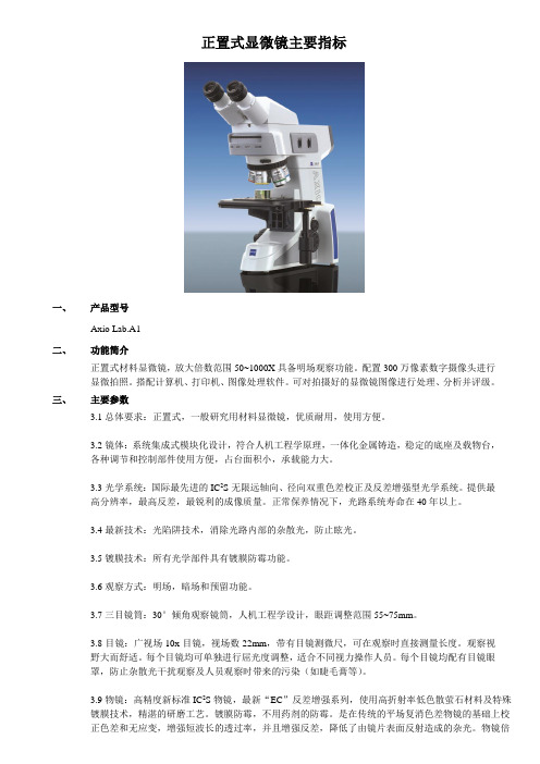
正置式显微镜主要指标一、产品型号Axio Lab.A1二、功能简介正置式材料显微镜,放大倍数范围50~1000X具备明场观察功能。
配置300万像素数字摄像头进行显微拍照。
搭配计算机、打印机、图像处理软件。
可对拍摄好的显微镜图像进行处理、分析并评级。
三、主要参数3.1总体要求:正置式,一般研究用材料显微镜,优质耐用,使用方便。
3.2镜体:系统集成式模块化设计,符合人机工程学原理,一体化金属铸造,稳定的底座及载物台,各种调节和控制部件使用方便,占台面积小,承载能力大。
3.3光学系统:国际最先进的IC2S无限远轴向、径向双重色差校正及反差增强型光学系统。
提供最高分辨率,最高反差,最锐利的成像质量。
正常保养情况下,光路系统寿命在40年以上。
3.4最新技术:光陷阱技术,消除光路内部的杂散光,防止眩光。
3.5镀膜技术:所有光学部件具有镀膜防霉功能。
3.6观察方式:明场,暗场和预留功能。
3.7三目镜筒:30°倾角观察镜筒,人机工程学设计,眼距调整范围55~75mm。
3.8目镜:广视场10x目镜,视场数22mm,带有目镜测微尺,可在观察时直接测量长度。
观察视野大而舒适。
每个目镜均可单独进行屈光度调整,适合不同视力操作人员。
每个目镜均配有目镜眼罩,防止杂散光干扰观察及人员观察时带来的污染(如睫毛膏等)。
3.9物镜:高精度新标准IC2S物镜,最新“EC”反差增强系列,使用高折射率低色散萤石材料及特殊镀膜技术,精湛的研磨工艺。
镀膜防霉,不用药剂的防霉。
是在传统的平场复消色差物镜的基础上校正色差和无应变,增强短波长的透过率,并且增强反差,降低了由镜片表面反射造成的杂光。
物镜倍数为5X、10X、20X、50X、1000X。
3.10功能转换盘:具备4位功能转换盘。
有预留位置,便于日后升级。
3.11照明装置:采用特制照明系统并使用长寿命高亮度卤素灯及特殊反光碗装置的柯拉照明方式,带调光装置和显示。
增强光的色还原性,进而提高分辨率。
Axio vert.A1技术说明

显微镜及图像分析系统技术文件仪器型号:Axio Vert.A12014年4月21日目录附件一、品牌介绍附件二、设备用途附件三、技术指标附件四、供货范围及报价附件五、计划进度及培训附件六、环境要求附件七、质保及其它服务附件一:聚焦CARL ZEISS世界顶级品牌,可见光及电子光学领导企业-----蔡司是一家致力於应用研究,对於光学、玻璃技术、精密技术以及电子等高品质的产品开发、制造、销售有着突出贡献的德国军工企业。
自1846 年开始,carl zeiss已有160多年的传统与创新。
百年历史缔造了蔡司在光学领域不可撼动的领导地位,至今显微镜的生产标准中的83%是以蔡司厂标为基准。
国际物镜的检测标准是以蔡司物镜为基准。
蔡司显微镜以其不断领先的技术和可靠的质量推动了世界材料科学的发展同时也受到知名科学家和诺贝尔奖得主的青睐!作为显微镜的鼻祖国际标准的缔造者,蔡司公司将以更新的技术延续carl zeiss成功的传奇故事!北京普瑞赛司仪器有限公司BEIJING PRECISE INSTRUMENT CO.,LTD,是一家专业从事理化测试仪器及相关产品的研发、销售、技术咨询的高新技术企业,是德、英、美、瑞士等多家仪器生产商在中国地区的总代理。
2001年德国卡尔.蔡司(Carl.Zeiss )基金会特别授权普瑞赛司公司为蔡司(Zeiss)材料显微镜中国独家代理商;负责蔡司(Zeiss)材料显微镜系列产品、相关设备和附件在中国地区的销售、服务与技术咨询业务。
与分布在世界各地的经销商一样,普瑞赛司在中国建立了完善的研发、销售、及服务一体化的现代化管理体系,为中国用户带来国际品质的尖端产品和专业服务。
为了给中国客户提供一个了解材料显微镜产品,网上技术交流的平台,2006年1月,我们开通了“中国材料显微镜网”(),它强大的产品和专家资源为客户建立了一个完善的技术和商务合作环境.我们将秉承“把世界上最好的仪器介绍到中国”的经营理念,为提高中国材料科学的发展提供先进的,高精度的,高质量的测试工具及手段,并希望与国内有识之士携手在质量控制,科研及前沿技术领域共创辉煌。
6082铝合金圆管氧化黑线缺陷原因分析
面,目前尚未看到公开文献报道,国内对 6××× 系合金氧化黑线方面的研究也不多。因此,本文从 生产实践出发,分析了 6082 铝合金圆管表面氧化黑 线的形貌和产生原因,提出了消除氧化黑线的有效 措施,以期为广大铝型材挤压同行提供有益参考。
晶粒组织,金相显微镜的型号为倒置型 ZEISS Axio vert.A1;采用蔡司扫描电子显微镜 (标配能谱仪) 进行能谱和形貌分析。
2 表面黑线现象的组织分析
2.1 表面黑线现象 6082 圆管氧化后的表面黑线缺陷典型形貌如
图 1 所示。圆管表面的黑线往往有很多条,且黑线 会贯穿整个圆管的挤压长度。实际检测发现,黑线 出现在圆管的具体位置并不固定。
黑线
1mm
图 1 6068 铝合金圆管表面黑线
2.2 晶粒组织分析 对 6082 成 品 圆 管 沿 着 挤 压 方 向 取 样 进 行 分
析,从正常区域和黑线处分别取样,其晶粒组织如 图 2 所示。
(a) 正常区域形貌
100 μm
(a) 正常区域晶粒组织
100 μm
(b) 黑线区域晶粒组织 图 2 6082 铝合金圆管高倍组织
对于在室外恶劣环境下使用的 6082 铝合金管 型棒材,往往采取阳极氧化处理在产品表面生成一 层致密的氧化膜,防止产品进一步被腐蚀,以提高 其使用寿命。但阳极氧化后,某些产品表面会出 现黑线从而影响产品防护效果和美观度。为了消 除 6 × × × 系合金产品氧化黑线,国内的研究人员 对此缺陷的形成原因进行了一些研究和探讨。郭世 杰等人[1]分析认为产品表面黑线主要是熔体处理过 程中残留的夹杂物,在挤压过程中受到剪切作用脱 落所致;通过改进熔体处理工艺、控制颗粒物种类 和含量能够降低挤压材黑线缺陷的出现概率。王奕 雷[2]针对水气对熔炼合金品质的影响,分析了水气 造成铸锭黑线缺陷率偏高的原因,认为通过加强精 炼减少氧化夹杂物可以控制黑线缺陷。陈秀琴[3]认 为黑线是因该处的金属与模具棱角等发生局部强烈 摩擦,局部变形温升,发生再结晶而引起,黑线与 模具结构关联较大。宋冰[4]研究了造成材料“黑线” 不良的原因,认为黑线部位成分与正常部位成分存 在差异并提出了消除黑线的措施。
- 1、下载文档前请自行甄别文档内容的完整性,平台不提供额外的编辑、内容补充、找答案等附加服务。
- 2、"仅部分预览"的文档,不可在线预览部分如存在完整性等问题,可反馈申请退款(可完整预览的文档不适用该条件!)。
- 3、如文档侵犯您的权益,请联系客服反馈,我们会尽快为您处理(人工客服工作时间:9:00-18:30)。
研究级倒置式万能材料显微镜
Axio Vert.A1
品牌:卡尔·蔡司
型号:Axio Vert.A1 制造商:德国卡尔蔡司公司
经销商:北京普瑞赛司仪器有限公司 产地:德国 联系方式:800-890-0660
蔡司公司最新推出的全新一代研究级倒置万能材料显微镜Axio Vert.A1开创了研究级倒置式显微镜产品的新纪元,完善的科勒式照明系统将光学系统的功能发挥得淋漓尽致,为您提供前所未有图像质量,稳固的产品设计与人机工程学理念相融合,使得Axio Vert.A1在满足您繁重的科研工作的同时为您提供最舒适、便捷的操作方式。
参数:
光学系统:ICCS 光学系统,镜体:FEM 设计、ACR 位置编码
1、 物镜:5x ,10x ,20x ,50x ,100x ,可选1.25x ,2.5x ,150x
2、 目镜10x
3、 物镜转盘:研究级5孔明暗场物镜转盘
4、 观察功能转盘:4位
5、 光源:12V100W 卤素灯,带有反光碗。
6、 光学附件:目镜测微尺,台尺,各种滤色片。
7、 数字化平台:可配图像分析系统(数码相机、摄像头、图像分析软件)
8、 可配热台(用于高温金相分析)
9、 可配自动扫描台。