2013 HBV检测
HIVID技术检测HBV病毒整合
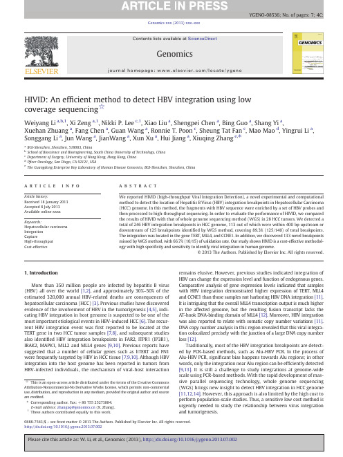
HIVID:An ef ficient method to detect HBV integration using low coverage sequencing ☆Weiyang Li a ,b ,1,Xi Zeng a ,1,Nikki P.Lee c ,1,Xiao Liu a ,Shengpei Chen a ,Bing Guo a ,Shang Yi a ,Xuehan Zhuang a ,Fang Chen a ,Guan Wang a ,Ronnie T.Poon c ,Sheung Tat Fan c ,Mao Mao d ,Yingrui Li a ,Songgang Li a ,Jun Wang a ,JianWang a ,Xun Xu a ,Hui Jiang a ,Xiuqing Zhang e ,⁎aBGI-Shenzhen,Shenzhen,518083,ChinabSchool of Bioscience and Bioengineering,South China University of Technology,China cDepartment of Surgery,University of Hong Kong,Hong Kong,China dP fizer Oncology,San Diego,CA 92121,USA eThe Guangdong Enterprise Key Laboratory of Human Disease Genomics,BGI-Shenzhen,Shenzhen,Chinaa b s t r a c ta r t i c l e i n f o Article history:Received 18January 2013Accepted 8July 2013Available online xxxx Keywords:Hepatocellular carcinoma Integration CaptureHigh-throughput Cost-effectiveWe reported HIVID (high-throughput Viral Integration Detection),a novel experimental and computational method to detect the location of Hepatitis B Virus (HBV)integration breakpoints in Hepatocellular Carcinoma (HCC)genome.In this method,the fragments with HBV sequence were enriched by a set of HBV probes and then processed to high-throughput sequencing.In order to evaluate the performance of HIVID,we compared the results of HIVID with that of whole genome sequencing method (WGS)in 28HCC tumors.We detected a total of 246HBV integration breakpoints in HCC genome,113out of which were within 400bp upstream or downstream of 125breakpoints identi fied by WGS method,covering 89.3%(125/140)of total breakpoints.The integration was located in the gene TERT,MLL4,and CCNE1.In addition,we discovered 133novel breakpoints missed by WGS method,with 66.7%(10/15)of validation rate.Our study shows HIVID is a cost-effective methodol-ogy with high speci ficity and sensitivity to identify viral integration in human genome.©2013The Authors.Published by Elsevier Inc.All rights reserved.1.IntroductionMore than 350million people are infected by hepatitis B virus (HBV)all over the world [1,2],and approximately 30%–50%of the estimated 320,000annual HBV-related deaths are consequences of hepatocellular carcinoma (HCC)[3].Previous studies have discovered evidence of the involvement of HBV in the tumorigenesis [4,5],indi-cating HBV integration in host genome is suspected to be one of the most important etiological events in HBV-induced HCC [6].The recur-rent HBV integration event was first reported to be located at the TERT gene in two HCC tumor samples [7,8],and subsequent studies also identi fied HBV integration breakpoints in FAR2,ITPR1(IP3R1),IRAK2,MAPK1,MLL2and MLL4genes [9,10].Previous reports have suggested that a number of cellular genes such as hTERT and FN1were frequently targeted by HBV in HCC tissue [7,9,10].Although HBV integration into the host genome has been reported in tumors from HBV-infected individuals,the mechanism of viral-host interactionremains elusive.However,previous studies indicated integration of HBV can change the expression level and function of endogenous parative analysis of gene expression levels indicated that samples with HBV integration demonstrated higher expression of TERT,MLL4and CCNE1than those samples not harboring HBV DNA integration [11].It is intriguing that the overall MLL4transcription output is much higher in the affected genome,but the resulting fusion transcript lacks the AT-hook DNA-binding domain of MLL4[12].Moreover,HBV integration was also reported to relate with somatic copy number variations [11].DNA copy number analysis in this region revealed that this viral integra-tion colocalized precisely with the junction of a large DNA copy number loss [12].Traditionally,most of the HBV integration breakpoints are detect-ed by PCR-based methods,such as Alu-HBV PCR.In the process of Alu-HBV PCR,signi ficant bias happens towards Alu regions;in other words,only the integration near Alu region can be ef ficiently detected [9,13].It is still a challenge to study integrations at genome-wide scale using PCR-based methods.With the rapid development of mas-sive parallel sequencing technology,whole genome sequencing (WGS)brings new insight to detect HBV integration in HCC genome [11,12,14].However,this approach is also limited by the high cost to perform population-scale studies.Thus,a sensitive low cost method is urgently needed to study the relationship between virus integration and tumorigenesis.Genomics xxx (2013)xxx –xxx☆This is an open-access article distributed under the terms of the Creative Commons Attribution-Noncommercial-No Derivative Works License,which permits non-commercial use,distribution,and reproduction in any medium,provided the original author and source are credited.⁎Corresponding author.Fax:+8675525273884.E-mail address:zhangxq@ (X.Zhang).1These authors contributed equally to this work.YGENO-08536;No.of pages:7;4C:0888-7543/$–see front matter ©2013The Authors.Published by Elsevier Inc.All rights reserved./10.1016/j.ygeno.2013.07.002Contents lists available at ScienceDirectGenomicsj o u r n a l h o m e p a ge :w ww.e l s e v i e r.c o m/l o c a t e /y g e n oHere,we present a novel experimental and computational meth-od,HIVID (high-throughput viral integration detection),to effectively detect hbv integration at a single base pair resolution using HBV cap-ture sequencing (Fig.1).Probes were prepared to capture the HBV inserted fragments in host genome and these fragments were further sequenced on Illumina HiSeq 2000platform.Based on the sequence data of enriched DNA,we recruited a pair-read assemble strategy to locate the integration breakpoints.To examine the performance of HIVID,this method was tested in 28HCC samples that were previously sequenced at the whole-genome level for identifying HBV integrations [11].The integration hotspots of human genome were located in the genes TERT,MLL4and CCNE1.Most of the breakpoints were signi ficantly enriched in a 500bp region on HBV genome from 1500bp to 2000bp.We compared the results of HIVID and WGS method,and validated the novel HBV integration using PCR and Sanger sequencing.Our study provided an accurate and cost-effective method for HBV integration re-search,which can also be adapted to other virus integration studies.2.Materials and methodsBased on our previous study [11],28Chinese HCC samples from Queen Mary Hospital,Hong Kong were selected.All patients had been diagnosed with HCC with concurrent HBV rmed written consents were obtained from each patient.Approvals were acquired from the BGI Ethics Committee and the Institutional Review Board of The University of Hong Kong/Hospital Authority Hong Kong West Cluster.All samples had been previously used to discover HBV integration breakpoints.The results from 30×WGS data were used to evaluate sensitivity and speci ficity of HIVID.2.1.HBV probes preparationFull-length HBV genome of type B and type C were ampli fied by PCR using biotin-labeled dNTPs.PCR mix was prepared as follows:5μl of HBV DNA (1ng/μl);5μl of 10*LA Taq Buffer;26.5μl of H 2O;4μl of 2.5mM dNTPs (1/4dNTPs biotin-labeled);2.5μl of 10pmol P1(TTT TTC ACC TCT GCC TAA TCA);2.5μl of 10pmol P2(AAA AAGTTG CAT GGT GCT GG);and 0.5μl of LA Taq enzyme (Takara Bio,Inc.).PCR was subjected to the following cycling condition:initial denaturation for 3min at 94°C;32cycles of denaturation for 30s at 94°C,annealing for 50s at 56°C,and extension for 2.5min at 68°C;final extension for 10min at 72°C.Amplicons were puri fied by Ampure beads (Beckman Coulter,Inc.)and were then fragmented by Covaris E-210(Covaris,Inc.,Woburn,MA)to 250bp –500bp through the 2%agarose electrophoresis.Single-stranded HBV probes were generated by high temperature denaturation at 94°C for 5min.2.2.HBV-inserted fragments enrichment and sequencingSequencing libraries of 170bp insert size were constructed for 28samples following the instruction of Illumina.Genomic DNA was sheared to around 150bp –200bp DNA fragments by Covaris E-210(Covaris,Inc.,Woburn,MA).These fragments were puri fied,end blunted,“A ”tailed,and adaptor ligated.10cycles of PCR were performed after size selection in 2%agarose gel.The concentration of libraries was quanti fied by Bioanalyzer 2100(Agilent Technologies,Santa Clara,CA).Libraries were hybridized with HBV probes at 47°C for 24hours and then washed to remove un-captured fragments.The eluted fragments were ampli fied by 16cycles of PCR to generate libraries for sequencing (Fig.2).Libraries were quanti fied and proceeded to 101cycles paired-end index sequencing in the Illumina HiSeq 2000sequencer according to manufacturer's instructions (Illumina Inc.,San Diego,CA).2.3.HBV integration breakpoints detectionThe whole pipeline for data analysis in HIVID consists of six steps (Fig.1).After we isolated the paired-end reads which may contain the signals of HBV integration,paired-end read assembly was conducted to obtain reconstructed inserted fragment and detected HBV integra-tionbreakpoints.Fig.1.Overall work flow of HIVID.The pipeline includes the work flow of experiment and bioinformatics process.In the process,we do Raw data filter first,then perform Raw-mapping with filtered raw data.After that we pick out the chimeric paired-end reads to do Paired-end read assembly.PE-assembled reads are conducted to go through Re-mapping to locate the HBV integrations sites.Finally,Signal merging and Signal filter are performed to obtain the final results.To do the step of Paired-end reads assembly,we used a program developed by ourselves.2W.Li et al./Genomics xxx (2013)xxx –xxx2.3.1.Raw data filterLow quality reads and duplication reads,as well as adaptor contami-nation reads,was first removed to obtain clean reads for subsequent analysis.If a read whose bases with quality value (b =5)occupy 50%of the read length,the read is considered as a low quality read.We then had the clean reads for detecting the HBV integration after the low qual-ity reads and duplication reads removal.2.3.2.Raw-mappingClean reads were mapped to human (NCBI build 37,HG19)and HBV genome using SOAP2(-l 40-v 5-r 1)[15].We removed reads that perfectly paired-end aligned to human or HBV genome and reserved chimeric paired-end reads (partial read sequence aligned to human genome and partial aligned to HBV genome)for assembly.This might help identify HBV integration breakpoint.2.3.3.Paired-end reads assemblyPaired-end reads assembly was used to reconstruct around 170bp of fragment sequences,assembly which might increase the potential to locate the exact position of breakpoints.The paired-end reads were first changed to the same strand.In case that the tail of an upstream end (read1)and the head of a downstream end (read2)overlap by more than 5bp where the mismatch rate was less than 0.2,the two ends would be spliced into one continuous sequence,called PE-assembled read,which was reconstructed inserted fragment (Fig.3A).2.3.4.Re-mappingTo determine the exact location of HBV integration breakpoint,the PE-assembled reads were re-mapped to human and HBV genome using BWA 0.5.9-r16(-a 1,-b 2,-q,5,-r,2)[16].If the match length on both HBV genome reference and human genome reference for one PE-assembled read was larger than 30bp,the PE-assembled read alignment result was reported to detect a precise breakpoint.The joint position of human and HBV sequence was the breakpoints for HBV inte-gration (Fig.3B).2.3.5.Signal mergingWe also calculated the total number of support PE-assembled reads (NSS)of each HBV integration breakpoint to examine its reliability.Considering errors in the procedure of experiment and bioinformatics analysis and highly heterogeneous nature of tumors,we merged breakpoints within 20bp to select the breakpoint with largest NSS as the representative,and reset its NSS as sum of NSS of all breakpoints in this region.2.3.6.Signal filterTo minimize the impact of total sequencing data for each breakpoint frequency and remove the noise signals,we normalized NSS to Normal-ized number of support PE-assembled reads (NNSS)using its number of effective paired-reads (EFR)as following:NNSS ¼NSS Â106=EFREffective paired-reads are de fined as reads used to search breakpoints except contaminated data.NNSS could be considered as the number of supporting reads of each HBV integration breakpoint out of every million read-pairs and represents the reliability and data utilization with no bias.In this study,HBV breakpoints with NNSS N =1was regarded as true signal.2.4.PCR and Sanger sequencing validationPCR and Sanger sequencing was used to verify the selected HBV integration breakpoints from HIVID.PCR primers were designed based on the PE-assembled fragment,in which one primer located in human genome and the other in HBV genome.PCR were performed by GeneAmp®PCR System 9700thermal cycler and then preceded to Sanger sequencing on Applied Biosystems 3730x DNA analyzer (Life Technologies,Inc).3.Results3.1.HBV integration breakpoints detectionTo get the overall evaluation of the accuracy of breakpoint detection using HIVID approach,we compared our results to that of previously re-ported ~30×WGS approach [11].Sung et al.reported a total of 399HBV integration breakpoints in 81HBV-positive and 7HBV-negative HCC and adjacent non-tumor samples,with a validation rate of 82.0%[11].We randomly selected 28samples for HIVID to conduct HBV capture sequencing on Illumina HiSeq 2000,generating ~1.43Gb on average (Table 1).The effect of paired-end reads for the following analysis were about 6millions,among them around 83.72%and 0.08%can be aligned to human genome and HBV genome,respectively.After paired-end assembly and re-mapping,we finally obtained an average of 1654reads supporting HBV integration breakpoints for each sample.A total of 246HBV integration breakpoints were detected within these 28sam-ples,in average 8.8breakpoints per sample.The number of integration breakpoints varied from 1to 27among samples (Table S1).3.2.HBV integration breakpoint detection comparisonWe firstly compared HBV integration breakpoints between HIVID and WGS approach in 28selected samples.To minimize the impact of different work flows we de fined a shared breakpoint in HIVID and WGS method when the distance of two breakpoints from the same sam-ple was less than 400bp.There were 140HBV integration breakpoints from Sung's study with at least two paired-end reads supporting and 89.3%of them (125/140)was shared with HIVID test (Tables S2and S3),suggesting that the current approach has high speci ficity when com-pared with WGS approach.In contrast,only 45.9%of HBV integration breakpoints (113/246)from HIVID approach were shared with WGS approach (Fig.4).Fourteen of 113HBV integration sites that were select-ed for validation were all validated in previous WGS study,showing 100%validation rate [11].Further analysis for the average NNSSvalue,Fig.2.HBV capture work flow.The different colored images marked represented differ-ent meaning.The dark yellow color represented the HBV fragments,while the glaucous color represented the human genome fragments.DNA libraries were hybridized with HBV probes at 47°C for 24hours and then washed to remove un-captured fragments.The eluted fragments were ampli fied by 16cycles of PCR to generate libraries for se-quencing.(For interpretation of the references to color in this figure legend,the reader is referred to the web version of this article).3W.Li et al./Genomics xxx (2013)xxx –xxxwhich was 13.6for 113WGS-shared HBV integration breakpoints and 8.5for all breakpoints,indicated WGS approach might tend to identify high frequency HBV integration breakpoints due to a relatively limited sequencing depth.The comparative analysis indicated HIVID test could detect almost the same HBV integration breakpoints as WGS approach with much less sequencing data.In the 133novel breakpoints we randomly picked 15novel HBV integration breakpoints from six samples for PCR and Sanger sequencing validation test.66.7%(10/15)of novel HBV integration sites were suc-cessfully con firmed (Table S4),which is signi ficantly lower than the validation rate for WGS-shared breakpoints.We then investigated the average NNSS value of the 133novel breakpoints,which were only 4.2,compared to 13.6for WGS-shared breakpoints and 8.5for the total ones.It was obvious that HIVID showed a notable advantage on breakpoints enrichment ef ficiency.On average,the HIVID approach generated 6mil-lion paired-end reads,compared to 600million read pairs for WGS meth-od.Moreover,the average NNSS values of two methods were 8.5and 0.0142,representing that HIVID approach could enrich HBV integration fragments close to 600times than WGS approach.This high ef ficiency for HBV integration sites enrichment would be bene fit for large-scale virus-related tumor development studies.3.3.Characteristics of HBV integration breakpointsWe also investigated the characteristics of HBV integration breakpoints,which might be related with HCC development.Integra-tion events were detected in HBV subtype C (AB014381.1).Among them,33.5%(68/203;P =2.7×10⁻⁵)were signi ficantly enriched in a 500bp region on HBV genome from 1500bp to 2000bp (Supplemen-tary Fig.S1),which was consistent with the results of the WGS study [11,12].Additionally,we also surveyed the frequency of integration breakpoints around HBV genes,showing 38.9%(79/203)breakpoints were located in the region of pre-S1(Table S5).The analysis for recurrent genes with HBV integration breakpoints in those 28samples was also consistent with previous studies (Fig.5)[10,17–19].Using HIVID approach,HBV integration breakpoints were detected in TERT ,MLL4and CCNE1genes,affecting 25%,17.9%,and 7.1%of HCC samples (Table 2).Notably,we identi fied HBV integration breakpoint in TERT gene for sample 90T and 41T,which was missed in previous WGS approach.Besides the frequent bias of HBV integration sites in HBV and human genome,we also noticed that HBV integration events with the deletion of human genome.For example,a 49bp human genome sequence in sample 43T was replaced by a 41bp HBV sequence on ChrX:20,156,148(Table S4).In sample 49T the human genome fragment of 728,975bp in chromosome 2were replaced by 127bp HBV sequence (Fig.6).The abundant supporting reads for breakpoints provided by HIVID may be helpful to explore the complex relation.4.DiscussionIn this study,we developed a novel approach for HBV integration breakpoint detection and employed 28HCC samples to estimate the performance on integration sites with high frequency and low frequen-cy.With only 1.5Gb HBV capture sequencing data,HIVID approach can identify about 90%of HBV integration breakpoints identi fied from ~30×WGS data,and over 50%of novel low frequent integration sites missed by WGS approach.HIVID approach shows its strength on several aspects compared with previously reported approaches.Unlike the widely used HBV-Alu PCR method,HIVID can be uti-lized to detect integration sites at the genome-wide level instead of the regions close to Alu repeats.In fact,83.3%(205/246)of the discov-ered HBV integration breakpoints were more than 10,000bp away from Alu repeats (Table S2),signi ficantly larger than 7.8%of WGS ap-proach [11].Although both HIVID and WGS approach are based on mas-sively parallel sequencing platform,HIVID approach has dramatically higher ef ficiency on virus integration detection.First,HIVID approach requires much less sequencing data due to the process of HBVfragmentFig.3.Insight of computational process of HIVID.A,principle of pair-end reads assembly.The figure shows that the tail of an upstream end (read1)and the head of a downstream end (read2)were spliced into one continuous sequence,called PE-assembled read;B,determination of breakpoint.The figure shows that one PE-assembled read consists of the part of human sequence and part of HBV sequence.The joint position was the breakpoint for HBV integration.4W.Li et al./Genomics xxx (2013)xxx –xxxcapture,but with an enrichment close to 600times compared with WGS approach.HIVID could also be suitable for detecting the breakpoints of other types of viruses,such as human papillomavirus,human immuno-de ficiency virus.Moreover,it also could decrease the cost and shorten the time to generate the results.Secondly,HIVID can detect 89.3%(125/140)integration breakpoints which had been identi fied by WGS method,and those integration sites are also characterized by higher NNSS and validation rate.These results demonstrate the HIVID method has a high speci ficity of detecting viral integration breakpoints.Thirdly,HIVID method is more sensitive for detecting low frequency HBV inte-gration breakpoints.Although the validation rate was 66.7%(10/15),HIVID methods could identify 50%more HBV integration sites.The reduced average NNSS value of the novel sites (4.2)compared with the WGS shared ones (13.6)indicates HBV integration in these sites may be less frequent among the tumor cells.The other cause for the lower veri fication rate of new breakpoints could be attributed to the primer design dif ficulty for the shorter support reads.The validationrate might be improved if we adopt the 454long reads sequencing.The long reads also will help us to get more information about the HBV insertion sequence.The major limitation of this approach com-pared with WGS is that we can only target speci fically one or more virus that have genome references,and identify their integrations.This is a hypothesis driven approach.Another technical limitation is that human genome reads and free virus reads take up a high propor-tion of the data.The method had a low capture speci ficity,although it had an obvious enrichment.We will get more integration breakpoints and need less sequencing data if the capture speci ficity could get im-proved.The single cell study for virus integration breakpoints could fig-ure out monoclonal evolution and the roles of high and low frequency integration events.Since we used part of samples from our previous study,the similar hotspot regions in human and HBV genome were de-tected with these two methods.However,with more supporting reads we annotated the complicated types of HBV integration events,which were omitted in our previous study.Herein,we found a trend of dele-tion in human genome accompanying with HBV fragments insertion,which might induce chromosomal instability and further carcinogene-sis.Genetic instability triggered by HBV integration has been considered in some reports to be an important contributing factor in the pathogen-esis of HCC [20,21].HBV insertions are commonly associated with large genetic alterations:deletions,duplications and chromosomal transloca-tions,which might re flect the abrogation of control mechanisms that safeguard chromosomal integrity [22].However,the impact of those human structure variations on molecular mechanism of HCC still re-mains unclear.In summary we reported the HIVID approach to detect HBV inte-gration breakpoints with high speci ficity and sensitivity using only 1.5Gb sequencing data,for which this approach can be used to screen virus integration in a large cohort of samples and leads to a systematic study for its relationship with disease etiology and tumorigenesis in a comprehensive and unbiased way.Supplementary data to this article can be found online at /10.1016/j.ygeno.2013.07.002.Table 1Data production of 28tested samples.Sample Total Bases Reads number EFR (M paired-reads)Human alignment (pair end)HBV alignment (pair end)Integration rate 101T 1.50G 7.51M;7.51M 5.8895.58%0.03%738(0.01%)43T 1.32G 6.58M;6.58M 5.6686.30%0.04%2520(0.02%)71T 1.49G 7.45M;7.45M 6.4685.05%0.11%1858(0.01%)58T 1.47G 7.33M;7.33M 6.1684.35%0.05%1230(0.01%)34T 1.39G 6.93M;6.93M 5.9884.35%0.02%480(0.00%)55T 1.53G 7.63M;7.63M 6.4782.91%0.10%602(0.00%)35T 1.64G 8.22M;8.22M 7.0585.13%0.02%188(0.00%)41T 1.15G 5.77M;5.77M 4.9784.23%0.12%1042(0.01%)32T 0.70G 3.52M;3.52M 3.1287.08%0.08%424(0.01%)186T 1.99G 9.94M;9.94M 8.5986.70%0.01%374(0.00%)49T 1.17G 5.83M;5.83M 4.9582.61%0.11%2026(0.02%)26T 1.40G 7.01M;7.01M 5.9481.78%0.03%442(0.00%)90T 1.34G 6.70M;6.70M 5.8188.05%0.20%1212(0.01%)182T 1.55G 7.74M;7.74M 6.6887.93%0.03%474(0.00%)266T 1.36G 6.80M;6.80M 5.7786.27%0.03%860(0.01%)145T 1.41G 7.03M;7.03M 5.7486.09%0.08%1398(0.01%)174T 1.35G 6.75M;6.75M 5.7887.22%0.03%290(0.00%)122T 1.52G 7.59M;7.59M 6.4885.11%0.15%1086(0.01%)114T 1.85G 9.25M;9.25M 7.5986.53%0.01%176(0.00%)17T 1.47G 7.36M;7.36M 6.4386.35%0.23%584(0.00%)200T 1.45G 7.25M;7.25M 5.6274.33%0.18%1546(0.01%)95T 1.24G 6.21M;6.21M 5.1877.77%0.04%1546(0.01%)23T 1.17G 5.83M;5.83M 4.8879.94%0.08%2634(0.03%)64T 1.17G 5.83M;5.83M 4.8777.04%0.01%136(0.00%)30T 1.66G 8.28M;8.28M 6.9380.35%0.05%572(0.00%)65T 1.80G 9.01M;9.01M 7.5677.03%0.05%482(0.00%)70T 1.04G 5.19M;5.19M 4.3479.30%0.30%1752(0.02%)82T1.90G9.51M;9.51M8.0278.64%0.03%288(0.00%)This table shows the data production of 28samples.Including human alignment ratio of pair-end reads,HBV alignment ratio of pair-end reads,raw data quantity and integration rate,etc.Fig.4.Overview of breakpoints detected by HIVID.The figure shows that HIVID detect-ed 246breakpoints on human genome in total.There were 113novel breakpoints and 133breakpoints shared with WGS method.The shared 133breakpoints covered 89.3%of WGS result.In addition the figure also provides the information of NNSS value and validated rate.5W.Li et al./Genomics xxx (2013)xxx –xxxFig.5.Distribution of breakpoints on human genome.In this CIRCOS figure,we showed the human chromosomes,the integration frequency of each breakpoint on the level of supporting reads and on the level of samples.The outer circle represents 24chromosomes and mitochondria with different color-code and number labeled.In the middle circle,the integration frequency of each breakpoint one the level of supporting reads is displayed as height of each blue point.In the inner circle,the height of each red point represents the integration frequency of each breakpoint on the level of samples.To explore the distribution of frequency of all integration breakpoints on the level of supporting reads,we pooled all breakpoints from all samples.We merged those breakpoints which appeared in several different samples but in the same location and reset NNSS of the new merged breakpoint as sum of NNSS of all breakpoints in this location.The new NNSS could be seen as the integration frequency of each merged breakpoint on the level of supporting reads.The middle circle was figured based on the process above and the NNSS of each merged breakpoint is displayed as the height of each blue point.To explore the distribution of frequency of integration breakpoints on the level of samples,the human genome sequence was divided into about three million windows,each with length of 1000bp.The total number of samples that the breakpoints in each window belonged to,was calculated.Each window was considered as one new breakpoint and the number of samples in each win-dow calculated above was considered as supporting samples that supported the window as a new breakpoint.The number could be seen as the integration frequency of each breakpoint on the level of samples.The inner circle was figured based on the process above and the height of each red point represents the integration frequency of each breakpoint on the level of samples.(For interpretation of the references to color in this figure legend,the reader is referred to the web version of this article).Table 2The enriched region of HBV integration.Sample ID PositionNSS NNSS Gene266T chr5:1,295,4639716.82TERT:Promoter 34T chr5:1,295,3759616.05TERT:Promoter 58T chr5:1,295,3398012.98TERT:Promoter 65T chr5:1,295,4429712.82TERT:Promoter 90T chr5:1,295,6397212.38TERT:Promoter41T chr5:1,295,12119 3.82TERT:NM_001193376:5-UTR;TERT:NM_198253:5-UTR;64T chr5:1,295,2959 1.85TERT:Promoter49T chr19:36,212,74121242.83MLL4:NM_014727:Intron;71T chr19:36,213,142467.12MLL4:NM_014727:Intron;186T chr19:36,214,00716619.34MLL4:NM_014727:CDS;70T chr19:36,212,313465107.03MLL4:NM_014727:CDS;95T chr19:36,212,5665711.01MLL4:NM_014727:CDS;145T chr19:30,303,49923140.24CCNE1:NM_001238:Intron;200Tchr19:30,315,3669416.72CCNE1:DownstreamThis table shows the hotspots of HBV integration in the 28samples.The relative genes of these integration positions have been shown in the table.6W.Li et al./Genomics xxx (2013)xxx –xxx。
实时荧光定量PCR检测HBV DNA室内质控分析

实时荧光定量PCR检测HBV DNA室内质控分析目的探讨实时荧光定量PCR在HBV DNA定量检测过程中的室内质控问题。
方法用实时荧光定量PCR法检测2013年1~7月自制的乙肝阳性室内质控血清,与临床标本同步检测,计算每次阳性质控结果的常用对数、标准曲线的斜率、截距和相关系数(r)及相关结果的均值(x)、标准差(s)和变异系数(cv),利用EXCEL折线散点图画出质控图进行质控。
结果本室2013年1~7月测定自制的阳性室内质控结果测定值均在控,标准差和变异系数均在允许范围内,符合要求。
结论本实验室采用实时荧光定量PCR检测HBV-DNA定量检测过程中,选用的质控方法和自制的质控品稳定性良好,能可靠有效地为临床提供准确的检验结果。
Abstract:ObjectiveTo explore the indoor quality controlproblems in HBV DNA quantitative detection of real-time fluorescence quantitative PCR. Indoor quality control serum HBsAg positive self detection.MethodsFor 1~7 months of 2013 by real-time fluorescence quantitativePCR method and clinical specimens, synchronous detection, curve of standard logarithm, calculated for eachpositive quality control results of the slope, intercept and correlation coefficient (R) value and related results ,standard deviation (s) and the coefficient of variation(CV), scatter picture quality control chart for quality control by using the EXCEL line. ResultsThe 1~7 months of 2013 were positive results of internal quality control of homemade measured values are in control, the standard deviation and variation coefficient were within the allowable range, meet the requirements. ConclusionThis laboratory detection of HBV-DNA quantitative real-time fluorescence quantitative PCR detection process,quality control methods and quality control of homemadegood stability, can provide valid and reliable accurate test results for clinical.Key words:Real time fluorescence quantitative PCR; Polymerase chain reaction; HBV-DNA; Quality controlHBV DNA含量是诊断和监测乙型病毒性肝炎的重要指标[1]。
HBsAg与HBV DNA定量测定的临床信息互补

HBsAg与HBV DNA定量测定的临床信息互补武建国【摘要】2013年是HBV表面抗原(HBsAg)发现五十周年.HBsAg的定性测定一直是判定HBV感染的依据,但近年来其定量检查受到临床高度重视,主要原因是定性检查只能反映HBV感染是否存在,而定量检测才能反映病情,指导治疗,判断预后.HBsAg与反映病毒复制的HBV DNA合成途径不同,二者在血清中的表达水平并不平行,这在核苷酸类似物(NAs)的抗病毒治疗时表现尤为突出.因此,为有效控制慢性乙肝(CHB)的病情发展,优化抗病毒治疗策略,追求患者临床结局的改善,HBsAg 定量与HBV DNA定量的联合检测已成为肝病专家的共识,并被纳入各种诊断、治疗的方案中.【期刊名称】《临床检验杂志》【年(卷),期】2013(031)011【总页数】4页(P801-804)【关键词】乙型肝炎病毒;表面抗原;乙肝病毒脱氧核糖核酸;定量【作者】武建国【作者单位】南京军区南京总医院临床中心实验科,南京210002【正文语种】中文【中图分类】R446.61963年,美国国立卫生研究院的Baruch S. Blumberg偶然发现澳大利亚一原住民的血清与多次输血的血友病患者血清在琼脂双向扩散试验时出现一条沉淀线,对该不明物质取名为“澳大利亚抗原”(Australia antigen,Au抗)。
其后研究发现,Au抗与肝炎有关。
1968年Alfred Prince确认Au抗是HBV的一部分,并正名为HBV表面抗原(HBsAg)。
1970年Dane DS在电镜下观察到完整的HBV颗粒(Dane颗粒)。
研究发现,HBV感染与输注污染血密切相关。
1972年在建立血清中HBsAg的测定法(Abbott公司放射免疫法,AUSRIA-125)后,世界卫生组织(WHO)正式建议对献血员进行筛查,此举使美国因输血感染HBV的概率下降了75%[1]。
上世纪80年代,酵母菌重组乙肝疫苗(HBsAg亚单位疫苗)获美国食品和药物管理局(FDA)批准,广泛用于新生儿和普通人群预防接种,使乙肝的流行得到了很大程度的控制。
荧光定量PCR检测HBV

荧光定量PCR检测HBV[摘要] 目的探讨荧光定量pcr法检测hbv-dna与elisa法乙肝免疫学指标的相关性。
方法采用elisa 法和荧光定量pcr技术分别对353 例乙型肝炎患者血清进行免疫学指标的检测及hbv-dna 的定量检测。
结果 elisa法呈大三阳组患者的hbv-dna阳性率及病毒平均拷贝数均高于小三阳组;hbeag(+)组的阳性率及病毒平均拷贝数高于hbeag(-)组;hbeab(+)组有较低的hbv-dna阳性率及病毒平均拷贝数。
结论 hbv-dna 的含量与乙肝免疫学检测结果具有相关性。
临床上应注意两者联合应用。
[关键词] hbv-dna;elisa法;乙肝;免疫标志物[中图分类号] r512.62 [文献标识码] b [文章编号] 2095-0616(2013)06-110-03乙型肝炎是目前最常见的威胁人类健康的传染病之一。
我国是乙型肝炎的高发区,并且近年来其发病率有上升趋势[1]。
因此做好乙肝的检测对于传染病的防控及临床诊治有重要的意义。
目前临床最常用的乙肝筛查及诊断治疗的方法是检测乙肝免疫学指标,即乙肝五项指标。
而在免疫指标检测方面最常用的是酶联免疫吸附试验(elisa)法,它反映人体对hbv的免疫反应状态,具有快速、价廉、简便的特点,适合乙肝的大批量筛查及快速诊断;但是不能直接反映hbv在患者体内的复制情况。
而荧光定量pcr检测hbv-dna 法可直接监测血清中乙肝病毒核酸的载量,是判断病毒复制活跃程度及是否具有传染性的可靠依据,对临床抗病毒治疗及判断预后均具有指导意义。
1 资料与方法1.1 一般资料收集2011年6月~2012年6月在本院门诊、住院及健康查体的hbsag(+)标本共353例,其中女157例,男196例。
按患者的乙肝病毒血清免疫学标志物组合分为6组:a组(大三阳)hbsag(+)、hbeag(+)、hbcab(+),共112例;b组(小三阳)hbsag(+)、hbeab (+)、hbcab(+),共93例;c组 hbsag(+)、hbeag(+),共45例;d组 hbsag(+)、hbeag(-),共31例;e组 hbsag(+)、hbeab (+),共53例。
检测乙肝患者血清标志物与HBV—DNA含量浅析
检测乙肝患者血清标志物与HBV—DNA含量浅析作者:车梦楠来源:《中国保健营养·中旬刊》2013年第10期【摘要】目的:探讨乙型肝炎患者血清标志物与HBV-DNA定量检测之间的关系及其临床意义;方法:选择258例经我院确诊的乙肝患者,采用酶联免疫吸附试验法检测患者体内血清标志物,采用荧光定量聚合酶链反应检测HBV-DNA含量,比较血清标志物与HBV-DNA检测量之间的关系;结果:HBsAg+/HBeAg+组与HBsAg+/HBeAg+/抗-HBc+组阳性检出率分别为100%(16/16)与95.45%(84/88),HBV-DNA对数值分别为7.85±2.12拷贝数/ml和7.08±2.65拷贝数/ml;27例HBsAg+/HBc +组、85例HBsAg+/抗-HBe+/抗-HBc+组、11例抗-HBs +/抗HBe+/抗-HBc +组、8例抗-HBe +/抗-HBc+组、23例抗-HBs+组阳性检出率分别为62.96%、52.94%、37.50%、27.27%、4.35%;结论:乙肝患者血清HBV-DNA阳性率与血清标志物存在相关性;同时进行血清标志物的检测与HBV-DNA定量检测能较准确直接地反应病毒复制程度及传染强度,对于早期诊断、治疗及预后判断具有指导意义。
【关键词】乙肝患者;血清标志物;HBV-DNA【中图分类号】R440 【文献标识码】A 【文章编号】1004—7484(2013)10—0087—02乙型肝炎是一种以肝脏炎性病变为主累及器官损害的传染性病症疾病,常用的诊断指标有HBV-DNA与血清标志物(乙肝二对半)。
为探讨乙肝患者体内血清标志物与HBV-DNA定量之间的关系,本文选择258例乙肝患者,采用酶联免疫吸附试验(ELISA)检测血清标志物,采用荧光定量聚合酶链反应(FQ-PCR)技术检测其血清HBV-DNA的含量,旨在为临床诊断、治疗及预后判断提供参考数据。
北京市卫生局关于同意卫生部北京医院等44家医疗机构开展临床基因扩增检验技术的通知

北京市卫生局关于同意卫生部北京医院等44家医疗机构开展临床基因扩增检验技术的通知文章属性•【制定机关】北京市卫生局•【公布日期】2013.12.31•【字号】京卫医字[2013]221号•【施行日期】2013.12.31•【效力等级】地方规范性文件•【时效性】现行有效•【主题分类】健康促进正文北京市卫生局关于同意卫生部北京医院等44家医疗机构开展临床基因扩增检验技术的通知(京卫医字[2013]221号)各区县卫生局,各有关医疗机构:根据原卫生部《医疗机构临床基因扩增实验室管理办法》(卫办医政发〔2010〕194号)和《北京市卫生局关于印发北京市医疗机构临床基因扩增实验室技术审核相关文件的通知》(京卫医字〔2012〕160号),我局委托北京市医学检验质量控制与改进中心组织专家对我市申请开展临床基因扩增检验技术的医疗机构进行了技术审核和现场评估。
经审核研究,现就有关工作通知如下:一、同意给予卫生部北京医院等44家医疗机构开展的临床基因扩增检验项目登记备案(机构名单和检验项目见附件)。
二、各医疗机构应按照《北京市卫生局转发国家卫生计生委关于印发医疗机构临床检验项目目录(2013版)的通知》(京卫医字〔2013〕156号,以下简称《目录》)要求认真核查准予开展的检验项目。
涉及因《目录》调整等而导致检验项目超出已登记的诊疗科目专业范围的,应当按照有关规定先行办理相关诊疗科目的变更登记手续。
三、符合条件的医疗机构可携《医疗机构执业许可证副本》到准予登记注册的卫生行政部门办理临床基因扩增检验技术的登记手续。
各卫生行政部门审核后,在医疗机构《医疗机构执业许可证》副本备注栏予以登记“允许开展临床基因扩增检验技术”。
四、完成相关诊疗科目及临床基因扩增检验技术登记后的医疗机构可在备案项目范围内开展临床基因扩增检验工作。
请各区县卫生局将本文转发至辖区有关医疗机构。
北京市卫生局2013年12月31日附件:。
电化学发光定量检测乙肝五项指标分析及临床意义
电化学发光定量检测乙肝五项指标分析及临床意义发表时间:2013-04-16T10:04:54.967Z 来源:《医药前沿》2013年第5期供稿作者:孙学青韩景银刘兆军[导读] HBV感染人体后在组织细胞与血液中会出现各种类型的感染标志物,通过对这些感染标志物的检测分析。
孙学青韩景银刘兆军(山东省东营市人民医院检验科山东东营 257091)【摘要】目的分析电化学发光定量检测乙肝五项指标与定性检测结果的差异及定量检测的临床意义。
方法对292份血标本分别采用ELISA 法定性检测和电化学发光法定量检测乙肝五项指标。
结果 ELISA法和电化学发光法分别检出9种和12种乙肝五项指标模式,电化学发光法HBsAg、HBsAb、HBeAg、HBeAb、HBcAb的阳性检出率均高于ELISA法(P均<0.05)。
结论电化学发光定量检测乙肝五项指标敏感性高于定性法并且具有良好的重复性,适合用于HBV感染的早期诊断以及临床抗病毒药物治疗疗效预测和评估。
【关键词】定量检测乙肝五项指标临床意义【中图分类号】R446 【文献标识码】A 【文章编号】2095-1752(2013)05-0389-03 Electrochemiluminescence analysis and clinical significance of the quantitative detection of hepatitis B five indicators Sun Xue-qing, Han Jing-yin, Liu Zhao-jun (Dongying City, Shandong Province, People's Hospital Laboratory shandong Dongying 257091) 【Abstract】Objective To analyze the the electrochemiluminescence quantitative detection of hepatitis B five indicators and qualitative differences and quantitative detection of the test results of clinical significance. Methods 292 blood samples using ELISA the legal detection and electrochemiluminescence quantitative detection of hepatitis B five indicators. Results The ELISA and ECLIA were detected 9 and 12 hepatitis B five indicators mode, the ECLIA HBsAg , HBsAb, HBeAg, HBeAb, HBcAb positive were higher than the ELISA method (P<0.05). Conclusion Electrochemiluminescence quantitative detection of hepatitis B five indicators more sensitive than the qualitative method with good reproducibility, and suitable for use in the early diagnosis of HBV infection and the clinical antiviral therapy prediction and assessment. 【Key words】Quantitative detection Hepatitis B five indicators Clinical significance 乙肝病毒(HBV)在我国以及整个亚洲地区有很高的感染率,根据世界卫生组织(WHO)的统计,全世界1/3的人曾感染过HBV,其中约2%-5%为慢性感染[1]。
HBV-DNA荧光定量与ELISA测定乙肝五项指标及肝功能相关性分析
HBV-DNA荧光定量与ELISA测定乙肝五项指标及肝功能相关性分析发表时间:2013-08-05T15:45:20.967Z 来源:《中外健康文摘》2013年第26期供稿作者:容文潮苏明茂黄秀华赵一帆[导读] HBV-DNA定量与HBV-M关系密切,与ALT、AST和r-GT有一定的相关性,全面检测各项指标有利于乙肝患者的诊断和治疗。
容文潮苏明茂黄秀华赵一帆(台山市人民医院检验科广东江门 529200)【摘要】目的探讨HBV-DNA定量与不同血清学标志物(HBV-M)及肝功能的关系;方法对352例HBsAg阳性患者血清用RQ-PCR定量法测定HBV-DNA含量,用ELiSA法测定HBV-M,同时进行肝功(ALT、AST、r-GT)测定。
结果 HBV-DNA定量大三阳组显著高于小三阳组和(HBsAg+HBcAb)组(P<0.05);HBV-DNA定量与ALT、AST、r-GT含量呈一定的正相关性(P<0.05)。
结论 HBV-DNA定量与HBV-M 关系密切,与ALT、AST和r-GT有一定的相关性,全面检测各项指标有利于乙肝患者的诊断和治疗。
【关键词】 HBV-DNA HBV-M 肝功三酶检测乙肝病毒是引起乙型肝炎的病原体,是我国常见的传染病,其传染性和病死率均居高不下,我国每年约有1.2亿人感染乙型肝炎病毒(HBV),每年约有30万人死于慢性乙型肝炎、肝硬化、肝癌等相关性疾病,严重危害人们的健康[1],目前,乙型肝炎病毒感染诊断检测手段较多,主要是血清学标志物(HBV-M)的检测和HBV-DNA含量的检测。
荧光定量聚合酶链式反应(FQ-PCR)具有较高的特异性和灵敏度,克服了常规PCR假阳性的特点,实现对HBV-DNA的定量检测可直接监测血清中HBV-DNA的复制情况,能为乙肝患者提供准确的病原诊断和抗病毒治疗的正确评估。
酶免疫吸附试验(ELISA)法检测乙肝血清学标志物(HBV-M)是目前诊断乙型肝炎的免疫状态[2],是一种定性分析。
- 1、下载文档前请自行甄别文档内容的完整性,平台不提供额外的编辑、内容补充、找答案等附加服务。
- 2、"仅部分预览"的文档,不可在线预览部分如存在完整性等问题,可反馈申请退款(可完整预览的文档不适用该条件!)。
- 3、如文档侵犯您的权益,请联系客服反馈,我们会尽快为您处理(人工客服工作时间:9:00-18:30)。
Rx
Immune Active
Non-replicative
Rx
Resolve
Hepatitis B
三、乙型肝炎其它标志物的检测
Hepatitis B
(一) pre-S和抗-preS检测
1.pre-S蛋白
HBV外膜蛋白成份 Pre-S1 肝细胞膜受体
Pre-S2 聚合人血清白蛋白受体
HBcAg = inner core protein
Hepatitis B
HBV感染过程中抗原、抗体的产生
HBV
HBeAg
HBsAg
Pre-Core HBX
Small HBs Medium HBs Large HBS HBcAg Poly DNA-
RNA
RNA
DNA-
Hepatitis B
HBV的抗原、抗体
感染过程空白期
抗-HBs
被动获得
0 4 8 12 16 20 24 28 32 36 52 100 w
Hepatitis B
乙型肝炎自然病程的相关指标
HBsAg
Serology ALT level HBV DNA level Chronicity Stage Immune tolerant (phase I)
Hepatitis B
HBeAg阴性的乙型肝炎及其HBV
一般出现在感染后期 表现为进程性肝脏损害 预后很差
编码HBeAg的基因preC启动子区发生突变
Hui CK. Curr Treat Options Gastroenterol. 2007 Dec;10(6):474-82.
Hepatitis B
抗-HBs
0
4
8
12 16 20 24 28 32 36
52
100 w
Hepatitis B
2.临床意义 (1) 感染过HBV、康复和获免疫力的标志 (2) 中和抗体,可维持数年至10余年
(3) 接种疫苗效果观察
William Rutter
Hepatitis B
抗-HBs 与 乙肝疫苗
Pediatr Infect Dis J 23(7):650-655, 2004
Baruch Blumberg
低浓度HBsAg不容忽视
HBsAg浓度在5ng/mL以下的有189例, 占总数的2.34%,占HBsAg阳性人群的23.16% 1ng/mL以下者: 占总数的1.04% 占HBsAg阳性人群的10.3%
0.5ng/mL以下者 占HBsAg阳性人群的约5%
陈瑜等,中华检验医学杂志,2001,第3期 楼滨等,中华临床感染病杂志,2009,第3期
HBeAg与HBV感染状况
V.Rafalski. 4th European Congress of Chemotherapy and Infection 2002;May 4
Hepatitis B
2.临床意义
(1) HBV复制活跃和传染性强的标志
(2) 疗效观察的指标 (3) 持续阳性表明患者易转为慢性乙肝
Centers for Disease Control. /hepatitis. Accessed January 31, 2006.
Hepatitis B
我国乙肝患者的基本情况
五大关键发现 乙肝患者的生活受到全方位负面影响
乙肝患者缺乏乙肝的疾病知识
乙肝患者对治疗目标缺乏正确的认识 乙肝患者的治疗药物、方法欠规范 乙肝患者缺乏对疾病知识的信息来源
教学目的
主要标志物的临床意义 检测结果的分析方法 危害和防护措施
正确的医学观念
Hepatitis B
一、乙型肝炎病毒概述
Hepatitis
病毒类型
甲型肝炎病毒(HAV)
基因组类型 传播途径
ssRNA 粪 -口途径
dsDNA 肠道外途径 肠道外途径 ssRNA
乙型肝炎病毒(HBV)
Hepatitis B
Hepatitis B
HBV的基本性质
D.S Dane. Lancet 1970
Hepatitis B
Pasek M. Nature 1979
Hepatitis B
DNA DNA聚合酶 Polymerase
HBsAg = surface (coat) protein HBeAg = secreted protein e
丙型肝炎病毒(HCV)
丁型肝炎病毒(HDV)
戊型肝炎病毒(HEV) 其它肝炎病毒
肠道外途径 ssRNA
粪 -口途径 ssRNA
Hepatitis B
乙型肝炎的世界发病情况
HBsAg Prevalence > 8% - High 2-8% - Intermediat
12 16 20 24 28 32 36
52
100 w
Hepatitis B
2.临床意义
(1) 感染早期诊断指标
(2) HBV复制活跃、具有强传染性 (3) 多见于急性肝炎
Hepatitis B
(六) 抗-HBc IgG检测
1.参考值 ELISA 阴性
症状
抗-HBe HBeAg 抗-HBc IgG HBsAg 抗-HBc IgM
HBs Ag 抗HBs HBe Ag 抗HBe 抗 -HBc IgG
临床意义
+ +
-
+ -
+
+
+
???
???
Hepatitis B
乙型肝炎五项标志物
症状 抗-HBe HBeAg HBsAg 抗-HBc IgM 抗-HBs 抗-HBc IgG
0
4
8
12
16
20
24 28
32
36
52
100 w
Hepatitis B
Hepatitis B
HBV 与 肝细胞性肝癌(HCC)
Percent cumulative incidence 12 HBsAg+, HBeAg+
10
8 6 4 2 0 0 1 2 3 4 5 6 7 8 HBsAg+, HBeAgHBsAg-, HBeAg9 10 Year
Yang HI, et al. N Engl J Med. 2002;347:168-174.
低水平的HBsAg检出率存在差异
ELISA 0.8~1.0 1.0~1.5 1.5~3.0 Sample 44 51 62 COI<1(阴性) n 28 25 9 % 63.64 49.02 14.52 COI≥1(阳性) n 16 26 53 % 36.37 50.98 85.48
3.0~6.0
疫苗接种,有正常的 HBsAb 应答;同时感染免
疫逃避变异株
Hepatitis B
(三) HBeAg检测
1.参考值
症状
抗-HBe HBeAg 抗-HBc IgG HBsAg 抗-HBc IgM
ELISA 阴性
抗-HBs
0
4
8
12 16 20 24 28 32 36
52
100 w
Hepatitis B
Hepatitis B
2. 临床意义 Pre-S1
(1) 早期感染的指标 (2) 病毒复制活跃标志
抗Pre-S1
中和抗体 预后良好的指标
Hepatitis B
2. 临床意义
Pre-S2
病毒复制和较强传染性的标志 抗-Pre-S2 (1) 中和抗体 (2) 病毒清除的标志
对HBsAg阴性、抗HBc阳性患者,在给予长期或大剂量免
疫抑制剂或细胞毒药物(特别是针对B或T淋巴细胞单克 隆抗体)治疗时,应密切监测HBV DNA和HBsAg,若出 现阳转则应及时加用抗病毒治疗
Hepatitis B
(二) 抗-HBs检测
1.参考值 ELISA 阴性
症状
抗-HBe HBeAg 抗-HBc IgG HBsAg 抗-HBc IgM
合计
32
189
2
64
6.25
33.86
30
125
93.75
66.14
HBsAg ELISA法弱阳性标本ECLIA结果
低浓度HBsAg的准确检测对于临床确诊和流 行病学监控具有重要意义
血清HBsAg是诊断HBV感染的直接证据 所谓“HBsAg阴性”的HBV感染者中,有相当一
部分为血清HBsAg呈低水平而被漏检的假阴性
LABORATORY DIAGONOSIS
乙型肝炎标志物检测
Serologic Examination of Hepatitis B
马越云
Hepatitis B
教学内容
概述(10min)
乙肝五项标志物(30min) 其它标志物(15min)
临床讨论 (15min)
Hepatitis B
HBsAg Pre-S1 Anti-Pre-S1 Pre-S2 Anti-Pre-S2
Anti-HBs
HBeAg Anti-HBe
HBcAg Anti-HBc DNA
Hepatitis B
什么是乙型肝炎两对半?
HBsAg
抗-HBs
HBeAg
抗-HBe
抗-HBc
Hepatitis B
什么是大三阳、小三阳?
(四) 抗-HBe检测
1.参考值
ELISA 阴性
症状
抗-HBe HBeAg 抗-HBc IgG HBsAg 抗-HBc IgM
抗-HBs
0
4
8
12 16 20 24 28 32 36