实验设计思路及思考问题
小学六年级科学实验的设计思路与方法

小学六年级科学实验的设计思路与方法在小学六年级的科学课堂上,实验设计是一项激发学生兴趣和创造力的重要任务。
实验不仅能帮助学生理解科学原理,还能培养他们的观察力、动手能力和逻辑思维能力。
设计一个有效的科学实验,需要从多个方面进行考虑和规划,以确保实验既有趣又富有教育意义。
以下是一些设计思路与方法,帮助教师们为学生制定富有启发性的科学实验。
首先,明确实验的教育目标是设计实验的第一步。
教师需要考虑实验的核心目标是什么,是为了让学生理解一个科学概念,还是为了让他们掌握实验技能。
例如,在一个关于植物生长的实验中,教育目标可能是让学生了解植物对光照、水分和土壤的需求。
明确了目标之后,教师可以围绕这一目标设计相应的实验步骤和活动。
其次,选择适当的实验主题和材料至关重要。
实验的主题应与学生的知识水平和兴趣相匹配,以激发他们的好奇心和探索欲。
比如,针对六年级学生,可以选择生活中常见的现象作为实验主题,如“为什么冰块会浮在水面上?”或者“不同液体的溶解速度”。
选择材料时,尽量选用学生能够轻松获取和操作的常见物品,比如水、盐、糖、纸杯等,以降低实验的复杂度和风险。
设计实验步骤时,要确保每个步骤清晰且易于操作。
步骤应该按逻辑顺序排列,从实验准备、操作到结果记录,每一步都要详细说明。
例如,如果设计一个关于液体密度的实验,步骤可以包括:准备不同种类的液体(如水、油、糖水)、将这些液体倒入透明容器中、观察液体的分层现象,并记录观察结果。
这样不仅能让学生理解实验过程,还能让他们学会如何系统地进行科学观察。
为了增强实验的互动性和趣味性,可以在实验中加入一些学生参与的环节。
例如,在探究植物生长的实验中,可以让学生自己种植植物、记录生长情况,并讨论观察到的现象。
这种参与感不仅能增加他们的实验兴趣,还能帮助他们更好地理解科学原理。
教师可以设计一些简单的实验问题,引导学生思考和讨论,例如“为什么有些植物在阳光下生长得更快?”。
安全问题是实验设计中不可忽视的一部分。
初中生物实验设计思路(含学习方法技巧、例题示范教学方法)

初中生物实验设计思路第一篇范文:初中生物实验设计思路生物实验是生物学教学的重要组成部分,它有助于学生深入理解生物学知识,培养学生的实践能力和创新精神。
初中生物实验设计应遵循科学性、严谨性、安全性和教育性原则,以提高学生的实验兴趣和实验技能。
本文将从以下几个方面阐述初中生物实验设计的思路。
二、实验选题1.贴近生活:选择与学生生活密切相关的实验题材,激发学生的兴趣和探究欲望。
例如,研究植物的生长、人体的生理功能等。
2.注重基础:注重生物学基本概念和原理的实验教学,为学生后续学习打下坚实基础。
例如,观察细胞的结构、探究遗传与变异等。
3.突出实践:强调学生的动手操作和实践能力,培养学生的实验技能。
例如,植物的组织培养、生态瓶的制作等。
4.注重创新:鼓励学生开展小发明、小制作等创新性实验,培养学生的创新能力。
例如,设计并制作一个简易的生态循环系统等。
三、实验原理与步骤1.实验原理:明确实验所涉及的生物学原理,为学生提供实验的理论依据。
例如,植物光合作用的原理、细胞呼吸的原理等。
2.实验步骤:详细阐述实验的操作流程,确保学生能够顺利进行实验。
例如,制作临时装片观察细胞、测定植物的光合速率等。
3.数据处理:引导学生运用科学方法对实验数据进行处理和分析,培养学生的数据分析能力。
例如,计算植物的光合速率、制作柱形图分析实验结果等。
四、实验注意事项1.安全第一:强调实验安全,确保学生在实验过程中的人身安全。
例如,使用仪器设备时要注意操作规范、实验药品要妥善存放等。
2.严谨实验:培养学生严谨的实验态度,克服实验过程中的马虎行为。
例如,准确记录实验数据、遵守实验纪律等。
3.环保意识:教育学生在实验过程中注意环境保护,减少对环境的污染。
例如,合理使用实验药品、妥善处理实验废弃物等。
五、实验评价与反思1.学生自评:鼓励学生对自己的实验过程和结果进行评价,提高学生的自我认知能力。
例如,分析自己在实验中的优点和不足,提出改进措施等。
(完整版)实验设计的思路与步骤
(完整版)实验设计的思路与步骤引言实验设计是科学研究中的重要环节,合理的实验设计能够确保实验的可靠性和可重复性,进而保证实验结果的可信度和准确性。
本文将介绍实验设计的思路与步骤,帮助读者在进行实验设计时能够做到有条不紊、系统化的进行。
实验设计的思路实验设计的思路是指在进行具体实验前需进行的全面思考和规划。
它包括以下几个方面:1. 确定实验目的:在进行实验设计之前,首先需要明确实验的目的和研究问题。
实验目的的明确有助于确定实验的设计和评估实验结果的有效性。
2. 收集背景资料:在实验设计之前,需要对相关领域的背景知识进行充分的了解和收集,包括已有的研究成果、理论基础和实验方法等。
这有助于我们在实验设计中避免重复已有工作,并更好地选择适合的实验方法和工具。
3. 确定研究假设:实验设计需要建立在明确的研究假设的基础上。
研究假设是对研究问题的假设性回答,能够指导实验设计和数据分析的方向。
4. 设计实验方案:实验方案是实验设计的核心部分,包括实验的内容、实验对象、实验变量和实验步骤等。
在设计实验方案时,需要充分考虑实验目的和研究假设,合理安排实验步骤和控制实验变量,以确保实验的可靠性和有效性。
5. 制定实验计划:在进行实验设计之前,需要制定详细的实验计划,明确实验的时间、地点、参与人员和预算等。
实验计划的制定有助于提前预见和解决实验过程中的潜在问题,确保实验进程的顺利进行。
实验设计的步骤实验设计的步骤是指在具体的实践中,我们按照一定的程序和方法进行实验设计。
以下是实验设计的基本步骤:1. 确定研究问题:在进行实验设计之前,需要明确研究问题,它将成为我们实验的出发点和基础。
2. 制定实验目标:根据研究问题,明确实验的目标和预期成果。
实验目标应该与研究问题紧密相关,并且具有明确的可衡量标准。
3. 确定实验变量:实验变量是对实验过程中可能影响实验结果的因素进行控制和调整的对象。
需要明确独立变量和因变量,并进行操作性定义。
初中科学实验设计思路分享

初中科学实验设计思路分享科学实验是初中学生学习科学知识、培养科学思维的重要环节。
通过实验,学生可以亲自动手操作,观察现象,探索问题,培养实验设计能力和科学探究精神。
本文将分享一些初中科学实验设计的思路,帮助学生更好地进行科学实验。
一、确定实验目的每个实验都有一个明确的目的,学生在进行实验设计时首先要明确实验目的。
实验目的应该与所学科学知识密切相关,能够帮助学生巩固所学知识,理解科学原理。
同时,实验目的也应该具有一定的实践意义,能够引发学生的兴趣,激发他们的科学探索欲望。
例如,学生学习了光的反射定律,可以设计一个实验来验证光的反射定律。
实验目的可以是:通过实验观察和测量,验证光的反射定律是否成立,加深对光的反射定律的理解。
二、选择适当的实验材料和仪器实验材料和仪器的选择对于实验结果的准确性和可靠性至关重要。
学生在进行实验设计时,应根据实验目的选择适当的实验材料和仪器,确保实验能够顺利进行。
例如,设计一个实验来研究植物的光合作用。
学生可以选择一些常见的绿色植物叶片作为实验材料,使用光合作用测定仪器来测量光合作用的速率。
这样可以保证实验结果的准确性和可靠性。
三、合理安排实验步骤实验步骤的合理安排对于实验的顺利进行和结果的准确性有着重要的影响。
学生在进行实验设计时,应根据实验目的和实验材料的特点,合理安排实验步骤,确保实验能够按照预期进行。
例如,设计一个实验来观察物体的密度。
学生可以按照以下步骤进行实验:首先,准备好各种不同材质的物体和一定容量的水;然后,测量物体的质量和体积,并计算出物体的密度;最后,将物体放入水中观察其浮沉情况,验证密度计算的准确性。
四、注意实验条件的控制实验条件的控制对于实验结果的准确性和可靠性至关重要。
学生在进行实验设计时,应注意对实验条件进行控制,排除干扰因素,确保实验结果的准确性。
例如,设计一个实验来研究酵母发酵的速率。
学生可以控制发酵液的温度、酵母的数量和发酵时间等因素,确保实验条件的一致性,排除其他因素对实验结果的影响。
中学生科学实验设计思路

中学生科学实验设计思路科学实验是培养学生科学素养和创新能力的重要途径之一。
通过科学实验,学生可以亲自动手、观察现象、分析问题、进行假设,并通过实验验证假设,从而培养自己的科学思维和实践能力。
本文将从实验选题、设计思路、实验步骤等方面,结合具体实例,探讨中学生科学实验的设计思路。
一、确定实验选题科学实验选题的选择需要考虑实用性、可行性和科学性等因素。
对于初中学生来说,可以选择一些基础的实验题目,如化学反应、物理力学、生物观察等。
具体选题的时候,可以从自身兴趣出发,选择激发自己好奇心的问题。
二、明确实验目的实验目的是科学实验设计的核心。
学生需要明确自己进行实验的目的,并围绕此目的进行实验设计。
例如,如果实验目的是观察化学反应的速度与温度的关系,那么实验设计就需要着重考虑温度的变化,以及如何测量反应速度。
三、确定实验设计思路在实验设计中,学生要有一定的创新思维,可以通过提出假设、进行推理、制定实验计划等方式,从而决定实验的步骤和方法。
学生需要结合实际情况和实验目的,考虑实验的可行性和实用性。
四、制定实验步骤实验步骤是实验设计的具体实施过程,学生需要条理清晰地制定实验步骤。
在制定实验步骤时,学生可以参考相关实验指导书,了解实验过程中应该注意的事项,并根据实验目的和设备材料的要求进行适当的调整。
五、调整实验参数在实验设计中,学生需要思考如何调整实验参数,以探究问题背后的原理。
实验参数的调整可以通过改变实验条件、设备材料等途径实现。
调整实验参数可以让学生从不同的角度观察和分析问题,从而加深对科学原理的理解。
六、数据采集和处理在实验过程中,学生需要准确地采集实验数据,并进行数据处理和分析。
数据采集可以通过观察、测量、记录等方式进行,学生需要把握好实验时机和方法,保证数据的准确性和可靠性。
在数据处理和分析的过程中,学生需要运用一些数学和统计方法,对实验结果进行整理和解读。
七、结果验证和讨论实验结果的验证和讨论是科学实验设计的重要环节。
对小学科学实验课堂设计的思考

对小学科学实验课堂设计的思考小学科学实验课是培养学生科学素养和培养动手能力的重要环节。
设计一堂生动有趣的小学科学实验课,需要注重以下几个方面的思考:一、选取适合的实验内容实验内容要符合小学生的认知水平和学习需求,内容要有趣且容易理解。
可以选择生活中常见的实验,如水溶液的颜色变化、物体的运动规律等。
也可以选择小学阶段的常见科学原理进行实验,如测量温度、观察光线传播等。
通过实验,让学生亲自动手、亲眼观察,提高他们对科学知识的理解和记忆。
二、设置明确的实验目标每堂实验课都应有明确的实验目标,学生知道他们将通过实验学到什么,并能掌握哪些实验操作技能。
在学习“水的沸点”这个实验时,实验目标可设为:了解水的沸点是多少度,学会使用温度计测量水的温度。
通过明确的实验目标,学生能够更有目的地进行实验,提高实验的效果。
三、设计简单易操作的实验流程实验课堂时间有限,为了让学生能顺利完成实验,实验流程应尽量简单明了。
实验前,教师应向学生详细讲解实验流程,并提前演示一遍。
为了保证实验能够进行,要提前准备好所需的实验器材和实验材料,并进行合理安排,方便学生进行操作。
如果实验流程复杂,可将一些操作步骤进行整合,让学生通过少量的步骤完成实验,以提高实验效率。
四、提供实验材料的安全保障安全是实验课的首要考虑因素。
在设计实验的过程中,要考虑学生的安全保障,尤其是小学生。
实验材料要选择无害、不易引起危险的材料,避免使用有毒或易燃易爆物品。
在实验过程中,要对学生进行安全警示,教育学生如何正确使用实验器材,并严格遵循实验室的安全规则。
五、引导学生观察和思考科学实验的目的不仅是为了让学生进行实验操作,更重要的是要培养学生的观察研究能力和思考能力。
在实验过程中,教师要引导学生仔细观察实验现象、记录实验数据,并与他们一起分析实验结果。
通过教师的引导和学生的思考,让学生能够从实验中发现科学的道理,激发他们对科学的兴趣和探索欲望。
六、进行实验结果的总结与讨论实验结束后,教师要组织学生进行实验结果的总结与讨论。
实验设计题的解题思路和方法
实验设计题的解题思路和方法1. 思考问题的顺序(1)围绕主要问题思考。
例如:选择适当的实验路线、方法;所用药品、仪器简单易得;实验过程快速、安全、环保;实验现象明显。
(2)思考有关物质的制备、净化、吸收和存放等有关问题。
例如:制取二氧化碳并验证二氧化碳的化学性质时,因为制取的二氧化碳气体中常含有盐酸挥发出来的氯化氢气体和水蒸气,所以往往在验证性质装置前加接一个气体净化装置和一个干燥装置,以防止杂质气体和水蒸气影响实验结论。
2. 仪器连接的顺序(1)所用仪器是否恰当,所给仪器是全用还是选用。
(2)仪器是否齐全。
例如:制取有毒气体及涉及有毒气体的实验是否有尾气的吸收装置。
(3)安装顺序是否合理。
例如:是否遵循“自下而上,从左到右”;气体净化装置中不应先经干燥,后又经过水溶液洗气。
(4)仪器间连接顺序是否正确。
例如:洗气时“进气管长,出气管短”;干燥管除杂质时“大进小出”等。
3. 实验操作的顺序(1)连接仪器。
按“气体发生→除杂质→干燥→主体实验→尾气处理”顺序连接好实验仪器。
(2)检查气密性。
在整套仪器连接完毕后,应先检查装置的气密性,然后装入药品。
检查气密性的方法要依装置而定。
(3)装药品进行实验操作。
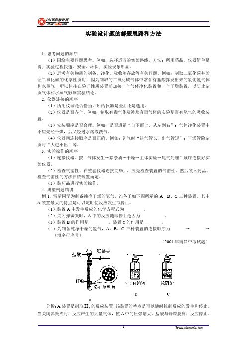
4. 典型例题精讲例1. 雪晴同学为制备纯净干燥的氢气,准备了如下图所示的A、B、C三种装置。
其中A装置最大的特点是可以随时使反应发生或停止。
(1)装置A中发生反应的化学方程式为_________。
(2)关闭弹簧夹时,A中的反应随即停止是因为___________。
(3)装置B的作用是_________,装置C的作用是________。
(4)为制备纯净干燥的氢气,A、B、C三种装置的连接顺序为_______→_______→_______(填字母序号)(2004年南昌中考试题)分析:A装置是制取H2的反应装置,该装置的特点是可以随时控制反应的发生和停止。
当关闭弹簧夹时,反应产生的大量气体,使A中的压强增大,盐酸与锌粒脱离,反应停止。
科学实验的设计思路
科学实验的设计思路在科学的探索之旅中,科学实验就像是一把神奇的钥匙,能够帮助我们打开未知世界的大门,揭示事物的本质和规律。
而一个成功的科学实验,其关键在于精心设计的思路。
这不仅需要我们有敏锐的观察力和强烈的好奇心,更需要有严谨的逻辑和系统的方法。
首先,明确实验目的是设计思路的基石。
我们需要清晰地知道自己想要解决什么问题,或者想要验证什么样的假设。
这就好比我们在旅行前要确定目的地一样,只有明确了方向,才能规划出正确的路线。
例如,如果我们想研究某种药物对某种疾病的治疗效果,那么实验目的就是评估这种药物是否能够有效缓解症状、改善患者的健康状况,以及探究其可能的作用机制。
如果实验目的不明确,就会像在黑暗中摸索,不仅浪费时间和资源,还可能得出错误的结论。
有了明确的目的,接下来就要提出合理的假设。
假设是基于已有的知识和经验,对实验结果的一种推测。
它为实验设计提供了一个初步的框架。
比如,在上述药物研究的例子中,我们可能假设这种药物能够通过抑制某种生物通路来发挥治疗作用。
假设的提出既要大胆创新,又要有一定的依据,不能凭空想象。
同时,我们也要做好假设不成立的准备,因为科学的探索往往是在不断的否定和修正中前进的。
实验变量的选择和控制是实验设计中至关重要的环节。
变量可以分为自变量、因变量和控制变量。
自变量是我们主动改变或操纵的因素,因变量是随着自变量的变化而产生相应变化的结果,控制变量则是那些可能影响实验结果但我们希望保持恒定的因素。
以植物生长实验为例,如果我们想研究光照时间对植物生长高度的影响,光照时间就是自变量,植物的生长高度就是因变量,而土壤质量、温度、湿度等则是需要控制的变量。
通过精确地选择和控制变量,我们可以更准确地揭示自变量和因变量之间的关系。
样本的选择也是不容忽视的一点。
样本要具有代表性和随机性,这样才能保证实验结果能够推广到更广泛的群体。
如果我们研究的是某种新型教学方法对学生成绩的影响,那么选择的样本应该涵盖不同学习能力、不同背景的学生,而不能只局限于某个特定的班级或群体。
小学生如何正确进行科学实验的设计与思考
小学生如何正确进行科学实验的设计与思考科学实验是小学生学习科学知识和培养科学思维的重要途径之一。
正确进行科学实验的设计与思考不仅可以帮助小学生更好地理解科学原理,还能培养他们的观察、思考和解决问题能力。
本文将介绍小学生如何正确进行科学实验的设计与思考。
首先,科学实验的设计需要明确实验目的。
小学生在进行科学实验之前,首先要明确实验的目的是什么,想要通过实验了解或验证什么知识点。
例如,可以设计一个实验来观察和了解植物的生长过程,或者验证不同材料的导电性能。
其次,科学实验的设计需要明确实验的步骤。
根据实验目的,小学生需要列出实验所需的材料和步骤,确保实验的可重复性和可评估性。
例如,在观察植物生长的实验中,小学生需要准备种子、土壤和水等材料,并且明确种子的播种、浇水和放置的步骤。
在进行科学实验的过程中,小学生还需要注意实验的条件控制。
为了保证实验结果的准确性,小学生需要尽量控制实验条件的一致性。
例如,在进行导电性实验时,如果使用的导线长度不同,结果可能会受到影响。
因此,小学生需要尽量保证实验条件的统一,只改变一个变量进行实验。
与此同时,小学生在进行科学实验时应该注重安全。
在进行实验前,小学生应该了解实验过程中可能存在的风险,并采取相应的安全措施。
例如,如果实验中涉及使用化学试剂或火源,小学生应该戴上实验手套和护目镜,并在老师或家长的指导下进行。
在科学实验的过程中,小学生还需要记录观察结果并进行数据分析。
小学生可以使用图表、文字或图片等形式来记录实验的观察结果,并分析这些结果是否与自己的预期相符。
例如,在植物生长实验中,小学生可以记录每天观察到的植物的生长情况,并绘制出折线图或柱状图来展示植物的生长变化。
科学实验的设计与思考并不是一次性的过程,小学生需要通过实验的反思和总结来提高自己的科学能力。
在实验结束后,小学生可以回顾整个实验的过程,思考实验中遇到的问题和解决方法,并分析实验结果的合理性和可靠性。
此外,小学生还可以尝试设计自己的科学实验。
小学科学实验设计与思考

小学科学实验设计与思考科学实验是小学科学教育中非常重要的一部分,通过实验可以让学生亲自动手、观察、实践,培养他们的观察力、实验能力和解决问题的能力。
在小学科学实验设计中,我们应该注重培养学生的思考能力和创新意识。
本文将探讨小学科学实验设计与思考的相关问题。
一、科学实验设计的基本原则科学实验设计需要遵循一定的原则,才能保证实验的可靠性和有效性。
首先,实验设计要有明确的目的和问题。
学生在设计实验时,应该明确自己的目的是什么,要解决什么问题。
其次,实验设计要有可操作性。
学生在设计实验时,要考虑到实验材料和设备的可获得性和可操作性,确保实验能够顺利进行。
再次,实验设计要有可比性。
学生在设计实验时,要考虑到控制变量的重要性,只有在变量控制得当的情况下,才能比较实验结果的差异。
最后,实验设计要有可重复性。
学生在设计实验时,要考虑到实验的可重复性,只有实验能够被其他人重复进行,才能验证实验结果的可靠性。
二、科学实验设计的步骤科学实验设计包括以下几个步骤:观察现象、提出问题、做实验、记录数据、分析数据、得出结论。
首先,学生需要观察周围的自然现象,发现问题。
例如,观察到水在不同温度下的沸腾点不同,就可以提出问题:水的沸腾点与温度有关吗?然后,学生需要设计实验来验证自己的问题。
例如,可以将水分别加热到不同的温度,观察水的沸腾点。
接下来,学生需要记录实验数据,例如记录每个温度下水的沸腾点。
然后,学生需要分析实验数据,比较不同温度下水的沸腾点的差异。
最后,学生可以得出结论,回答自己的问题:水的沸腾点与温度有关。
三、科学实验设计中的思考科学实验设计中的思考是培养学生科学思维和创新意识的重要环节。
首先,学生在设计实验时需要思考如何提出一个有挑战性的问题。
一个好的问题可以激发学生的思考和研究兴趣。
例如,学生可以思考如何设计一个实验来验证植物对光的需求量。
其次,学生在设计实验时需要思考如何控制变量。
变量的控制是保证实验结果可靠性的关键。
化学实验设计思路如何合理设计实验流程与参数

化学实验设计思路如何合理设计实验流程与参数化学实验是学习和应用化学知识的重要环节之一,而合理设计实验流程和参数则是实验成功的关键。
本文将探讨如何合理设计化学实验的思路以及实验流程和参数的合理选择。
一、思路合理设计1. 确定实验目的:在设计化学实验之前,首先要明确实验目的。
实验目的可以是检验某一化学原理的有效性、验证某一假设或推导某一公式等,只有明确了实验目的,才能更好地设计实验方案。
2. 参考相关文献:在实验设计之前,可以参考相关科学文献,了解已有的实验方法和结果,从而为实验设计提供有力的依据。
相关文献的参考还可以帮助我们了解实验材料和仪器的可行性以及可能出现的问题。
3. 确定实验步骤:在设计实验步骤时,需要根据实验目的和实验材料的特性来确定。
一般来说,实验步骤可以按照时间顺序排列,确保实验的逻辑性和连贯性。
4. 考虑安全问题:在进行化学实验时,安全问题是至关重要的。
在设计实验流程时,要合理考虑实验材料的危险性,选择适当的防护措施,确保实验过程的安全性。
二、合理设计实验流程1. 准备实验材料:在进行化学实验之前,需要准备实验所需的各种材料,包括试剂、溶剂、仪器设备等。
实验材料的准备要根据实验的目的和步骤进行合理选择,并确保其质量和纯度。
2. 检查实验仪器:在实验之前,要对实验所需的仪器进行仔细检查,确保其正常工作。
如有需要,还可以进行标定和调整,以保证实验的准确性和可重复性。
3. 确定实验条件:在实验设计中,需要根据实验的目的和需要确定实验的条件,包括温度、压力、浓度等。
实验条件的选择应该合理,能够满足实验的要求,并且易于实施和控制。
4. 设计实验控制对照组:在进行化学实验时,常常需要设置对照组以进行对比分析。
因此,在实验设计中要充分考虑对照组的选择和设计,确保实验结果的准确性和可靠性。
三、合理选择实验参数1. 温度控制:温度是化学反应速率的重要影响因素之一,因此在设计实验时要合理选择温度参数。
(完整版)实验设计的思路与步骤

(完整版)实验设计的思路与步骤引言实验设计是科学研究中至关重要的一环,它的合理性和严谨性直接影响到实验结果的可靠性。
在本文中,我们将详细介绍实验设计的思路和步骤,希望能够帮助读者进行有效的实验设计。
实验设计的思路实验设计的思路可以总结为以下几点:1. 确定研究目的在进行实验设计之前,我们首先需要明确研究的目的是什么。
研究目的可以是探索某个现象的原理,验证某个理论的有效性,或者寻找改进某个产品性能的方法等。
明确研究目的有助于我们在实验设计中有针对性地选择实验因素和设置实验方案。
2. 确定实验因素实验因素是指在实验中可以被操作和改变的变量,它们会对实验结果产生影响。
确定实验因素的关键在于全面考虑可能影响实验结果的各种因素,并将其转化为实验因素。
实验因素可以分为自变量和因变量,其中自变量是被实验者操作的变量,而因变量是受实验因素影响而改变的变量。
3. 设计实验方案在设计实验方案时,我们需要考虑如下几个方面:a. 控制变量为了保证实验结果的可靠性和准确性,我们需要控制实验中除了自变量之外的其他因素。
这可以通过设定实验组和对照组、随机分配实验对象等方式来实现。
控制变量有助于消除其他因素对实验结果的干扰。
b. 确定实验样本在实验设计中,我们需要确定实验所涉及的样本数量。
样本数量的确定需要根据实验目的、实验因素和统计学方法进行合理估计。
样本数量的大小直接影响到实验结果的可靠性以及对总体的推广程度。
c. 确定实验方法实验方法是指在实验过程中具体操作的步骤和技术。
在确定实验方法时,我们需要考虑实验操作的可行性、时间成本、资源限制等因素。
合理的实验方法有助于提高实验的精确性和效率。
4. 分析实验数据实验设计完成后,我们需要对实验数据进行分析,以得出准确的结论。
在分析实验数据时,我们可以采用统计学方法进行数据处理和结果的验证。
统计学方法可以帮助我们了解实验结果的可靠性和有效性。
实验设计的步骤实验设计的具体步骤可以总结为以下几点:1. 确定研究目的。
高三化学总结化学实验中的实验设计与数据分析思路

高三化学总结化学实验中的实验设计与数据分析思路在高三化学中,实验是一项重要的学习活动,通过实验可以培养学生的观察、实验设计和数据分析能力。
本文将总结化学实验中的实验设计与数据分析思路,并探讨其重要性。
一、实验设计思路在进行化学实验时,实验设计是十分重要的环节,一个好的实验设计能够确保实验的准确性和可靠性。
以下是一些实验设计的思路:1.明确实验目的和预期结果在进行实验之前,首先需要明确实验的目的和预期结果。
实验目的可以是验证一个化学原理、分析一个化学性质,或者合成某种物质等。
预期结果则指在实验条件下,我们期望观察到的现象或得到的数据。
2.选择合适的实验方法和仪器设备根据实验的目的和预期结果,选择合适的实验方法和仪器设备是非常重要的。
不同实验方法和仪器设备适用于不同的实验目的和预期结果。
例如,如果我们的实验目的是测定溶液的浓度,那么我们可以选择使用滴定法或分光光度法等。
3.控制实验条件和实验参数在进行实验前,需要仔细考虑实验条件和实验参数的选择。
实验条件包括环境温度、压力、湿度等,而实验参数则包括反应物质的种类和比例、反应时间、反应温度等。
合理控制实验条件和实验参数可以保证实验结果的准确性和可重复性。
4.安全性考虑在进行化学实验时,安全性是至关重要的。
必须时刻注意实验过程中的安全问题,采取有效的安全措施,例如佩戴安全眼镜、手套等,避免接触有毒、腐蚀性物质。
另外,实验过程中还需妥善管理实验废液和废品,避免对环境造成污染。
二、数据分析思路实验数据的分析是实验结果的重要组成部分,通过对数据的合理处理和分析,可以得出有意义的结论。
以下是一些数据分析的思路:1.数据收集和整理在实验过程中,准确地记录和收集实验数据是非常重要的。
需要将实验数据整理成表格或图表的形式,以便进行后续分析和对比。
此外,还可以进行数据的标准化处理,以确保数据的可比性。
2.数据可视化和描述对于实验数据,可以使用图表的形式进行可视化展示,例如柱状图、曲线图等。
化学实验设计思路如何合理设计实验方案

化学实验设计思路如何合理设计实验方案化学实验的设计是一个重要的环节,它涉及到实验的目的、步骤、材料选择、变量控制等多个方面。
一个合理的实验设计能够确保实验结果的准确性和可靠性。
本文将探讨如何合理设计化学实验方案,并提供一些建议和思路。
1. 实验目的的明确在设计化学实验方案之前,首先要明确实验的目的。
实验目的应该具体明确,并与所学的化学知识和课程内容相符合。
清晰的实验目的有助于指导实验的设计,确保实验的目标能够得到实现。
2. 实验步骤的合理性实验步骤的设计应该合理,并按照一定的逻辑顺序进行。
具体来说,实验步骤应该按照从简单到复杂的顺序进行,以确保学生能够逐步掌握实验技能和相关知识。
此外,实验步骤的顺序应该符合化学反应的反应速率和相关条件的要求。
3. 材料的选择与准备在实验方案中,选择和准备适当的材料是非常重要的。
材料的选择应该符合实验的要求,并且能够提供可靠的数据和结果。
同时,材料的准备要求要细致入微,确保实验中每个材料的质量和纯度。
4. 控制变量的重要性在化学实验中,正确地控制变量可以确保实验结果的可靠性和可重复性。
因此,在实验方案中应该明确并控制对实验结果影响较大的变量。
同时,需要注意的是,在改变和控制变量的过程中,要确保实验的安全性和实施的可行性。
5. 安全措施的考虑化学实验属于高风险的实践活动,因此在设计实验方案时,应该充分考虑实验的安全性。
合理的安全措施包括但不限于使用个人防护装备、控制危险物质的使用量、合理安排实验室操作时间等。
实验方案中应该明确指出相应的安全措施并确保实验操作过程中能够严格遵守。
6. 数据处理和结果分析设计实验方案时,需要考虑实验数据的处理和结果的分析。
合理的数据处理方法和结果分析方法有助于得出结论,并对实验结果进行科学的解释。
因此,在实验方案中应该明确指出数据处理和结果分析的方法,并合理使用相关的统计分析方法。
综上所述,化学实验的设计要合理考虑实验目的、步骤、材料选择、变量控制、安全措施、数据处理和结果分析等多个方面。
初中化学实验设计思路范文

初中化学实验设计思路第一篇范文:初中化学实验设计思路化学实验是初中教育中不可或缺的一部分,它能够帮助学生更好地理解化学知识,培养他们的实验操作能力和科学思维。
本文将从以下几个方面阐述初中化学实验设计的思路。
一、实验目标明确在设计初中化学实验时,首先要明确实验的目标。
实验目标应与教学大纲和课程内容紧密结合,确保学生在实验过程中能够掌握相关的化学知识和技能。
例如,在进行“酸碱中和反应”的实验时,实验目标可以设定为让学生了解酸碱中和反应的原理,掌握中和反应的实验操作方法。
二、实验原理透彻在实验设计过程中,教师需要对实验原理进行深入研究,确保实验过程能够准确地反映出化学反应的本质。
例如,在设计“铁丝在氧气中燃烧”的实验时,教师应详细研究铁丝燃烧的化学反应方程式,了解燃烧过程中各元素的转化关系,以便在实验中能够准确地观察和解释现象。
三、实验步骤详细实验步骤应详细明了,便于学生按照要求进行实验操作。
在实验步骤中,应包括实验材料的准备、实验仪器的摆放、实验操作的顺序以及实验数据的记录等方面。
例如,在进行“制作二氧化碳气泡”的实验时,实验步骤可以分为以下几个部分:1.准备实验材料:澄清石灰水、醋、气球等。
2.摆放实验仪器:烧杯、滴管、澄清石灰水、醋等。
3.实验操作:将醋滴入澄清石灰水中,观察气泡的产生。
4.记录实验数据:观察气泡的形状、大小、颜色等特征。
四、实验现象生动实验现象是学生最容易感知和记住的部分,因此在实验设计中应注重实验现象的生动性和直观性。
通过观察实验现象,学生可以更好地理解化学反应的原理。
例如,在设计“电解水”的实验时,可以通过观察氢气和氧气的生成,让学生深刻理解电解水的原理。
五、实验安全可靠实验安全是化学实验设计中必须考虑的重要因素。
在设计实验时,应确保实验过程安全无害,避免使用有毒、易燃、易爆等危险品。
同时,教师还需加强对学生的安全教育,确保学生在实验过程中能够遵循安全操作规程。
例如,在进行“制取氧气”的实验时,应确保实验操作过程中不会引起火灾或爆炸。
化学实验设计解题思路和方法
化学实验设计解题思路和方法化学实验设计解题思路和方法实验设计题主要考查学生对实验仪器的应用能力、实验原理的迁移能力和实验方案的选择能力。
其基本原则是:原理正确、操作简便、现象明显、药品经济、安全可靠。
其基本解题思路和方法是:一. 思考问题的顺序1. 围绕主要问题思考。
例如,选择适当的实验路线、方法;所用药品、仪器简单易得;实验过程快速、安全;实验现象明显。
2. 思考有关物质的制备、净化、吸收和存放等有关问题。
例如,抽取在空气中易潮的物质时,往往在装置末端再接一个干燥装置,以防止空气中水蒸气进入。
3. 思考实验的种类及如何合理地组装仪器,并将实验与课本实验比较、联靠。
例如,实验室用加热氯化铵和碱石灰的固体混合物制取氨气,可通过迁移实验室制取O2的实验原理,选用与制O2的装置制取NH3,用向下排空气法收集NH3。
二. 仪器连接的顺序1. 所用仪器是否恰当,所给仪器是全用还是选用。
2. 仪器是否齐全。
例如,制有毒气体及涉及有毒气体的实验是否有尾气的吸收装置。
3. 安装顺序是否合理。
例如,安装仪器是否遵循“自下而上,从左到右”;气体净化装置中不应该先经干燥,后又经过水溶液洗气。
4. 仪器间连接顺序是否正确。
例如,洗气时“进气管长、出气管短”;干燥管除杂质时“大进小出”等。
三. 实验操作的顺序1. 连接仪器。
按“气体发生→除杂质→干燥→主体实验→尾气处理”顺序连接好实验仪器。
2. 检查气密性。
在整套仪器连接完毕后,应先检查装置的气密性,然后装入药品。
检查气密性的方法要依装置而定。
3. 装药品进行实验操作。
例:在实验室里制氧气时常用氯酸钾做原料,用二氧化锰做催化剂,根据催化剂的涵义,二氧化锰的质量和化学性质都不发生改变。
试设计一个实验,证明二氧化锰在氯酸钾分解前后的质量不变,说明实验程序和主要操作步骤。
分析:要证明二氧化锰在氯酸钾分解前后质量不变,就必须测定两个质量,一个是加到反应器中的MnO2的质量,一个是反应后剩余固体中的MnO2的质量。
初中化学实验设计思路分析

初中化学实验设计思路分析化学实验是化学学习中的重要环节,能够帮助学生巩固理论知识,培养实验技能和科学思维。
在初中化学实验设计中,我们需要考虑实验的安全性、操作性和教育性,以确保实验的有效性和教学效果。
本文将从实验设计思路的分析、实验目的的明确、实验条件的准备等几个方面展开讨论。
一、实验设计思路的分析在考虑实验设计时,首先需要分析实验的目的和内容,确定实验的步骤和方法。
例如,在学习溶液的制备和浓度计算时,可以设计酸碱中和反应的实验。
通过控制反应物的摩尔比例和反应条件,让学生了解溶液的配制和酸碱反应的特点。
二、实验目的的明确在实验设计中,明确实验的目的是十分重要的。
实验目的要求明确具体,能够与课堂所学的理论知识相结合,让学生在实践中加深对理论的理解。
以酸碱反应实验为例,可以明确实验的目的是探究酸碱中和反应的化学方程式、计算方程式中溶液的浓度等。
三、实验条件的准备在进行化学实验时,实验条件的准备是非常重要的。
首先需要准备好所需的实验设备和试剂,确保实验过程的顺利进行。
其次,要对实验的安全措施进行充分考虑,保证实验的安全进行。
例如,在酸碱反应实验中,需佩戴安全眼镜、实验手套等,以保护实验人员的安全。
四、实验步骤的设计实验步骤的设计要简洁明了,使学生能够清楚地理解和掌握实验的操作过程。
可以将实验步骤按照时间顺序进行排列,确保学生能够正确地操作实验设备和试剂。
在酸碱中和实验中,可将实验步骤设计为:配制酸碱溶液、称取反应所需的试剂、反应过程的观察和记录等。
五、实验数据的处理在化学实验中,数据处理是一个重要的环节。
通过对实验数据的处理和分析,能够帮助学生得出结论,并加深对理论知识的理解。
在酸碱中和实验中,可以通过实验数据的处理,计算出反应的摩尔比例和浓度,进一步说明溶液的制备和计算方法。
六、实验结果的呈现在进行化学实验后,需要将实验结果进行呈现。
可以通过文字描述、表格、图表等形式进行展示,并对结果进行分析和解释。
小学科学实验设计思路培养教学反思

小学科学实验设计思路培养教学反思一、科学实验设计的重要性科学实验设计是小学科学教学中不可或缺的一环。
通过实验设计,学生能够亲自动手进行实验操作,观察和体验科学现象,培养科学思维和动手能力。
因此,科学实验设计对于培养学生的科学素养具有重要意义。
二、培养学生的科学思维1. 引导学生提出科学问题为了培养学生的科学思维,科学实验设计的第一步是引导学生提出科学问题。
例如,可以通过观察、调研或者背景知识引发学生的好奇心,激发他们提出精确、明确的科学问题。
2. 鼓励学生进行科学探究和推理科学实验设计要鼓励学生进行科学探究和推理。
通过提供合适的材料和实验条件,学生可以自主设计实验并进行验证。
在实验过程中,鼓励学生观察、记录和分析数据,并通过推理和总结得出科学结论。
3. 培养学生的创新思维科学实验设计还要培养学生的创新思维。
鼓励学生提出新颖的实验方案和创新的解决方法。
同时,也要在实验设计过程中给予学生一定的自由度,让他们有机会发挥创意和想象力。
三、实验设计过程中的教学反思1. 赋予学生更多的自主权在实验设计过程中,教师要适度放手,赋予学生更多的自主权。
尽量不事先给出解决方案,而是通过引导学生分析问题、制定实验方案,并充分讨论和交流,鼓励学生独立思考和解决问题。
2. 鼓励学生合作与分享实验设计不仅仅是单个学生的任务,更应该是学生之间的合作与分享。
教师可以组织学生进行小组合作,在小组内共同讨论和设计实验方案。
通过合作与分享,可以促进学生之间的交流与学习,提高实验设计的质量。
3. 注意实验安全与教学效果的平衡在实验设计中,安全始终是重要的因素之一。
教师要引导学生注重实验的安全措施,并提前预防可能的危险。
但是,过分追求安全可能会削弱实验教学的效果。
因此,在实验安全与教学效果之间要做一个平衡,确保学生的实验体验和学习效果。
四、实验设计的评估与反馈教师在学生完成实验设计后,可以进行评估与反馈。
这有助于学生对实验设计的认识和提高,并能够及时纠正不足之处。
初中化学实验探究设计与思考

初中化学实验探究设计与思考化学实验是初中化学教学中不可或缺的一部分,通过实际操作,学生可以亲身体验化学现象,加深对化学原理的理解。
然而,仅仅进行实验并不能达到最佳的教学效果,探究设计与思考是化学实验的重要环节。
本文将探讨初中化学实验的探究设计和思考过程,并提供一些实验设计的思路。
一、探究设计的重要性探究设计是指在进行化学实验时,通过设计一系列的实验步骤和问题,引导学生主动思考、探索和发现。
相比于传统的“教师说,学生听”的教学方式,探究设计能够激发学生的主动性和创造性,培养他们的科学思维和实验技能。
此外,探究设计还能够增强学生对化学知识的记忆和理解,提高他们的问题解决能力和分析能力。
二、实验设计的思路1.确定实验目的和问题在进行实验设计时,首先需要明确实验的目的和问题。
实验目的可以是验证某个化学原理,探究某个化学现象的原因,或者解决某个实际问题。
问题的提出需要具有启发性和挑战性,能够引导学生进行思考和实验探究。
2.选择实验方法和步骤在确定实验目的和问题后,需要选择适当的实验方法和步骤。
实验方法应该简单明了,容易操作,能够达到预期的实验效果。
实验步骤应该清晰具体,包括所需材料、操作顺序和注意事项等。
3.设计实验变量和对照组在进行实验设计时,需要考虑实验变量和对照组的设置。
实验变量是指在实验过程中被改变的因素,对照组是指与实验组相比较的参照物。
通过设置实验变量和对照组,可以排除其他因素的干扰,准确地观察和分析实验结果。
4.收集和分析实验数据在进行实验时,需要准确地记录实验数据,并进行数据的分析和比较。
可以使用表格、图表等形式整理和展示实验数据,通过对数据的分析,可以得出结论和解答实验问题。
5.思考和总结实验结果实验结束后,需要对实验结果进行思考和总结。
可以回答实验问题,解释实验现象,探讨实验结果的意义和应用。
同时,也可以提出新的问题和进一步的实验探究方向。
三、实验设计的案例以下是一个初中化学实验设计的案例,以探究酸碱中和反应的速率为例。
- 1、下载文档前请自行甄别文档内容的完整性,平台不提供额外的编辑、内容补充、找答案等附加服务。
- 2、"仅部分预览"的文档,不可在线预览部分如存在完整性等问题,可反馈申请退款(可完整预览的文档不适用该条件!)。
- 3、如文档侵犯您的权益,请联系客服反馈,我们会尽快为您处理(人工客服工作时间:9:00-18:30)。
实验设计思路及思考问题?--异体类血提取PRF的性能对比1.首先现在所用的都是自体血中提取到的PRF,但现在考虑到伦理问题或者来源量有限的问题所以考虑用异体的动物血。
Q但问题是异体血存在immune compatibility 问题以及如果证明各方面都合适要制成成品后要如何保存?2.Q:其实还是自体血好吧~那除了用异体血来代替能不能通过PCR 的方法进行增量?3.各种血中(同样是10ML)可提取到的PRF量的比较--PRF提取到的体积或者重量的百分比比较。
Ps:多取样求平均使用的是glass coated plastic tube4.各种血中提取到的PRF质的比较--PRF中各种有用成分的含量比较Ps:有用成份主要来自α-platelet ( 主要是一些生长因子)Q:如果要比较的话是要用什么方法?A:用分子生物学的方法--电泳?有一篇文章是这样的。
或者用免疫学的方法-Elisa/IMMUNOSORBENT ASSAY KITS?300 mins after colt formation detect GF in PRF releasates in the supernatant serum.5.对于性能的分析方向/检测项目:①组织学染色--(有好多种)体内培养后处死然后切片检测② SEM 扫描的纤维结构体外第一次提取之后就可以直接检测fibrin network architecture③细胞学(使用流式细胞仪检测)进行细胞计数主要是体外,体外就在培养瓶/皿里面培养细胞+ 上面覆盖PRF 然后看对于细胞的proliferation+ differentiation的影响具体方法参考Q 哪些是主要的功能细胞?A: WBC+ fibroblasts+ osteoblastes+ osteoclasts+ periosteal(骨膜)cells.etc4. 抗菌性/抑菌性:可以检测其MIC?可以参照其他组实验5. 观察软组织形成情况--体内可以放入后切片或者直接表观观察比如进行牙周检查项目(BI/ BOP 等)Ps:此处的软组织主要指gingival tissue6. ALP activity7. 检测not only bone formation but also bone to implants contact 体内实验/从临床学上检测8. 检测mineralization (有好多种)5.对于制备PRF的离心参数问题不一样:主流的是用glass coated plastic tube centrifuge 2700rpm for 12 min6.数据处理的问题--用Tukey’s test 以及对数据处理结果的表示问题(柱状图<突出数量对比>还是曲线图<突出时间变化>要具体情况具体分析)7.联想--骨刺的发病机制是什么?能否从中或者一些osteoinductive/ bone regeneration 的方法/切入点8.PRF 对四种细胞的调控--Dermal prekeratinocytes from earGingival fibroblasts from alveolar ridgePreadipocyte from inner face of kneeOB from mandibular boneQ: 选取这些地方作为取材源是因为这里细胞多?为什么不是原位取?这样能说明其真正的clinical effects to imitation.参考文献的内容要点总结:体内实验:24只新西兰雄兔(8-10个月大)拔除上中切牙后填充PRF后缝合后在1,2,3,4周后分别进行组织学放射学检查。
组织学检查方法1Premaxilla were resected cutting till the end of dental canal and all soft tissue removed then fixed in 10% buffered formalin solution 48hours to fix it. And then 10% formic acid 10-15days 每48小时换液以脱钙。
长流水冲洗24小时后通过series of alcohol 以脱水,然后每5微米切片用HE染色观察计数osteoblasts/osteoclasts/bone trabecular number and trabecular width/ trabecular separation/cortical width/ vessel number/ bone marrow space volume.Results:2w--apposition of extracellular osteoid matrix surrounded by osteoblasts3w--primitive bone formation with feature of woven bone.4w--bone trabecular filled the cervical socket covered with hazy epithelial tissue, immature bone fibrous connective tissue with fibroblast cells and numerous capillaries.PS : coronal / middle/ apical三区取材GF released from PRF periodically( per 7days) includes IL-1β/ IL-6/ TNF-α/TGF-β1/ PDGF/EGF/VEGF stimulating cell migration and differentiation.详细作用机制见笔记本组织学检查方法2Bone fragment fixed in formal dehydesolution and then put it in series alcohol until well-dehydrate and then immerse into methylmethacry resin.After that, the sample is stained by two methods--PAS and Masson trichrome and then we can observe them under light microscope--mineralized bone trabecular( blue in Pas and green in Masson ) osteoid border ( red in PAS and Masson) medullary space( orange-pink in Pas and pink in Masson)SEM 方法:To detect the fibrin network architecture of PRF membrancePRF is fixed in 2.5% 戊二醛内1小时以脱水然后切割,表面sputter-coated with 20nm gold photographs were taken at 15-25KV using 15-3500 magnifications.细胞培养方法--体外培养如何取材?A:干细胞可以取自小鼠胫骨骨髓内OB were isolated from calvariae of 1-day-old sprague-dawley rats.Cell culture condition 在培养皿中上面覆盖PRF(30 culture plates with D=60mm and 20000 cells per plate 很难定量吧①standard condition: incubation at 37℃+ 5%CO2 + 高糖DMEM+ 1% antibiotics (penicillin-streptomycin)+ glutamine( 200mm)+ FCS 10%②differentiation condition: incubation at 37℃+ 5%CO2 + 高糖DMEM+ 1% antibiotics(penicillin-streptomycin)+ glutamine( 200mm)+ FCS 10% + Vc( 50 微克/ml)+ B-glycerophosphate( 10 mmol/L) + dexamethasone (0.1微毫升/L)Calvariae were minced and incubated at room temperature for 20mins with gentle shaking of an enzymatic solution containing 0.1% collagenase,0.05% trypsin 4mmol/l Na2-rthylenediaminetetra acetic acid in calcium and PBS.MEM containing 10% FBS and antibiotics (100mg penicillin +100 IU/ml of streptomycin 37℃培育箱with 95% air 5% co2 100% relative humidity.OB pretreat in α-MEM supplemented with 10% FBS, 100ug/ml streptomycin and 100 U/ml penicillin for 24hours. Then they were cultured in α-MEM supplemented with 1% FBS 100ug/ml streptomycin 100U/ml penicillin for another 24 hours before use.MTT assay: proliferation assessmentAbsence of cytotoxityCell suspension was diluted to the concentration of 5000 cells/ml, 200微升of cells suspension was seeded into a 96 well tissue culture plate.( control group and test group). Cells were placed in the incubator for 24hours to obtain monolayer cell growth.And then rinse it with PBC ( phosphate buffer saline solution) 2 times.This experiment were repeated 3 times to ensure reproducibility.The spectrophotometric absorbance( optical density) was read at 540nm using a microplate reader.Mineralization nodules : differentiation assessmentThree series was fixed with a neutral phosphate buffered 2% glutaraldehyde solution during 1hour at 4 ℃.A Vonkossa staining was then performed to detect mineralization nodules--dark brown/ black spots.PS: large nodules was counted in the 3 cm2 middle in the culture plate.Mineralization assessment 2:After 2 weeks culture, cells were washed 3 times with PBS ( ph= 7.4)then stained with 0.5% alzarin reds in H2O PH 4.0 for 1h at room temperature.And after staining, wash cells 3 times with H2O followed by 70% ethanol and then culture cells in 100mmol/l cety pyridinium chloride 1 hour waiting for solution and release. Ca-bonding alizarin reds 进入溶液中可以测吸光度。
