局解总结
(完整word版)局部解剖学重点总结
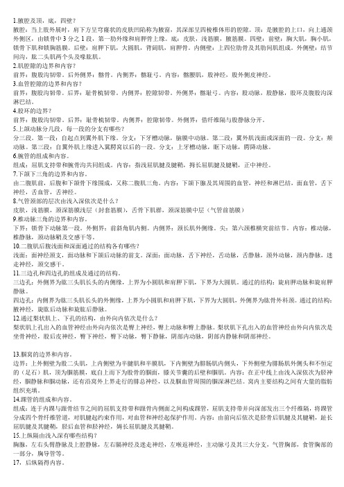
1.腋腔及顶,底,四壁?腋腔:当上肢外展时,肩下方呈穹窿状的皮肤凹陷称为腋窗,其深部呈四棱椎体形的腔隙。
顶:是腋腔的上口,向上通颈外侧区,由锁骨中3分之1段,第一肋外缘和肩胛骨上缘。
底:皮肤,浅筋膜,腋筋膜。
四壁:前壁:胸大肌,胸小肌,锁骨下肌和锁胸筋膜。
后壁:肩胛下肌,大圆肌,背阔肌,肩胛骨。
内侧壁:上四位肋骨及其肋间肌组成。
外侧壁:结节间沟,肱二头肌两个头及喙肱肌。
2.肌腔隙的边界和内容?前界:腹股沟韧带。
后外侧界:髂骨。
内侧界:髂耻弓。
内容:髂腰肌,股神经,股外侧皮神经。
3.血管腔隙的边界和内容?前界:腹股沟韧带。
后界:耻骨梳韧带。
内侧界:腔隙韧带。
外侧界:髂耻弓。
内容:股动脉,股静脉,股环及腹股沟深淋巴结。
4.股环的边界?前界:腹股沟韧带。
后界:耻骨梳韧带。
内侧界:腔隙韧带。
外侧界:借纤维隔与股静脉分开。
5.上颌动脉分几段,每一段的分支有哪些?分三段。
第一段:自起点到翼外肌下缘。
分支:下牙槽动脉,脑膜中动脉。
第二段:翼外肌浅面或深面的一段。
分支:颊动脉。
第三段:自翼外肌上缘进入冀腭窝以后的一段。
分支:上牙槽动脉,眶下动脉,腭降动脉。
6.腕管的组成和内容。
组成:屈肌支持带和腕骨沟共同组成。
内容:指浅屈肌腱及腱鞘,拇长屈肌腱及腱鞘,正中神经。
7.下颌下三角的边界和内容。
由二腹肌前,后腹和下颌骨下缘围成,又称二腹肌三角。
内容:下颌下腺及其周围的血管,神经和淋巴结,面血管,舌下神经,舌血管,舌神经。
8.气管颈部的层次由浅入深依次是什么?皮肤,浅筋膜,颈深筋膜浅层(封套筋膜),舌骨下肌群,颈深筋膜中层(气管前筋膜)9.椎动脉三角的边界和内容。
下界:锁骨下动脉第一段。
外侧界:前斜角肌内侧。
内侧界:颈长肌外侧缘。
尖:第六颈椎横突前结节。
内容:椎动脉,椎静脉,颈动脉鞘及交感干等。
10.二腹肌后腹浅面和深面通过的结构各有哪些?浅面:面神经颈支,面动脉和下颌后动脉的前支。
深面:面动脉,舌下神经,舌动脉,舌静脉,颈外动脉,颈内静脉,迷走神经,颈交感干。
司法局人民调解工作总结

发现问题与不足
分析工作中遇到的问题和不足,为今 后的工作提供改进方向。
交流经验
总结有助于不同地区、不同部门之间 交流人民调解工作的经验和做法,促 进共同进步。
提升工作水平
通过对比以往工作,可以激励工作人 员不断提高自身的业务水平和综合素 质。
工作总结的时间和范围
时间范围
本次总结的时间范围为XXXX年XX月XX日至XXXX年XX月XX日。
激励和鼓舞全局同志继续努力,为人民调解事业做出更大
的贡献。
表彰先进典型
我们将对在工作中表现突出的调解员进行表彰和奖励,树立榜样 ,激励全局同志向他们学习。
加强团队建设
通过举办团队建设活动,增强团队凝聚力和向心力,形成团结、 奋进的工作氛围。
宣传人民调解事业
加大人民调解工作的宣传力度,提高公众对人民调解工作的认知 度和认同感,增强调解员的荣誉感和使命感。
范有序进行。
加强人民调解员队伍建设
选拔培训
严格选拔调解员,确保他们具备 法律知识和调解技能,并定期进 行培训,提高调解员的业务水平
和综合能力。
职业道德
加强调解员职业道德建设,树立公 正、公平、公信的形象,确保调解 工作的公正性和权威性。
激励保障
完善调解员的激励保障机制,包括 薪酬制度、晋升渠道、保险福利等 ,激发调解员的工作积极性和责任 心。
• 邻里纠纷:XX件,占总案件的XX%。 主要包括噪音扰民、占用公共空间、 宠物管理等。
案件分类
• 婚姻家庭纠纷:XX件,占总案件的 XX%。主要涉及夫妻矛盾、家庭暴力 、财产分割等问题。
人民调解成功率统计
调解成功率:成功调解案件数占 总案件数的XX%。
• 调解员的专业能力和经验:调 解员具备丰富的法律知识和调 解技巧,能够有效沟通并引导 当事人达成共识。
局解知识总结2.0版(更正版)
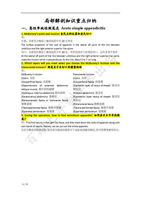
局部解剖知识重点归纳一、急性单纯性阑尾炎Acute simple appendicitis1. McBureny’s point and incision麦氏点的位置和麦氏切口答:位置:在脐至右髂前上棘连线的中外1/3交界处The surface projection of the root of appendix is the lateral 1/3 point of the line between umbilicus and the right anterior superior iliac spine.切口:从脐到右髂前上棘连线的中外1/3处,所作的垂直于此线的切口,长约5到7厘米At the lateral 1/3 point of the line between umbilicus and the right anterior superior iliac spine, make the incision which is perpendicular to this line. About 5 to 7 cm long.2. Which layers will you meet when you choose the McBureny’s incision and the transcrectal incision? 阑尾炎手术切口的腹壁结构答:McBureny’s incision(1)skin 皮肤(2)superficial fascia 浅筋膜(3)aponeurosis of extermal abdominal oblique muscle 腹外斜肌腱膜(4)obliquusinternusabdominis腹内斜肌(5)transversusabdominis腹横肌(6)transversalis fascia or transverse fascia 腹横筋膜(7)extraperitoneal fascia 腹膜外筋膜(8)parietal peritoneum 壁腹膜Transcrectal incision(1)skin 皮肤(2)superfacial fascia 浅筋膜(3)anterior layer of rectus of sheath 腹直肌鞘前层(4)rectus abdominis腹直肌(5)posterior layer rectus of sheath 腹直肌鞘后层(6)transverse fascia腹横筋膜(7)extra-peritoneal fascia 腹膜外筋膜(8)parietal peritoneum 壁腹膜3. During the operation, how to find vermiform appendix? 如何在手术中寻找阑尾?答:First find cecum in the right iliac fossa, and then track down the roots of appendix along with colic bands of cecum. Namely, we can put out the whole appendix.先在右髂窝内找到盲肠,再沿着盲肠的结肠带往下追踪找到阑尾根部,即可将整条阑尾牵出。
局解实验报告实验总结
一、实验目的1. 了解人体局部解剖学的结构和功能;2. 培养学生观察、分析、归纳的能力;3. 提高学生对人体解剖学的认识和兴趣。
二、实验时间与地点时间:2021年X月X日地点:XX大学解剖实验室三、实验对象男性尸体一具四、实验内容1. 头部解剖:观察颅骨、脑、血管、神经等结构;2. 颈部解剖:观察颈椎、甲状腺、血管、神经等结构;3. 上肢解剖:观察肩关节、肘关节、腕关节、肌肉、血管、神经等结构;4. 躯干部解剖:观察胸壁、胸膜、肺、心脏、血管、神经等结构;5. 下肢解剖:观察髋关节、膝关节、踝关节、肌肉、血管、神经等结构。
五、实验步骤1. 头部解剖:(1)将头部皮肤、肌肉、脂肪层等软组织剥离;(2)观察颅骨、脑、血管、神经等结构;(3)用剪刀剪开颅骨,观察脑的结构;(4)观察颈动脉、椎动脉、颈内静脉等血管结构;(5)观察颅神经、颈神经等神经结构。
2. 颈部解剖:(1)将颈部皮肤、肌肉、脂肪层等软组织剥离;(2)观察颈椎、甲状腺、血管、神经等结构;(3)观察颈动脉、椎动脉、颈内静脉等血管结构;(4)观察颈神经、迷走神经等神经结构。
3. 上肢解剖:(1)将上肢皮肤、肌肉、脂肪层等软组织剥离;(2)观察肩关节、肘关节、腕关节等关节结构;(3)观察肱二头肌、肱三头肌、三角肌等肌肉结构;(4)观察肱动脉、桡动脉、尺动脉等血管结构;(5)观察正中神经、桡神经、尺神经等神经结构。
4. 躯干部解剖:(1)将躯干部皮肤、肌肉、脂肪层等软组织剥离;(2)观察胸壁、胸膜、肺等结构;(3)观察心脏、血管、神经等结构;(4)观察肋间神经、胸神经等神经结构。
5. 下肢解剖:(1)将下肢皮肤、肌肉、脂肪层等软组织剥离;(2)观察髋关节、膝关节、踝关节等关节结构;(3)观察大腿肌肉、小腿肌肉等肌肉结构;(4)观察股动脉、腘动脉、胫前动脉、胫后动脉等血管结构;(5)观察坐骨神经、腓总神经等神经结构。
六、实验结果与分析1. 头部解剖:观察到了颅骨、脑、血管、神经等结构,了解了它们的形态和功能;2. 颈部解剖:观察到了颈椎、甲状腺、血管、神经等结构,了解了它们的形态和功能;3. 上肢解剖:观察到了肩关节、肘关节、腕关节等关节结构,了解了它们的形态和功能;观察到了肱二头肌、肱三头肌、三角肌等肌肉结构,了解了它们的形态和功能;观察到了肱动脉、桡动脉、尺动脉等血管结构,了解了它们的形态和功能;观察到了正中神经、桡神经、尺神经等神经结构,了解了它们的形态和功能;4. 躯干部解剖:观察到了胸壁、胸膜、肺等结构,了解了它们的形态和功能;观察到了心脏、血管、神经等结构,了解了它们的形态和功能;观察到了肋间神经、胸神经等神经结构,了解了它们的形态和功能;5. 下肢解剖:观察到了髋关节、膝关节、踝关节等关节结构,了解了它们的形态和功能;观察到了大腿肌肉、小腿肌肉等肌肉结构,了解了它们的形态和功能;观察到了股动脉、腘动脉、胫前动脉、胫后动脉等血管结构,了解了它们的形态和功能;观察到了坐骨神经、腓总神经等神经结构,了解了它们的形态和功能。
司法局调解员个人工作总结

调解员个人工作总结时光荏苒,岁月如梭。
转眼间,一个学年已接近尾声。
作为一名调解员,我深感责任重大,使命光荣。
回顾过去的一年,我始终坚持“调解为民”的宗旨,努力做好本职工作,积极参与调解工作,取得了一定的成绩。
现将个人工作总结如下:一、学习法律知识,提高业务能力作为一名调解员,我深知法律知识的重要性。
在过去的一年里,我认真学习了国家法律法规,特别是关于调解工作的相关法律知识。
通过学习,我不断提高自己的业务能力,为更好地服务群众奠定了基础。
二、积极参与调解,维护社会和谐在工作中,我积极参与各类调解案件,努力化解矛盾纠纷。
今年共参与了50余起案件的调解工作,其中成功调解40余起,调解成功率达到80%以上。
在调解过程中,我始终坚持以事实为依据,以法律为准绳,公正公平地处理案件。
同时,我还注重发挥调解工作的优势,积极引导当事人双方化解矛盾,达到和谐共赢的目的。
三、注重调解方法,提高调解效果在调解工作中,我注重运用多种调解方法,根据案件的特点和当事人的需求,灵活运用调解技巧。
在处理婚姻家庭纠纷时,我注重从感情入手,引导当事人珍惜家庭幸福;在处理财产纠纷时,我注重公平公正,确保当事人双方的合法权益得到保障。
此外,我还注重与当事人建立良好的沟通,倾听他们的诉求,关心他们的困难,以真情实感打动他们的心灵。
四、加强沟通协调,形成工作合力在调解工作中,我注重与相关部门的沟通协调,形成工作合力。
在与公安、法院等部门的协作中,我积极向他们学习,不断提高自己的调解水平。
同时,我还加强与镇村两级调解组织的联系,共同化解矛盾纠纷,为维护社会和谐稳定做出了贡献。
五、总结经验教训,不断提高自己在调解工作中,我不断总结经验教训,认识到自己的不足之处,并努力改进。
在处理一些复杂案件时,我认识到单靠调解员的力量是有限的,需要发动社会各界力量共同参与。
因此,我在工作中注重调动各方积极性,形成合力,共同化解矛盾纠纷。
总之,过去的一年,我在司法局领导和同事们的关心支持下,取得了一定的成绩。
局部解剖知识点总结

局部解剖知识点总结一、身体表面解剖身体表面解剖是解剖学的基础,它主要研究人体表面的各种标志物和表面标志,以及表面解剖和内部解剖的关系。
在学习身体表面解剖的时候,要理清楚肌肉、骨骼和器官在体表的表现形式,熟练掌握各种部位的解剖标志。
比如腋窝、胸骨下缘、肱三头肌区、髌骨上部等。
此外,还要了解体表各个部位的测量方法及其临床应用。
二、头部解剖头部解剖主要研究头颅、面部、颈部等区域的解剖结构。
头颅是保护脑部的重要组成部分,它主要由颅骨和颅面骨组成。
颅骨有颅顶骨、颞骨、颧骨、枕骨等组成,颅面骨主要有上颌骨、下颌骨等。
面部主要由眼、鼻、口等组成,颈部主要是通过颈椎与头部相连。
在学习头部解剖的时候,需要了解头颅和颅面骨的名称、数量、形态以及各个部分的关系,掌握头颅颅面骨的解剖标志以及各种孔窗、突起的位置和功能。
三、胸部解剖胸部是人体的重要部位,它主要包括胸骨、肋骨、胸骨软骨等组成。
在学习胸部解剖的时候,要理清楚胸骨、肋骨、胸骨软骨、锁骨以及肋间肌、膈肌等肌肉的位置和形态。
此外,还应了解胸部的器官,比如心脏、肺脏、气管、食管等器官的位置和结构。
四、腹部解剖腹部是人体的消化系统和泌尿系统的主要部位,它包括腹膜、腹腔、腹壁、腹内器官等。
在学习腹部解剖的时候,要了解腹壁的层次结构,包括皮肤、皮下组织、腹直肌、腹内斜肌和腹横肌等,掌握各种解剖标志的位置和形态,以及腹腔内器官的位置和结构。
五、四肢解剖四肢是人体的重要运动部位,它主要包括上肢和下肢。
在学习四肢解剖的时候,要了解上肢和下肢的骨骼、肌肉、关节、血管、神经等结构,掌握各种部位的解剖标志,熟练掌握四肢的基本动作和功能,以及四肢与躯干的连接方式。
六、神经系统解剖神经系统解剖主要研究中枢神经系统和周围神经系统的结构与功能。
中枢神经系统包括大脑、小脑、脑干和脊髓,周围神经系统包括脑神经和脊神经。
在学习神经系统解剖的时候,要了解各种脑叶、脑室、脑神经的出口和入口,脊髓的各段的特点以及脊神经的部位和分布。
局解学习小结

头面部
1. 面部皮肤、肌肉的神经分布
2. 穿腮腺的结构
3. 腮腺床
4. 额顶枕区和颞区的层次结构及颅顶部的“危险区”
颈部
1、颈部的筋膜间隙
2、颈动脉三角、
3、肌三角、甲状腺(被膜、从皮肤到甲状腺经过的层次、甲状腺的动脉与神经的位置关系)纵膈
胸部
1、肋间隙穿过的结构排列
2、膈上三个孔的名称及位置
3、熟悉乳房的淋巴回流
4、胸膜腔、壁胸膜的四部分肺和胸膜下界的体表投影
5、肺根内结构的排列
6、食管上、下三角、动脉导管三角、心包、心包横窦及斜窦
7、上纵隔、后纵隔的结构
8、纵膈侧面观
腹部
1、腹部不同位置切口的层次
2、腹股沟管、腹股沟三角
腹腔血管
1.胃的毗邻、动脉供应及来源
2.肝蒂结构的位置关系
3.Calot三角及胆总管的分段
4.肝门静脉的属支
5.十二指肠和结肠的分部
腹膜后隙
1、肾的位置和毗邻
2、肾的被膜、肾蒂结构的排列
3、输尿管
盆部和会阴、脊柱区
1、盆筋膜间隙
2、坐骨直肠窝
3、会阴部两个三角
4、会阴浅隙、会阴深隙
5、腰上三角、腰下三角、听诊三角
6、背部浅层肌
上肢
1、腋窝四壁
2、腋动脉分段、臂丛主要分支、腋窝淋巴结五群。
3、肌腱袖、三边孔、四边孔
4、肘窝的边界及内容
5、前臂前区四组血管神经束的组成
6、腕管的组成及穿过的结构
7、手掌筋膜间隙及手部神经分布
8、掌浅弓、掌深弓
下肢
1梨状肌上、下孔及其穿行的结构
2、大隐静脉注入股动脉之前接受五大属支
3、肌腔隙、血管腔隙、股三角、收肌管、腘窝、踝管。
局部解剖学 的总结
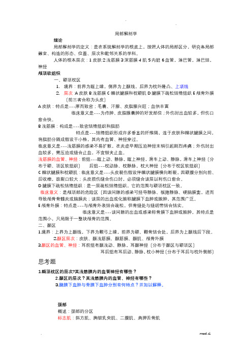
局部解剖学绪论局部解剖学的定义:是在系统解剖学的根底上,按照人体的局部区分,研究各局部器官、构造的形态、位置、层次和毗邻关系的学科。
人体的根本层次:1皮肤2浅筋膜3深筋膜4肌5内脏6血管,淋巴管,淋巴结,神经颅顶软组织一、额顶枕区1.境界:前界为眶上缘,侧界为上颞线,后界为枕外隆凸、上项线2.层次:A皮肤B浅筋膜C帽状腱膜和枕额肌D腱膜下疏松结缔组织E颅骨外膜〔前三者合称为头皮〕A皮肤:特点是---厚而致密;毛囊、汗腺、皮脂腺兴旺;血供丰富临床意义是---为疖肿、皮脂腺囊肿的好发部位;外伤时出血较多,但伤口愈合快。
B浅筋膜:构成是---致密结缔组织和脂肪特点是---结缔组织形成许多垂直的纤维隔,连于皮肤和帽状腱膜之间,将脂肪分隔成假设干小格,其内有血管、神经穿过。
临床意义是---浅筋膜的感染不易扩散,在炎症早期压迫神经末梢引起剧烈疼痛;外伤时出血较多,需压迫或缝合止血,不宜钳夹止血。
浅筋膜的血管、神经:前组---眶上动、静脉,眶上神经,滑车上动、静脉,滑车上神经〔分布于额、顶区软组织〕后组---枕动脉、枕静脉,枕大神经〔分布于枕区软组织〕C帽状腱膜和枕额肌:临床意义是---头皮裂伤假设伴帽状腱膜横向断裂,因额腹分别向前、后收缩,故裂口较大;头皮损伤缝合伤口时,必须缝合该层以利伤口愈合。
D腱膜下疏松结缔组织:是一层疏松结缔组织,它的范围与额顶枕区一致。
临床意义:是颅顶部的危险区〔因该间隙的感染可经导静脉、板障静脉、硬脑膜窦,进而导致颅骨骨髓炎或脑膜炎;该层的出血或化脓称腱膜下血肿或脓肿,其范围广泛。
E颅骨外膜:特点是---与颅骨外表结合疏松,但骨缝处与缝纫带结合结实。
临床意义是---该间隙的出血或感染称骨膜下血肿或脓肿,其特点是范围小,只局限于一整块颅骨的范围。
二、颞区1.境界:上界为上颞线,下界为颧弓上缘,前界为额、颧骨结合处,后界为上颞线后下段。
2.颞区层次:皮肤、颞浅筋膜、颞筋膜、颞肌、颅骨外膜3.颞区的血管、神经:耳前组有颞浅动、静脉,耳颞神经〔分布于颞区与额顶区〕耳后组有耳后动、静脉,枕小神经〔分布于耳后与枕外侧部〕思考题1.额顶枕区的层次?其浅筋膜内的血管神经有哪些?2.颞区的层次?其浅筋膜内的血管、神经有哪些?3.腱膜下血肿与骨膜下血肿分别有何特点?并加以解释。
局解重点总结范文
局解重点总结范文首先,局解重点总结是在分析和解决问题时非常重要的一个环节。
通过对问题进行局解,可以帮助我们更好地理解问题的本质和原因,从而制定出更有效的解决方案。
其次,在进行局解重点总结时,需要注意以下几个方面。
首先是对问题的全面性进行考虑,不能只从一个角度出发,而是要从多个角度综合考虑问题的各个方面。
其次是要注意问题的实质,不要被表面现象所迷惑,要深入分析问题的本质和原因。
再次是要注重问题的关联性,要考虑问题与其他问题之间的关系,以及问题与环境的相互作用。
最后是要有足够的逻辑思维能力和分析能力,通过分析问题之间的逻辑关系和因果关系,找出问题的关键要点。
首先,可以采用分析法来找出关键要点。
分析法是指将问题进行逐步分解和分析,从而找出问题的关键要点。
通过将问题进行分解,可以将复杂的问题变得简单明了,从而更容易找出关键要点。
其次,可以采用对比法来找出关键要点。
对比法是指将问题与其他相关问题进行对比,通过对比找出问题的关键要点。
通过对比不同问题之间的差异和相似之处,可以更好地理解问题的本质和原因,从而找出关键要点。
再次,可以采用逻辑思维来找出关键要点。
逻辑思维是指通过分析问题之间的逻辑关系和因果关系,找出问题的关键要点。
通过分析问题的逻辑关系和因果关系,可以更准确地找出问题的关键要点。
最后,可以采用归纳法来找出关键要点。
归纳法是指通过对问题进行归纳和总结,找出问题的关键要点。
通过将问题进行归纳和总结,可以将问题的复杂性降低,从而更容易找出关键要点。
总之,局解重点总结是在分析和解决问题时非常重要的一个环节。
通过对问题进行全面、实质、关联和逻辑的分析,可以找出问题的关键要点,从而确定解决问题的重点方向。
在进行局解重点总结时,可以采用分析法、对比法、逻辑思维和归纳法等方法和技巧,来帮助我们更好地找出关键要点。
只有找出了问题的关键要点,才能制定出更有效的解决方案。
局解操作心得体会7篇

局解操作心得体会7篇大家要知道富有真情实感的心得体会才能得到读者们的认可的,随着社会的发展,我们有越来越多的机会需要写心得体会,以下是作者精心为您推荐的局解操作心得体会7篇,供大家参考。
局解操作心得体会篇1作为一个刚刚来到学校不久的老师,就要成为班主任,想想内心还是有很大的压力的。
从上学期接到要当班主任的通知到现在培训结束,我的内心从开始的慌乱紧张,渐渐的有了一些自信。
通过这次培训,让我多多少少的了解了我们学校班主任工作的一些内容,增长了自信。
六月二十八号下午,刚刚来到杭州,就是一场大雨,给紧张的心里增添了几分压抑,加上大多数老师都不认识,当时的心情有些紧张。
在简短的开班仪式后,就开始了第一堂课——《班级团体心理辅导技术》。
这节课由高亚兵老师给我们讲解。
没想到上来就是一节心里课,没有什么班主任工作的条条框框。
这样的切入不免让我紧张的心里有了一些放松。
在接下来的几天里,分别有六位专家学者给我们讲授了班主任日常工作的经验方法。
这些专家有的从专业的学科层次讲解班主任工作的理论方法,有的则从自己多年的工作经验,自己的切身体会,动情的讲解在从事班主任工作时的一些感人故事。
当然,除了这些令我深受佩服的专家们的经验分享,给我留下更加深刻印象的是我们本校的前辈们,他们的一些故事才是真正触动心灵的分享,让我感到,在我身边的平凡岗位上的不平凡的前辈们。
在听过各位前辈们毫无保留的分享之后,不仅在班主任日常工作方法的理论层面上重新认识的班主任工作的内涵,更从我校各位班主任的分享中,在实践层面给了我启示。
通过本次学习,使我认识到作为一个班主任要做到以下几个方面:一、做一个有爱的班主任郑州信息技术学校作为一个传承一百多年的学校,文化底蕴丰富。
听了各位前辈的分享,我深深体会到了,在这样一个学校,班主任应该是一个带着爱心的老师的重要性。
中职类学校学生入学成绩低是一个事实,这也让许多老师轻描淡写的推卸责任说生源差是学校不出承接的原因。
局解重点归纳整理

头颈部名词解释:1.头皮:皮肤、浅筋膜(皮下组织)、帽状腱膜及颅顶肌(额,枕肌)三层合称头皮。
2.颅顶腱膜下间隙(腱膜下疏松结缔组织):位于帽状腱膜与骨膜之间的薄层疏松结缔组织。
头皮借此层与颅骨外膜疏松连接,故移动性大,开颅时可经此间隙将皮瓣游离后翻起,头皮撕脱伤也多沿此层分离。
3.腮腺床:腮腺的深面与茎突诸肌及深部血管神经相邻。
这些肌、血管神经包括颈内动、静脉,舌咽、迷走、副神经及舌下神经共同形成“腮腺床”。
4.面部危险三角:面静脉与颅内的海绵窦借多条途径向交通,因此面部感染有向颅内扩散的可能,尤其是口裂以上两侧口角至鼻根的三角形区域,感染向颅内扩散的可能性更大,被称为“危险三角区”。
5.封套筋膜:即颈筋膜浅层。
向上附于头颈交界处,向下附于颈、胸和上肢交界线,向前在颈前正中线处左、右相延续,向两侧包绕斜方肌和胸锁乳突肌并形成两肌的鞘,向后附于项韧带和第7颈椎棘突,形成完整的封套结构。
6.胸骨上间隙:封套筋膜在距胸骨柄上缘约3~4cm处,分为深浅两层,向下分别附于胸骨柄前、后缘,两层之间为胸骨上间隙。
内有颈静脉弓,颈前静脉下段、胸锁乳突肌胸骨头、淋巴结及脂肪组织等。
7.咽后间隙:位于椎前筋膜与颊咽筋膜之间,其延伸至咽外侧壁的部分为咽旁间隙。
8.气管前间隙:位于气管前筋膜与气管颈部之间。
内有甲状腺最下动脉、甲状腺下静脉和甲状腺奇静脉丛等。
小儿还有胸腺上部、左头臂静脉和主动脉弓等。
9.椎前间隙:位于脊柱、颈深肌群与椎前筋膜之间。
颈椎结核脓肿多积于此间隙,并经腋鞘扩散至腋窝。
10.颈动脉鞘:颈动脉鞘上起自颅底,下续纵隔。
鞘内全长有颈内静脉和迷走神经,鞘内上部有颈内动脉,下部为颈总动脉。
在颈动脉鞘下部,颈内静脉位于前外侧,颈总动脉位于后内侧,在二者之间的后外方有迷走神经。
鞘的上部,颈内动脉居前内侧,颈内静脉在其后外方,迷走神经行于二者之间的后内方。
11.锁骨上大窝:又名锁骨上三角,位于锁骨上方,在体表呈明显凹陷。
局解实验心得体会5篇

局解实验心得体会5篇(经典版)编制人:__________________审核人:__________________审批人:__________________编制单位:__________________编制时间:____年____月____日序言下载提示:该文档是本店铺精心编制而成的,希望大家下载后,能够帮助大家解决实际问题。
文档下载后可定制修改,请根据实际需要进行调整和使用,谢谢!并且,本店铺为大家提供各种类型的经典范文,如述职报告、调研报告、策划方案、活动方案、心得体会、应急预案、规章制度、教学资料、作文大全、其他范文等等,想了解不同范文格式和写法,敬请关注!Download tips: This document is carefully compiled by this editor. I hope that after you download it, it can help you solve practical problems. The document can be customized and modified after downloading, please adjust and use it according to actual needs, thank you!Moreover, our store provides various types of classic sample essays, such as job reports, research reports, planning plans, activity plans, personal experiences, emergency plans, rules and regulations, teaching materials, complete essays, and other sample essays. If you want to learn about different sample formats and writing methods, please pay attention!局解实验心得体会5篇优秀的心得体会是我们成长道路上的重要经验总结,通过反思自己的行动,我们能够从中汲取深刻的心得体会,下面是本店铺为您分享的局解实验心得体会5篇,感谢您的参阅。
局解实验心得体会7篇
局解实验心得体会7篇局解实验心得体会篇1?教育部高职高专规划教材:工程数学(建工类)》包括了线性代数、概率论、数理统计的基本内容,还介绍了matlab和sas,2个软件系统,8个数学建模问题,18个数学实验,66个建工专业的例题与习题。
这本教材是湖南省普通高等教育面向21世纪教学内容和课程体系改变计划立项项目的成果之一.编者以再设计的思想,按照高职高专工科基础课内容以应用为目的,以必需够用为度的原则,全面审视了工程数学传统的教学内容,以及当代科学技术的发展水平和前景,提出了[基础理论]+[数学建模]+[数学软件]三大模块有机结合的工程专科数学教学内容的设计方案,并以此编成了这本书.它有以下3个特点:1.充分注意了工程数学基础理论的重要地位.全书以2/3的篇幅介绍了建工类高职高专学生所必需的线性代数、概率与数理统计方面的基础知识,仅删去一些烦琐的证明、神奇的运算技巧和少数几个概念.2.强调以培养创新精神和应用能力为重点的指导思想.介绍了matlab和sas 2个软件系统,讨论了8个数学建模问题,列出了18个数学实验,有66个例题或习题具有鲜明的建工类专业色彩,使学生能感受到工程氛围,注意基础知识用于工程实践,并能在建模训练中培养探索、创新能力.3.内容处理新颖.本书在强调数学概念与基础理论的基础上,进行了6个方面的渗透:(1)渗透数学在工程技术中应用的实例;(2)渗透数学建模思想;(3)渗透数学实验方法;(4)渗透数学软件应用;(5)渗透经济效益意识;(6)渗透科学思维方法.这样,三大模块有机结合起来,互相渗透,融为一体,成为一个新的课程体系.这种体系以数学知识为基础,实际问题为背景,数学建模为手段,数学软件为工具,既有利于教学手段、教学方法的改变,更有利于学生素质的综合提高。
本书大部分内容在湖南城建高等专科学校试讲多年,编者做过大量的跟踪调查,召开座谈会、调查会,与会人数累计上百人次,问卷调查不下千人,收集读书报告(或数学学习心得)600多份.这些调查充分证明,本书的内容设计与讲述方法,有利于提高学生的应用能力,有利于培养学生的数学意识,而且在后续课程学习中,数学知识也基本够用.这本书是为房屋建筑工程、道路桥梁、给水排水、规划设计、风景园林、工程造价、房地产管理等建工类专业的高职高专学生编写的,也可供其他专业的高职高专学生和教师参考.讲授本书内容约需50~70课时,目录中打*号的可作选学.本书是湖南城建高等专科学校信息工程系数学教研室集体研究的成果.李天然副教授担任主编,张新宇、田罗生两位副教授担任副主编,参编人员分工如下:李天然编写第三、四、十一、十二章,张新宇编写第六、八章,田罗生编写第一、二章,龚卫明副教授编写第九、十章,龙韬讲师编写第五章,李俊锋讲师编写第七章.此外,何孟义教授、金庆华副教授、彭德权副教授、肖劲松讲师、郭冰阳讲师等也参加了本书大部分内容的教学研究. --此文字指本书的不再付印或绝版版本。
局部解剖重点

局解知识点总结:1. 肱动脉的体表投影;肱动脉的 3 个分支:肱动脉的体表投影:上肢外展 90 度,手掌向上,由锁骨中点至肘前横纹终点远侧2cm处的连线,为腋动脉和肱动脉的体表投影。
二者以大圆肌下缘为界,大圆肌下缘以上为腋动脉,以下为肱动脉。
肱动脉的 3 个分支:(1)肱深动脉:在大圆肌下缘的稍下方起于肱动脉后内壁,与桡神经一同经肱三头肌内侧头和外侧头之间转入臂后区的桡神经沟中。
(2)尺侧下副动脉:在肱深动脉起点稍下方自肱动脉发出,陪伴尺神经穿过内侧肌间隔行向内上髁背侧面,与尺侧返动脉和尺侧下副动脉符合。
(3)尺侧上副动脉:在肱骨内上髁上方约 5 厘米处起于肱动脉,散布于内上髁的前,后边,参加肘关节动脉网的构成。
上肢动脉的骨干:锁骨下动脉 - 腋动脉 - 肱动脉 - 桡动脉、尺动脉 - 掌浅弓、掌深弓大隐静脉在隐静脉裂孔周边的五条属支是:旋髂浅静脉、腹壁浅静脉、阴部外静脉、股内侧浅静脉、股外侧浅静脉注意上肢的深静脉和浅静脉都有哪些,怎么走形!2.腹部的体表标记1.肋弓下缘: 8-10 肋软骨连结形成的肋缘和第 11-12 浮肋构成,用于腹部分区、肝脾丈量和胆囊定位2.剑突:胸骨下端软骨,肝脏丈量标记3.腹上角:双侧肋弓至剑突根部的交角,判断体型及肝脏丈量。
4.脐:腹部中心,投影至 3-4 腰椎之间,为腹部四分区法的标记,易有脐疝发生。
5.髂前上棘:髂棘前方突出点,腹部九分区法标记,骨髓穿刺部位6.腹直肌外缘:位于锁骨中线处,用于手术切口和胆囊点定位。
7.腹中线:腹部四分区法的垂直线,易有白线疝。
8.腹股沟韧带:找寻股动脉、股静脉的标记,常为腹股沟疝的经过部位和所在。
9.耻骨联合:两耻骨间的纤维软骨连结。
10.肋脊角:背部双侧第 12 肋骨与脊柱的交角,是检查肾脏压、叩痛的地点。
3.腹部的分区(一)四区法:即十字型法,以脐为中心划一水平线和一垂直线,两线订交,把腹部分红四区(二)九区法井字型分区,用两条水平线和两条垂直线将腹部分红九个区,上水平线又称肋线,为双侧肋缘最低点(相当于第十肋骨)的连线;下水平线又称髂棘线,为双侧髂前上棘的连线;左、右两条垂直是在髂前上棘至腹正中线的水平线的中点上所作的垂直线。
(完整版)局部解剖学总结+分区超级整理
局部解剖学笔记整理层次结构类一,额顶枕区:(冠状面至脑实质)1、皮肤,2、浅筋膜,3、帽状腱膜(1,2,3合称头皮),4、帽状腱膜下疏松结缔组织,5、颅骨外膜,6、颅骨外板,7、板障,8、内板,9、硬膜外腔,10、硬脑膜,11、硬膜下隙,12、蛛网膜,13、蛛网膜下腔,14、软膜,15、脑实质二,颞区:1、皮肤,2、浅筋膜,3、颞浅筋膜,4、颞深筋膜,5、颞肌,6、颅骨外膜,7(后同额顶枕区)三,手掌掌心部:1、皮肤,2、浅筋膜,3、掌腱膜,4、掌浅弓及其分支,5、正中神经及尺神经分支,6、四条指浅屈肌肌腱及腱鞘,7、指深屈肌肌腱及腱鞘(7的桡侧:四条蚓状肌发出到达2345指背腱膜),8、掌中隔,9、掌中间隙,鱼际间隙,10、外侧骨间伸肌肌腱及内侧骨间伸肌及肌腱,11、掌深弓,12、指间掌侧肌3块,指间背侧肌4块,13、掌骨四,腮腺咬肌区:1、皮肤,2、浅筋膜,3、腮腺鞘(颈封筋膜向上延续及咬肌筋膜构成),4、腮腺浅部,5、峡部,6、下颌支,7、深部五,阴囊及睾丸的被膜:1、皮肤,2、浅筋膜(肉膜,有平滑肌),3、精索外筋膜,4、提睾肌,5、精索内筋膜,6、睾丸固有鞘膜的壁层,腔隙,内脏层(腹膜形成的结构),7、白膜六,腹壁:1、皮肤,2、浅筋膜(脐以上一层,脐以下分两层,浅层脂肪层camper筋膜和深层的膜层scarpa 膜层),3、腹外斜肌及腱膜,4、腹内斜肌,5、腹横肌,6、腹横筋膜,7、腹外筋膜,8、壁腹膜七,胸壁:1、皮肤,2、浅筋膜,3、深筋膜浅层,4、胸大肌,5、深筋膜深层,6、胸小肌,7、肋及肋间隙,8、胸内筋膜,9、壁胸膜床类:组织器官深面的结构一,腮腺床:由三组结构组成:1、颈内血管(a v),2、起于茎突(乳突后内侧)的茎突舌骨肌,茎突舌肌,茎突咽肌,3、后四对脑神经:舌咽,迷走,副,舌下神经二,胃床:胃后壁,膈,脾,左肾上腺,左肾,胰,横结肠鞘类一,腮腺鞘:腮腺咬肌筋膜在腮腺前缘分成深浅两层包被腮腺形成。
局部解剖个人感想总结

局部解剖个人感想总结前言局部解剖作为医学中重要的基础课程之一,是学习医学知识的重要一环。
在学习和实践之后,我对局部解剖有了更深刻的理解和感受。
本文将通过对局部解剖的个人感想总结,探讨其对于医学学习的重要性以及对我个人的影响。
局部解剖的重要性1. 了解人体结构和组织局部解剖是研究人体构造和组织关系的基础,通过学习局部解剖,我们可以了解人体不同部位的结构特点、器官位置及其功能。
只有通过深入了解人体解剖,才能正确地评估和处理疾病以及进行手术操作。
2. 建立三维空间思维学习局部解剖需要我们通过二维图像进行三维空间思维的构建和理解。
这对于医学学生来说,是一项重要任务。
通过练习,我们可以逐渐掌握三维空间思维,提高对解剖结构的抽象和空间理解能力。
3. 综合运用多学科知识局部解剖不仅涉及解剖学,还需要综合运用生理学、病理学等多个学科的知识。
通过结合多学科知识,我们可以更好地理解人体不同部位的解剖结构与其功能的关联性,为日后的临床实践打下坚实的基础。
学习局部解剖的感受1. 探索身体奥秘的乐趣学习局部解剖的过程就如同一场身体的探险之旅,在不断探索和研究中,你会对自己的身体结构有更深入的了解。
了解身体的构造,不仅让我们对自己的身体有更多的认识,也增加了对生命的敬畏和对医学的热爱。
2. 培养耐心和细致的态度局部解剖的学习需要详细的观察和细致的分析,这对学生们的耐心和细致要求非常高。
在课堂上,我们需要认真听讲,用心观察和描绘每个解剖结构,这培养了我们仔细观察的能力,对于未来的临床实践非常重要。
3. 培养团队合作能力学习局部解剖时,我们往往需要在小组中进行解剖实验或讨论。
这样的学习方式强调团队合作和集体智慧,促进了学生之间的交流与合作,提高了我们的团队合作能力。
4. 锻炼空间想象能力学习局部解剖的过程中,我们经常需要通过二维图像还原三维空间结构。
这要求我们具备较强的空间想象能力。
通过不断练习,我们可以逐渐培养和提高空间想象能力,这对于个人的综合素质也有积极的影响。
局部解剖实验总结
局部解剖实验总结导言:一、实验目的:本次局部解剖实验的目的是帮助学生熟悉人体头,颈部和上肢的解剖结构,了解其组织和器官的位置关系,培养学生观察和分析的能力。
二、实验过程:在本次实验中,我们采用了有机解剖实验的方法,并配合使用了解剖模型和标本,以帮助学生更好地理解和记忆解剖结构。
1.头部解剖:在头部解剖实验中,我们首先观察了头皮,骨骼和肌肉的结构。
通过解剖模型的辅助,我们发现头部主要由颅骨和面部骨组成,头皮下有颈动脉和其他重要血管,头皮上覆盖着不同的肌肉。
在解剖实验中,我们还学习了头骨骨骼的各个构成部分,并观察了各个骨骼之间和其他组织的连接关系。
2.颈部解剖:在颈部解剖实验中,我们主要关注颈椎和颈部肌肉的结构。
我们通过解剖模型和标本观察到颈部有七个颈椎,呈现出一定的弧度。
颈部肌肉的覆盖不仅保护了颈椎,还参与了呼吸和咽喉的活动。
我们还学习了气管和食管的位置关系,理解了它们在颈部的解剖结构。
3.上肢解剖:在上肢解剖实验中,我们学习了上肢骨骼和肌肉的结构。
我们通过解剖标本和解剖模型,观察到上肢有胸骨、肩胛骨、肱骨和尺骨等骨骼组成。
肩部和手部的肌肉参与了上肢的运动和功能。
我们还学习了上肢动脉和静脉的分布,了解了它们在上肢的供血和排血功能。
三、实验收获:通过本次局部解剖实验,我们对头,颈部和上肢的解剖结构有了初步的了解。
通过实际操作和观察,我们更加直观地认识到人体解剖结构之间的相互关系,为后续学习提供了重要的基础。
在实验中,我们也培养了观察和分析的能力,提高了学习解剖学知识的兴趣和积极性。
四、实验反思:在本次实验中,我们发现对于初学者来说,人体解剖结构的复杂性和多样性是一种挑战。
在解剖实验中,我们可能会遇到一些困难,比如找不到特定的结构,或者难以理解结构之间的关系。
因此,我们需要更多的实践和指导来加深对解剖学知识的理解。
结语:通过本次局部解剖实验,我们不仅加深了对头部,颈部和上肢解剖结构的了解,还提高了实际操作和观察的能力。
(完整版)局部解剖学考试重点总结超级完整

第一章头颈部一、名词解释1、头皮:颅顶的额顶枕区皮肤、浅筋膜、帽状腱膜和枕额肌三层紧密附着,组成“头皮”。
2、面部“危险三角”:指两侧口角至鼻根连线所形成的三角区,此区的面静脉无静脉瓣,并经眼静脉与海绵窦交通。
若发生化脓性感染,易循上述途径逆行至海绵窦,导致颅内感染。
3、腮腺床:腮腺深面有起自茎突的诸肌、颈内动脉和静脉、第四—第七脑神经共同组成的腮腺床。
4、枕三角:由胸锁乳突肌后缘、斜方肌前缘和肩胛舌骨肌下腹围成。
内有副神经外支、淋巴结及颈丛皮支等。
5、下颌管:位于下颌体内,由下颌孔到颏孔的骨性管道,内有下牙槽动脉、静脉、神经。
6、颈动脉鞘:颈部深筋膜中层,气管前筋膜向两侧延续,包裹颈总动脉、颈内动静脉和迷走神经形成。
7、下颌下三角(二腹肌三角):由二腹肌前、后腹和下颌体下缘围成。
三角内有下颌下腺、下颌下淋巴结、面血管、舌血管、舌下神经和舌神经。
8、甲状腺假被膜:包裹甲状腺的气管前筋膜,即甲状腺鞘。
9、甲状腺悬韧带:在甲状腺两侧叶的内侧部和峡的后面甲状腺假被膜增厚形成甲状腺悬韧带,喉返神经在其后方上行,手术时应该注意保护。
10、颈袢:上根:自舌下神经发出,为来自第1颈神经前支的纤维,沿颈内动脉、颈总动脉下降。
下根:由颈丛的第2、3颈神经前支纤维组成,在颈内静脉内侧下行。
上下根在颈内静脉的后内侧或前外侧联合成颈袢。
11、肌三角:底为颈深筋膜深层,顶为颈深筋膜浅层。
三角内含有胸骨舌骨肌、胸骨甲状肌、甲状舌骨肌和肩胛舌骨肌上腹,以及气管前筋膜和位于其深部的甲状腺、气管颈部、食管颈部等结构。
12、气管前间隙:气管前筋膜与气管颈部之间借疏松结缔组织相连形成气管前间隙。
13、颞下颌关节:由下颌窝、关节结节、下颌头、关节盘、关节囊及韧带组成。
它是颌面部唯一既稳定又灵活的联合关节,参与完成咀嚼、吞咽、语言及表情等功能。
14、翼点:位于颞窝前下部,为额骨、顶骨、颞骨、蝶骨四骨汇合处,多呈“H”形,其内面有脑膜中动脉前支经过,翼点是颅骨的薄弱部位。
局解知识点总结期末
局解知识点总结期末
博弈论是局解的一个重要分支,研究的是对策论中的竞争和决策问题。
博弈论的基本思想
是通过对策分析,来求出最优的决策方案。
常见的博弈论模型包括零和博弈、合作博弈、
多人博弈等。
通过对策分析,可以得出最优的决策方案,从而在竞争和决策中取得更好的
效果。
最优化是局解的另一个重要分支,研究的是如何寻找问题的最优解。
最优化的问题可以是
线性规划、非线性规划、整数规划等。
通过对问题进行数学建模,可以利用最优化工具求
解出最优解,从而在实际问题中取得更好的效果。
概率论也是局解的重要应用领域,研究的是不确定性的问题。
概率论的基本思想是通过对
不确定性进行建模和分析,来求出问题的解决方案。
常见的概率论模型包括随机变量、概
率分布、统计推断等。
通过概率论的方法,可以对不确定性进行建模和分析,从而求解问
题的解决方案。
局解的基本思想是将不完全信息的问题转化为数学模型,然后通过数学方法来求解问题的
解决方案。
局解的方法和技巧可以应用到各种不完全信息的问题中,如金融投资、生产调度、市场竞争等。
通过局解的方法,可以更好地理解和分析不完全信息的问题,为决策提
供更好的支持。
总而言之,局解是一个非常重要的数学理论和方法,可以应用到各种不完全信息的问题中。
通过局解的方法和技巧,可以更好地理解和分析问题,并找到最优的解决方案。
局解的研
究既有理论意义,也具有实际应用价值,对于提高决策效果和解决复杂问题有着重要的意义。
局解实验心得体会5篇
局解实验心得体会5篇局解实验心得体会篇1一个长学期的电路原理,让我学到了很多东西,从最开始的什么都不懂,到此刻的略懂一二。
在学习知识上方,开始的时候完全是老师讲什么就做什么,感觉速度还是比较快的,跟理论也没什么差距。
但是之后就觉得越来越麻烦了。
从最开始的误差分析,实验报告写了很多,但是真正掌握的确不多,到最后的回转器,负阻,感觉都是理论没有很好的跟上实践,很多状况下是在实验出现象以后在去想理论。
在实验这门课中给我最大的感受就是,必须要先弄清楚原理,在做实验,这样又快又好。
在养成习惯方面,最开始的时候我做实验都是没有什么条理,想到哪里就做到哪里。
比如说测量三相电,有很多种状况,有中线,无中线,三角形接线法还是y形接线法,在这个实验中,如果选取恰当的顺序就能够减少很多接线,做实验就应要有良好的习惯,就应在做实验之前想好这个实验要求什么,有几个步骤,就应怎样安排才最合理,其实这也映射到做事情,不管做什么事情,就应都要想想目的和过程,这样才能高效的完成。
电原实验开始的几周上课时光不是很固定,实验报告也累计了很多,第一次感觉有那么多实验报告要写,在交实验报告的前一天很多同学都通宵了的,这说明我们都没有合理的安排好自我的时光,我就应从这件事情中吸取教训,合理安排自我的时光,完成就应完成的学习任务。
这学期做的一些实验都需要严谨的态度。
在负阻的实验中,我和同组的同学连了两三次才把负阻链接好,又浪费时光,又没有效果,在这个实验中,有很多线,很容易插错,所以要个性仔细。
在最后的综合实验中,我更是受益匪浅。
完整的做出了一个红外测量角度的仪器,虽然不是个性准确。
我和我组员分工合作,各自完成自我的模块。
我负责的是单片机,和数码显示电路。
这两块都是比较简单的,但是数码显示个性需要细致,由于我自我是一个粗心的人,所以数码管我检查了很多遍,做了很多无用功。
总结:电路原理实验最后给我留下的是:严谨的学习态度。
做什么事情都要认真,争取一次性做好,人生没有太多时光去浪费。
- 1、下载文档前请自行甄别文档内容的完整性,平台不提供额外的编辑、内容补充、找答案等附加服务。
- 2、"仅部分预览"的文档,不可在线预览部分如存在完整性等问题,可反馈申请退款(可完整预览的文档不适用该条件!)。
- 3、如文档侵犯您的权益,请联系客服反馈,我们会尽快为您处理(人工客服工作时间:9:00-18:30)。
脊柱区Muscles of BackSuperficial group–Trapezius 斜方肌–Latissimus dorsi 背阔肌–Levator scapulae 肩胛提肌–Rhomboideus 菱形肌Deep group–Splenius 夹肌–Erector spinae 竖脊肌Triangle of auscultation 听诊三角(肩胛旁三角)Boundaries•Latissimus dorsi•Trapezius•The medial border of thescapulaInferior lumbar triangle 腰下三角–Boundaries•Lower part of the lateralmargin of latissimusdorsi•Posterior free border ofthe obliquus externusabdominis•Iliac crestSuperior lumbar triangle腰上三角–Boundaries•Serratus posteriorinferior•Erector spinae•Obliquus internusabdomins上肢Cephalic vein 头静脉•Arises from the lateral side of the dorsal venous rete on the back of hand •Winds around the lateral border of the forearm; it then ascends into thecubital fossa and up the front of the armon the lateral side of the biceps.•It continues up in the deltopectoralgroove and then to the infraclavicularfossa, where it pierces clavipectoralfascia to drain into axillary vein. Basilic vein 贵要静脉•Arises from the medial side of the dorsal venous rete of hand•Winds around the medial border of the forearm•Then ascends into the cubital fossa and up the front of the arm on the medialside of the biceps to middle of the armwhere it pierces the deep fascia andjoins the brachial vein or axillary vein Median cubital vein 肘正中静脉•Links cephalic vein and basilic vein in the cubital fossa•It is a frequent site for venipuncture to remove a sample of blood or add fluidto the bloodAxillary fossa 腋窝Boundaries•Apex–Middle 1/3 of clavicle–Lateral border of first rib–Upper border of the scapula •Base–Skin–Superficial fascia–Axillary fascia 腋筋膜(cribriform fascia 筛状筋膜)•The medial wall–Serratus anterior–Upper four ribs and intercostalspaces•The lateral wall–Intertubercular groove–Long head and short head ofbiceps brachii–Coracobrachialis•The anterior wall–Pectoralis major–Pectoralis minor–Subclavius–Clavipectoral fascia 锁胸筋膜•The posterior wall–Subscapularis–Teres major–Latissimus dorsi–ScapulaContents•Axillary a. and principal branches•Axillary v. and tributaries•Brachial plexus and branches•Axillary lymph nodes•Loose connective tissue Trilateral foramen 三边孔(trilateral space 三边隙)•Boundaries–Superior: teres minorsubscapularislateral border of scapula,articular capsule of shoulder joint–Inferior: teres major–Lateral: long head of tricepsbrachii•Structures pass through the trilateral foramen–Circumflex scapular a. and v. Quadrilateral foramen 四边孔(quadrilateral space 四边隙)•Boundaries–Superior: teres minorsubscapularisarticular capsule of shoulder joint–Inferior: teres major–Medial: long head of tricepsbrachii–Lateral: surgical neck ofhumerus•Structures pass through the quadrilateral foramen–Axillary n.–Posterior humeral circumflex a.and v.Axillary artery•Begins at the at lateral border of first rib as a continuation of subclavianartery•At the lower border of teres major it becomes the brachial artery.•Divided into three parts by overlying pectoralis minorBranches•First part–superior thoracic a.–thoracoacromial a.•Second part–lateral thoracic a.•Third part– subscapular a.•throcodorsal a.•circumflex scapular a.–anterior humeral circumflex a.–posterior humeral circumflex a. Brachial artery 肱动脉–Begins at the lower border of theteres major as a continuation ofaxillary artery–Terminates opposite the neck ofradius by dividing into radialand ulnar arteriesBranches•Deep brachial a. 肱深动脉–Follows theradial nerve intothe spinal grooveof the humerus•Superior ulnar collaerala. 尺侧上副动脉–follows the ulnarnerve•Inferior ulnarcollateral a.尺侧下副动脉–Takes part in theanastomosisaround the elbowjoint.Median nerve 正中神经–Origin•Arises from the medialand lateral cord of thebrachial plexus–Course•Descends on the lateralside of brachial artery•Halfway down the arm, itcrosses the brachialartery to reach itsmedial side Humeromuscular tunnel 肱骨肌管•Composition–Formed by triceps brachii肱三头肌and groove for radial nerveof humerus 肱骨桡神经沟•Structures passing through the tunnel–Radial nerve 桡神经–Deep brachial a. and v. 肱深动静脉•Radial collateral a. 桡侧副动脉•Middle collateral a. 中副动脉Cubital fossa 肘窝Boundaries–Laterally: brachioradialis肱桡肌–Medially: pronator teres旋前圆肌–Roof: skin, superficial facia,deep fascia and aponeurosis ofbiceps–Floor: brachialis 肱肌,supinator 旋后肌and capsule ofelbow joint 肘关节囊Contents•biceps brachii tendon 肱二头肌腱•Medial to biceps brachii tendon–Brachial a. 肱动脉-dividesinto radial and ulnar a.,usually at apex of fossa–Brachial v. 肱静脉–Median n. 正中神经•Lateral to the biceps brachii tendon–Radial n. 桡神经–Lateral antebrachial cutaneousn. 前臂外侧皮神经•Deep cubital lymph nodesCarpal canal 腕管–Composition:Formed by flexorretinaculum 屈肌支持带andcarpal groove 腕骨沟–Structures passing through thecarpal tunnel•Median n. 正中神经•Tendons of flexordigitorum superficialis指浅屈肌and flexordigitorum profundus 指深屈肌enclosed by commonflexor synovial sheath屈肌总腱鞘•Tendon of flexorpollicis longus 拇长屈肌enclosed by synovialsheath for flexorpollicis longus 拇长屈肌腱鞘Superficial palmar arch 掌浅弓•Formed by ulnar artery and superficial palmar branch of radial artery•The curve of arch lies across the palm, level with the distal border of fullyextended thumb•Gives rise to three common palmar digital arteries 指掌侧总动脉 (eachthen divides into twoproper palmar digital arteries 指掌侧固有动脉) and one ulnar palmar artery ofquinary finger 小指尺掌侧动脉Deep palmar arch 掌深弓•Formed by radial artery and deep palmar branch of ulnar artery•The curve of arch lies across upper part of palmar at level with proximal borderof extended thumb•Gives rise to three palmar metacarpal arteries 掌心动脉to joint the distalends of the corresponding common palmardigital arteriesMedian n. 正中神经–Muscular branches: supplythenar except adductor pollicis,first two lumbricales–Cutaneus branches: supply skinof thenar, central part of palm,palmar aspect of radial threeand one-half fingers, includingmiddle and distal fingers ondorsum–Recurrent branch of the mediannerve正中神经返支–lies deep to the skin anddeep fascia overlyingthe anterior margin ofthe thenar near themidline of the palm Ulnar n. 尺神经–Muscular branches supplyhypothenar muscles, interossei,3rd and 4th lumbricales andadductor pollicis–Cutaneus branches supply skin ofhypothenar, palmar surface ofulnar one and one-half fingers,ulnar half of dorsum of hand,posterior aspect of ulnar twoand one-half fingers下肢Landmarks of lower limb•Gluteal region and thigh–anterior superior iliac spinesanterior inferior iliac spines髂前下棘–tubercle of iliac crest 髂结节–ischial tuberosity 坐骨结节–greater trochanter 大转子–pubic tubercle 耻骨结节–pubic crest 耻骨嵴–superior border of pubicsymphysis•Knee–patella ligament 髌韧带–tuberosity of tibia 胫骨粗隆–medial and lateral condyles 内外侧髁–medial and lateral epicondyles内、外上髁–tendon of biceps femoris 股二头肌腱–tendons of semitendinosus andsemimembranosus 半腱、半膜肌腱•Leg–anterior border of tibia 胫骨前缘–head of fibula 腓骨头–neck of fibula 腓骨颈•Ankle and foot–medial and lateral malleolus内、外侧踝–calcaneal tuberosity 跟骨结节–tuberosity of navicular bone 舟骨粗隆–tuberosity of fifth metatarsalbone 第五跖骨粗隆Superficial veins:–Great saphenous vein 大隐静脉•Drains the medial end ofdorsal venous arch offoot and passes upwarddirectly in front of themedial malleolus.•Then ascends in companywith the saphenous n. inthe superficial fasciaover the medial side ofthe leg.Small saphenous v. 小隐静脉–Arises from the lateral part ofthe dorsal venous arch of foot–Ascends behind lateralmalleolus and then runs up themidline of the back of the leg –Pierces the deep fascia andenters the popliteal v.–Drains the lateral side of thefoot and ankle and the back ofthe legStructures passing through suprapiriform foramen (from lateral to medial side)–Superior gluteal n. 臀上神经–Superior gluteal a. 臀上动脉–Superior gluteal v. 臀上静脉Structures passing through infrapiriform foramen (from lateral to medial side)–Sciatic n. 坐骨神经–Posterior femoral cutaneous n.股后皮神经–Inferior gluteal n. 臀下神经–Inferior gluteal a., v. 臀下动、静脉–Internal pudendal v., a.阴部内动、静脉–Pudendal n. 阴部神经Structures passing through small sciatic foramen (from lateral to medial side)–Internal pudendal v., a.阴部内动、静脉–Pudendal n. 阴部神经Superficial inguinal lymph nodes–Superior group:•Lies just distal to theinguinal ligament•Receive lymph vesselsfrom anterior abdominalwall below umbilicus,gluteal region, perinealregion, external genitalorgans–Inferior group:•Lies vertical along theterminal great saphenousv.•Receives all superficiallymph vessels of lowerlimb, except for thosefrom the posterolateralpart of calf–Efferent vessels drain into thedeep inguinal ln. or externaliliac ln.Fascia lata 阔筋膜–Iliotibial tract 髂胫束•laterally the deepfascia forms a thick band•from the iliac tubercleto the lateral condyle oftibial–Saphenous hiatus 隐静脉裂孔• A gap in the deep fasicawhich lies about 4 cmbelow and lateral to thepubic tubercle. Thefalciform margin 镰状缘is the lower lateralborder of the opening,which lies anterior tothe femoral vessels.•Filled with looseconnective tissue calledthe cribriform fascia筛筋膜Lacuna musculorum 肌腔隙–Boundaries:•Anteriorly: lateralportion of inguinalligament•Posterolaterally: ilium•Medially: iliopectinalarch–Contents:Iliopsoas 髂腰肌•femoral n. 股神经•lateral femoralcutaneous n. 股外侧皮神经Lacuna vasorum 血管腔隙–Boundaries:•Anteriorly: medialportion of inguinalligament•Posteriorly: fascia ofpecteineus and pectinealligament•Medially: lacunaligament•Laterally: iliopectinalarch–Contents:•Femoral sheath 股鞘•Femoral a. and v. 股动、静脉•Femoral branch ofgenitofemoral n. 生殖股神经股支•Lymphatic vesselsFemoral triangle 股三角–Boundaries•Superiorly (base) :inguinal ligament 腹股沟韧带•Laterally: medial borderof sartorius 缝匠肌内侧缘•Medially: medial borderof adductor longus 长收肌内侧缘•Apex: continuous withadductor canal 收肌管•Anterior wall: fascialata 阔筋膜•Posterior wall: consistsof iliopsoas 髂腰肌,pectineus 耻骨肌andadductor longus 长收肌from lateral to medialside–Contents:from lateral to medial•Femoral n. 股神经•Femoral sheath 股鞘•Femoral a. 股动脉•Femoral v. 股静脉•Femoral canal 股管•Deep inguinallymph nodes 腹股沟深淋巴结•Fatty tissue 脂肪组织Femoral canal 股管–Femoral ring 股环•Anteriorly:inguinalligament•Medially: lacuna lig. 腔隙韧带•Posteriorly: pecteneallig. 耻骨梳韧带•Laterally: femoral v. 股静脉•Superior: covered byfemoral septum 股环隔Adductor canal 收肌管–Boundies•Anterior wall: adductorlamina大收肌腱板andsartorius 缝匠肌•Lateral wall: vastusmedialis 股内侧肌•Posteomedial wall:adductor longus 长收肌and adductor magmus 大收肌–Contents•Saphenous n. 隐神经•Femoral a. and femoral v.股动、静脉•lymphatic vessels andloose connective tissue Sciatic nerve 坐骨神经•Course–Arises from the sacral plexus–Leaves pelvis throughinfrapiriform foramen to entergluteal region–Runs inferiorly laterally deepto gluteus maximus–Passing midway between thegreater trochanter of femur andischial tuberosity to back ofthigh–Lying deep to long head of bicepsfemoris,–Normally divided into tibial andcommon peroneal nerves justabove popliteal fossa•Innervates–Semitendinosus 半腱肌–Semimembranosus 半膜肌–Biceps femoris 股二头肌–Hip and knee joints 髋关节和膝关节Popliteal fossaBoundaries:–Superolaterally: Biceps femoris股二头肌–Superomedially: semitendinosusand semimembranosus 半腱肌和半膜肌–Inferiorly: lateral and medialheads of gastrocnemius 腓肠肌内、外侧头–Roof: deep fascia–Floor:•popliteal surface of thefemur•posterior capsule of theknee joint•fascia coveringpopliteus–Contents–Nerves•Tibial nerve 胫神经•Common peroneal nerve 腓总神经–Popliteal vein and itstributaries 腘静脉及属支–Popliteal artery and itsbranches 腘动脉及分支–Popliteal lymph nodes 腘淋巴结Malleolar canal 踝管•Boundaries–Formed by medial surface ofcalcaneus, flexor retinaculumand medial malleolus•Structures passing through the malleolar canal–Tibialis posterior 胫骨后肌–Flexor digitirum longus 趾长屈肌–Posterior tibial a. v. and n. 胫后动脉、静脉–Tibial n. 胫神经–Flexor hallucis longus 长屈肌头部•眉弓 superciliary arch•眶上切迹 supraorbital notch•眶下孔 infraorbital foramen•颏孔 mental foramen•翼点 pterion•颧弓 zygomatic arch•耳屏 tragus•髁突 condylar process•乳突 mastoid process•前囟点 bregma•人字点 lambda•枕外隆突external occipital protuberanceParotid duct 腮腺管•Arises front anterior border ofgland•Lies 1.5 cm below and parallel tozygomatic arch•Passes forward over masseter,pierces the buccinator and oralmucosa to open opposite secondupper molar toothStructures vertical passing through the parotid gland•External carotid a. 颈外动脉•Superficial temporal a.颞浅动脉•Superficial temporal v.颞浅静脉•Retromandibular vein下颌后静脉•Auriculotemporal n.耳颞神经Structures transversal passing through the parotid gland•Maxillary a. & v.上颌动、静脉•Transverse facial a. & v.面横动、静脉•Branches of facial n.面神经的分支The structures from superficial to deep•Branches of facial nerve 面神经的分支•Retromandibular vein 下颌后静脉•External carotid a. 颈外动脉and Auriculotemporal n. 耳颞神经Layers of frontoparietooccipital region Consists of five layers The superficial 3 layer are closely knit together, called scalp 头皮1. Skin 皮肤thick and hair bearing and contains numerous sebaceous glands皮厚、腺多、血运丰富2. The superficial fascia 浅筋膜•Dense connective tissue that binds the skin to the underlying epicranialaponeurosis•The vasculature of the scalp runs primarily in this layer. It is rich andwidely anastomosis.•Wounds of the scalp bleed profusely but heal well.a. v. and n. in superficial fascia•Anterior group–Supratrochlear a. v. n. 滑车上动、静脉和神经–Supraorbital a. v. n. 眶上动、静脉和神经•Posterior group–Occipital a. v. 枕动、静脉–Greater occipital n. 枕大神经3. Epicranial aponeurosis and occipitofrontalis 帽状腱膜和枕额肌•It is interposed between the frontalis and occipitalis portions of theoccipitofrontalis muscle.•These muscles place the aponeurosis under tension so that deep transverselacerations of the scalp gape widely •坚韧致密,前连额腹,后连枕腹,4. Subaponeurotic loose connective tissue(space)腱膜下疏松结缔组织(间隙)•Contains a rich network of deep arteries and veins. Therefore, thislayer has been called the “dangerousarea”.•Infection may spread to the substance of the bones, to venous channels within thecranial cavity, or to the brain.5. Pericranium 颅骨外膜•Fuses firmly with bone at the sutures and with the periosteum of the adjacentbone, thus limiting the subperiostealspace.•薄而致密,易于颅骨分离,如有血肿,与骨一致Hypophysis and hypiphyseal fossa 垂体与垂体窝•Shape and position–Pea-sized organ, attached byinfundibulum to hypothalamus,lies in hypophysial fossa–Consists of two parts:•Adenohypophysis•Neurohypophysis•Relationship–Anterior-tuberculum sellae–Posterior-dorsum sellae–Anterolateraly-optic canal–Above-diaphragm sellae, opticchiasma and optic nerve–Laterally-cavernous sinus–Below-sphenoid sinus Cavernous sinus 海绵窦•Position: lies on each side of sella turcica•Traversing the cavernous sinus–internal carotid artery 颈内动脉–abducent nerve 展神经•Traversing the lateral wall of the cavernous sinus–oculomotor nerve 动眼神经–trochlear nerve 滑车神经–ophthalmic nerve 眼神经–maxillary nerve 上颌神经颈部Landmarks of the neck⏹Hyoid bone 舌骨⏹Thyroid cartilage 甲状软骨⏹Cricoid cartilage 环状软骨⏹Catotid tubercle 颈动脉结节⏹Sternocleidomastoid 胸锁乳突肌⏹Suprasternal fossa 胸骨上窝⏹Greater supraclaviclar fossa 锁骨上大窝Cervical fascia 颈筋膜Superficial layer of cervical fascia 颈筋膜浅层 (investing fascia 封套筋膜)⏹Encloses trapezius,sternocleidomastoid, posterior bellyof digastric and parotid andsubmandibular glands⏹Attached to bony landmarks of upper andlower boundaries of neck and zygomaticarch of facePretracheal layer 气管前层⏹Lies deep to the infrahyoid muscle⏹Encloses viscera of neck: pharynx,larynx, trachea, esophagus, thyroidgland and parathyroid glands⏹Completely surrounds thyroid gland,forming a sheath for it, and bind thegland to larynx to form suspensoryligament of thyroid gland 甲状腺悬韧带⏹Extends from arch of cricoid cartilage,thyroid cartilage and hyoid bone tofibrous pericardium of superiormediastinumPrevertebral layer 椎前层⏹Lies anterior to bodies of cervicalvertebrae and prevertebral muscles;extends from base of skull downward intothe superior mediastinum, continuouswith anterior longitudinal lig. andendothoracic fascia⏹Covers subclavian vessels and roots ofbrachial plexus⏹Extends into upper limb as axillarysheath★ Carotid triangle 颈动脉三角⏹Boundaries❑Anterior border ofsternocleidomastoid❑Superior belly of omohyoid❑Posterior belly of digastic⏹Covered by skin, superficial fascia,platysma and investing fascia⏹Deep-prevertebral fascia⏹Medial - lateral wall of pharynxContents❑Common carotid a. and itsbranches❑Internal jugular v. and itstributaries❑Hypoglossal n. with itsdescending branches❑Vagus nerve❑Accessory nerve❑Deep cervical lymph nodes Muscular triangle 肌三角•Bounded by midline of the neck, superior belly of the omohyoid and anteriorborder of the sternocleidomastoid.•Covered by skin, superficial fascia, platysma, anterior jugular v., andinvesting fascia•Deep-prevertebral fasciaThyroid gland 甲状腺•Shape and position–H-shape–Left and right lobes: lie oneither side of inferior part oflarynx and superior part oftrachea, extend from middle ofthyroid cartilage to level ofsixth trachea cartilage–Isthmus: overlies 2nd to 4thtracheal cartilage–Pyramidal lobe: some timesarises from isthmusCoverings of the thyroid gland•False capsule: a sheath of pretracheal fascia which is attached to arch ofcricoid and thyroid cartilages to formthe suspensory ligament of thyroidgland, hence, the thyroid gland moveswith larynx during swallowing andoscillates during speaking•True capsule: fibrous capsule•Space between sheath and capsule of thyroid gland: there are looseconnective tissue, vessels, nerves andparathyroid glandsRelations of the thyroid gland•Anteriorly:–Skin–superficial fascia–investing fascia–Infrahyoid muscles andpretracheal fascia•Posteromedially:–Larynx and trachea–Pharynx and esophagus–Recurrent laryngeal nerve •Posterolaterally:–Carotid sheath with commoncarotid a., internal jugularv., and vagus n.–Cervical sympathetic trunk Arteries of the thyroid gland•Superior thyroid a. 甲状腺上动脉–Branch of external carotid a.–Runs superficial and parallel tothe external branch of superiorlaryngeal n. to reach the upperpole of thyroid gland–Gives off superior laryngeal a.in company with internal branchof superior laryngeal n.•Inferior thyroid artery 甲状腺下动脉–Branch of thyrocervical trunk ofsubclavian a.–Turns medially and downward,reaches the posterior border ofthe thyroid gland, where it isclosely related to the recurrentlaryngeal n.–Supplies inferior pole ofthyroid gland•Arteria thyroidea ima 甲状腺最下动脉–May arise (4%) from thebrachiocephalic a. or aorticarchSuperior laryngeal n. 喉上神经•Internal branch 内支:which pierces thyrohyoid membrane to innervatesmucous membrane of larynx above fissureof glottis•External branch 外支:is fine n., which descends in company with the superiorthyroid a. and supplies cricothyroid Recurrent laryngeal nerves 喉返神经•Ascend in tracheo-esophageal groove•Pass deep to the lobe of the thyroid gland and come into close relationshipwith the inferior thyroid a.•Cross either in front of or behind the artery•Nerves enter larynx posterior to cricothyroid joint, the nerve is nowcalled inferior laryngeal nerve喉下神经•Innervations: laryngeal mucosa below fissure of glottis, all laryngeallaryngeal muscles except cricothyroid Relationship of arteries to the laryngeal nerves•The superior thyroid a. is closely related near its origin to the superiorlaryngeal n.•Section of the nerve would anaesthetise the laryngeal mucosa and abolish thecough reflex, so increasing the risk offood or a foreign body entering thetrachea.•The inferior thyroid a. is closely related to the recurrent laryngeal n. asit enters the gland.•Section of the nerve would result in paralysis of muscles that move thevocal cords on that side. Parathyroid gland 甲状旁腺•Position–Two superior parathyroid glands:lie at junction of superior andmiddle third of posterior borderof thyroid gland–Two inferior parathyroid glands:lie near the inferior thyroidartery, close to the inferiorpoles of thyroid glandRelations of cervical part of trachea •Anteriorly–Skin–Superficial fascia–Investing fascia–Suprasternal space and jugulararch–Infrahyoid muscles andpretracheal fascia–Isthmus of thyroid gland ( infront of the 2nd to 4thtracheal cartilage)–Inferior thyroid v. and unpairedthyroid venous plexus–Arteria thyroid ima (if present)–Thymus, left brachiocephalic v.and aortic arch in child•Superolaterally–lobes of the thyroid gland (downas far as the sixth ring) •Posteriorly–Esophagus–R. & L. recurrent laryngealnerves•Posterlaterally–Cervical sympathetic trunk–Carotid sheathCarotid sheath 颈动脉鞘⏹Formed by components of all three layersof deep cervical fascia⏹Contains common and internal carotidarteries, internal jugular vein, andvagus nerve胸部Landmarks of thorax•Jugular notch corresponds with–The 2th thoracic vertebra inmale, the 3th thoracic vertebrain female•Sternal angle corresponds with–Connects 2nd costal cartilagelaterally–The lower border of 4th thoracicvertebra–The bifurcation of trachea inthe adult–The beginning of aortic archwhich ends posteriorly at thesame level–The esophagus is crossed by theleft main bronchus•Xiphoid process-xiphisternal synchondrosis lies opposite the body ofthe 9th thoracic vertebra•Clavicle–Inferior fossa of clavicle–Coracoid process•Ribs and intercostal spaces•Costal arch–Infrasternal angle 胸骨下角–Xiphocostal angle 剑肋角•Mammary papilla 乳头Thoracic wall 胸壁(层次)Superficial structures•Skin•Superficial fascia–Superficial n.•Supraclavicular n.•Anterior and lateralcutaneou sbranches ofintercostal n.T2 Sternal angleT4 NippleT6 Xiphoid processT8 Costal archT10 UmbilicusT12 Midpoint between umbilicusand symphysis pubis–Superficial a.–Superficial v.•Thoracoepigastric v.胸腹壁静脉•Lateral thoracic v. Mamma 乳房Structures•Contains skin, mammary glands and adipose tissue•Consists of 15 to 20 Lobes of mammary gland 乳腺小叶that radiate outwardfrom the nipple•lactiferous duct 输乳管•lactiferous sinus 输乳管窦•Suspensory ligaments of breast 乳房悬韧带(cooper’s ligaments): connectivetissue septa that extend from the skinto the deep fasciaLymphatic drainage of breast•Into pectoral ln. from lateral and central parts of breast•Into apical and supraclavicular ln.from superior part of breast•Into parasternal ln. from medial part of breast•Into interpectoral ln. from deep part of breast•The lymphatic capillaries of breast form an anastomosing network which iscontinuous across the midline with thatof the opposite side and with that of theabdominal wallDeep structures•Deep fascia–Superficial layer–Deep layer—clavipectoralfascia•Muscles of thorax–Pectoralis major 胸大肌–Pectoralis minor 胸小肌–Subclavius 锁骨下肌–Serratus anterior 前锯肌–Intercostales externi 肋间外肌–Intercostales interni 肋间内肌–Intercostales intimi 肋间最内肌–Transverses thoracis 胸横肌Intercostal space•Eleven spaces between ribs•Intercostal muscles–Intercostales externi 肋间外肌•Extends down andanteriorly from ribabove to rib below•Replaced anteriorly byexternal intercostalmembrane•Action: raise ribs forforced inspiration–Intercostales interni 肋间内肌•Extends up andanteriorly from ribbelow to rib above•Replaced posteriorly byexternal intercostalsmembrane•Action: depress ribs forforced expiration–Intercostales intimi 肋间最内肌•Incomplete, thin,closely applied tointercostales interni •Intercostal a. and v.–Posterior intercostals arteries肋间后动脉–subcostal artery 肋下动脉•Intercostal n.–Intercostal nerves 肋间神经(anterior rami of T1- T11)– Subcostal nerve 肋下神经(anterior ramus of T12)–from superior to inferior: vein,artery, nerve (VAN)Internal thoracic vessels•Internal thoracic a. 胸廓内动脉•Internal thoracic v. 胸廓内静脉Endothoracic fascia 胸内筋膜– A thin layer of connectivetissue–Separates the parietal pleurafrom the thoracic wallCupula of pleura 胸膜顶: extends up into theneck, over the apex of lung, 2.5cm above the medial third of claviclePleural cavity 胸膜腔–Potential space betweenvisceral and parietal pleural–Contains a small amount ofpleural fluid–Subatmospheric pressure in itPleura recesses 胸膜隐窝•Potential spaces of pleural cavitywhich lungs are not occupied in quietrespiration•Costodiaphragmatic recess 肋膈隐窝–slit-like space between costaland diaphragmatic pleurae oneach side–The lowest area of pleuralcavity•Costomediastinal recess 肋纵隔隐窝–On the left side between themediastinal pleural and costalpleuraLower margins of pleura cross the 8th,10th, 11th ribs, and 12th ribs at the midclavicular lines, the midaxillary lines,scapular line, and the sides of the vertebralcolumn, respectively.Hilum of lung 肺门middle of medial surface, which isa depression where the bronchi,vessels, and nerves enter thelungRoot of lung 肺根ContentsPrincipal bronchusPulmonary artery and veinNerves and lymphatic vesselsSurrounded by connective tissueOrder of structures in the root oflungFrom before backward: V. A. B.From above downward:L: A. B. V.R: B. A. V. Relations of left root of lungAnterior: phrenic n .andpericardiacophrenic vesselsPosterior: thoracic aorta and vagusn.Superior: aortic archInferior: pulmonary lig.Relations of right root of lungAnterior: phrenic n.,pericardiacophrenic vessels,superior vena cava, partpericardium, and right atriumPosterior: vagus n.Superior: azygos v.Inferior: pulmonary lig.The Mediastinum 纵隔Concept• A broad central partition that separates the two laterally placedpleural cavities.•It extends from the sternum to the bodies of the vertebrae, and from thesuperior thoracic aperture to thediaphragm.Subdivisions of mediastinumSuperior mediastinum 上纵隔Locating-from inlet of thorax to plane extending from level of sternal angle anteriorly to lower border of T4 vertebra posteriorlyContentsSuperficial layerThymusThree veinsLeft brachiocephalic v.Right brachiocephalic v.Superior vena cavaMiddle layerAotic arch and its three branchesPhrenic n.Vagus n.Posterior layerTracheaEsophagusThoracic ductInferior mediastinum下纵隔Anterior mediastinum 前纵隔Location Posterior to body ofsternum and attached costalcartilages, Anterior to heartand pericardiumMiddle mediastinum 中纵隔Location-between anterior mediastinumand posterior mediastinumPosterior mediastinum 后纵隔Location Posterior to heartand pericardium, Anterior tovertebrae T5-T12Relations of aortic arch•Anteriorly and to the left–Pleura–Lung–Phrenic n.–Pericardiacophrenic vessels–Vagus n.•Posteriorly and to the right–Trachea–Esophagus–Left recurrent laryngeal n.–Thoracic duct–Deep cardiac plexus。
