Immobilization of Pronase on Sepharose Beads and Digestion of Glycopeptides_v1
《临床肝胆病杂志》推荐使用的规范医学名词术语

临床肝胆病杂志第40卷第3期2024年3月J Clin Hepatol, Vol.40 No.3, Mar.2024[3]XIA SL, LIU ZM, CAI JR, et al. Liver fibrosis therapy based on biomi⁃metic nanoparticles which deplete activated hepatic stellate cells[J]. J Control Release, 2023, 355: 54-67. DOI: 10.1016/j.jconrel.2023.01.052.[4]LIU YW, DONG YT, WU XJ, et al. The assessment of mesenchymalstem cells therapy in acute on chronic liver failure and chronic liver disease: A systematic review and meta-analysis of randomized con⁃trolled clinical trials[J]. Stem Cell Res Ther, 2022, 13(1): 204. DOI:10.1186/s13287-022-02882-4.[5]ZHANG ZL, SHANG J, YANG QY, et al. Exosomes derived from hu⁃man adipose mesenchymal stem cells ameliorate hepatic fibrosis by inhibiting PI3K/Akt/mTOR pathway and remodeling choline me⁃tabolism[J]. J Nanobiotechnology, 2023, 21(1): 29. DOI: 10.1186/ s12951-023-01788-4.[6]ZHAO T, SU ZP, LI YC, et al. Chitinase-3 like-protein-1 function andits role in diseases[J]. Signal Transduct Target Ther, 2020, 5(1): 201. DOI: 10.1038/s41392-020-00303-7.[7]YANG H, ZHAO LL, HAN P, et al. Value of serum chitinase-3-likeprotein 1 in predicting the risk of decompensation events in patients with liver cirrhosis[J]. J Clin Hepatol, 2023, 39(7): 1578-1585. DOI:10.3969/j.issn.1001-5256.2023.07.011.杨航, 赵黎莉, 韩萍, 等. 血清壳多糖酶3样蛋白1(CHI3L1)对肝硬化患者发生失代偿事件风险的预测价值[J]. 临床肝胆病杂志, 2023, 39(7): 1578-1585. DOI: 10.3969/j.issn.1001-5256.2023.07.011.[8]MA L, WEI J, ZENG Y, et al. Mesenchymal stem cell-originated exo⁃somal circDIDO1 suppresses hepatic stellate cell activation by miR-141-3p/PTEN/AKT pathway in human liver fibrosis[J]. Drug Deliv, 2022, 29(1): 440-453. DOI: 10.1080/10717544.2022.2030428. [9]NISHIMURA N, DE BATTISTA D, MCGIVERN DR, et al. Chitinase 3-like 1 is a profibrogenic factor overexpressed in the aging liver and in patients with liver cirrhosis[J]. Proc Natl Acad Sci U S A, 2021, 118(17): e2019633118. DOI: 10.1073/pnas.2019633118.[10]WANG CG, LI SZ, SHI JM, et al. Research progress in differentia⁃tion, identification, and purification methods of human pluripotent stem cells to mesenchymal-like cells in vitro[J]. J Jilin Univ Med Ed, 2023, 49(6): 1655-1661. DOI: 10.13481/j.1671-587X.20230634.王成刚, 李生振, 史嘉敏, 等. 体外人多能干细胞向间充质样细胞分化、鉴定和纯化方法的研究进展[J]. 吉林大学学报(医学版), 2023, 49(6): 1655-1661. DOI: 10.13481/j.1671-587X.20230634.[11]LI TT, WANG ZR, YAO WQ, et al. Stem cell therapies for chronicliver diseases: Progress and challenges[J]. Stem Cells Transl Med, 2022, 11(9): 900-911. DOI: 10.1093/stcltm/szac053.[12]YANG X, LI Q, LIU WT, et al. Mesenchymal stromal cells in hepaticfibrosis/cirrhosis: From pathogenesis to treatment[J]. Cell Mol Im⁃munol, 2023, 20(6): 583-599. DOI: 10.1038/s41423-023-00983-5. [13]ZHAO SX, LIU Y, PU ZH. Bone marrow mesenchymal stem cell-derived exosomes attenuate D-GaIN/LPS-induced hepatocyte apop⁃tosis by activating autophagy in vitro[J]. Drug Des Devel Ther, 2019, 13: 2887-2897. DOI: 10.2147/DDDT.S220190.[14]LEE CG, HARTL D, LEE GR, et al. Role of breast regression protein39 (BRP-39)/chitinase 3-like-1 in Th2 and IL-13-induced tissue re⁃sponses and apoptosis[J]. J Exp Med, 2009, 206(5): 1149-1166.DOI: 10.1084/jem.20081271.[15]HIGASHIYAMA M, TOMITA K, SUGIHARA N, et al. Chitinase 3-like 1deficiency ameliorates liver fibrosis by promoting hepatic macro⁃phage apoptosis[J]. Hepatol Res, 2019, 49(11): 1316-1328. DOI:10.1111/hepr.13396.收稿日期:2023-06-09;录用日期:2023-08-17本文编辑:邢翔宇引证本文:LIU PJ, YAO LC, HU X, et al. Effect of human umbilical cord mesenchymal stem cells in treatment of mice with liver fibrosis and its mechanism[J]. J Clin Hepatol, 2024, 40(3): 527-532.刘平箕, 姚黎超, 胡雪, 等. 人脐带间充质干细胞(hUC-MSC)对肝纤维化小鼠模型的治疗作用及其机制分析[J]. 临床肝胆病杂志, 2024, 40(3): 527-532.读者·作者·编者《临床肝胆病杂志》推荐使用的规范医学名词术语有关名词术语应规范统一,以全国自然科学名词审定委员会公布的各学科名词为准。
低氧微环境促进人嗅黏膜间充质干细胞增殖研究学习课件
OM-MSCs可表达间充质干细胞的标记物,同时也具有自己的特点
Lindsay, S.L., Johnstone, S.A., Mountford, J.C., Sheikh, S., Allan,D.B., Clark, L., and Barnett, S.C. (2013). Human mesenchymal stem cells isolated from olfactory biopsies but not bone enhance CNS myelination in vitro. Glia 61, 368–382.
背景—干细胞春天
背景—间充质干细胞
干细胞移植治疗神 经损伤及修复一直是神 经学科界研究的热点。 目前,有研究证实胚胎 干细胞、神经干细胞、 多能干细胞等均对神经 修复治疗有效,而这些 细胞仍然存在着一些如 :伦理、免疫排异及细 胞来源不足等问题。
间充质干细胞(MSCs)
种子细胞 Seed
背景—OM-MSCs
背景—低氧微环境
通常将小于10%的氧气浓度称为低氧,但此浓度仍比人体 正常组织及组织间隙的氧气浓度要高,人体正常组织及组织间 隙中的氧气浓度约为3%-9%,血液中氧浓度约为1%-13%,而 骨髓腔内氧气浓度仅1%-7%, 所以本实验我们选取3%O2的 微环境来培养OM-MSCs, 同时观察研究低氧对其细胞 增殖能力及细胞活性的影响。
背景—OM-MSCs安全性
通过慢病毒转染绿色荧光蛋白,转染后>98%细胞转染绿色荧光蛋白。 体内和体外致瘤的实验,在体外的软琼脂。OM-MSCs不会在软琼脂增 殖,而MKN45肿瘤细胞系会出现典型的细胞克隆球。在体外注射OMMSCs和MKN45肿瘤细胞系到免疫缺陷的小鼠上,MKN45细胞可出现 明显的肿瘤发生,而OM-MSCs注射部位未见明显肿瘤发生。
血细胞形态学分析中国专家共识(2013年版)
4.原始细胞非红系细胞计数:原始细胞非红系 细胞计数(NEC)是 1985 年 FAB 协作组 AML 修订诊 一、名词与定义 断标准中为了鉴别 AML 与 MDS 而提出的。为了鉴 1.髓系原始细胞:髓系原始细胞包括原粒细胞、 别AML-M6和MDS,在骨髓有核细胞分类计数时, 原单核细胞和原巨核细胞。原粒细胞分为两类:一 当红系有核细胞占骨髓有核细胞比例≥0.50 时,原始 类是无嗜天青颗粒的原粒细胞(亦称原粒细胞I型), 细胞比例应按 NEC(除去红系有核细胞以及淋巴细 另一类是有嗜天青颗粒的原粒细胞(亦称原粒细胞 胞和浆细胞等非造血细胞)计算,如果原始细胞比 Ⅱ型) [2]。原粒细胞与早幼粒细胞的鉴别点是早幼粒 例≥0.20 则应诊断 M6,否则诊断 MDS。当确定患者 细胞有清晰可辨的高尔基区(油镜下表现为核旁空 是诊断AML或 MDS 后,在进一步进行分型诊断时, 晕区)。幼单核细胞、急性早幼粒细胞白血病的粗颗 AML 是按原始细胞占 NEC 的比例,而 MDS 则是 按原始细胞占有核细胞的比例。 粒和细颗粒早幼粒细胞、M2b 的异常中幼粒细胞为 原始细胞等同细胞(应归人原始细胞计数中)。原红 细胞只有在诊断纯红系白血病时视为原始细胞等同 二、血细胞形态学分析步骤 细胞归入原始细胞计数中。不成熟单核细胞[3]以及 1.标本收集与涂片制备: 小的发育异常巨核细胞和微巨核细胞不是原始细胞 取髂骨(一般取自髂后上嵴,如果患者只能平 等同细胞,不应计人原始细胞计数中。 卧或根据病情需要,必要时取自髂前上嵴或胸骨, 儿童还可以选择胫骨)骨髓液涂于载玻片上,涂片 2.不成熟单核细胞:单核细胞包括原单核细胞、 上血膜应位于涂片中央位置,呈“舌头状”,分为头、 幼单核细胞、不成熟单核细胞和单核细胞[3]。不成 体、尾三部分,一般涂片 10 张[5-6]。每张骨髓涂片应 熟单核细胞胞体比单核细胞小,胞质嗜碱性比幼单 用铅笔清晰注明患者姓名、标本来源、取材日期,并 核细胞弱但比单核细胞强,胞核转曲/有切迹,核 附有唯一的条形码标识。血膜过厚、涂片过于弥漫 染色质比幼单核细胞更浓集,极少可见核仁[3]。从形 分布、出现凝块,均属于不合格标本。 态上区分幼单核细胞、不成熟单核细胞和单核细胞 有利于急性髓系白血病(AML)M5b 与慢性粒一单核 2.涂片染色: 细胞白血病的鉴别诊断。 选取 2 张合格骨髓涂片进行染色,染液选取瑞 3.发育异常:发育异常( dysplasia)[4]是指形态 学上异常发育,并不是与骨髓增生异常综合征(MDS) 同义。这一名词只能用于 3 个髓系系别,其他系别 细胞表现相似而形态学上异常发育者,如淋巴细胞 和浆细胞,应依惯例冠以“不典型(atypical)”,“不典 注意事项: 型”也适用于肥大细胞。造血发育异常可产生细胞学 ①未干透的血膜不能染色,否则染色时血膜易 异常的红细胞[如异形红细胞或二形性(dimorphic)红 脱落; 细胞组群]或血小板(如巨型、少颗粒或有异常颗粒), ②注意染色时间与染液浓度、染色时温度成反 氏一姬姆萨复合染液[7],通常情况下,室温染色 15~30min,流水冲去染液,待干燥后贴上纸质标签, 注明患者姓名、标本来源、取材日期及唯一的条形 码标识。
牙龈素促进牙龈卟啉单胞菌免疫逃逸的机制
牙龈素促进牙龈卟啉单胞菌免疫逃逸的机制李格格;潘佳慧;唐秋玲;刘歆婵;侯玉帛;于维先【摘要】The keystone-pathogen hypothesis about the etiology ofperiodontitis has attracted the attention of scholars in recent years.In this view,Porphyromonas gingivalis is considered as a keystone pathogen in the pathogenic progression of periodontitis.Gingipains generated by Porphyromonas gingivalis are key virulence factors,which could assist Porphyromonas gingivalis to escape the killing effects of macrophages,neutrophils and complement system.The aim of this literature review is to discuss the immune evasion strategies of Porphyromonas gingivalis via gingipains.The exposition of this important mechanism may provide a further understanding of the pathogenic mechanism of gingipains,and lay a foundation for exploring new methods for the prevention and treatment of periodontitis.%有关牙周炎病因的关键致病菌假说近年来引起学者们的关注,该假说认为牙龈卟啉单胞菌在牙周炎的发病过程中发挥着重要作用,是牙周炎的关键致病菌.牙龈素是牙龈卟啉单胞菌产生的重要致病因子之一,它可以协助牙龈卟啉单胞菌逃逸巨噬细胞、中性粒细胞及补体系统的杀伤作用.本文就牙龈素促进牙龈卟啉单胞菌免疫逃逸的机制相关研究进展作一综述,对这一机制的深入了解有助于进一步理解牙龈素的致病机制,同时也可以为牙周炎的防治探索新的途径.【期刊名称】《国际口腔医学杂志》【年(卷),期】2017(044)005【总页数】4页(P519-522)【关键词】牙周炎;牙龈卟啉单胞菌;牙龈素;免疫逃逸【作者】李格格;潘佳慧;唐秋玲;刘歆婵;侯玉帛;于维先【作者单位】吉林大学口腔医院牙周病科吉林省牙发育及颌骨重塑与再生重点实验室长春130021;吉林大学口腔医院牙周病科吉林省牙发育及颌骨重塑与再生重点实验室长春130021;吉林大学口腔医院牙周病科吉林省牙发育及颌骨重塑与再生重点实验室长春130021;吉林大学口腔医院种植科吉林省牙发育及颌骨重塑与再生重点实验室长春130021;吉林大学口腔医院牙周病科吉林省牙发育及颌骨重塑与再生重点实验室长春130021;吉林大学口腔医院牙周病科吉林省牙发育及颌骨重塑与再生重点实验室长春130021【正文语种】中文【中图分类】R780.2牙周炎是一种主要由牙菌斑生物膜引起的免疫炎症性疾病。
211170285_微生物固定化技术及其强化生物脱氮研究进展
DOI :10.19965/ki.iwt.2022-0159第 43 卷第 4 期2023年 4 月Vol.43 No.4Apr.,2023工业水处理Industrial Water Treatment 微生物固定化技术及其强化生物脱氮研究进展慕浩1,胡凯耀1,朱红娟1,彭钰卓1,王倩1,王亚娥1,李杰1,2(1.兰州交通大学环境与市政工程学院,甘肃兰州 730070;2.甘肃省膜科学技术研究院有限公司,甘肃兰州 730020)[ 摘要 ] 许多研究致力于用生物脱氮技术去除污染水体中的氮。
微生物固定化是采用物理或化学的方法,将微生物截留在某一特定区域的技术。
该技术既可保证功能微生物在适宜条件下快速增殖,使其具有较高的抵御外界不利环境因素的优势,同时可提高功能微生物与本土微生物的竞争力。
生物脱氮技术与微生物固定化技术相结合具有很大的应用潜力。
综述了几种传统微生物固定化方法和新型微生物固定化方法的分类、原理、优缺点、应用范围及前景。
在此基础上,以凝胶包埋法为例,介绍了微生物固定化技术强化生物脱氮的机理,如为微生物提供相应保护,加快微生物生长富集速度,在凝胶球内外形成不同浓度的溶解氧,以及额外提供功能微生物和营养物质等。
以凝胶包埋法加快厌氧氨氧化菌生长富集速度,利用凝胶球内外溶解氧浓度差实现短程硝化-厌氧氨氧化为实例进行阐述。
最后对微生物固定化技术强化生物脱氮目前存在的问题进行总结并提出展望,开发成本低廉且稳定性强的固定化材料具有重要意义。
[关键词] 固定化;微生物;生物脱氮;凝胶包埋法[中图分类号] X703.1 [文献标识码]A [文章编号] 1005-829X (2023)04-0028-08Research progress on microbial immobilization technology andits enhanced biological nitrogen removalMU Hao 1,HU Kaiyao 1,ZHU Hongjuan 1,PENG Yuzhuo 1,WANG Qian 1,WANG Yae 1,LI Jie 1,2(1.School of Environmental and Municipal Engineering ,Lanzhou Jiaotong University ,Lanzhou 730070,China ;2.Gansu Membrane Science and Technology Research Institute Co., L td., L anzhou 730020,China )Abstract :Many studies have been devoted to remove nitrogen from polluted water bodies by biological nitrogen re⁃moval techniques. Immobilization is a technique that uses a physical or chemical method to trap microorganisms in a specific area. The technique ensures rapid proliferation of microorganisms under suitable conditions , giving them the advantage against external adverse environmental factors , while improving the competitiveness of functional mi⁃croorganisms with local microorganisms. The combination of biological nitrogen removal technology and microbial immobilization technology has great potential for application. The classification , principles , advantages and disad⁃vantages , application and prospects of several traditional and new microbial immobilization methods were reviewed. On the basis , the mechanism of enhanced biological nitrogen removal by by gel embedding method in microbial im ⁃mobilization technology were introduced , such as providing protection for microorganisms , accelerating the growth and enrichment rate of microorganisms , forming different concentrations of dissolved oxygen inside and outside the bulb , and providing additional functional microorganisms and nutrients. The gel embedding method was used as an example to illustrate the accelerated growth and enrichment of Anammox bacteria , and partial nitrification -anammox by using the difference in dissolved oxygen between the inside and outside of the bulb. Finally , the cur⁃rent problems of microbial immobilization technology to enhance biological nitrogen removal were summarized andthe prospect was proposed. It is important to develop low -cost and stable immobilization materials.Key words :immobilization ;microorganism ;biological nitrogen removal ;gel embedding[基金项目] 国家自然科学基金项目(51768032)工业水处理 2023-04,43(4)慕浩,等:微生物固定化技术及其强化生物脱氮研究进展过量的氮排放会导致水体富营养化,使水生生态系统恶化,进而危害人类健康〔1〕。
免疫学-第六章-白细胞分化抗原和黏附分子

AM
分子(CD4、CD8)
二、黏附分子的生物学作用
(一)参与免疫细胞间相互作用 (二)参与炎症反应 (三)参与淋巴细胞归巢 (四)其他作用
黏附分子还具有其他多种重要的生物学功能。
APC与T细胞间协同刺激分子及相应配体相 互作用产生第二信号
参与淋巴细胞归巢(lymphocyte homing)
参与B细胞识别、黏附和活化过程的CD分 子主要有:
CD79a、CD79b、CD19、CD21、CD81、 CD80、CD86和CD40等。
三、参与免疫效应的CD分子
(一)构成免疫球蛋白Fc段受体的CD分子 属于CD分子的Fc受体有FcγR、FcαR和
FcεR。其中: FcγR分为FcγRⅠ、FcγRⅡ和FcγRⅢ三类; FcεR分为FcεRⅠ和FcεRⅡ两类。
为反复发作的严重感染。 分为: LAD-1, LAD-2; LAD-1 病因:CD18基因缺陷。
病例
廖某,男,14岁,因左足掌外伤后肿痛而入院。患者 自出生后6天起出现发热、脐部感染,随后反复发 热、腹泻、牙龈红肿和口腔溃疡。3岁后全身多处 软组织反复感染、慢性牙周炎,一点轻微外伤也 会引起感染。14年来曾多次就诊,但始终无法确 诊。体查:体温38.1℃,脉搏103次/分,发育稍差, 贫血貌,左侧腹股沟淋巴结肿大,咽部稍红。实 验室检查:外周血白细胞数目明显增高,流式细 胞分析外周血白细胞表面CD18分子阳性率小于1% (正常值≥95%)
指淋巴细胞的定向移动。包括:淋巴细胞再循 环,白细胞向炎症部位迁移以及成熟淋巴细胞 迁往外周淋巴器官;
分子基础:淋巴细胞归巢受体(lymphocyte homing receptor,LHR),血管地址素。
第三节 CD和黏附分子的临床应 用
医学免疫学名词解释部分
医学免疫学名词解释部分免疫(immunity):是指机体识别“自己”与“非己”抗原,对自身抗原形成天然免疫耐受,对非己抗原发生排斥作用的一种生理功能。
正常情况下,对机体有利;免疫功能失调时,会产生对机体有害的反应。
1.固有免疫应答(innate immune response):也称非特异性或获得性免疫应答,是生物体在长期种系发育和进化过程中逐渐形成的一系列防御机制。
此免疫在个体出生时就具备,可对外来病原体迅速应答,产生非特异性抗感染免疫作用,同时在特异性免疫应答过程中也起作用。
2.适应性免疫应答(adaptive immune response):也称特异性免疫应答,是在非特异性免疫基础上建立的,该种免疫是个体在生命过程中接受抗原性异物刺激后,主动产生或接受免疫球蛋白分子后被动获得的。
3.免疫防御(immunologic defence):是机体排斥外来抗原性异物的一种免疫保护功能。
该功能正常时,机体可抵御病原微生物及其毒性产物的感染和损害,即抗感染免疫;异常情况下,反应过高会引起超敏反应,反应过低或缺失可发生免疫缺陷。
4.免疫自稳(immunologic homeostasis):是机体免疫系统维持内环境稳定的一种生理功能。
该功能正常时,机体可及时清除体内损伤、衰老、变性的细胞和免疫复合物等异物,而对自身成分保持免疫耐受;该功能失调时,可发生生理功能紊乱或自身免疫性疾病。
5.免疫监视(immunologic surveillance):是机体免疫系统及时识别、清除体内突变、畸变细胞和病毒感染细胞的一种生理功能。
该功能失调时,有可能导致肿瘤发生,或因病毒不能清除而出现持续感染。
6.MALT(mucosal-associated lymphoid tissue):即黏膜伴随的淋巴组织。
是指分布在呼吸道、肠道及泌尿生殖道的粘膜上皮细胞下的无包膜的淋巴组织。
除执行固有免疫外,还可执行局部特异性免疫。
白藜芦醇对脑出血大鼠铁死亡的影响
doi:10.16252/j. cnki. issn1004-0501-2021.03.009!"白藜芦醇对脑出血大鼠铁死亡的影响佘仁夏,张潘,何晓英!(西南医科大学附属医院神经内科,神经系统疾病与脑功能实验室,四川泸州646000)【摘要】目的探讨白藜芦醇对脑出血(IC H)大鼠铁死亡的影响及可能的作用机制。
方法自体血注射法建立I C H模型。
通过观察改良神经功能缺失评分(m N S S评分)评估大鼠神经功能,H E染色观察脑组织病理改变,比色法等检测铁含量、丙二醛(M D A)、谷胱甘肽含量(G S H)以观察铁死亡情况,Westem-Blott法检测GPX4、x C T蛋白表达水平。
结果与sham组相比,I C H组术后各时间点神经功能障碍差异有统计学意义(2 < 0.01 ),脑组织病理损伤严重,GP X4表达减少(2 <0.01),且在1d时表达最低后缓慢升高;与I C H组相比,Res组神经功能障碍减轻(2 <0.01或2 <0.05),脑组织病理损伤减少,G PX4表达趋势与I C H相同,但各时间点均较I C H组表达增多%2 < 0.01 )。
在1d时间点,与sham组相比,I C H组铁离子增多,x C T表达低下,抗氧化剂消耗,脂质氧化严重;R e s组铁离子减少,x C T表达升高,抗氧化剂表达增加,脂质氧化减少,差异具有统计学意义(2 <0.01)。
结论白藜芦醇可以上调GP X4、x C T表达,增强抗氧化能力,减少ICH后脂质氧化,抑制铁死亡起到神经保护作用。
【关键词】脑出血(白藜芦醇;氧化应激;铁死亡【中图分类号】R 743.34 【文献标志码】A【文章编号】1004 0501(2021)03025406Effect of Resveratrol onFerroptosis inRats withCerebral Hemorrhage.She Renxia,Zhang 2a n,He Xiaoying. Department of Nenrol〇i5y,the Affiliatecd H ospital of Southwest Mediccil University,Neurologic# Diseases and Brain Function Laboratory,Luzhou,Sichuan646000,China.【Abstract】Objective To investigate the e f f e c t of resveratrol on Ferroptosis in r a t s with cerebral hemorrhage (ICH)andi t s mechanism.Methods The model of ICH was establ i s l i e d by injecting autologous blood.The neurological d e f i c i t s were evaluated by neurobehavioral score(Modified Neurological Severity Score,m N S S).The p a t h i o l o g i c a l changes were observed by ning.Iron,M D A and G S H were detected by Colorimetry t o evaluate the f e r o r j D t o s i s.GP X4 protein and xCT pro were detected by western-blotting.Results Compared with the shamgroup,the ICHgroup had significant neurological dysfunctiona t various time p oints a f t e r ICH(2 < 0.01 ),the pathological injury t o brain tissue was severe.GPX4 expression decreased!2 <0.01 ),and slowly increased a f t e r1d was the lowest expression'compared with the ICH group,the Res group neuObstructions were alleviated!2 < 0.01 or2 < 0.05 ),brain tissue pathological damage was reduced,GPX4 expression trend wasthe same as I C H,but each time point was increased compared with ICH group!2 < 0•01 )•At the 1d time point,compared withthe shamgroup,the ICH group showed that the iron increased,xCT expression decreased,antioxidant consumption,and severe l i p i doxidation;compared w i t h i the ICH group,the Res group showed that t h i e iron had reduced,xCT expression increased,antioxidant expression increased,and l i f)i d Oxidation decreased,and the diference has s t a t i s t i c a l l y s i g n ificant(2 < 0.01) .Conclusion Resv e ratrol can enhance t he expression of GPX4 and xCT,enhance antioxidant capacity,reduce l i j D i d oxidation ater IC H,and i n h ibitferroptosis t o play a neuroprotective role.【Key words】cerebral hemorrhage;resveratrol;oxidative stress;f e r r o ptosis铁死亡(Ferroptosis)是2012年D i x o n等[1]提出的 导活性氧(reactive oxygen species,R O S)过量生成,继发一种新的细胞死亡方式,主要特征为铁离子沉积并诱 细胞内抗氧化剂谷胱甘肽(glutathione,G S H)耗竭、脂!通信作者:E-mail & 102050228 @qq. com质R O S清除剂谷胱甘肽还原酶4 (glutathione peroxidase4 ,G P X4 )表达减少,从而导致氧化应激增强细胞 氧化损伤甚至死亡[2]。
国际上著名的从事药剂学研究的专家

Intra Oral Delivery (口腔内传递)直接由口腔黏膜吸收,瞬间进入血液循环,有效成分不流失。
Universities, Departments,FacultiesResearchersButler University College of Pharmacy and Health Sciences Health Sciences USA Associate Professor Nandita G. DasMain focus on her research facilities are about peformulation, biopharmaceutics, drug targeting, anticancer drug delivery.Purdue University School of Pharmacy and Pharmacal Sciences Department of Industrial and Physical Pharmacy (IPPH) USA Professor Kinam ParkControlled Drug Delivery, Glucose-Sensitive Hydrogels for Self-Regulated Insulin Delivery, Superporous Hydrogel Composites, Oral Vaccination using Hydrogel Microparticles, Fractal Analysis of Pharmaceutical Solid Materials.St. John's University School of Pharmacy and Allied Health ProfessionsUSA Professor Parshotam L. MadanControlled and targeted drug delivery systems; Bio-erodible polymers as drug delivery systemsThe University of Iowa College of Dentistry Department of Oral Pathology, Radiology, and Medicine USA Professor Christopher A. Squierpermeability of skin, and oral mucosa to exogenous substances, including alcohol and tobacco, and drug deliveryThe University of Iowa College of Pharmacy Department of Pharmaceutics USA Associate Professor Maureen D. DonovanMucosal drug delivery especially via the nasal, gastrointestinal and vaginal epithelia; and mechanisms of drug absorption and disposition.The University of Texas at San Antonio College of Engineering Department of Biomedical Engineering USA Professor Jeffrey Y. ThompsonDental restorative materials and implantsThe University of Utah Pharmaceutics & Pharmaceutical Chemistry USA Professor John W. MaugerDr. Maugner is mainly focused on dissolution testing and coating technology of orally administered drug products with bitter taste about which he is one of the inventors of a filed patent.University of Kentucky College of Pharmacy Pharmaceutical Sciences USA Professor Peter CrooksDr. Crooks is internationally known for his research work in drug discovery, delivery, and development, which includes drug design and synthesis, pharmacophore development, drug biotransformation studies, prodrug design, and medicinal plant natural product research. His research also focuses on preclinical drug development, including drug metabolism and pharmacokinetics in animal models, dosage form development, and drug delivery assessment using both conventional and non-conventional routes, and preformulation/formulation studies.Associate Professor Russell MumperDr. Mumper's main research areas are thin-films and mucoadhesive gels for (trans)mucosal delivery of drugs, microbicides, and mucosal vaccines, and nanotemplate engineering of nano-based detection devices and cell-specific nanoparticles for tumor and brain targeting, gene therapy and vaccines.West Virginia University School of Pharmacy Department of Basic Pharmaceutical Sciences USA Associate Professor Paula Jo Meyer StoutDr. Stout's research areas are composed of dispersed pharmaceutical systems, sterile product formulation DDS for dental diseases and coating of sustained release formulations.Monash University Victorian College of Pharmacy Department of Pharmaceutics Australia Professor Barrie C. FinninTransdermal Drug Delivery. Physicochemical Characterisation of Drug Candidates. Topical Drug Delivery. Drug uptake by the buccal mucosaProfessor Barry L. ReedTransdermal Drug Delivery. Topical Drug Delivery. Formulation of Dental Pharmaceuticals.University of Gent Faculty of Pharmaceutical Sciences Department of Pharmaceutics Belgium Professor Chris Vervaet-Extrusion/spheronisation - Bioadhesion - Controlled release based on hot stage extrusion technology - Freeze-drying - Tabletting and - GranulationPh.D. Els AdriaensMucosal drug delivery (Vaginal and ocular) Nasal BioadhesionUniversity of Gent Faculty of Pharmaceutical SciencesLaboratory of Pharmaceutical Technology Belgium Professor Jean Paul Remonbioadhesive carriers, mucosal delivery, Ocular bioerodible minitablets, Compaction of enteric-coated pellets; matrix-in-cylinder system for sustained drug delivery; formulation of solid dosage forms; In-line monitoring of a pharmaceutical blending process using FT-Raman spectroscopy; hot-melt extruded mini-matricesDanish University of Pharmaceutical Sciences Department of Pharmaceutics Denmark Associate Professor Jette JacobsenLow soluble drugs ?in vitro lymphatic absorption Drug delivery to the oral cavity ?in vitro models (cell culture, diffusion chamber) for permeatbility and toxicity of drugs, in vivo human perfusion model, different formulation approaces, e.g. iontophoresis.。
免疫冲刺
名词解释1、免疫(Immunity):机体识别自我(self)与非我(nonself)物质,通过免疫应答排除非我物质,以维持自身稳定的生物学功能。
2、固有免疫(innate immunity)/天然免疫(natural immunity)/非特异性免疫(nonspecific immunity) ):个体在长期种系发育进化中形成的防御功能,具有遗传获得、无特异性、无记忆性、作用快而弱的特点。
包括皮肤、黏膜物理屏障、局部细胞分泌的杀菌物质;非特异性效应细胞(中粒、单核、NK);血液和体液中效应分子(补体、溶菌酶、细胞因子)。
3、适应性免疫(adaptive immunity)/获得性免疫(acquired immunity)/特异性免疫(specific immunity) :个体在发育过程中接触特定抗原产生的仅针对特定抗原而发生的反应。
后天获得具有特异性、记忆性、作用慢而强、耐受性。
在固有性免疫之后产生,由T\B细胞执行,他们识别病原体成分被活化、增值、分化等免疫应答后产生效应细胞杀伤清除病原体。
4、抗原(antigen, Ag):能够刺激机体产生抗体或致敏淋巴细胞,并能与抗体或致敏淋巴细胞发生特异性结合的物质。
5、免疫原性(Immunogenicity) :刺激机体产生特异性抗体或致敏淋巴细胞的性能。
6、免疫反应性(Immunoreactivity):与相应的抗体或致敏淋巴细胞发生反应,特异性相结合的性能。
7、半抗原(hapten):能与相应的抗体结合而具有免疫反应性,但不能诱导免疫应答,而无免疫原性。
8、表位(epitope)抗原决定基(antigenic determinant)。
抗原表面决定抗原特异性的特殊化学基团。
一般由氨基酸、单糖或核苷酸残基组成。
其性质、位置决定着抗原的特异性。
9、构象表位(comformational epitope):位于抗原表面,由序列上不相连,但在空间结构上相互连接的氨基酸或多糖构成的表位。
艾拉莫德治疗中轴型脊柱关节炎的临床疗效观察
(HIV)抗体阳性,或患严重感染性疾病,需持续抗 感染治疗;有恶性肿瘤、淋巴增殖性疾病史;外周
2 结 果
血白细胞计数或中性粒细胞比例低于正常值下 2.1 一般临床资料 入组的21例axSpA患者
限;肝肾功能受损[ALT或AST≥2倍正常值上 中,男16例、女5例,年龄为(27.61±7.17)岁,病
床试验;入选前3个月使用过生物制剂;研究者认 ASAS5/6患者比例仅为4/18、2/18和3/18。
为有不宜入选的其他原因。本研究符合2013年 在16周的随访期间,无患者达到ASAS部分缓解。
修订的《赫尔辛基宣言》。患者及其家属均签署知 2.2.2 疗效观察指标 艾拉莫德治疗第8周,患
情同意书。
犆犾犻狀犻犮犪犾犲犳犳犲犮狋狅犳犻犵狌狉犪狋犻犿狅犱犳狅狉狋狉犲犪狋犿犲狀狋狅犳犪狓犻犪犾狊狆狅狀犱狔犾狅犪狉狋犺狉犻狋犻狊 犡犝犢犻狑犲狀,犜犃犗犢犻犾犻,犣犎犃犖犌犎狌犪,犇犃犐 犛犺犲狀犵犿犻狀犵. 犇犲狆犪狉狋犿犲狀狋狅犳犚犺犲狌犿犪狋狅犾狅犵狔犪狀犱犐犿犿狌狀狅犾狅犵狔,犛犺犪狀犵犺犪犻犛犻狓狋犺犘犲狅狆犾犲’狊犎狅狊狆犻狋犪犾,犛犺犪狀犵犺犪犻犑犻犪狅犜狅狀犵 犝狀犻狏犲狉狊犻狋狔,犛犺犪狀犵犺犪犻200233,犆犺犻狀犪 犆狅狉狉犲狊狆狅狀犱犻狀犵犪狌狋犺狅狉:犇犃犐犛犺犲狀犵犿犻狀犵.犈犿犪犻犾:狊犺犲狀犵犿犻狀犵犱犪犻@163.犮狅犿 【犃犫狊狋狉犪犮狋】 犗犫犼犲犮狋犻狏犲 Toobservetheeffectivenessofiguratimodinthetreatmentofaxialspondyloarthritis(axSpA). 犕犲狋犺狅犱狊 TwentyonepatientswithactiveaxSpAwhodidnotrespondwelltoNSAID(≥4weeks)wereincludedinthis study.OnthebasisofNSAID,thepatientsweretreatedwithiguratimod(50mg/d)for16weeks.Clinicalandlaboratory indicatorswererecordedbeforetreatment,8and16weeksaftertreatment.Theprimaryefficacyindexwastheproportionof patientswhomet20%improvementleveloftheinternationalsocietyclassificationcriteriaforaxialspondyloarthritis (ASAS20).ThesecondaryefficacyindexwastheproportionofpatientswhometASAS40andASAS5/6oftheASAS standard.犚犲狊狌犾狋狊 Onepatienteachwaslostduringfollowupat8,12and16weeksaftertreatment.Theoverallvisual analguescale(VAS)scoreandpainVASscoreweremildlyreducedafter8weeksoftreatment(both犘<0.05),buttheydid notlastfor16weeks.erythrocytesedimentationrate(ESR)andCreactiveprotein(CRP)didnotdecreasesignificantly8and 16weeksaftertreatment(all犘>0.05).Atthe8thweekaftertreatment,35.0%(7/20),20.0%(4/20)and30.0%(6/20) ofthepatientsachievedASAS20,ASAS40andASAS5/6,respectively.Atthe16thweekaftertreatment,theywere4/18, 2/18and3/18,respectively.犆狅狀犮犾狌狊犻狅狀 IguratimoddoesnotshowsignificantshorttermclinicaleffectonaxSpA. 【犓犲狔狑狅狉犱狊】 Axialspondyloarthritis;Spondylitis,ankylosis;Iguratimod;Curativeeffect
牛骨髓蛋白的酶解工艺优化及其理化性质和抗氧化特性

古丽米热·阿巴拜克日,帕尔哈提·柔孜,则拉莱·司玛依,等. 牛骨髓蛋白的酶解工艺优化及其理化性质和抗氧化特性[J]. 食品工业科技,2023,44(20):171−181. doi: 10.13386/j.issn1002-0306.2022100246ABABAIKERI Gulimire, ROZI Parhat, SEMAYI Zelalai, et al. Optimization of Enzymatic Hydrolysis of Bovine Bone Marrow Protein and Its Physicochemical and Antioxidant Properties[J]. Science and Technology of Food Industry, 2023, 44(20): 171−181. (in Chinese with English abstract). doi: 10.13386/j.issn1002-0306.2022100246· 工艺技术 ·牛骨髓蛋白的酶解工艺优化及其理化性质和抗氧化特性古丽米热·阿巴拜克日,帕尔哈提·柔孜*,则拉莱·司玛依,阿力木·阿布都艾尼,曹 博,杨晓君(新疆农业大学食品科学与药学学院,新疆乌鲁木齐 830052)摘 要:探讨牛骨髓蛋白(Bovine bone marrow protein, BBMP)酶解工艺并评价其理化性质和抗氧化活性,挖掘其潜在药用和保健功效物质基础,提升牛骨的综合利用价值。
以水解度(DH )、蛋白含量、1,1-二苯基-2-三硝基苯肼自由基(DPPH·)清除率为评价指标,结合结构表征,筛选最佳酶种。
以酶解时间、酶添加量、pH 、酶解温度为自变量,采用响应面法优化牛骨髓蛋白的酶解工艺,并研究其酶解物的理化性质和抗氧化活性。
替加环素与泰能比较治疗鲍曼
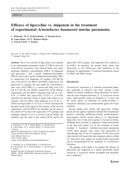
ARTICLEEfficacy of tigecycline vs.imipenem in the treatmentof experimental Acinetobacter baumannii murine pneumoniaC.Pichardo &M.E.Pachón-Ibañez &F.Docobo-Perez &R.López-Rojas &M.E.Jiménez-Mejías &A.Garcia-Curiel &J.PachonReceived:31July 2009/Accepted:1February 2010/Published online:25February 2010#Springer-Verlag 2010Abstract The in vivo activity of tigecycline was evaluated in an experimental pneumonia model (C57BL/6mice)by Acinetobacter baumannii .Two clinical strains were used:minimum inhibitory concentrations (MICs)of imipenem and tigecycline 1and 2µg/mL (imipenem-susceptible,IPM-S),and 8and 2µg/mL (imipenem-intermediate,IPM-I),respectively.For imipenem (30mg/Kg),∆T/CMI (h)were 1.04and 0.51for IPM-S and IPM-I,respectively.For tigecycline (5mg/Kg),the area under the concentration –time curve (AUC)/MIC 0–24h (serum and lung)were 9.24and 4.37(for the two strains),respectively.In the efficacy experiments with the IPM-S,imipenem (log CFU/g 3.59±0.78,p =0.006)and tigecycline (2.82±1.2,p =0.054)decreased the bacterial counts in lungs with respect to its controls;with the IPM-I,both imipenem (1.21±0.52,p =0.002)and tigecycline (3.21±0.28,p =0.035)decreased the bacterial counts with respect to the controls.In the survival experiments,with the IPM-S,the mortality was the same in the control (67%)and in the tigecycline (77%)groups,and imipenem reduced it (21%,p =0.025);with the IPM-I,the mortality was the same in the control (87%)and in thetigecycline (85%)groups,and imipenem (0%)reduced it (p <0.001).In summary,the present study shows that tigecycline is less efficacious than imipenem in the treatment of experimental A.baumannii pneumonia caused by IPM-S and IPM-I strains.IntroductionAcinetobacter baumannii is a common nosocomial patho-gen,especially in intensive care units,causing a great number of clinical conditions,being pneumonia or bacter-emia the more frequent infections [1,2].In the last several years,multi-resistant strains are frequent [1,3]and,with the recent reports of outbreaks of colistin-resistant A.baumannii isolates[4],new antimicrobial agents have been searched.Recent studies have shown that tigecycline exhibits potent activity against organisms isolated from hospitalized patients,including multidrug-resistant nonfermentative gram-negative bacilli,among others [5,6].Specifically,tigecycline was in vitro-active against A.baumannii strains,including those resistant to imipenem,being bacteriostatic in the time –kill studies [7].In the treatment of multi-resistant A.baumannii ventilator-associated pneumonia (VAP),three patients were cured with tigecycline in monotherapy and 15out of 19patients (78.9%)were cured when treated with tigecycline plus imipenem and/or colistin [8].Data from more studies are needed before tigecycline can be recommended for the treatment of Acinetobacter infections.The purpose of this study is to compare the efficacy of tigecycline and imipenem in a murine pneumonia model caused by A.baumannii strains susceptible and intermedi-ate to imipenem.C.Pichardo (*):M.E.Pachón-Ibañez :F.Docobo-Perez :R.López-Rojas :M.E.Jiménez-Mejías :A.Garcia-Curiel :J.PachonInstituto de Biomedicina de Sevilla,Hospital Universitario Virgen del Rocío/CSIC/Universidad de Sevilla,Avda.Manuel Siurot s/n,41013Seville,Spaine-mail:cristina.pichardo.exts@juntadeandalucia.esC.Pichardo :M.E.Pachón-Ibañez :F.Docobo-Perez :R.López-Rojas :M.E.Jiménez-Mejías :A.Garcia-Curiel :J.PachonSpanish Network for Research in Infectious Diseases(REIPI —ISCIII —RD06/0008),Instituto de Salud Carlos III,Madrid,SpainEur J Clin Microbiol Infect Dis (2010)29:527–531DOI 10.1007/s10096-010-0890-6Materials and methodsWe used two A.baumannii strains collected from blood cultures,corresponding to the more prevalent clones isolated in our hospital:imipenem-susceptible strain (IPM-S,953,minimum inhibitory concentration[MIC] imipenem=1mg/L,tigecycline=2mg/L),and imipenem-intermediate strain(IPM-I,1514,MIC imipenem=8mg/L, tigecycline=2mg/L).Imipenem(laboratory standard powder)and imipenem plus cilastatin(IPM)were obtained from Merck Sharp& Dohme(Madrid,Spain)for the in vitro and the in vivo experiments,respectively,and tigecycline(TGC)from Wyeth-Ayerst(Pearl River,NY,USA).The animals used were immunocompetent C57BL/6 female mice weighing14–16g(University of Seville’s facility)and had a sanitary status of murine pathogen-free (MPF).Drug pharmacokineticsThe plasma levels of imipenem were determined after the administration of a single intramuscular(im)dose of 30mg/Kg.The plasma and lung(homogenate)levels of tigecycline were determined after the administration of a single subcutaneal(sc)dose of5mg/Kg.Blood and lungs were extracted from three anesthetized mice per time-point. The total plasmatic drug concentrations and the total drug concentrations in the lung were measured for triplicate by the bioassay method,using Micrococcus luteus ATCC9341 for imipenem and Bacillus cereus ATCC9634for tigecy-cline as the indicator strains.The intraday and interday variation of the assays were2.6%±2.4%and3.2%±1.9% for imipenem,and 3.5%±2.6%and 4.5%±1.1%for tigecycline;the linearity(r2)of the assay was0.91±0.02 and0.92±0.32,respectively,and the lower limits of detection were0.01and0.1mg/L.The maximum plasma concentration(C max,mg/L)and terminal half-life(t1/2,h) were calculated.The time during which the plasma concentration remained above the MIC(ΔT/MIC,h)was estimated by extrapolation from the regression line of plasma elimination.Experimental pneumonia model in miceA modification of the Esposito and Pennington model performed by our group[9]was used to produce pneumo-nia.The animals were inoculated with50μl of the A. baumannii bacterial suspension,with a final inoculum size of approximately108CFU/mL.The treatment was commenced at4h after the inoculation,grouping the mice in each of the following treatment groups over a period of 72h:(a)controls,no treatment;(b)tigecycline,10mg/Kg/d/sc,b.i.d.;and(c)imipenem,120mg/Kg/d/im,t.i.d.(a quarter of the total dose at8:00and14:00h,and the half of the total dose at20:00h).Two types of experiments were performed:survival and bacterial clearance from lung studies.Survival experimentsBecause in this model the mortality in the control group is near100%at72h,the influence on the mortality of the different treatments was evaluated in this period,in groups of15mice.Efficacy experimentsTo evaluate the efficacy of antimicrobials in the clearance of bacteria from lungs,efficacy experiments were per-formed.Groups of five mice were sacrificed at4h after the infection,before the beginning of treatments(control groups),and groups of five mice were sacrificed every 24h,4h after the last dose in the case of IPM and12h after the last dose in the case of tigecycline(treatment groups).At these time-points,the surviving mice were sacrificed and thoracotomy was carried out;the lungs were removed,weighed,and processed for quantitative cultures. The results were expressed as the log10CFU/g of tissue.In order to confirm that imipenem and tigecycline were not toxic to the animals,groups of ten non-infected mice were each given the antibiotics for72h.The use of the experimental pneumonia model was approved by the Ethics Committee of the Hospital Universitario Virgen del Rocío, Sevilla,Spain.Statistical analysisThe numbers of surviving animals were evaluated with Fisher’s exact test.The CFU/g of lung tissue was analyzed using the Kruskal–Wallis test.The SPSS v13.0statistical package(SPSS Inc.,Chicago,IL,USA)was used.A p-value<0.05was considered to be significant.Results and discussionThe pharmacokinetic/pharmacodynamic parameters of each antimicrobial drug(C max,t1/2,AUC,∆T/MIC,and AUC0–24h/ MIC)are shown in Table1.In the survival experiments(Table2),with both strains IPM-I and IPM-S,only imipenem reduces the mortality with respect to the control groups(0and21%in the imipenem groups and87and67%in the controls, respectively,p≤0.025);imipenem was better than tigecy-cline in both groups(p≤0.007).In the bacterial clearance from the lungs experiments (Fig.1),with the IPM-I strain,for the imipenem-treated group,there was a decrease of the bacterial counts from controls(p=0.002).In the tigecycline-treated group,the treatment cleared the lungs compared to the control group (p=0.035).With the IPM-S strain,in the imipenem-treated group,there was a decrease of the bacterial counts(p= 0.006)and in the tigecycline-treated group,the bacterial lung concentration decreased to2.82log CFU/g(p=0.054).These results show that,in general,imipenem is more active than tigecycline in this experimental pneumonia model caused by A.baumannii with the doses examined. Tigecycline decreased the bacterial count in the lung for the strain intermediate to imipenem with respect to the controls. However,in the in vivo experiments performed with the strains susceptible and intermediate to imipenem,imipenem was better than tigecycline regarding the mortality and clearance of bacteria from the lungs.Different studies in experimental A.baumannii pneumo-nia models had shown that imipenem was the most active antimicrobial,including infections caused by susceptible and intermediate strains[9,10].Different works showed the high in vitro activity of tigecycline from multi-resistant pathogens,including A.baumannii[5,6],which is of particular importance because of the high frequency of multi-resistant strains[1,3],with a high number of strains only susceptible to colistin[1,11,12]or the appearance of pan-resistant A.baumannii isolates[13,14].In a previous in vitro study on the activity of imipenem and tigecycline against49isolates of A.baumannii,the MIC90values were 128and2mg/L,respectively;however,in the time–kill studies,tigecycline was bacteriostatic[7].The limited efficacy of tigecycline in the experimental pneumonia model may be explained by the in vitro bacteriostatic instead of bactericidal activity against A.baumannii.Tigecycline has high penetration in tissues[15,16];thus, we found a serum and lung C max of1.92mg/L and8.29μg/ g,respectively.These figures are higher than those found in humans after doses of50and100mg,which show serum C max values of0.38and0.91mg/L[15,17],respectively, and the mean and median concentration values of tigecy-cline in the lungs after100mg were0.50and0.31μg/g (range:0.11–1.89μg/g)[18].There are no definitive data regarding the pharmacodynamic variables predicting the in vivo efficacy of tigecycline.Several studies had addressed this issue,both in clinical trials and in experimental models of infection[15,16].Some authors suggest that,although tetracyclines do not exhibit concentration-dependent kill-ing,if the antimicrobial agent has a moderate to prolonged postantibiotic effect(PAE),the time of exposure is less important and the AUC/MIC ratio is the best pharmacoki-netic/pharmacodynamic parameter correlating with the therapeutic efficacy of these drugs[19].Data obtained in animal models[17,20]suggest that tigecycline activityTable1Pharmacokinetic/pharmacodynamic parametersC max t1/2(h)AUC(mg.h/L)AUC/MIC0–24h∆T/MIC(h)Strain1514IPM-I Strain953IPM-S Strain1514IPM-IStrain953IPM-STigecycline(serum)5mg/Kg 1.92mg/L 2.339.249.249.24 1.70 1.70 Tigecycline(lung)5mg/Kg8.29μg/g0.23 4.37 4.37 4.370.970.97 Imipenem(serum)30mg/Kg16.9mg/L0.158.25 4.12330.51 1.04C max:maximal drug concentration;t1/2:half-life;AUC:area under the concentration–time curve;AUC/MIC0–24h:area under the concentration–time curve at24h to the MIC;∆T/MIC:the time that a drug concentration remains above the MICTable2Survival study using three strains with different susceptibilities to imipenemStrain1514(IPM-I)Strain953(IPM-S)n n died%n n died% Controls151387151067 Imipenem,120mg/Kg/d/im1500a14321b Tigecycline,10mg/Kg/d/sc131185131077 IPM:imipenem,I:intermediate,S:susceptiblea p<0.001compared to the controls and tigecycline.b p=0.025and p=0.007compared to the controls and tigecycline,respectivelydepends on the time above the MIC and on the AUC/MIC.On the other hand,the relationship between the AUC,MIC,and the microbiologic results in clinical trials in complicat-ed skin and soft tissue infections were revised recently [21,22],showing that an AUC/MIC ratio between 6.96and 12.3was necessary for a good clinical and microbiological response,with the predominant pathogens being Staphylo-coccus aureus and Streptococcus spp.However,although in our experiments the serum AUC/MIC of tigecycline was 9.24,we did not obtain the same favorable results in the pneumonia by A.baumannii .The clinical information on the relevance of tigecycline as an antimicrobial agent against A.baumannii is scarce.A clinical case of septic shock due to a multidrug-resistant A.baumannii strain in a patient with intra-abdominal abscess after acute pancreatitis,with failure during treatment with colistin plus meropenem,cured after the addition of tigecycline [23].In the treatment of multi-resistant A.baumannii V AP in a recent study [8],three patients were cured with tigecycline in monotherapy and 15out of 19patients (78.9%)cured when treated with tigecycline plus imipenem and/or colistin.In other study,only two out of five patients with A.baumannii V AP were cured with tigecycline in monotherapy or in combination [24].Peleg et al.[25]show the first clinical description of bloodstream infection caused by tigecycline-non-susceptible A.baumannii .In their work,two patients developed breakthrough A.baumannii bacteraemia while receiving tigecycline for other indications.The MICs oftigecycline for these strains were 4and 16mg/L,respectively,and the resistance appears to be at least partly attributable to an efflux pump mechanism,since the exposure to an efflux pump inhibitor diminished the MIC of tigecycline 4to 1mg/L and from 16to 4mg/L,respectively.The authors conclude that,given the facility of A.baumannii to acquire resistance to other antimicrobials,exposure to sub-therapeutic levels of tigecycline for even short periods of time may promote the rapid emergence of tigecycline resistance.In this sense,a recent multicenter Spanish study obtained an MIC 90of tigecycline for A.baumannii of 8mg/L [26],which is higher than that found previously by us [7].The results of the present experimental study and those from the clinical experience pointed to the necessity of evaluating tigecycline combined with other antimicrobials when treating severe A.baumannii infections.Thus,in a model of A.baumannii experimental murine pneumonia caused by imipenem-susceptible strains [9],the treatment with other tetracyclines,such as doxycycline plus amikacin,was as efficacious as imipenem in reducing the mortality and the clearance of bacteria from the lungs,this combina-tion being synergic in vitro.However,in an in vitro study using time –kill curves [27],the possible synergy of tigecycline plus amikacin,meropenem,imipenem,ciprofloxacin,levofloxacin,ampicillin-sulbactam,and rifampin has been evaluated against A.baumannii strains intermediate or resistant to carbapenems (MIC 90of tigecycline of 2mg/L),showing indifference for tigecycline in combination with these antimicrobials.Also,concentration escalation studies dem-onstrate that tigecycline may need to approach serum concentrations higher than those currently achieved to treat multidrug-resistant A.baumannii .In the same way,using checkerboard testing,tigecycline synergy was observed in only 1of 5strains when combined with cefepime or amikacin,and no synergy was shown with the combination of tigecycline with amoxicillin/cavulanate,piperacillin/tazobactam,ceftriaxone,ceftazidime,imipenem,merope-nem,aztreonam,trimethoprim/sulfamethoxazole,or cipro-floxacin [28].In summary,the present study,taking into account the limitations of the small size of the groups in the efficacy experiments,shows that tigecycline in monotherapy is less efficacious than imipenem in the treatment of experimental A.baumannii pneumonia caused by susceptible or inter-mediate to imipenem strains.However,due to its in vitro activity against multi-resistant A.baumannii ,the high mortality of severe infections caused by this bacterium,such as V AP and bacteremia,and the paucity of therapeutic alternatives for these severe infections,new in vivo studies are needed to evaluate its efficacy in combination with otherantimicrobials.Fig.1Bacterial clearance from lung tissue using two strains with different susceptibilities to imipenem (treatment during a period of 72h).Values are expressed as mean ±standard deviation (SD)(log CFU/g).*p <0.05,#p =0.054Acknowledgments This work was partly supported by a grant fromthe Wyeth-Ayerst Research(Pearl River,NY,USA).The authorsdeclare no conflict of interest with respect to this article. References1.Cisneros JM,Reyes MJ,Pachón J,Becerril B,Caballero FJ,García-Garmendía JL,Ortiz C,Cobacho AR(1996)Bacteremiadue to Acinetobacter baumannii:epidemiology,clinical findings,and prognostic features.Clin Infect Dis22:1026–10322.Garnacho-Montero J,Ortiz-Leyba C,Jiménez-Jiménez FJ,Barrero-Almodóvar AE,García-Garmendia JL,Bernabeu-WittellM,Gallego-Lara SL,Madrazo-Osuna J(2003)Treatment ofmultidrug-resistant Acinetobacter baumannii ventilator-associatedpneumonia(V AP)with intravenous colistin:a comparison withimipenem-susceptible V AP.Clin Infect Dis36:1111–11183.Fernández-Cuenca F,Pascual A,Ribera A,Vila J,Bou G,Cisneros JM,Rodríguez-Baño J,Pachón J,Martínez-Martínez L;Grupo de Estudio de Infección Hospitalaria(2004)Diversidadclonal y sensibilidad a los antimicrobianos de Acinetobacter baumannii aislados en hospitales españoles.Estudio multicéntrico nacional:proyecto GEIH-Ab2000.Enferm Infecc Microbiol Clin22(5):267–2714.Vila J,Pachón J(2008)Therapeutic options for Acinetobacterbaumannii infections.Expert Opin Pharmacother9(4):587–599 5.Bouchillon SK,Hoban DJ,Johnson BM,Johnson JL,Hsiung A,Dowzicky MJ;Tigecycline Evaluation and Surveillance Trial(TEST)Group(2005)In vitro activity of tigecycline against3989gram-negative and gram-positive clinical isolates from the UnitedStates Tigecycline Evaluation and Surveillance Trial(TESTProgram;2004).Diagn Microbiol Infect Dis52(3):173–1796.Fritsche TR,Sader HS,Stilwell MG,Dowzicky MJ,Jones RN(2005)Potency and spectrum of tigecycline tested against aninternational collection of bacterial pathogens associated with skinand soft tissue infections(2000–2004).Diagn Microbiol InfectDis52(3):195–2017.Pachón-Ibáñez ME,Jiménez-Mejías ME,Pichardo C,Llanos AC,Pachón J(2004)Activity of tigecycline(GAR-936)against Acinetobacter baumannii strains,including those resistant to imipenem.Antimicrob Agents Chemother48:4479–44818.Schafer JJ,Goff DA,Stevenson KB,Mangino JE(2007)Earlyexperience with tigecycline for ventilator-associated pneumoniaand bacteremia caused by multidrug-resistant Acinetobacter baumannii.Pharmacotherapy27(7):980–9879.Rodríguez-Hernández MJ,Pachón J,Pichardo C,Cuberos L,Ibáñez-Martínez J,García-Curiel A,Caballero FJ,Moreno I,Jiménez-Mejías ME(2000)Imipenem,doxycycline and ami-kacin in monotherapy and in combination in Acinetobacter baumannii experimental pneumonia.J Antimicrob Chemother 45:493–50110.Wolff M,Joly-Guillou ML,Farinotti R,Carbon C(1999)In vivoefficacies of combinations ofβ-lactams,β-lactamase inhibitors,and rifampin against Acinetobacter baumannii in a mousepneumonia model.Antimicrob Agents Chemother43:1406–141111.Bergogne-Bérézin E,Towner KJ(1996)Acinetobacter spp.asnosocomial pathogens:microbiological,clinical,and epidemio-logical features.Clin Microbiol Rev9:148–16512.Bou G,CerveróG,Domínguez MA,Quereda C,Martínez-BeltránJ(2000)Characterization of a nosocomial outbreak caused by amultiresistant Acinetobacter baumannii strain with a carbapenem-hydrolyzing enzyme:high-level carbapenem resistance in A.baumannii is not due solely to the presence ofβ-lactamases.J Clin Microbiol38:3299–330513.Henwood CJ,Gatward T,Warner M,James D,Stockdale MW,Spence RP,Towner KJ,Livermore DM,Woodford N(2002) Antibiotic resistance among clinical isolates of Acinetobacter in the UK,and in vitro evaluation of tigecycline(GAR-936).J Antimicrob Chemother49:479–48714.Urban C,Mariano N,Rahal JJ,Tay E,Ponio C,Koprivnjak T,Weiss J(2000)Polymyxin B-resistant Acinetobacter baumannii clinical isolate susceptible to recombinant BPI21and cecropin P1.Antimicrob Agents Chemother45:994–99515.Meagher AK,Ambrose PG,Grasela TH,Ellis-Grosse EJ(2005)Pharmacokinetic/pharmacodynamic profile for tigecycline—a new glycylcycline antimicrobial agent.Diagn Microbiol Infect Dis52(3):165–17116.Muralidharan G,Micalizzi M,Speth J,Raible D,Troy S(2005)Pharmacokinetics of tigecycline after single and multiple doses in healthy subjects.Antimicrob Agents Chemother49(1):220–229 17.Rubinstein E,Vaughan D(2005)Tigecycline:a novel glycylcy-cline.Drugs65(10):1317–133618.Rodvold KA,Gotfried MH,Cwik M,Korth-Bradley JM,DukartG,Ellis-Grosse EJ(2006)Serum,tissue and body fluid concen-trations of tigecycline after a single100mg dose.J Antimicrob Chemother58(6):1221–122919.Craig WA(1998)Pharmacokinetic/pharmacodynamic parameters:rationale for antibacterial dosing of mice and men.Clin Infect Dis 26:1–1220.van Ogtrop ML,Andes D,Stamstad TJ,Conklin B,Weiss WJ,Craig WA,Vesga O(2000)In vivo pharmacodynamic activities of two glycylcyclines(GAR-936and WAY152,288)against various gram-positive and gram-negative bacteria.Antimicrob Agents Chemother44(4):943–94921.Agwuh KN,MacGowan A(2006)Pharmacokinetics and phar-macodynamics of the tetracyclines including glycylcyclines.J Antimicrob Chemother58(2):256–26522.Doan TL,Fung HB,Mehta D,Riska PF(2006)Tigecycline:aglycylcycline antimicrobial agent.Clin Ther28(8):1079–1106,Review 23.Taccone FS,Rodriguez-Villalobos H,De Backer D,De Moor V,Deviere J,Vincent JL,Jacobs F(2006)Successful treatment of septic shock due to pan-resistant Acinetobacter baumannii using combined antimicrobial therapy including tigecycline.Eur J Clin Microbiol Infect Dis25:257–26024.Anthony KB,Fishman NO,Linkin DR,Gasink LB,Edelstein PH,Lautenbach E(2008)Clinical and microbiological outcomes of serious infections with multidrug-resistant gram-negative organ-isms treated with tigecycline.Clin Infect Dis46:567–57025.Peleg AY,Potoski BA,Rea R,Adams J,Sethi J,Capitano B,Husain S,Kwak EJ,Bhat SV,Paterson DL(2007)Acinetobacter baumannii bloodstream infection while receiving tigecycline:a cautionary report.J Antimicrob Chemother59(1):128–13126.Betriu C,Rodriguez-Avial I,Gómez M,Culebras E,López F,Alvarez J,Picazo JJ;Spanish Tigecycline Group(2006)Antimi-crobial activity of tigecycline against clinical isolates from Spanish medical centers.Second multicenter study.Diagn Micro-biol Infect Dis56(4):437–44427.Scheetz MH,Qi C,Warren JR,Postelnick MJ,Zembower T,Obias A,Noskin GA(2007)In vitro activities of various antimicrobials alone and in combination with tigecycline against carbapenem-intermediate or-resistant Acinetobacter baumannii.Antimicrob Agents Chemother51(5):1621–162628.V ouillamoz J,Moreillon P,Giddey M,Entenza JM(2008)In vitroactivities of tigecycline combined with other antimicrobials against multiresistant gram-positive and gram-negative pathogens.J Antimicrob Chemother61:371–374。
医学免疫学英文词汇

AAcquired immune deficiency syndrome, AIDS 获得性免疫缺陷综合征(艾滋病) Acquired immunity 获得性免疫Activation induced cell death, AICD 活化诱导的细胞死亡Active immunotherapy 主动免疫治疗Acute phase protein 急性时相蛋白Adapter 转接蛋白Adaptive immunity 适应性免疫Addressin 地址素Adenosine deaminase 腺苷脱氨酶Adjuvant 佐剂Adoptive immunity 过继免疫Adoptive immunotherapy, AIT 过继免疫治疗Affinity 亲和力Affinity maturation 亲和力成熟Agglutination 凝集反应Allelic exclusion 等位排斥Allergen 变应原Allergin 变应素Allergy 变态反应Allogenic antigen 同种异型抗原Allograft 同种异型移植Allotype 同种异型Allorecognition 同种异型识别Alpha-fetoprotein, AFP 甲种胎儿球蛋白Alternative pathway 旁路途径Anamnestic response 回忆应答Anaphylactic shock 过敏性休克Anaphylactogen 过敏原Anaphylaxis 过敏反应Anchor residue 锚定残基Ankylosing spondylitis, AS 强直性脊柱炎Antibacterial immune serum 抗菌免疫血清Antibody, Ab 抗体Antibody-dependent cell-mediated cytotoxicity, ADCC 抗体依赖性细胞介导的细胞毒作用Antiviral immune serum 抗病毒免疫血清Antigen, Ag 抗原Antigenic determinant 抗原决定簇Antigen internal image 抗原内影像Antigen-presenting cells, APC 抗原提呈细胞Antigen specific immune response 抗原特异性免疫应答Antigenicity 抗原性Anti-idiotype 抗独特型Antitoxic serum 抗毒素血清Apoptosis 细胞凋亡Apoptosis cell associated molecular pattern, ACAMP凋亡细胞相关的分子模式 Apoptotic body 凋亡小体Artificial active immunization 人工主动免疫Artificial passive immunization 人工被动免疫Ataxia telangiectasia syndrome, AT 毛细血管扩张共济失调综合征 Atopic dermatitisAutocrineAutograftAutoimmune antibodyAutoimmune disease, AIDAutoimmune hemolytic anemiaAutoimmune thrombocytopenic purpuraAutoimmunityBBacillus Calmette Guerin, BCGBare lymphocyte syndrome, BLSBasophilB cell epitope BB cell hybridoma BB cell linker ptotein, BLNKB cell receptor, BCRBiotin-avidin system, BASBispecific antibody, BsAbBlocking antibodyB lymphocyteBone marrowBone marrow transplantation, BMTBradykininCC1 inhibitor, C1 INHC4 binding protein,C8 binding protein, C8bpC-reactive protein,CRPCadherinCalcineurinCalmodulinCarcinoembryonic antigen, CEACarrierCecropinsCell adhesion molecules, CAM 特应性皮炎自分泌自体移植自身抗体自身免疫性疾病自身免疫性溶血性贫血自身免疫性血小板减少性紫癜自身免疫卡介苗裸淋巴细胞综合征嗜碱性粒细胞细胞表位细胞杂交瘤 B 细胞连接蛋白 B 细胞(抗原识别)受体生物素-亲和素系统双特异性抗体封闭抗体 B淋巴细胞骨髓骨髓移植缓激肽C1抑制分子 C4bp C4 结合蛋 C8 结合蛋白 C-反应蛋白钙粘蛋白钙神经素(钙调磷酸酶)钙调蛋白癌胚抗原载体杀菌肽细胞粘附分子hediak-Higashi syndrome Chediak-Higashi综合征 Chemokine 趋化性细胞因子 Chimeric antibody 嵌合抗体Chronic granulomatous disease, CGD 慢性肉芽肿病 Class Ⅱtransactivator, C Ⅱ TA Ⅱ类反式活化子 Class switch 类别转换Classical pathway 经典途径Clonal anergy 克隆无能Clonal deletion 克隆删除Clonal eliminationClonal expansionCluster of differentiation, CDColony stimulating factor, CSFCommon variable immunodeficiencyComplementComplement deficiencyComplement receptorComplementarity determining region, CDRComplete antigenConcanavalin A, Con AConformational determinantConstant regionCo-receptorCo-stimulating signalCo-stimulatory molecules, CMCo-stimulatory molecule receptor, CMR Cross reactionCryptic determinantCytokine, CKCytolytic typeCytotoxic typeCytotoxic T lymphocytes, CTL 或 Tc Cytotoxic T lymphocytes antigen-4, CTLA-4 DDecay accelerating factor, DAFDefensinsDelayed type hypersensitivity, DTHDelayed type hypersensitivity T cell, T DTH Dendritic cells, DCDiGeorge syndromeDiversity geneDNA vaccineDonorDouble immunodiffusion 克隆消除克隆扩增分化群集落刺激因子普通变化型免疫缺陷病补体补体缺陷补体受体互补性决定区完全抗原刀豆蛋白A 构象决定基恒定区,C区辅助受体协同刺激信号协同刺激分子协同刺激分子受体交叉反应隐蔽决定基细胞因子细胞溶解型细胞毒型细胞毒性T细胞细胞毒性T淋巴细胞(相关)抗原 4 衰变加速因子防御素迟发型超敏反应迟发型超敏反应性T细胞树突状细胞 DiGeorge综合征 D(多样化)基因 DNA疫苗供者双向免疫扩散Double negative cell, DN cell 双阴性细胞Double positive cell, DP cell 双阳性细胞EE rosette test E 花结试验Early phase reaction 早期相反应Endocytosis 胞吞作用Enzyme immunoassay, EIA 酶免疫测定Enzyme linked immunosorbent assay, ELISA 酶联免疫吸附试验Eosinophil, Eos 嗜酸性粒细胞Eosinophil chemotactic factor, ECF 嗜酸性粒细胞趋化因子Eosinophil peroxidase, EPO 嗜酸性粒细胞过氧化物酶 Epitope 表位Erythropoietin, EPO 红细胞生成素Extracellular matrix, ECM 细胞外基质F Fc receptor, FcR 结晶片断受体,Fc受体 Flow cytometry, FCM 流式细胞术Fluorescence-activated cell sorter, FACS 荧光活化细胞分类器 Follicular dendritic cells, FDC 滤泡树突状细胞 Fragment antigen binding, Fab 抗原结合片断Fragment crystallizable, Fc 结晶片断Fragment of variable region Fv 片断Framework region 骨架区Freund complete adjuvant 弗氏完全佐剂GGenetic engineering antibody 基因工程抗体Germ line gene 胚系基因Germinal center 生发中心Glycosylphosphatidylinositol, GPI 糖磷脂酰肌醇Graft versus host reaction, GVHR 移植物抗宿主反应 Granule exocytosis 颗粒胞吐Granulocyte colony stimulating factor, G-CSF 粒细胞集落刺激因子 Granulocyte-macrophage colony stimulating factor, GM-CSF 粒细胞-巨噬细胞集落刺激因子 Granzyme, Gz 颗粒酶Grave disease 毒性弥漫性甲状腺炎 Growth factor 生长因子Growth factor receptor binding protein-2,Grb-2 生长因子受体结合蛋白 2 Guanine nucleotide exchange factor, GEF 鸟苷酸置换因子Gut-associated lymphoid tissue, GALT 肠伴随(相关)淋巴组织 HHaplotype 单元型Hapten 半抗原Hashimoto’s thyroiditis 桥本甲状腺炎Heat shock protein, HSP 热休克蛋白Heavy chain 重链,H 链Helper T cells(lymphocytes), Th 辅助性 T 细胞 Hemolytic plaque assay 溶血空斑试验 Hemopoietic stem cell, HSC 造血干细胞 Heterophil antigen 异嗜性抗原 Hidden antigen 隐蔽抗原High endothelial venule, HEV 高内皮细胞小静脉 Hinge region 铰链区Histamin 组胺Histocompatibility antigen-2, H-2HLA genotypingHomologous restriction factor, HRFHost versus graft reaction, HVGRHuman immunodeficiency virus, HIVHuman leukocyte antigen,HLAHumanized antibodyHumoral immunity Hypersensitivity Hypervariable region, HVRIIdiotype, IdIdiotype networkImmediate hypersensitivity Immune adherent, IAImmune complex, ICImmune function related gene Immune regulationImmune responseImmune response region Immune serumImmune surveillanceImmune systemImmunityImmunocyteImmunodeficiency disease, IDD Immunofluorescence ImmunogenicityImmunoglobulin, IgImmunoglobulin superfamily, IgSFImmunohistochemistry techniqueImmunological competenceImmunological ignorance , ;Immunological toleranceImmunological non-responsiveness 小鼠的组织相容性抗原HLA 基因分型同源限制因子宿主抗移植物反应人类免疫缺陷病毒人类白细胞抗原人源化抗体体液免疫超敏反应高变独特型独特型网络速发型超敏反应免疫粘附免疫复合物免疫功能相关基因免疫调节免疫应答免疫应答区免疫血清免疫监视免疫系统免疫免疫细胞免疫缺陷病免疫荧光法免疫原性免疫球蛋白免疫球蛋白超家族免疫组化技术免疫适能免疫忽视免疫耐受免疫不应答Immunologically privileged sites 免疫隔离部位Immunology 免疫学Immunoreceptor tyrosine-based activation motifs, ITAM 免疫受体酪氨酸活化基序 Immunoreceptor tyrosine-based inhibitory motifs, ITIM 免疫受体酪氨酸抑制基序 Immunotherapy 免疫治疗Inactivated vaccine 灭活疫苗Inducible nitric oxide synthase, iNOS 诱导型一氧化氮合成酶Inflammatory cell 炎症细胞Innate immunity 固有(性)免疫Inositol-1, 4, 5-trisphosphate,IP3Insulin-dependent diabetes mellitus, IDDM Intercellular adhesion molecular,ICAMIntegrinInterferon, IFNInterleukin, ILInternalizationImtraepithelial lymphocytes, IELJJoining chainJoining geneKKiller activatory receptor, KARKiller inhibitory receptor, KIRKiller immunoglobulin like receptor KininogenaseLLangerhans cells, LCLarge granular lymphcytes, LGLsLate phase reactionLectinLectin-like carbohydrate recognition domain, CRDLeucine rich repeat, LRRLeukinLeukocyte adhesion deficiency, LADLeukocyte common antigen, LCALeukocyte differentiation antigen, LDALeukotrienes (Leucotrienes), LTsLigandLight chainLinker for activation of T cell, LATLinear determinantLinkage disequilibriumLipoteichoic acid, LTALipoxygenase pathway 三磷酸肌醇胰岛素依赖型糖尿病细胞间粘附分子整合素干扰素白细胞介素内化上皮细胞间淋巴细胞 J链J基因杀伤细胞活化受体杀伤细胞抑制受体杀伤细胞免疫球蛋白样受体激肽原酶郎格汉斯细胞大颗粒淋巴细胞晚期相反应凝集素凝集素样糖识别结构域富含亮氨酸的重复序列白细胞素白细胞粘附缺陷白细胞共同抗原白细胞分化抗原白三烯配基,配体轻链,L 链 T细胞活化连接蛋白线性决定基连锁不平衡磷壁酸脂氧合酶途径Live-attenuated vaccine 减毒活疫苗Long-acting thyroid stimulator, LATS 长效甲状腺刺激素Low molecular-weight polypeptide, LMP 低分子量多肽LPS binding protein, LBP LPS结合蛋白Lymphocyte 淋巴细胞Lymphocyte function associated antigen, LFA 淋巴细胞功能相关抗原 Lymphocyte homing 淋巴细胞归巢Lymphocyte homing receptor, LHR 淋巴细胞归巢受体Lymphoid DC 淋巴系树突状细胞Lymphoid progenitor 淋巴样祖细胞Lymphoid stem cells, LSC 淋巴样干细胞Lymphokine, LK 淋巴因子Lymphokine activated killer cell, LAK 淋巴因子激活的杀伤细胞 Lymphotoxin, LT 淋巴毒素β -lysin 乙型溶素Lysosome-associated membrane proteins-1,LAMP-1 溶酶体相关膜蛋白1 Lysozyme 溶菌酶MMacrophages, M φ巨噬细胞Macrophage colony stimulating factor, M-CSF 巨噬细胞集落刺激因子 Macropinocytosis 巨吞饮Magainins 爪蟾抗菌肽Major histocompatibility complex, MHC 主要组织相容性复合体 Mannan-binding lectin, MBL 甘露糖结合凝集素Mannose binding protein, MBP 甘露糖结合蛋白Mannose receptor, MR 甘露糖受体Mast cell, MC 肥大细胞MBL-associated serine protease, MASP MBL 伴随的丝氨酸蛋白酶 Membrane attack complex, MAC 膜攻击复合物Membrane cofactor protein, MCP 膜辅助因子蛋白Membrane Ig, mIg 膜表面免疫球蛋白Membrane inhibitor of reactive lysis, MIRL 膜反应性溶解抑制物 Memory cells 记忆细胞MHC class Ⅰ gene MHC Ⅰ类基因MHC class Ⅱ gene MHC Ⅱ类基因MHC class Ⅲ gene MHC Ⅲ类基因MHC restriction MHC 限制性Microfold cell M细胞,微小褶皱细胞β 2-Microglobulin, β2 -m β2 微球蛋白Minor histocompatibility antigen 次要组织相容性抗原 Mitog en 丝裂原Mitogen-activation protein kinase, MAPK 丝裂原激活蛋白激酶 Molecular mimicry 分子模拟Monoclonal antibody, McAb 单克隆抗体第7 / 11页Monocyte 单核细胞Monocyte chemotactic protein, MCP 单核细胞趋化蛋白Monokine, MK 单核因子Mononuclear-phagocyte system, MPS 单核-巨噬细胞系统Mucosal-associated lymphoid tissue, MALT 粘膜伴随(相关)淋巴组织 Mucosal mast cell, MMC 粘膜肥大细胞Multiple hematopoietic stem cells, HSC 多能造血干细胞Multiple sclerosis, MS 多发性硬化症Myasthenia gravis, MG 重症肌无力Myeloid DCMyeloid stem cellMyeloperoxidase, MPONNaive T(B) cellsNatural cytotoxic cellNature killer cell, NK cellNeutrophilsNitric oxide, NONitroblue tetrazolium, NBTNon-classical MHC class Ⅰ geneNon-organ specific autoimmune diseaseNon-specific immunityNude miceOOpsonizationOrgan specific autoimmune diseasePParacrineParoxysmal nocturnal hemoglobineria, PHNPassive immunotherapy ,Passive transfer of lymphocytePathogen associated molecular pattern, PAMPPattern recognition receptor, PRRPeptide antibioticsPeptidoglycan, PGNPerforinPeripheral lymphoid organPeyer’s patchesPhagocytePhagocytosisPhagolysosomePhosphatidylinnosital pathway 髓系树突状细胞髓样干细胞髓过氧化物酶初始T(B)细胞自然细胞毒性细胞自然杀伤细胞嗜中性粒细胞一氧化氮氮蓝四唑非经典性Ⅰ类基因非器官特异性自身免疫病非特异性免疫裸小鼠,裸鼠调理作用器官特异性自身免疫病旁分泌夜间血红蛋白尿被动免疫治疗淋巴细胞被动转移病原相关分子模式模式识别受体肽抗菌肽聚糖穿孔素外周淋巴器官派氏集合淋巴结吞噬细胞吞噬作用吞噬溶酶体 PI途径第8 / 11页Phosphatidylinositol-3-kinase, PI3-K 磷脂酰肌醇3激酶Phosphatidylinositol-4,5-bisphosphate, PIP 2 磷脂酰肌醇4,5二磷酸 Phosphatidylinositol-3,4,5-trisphosphate 磷脂酰肌醇3,4,5三磷酸 Phosphatidylserine, PS 磷脂酰丝氨酸Phosphoinositides 磷酸肌醇Phospholipase 磷脂酶Phospholipid bilineurine 磷脂胆碱Phosphotylinositide-3 kinase 磷酸肌醇-3激酶Phytohemagglutinin, PHA 植物血凝素PinocytosisPinocytotic vesiclePlacental γ globulinPlaque forming cell, PFCPlasma cellsPlatelet activating factor, PAFPolymeric Ig receptor, pIgRPolymorphismPolymorphic genesPolymorphonuclear neutrophils, PMN PrecipitationPrimary immunodeficiency disease, PIDD Primary responsePro-B cellprofessional antigen presenting cells Programmed cell death, PCDProperdin, PProstaglandin, PGProtein kinase C, PKCProtein tyrosine kinase, PTKProtein tyrosine phosphatase, PTP/PTPase Proteolytic enzyme complexProteosomePurine nucleotide phosphorylase, PNPRγδ+T cellRadioimmunoassay, RIAReactive nitrogen intermediates, RNIs Reactive oxygen intermediates, ROIs RearrangementReceptor editingReceptor-mediated endocytosisRecipientRecombinant antigen vaccineRecombinant vector vaccine 胞饮作用吞饮泡胎盘丙种球蛋白空斑形成细胞浆细胞血小板活化因子多聚免疫球蛋白受体多态性多态性基因多形核嗜中性粒细胞沉淀反应原发性免疫缺陷病初次应答祖B细胞专职抗原提呈细胞程序性细胞死亡备解素前列腺素蛋白激酶C 蛋白酪氨酸激酶蛋白酪氨酸磷酸酶蛋白水解酶复合体蛋白酶体嘌呤核苷磷酸化酶γδ+T细胞放射免疫测定法反应性氮中间物反应性氧中间物(基因)重排受体编辑受体介导的胞吞作用受者重组抗原疫苗重组载体疫苗第9 / 11页Recombinase 重组酶Recombination activating genes, RAG 重组活化基因Recombination signal sequences, RSS 重组信号序列Rejection (移植物的)排斥Rheumatoid arthritis, RA 类风湿性关节炎Rheumatoid factor, RF 类风湿因子SScavenger receptor, SR 清杂受体Secondary immunodeficiency disease, SIDD 继发性免疫缺陷病Secondary response 再次应答Secretory component, SC 分泌成分,分泌小体Secretory IgA, sIgA 分泌型免疫球蛋白 ASecretory piece, SP 分泌片Selectin 选择素Selective IgA deficiency 选择性IgA缺陷Serin/threonine phosphatase 丝/苏氨酸磷酸酶Serum amyloid pretein A, SAA 血清淀粉样蛋白 ASevere combined-immunodeficiency disease, SCID 重症联合免疫缺陷病 Signal transduction 信号转导Signal transducers and activator of transcription, STAT 信号转导和活化转录因子 Signalling complex 信号复合体Single immunodiffusion 单向免疫扩散Small G protein 小 G 蛋白Sneaking through 漏逸Soluble TNF receptor, sTNFR 可溶性TNF受体Somatic hypermutation 体细胞高(频)突变Specific immunity 特异性免疫Split tolerance 耐受分离Src family kinase Src家族激酶Staphylococcus enterotoxin, SE 葡萄球菌肠毒素Staphylococcus protein A, SPA 葡萄球菌蛋白 AStem cell factor, SCF 干细胞(生长)因子Subunit vaccine 亚单位疫苗Superantigen, SAg 超抗原Suppressor T cells, Ts 抑制性T细胞Syngraft 同种基因移植,同型移植 Synthetic peptide vaccine 合成肽疫苗Systemic lupus erythematosus, SLE 系统性红斑狼疮TT cell epitope T细胞表位T cell receptor, TCR T细胞(抗原识别)受体 T lymphocyte T 淋巴细胞TCR/CD3 complex TCR/CD3 复合物Terminal deoxynucleotidyl transferase, TdT 末端脱氧核苷酸转移酶第10 / 11页Terminal pathway 末端通路Thrombopoietin 血小板生成素Thymic stromal cells, TSC 胸腺基质细胞Thymocyte 胸腺细胞Thymus 胸腺Thymus dependent antigen, TD-Ag 胸腺依赖抗原Thymus epithelial cells, TEC 胸腺上皮细胞Thymus independent antigen, TI-Ag 非胸腺依赖抗原Thyroid stimulating hormone, TSH 促甲状腺激素Tolerogen 耐受原Toll like receptor, TLR Toll样受体Toxoid &nbs, p; 类毒素Transforming growth factor, TGF 转化生长因子Transporters associated with antigen processing,TAP 抗原处理相关转运蛋白 Tumor-associated antigen, TAA 肿瘤相关抗原Tumor necrosis factor, TNF 肿瘤坏死因子Tumor-specific antigen, TSA 肿瘤特异性抗原VVariable folding 可变折叠Variable geng V基因Variable region ( V region ) 可变区,V区Vitronectin 玻璃连接蛋白,玻连蛋白 Very late appearing ant igen, VLA 迟现抗原WWestern blotting 免疫印迹法Wiskott-Aldrich syndrome, WAS 伴湿疹血小板减少性免疫缺陷病 XX-linked agammaglobulinemia, XLA 性联无丙种球蛋白血症 X-l inked hyperimmunoglobulin M syndrome, HIM 性联高IgM综合征 X-l inked SCID, XSCID 性联重症联合免疫缺陷病。
血小板在脓毒症中的研究进展
•综述.血小板在脓毒症中的研究进展陈秋杉王兴勇重庆医科大学附属儿童医院急诊科400014通信作者:王兴勇,Email:wxy991819@【摘要】脓毒症为感染致全身免疫失调的炎症反应综合征,血小板在脓毒症发生发展中起着重要作用。
血小板可通过黏附、聚集、活化、脱颗粒作用与病原微生物相反应,以达到保护机体的作用。
同时,脓毒症时血小板还可与中性粒细胞等免疫细胞相互作用,参与微血栓、炎症的形成。
因此,血小板有抗微生物效应及与其他先天免疫细胞协作,形成复杂的血管内免疫防御系统,防止细菌传播的功能。
但其介导的免疫功能如果失调,可致宿主细胞和组织受到严重的附带损害,导致脓毒症相关器官功能障碍。
本文对血小板在脓毒症及其器官功能障碍中的免疫机制作一综述。
【关键词】脓毒症;血小板;免疫系统DOI : 10. 3760/cma. j. issn. 1673-4912. 2021.02.014Research progress of platelets in sepsisChen Qiushan, Wang XingyongEmergency Department of Children's Hospital Affiliated to Chongqing Medical University .Chongqing 400014 ^ChinaCorresponding author: Wang Xingyong 1Email:wxy9918J9@ sina. com【A bstract] Sepsis is an inflammatory response syndrome caused by systemic immune disorderscaused by infection. Platelets play an important role in the development of sepsis. Platelets can react withpathogenic microorganisms through adhesion, aggregation, activation, threshing, and have achieved the role ofprotecting in the body. At the same time,during sepsis,platelets can also interact with immune cells such asneutrophils and participate in formation of microthrombi and inflammation. Therefore, platelets have an antimicrobial effect and cooperate with other innate immune cells to form a complex intravascular immune defense system to prevent the spread of bacteria. However, if the internal immune function mediated by it is dys-regulated, the host cells and tissues may be seriously collaterally damaged, resulting in sepsis-related organdysfunction. This article reviewed the research progress of platelet immune mechanism and organ dysfunctionin sepsis.【Key words 】Sepsis; Platelet ;Immune systemDOI : 10. 3760/cma. j. issn. 1673-4912. 2021.02.014脓毒症是指机体对于感染失控反应所导致的 器官功能衰竭[1],血小板减少在脓毒症中较为常 见,是脓毒症发病率及病死率相关的独立指标。
坚忍肠球菌6-24和枯草芽孢杆菌B9对罗氏沼虾免疫水平与肠道环境的改善作用
坚忍肠球菌6-24和枯草芽孢杆菌B9对罗氏沼虾免疫水平与肠道环境的改善作用朱成坤,秦振宁,韩双艳,梁书利,林影(华南理工大学生物科学与工程学院,广东省发酵与酶工程重点实验室,广东广州 510006) 摘要:本研究通过抑菌试验、粘附性实验和特定水解酶活测定,结合16S rDNA和生理生化鉴定,从虾肠道中分离出坚忍肠球菌(Enterococcus durans)6-24和枯草芽孢杆菌(Bacillus subtilis)B9,并初步研究他们在饲喂罗氏沼虾(Macrobrachium rosenbergii)的益生作用。
两株菌按1×10⁷ cfu/kg添加到饲料并进行为期4周的投喂实验后,本研究测定了罗氏沼虾的总血球细胞,酚氧化酶原系统(proPO),抗氧化系统以及溶菌酶变化情况。
研究结果显示,罗氏沼虾投喂含有6-24和B9的饲料后,血淋巴中总血球细胞计数(total haemocyte count)显著提高(p<0.05),酚氧化酶(phenol oxidase)、超氧化物歧化酶(superoxide dismutase)以及溶菌酶(lysozyme)的活性同时也有所提高(p<0.05),其中酚氧化酶上升48.24%,溶菌酶上升198.00%。
罗氏沼虾肠道消化酶的影响实验显示,添加混合益生菌的实验组虾肠道消化酶(淀粉酶、蛋白酶、脂肪酶)的活性与对照组相比有显著提高(p<0.05),其中蛋白酶活力更是上升84.80%。
最后,研究还发现,饲喂坚忍肠球菌6-24和枯草芽孢杆菌B9后,罗氏沼虾肠道中可培养弧菌数量下降59.76%(p<0.05)。
研究结果表明,坚忍肠球菌6-24与枯草芽孢杆菌B9能在一定程度上改善罗氏沼虾的肠道环境和免疫水平,具有虾饲用益生菌开发的潜力。
关键词:益生菌;发酵饲料;罗氏沼虾;生长性能;免疫;肠道环境文章篇号:1673-9078(2021)03-23-31 DOI: 10.13982/j.mfst.1673-9078.2021.3.0302 Improving the Immunity and Intestinal Tract of Macrobrachiumrosenbergii by Enterococcus durans 6-24 and Bacillus subtilis B9ZHU Cheng-kun, QIN Zhen-ning, HAN Shuang-yan, LIANG Shu-li, LIN Ying(Guangdong Key Laboratory of Fermentation and Enzyme Engineering, School of Biology and Biological Engineering,South China University of Technology, Guangzhou 510006, China)Abstract:Enterococcus durans6-24 and Bacillus subtilis B9 were isolated from the intestine of shrimp through antibacterial tests,adhesion tests and specific hydrolase activity determinations,in combination with16S rDNA and physiological and biochemical identification.A preliminary study was carried out on the probiotic effects of 6-24 and B9 in feeding Macrobrachium rosenbergii.After the two strains wereadded to the feed at 1×10⁷cfu/kg,a 4-weekfeeding experiment started, and the changes in total blood cells, prophenoloxidase system (proPO),antioxidant system and lysozyme of M.rosenbergii were determined. The research results showed that after M.Rosenbergii was fed with thefeed containing 6-24 and B9, the total hymphocyte count increased significantly(p<0.05), with the activities of phenoloxidase, superoxidedismutase and lysozyme increased simultaneously(phenol oxidase increased by 48.24%; lysozyme increased by 198.00%). The experiments onthe intestinal digestive enzymes of Macrobrachium rosenbergii revealed that compared to the control, the activities of digestive enzymes(amylase, protease, lipase) of the experimental group fed with mixed probiotics increased significantly (p<0.05) (among which, the protease 引文格式:朱成坤,秦振宁,韩双艳,等.坚忍肠球菌6-24和枯草芽孢杆菌B9对罗氏沼虾免疫水平与肠道环境的改善作用[J].现代食品科技, 2021,37(3):23-31ZHU Cheng-kun, QIN Zhen-ning, HAN Shuang-yan, et al. Improving the immunity and intestinal tract of Macrobrachium rosenbergii byEnterococcus durans 6-24 and Bacillus subtilis B9 [J]. Modern Food Science and Technology, 2021, 37(3): 23-31投稿日期:2020-04-02基金项目:广东省重点领域研发计划项目(2018B020205002)作者简介:朱成坤(1994-),男,硕士研究生,研究方向:益生菌在水产养殖上应用通讯作者:林影(1964-),女,博士,教授,研究方向:微生物分子改造及酶工程应用increased by 84.80%). Finally, the research revealed that the number of cultivable Vibrio in the intestine of Macrobrachium rosenbergii fedwith6-24 and B9 decreased by 59.76% (p<0.05). The results showed that Enterococcus durans 6-24 and Bacillus subtilis B9 can improve the intestine environment and immunity level of M.rosenbergii, and have the potential to become probiotic shrimp feed additives.Key words: probiotics; fermented feed; Macrobrachium rosenbergii; growth performance; immunity; intestinal environment罗氏沼虾(Macrobrachium rosenbergii)属于长臂虾科、沼虾属动物,又名马来西亚大虾、淡水长臂大虾、大头虾等,是一种大型淡水经济虾类,素有淡水虾王之称。
裂隙淋巴细胞协诊婴儿百日咳1例并文献复习
Clinical Journal of Chinese Medicine 2020 V ol.(12) No.35-122-裂隙淋巴细胞协诊婴儿百日咳1例并文献复习One case of fissure lymphocytes in aid-diagnosing infantile pertussis and related literatures review彭 税1 李 俊2 邵丽丽2 杨丽丽2 杨 伟2(1.长治医学院,山西 长治,046000;2.长治市人民医院,山西 长治,046000)中图分类号:R378.4+2 文献标识码:A 文章编号:1674-7860(2020)35-0122- 证型:IAD 【摘 要】目的:探讨裂隙淋巴细胞协诊婴儿百日咳的可能性,为百日咳的诊断提供新思路。
方法:分析1例裂隙淋巴细胞协诊婴儿百日咳的患儿临床资料,并复习相关文献。
结果:患儿甲,男,1个月,咳嗽5天,末梢血涂片发现裂隙淋巴细胞(>5%),聚合酶链式反应(Polymerase Chain Reaction ,PCR )技术检出百日咳鲍特菌核酸,诊断为百日咳。
文献复习:近年来,婴儿百日咳发病率呈现逐渐升高的趋势,大部分婴儿百日咳以“支气管炎”或“肺炎”起病,末梢血涂片发现裂隙淋巴细胞可提示临床医务工作者注意有无百日咳(特别是对于分子生物实验室条件受限的医院),预防漏诊或误诊,可及早进行诊治,尽量避免并发症的发生。
结论:婴儿百日咳可通过末梢血涂片发现裂隙淋巴细胞来协诊,临床上可避免误诊或漏诊,减免并发症的发生,保证患儿的正常发育。
【关键词】 婴儿;百日咳;裂隙淋巴细胞【Abstract 】Objective: To explore the possibility of fissure lymphocytes in aid-diagnosing infantile pertussis, and provide a new idea for the diagnosis of pertussis. Methods: The clinical data of 1 case was analyzed and the relevant literatures were reviewed. Results: The patient A, male, 1 month old, coughed for 5 days. Fissure lymphocytes (>5%) were found on the peripheral blood smear. Bordetella pertussis nucleic acid was detected by PCR technology, so pertussis was diagnosed. Literatures review: In recent years, the incidence of infantile pertussis has shown a gradual increase. Most of the infantile pertussis starts with bronchitis or pneumonia. The discovery of fissure lymphocytes in the peripheral blood smear can prompt clinical medical workers to pay attention to the presence or absence of pertussis (especially for hospitals with limited molecular biology laboratory conditions), to prevent missed diagnosis or misdiagnosis, make a diagnosis and give treatment as soon as possible, and try to avoid complications. Conclusion: Infantile pertussis can be aid-diagnosed by finding fissure lymphocytes in peripheral blood smear, to avoid misdiagnosis or missed diagnosis in clinic, reduce complications, and ensure the normal development of children.【Keywords 】Infants; Pertussis; Fissure lymphocyte doi:10.3969/j.issn.1674-7860.2020.35.043百日咳是由百日咳鲍特菌引起的一种高度急性呼吸道传染性疾病,主要通过飞沫传播,人群普遍易感,以未到免疫接种年龄或未完成全程接种的婴幼儿[1]最为高发,其特征性临床症状为阵发性、痉挛性咳嗽,咳嗽终末出现深长的“鸡鸣”样吸气性吼声,夜间为著,咳嗽虽重而肺部多无明显异常体征(症状与体征不符),咽拭子及痰培养百日咳鲍特菌阳性可确诊。
- 1、下载文档前请自行甄别文档内容的完整性,平台不提供额外的编辑、内容补充、找答案等附加服务。
- 2、"仅部分预览"的文档,不可在线预览部分如存在完整性等问题,可反馈申请退款(可完整预览的文档不适用该条件!)。
- 3、如文档侵犯您的权益,请联系客服反馈,我们会尽快为您处理(人工客服工作时间:9:00-18:30)。
Analytical Glycomics Laboratory Office (530) 752-5504 SOP: Procedure Study Revision Date: 3/3/2014, edited by Lauren
Procedure: Immobilization of Pronase on Sepharose Beads and Digestion of Glycopeptides
Purpose: To cleave peptide bonds and form glycopeptides. Trainer: Charles Nwosu
Author: Charles Nwosu, edited by Lauren
Supplies and samples required:
∙ CNBr activated sepharose 4B beads (GE Healthcare) –Sigma #C9142-5G
∙1mM HCl
∙Phosphate buffer solution (0.1M, Sigma, #P5244) pH 7.5∙Pronase E (Sigma- Protease, type XIV #P5147-1G)
∙1M aqueous ethanolamine - pH 9.0
∙Microcentrifuge (low speed to gently centrifuge beads to bottom of vial)
∙Gel Tips
∙0.05% (w/w) NaN3 in 100mM NH4OAc(optional –only for storage)
PPE: Lab coat and gloves at all times.
Instrument(s) that will be used to run samples:
∙Chip/TOF, Chip/Q-TOF
∙QQQ
Methods:
1.Weigh 150mg CNBr activated sepharose 4B beads into a
1.5 mL microfuge tube
2.Add 1 mL of 1mM HCl to suspend the beads. Agitate
gently for approximately 30 min.
3.Briefly centrifuge the suspension using a microfuge to
separate beads from solution.
4.Remove and discard the supernatant.
5.Wash the beads with 500uL of phosphate buffer (pH 7.4).
Repeat, for a total of three washings (3 cycles).
6.Prepare Pronase E solution:
a.Weigh 1mg of Pronase E.
b.Add 100uL 50:50 Phosphate buffer:H2O.
7.Add 500uL of fresh phosphate buffer and 100uL of the
prepared Pronase E solution to the sepharose bead suspension.
8.Allow the pronase bead coupling to continue overnight
(or for 2 hours) with gentle agitation at room temperature. This ensures quantitative covalent coupling of proteases to the beads via reaction of 1˚amino groups with the CNBr activated sepharose support.
9.Briefly centrifuge the suspension using a
microcentrifuge. Remove and discard the supernatant. 10.Wash the beads 3-5 times (using a very low vortexing
speed, or just with repeated inversion) with 500uL of phosphate buffer to remove excess enzymes.
11.After removing the last wash supernatant, add 500uL of
1M aqueous ethanolamine (pH 9.0) to block all remaining active sites of the beads.
12.Allow blocking of residual active sites to proceed for 2
hours with gentle agitation at room temperature.
13.Centrifuge the suspension using a microcentrifuge for ~1
minute, and remove the supernatant.
14.Wash the beads with 500uL of phosphate buffer. Repeat,
for a total of 4 washings, to remove residual ethanolamine.
15.(OPTIONAL–for storage if digestion isn’t ready).After
removing the supernatant from the last washing, add approximately 500uL of 100mM NH4OAc (pH 7.4) with
0.05% (w/w) NaN3. Store at 4˚C.
16.Mix 1mg of substrate/protein to 300uL of phosphate
buffer (pH 7.4).
17.Add substrate solution to pronase beads.
18.Place samples in the incubator with gentle agitation using
these conditions:
a.Temperature –37˚C, Speed- 10, Time –Allow digestion
for pre-determined time (3-48hrs) depending on peptide length expected.
19.Centrifuge the suspension using a microfuge (~1
minute), and recover the supernatant in a new microfuge tube. Use great care to avoid transferring any beads along with the glycopeptides supernatant.
20.Continue with SPE clean up using the following
conditions:
a.Condition –9mL H2O, 9mL 80% ACN in 0.1% TFA,
15mL H2O
b.Load Sample
c.Desalt with 9mL H2O
d.Elute with 9mL 40% ACN in 0.05% TFA
21.Concentrate/Dry using a centrifugal evaporator.
22.Analysis
a.If using 1mg of standard protein, sample can be
analyzed using Chip-TOF/Q-TOF after a 5x dilution.
Injection volume should be 2-3µL.
**NOTE 1: Do not use buffers or reagents that introduce free amine groups (e.g. Tris buffers) until after the ethanolamine blocking step to prevent interference with successful enzyme immobilization.**NOTE 2: The coupling efficiency and the stability of enzyme coupled beads are pH sensitive. Always ensure that all solutions are buffered at the appropriate pH.。