腕管综合征的超声特征研究
腕管综合征的诊治进展

腕管综合征的诊治进展标签:腕管综合征;诊断;治疗方法腕管综合征(Carpal tunnel syndromeCTS)是较常见的周围神经卡压综合征,以手部麻痛、桡侧三指感觉改变和鱼际肌萎缩三大症状及夜间痛醒史为典型特征,起病缓慢,易被误诊(如颈椎病、进行性肌萎缩等)为特点的一种疾病,如不及时防治可使手致残,早期诊断和治疗十分重要。
1诊断1.1 症状1.1.1感觉异常手部有蚁走感或麻刺痛,开始为间歇性,渐呈持续性且进行性加重,以夜间为甚;还常伴有烧灼痛,肿胀及紧张感。
疼痛主要在手的桡侧,以中指、食指和拇指最多,有此症状者约占97%,疼痛偶尔放射至腕部和前臂下部(47%),甚至肘部或肩部(18%)[1]。
常在夜间或清晨加重,患者常有“麻醒”或“痛醒”史,造成这种症状的原因是因为睡眠时手活动减少,血管扩张,静脉淤滞,从而增加了腕管骨的容积所致。
而李荣祝[1]则认为是由于睡觉姿势改变了体液分布或为了体温调节而使肢体血流增加所致。
而甩手、局部按摩或上肢悬垂床边等常使症状得以缓解,而过伸或屈手腕可加重症状。
1.1.2 肌肉无力可出现轻度拇短展肌的软弱,严重者有拇短展肌及拇对掌肌消瘦。
一般病史在4个月以内者少见[2]。
1.1.3 营养改变少数患者有拇指和食指的严重发绀、指尖溃疡及拇、食、中指指髓的萎缩。
有此症状者病史均在3年以上[2]。
1.2 体征1.2.1 感觉减退较为常见,轻则减退,重则消失。
但不一定累及整个正中神经支配区。
主要侵犯浅感觉,尤其是痛觉,多数是痛觉减退,少数呈过敏。
以食、中指的末节掌面为多,拇指较少,小指一般不受累。
温觉和轻触觉受累不明显,位置觉正常[2]。
但卢祖能[3,4]经262 例患者观察后认为CTS 病按典型正中神经分布区(桡侧3个半指)出现者较少,大多数患者为所有5个手指的不适,故认为正中神经分布区外的症状或体征为CTS的重要特点之一。
也有作者认为偶可累及5指[1]。
1.2.2 肌力减退一般患者肌力减退不明显,但有的重症患者可出现拇指展肌的肌力减退。
高频超声检查对腕管综合征的诊断价值

Va u f h g f e u n y u t a o n x m i a i n i l e o i h—r q e c lr s u d e a n to n
d a n ss o a p lt n e y d o e i g o i f c r a u n ls n r m
・
5 2・ 0
安 徽 医 药
A h i d a n h r aet a J unl 02A r1 ( ) n u Mei l dP am cui l o ra 2 1 p ;6 4 c a c
高频超声检 查对腕 管综合征 的诊断价值
陈 光, 陶仁 好 , 吴 洁, 陈 娟 , 明贞 , 范 杨 霞
( T ) M e o s T et・i t ai t 4 ie )o cncl i nsdC Sw r i ddi oer tg , t mei es g ,n C S . t d w nye h pt ns(7s s f l i l da oe T e d ie t al s e i e d t t e ad h g e d i ay g e v n y a nr a a
( 武警 安 徽 省 总 队 医 院特 检 科 , 徽 合 肥 安 2 04 ) 30 1
摘要 : 目的 探讨高频超声检查对腕管综合征( T ) C S 的诊断价值 。方法 对 2 8例( 7侧) 4 临床诊断为 C S的患者 , T 按照电生理 分期 诊断标准分为早、 晚三期 。应用高频超声检查测量三期患者正 中神 经 内径 、 肌支持带厚度 , 中、 屈 桡尺关节 、 豌豆骨 及钩骨 钩平 面正中神经截 面积 , 并与 2 (0侧 ) 0例 4 正常腕管超声结果进行对 比研究 。结果 ± .3 a .( .8- .2 m , <0 0 ] 0 0 )m 0 0 1 0 )a P - 0 .5 。结论 临床 诊断价值 较大。
高频超声在腕管综合征诊断中的价值

高频超声在腕管综合征诊断中的价值作者:卢芙蓉常凤玲冯俊来源:《中国实用医药》2013年第23期【摘要】目的探讨高频超声在腕管综合征诊断中的价值。
方法选择30例腕管综合征患者,30例健康对照者,采用高频超声检测其钩骨钩水平腕横韧厚度和正中神经在豌豆骨的截面积。
结果 CTS组正中神经在豌豆骨的截面积明显大于对照组,两组比较差异有统计学意义(P【关键词】高频超声;腕管综合征;诊断腕管综合征(carpal tunnel syndrome, CTS )是正中神经在腕管内受到嵌压而表现为支配区功能障碍的一组症状和体征,也是临床上最常见的外周神经卡压综合征之一,又称为迟发性正中神经麻痹。
目前,电生理检测是诊断CTS最主要的辅助检查手段,但有其局限性,它只能评价正中神经功能状况,不能反映其本身及周围的解剖结构改变。
随着高频超声诊断技术在临床上的应用,特别是对手腕部神经和肌腱病变的诊断有重要价值。
1 资料与方法1.1 一般资料1.1.1 CTS组收集2010 年1 月至2012年1 月本院就诊CTS患者30例46侧,其中男14例,女16例,年龄18~52岁,平均33岁。
1.1.2 对照组选择性别、年龄与之相匹配的30例正常人作为对照组,其中男12例、女18例,年龄20~62岁,平均36岁。
两组年龄、性别构成等一般资料比较,差异无统计学意义(P>0.05)。
1.2 仪器采用GE公司LOGIQ7型彩色多普勒超声诊断仪,探头频率范围为7.5MHz~12MHz。
所有患者均采用相同参数设置(增益、深度、聚焦等),检查条件设置为骨骼肌肉。
1.3 扫查方法手臂伸直,手心向上,充分暴露被检肢体,在腕部连续动横断面扫查,并了解正中神经的位置、形态、走向、回声的改变及其临近组织和血管的解剖关系,确定腕管入口(豌豆骨水平:腕掌侧面中间腕横纹远侧约1 cm,对向伸直小指的纵轴处为豌豆骨)及腕管出口(钩骨钩水平:豌豆骨下外侧1 cm处相当于环尺侧缘延长线为钩骨钩)位置。
腕管综合征的超声定量检测

腕管综合征的超声定量检测发表时间:2019-08-06T15:02:13.577Z 来源:《生活与健康》2019年第06期作者:谭春梅[导读] 腕管综合征是临床表现上最常见的一种周围神经卡压型疾患,在周围神经压迫性疾病中占第一位。
合江县人民医院四川泸州 646200腕管综合征是临床表现上最常见的一种周围神经卡压型疾患,在周围神经压迫性疾病中占第一位。
弯管综合征是正中神经在弯管内受压后而引发出的支配区功能障碍的一些症状,最明显的症状就是手指麻木,该疾病发生的主要原因是腕管内压力增高,最终导致正中神经受卡压。
腕管综合征的主要病因包括反复屈伸手腕手指活动、腕部慢性劳损的原因导致腕关节滑膜炎等。
腕管是由腕骨和屈肌支持带组成的骨纤维管道,在腕管通过的有正中神经和一系列的屈肌腱,屈肌腱中就包括屈拇长肌腱、屈指浅肌腱、屈指深肌腱一共9条,正中神经紧贴在屈肌支持带的下方。
腕管内的组织液压力是相对稳定的,但是由于腕管内的内容物增加或者是腕管容积减小,都能够导致腕管内的压力增高。
导致腕管内的压力增高最常见的原因是腕管内腱周滑膜增生和纤维化。
有时屈肌肌腹过低、类风湿等滑膜炎症、腕管内的软组织肿物等等原因都有可能导致腕管综合征的发生。
当然也有的人会以为是长期过度使用手指造成的,例如说长期的写字、用鼠标或者是电脑打字等原因也能造成弯管综合征,但是这种观点还是存在着很大的争议。
因为腕管综合征在电脑发明的前期就已经出现,而且高发人群也并不是常用电脑的人。
在临床治疗记录上来看,女性的发病率相对于男性较高,且容易出现在孕期和哺乳期的妇女,原因尚不明确。
有人认为是与雌激素的变化导致的水肿有关,但是许多患者在哺乳期结束后仍然没有得到缓解。
从临床表现来看,也可能和风湿病、类风湿病、糖尿病等疾病有一定的关系。
弯管综合征的主要症状包括拇指、食指、中指和环指桡侧半感觉异常或者是麻木。
夜晚的发病率比白天的更高,很多人在夜晚入睡后因为手指麻木,而被麻醒的情况特别多,患者需要起床活动或者甩手得到一定的缓解后才可以重新入睡,这与患者入睡时手腕多呈垂腕的姿势有关。
高频超声诊断腕管综合征的应用价值

录 在 子 宫 内 膜异 位 症 发 病 中 的 作 用 [ J ] . 中 国 免疫 学 杂 志 , 2 0 0 5 ,
2 1: 7 0 5 .
[ 5 ] 陆 品红 , 刘嘉茵. 子 宫 内 膜 异 位 症 患 者 腹 腔 液 TNF — a 、 s I ’ NF R 的 检测E J ] . 哈尔滨医药 , 2 0 0 5 , 2 5 ( 5 ) : 5 . [ 6 ] 李建霞 , 戴淑真 , 刘 红 . 子 宫 内 膜 异 位 症 患 者 辅 助 性 T 细 胞 亚
李 丹
( 长 春 市 中一 t l , 医院 电诊科 , 吉林 长春 1 3 0 0 5 1 )
腕 管综合 征 ( C a r p a l T u n n e l S y n d r o me ) 又称 腕 管狭 窄症 , 是 正 中神 经在 腕 管 内受 压 而 表 现 出 的一
经周期 , 可溶 性肿瘤 坏 死 因子 受体 Ⅱ的水 平也 在 发 生改变 , 在月 经 的增 殖 期水 平 要 高 于 分泌 期 。可 溶
性肿瘤 坏死 因子 受体 Ⅱ大部 分是在 子宫 内膜 的的腺 细胞 中 , 所 以其水 平 随之升 高 。 本 文 结 果显 示 , 观 察组 患 者 血清 可溶 性 肿 瘤坏
死 因子受体 Ⅱ之 间失衡 可 能参与 子宫 内膜异 位症 的
发病 。
( 收 稿 日期 : 2 0 1 3 0 2 1 4 )
文章编号 : 1 0 0 7 —4 2 8 7 ( 2 0 1 3 ) 1 1 —2 0 4 2 —0 2
高频 超 声诊 断腕 管 综合 征 的应 用 价值
组症状 和体 征 , 是 周 围神 经卡 压 性 疾 患 。多种 原 因
超声定量测定正中神经诊断腕管综合征

dan s f aplu nly do eC S a dt l kf a al D aya cc t a n igot re a M ehd i oio ra tn e sn rm (T ) n o r l be i nmi re ddanscc tr . g s c , oo ov u d i r a i i i i to s
LU amig XU La g AO ig u , ta.D p r n fDig ot tao n ,Ha gh u FrtP o l s i l I Xio n , in ,B Ln y n e 1 e at me t a n s cUl su d o i r n z o i e peSHopt , s a
Th ry- wo p te t t a a u e y r me c n r d b lc r my g a h n t it s mptma i c nto s we e it t ai n swi c r lt nn ls nd o o f me y ee to o r p y a d h ry a y h p i o tc o r l r
i cu e n t e su y a d u d r e th g - e o u in u r s n g a h ft e w ss T e a c r c ft e u r s n g a h c n l d d i t d n n e w n i h r s l t h a o o r p y o r t. h c u a y o h a o o r p i h o h i h
Ha g h u 3 0 0 , h n n z o 0 6 C ia 1
[ src] Obet e oea a es nf ac f hao orpi qa tai einnremesrm ns nte Abt t a jc v T v u t t i icneo u rsn gahc u n t v m da e aue e ti i l eh g i ite v h
超声和神经传导检查对腕管综合征的诊断价值比较

1 . m , 0 4m 超声诊 断 C S的敏感性和特异性分别为 7 %和 6 % , N S的敏感性为 8% , T 6 4 而 C 1 特异性为 8 %。超声检 4 查和 N S的敏感性相似 ( 0 5 ) 而特异性 比 N S显著降低 ( 0 0 ) C P= .9 , C P= . 3 。结论 在诊 断 C S方面 , T 超声与 N S C
确定 正 中神 经边缘 。
I 2 3 N S 采用 丹 麦 产 D ni K y o t 肌 电 . . C a t eP i 型 c n
考标准 , 对超声检查和 N S的诊断价值进行 比较。 C
1 资料 与方 法
1 1 临床 资料 .
选择 2 6例 ( 1 ) T 4 手 C S患者 , 4 男
腕管综 合征 ( T ) 由于正 中神 经在腕 管 内受 CS 是
司 比性 。
到嵌 压而 引起 的 , 最 常 见 的周 围神 经 病之 一 。神 是
12 方法 .
经传导检查 ( C ) N S 用于 C S T 诊断的特异性达 9% , 5 敏 感性 为 4 % ~8% … , 存 在 漏诊 、 诊 。据 报 9 8 仍 误
C S的一 种 可 选 方 法 。然 T
而, 大多数 研究是 用 N S结果作 为参 考标 准 对超 声 C 和 N S进行 比较 , C 并不 能准确反 映超声对 C S的诊 T
仪, 探头频率 5~1 z 检 查 条 件设 为 肌 肉骨骼 。 2MH ,
断 价值 。2 1 00年 1~ 4月 , 们 将 临 床诊 断作 为参 我
C S组在 腕管 入 口的 C A较 对 照 组 显 著 增 大 ( 0 0 ) 以腕 管 入 口 C A的 最 佳 截 断 值 为 T S P< . 1 。 S
腕管综合征(CTS)

腕管综合征(CTS)编者按:腕管综合征又叫“鼠标手”是手腕部的劳损性疾病,常常发生在妈妈手之后一段时间,近年来曾上升趋势,而且越来越年轻化。
此病是一种临床常见的以正中神经腕部受压造成的多个手指麻木、疼痛为主的周围神经卡压性疾病,女性发病高于男性,也是手外科常见的手术治疗的周围神经性疾病。
01历史 1913年,法国学者Marie和Foix医生首次报道了低位正中神经卡压症状患者的神经病理检查结果,并提出如果早期诊断并切开腕横韧带,或许可以避免出现神经的病变。
1933年,Learmouth报道了手术切开屈肌支持带治疗腕管神经卡压的病例。
1953年,Kremer首次在公开出版物中使用了“腕管综合征”来命名这一疾患,并一直被沿用至今。
常见症状:正中神经支配区(拇指、示指、中指和环指桡侧),感觉异常和麻木。
夜间指头麻木很多时候是首发症状,许多患者均有夜间麻醒经历,醒后甩手或搓手等活动后好转。
局部性疼痛常放射到肘部及肩部。
压迫或叩击腕管处、背伸腕关节时可使麻木及疼痛感加重。
寒冷季节患指发凉、发绀、手指活动不灵敏,拇指外展肌力差。
病变长期发展可导致肌肉萎缩(主要为拇指侧的大鱼际肌出现萎缩),精细动作受限,手部无力,不灵活,如拿硬币、系纽扣、拿筷子等动作完成困难等,往往会被误诊为颈椎病及糖尿病的神经损伤。
病因是是腕管内压力增高导致正中神经受卡压。
腕管,是一个由腕骨和屈肌支持带组成的骨纤维管道。
前者构成腕管的桡、尺及背侧壁,后者构成掌侧壁。
腕管顶部是横跨于尺侧的钩骨、三角骨和桡侧的舟骨、大多角骨之间的屈肌支持带。
正中神经和屈肌腱由腕管内通过(屈拇长肌腱,4条屈指浅肌腱,4条屈指深肌腱)。
尽管腕管两端是开放的入口和出口,但其内组织液压力却是稳定的。
无论是腕管内的内容物增加,还是腕管容积减小,都可导致腕管内压力增高。
有研究认为过度使用手指,尤其是重复性的活动,如长时间用鼠标或打字等,可造成腕管综合征。
常见以下情况:1.慢性损伤致腕管内的肌腱、滑膜及神经水肿,继发纤维增生。
超声引导下治疗腕管综合症参考PPT
超声引导下治疗腕管综合症
拇指不灵活,患手握力减弱,握物端物时, 偶有突然失手的情况,到了晚期,症状进 一步加重,大鱼际萎缩,手指运动和感觉 障碍。出现精细动作受限,如拿硬币、系 纽扣困难。病程长者,可有皮肤干燥、脱 屑、指甲脆变等现象。
超声引导下治疗腕管综合症
1、有外伤史或长期手工劳动史,多发于 中年女性,40--60岁好发。
2、拇、食、中指及环指桡侧疼痛和麻 木,夜间加重。
超声引导下治疗腕管综合症
3、腕管掌侧有明显压痛,或有条索状硬 块。早期可发现桡侧三指感觉过敏,其余 两指正常,病程长者,两手对比侧面观, 患手大鱼际萎缩,拇指无力。
4、叩诊试验阳性:轻叩腕管近端正中 部位(桡侧腕屈肌腱与掌长肌腱之间), 患者正中神经分布的手指有放射性触电样 刺痛感。
超声引导下治疗腕管综合症
6、X线检查:腕关节可无异常发现,某 些病例有骨质增生、陈旧性骨折影等,个 别患者可有腕横韧带的钙化影。
7、MRI检查 8、腕管内压力测定 9、超声检查--超声测量正中神经的截面
积是诊断腕管综合征的可靠方法
超声引导下治疗腕管综合症
1、颈椎病:神经根型颈椎病C6神经根受 压,临床表现容易与腕管综合征相混淆。 但臂丛牵拉试验、压顶、叩顶试验阳性, 屈腕试验则为阴性。
振感检查:用256频率的音叉击打坚硬物后, 用音叉的尖端置于检查指指尖,双手同指对照, 观察感觉变化。
超声引导下治疗腕管综合症
超声引导下治疗腕管综合症
5、肌电图检查: 肌电图检查对腕管综合征的辅助诊断具
有重要意义。在近侧腕横纹正中神经部位, 置一双极电极,测定拇对掌肌或拇短展肌 处的运动纤维传导时间。正常短于5毫秒, 本症患者测定可长达20毫秒。
超声引导下治疗腕管综合症
超声波配合电针治疗腕管综合征

超声波配合电针治疗腕管综合征目的探究腕管综合征患者运用超声波与电针联合治疗后所存在的应用价值。
方法方便选取100例在2015年12月—2017年4月来该院治疗的腕管综合征患者,分为观察组和对照组各50例,均根据自愿原则划分。
超声波与电针联合治疗和单纯电针治疗分别为对观察组和对照组采取的治疗方法。
结果观察组治疗总有效率96%高于对照组的88%,(P<0.05);观察组治疗满意度100%高于对照组的80%(P<0.05);对照组生存质量各指标得分低于观察组(P<0.05)。
结论腕管综合征患者运用超声波与电针联合治疗后腕管综合征患者运用超声波与电针联合治疗后,可有效提高治疗效果,改善患者生存质量,且患者对护理工作的满意度较高。
[Abstract] Objective This paper tries to explore the application value of combined use of ultrasound and electroacupuncture in patients with carpal tunnel syndrome. Methods A total of 100 patients with carpal tunnel syndrome treated in this hospital from December 2015 to April 2017 were convenient divided into the observation group and the control group,with 50 cases in each group according to the voluntary principle. Ultrasound combined with electroacupuncture and electroacupuncture alone were the treatment methods for the observation group and the control group respectively. Results The total effective rate was 96% in the observation group,significantly higher than the control group of 88%(P<0.05). The satisfaction rate of the observation group was 100%,higher than that of the control group of 80%(P<0.05). The score of the index was lower than that of the observation group(P<0.05). Conclusion The patients with carpal tunnel syndrome treated by ultrasonic combined with electroacupuncture can effectively improve the treatment effect,improve the quality of life of patients,and increase their satisfaction with nursing work.[Key words] Ultrasound;Electroacupuncture;Carpal tunnel syndrome;Clinical effect腕管綜合征是外科常见病,该病属于周围神经卡压性疾患,在女性及中老年人群中,该病的发病率较高。
高分辨力超声在腕管综合征诊断中的应用研究

中国民康 医学
Me ia o r a fC ie eP o l at dcl u n lo hn s e pesHe l J h
Aug 2 1 . 01
第2 3卷
下半月
第1 6期
V 12 S o. 3 HM No 1 .6
胞磷 胆碱为核苷衍 生 物 , 注入本 品可 迅速 进人 血流 , 并
义 , 表 3 见 。
3 讨 论
病 例组 患侧 上
肢桡 、 尺动脉 V a 、 I D与对 照组 比较 差异 无统计 学 意 m xR 及
表 3 两组桡 、 尺动脉 V a 、 I D比较 ( mx R 及 ±s )
( 下转第 23 02页 )
1 9 98
21 0 1年 8 月
病例组 C A增 大 , S 与对
12 仪器 .
探 头。
照组相 比差异有 统计 学 意义 。病 例组 E MR增大 , 两组 差 异
有 统计 学意义 。研究组 D ML延长与对 照组差别有 显著统计
学 意 义 , 表 2 见 。 表 2 两组 C A E S 、 MR及 D ML比较 ( ± ) s
张 志涛 周艳 玲’潘 , ,
(. 1吉林 市中心医院电诊科 , 吉林 吉林
丽 张铁 山 ,
12 1 ;. 3 0 12 北华大学附属 医院电诊科 )
【 关键词 】 高分辨力超声; 腕管综合征; 尺动脉 桡、
d i1 .9 9 ji n 17 0 6 .0 .6 0 0 o:0 3 6/.s .6 2— 39 2 1 1 .2 s 1
表面积 ( .2- .4 m 。其 中, 17 t 1 ) - O 右腕 2 6例 , 腕 1 , 侧 左 4例 双
肌电图、高频超声诊断正中神经腕管综合征价值比较

肌电图、高频超声诊断正中神经腕管综合征价值比较摘要目的比较肌电图和高频超声两种方法诊断患有腕管综合征患者的临床价值。
方法在2017年3月到2018年5月间选取30例疑似患有腕管综合征患者作为此次实验研究的对象,定为观察组,观察组患者分别给予肌电图和高频超声检查,观察两组检查方法检测的灵敏度及特异性。
同时再选取同时期内的健康志愿者30例作为实验的对照组,分别对两组患者或健康志愿者进行超声检查,测量两组患者或志愿者的豌豆骨水平正中神经左右径、前后径以及横截面积,并根据实际情况进行比较。
结果根据实验研究可知,肌电图检查观察组患者的灵敏度和特异性分别为25.00%(1/4)、92.31%(24/26);高频超声检查观察组患者的灵敏度和特异性分别为50.00%(2/4)、88.46%(23/26);经对比,并无明显差异,P>0.05。
另外,观察组患者和对照组健康志愿者的豌豆骨水平正中神经左右径、前后径以及横截面积均有明显差异,观察组均大于对照组,差异具有统计学意义(P<0.05)。
结论由此我们可以知道,用肌电图联合高频超声检查诊断腕管综合征患者可以更加清楚的判断腕管处正中神经的形态和功能,在临床上可以联合应用。
因此,在临床上可以大力的推广。
关键词肌电图;高频超声;正中神经腕管综合征;价值腕管综合征在临床上简称CTS,这种疾病的发病因素众多,疾病会使得患者腕管内的压力明显增高,压迫正中神经,从而导致患者感觉手指的麻木或者是疼痛,而且还会出现感觉功能的异常,患者的功能会出现障碍[1]。
腕管综合征在临床上多发病于中年女性,对于绝经期的女性来说,双腕都发生腕管综合征的概率可以达到百分之九十以上;如果是具有职业病史的男性,患有双侧腕管综合征的概率也会增加到百分之三十[2]。
对于腕管综合征患者,临床上常用肌电图在进行诊断,但是随着研究的进一步深入,科学家们发现,肌电图诊断出现了诸多不足,例如,肌电图检查疑似腕管综合征患者时并不能将患者正中神经周围的结构也显示出来,可以表达的信息有限,并不能为患者的手术提供更多准确的消息。
腕管综合征超声诊断方法概述
腕管综合征超声诊断方法概述近年来利用超声手段诊断腕管综合征的方法已获得国内外临床医生的广泛关注。
本文回顾并总结国内外文献中常用的超声诊断方法,将其对于腕管综合征的诊断准确性进行梳理和分析,为临床医生在此类疾病的诊疗过程中提供依据和参考。
标签:腕管综合征;超声;诊断腕管综合征(Carpal tunnel syndrome,CTS)是一类由于正中神经在腕部发生卡压所致的疾病。
这类疾病的人群发病率大约在5%左右,女性的发病率约是男性三倍,并且绝大多数是成年人,儿童CTS患者极少。
由于缺乏诊断标准,临床医生通常根据患者的症状和体征结合神经电生理检查来诊断CTS。
CTS患者正中神经的结构异常可以通过超声检查发现。
常规超声诊断CTS的方法主要为测量神经横截面积(Cross-sectional area,CSA)、或者是分析正中神经及腕横韧带的形态结构[1]。
用测量CSA的方法诊断CTS以被一些报道证实,然而如何确定最佳的诊断界值,仍是研究的难点。
尽管有研究评估了不同平面的CSA值用以诊断CTS,然而最近有报告对这种诊断方法表示怀疑。
有研究分析了正中神经横截面的扁平率及屈肌腱支持带的厚度,但用这两项参数诊断CTS的准确性较低。
弹性成像是近十年来新兴的一种超声技术。
某些器官和组织发生病变后,其组织弹性往往也发生一定程度的改变,弹性超声的原理就是在成像时对这些组织反复施加一定大小的压力,利用施压前后回声信号的改变来侦测其组织形变的大小,同时将信号转变为实时的彩色图像并评估组织弹性,红色表示弹性大,绿色表示弹性一般,蓝色表示弹性小,同时在图像上定量测得弹性值或弹性率。
弹性超声常被用于肝脏、乳房、胰腺、前列腺、甲状腺等部位肿块的良恶性鉴别。
用三元色谱分析法(Red-Green-Blue,RGB)来评估弹性超声的图像,已被一些文献报道,其诊断疾病的敏感性和特异性均超过90%。
此外,有文献报道用弹性超声中的弹性比率来诊断CTS,结合肌电图的结果,取得81%的敏感性和91%的特异性[2]。
高频超声在腕管综合征患者诊断中的应用研究
高频超声在腕管综合征患者诊断中的应用研究潘建春1,曾艾1,赛青2(1.新疆医科大学第六附属医院超声一科,新疆 乌鲁木齐 830000;2.新疆医科大学第六附属医院功能科,新疆 乌鲁木齐 830000)摘要:目的观察并探讨高频超声在腕管综合征(CTS)患者诊断中的临床应用效果。
方法选取2017年3月至2018年3月我院收治的疑似腕管综合征患者52例为CTS组,同时选取52例健康志愿者作为对照组,行高频超声检查对两组成员豌豆骨水平正中神经横截面积进行测量并比较。
CTS组分别给予高频超声与肌电图检查,并与手术结果比较分析。
结果患者组中肌电图检查诊断为CTS45例,灵敏度为95.7%,特异度为60.0%,高频超声检查异常诊断为CTS47例,灵敏度为94.0%,特异度为100.0%。
两组诊断准确率分别为92.3%,90.4%,CTS患者进行肌电图与高频超声检查诊断准确率差异无统计学意义(P>0.05)。
CTS组与对照组豌豆骨水平正中神经横截面积(CSA)比较,差异有统计学意义(P<0.05)。
结论高频超声在腕管综合征患者诊断中具有一定的临床应用价值,结合神经电生理检查可降低误诊率。
关键词:腕管综合征;高频超声;临床诊断中图分类号:R688 文献标识码:B DOI: 10.19613/ki.1671-3141.2018.69.121本文引用格式:潘建春,曾艾,赛青.高频超声在腕管综合征患者诊断中的应用研究[J].世界最新医学信息文摘,2018,18(69):158,162.0 引言应用效果腕管综合征(Carpal Tunnel Syndrome, CTS)是由多种因素引起腕管内压力升高而导致正中神经被挤压,造成手部正中神经支配区疼痛、麻木等感觉异常和大鱼际肌萎缩、肌力异常的综合征,是人体最常见的嵌压性神经病,也是最常见的周围神经病之一,重复性手工劳动者的发病率偏高,有研究表明女性较男性患病率高[1]。
腕管综合症的超声诊断与治疗策略

超声检查技术进展
新型超声设备功能
• 多普勒超声:应用多普勒效应原理,实时监测血流速度和方向,提高血管疾病的 诊断准确性。 • 高频超声:利用高频探头提供更高分辨率的图像,精细观察微小结构,增强病变 识别能力。 • 剪切波弹性成像:通过测量组织硬度,非侵入性地评估组织弹性,辅助鉴别良恶 性肿瘤及纤维化程度。
• 超声的实时成像: 提供腕管结构的动态视图,显示神经及Байду номын сангаас围组织的解剖关系,有 助于识别压迫点。 • 肌电图的功能性监测: 通过记录肌肉电活动,评估神经传导功能,检测神经损伤程 度及分布,为诊断提供量化依据。 • 联合应用的优势: 超声定位病变位置,肌电图评估神经功能,两者结合可提高诊断 准确性,指导个性化治疗方案。
Step 04
提供全面评估。
Thank you!
未来研究方向
Step 01
• 超声成像精度提升:研发高分 辨率超声设备,以更精细地观察 腕管内部结构,提高诊断准确性。
Step 03
• 超声与其他成像技术的结合应 用:研究超声与 MRI、CT 等成 像技术的互补作用,为复杂病例
Step 02
• 超声引导下的微创手术创新: 探索超声实时监测下的精准手术 技术,减少手术创伤,加快恢复 过程。
手术治疗考量
• 微创手术优势: 通过小切口进行,减少组织损伤,缩短恢复时间,降低感染风险。 • 开放手术适用场景: 当病变复杂或需广泛切除时,开放手术提供更直观的视野和操 作空间。 • 患者个体差异: 考虑患者的整体健康状况、病变的具体情况及个人偏好,选择最适 合的手术方法。
超声与肌电图的比较
两种检查的互补性
超声诊断指标解析
检查方法
高频线阵超声探头, 频率为(5 -12)MHz,受试者取坐位,上肢平放于诊床上,小臂呈 900角,腕部放松,掌心朝上,五指微张。将超声探头缓慢且小心地置于受试者的手腕处, 并以腕关节为起点,以肘为终点,依次进行连续扫描。主要对正中神经进行纵、横断面 检查,以判断有无变形或肿胀、神经的束膜、外膜和神经内的回声,并用描迹法测量 其横截面积。
腕管综合症超声诊断标准

腕管综合症超声诊断标准
对于腕管综合征超声的诊断,主要是看有没有明显的屈肌支持带水肿的情况,以及看正中神经走行以及走行区域有没有明显的局部水肿肥厚的情况。
如果做腕部的彩超检查有明显的屈肌支持带水肿肥厚,正中神经走行区域内出现局部的水肿以及增粗的情况,就说明有腕管综合征的情况,并且配合病人的临床症状,就可以确诊为综合征。
并且根据超声的检查可以指导腕管综合征的治疗,如果说出现了明显神经根受压,特别是正中神经受压引起的神经水肿肥厚的情况,这种情况就需要行手术切开增生肥厚的屈肌支持带,解除神经的受压情况。
早读|关于腕管综合征的诊治,看这篇就够了!
早读|关于腕管综合征的诊治,看这篇就够了!概述对于腕管综合征的认识最早始于19世纪末20世纪初。
1927年和1929年,Mayo Clinic的外科医生Meyerding和Learmonth开始对创伤后及关节炎所造成的腕管综合征患者进行正中神经松解术。
1946年,Canon和Love介绍了腕横韧带切开术,该术式成功治愈了正中神经的腕部卡压。
1950年和1951年,Phalen等发表的相关论文引起了医学界的关注,并将此病名为“腕管综合征”。
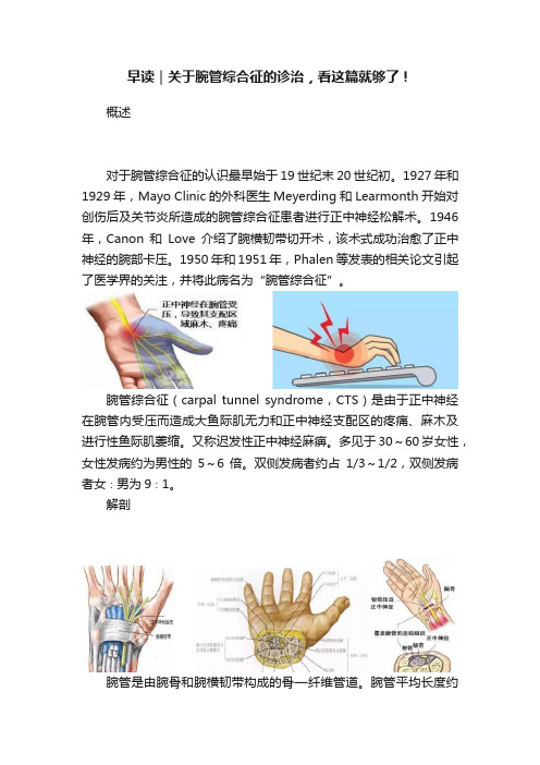
腕管综合征(carpal tunnel syndrome,CTS)是由于正中神经在腕管内受压而造成大鱼际肌无力和正中神经支配区的疼痛、麻木及进行性鱼际肌萎缩。
又称迟发性正中神经麻痹。
多见于30~60岁女性,女性发病约为男性的5~6倍。
双侧发病者约占1/3~1/2,双侧发病者女∶男为9∶1。
解剖腕管是由腕骨和腕横韧带构成的骨—纤维管道。
腕管平均长度约2.5cm。
腕管的顶部为腕横韧带,该韧带桡侧附着于大多角骨脊和舟骨结节,尺侧附着于豌豆骨和钩骨钩。
广义的腕横韧带还包括前臂深筋膜掌侧的远端部分。
腕管的内容物包括指浅屈肌腱、指深屈肌腱、拇长屈肌腱和正中神经(9条肌腱,1条神经)。
正中神经及其分支:正中神经在出腕管后,分为内侧束和外侧束。
外侧束包括鱼际支、拇指桡侧固有神经核拇指掌侧总神经;内侧束包括示指和中指的指掌侧总神经。
尽管腕管两端是开放的入口和出口,但其内组织液压力却是稳定的。
腕管内最狭窄处距离腕管边缘约50px,这种解剖特点与腕管综合症患者切开手术时正中神经形态学表现相符。
正中神经走行在屈肌支持带下方,紧贴屈肌支持带。
在屈肌支持带远端,正中神经发出返支,支配拇短展肌,拇短屈肌浅头,和拇对掌肌。
其终支是指神经,支配拇、示、中指和环指桡侧半皮肤。
病因任何能使腕管内容物增多、增大或使腕管容积缩小的因素均可导致本病。
多数病人病因不明,主要与下列因素有关:1.腕部外伤:包括骨折、脱位、扭伤、挫伤等,改变了腕管的形状,减少了腕管原有的容积。
超声对腕管综合征患者无症状手腕的评估研究
超声对腕管综合征患者无症状手腕的评估研究施杨;徐金锋;刘慧玉;华琪;邓克如【期刊名称】《中国医药科学》【年(卷),期】2016(006)017【摘要】目的:探讨超声在腕管综合征患者有症状及无症状手腕评估中的价值。
方法术前运用超声分别检查患者的患侧手腕和对侧无症状手腕,在豌豆骨和钩骨钩横截面分别测量:正中神经前后径、正中神经左右径、腕横韧带厚度,计算正中神经扁平率。
术中使用游标卡尺测量实体相应的数据。
对神经扁平率和腕横韧带厚度进行对比分析。
结果在豌豆骨平面和钩骨钩平面手术与超声测得的正中神经扁平率和腕横韧带厚度差异均无统计学意义(P >0.05),在豌豆骨平面和钩骨钩平面上,患侧手腕正中神经扁平率和腕横韧带厚度均显著高于对照组(P <0.05)。
结论超声检查可作为腕管综合症量化的诊断标准。
%Objective To investigate the value of ultrasound in assessment of symptom and without symptom in patients with carpal tunnel syndrome. Methods Both carpal canals of patients were diagnosed by ultrasound to measure the anteroposterion diameter and transverse diameter of the median nerve, and to measure the thickness of the transverse carpal ligament on pisiform bone and hamate bone planes. Then vernier caliper was used to measure the data during operations. The flat rate of nerve and the transverse ligament thickness of wrist were compared. Results The flat rate of nerve and the transverse ligament thickness of wrist had no significant difference between ultrasound and operation with the two ways (P > 0.05). The flat rate ofnerve and the transverse ligament thickness of wrist had a significant difference between the two wrists (P < 0.05). Conclusion Ultrasound can be diagnostic criteria for carpal tunnel syndrome.【总页数】4页(P149-151,182)【作者】施杨;徐金锋;刘慧玉;华琪;邓克如【作者单位】深圳市人民医院超声科,广东深圳518000;深圳市人民医院超声科,广东深圳 518000;深圳市人民医院超声科,广东深圳 518000;深圳市人民医院超声科,广东深圳 518000;深圳市人民医院超声科,广东深圳 518000【正文语种】中文【中图分类】R658.2【相关文献】1.高频超声在无症状型系统性硬化病患者腕管综合征早期诊断的应用 [J], 李园;宋烨;陆雯;王培军2.腕管综合征患者手指伸展/弯曲时正中神经位置和横截面面积变化情况的评估研究 [J], 邓永上;张云帆;谭耀灵3.超声诊断单侧尺神经病变:利用无症状侧尺神经横截面积作为参考值评估患侧病变的可靠性研究 [J], 胡培;朱昱亭;李治峰;郑光美;胡天鑫;曾小军;王云甫4.扩张型心肌病患者的无症状亲属接受超声心动图评估揭示临床前期病变 [J], Mahon N. G.;Murphy R. T.;MacRae C.A .;W. J. McKenna;韩瑞娟5.腕管综合征患者口服及局部注射类固醇疗效的临床和神经超声评估研究 [J], 朱明珍;初红;卢祖能因版权原因,仅展示原文概要,查看原文内容请购买。
超声模拟系统提高规培医师对腕管综合征诊断能力的初步研究①

超声模拟系统提高规培医师对腕管综合征诊断能力的初步研究①引言腕管综合征是一种常见的神经压迫症,主要由于腕管内结构的肿胀导致压迫正中神经而引起的,临床表现主要是手部麻木、疼痛和无力。
对于规培医师来说,准确地诊断和处理这种疾病是十分重要的,然而由于腕管综合征诊断的主要依赖于临床经验和技术水平,因此很容易发生误诊的情况。
本研究旨在探讨超声模拟系统对规培医师腕管综合征诊断能力的提高作用。
材料与方法研究对象:选择100名规培医师作为研究对象,均为腕管综合征诊断能力较弱的医师。
研究设计:采用对照实验设计,将研究对象随机分为实验组和对照组,每组50人。
实验组:接受超声模拟系统的训练,并进行腕管综合征的模拟诊断。
对照组:不接受任何训练,直接进行腕管综合征的模拟诊断。
超声模拟系统:采用先进的虚拟现实技术,模拟真实的超声检查情景和腕管综合征的影像。
通过系统的引导和模拟操作,规培医师可以获得更真实的超声检查体验,并提高诊断能力。
诊断准确性评价:采用对比组实验,对超声模拟系统训练前后的规培医师进行腕管综合征模拟诊断,评价其诊断准确性。
结果经过超声模拟系统的训练,实验组规培医师的腕管综合征模拟诊断准确性明显提高,在对比组实验中,实验组规培医师的诊断准确率达到90%,而对照组规培医师的诊断准确率仅为60%。
经统计学分析,实验组规培医师的诊断能力显著优于对照组。
讨论本研究结果表明,超声模拟系统可以有效提高规培医师对腕管综合征的诊断能力。
传统的临床经验和技术培训虽然可以提高医师的诊断能力,但由于腕管综合征的诊断主要依赖于影像学,因此超声模拟系统训练对规培医师的影像学诊断技能有显著的提高作用。
超声模拟系统的应用不仅可以提高规培医师的诊断准确性,还可以缩短诊断时间,提高工作效率,对临床工作具有积极的意义。
- 1、下载文档前请自行甄别文档内容的完整性,平台不提供额外的编辑、内容补充、找答案等附加服务。
- 2、"仅部分预览"的文档,不可在线预览部分如存在完整性等问题,可反馈申请退款(可完整预览的文档不适用该条件!)。
- 3、如文档侵犯您的权益,请联系客服反馈,我们会尽快为您处理(人工客服工作时间:9:00-18:30)。
SCIENTIFIC ARTICLEUltrasound features of carpal tunnel syndrome:a prospective case-control studyRenato A.Sernik&Claudia A.Abicalaf&Benedito F.Pimentel&Andresa Braga-Baiak&Larissa Braga&Giovanni Guido CerriReceived:19December2006/Revised:14July2007/Accepted:7August2007/Published online:8November2007 #ISS2007AbstractPurpose The purpose of the study was to examine the most adequate cut-off point for median nerve cross-sectional area and additional ultrasound features supporting the diagnosis of carpal tunnel syndrome(CTS).Material and methods Forty wrists from31CTS patients and63wrists from37asymptomatic volunteers were evaluated by ultrasound.All patients were women.The mean age was49.1years(range:29–78)in the symptomatic and45.1years(range24–82)in the asymptomatic group. Median nerve cross-sectional area was obtained using direct (DT)and indirect(IT)techniques.Median nerve echoge-nicity,mobility,flexor retinaculum measurement and the anteroposterior(AP)carpal tunnel distance were assessed. This study was IRB-approved and all patients gave informed consent prior to examination.Results In CTS the median nerve cross-sectional area was increased compared with the control group.Median nerve cross-sectional area of10mm2(DT)and9mm2(IT)had high sensitivity(85%and88.5%,respectively),specificity (92.1%and82.5%)and accuracy(89.3%and82.5%)in the diagnosis of CTS.CTS patients had an increased carpal tunnel AP diameter,flexor retinaculum thickening,reduced median nerve mobility and decreased median nerve echogenicity.Conclusion Ultrasound assists in the diagnosis of CTS using the median nerve diameter cut-off point of10mm2 (DT)and9mm2(IT)and several additional findings. Keywords Case-control.Ultrasound.Carpal tunnel syndrome.Median nerveIntroductionCarpal tunnel syndrome(CTS)is a common entrapment neuropathy of the median nerve.CTS is characterised by pain and sensory disturbance along the distribution of the median nerve,as well as thenar muscle atrophy in the advanced stages.The diagnosis may be made clinically[1] and with electromyography(EMG)[2].Ultrasound may also be valuable in the diagnosis of CTS[3].Several studies[2,4–6]have demonstrated that the combination of carpal tunnel ultrasound in combination with clinical features and EMG were more sensitive and specific than clinical evaluation or EMG in isolation.Ultrasound has proved to be able to depict normal and pathologic nerves, including anatomical variants[7,8]and abnormalities associated with CTS[9].Prior studies[10–12]have attempted to establish cut-off values for the median nerve cross-sectional area and haveSkeletal Radiol(2008)37:49–53DOI10.1007/s00256-007-0372-9R.A.Sernik(*):C.A.AbicalafDepartment of Radiology,University of São Paulo, Avenida Dr.Enéas de Carvalho Aguiar647,São Paulo,Brazil05403-900e-mail:Rasernik@.brB.F.PimentelDepartment of Orthopedics,University of Taubate, Sao Paulo,BrazilA.Braga-BaiakPost Graduation Program,Department of Radiology, University of Sao Paulo,Sao Paulo,BrazilL.BragaUniversity of Nebraska Medical Center,Omaha,NE,USAG.G.CerriDepartment of Radiology,University of Sao Paulo, Sao Paulo,Brazildescribed ultrasound features such as median nerve echo-genicity,mobility and flexor retinaculum bulging that can be useful in the diagnosis of patients with CTS.However,there is a lack of consensus in the medical literature regarding some of these topics,mainly the median nerve cut-off point.The purpose of this study was to evaluate the best cut-off value for the cross-sectional area of the median nerve and to describe additional ultrasound features that may assist in the diagnosis of CTS.Material and methods Study populationThis is a prospective case-control study comprising of 68participants and 103wrists that was carried out between September 1999and October 2000.All participants were women.The patients ’mean age was 49.1years (range:29–78)and that of the controls 45.1years (range:24–82).The 40CTS were diagnosed clinically and with EMG.They were considered to be candidates for surgery.There were 21right and 19left wrists.Of the 63asymptomatic wrists,30were on the right and 33on the left.All patients had been referred by the hospital ’s Department of Orthopaedic Surgery in a non-consecutive fashion.Participants were eliminated from the study if they had a history of underlying diseases (such as gout,rheumatoid arthritis and diabetes mellitus),pregnancy,previously performed wrist surgery or fracture,expansive lesions within the carpal tunnel and variants of the carpal tunnel (such as accessory muscles,bifid median nerve and persistent median artery).Among the originally evaluated 125wrists 22were excluded due to anatomical variations.Institutional Review Boardapproval was obtained for this rmed consent was provided by all participants prior to examination.Ultrasound imagingStudies were done using a Logic-700(General Electric,Milwaukee,WI,USA)with a linear array transducer ranging from 9to13MHz.A board certified radiologist (RAS)with expertise in musculoskeletal imaging and 6years of practical experience performed all examinations.Participants were seated with the forearm lying on a table,wrist in supination and extended fingers during the examination.Axial and transverse images were obtained at the proximal and distal carpal tunnel.All examinations followed the same protocol:a quick overview of the volar side was performed to rule out expansive lesions and anatomical variations.Subsequently,a detailed evaluation of the carpal tunnel was performed.Within the carpal tunnel,the median nerve was identified as a rounded hypoechoic structure with hyperechoic dots inside.The median nerve was qualitatively assessed considering its echogenicity.Meanwhile,the quantitative evaluation assessed median nerve cross-sectional area,flexor retinac-ulum thickness,median nerve mobility,and carpal tunnel anteroposterior (AP)distance.The median nerve cross-sectional area was obtained at the level of the pisiform bone using the direct (DT)and indirect (IT)technique.The DT was defined by tracing a continuous line around the inner hyperechoic rim of the median nerve with electronic calipers [4](Fig.1);and it was performed in all symptomatic (40out of 40)and asymptomatic wrists (63out of 63).Meanwhile,the IT resulted from the AP measurement (D1)and transverse distance (D2)of the inner median nerve calculated through the ellipsoid formula πD1ÂD2ðÞ=4½ [5](Fig.2).The IT was performed in 65%of symptomatic wrists (26out of40)Fig.1Ultrasound demonstrates the median nerve area calculated by the direct technique in a patient without carpal tunnel syndrome.A continuous line is traced around the inner hyperechoic rim of the median nerve with electronic calipers.(Median nerve cross-sectional area=0.07cm 2)Fig.2Ultrasound shows the AP and transverse distances to calculate the median nerve area using the indirect technique through the ellipsoid formula πD1ÂD2ðÞ=4½ ,in a patient without carpal tunnel syndrome.(Median nerve cross-sectional area=0.062cm 2)and 100%of asymptomatic wrists (63out of 63).The radiologist obtained three measurements of the median nerve for each technique.The mean value was used for statistical analysis.Median nerve mobility was calculated by measuring the distance between the radial margin of the median nerve and the radial margin of the ulnar artery,with and without passive flexion of the fingers (Fig.3).Median nerve mobility was assessed in 65%of the symptomatic (26out of 40)and 100%of the asymptomatic wrists (63out of 63).In addition,the AP distance of the flexor retinaculum (arched hyperechoic strip anterior to the median nerve)and the AP diameter of carpal tunnel (between the cortical bone of the capitate and the external border of the flexor retinaculum)were obtained during examinations (Fig.4).Statistical analysisThe cross-sectional area of the median nerve,thickness of the flexor retinaculum and AP diameter of the carpal tunnel of symptomatic and asymptomatic wrists were compared using a Mann –Whitney test.An unpaired Student ’s t test was used for comparing the mobility of the median nerve.The echogenicity of median nerve and the cut-off value established by DT and IT were evaluated with a Fisher ’s statistic test.Sensitivity,specificity,positive predictive value (PPV),negative predictive value (NPV)and accuracy demonstrated the efficacy of the IT values,using 15mm 2as the cut-off value [2].The coefficient of correlation was used to evaluate the relationship between DT and IT measurements.Various cut-off values were calculated to determine the best median nerve cross-sectional area.A significance level of 0.05wasused.Fig.3A 52-year-old woman with a clinical and electromyographic diagnosis of carpal tunnel syndrome.Median nerve mobility was calculated by measuring the distance between the radial margin of the median nerve and the radial margin of the ulnar artery,a with and b without passive flexion of the fingers.Measurements acquired were 0.69cm and 0.62cm respectively.MN median nerve,A ulnararteryFig.4Same patient as in Fig.3,demonstrating the AP distance of carpal tunnel measured between the capitate cortex and the external border of the flexor retinaculum.C capitate,FR flexorretinaculumFig.5A 47-year-old woman with symptoms of carpal tunnel syndrome and a positive electromyography test.Ultrasound demon-strated a hypoechogenic median nerve,with a mean cross-sectional area of 0.215cm 2ResultsThere was a statistically significant difference between the DT(15.1±7.3mm2in symptomatic and8.1mm2±1.6in asymptomatic patients;p value=0.001;Fig.5)and IT (15.51mm2±9.89in symptomatic and8.04mm2±1.50 patients;p value=0.001).The correlation between DT and IT was strong(r=0.99).Using a cut-off value of15mm2 for IT resulted in a sensitivity of34.6%,specificity of 100%,PPV of100%,NPV of78.8%and accuracy of80.9% in diagnosing CTS.With a cut-off value of9mm2for IT, the corresponding results were88.5%,82.5%,67.7%, 94.6%and82.5%respectively.With a cut-off value of 10mm2for DT the values were85.0%,92.1%,87.2%, 90.6%and89.3%respectively.In32of the40symptomatic wrists(80%)the median nerve was hypoechoic(p=0.001and p=0.004for DT and IT respectively).In addition,a statistically significant difference was found for the AP distance of the carpal tunnel(CTS=1.28±0.18cm,control=1.18±0.1cm;p=0.003),the flexor retinaculum thickness(CTS=0.88±0.23mm,control= 0.75±0.1mm;p=0.018),and mobility of the median nerve (CTS=0.96±0.66mm,range:0.07–3.1mm;control=1.29±0.7mm,range:0.13–3mm;p=0.041).DiscussionThis study demonstrates that a median nerve cross-sectional area of more than10mm2(DT),and more than9mm2(IT) are both sensitive and specific in diagnosing CTS. Furthermore,symptomatic wrists had a thicker flexor retinaculum,an increased AP diameter of the carpal tunnel, and reduced echogenicity and mobility of the median nerve, compared with the asymptomatic group.The cross-sectional area of the median nerve(DT and IT) at the pisiform bone level was higher in symptomatic wrists compared with the asymptomatic group.These findings concur with previous studies[2,4,5].In the present study both DT and IT were demonstrated to be accurate in diagnosing CTS,contrary to Duncan et al.[4]who reported that the DT had more accurate results than the IT. Moreover,a high correlation(r=0.99)between the areas calculated through DT and IT,in symptomatic and asymptomatic wrists,was found in our analysis.Kamolz et al.stressed the need for standardisation of median nerve cross-sectional area cut-offs[13].The majority of previously published studies addressed values ranging from9mm2to12mm2[3,4,6,10–12,14–17]. Our results demonstrated that10mm2(DT)is the most adequate cut-off value(specificity=92.1)this corroborates previous findings[10,15].In addition,our study demon-strated that decreased echogenicity of the median nerve was found in80%of symptomatic wrists.Only a few authors have considered qualitative changes of median nerve as a diagnostic criterion,mainly because the changes in the median nerve echogenicity are prone to angle beam artifacts,are operator-and equipment-dependent,and may be too subtle to be detected by all readers[2,4,18].We believe that such changes should be included in the diagnosis of CTS.To the best of our knowledge,this is the first series to demonstrate a difference in flexor retinaculum thickness and AP diameter of the carpal tunnel in symptomatic and asymptomatic wrists.Lee and colleagues[2]reported flexor retinaculum thickness and AP carpal tunnel distance measurements.However,they only reported values for asymptomatic wrists.Nakamichi and Tachibana[19]described patients with CTS having decreased median nerve transverse sliding compared with the control group,indicating that there is a restricted physiological motion of the nerve.The results from our study concur with this finding.We recognise some of the limitations of this study.First, it was a single institution study with a relatively small sample size.Second,ultrasound examinations were per-formed by a single radiologist and inter-observer agreement of the ultrasound findings was not attempted.Finally,our sample was composed solely of female participants. However,previous studies did not demonstrate gender-related differences in the ultrasound cross-sectional area among male and female CTS patients[4,11].In conclusion,ultrasound assists in the diagnosis of CTS using the median nerve diameter(cut-off point of10mm2 [DT]and9mm2[IT])and several additional findings. References1.Phalen GS.The carpal-tunnel syndrome.Clinical evaluation of598hands.Clin Orthop Relat Res1972;83:29–40.2.Lee D,van Holsbeeck MT,Janevski PK,Ganos DL,DitmarsDM,Darian VB.Diagnosis of carpal tunnel syndrome.Ultra-sound versus electromyography.Radiol Clin North Am1999;37: 859–872.3.Wiesler ER,Chloros GD,Cartwright MS,Smith BP,Rushing J,Walker FO.The use of diagnostic ultrasound in carpal tunnel syndrome.J Hand Surg[Am]2006;31:726–732.4.Duncan I,Sullivan P,Lomas F.Sonography in the diagnosis ofcarpal tunnel syndrome.AJR Am J Roentgenol1999;173:681–684.5.Buchberger W,Schon G,Strasser K,Jungwirth W.High-resolution ultrasonography of the carpal tunnel.J Ultrasound Med1991;10:531–537.6.Koyuncuoglu HR,Kutluhan S,Yesildag A,Oyar O,Guler K,Ozden A.The value of ultrasonographic measurement in carpal tunnel syndrome in patients with negative electrodiagnostic tests.Eur J Radiol2005;56:365–369.7.Beekman R,Visser LH.High-resolution sonography of theperipheral nervous system—a review of the literature.Eur J Neurol2004;11:305–314.8.Silvestri E,Martinoli C,Derchi LE,Bertolotto M,ChiaramondiaM,Rosenberg I.Echotexture of peripheral nerves:correlation between US and histologic findings and criteria to differentiate tendons.Radiology1995;197:291–296.9.Buchberger W,Judmaier W,Birbamer G,Lener M,SchmidauerC.Carpal tunnel syndrome:diagnosis with high-resolutionsonography.AJR Am J Roentgenol1992;159:793–798.10.Wong SM,Griffith JF,Hui AC,Lo SK,Fu M,Wong KS.Carpaltunnel syndrome:diagnostic usefulness of sonography.Radiology 2004;232:93–99.11.Ziswiler HR,Reichenbach S,V ogelin E,Bachmann LM,VilligerPM,Juni P.Diagnostic value of sonography in patients with suspected carpal tunnel syndrome:a prospective study.Arthritis Rheum2005;52:304–311.12.Yesildag A,Kutluhan S,Sengul N,et al.The role of ultrasono-graphic measurements of the median nerve in the diagnosis of carpal tunnel syndrome.Clin Radiol2004;59:910–915.13.Kamolz LP,Schrogendorfer KF,Rab M,Girsch W,Gruber H,Frey M.The precision of ultrasound imaging and its relevance for carpal tunnel syndrome.Surg Radiol Anat2001;23:117–121. 14.Sarria L,Cabada T,Cozcolluela R,Martinez-Berganza T,GarciaS.Carpal tunnel syndrome:usefulness of sonography.Eur Radiol 2000;10:1920–1925.15.Chen P,Maklad N,Redwine M,Zelitt D.Dynamic high-resolution sonography of the carpal tunnel.AJR Am J Roentgenol 1997;168:533–537.16.Kele H,Verheggen R,Bittermann HJ,Reimers CD.The potentialvalue of ultrasonography in the evaluation of carpal tunnel syndrome.Neurology2003;61:389–391.17.Beekman R,Visser LH.Sonography in the diagnosis of carpaltunnel syndrome:a critical review of the literature.Muscle Nerve 2003;27:26–33.18.Buchberger W.Radiologic imaging of the carpal tunnel.Eur JRadiol1997;25:112–117.19.Nakamichi K,Tachibana S.Restricted motion of the mediannerve in carpal tunnel syndrome.J Hand Surg[Br]1995;20: 460–464.。
