SERCA-review 02
三阴性乳腺癌的治疗现状

II (n=30)
Neoadjuvant TNBC
E-Cis-FP
pCR=40%; ORR=86%
Silver ()
II (n=28)
Neoadjuvant TNBC
Cis
pCR=22%
Leone ()
Retro (n=125)
Sikov ()
Platinum + D
pCR=34%, OS @ 5yr=55%, OS greater with cis vs carbo
Carbo=carboplatin; Cis=cisplatin; D=docetaxel; E=epirubicin; F=5-FU; H=trastuzumab; P=paclitaxel; retro=retrospective.
三阴性乳腺癌的治疗现状
第22页
(7) High dose chemotherapy(HDC ) for TNBC
五、TNBC-分子病理特征
三阴性乳腺癌的治疗现状
第8页
临床表现为侵袭性病程; 远处转移风险较高,内脏转移几率较骨转移高,脑转移几率也较高。预后较差,死亡风险较高。
六、TNBC-临床特征
三阴性乳腺癌的治疗现状
第9页
TNBC: Shorter Median Time from
Distant Relapse to Death
WSG AM 01试验 9个以上淋巴结阳性乳腺癌患者分为两组 A组: 密集EC× 2 序贯 HDC × 2 ( EPI 90 mg/m2,CTX 3 g/m2,塞替派400 mg/m2)B组: 密集EC × 4 序贯 密集CMF × 3结果表明,年轻三阴性乳腺癌患者从HDC中获益最多。
骨骼肌的兴奋-收缩耦联
骨骼肌的兴奋-收缩耦联Excitation-contraction coupling of a skeletal muscle【教学对象与学时】一、教学对象:五年制本科二、学时:4学时【预习要求】预习骨骼肌收缩-耦联的概念、步骤、肌丝滑行机制、单收缩及复合收缩【目的要求】一、操作:1.学会正确制备离体缝匠肌肌肉标本2.掌握同时记录骨骼肌动作电位和收缩的实验方法二、理论:1.引起组织兴奋的有效刺激必须具备的条件2.可兴奋组织兴奋一次其兴奋性的变化3.掌握兴奋-收缩耦联的定义、步骤、耦联因子和结构基础4.了解肌丝滑行的机制和骨骼肌收缩的总和【重点和难点】一、重点1. 掌握正确的制备离体缝匠肌肌肉标本的方法2. 理解不同频率的连续有效刺激对骨骼肌收缩的影响3. 理解横管在骨骼肌兴奋-收缩耦联中的作用二、难点1. 理解记录离体肌肉的放电情况与单个细胞的动作电位的异同【教学过程设计】1.课前提问和预习检查2.讲解同时记录离体肌肉放电和收缩活动的原理3.介绍肌肉标本和实验装置的连接和机械-电换能器的工作原理4.讲解BL-410生物机能实验系统的相关计算机操作5.演示制备缝匠肌标本6.学生以小组为单位完成实验,并观察不同强度和频率的有效刺激以及20%的高渗甘油任氏液对肌肉放电和收缩活动的影响7.实验结束前汇总全班实验结果,讨论比较刺激强度和频率变化时骨骼肌放电和收缩的变化以及它们之间的关系【课前预习检查或提问】一、何谓兴奋-收缩耦联?包括哪几个主要环节?二、什么是兴奋、兴奋性、阈刺激?三、正常情况下体内骨骼肌收缩是以何种形式进行的,有何生理意义?【课前讲解】一、实验设计的原理蟾蜍的一些基本生命活动和生理功能与温血动物相似,而其离体组织所需的生活条件比较简单,故选用蟾蜍缝匠肌标本来观察骨骼肌的收缩特性。
活的组织具有兴奋性,能接受刺激产生兴奋,在肌肉组织表现为收缩,但刺激要引起兴奋,其刺激强度、持续时间和强度-时间变化率均须达到某一最小值,即为有效刺激。
Critical Reviews in Oral Biology & Medicine
/Critical Reviews in Oral Biology & Medicine/content/14/2/128The online version of this article can be found at:DOI: 10.1177/1544111303014002062003 14: 128CROBM T.M.T. Waltimo, B.H. Sen, J.H. Meurman, D. Ørstavik and M.P.P. HaapasaloYeasts in Apical PeriodontitisPublished by: On behalf of:International and American Associations for Dental Research can be found at:Critical Reviews in Oral Biology & Medicine Additional services and information for/cgi/alerts Email Alerts:/subscriptions Subscriptions: /journalsReprints.nav Reprints:/journalsPermissions.nav Permissions:What is This?- Mar 1, 2003Version of Record >>IntroductionA pical periodontitis is an inflammatory process of the peri-apical area caused by an infection of the dental root canal system. In most cases, chemo-mechanical preparation of the root canal and local medication with calcium hydroxide fol-lowed by filling of the root canal with gutta-percha and sealer result in elimination of the infection and healing of the lesion. Occasionally, apical periodontitis does not respond favorably to root canal therapy, and periapical inflammation caused by the root canal infection may persist months or even years despite treatment. Several factors may contribute to the failure of the treatment of persistent cases. Most commonly, these fac-tors are related to difficulties in chemo-mechanical prepara-tion of the root canals. Occasionally, micro-organisms resistant to conservative therapy may also be involved (Byström, 1986; Sirén et al., 1997). Literature on the microbiological findings in persistent apical periodontitis not responding favorably to conservative therapy is limited (Bender and Selzer, 1952; Grahnen and K rasse, 1963; Engström, 1964; Goldman and Pearson, 1969; Haapasalo et al., 1983; Ranta et al., 1988; Sirén et al., 1997; Molander et al., 1998; Hancock et al., 2001; Kalfas et al., 2001; Love, 2001). However, it is known that a few species are more frequently isolated from persistent cases compared with primary cases. These include the Enterococcus faecalis/faecium group, enteric Gram-negative facultative rods, i.e., coliforms, and Pseudomonas species (Engström, 1964; Haapasalo et al., 1983; Ranta et al., 1988; Sirén et al., 1997; Hancock et al., 2001;Love, 2001). Recently, there has also been increasing interest in the presence and role of yeasts in infections resistant to con-servative root canal therapy (Nair et al., 1990; Sen et al., 1995, 1997a,b). This review presents the contemporary knowledge of the occurrence and biotypes of yeasts in endodontic infections, and susceptibility, in vitro, of yeasts to endodontic irrigants, local disinfectants, and antifungal agents.Taxonomy and General Characteristics of Yeasts Yeasts belong to a separate kingdom of living organisms, fungi. Contrary to bacteria, fungi are eukaryotic organisms, i.e., their genome is organized in a nucleus which is surrounded by a membrane. This membrane is continuous with the endoplas-mic reticulum, and organelles, such as mitochondria, ribo-somes, and different storage inclusions, are present. Fungal cell walls are rigid structures composed mainly of glucan, mannan, and chitin. For nutrition, fungal organisms are dependent on nitrogen and carbon compounds which are taken up through the cell wall (De Hoog and Guarro, 1995).Yeasts are present in various sites in the human body as members of the normal flora. They occur, e.g., in the gastro-intestinal tract, vagina, and perineal area (Jarvis, 1996). The oral cavity has suitable environmental conditions for yeast colo-nization. Oral yeasts belong to the division Ascomycota and class Endomycetes, which is divided further into four families: Saccharomycetaceae, Endomycetaceae, Dipodascaceae, and Lipomycetaceae. Clinically, the most important oral yeastsY EASTS IN A PICAL P ERIODONTITIST.M.T. Waltimo1*B.H. Sen2J.H. Meurman3D. Ørstavik4M.P.P. Haapasalo51Institute of Dentistry, University of Turku, Lemminkäisenkatu 2, 20520 Turku, Finland; 2Department of Restorative Dentistry and Endodontics, Ege University, Izmir, Turkey; 3Institute of Dentistry, University of Helsinki, and Department of Oral and Maxillofacial Diseases, Helsinki University Central Hospital, Finland; 4NIOM, Scandinavian Institute of Dental Materials, Haslum, Norway; and 5Department of Endodontics, Dental Faculty, University of Oslo, Norway; *corresponding author, tuomas.waltimo@utu.fiABSTRACT: Microbiological reports of apical periodontitis have revealed that yeasts can be isolated from approximately 5-20% of infected root canals. They occur either in pure cultures or together with bacteria. Almost all isolated yeasts belong to the genus Candida, and the predominant species is C. albicans. Pheno- and genotypic profiles of C. albicans isolates show hetero-geneity comparable with those of isolates from other oral sites. C. albicans expresses several virulence factors that are capable of infecting the dentin-pulp complex, including dentinal tubules. This causes, consequentially, an inflammatory response around the root apex, which suggests a pathogenic role for this organism in apical periodontitis. Yeasts are particularly associated with persistent root canal infections that do not respond favorably to conservative root canal therapy. This may be due to the resis-tance of all oral Candida species against a commonly used topical medicament, calcium hydroxide. However, other antimicro-bial agents may offer alternative therapeutic approaches and improve the treatment of these persistent cases of apical perio-dontitis.Key words.Apical periodontitis, Candida, endodontics, yeast infection.128Crit Rev Oral Biol Med14(2):128-137(2003)belong to the family Saccharomycetaceae and to the genus Candida . Reproduction of Candida is based on multilateral bud-ding, which may take place anywhere on the mother cell (de Hoog and Guarro, 1995).Oral Yeast SpeciesCandida albicans is the most dominant oral yeast species, fol-lowed by C. glabrata, C. krusei, C. tropicalis, C. guilliermondii, C.kefyr, and C. parapsilosis (Odds, 1988). Recent findings also sug-gest the occasional occurrence of C. dubliniensis, which is a species closely related to C. albicans (Hannula et al ., 1997). Other yeast genera have also been isolated from the oral normal flora,e.g., Saccharomyces spp. and Geotrichum spp. (Tawfik et al ., 1989;Stenderup, 1990; Heinic et al ., 1992). Isolation of other fungi from the oral cavity has also been reported, but they are usual-ly seen in association with systemic disease—for example, pul-monary cryptococcosis caused by Cryptococcus neoformans (Stenderup, 1990).Virulence Factors of CandidaThe transition of C. albicans from a harmless commensal to apathogenic organism appears to be dependent on minorchanges in predisposing conditions which cause the expres-sion of a variety of virulence factors (Shepherd, 1992; Sweet,1997). These factors include adherence, hyphal formation,thigmotropism, protease secretion, and phenotypic switch-ing phenomenon.Adherence of micro-organisms is a complex, multifactori-al process involving several types of cell-surface adhesinswhich are essential for colonization and infection of the host.The main adhesin molecules of C. albicans responsible foradhesion to host cells seem to be cell wall mannoproteins(Sweet, 1997). However, several other factors also contribute tothe adherence of yeasts, e.g., cell-surface hydrophobicity, envi-ronmental pH, and concentrations of iron, calcium, zinc, andcarbon dioxide (Ener and Douglas, 1992; K lotz, 1994;Samaranayake et al ., 1995; Sohnle et al ., 2001). Furthermore,environmental proteins from saliva and gingival crevicularfluid as well as extracellular matrix components affect thecomplex adherence of Candida to host cells and tissues(Calderone et al ., 2000; Holmes et al ., 2002). C. albicans is a pleo-morphic micro-organism demonstrating different growthforms such as germ tubes, yeasts (blastospores), pseudo- andtrue hyphae, and chlamydospores (Odds, 1988; de Hoog andGuarro, 1995). All growth patterns except chlamydosporesmay show conversion to each form of growth, depending onthe environmental conditions. Therefore, the term 'dimorphic',often used in the literature, is semantically inaccurate toexplain C. albicans morphogenesis. Although hyphal formation is not a prerequisite for pathogenicity of C.albicans , biopsies of candidal infections often revealhyphal adherence to and penetration through epithe-lial tissues, indicating increased pathogenicity in com-parison with ovoid yeast forms (Sweet, 1997). It seemsthat the hyphal penetration into tissues is enhanced by thigmotropism, i.e., contact sensing by hyphae to find intracellular junctions or microscopic breaks on mucosal surfaces (Sherwood et al ., 1992; Gow et al .,1994; Sweet, 1997). One of the key virulence determi-nants of Candida species is their ability to produce and secrete aspartyl proteases which digest a variety of host proteins. The virulence of these proteases has beendemonstrated with animal experiments showing that the amount of protease is directly comparable with the patho-genicity of the strain (MacDonald and Odds, 1983; K wong-Chung et al ., 1985; Okamoto et al ., 1993; Togni et al ., 1994).Therefore, the higher rate of protease activity of C. albicans in comparison with other Candida species also suggests higher virulence. In addition to these major virulence factors, C. albi-cans has a tendency to phenotypic alteration, which con-tributes to environmental adaptation. Phenotypic alterations include change of colony morphology and protease activity (Slutsky et al ., 1985; White and Agabian, 1995). This genetical-ly controlled phenomenon is known as phenotypic switching,and it may occur relatively frequently, especially under stress (Soll, 1988). Phenotypic switching may assist in survival of and colonization by the yeasts, and it may also lead to genetic selection of adaptive strains (Sweet, 1997). Virulence factors and their possible contributions to apical periodontitis are list-ed in Table 1.Oral Yeast Infections A characteristic feature of yeast infections is that they develop when the host provides the environmental conditions and nutrients essential for attachment, growth, and reproduction of fungi. In other words, yeasts are opportunistic pathogens.Thus, local or general predisposing factors are required for yeast infection to develop (Shepherd, 1992). These factors can be classified into four categories: (i) host factors, such as nor-mal and pathological changes in physiological status of the host; (ii) dietary factors, such as carbohydrate-rich diets and vitamin deficiencies; (iii) mechanical factors, such as denture-wearing; and (iv) iatrogenic factors, such as administration of broad-spectrum antibiotics and corticosteroids (Odds, 1988).The clinical form of candidosis is often related to a predispo-sing factor, e.g., acute pseudomembraneous candidosis (thrush) is often associated with natural factors such as immunological and microbiological instability at birth, angu-lar cheilitis with dietary factors, chronic atrophic candidosis (denture-associated stomatitis) with mechanical factors, and acute atrophic candidosis with iatrogenic factors (Lynch,1994). In dental practice, chronic atrophic candidosis (denture stomatitis) is perhaps one of the most frequently encountered oral Candida infections (Wilson, 1998). There has also been an increasing interest in the presence of yeasts in infected perio-dontal pockets and their possible role in the pathology of dif-ferent forms of periodontitis (Slots et al ., 1988; Zambon et al .,1990; Rams and Slots, 1991; Dahlén and Wickström, 1995;Hannula et al ., 1997, 2001).14(2):128-137 (2003)Crit Rev Oral Biol Med129TABLE 1Virulence Factors of C. albicans and Their Possible Contributions to Apical Periodontitis Virulence Factor Possible Contribution to Apical Periodontitis Adherence Colonization of dental hard tissues Hyphal formation Penetration into dentinal tubules Thigmotropism Penetration into dentinal tubulesProtease secretion Survival in conditions with limited nutrient supply Phenotypic alteration Adaptation in ecologically harsh conditionsMicrobiology of Apical PeriodontitisApical periodontitis is a host defense response to infection of necrotic pulp (Miller, 1894;Kronfeld, 1939; Kakehashi et al .,1965). The host has an array of defense mechanisms consisting of several types of inflammato-ry cells, such as polymorphonu-clear leukocytes and lympho-cytes, intercellular messengers,such as cytokines, and chemical weapons such as proteolytic enzymes (Nair, 1997). Despite these defenses, the body cannot eliminate the micro-organisms residing in the necrotic root canal, and therefore the inflam-matory process does not result in healing. The interaction between root canal infection and the host defense mecha-nisms eventually cause destruc-tion of periapical tissues and formation of apical periodonti-tis (Nair, 1997).More than 300 species of micro-organisms colonize the human oral cavity, but only a limited number of these have been isolated from infected root canals with apical periodontitis (Moore, 1987). Several factors contribute to the selection of micro-organisms. Primarily , the selection takes place among those micro-organisms entering the root canal, which depends on the pathway to the pulp. For example, a deep caries lesion may serve as a pathway and limit the number of possible microbial species. In addition,the host defense mechanisms in the infected but still vital pulp reduce the number of surviving species. Furthermore, environ-mental factors of the necrotic root canal, e.g., redox-potential and source of nutrients, give an advantage to species with pro-teolytic activity and ability to survive in anaerobic conditions.Finally, microbial interactions—either negative (such as compe-tition for nutrients, secreted toxic metabolites, and specific bacteriocins) or positive (i.e.,symbiosis of different spe-cies)—regulate the microflora of130Crit Rev Oral Biol Med14(2):128-137(2003)TABLE 2Micro-organisms Commonly Associated with Chronic Periodontitis, Apical Periodontitis, and Persistent Root Canal InfectionsDisease Group Micro-organismsReferencesChronic Gram-negative Fusobacterium nucleatum Moore and Moore, 1994periodontitisanaerobic rodsPorphyromonas gingivalis,Haffajee and Socransky, 1994Prevotella intermedia,Slots, 1999Campylobacter rectus,Hannula et al ., 2001Selenomonas spp.Treponema denticola Bacteroides forsythus Gram-negative Actinobacillusanaerobic rods actinomycetemcomitans Eikenella corrodens Gram-positive Peptostreptococcus micros anaerobic cocci Gram-positive Eubacterium spp.anaerobic rods Gram-positive Streptococcus intermedius facultative cocci YeastCandida albicans ApicalGram-negative Prevotella spp.Sundqvist, 1994periodontitisanaerobic rodsFusobacterium nucleatum,Sundqvist et al ., 1998Porphyromonas spp.Haapasalo, 1989Campylobacter rectus Selenomonas spp.Gram-positive Peptostreptococcus micros anaerobic cocci Gram-positive Eubacterium spp.anaerobic and Propionibacterium acnes facultative rods Actinomyces ctobacillus spp.Gram-positive Streptococcus spp.facultative cocciPersistent root Gram-positive Enterococcus faecalis Haapasalo et al ., 1983canal infectionfacultative cocci Streptococcus spp.Sirén et al ., 1997Waltimo et al ., 1997Gram-positive Peptostreptococcus spp.Sundqvist et al ., 1998anaerobic cocci Molander et al ., 1998Hancock et al ., 2001Gram-positive Actinomyces spp.anaerobic and facultative rods Gram-negative Bacteroides spp.anaerobic and facultative rods YeastCandida albicansthe infected root canal (Sundqvist, 1994).Apical periodontitis is a polymicrobial infection domina-ted by obligate anaerobes (Bergenholtz, 1974; Kanz and Henry, 1974; Sundqvist, 1976; Byström et al., 1985; Haapasalo, 1986; Sundqvist et al., 1989; Baumgartner and Falkler, 1991). Usually, the number of isolated species is between two and eight, and monoinfections are rare (K anz and Henry, 1974; Sundqvist, 1976, 1994; Haapasalo, 1986). Before root canal therapy, the most frequently isolated micro-organisms are: Gram-negative anaerobic rods, such as Prevotella spp., Porphyromonas spp., Fusobacterium nucleatum, Campylobacter rectus, and Selenomonas spp.; Gram-positive anaerobic cocci, such as Peptostreptococcus spp.; Gram-positive anaerobic and facultative rods, such as Eubacterium spp., Propionibacterium acnes, Actinomyces spp., and Lactobacillus spp.; and Gram-positive facultative Streptococcus species (Sundqvist, 1976; Haapasalo, 1986). The microbiology of root canal infections is still not clear in many regards, e.g., the data concerning the occurrence of uncultivable species such as spirochetes are scarce (Dahle et al., 1993).The literature on microbiological findings in persistent root canal infections is also relatively limited (Bender and Selzer, 1952; Grahnen and Krasse, 1963; Engström, 1964; Goldman and Pearson, 1969; Haapasalo et al., 1983; Ranta et al., 1988; Sirén et al., 1997; Molander et al., 1998). However, it is known that a few species are frequently isolated from persistent cases. These include the Enterococcus faecalis/faecium group, enteric Gram-negative facultative rods (i.e., coliforms), and Pseudomonas species (Engström, 1964; Haapasalo et al., 1983; Ranta et al., 1988; Molander et al., 1998; Sundqvist et al., 1998; Hancock et al., 2001; Love, 2001). Micro-organisms commonly associated with chronic periodontitis, apical periodontitis, and persistent root canal infections are listed in Table 2.Yeasts in Apical Periodontitis Microbiological investigations of apical periodontitis during the past 50 years have revealed that yeasts can be isolated from infected root canals (Grossman, 1952; Slack, 1953, 1957; Macdonald et al., 1957; Hobson, 1959; Goldman and Pearson, 1969; Matusow, 1981; Nair et al., 1990; Najzar-Fleger et al., 1992; Sen et al., 1995, 1997a,b; Waltimo et al., 1997; Molander et al., 1998). Slack (1953, 1957) reported that yeasts exist in about 5% of cases of apical periodontitis. According to Grossman (1952), as many as 17% of infected root canals may contain Candida species. Hobson (1959) reported that Candida albicans was often isolated from root canal infections, although their pathogenici-ty in the root canal was unclear. However, according to a case report, a pure culture of Candida albicans caused acute pulpal-alveolar cellulitis (Matusow, 1981).In another case report, C. albicans was found in root canals and in periapical granulomas of a patient suffering from chron-ic urticaria (Eidelmann et al., 1978). The complete cure of the patient was achieved only after the extraction of the infected teeth. Histological examination revealed that the granuloma exhibited an invasive Candida infection composed of acute and chronic granulation tissue along with hyphae and yeast cells. The root canal surfaces were covered by dense masses of yeast cells, and dentinal tubules were totally filled with hyphae. In addition to these cases, Damm et al. (1988) described two cases of cancer patients having dentinal candidosis. In the first case, carious dentin of the patient's deciduous teeth contained numerous oval to filamentous Candida cells. The teeth exhibit-ed either acute irreversible pulpitis or acute apical plete healing was accomplished after extraction of all deciduous teeth. The second case demonstrated exposed coro-nal dentin with heavy colonization by C. albicans. Pseudohyphae and yeast cells were present not only in pulp tissue but also in cervical and apical soft tissues. As seen in these cases, extensive invasion by fungi seems to be mostly associated with the immunocompromised state of the patients. However, Kinirons (1983) described a similar clinical case with no systemic illness.Nair et al. (1990) studied therapy-resistant root canal infec-tions and found micro-organisms in 6 of 9 specimens. Bacteria were shown in 4 of the 6 cases, while yeast-like organisms were found in 2 cases as judged by electron microscopy. The pres-ence of intraradicular fungi in the endodontically treated human teeth was associated with periapical lesions that per-sisted after treatment. Sen et al. (1995) observed bacteria and fungi with scanning electron microscopy in infected root canals and dentinal tubules associated with periapical lesions. They found that 4 out of 10 root canals were heavily infected with yeasts, confirming the association between yeasts and root canal infections. They formed dense but separate colonies, and, in one specimen, hyphal elements were also present. Since the patients in this study did not have any systemic disease, the presence of yeasts in root canals may be attributed to poor oral hygiene.In a report by Waltimo et al. (1997), the occurrence of yeasts was studied in 967 microbiological samples taken from cases of apical periodontitis not responding favorably to conventional treatment. Micro-organisms were found in 692 (72%) samples, whereas 275 (28%) showed no growth. Forty-eight fungal strains were isolated from 47 samples, which represented 7% of the culture-positive samples. The fungi were endomyceteous yeasts, and they were isolated either in pure culture (6 cases, 13%) or together with bacteria (41 cases, 87%). The identifica-tion of yeasts was carried out with conventional clinical labo-ratory procedures, showing results comparable with those of earlier studies. Almost all isolates belonged to the genus Candida, and C. albicans was the most common species. C.g labrata, C. g uilliermondii, C. inconspicua, and Geotrichum can-didum were also isolated.In studies of the initial microbial flora of root canal infec-tions, yeasts have usually not been found (Haapasalo, 1989; Sundqvist et al., 1989). However, according to a recent study of randomly selected patients with periapical radiolucencies, C. albicans was detected in 5 out of 24 samples (21%) taken from infected root canals by means of the polymerase chain-reaction-based (PCR) molecular detection technique (Baumgartner et al., 2000). The PCR was carried out conventionally with a detection limit of 10-4 ng of DNA. Although the material was limited, the high percentage may be due to the higher sensitivity of the method in comparison with detection of micro-organisms by conventional culture procedures. However, the finding indi-cates that yeasts may be present in low numbers at the start of root canal treatment, and that they may reach higher propor-tions during conventional treatment procedures. In another recent study, intact root canals with pulp necrosis were exam-ined microbiologically (Lana et al., 2001). C. tropicalis and S. cerevisiae were recovered from two canals (7.4%) before root canal therapy. C. guilliermondii and C. parapsilosis were cultivat-ed in the second and third collections, respectively, of root canal contents. According to these findings, it is also possible that yeasts which are common opportunistic pathogens of the oral14(2):128-137 (2003)Crit Rev Oral Biol Med131cavity gain access to the root canal as contaminants duringendodontic therapy (Sirén et al ., 1997). This emphasizes the importance of aseptic treatment procedures in the prevention of persistent infections. Regardless of the source and means of entry for yeasts into the root canal, their presence in cultivable numbers may have clinical importance in persistent cases.Influence of Necrotic Root Canalon Strain SelectionNecrotic root canals provide harsh ecological conditions for micro-organisms in comparison with other oral sites. The influ-ence of these conditions for yeast strain selection was examined in a recent study that compared the phenotypes and genotypes of C. albicans isolates from root canals and periodontal crevices (Waltimo et al ., 2001). Briefly , the phenotyping was based on the presence of 5 enzymes (valine arylamidase, phosphoamidase,alpha-glucosidase, beta-glucosidase, and N-acetyl-beta-glucosaminidase), ability of the strains to assimilate 11carbohydrates (glycerol, L-arabinose, xylose, adonitol,xylitol, sorbitol, methyl-D-glucoside, N-acetyl-D-glu-cosamine, sucrose, trehalose, and melezitose), and the resistance to boric acid (Williamson et al ., 1987).Genotyping was based on randomly amplified polymor-phic DNA (RAPD) profiles obtained with the use of two different primers.A total of 14 different phenotypes was found among the 37 root canal isolates of C. albicans . The majority of the isolates (26) were classifiable into three major phenotypes, described by Williamson et al . (1987):16 isolates (43.2%) belonged to phenotype A1R, 6(16.2%) to A1S, and 4 (10.8%) to B1S. Interestingly, C.albicans phenotype A1R, which was predominant, has been associated mainly with patients with symptomatic C. albicans infections but not with asymptomatic carri-ers. This may indicate a higher virulence of this pheno-type in comparison with other phenotypes (Xu and Samaranayake, 1995). The genotypic characterization with use of the combination of the two different primers yielded 32 different profiles for the 37 C. albicans strains,demonstrating high genotypic divergence of the iso-lates.Analysis of the current data implies genotypic het-erogeneity of C. albicans isolates from root canals in humans. However, frequently encountered phenotypes were similar to the ones reported from other oral and non-oral sources (Bostock et al ., 1993; Tsang et al ., 1995;Xu and Samaranayake, 1995; Matee et al ., 1996). This implies that phenotypically unusual strains of C. albicans are not frequently involved in root canal infections.Therefore, it seems that the root canal, an ecologically harsh niche with regard to redox-potential and nutrients supply, may not have an impact on strain selection that differs from those of other oral sites. Thus, it seems that a general characteristic of C. albicans is its ability to tolerate a wide variety of different environmental conditions.Accompanying Bacteria in Yeast InfectionsRecent studies have shown that yeasts can survive as a monoinfection of the root canal (Matusow, 1981; Waltimo et al ., 1997). However, they are usually found in mixed cultures together with bacteria. Yeasts may often be iso-lated together with facultative Gram-positive bacteria such as a- and non-hemolytic Streptococcus species, whereas Gram-negative isolates are rare (Waltimo et al ., 1997). The dom-inance of the facultative Gram-positive accompanying bacteria may be due to the special ecological conditions of the root canal during prolonged treatment, which could favor yeasts and streptococci. There may also be synergism between these micro-organisms. However, no or only negative association of Streptococcus spp. with other bacteria has been reported in root canals (Sundqvist, 1992). However, it has been reported that C.albicans may prolong the viability of -hemolytic streptococci (Burnet and Sherp, 1968). Furthermore, C. albicans co-aggre-gates with a variety of streptococci such as S. g ordonii, S.mutans, and S. sanguis (Holmes et al ., 1995; Nikawa et al ., 2001).This may promote their colonization and thus explain the con-comitant occurrence of these microbial species. Further investi-gations concerning microbial interactions are needed for a bet-132Crit Rev Oral Biol Med14(2):128-137(2003)Figure 1. Scanning electron micrograph of C. albicans blastospores on root canal surface in vitro . The bar indicates 10 m.Figure 2.Scanning electron micrograph of C. albicans hyphae penetrating a dentinal tubule. The bar indicates 2 m.。
thernostics under review -回复
thernostics under review -回复Title: Understanding and Analyzing the Role of TheragnosticsIntroduction:Theragnostics, an emerging field in medicine, combines therapeutics and diagnostics to provide personalized treatment options for patients. It revolutionizes the healthcare industry by tailoring treatment plans for individuals based on their unique genetic makeup, disease characteristics, and response to treatment. This article aims to explore the concept, development, challenges, and potential applications of theragnostics.I. Defining Theragnostics:Theragnostics, often referred to as theranostics, is a fusion of therapeutics and diagnostics. It encompasses the integration of diagnostic tools, such as medical imaging and biomarker analysis, with targeted therapy interventions. By integrating diagnosis and therapy, theragnostics ensures a more precise and individualized approach to healthcare.II. Evolution of Theragnostics:The concept of theragnostics can be traced back to the late 1990swhen researchers recognized the need for personalized medicine. Advances in genomics, proteomics, and imaging techniques laid the groundwork for the development of theragnostics. It was a paradigm shift from the traditional one-size-fits-all approach to a patient-centric model.III. Key Diagnostic Modalities in Theragnostics:a. Medical Imaging: Various imaging techniques, including positron emission tomography (PET), single-photon emission computed tomography (SPECT), and magnetic resonance imaging (MRI), are used to visualize and diagnose diseases. Imaging agents tagged with radioisotopes or paramagnetic substances enable accurate detection and localization of targets for subsequent therapy.b. Biomarkers: These are molecular indicators that provide specific information about a disease or its response to treatment. Biomarkers play a vital role in tailoring therapies for patients.IV. Therapeutic Approaches in Theragnostics:a. Targeted Drug Delivery: Theragnostics helps in delivering drugs directly to tumor sites, minimizing side effects. This is achieved through nanoparticles, liposomes, or antibody-drug conjugates, which are designed to specifically recognize and delivertherapeutics to diseased tissues.b. Radiopharmaceutical Therapy: Radioactive isotopes are attached to specific molecules, which selectively target cancer cells. Once targeted, the radioactive isotopes emit radiation, killing or damaging cancer cells while sparing healthy tissues.V. Challenges in Theragnostics:a. Regulatory Approval: Developing and validating tests, imaging agents, and therapeutic compounds is a complex process that requires regulatory approval. Ensuring accuracy, safety, and efficacy of theragnostics is essential for widespread adoption.b. Cost and Affordability: Theragnostics, being a relatively new and advanced field, can be expensive. Widespread adoption may be hindered due to high costs, especially in resource-constrained settings.c. Technology Integration: Integration of diagnostic and therapeutic approaches requires coordination between different disciplines, including radiology, pathology, and pharmaceuticals. Coordinated efforts are essential for seamless implementation and realization of its potential.VI. Potential Applications:a. Cancer Treatment: Theragnostics plays a crucial role in identifying tumor markers, determining response to treatment, and providing targeted therapy options. It aids in monitoring treatment response and adjusting therapies accordingly.b. Neurological Disorders: Theragnostics has the potential to help in early diagnosis and monitoring the progression of neurodegenerative diseases. It enables targeted drug delivery to specific brain regions, minimizing off-target effects.c. Cardiovascular Diseases: By identifying high-risk patients, tracking disease progression, and providing personalized treatment plans, theragnostics can significantly impact cardiovascular healthcare.Conclusion:Theragnostics represents an innovative approach revolutionizing personalized medicine. By integrating diagnostics with targeted therapeutics, theragnostics provides valuable opportunities for accurate disease diagnosis, prognosis, and treatment. Further research, technological advancements, and widespread adoption are necessary for maximizing its potential and improving patient outcomes.。
胆囊癌临床诊疗的新进展
胆囊癌临床诊疗的新进展中华外科杂志普外空间 2022-08-10 10:00 发表于北京作者:杨自逸,刘诗蕾,蔡晨,吴自友,熊逸晨,李茂岚,吴向嵩,全志伟,龚伟文章来源:中华外科杂志, 2022, 60(8)摘要胆囊癌的恶性程度极高,尚缺乏早期诊断方法和有效治疗手段,亟需高质量研究突破诊疗瓶颈。
本文回顾了2021年国内外发表的胆囊癌研究相关文献,对临床诊疗领域的重要进展进行综述,详细介绍了胆囊癌最新流行病学数据及危险因素、新兴的外周血实验室检查和影像学诊断方法、病理学类型新分类、外科治疗的热点与争议及系统性综合治疗动态。
这些研究结果有助于探索更有效的胆囊癌诊治方法,为改善胆囊癌患者的预后带来希望。
胆囊癌是胆道系统常见的恶性肿瘤,具有症状隐匿、发展迅速、早期转移、预后极差的特点。
我国是胆囊癌的高发地区之一,近年来发病率和病死率呈缓慢上升趋势。
目前仍缺乏特异度和灵敏度均较好的胆囊癌早期诊断手段,临床发现的胆囊癌多为中晚期。
尽管医学科技不断发展,早期诊断和根治性手术切除仍是可能治愈胆囊癌的手段,行之有效的系统性治疗方法依然在不断探索中。
本文展示了2021年胆囊癌临床诊疗领域的研究进展,以探索更好的胆囊癌诊疗方法。
一、流行病学特征(一)发病率与死亡率2020年全球癌症统计数据显示,全球胆囊癌新发115 949例(男性41 062例,女性74 887例),死亡84 695例(男性30 265例,女性54 430例)[1],均居消化系统肿瘤第6位。
胆囊癌全球发病率存在明显的地域差异,全球年龄标准化发病率平均为2.3/10万人,以东亚、南美最高,西欧、北美则发病率较低[2];且近年来男性和年轻群体的胆囊癌发病率呈升高趋势。
我国国家癌症中心数据显示,国内胆囊癌发病率为3.95/10万人(男性3.70/10万人,女性4.21/10万人),死亡率为2.95/10万人(男性1.9/10万人,女性2.1/10万人)[3]。
妊娠中晚期SD大鼠胰岛功能研究
妊娠中晚期SD大鼠胰岛功能研究陈焕珍;王晓;李凤英;刘赟;龙红梅;吴铃;张翠平;李果;罗敏【摘要】目的研究妊娠中晚期SD大鼠的葡萄糖耐受能力及胰岛素分泌状况.方法选择妊娠15 d的SD大鼠作为实验组(n=6),以同批次未妊娠雌性SD大鼠作为对照组(n=6).两组大鼠分别行腹腔内注射葡萄糖耐量试验(IPGTT).原位胶原酶灌注法分离SD大鼠胰岛,倒置相差显微镜观察胰岛形态;放射免疫学法测定葡萄糖刺激后胰岛细胞的胰岛素分泌水平.结果 IPGTT结果显示,实验组大鼠葡萄糖负荷后30、60、90和120 min时点的血糖值均明显低于对照组(P<0.05或P<0.01).与对照组比较,实验组大鼠胰岛体积明显增大,胰岛细胞致密度增加.含16.7 mmol/L葡萄糖的KRB缓冲液刺激1 h时,实验组大鼠胰岛素分泌水平明显高于对照组(P<0.01).结论妊娠中晚期SD大鼠的葡萄糖耐受能力及经葡萄糖刺激的胰岛素分泌能力均明显增强.【期刊名称】《上海交通大学学报(医学版)》【年(卷),期】2009(029)004【总页数】4页(P404-407)【关键词】妊娠;胰岛结构;腹腔内注射葡萄糖耐量试验;葡萄糖刺激的胰岛素分泌【作者】陈焕珍;王晓;李凤英;刘赟;龙红梅;吴铃;张翠平;李果;罗敏【作者单位】上海市内分泌代谢病研究所,上海交通大学,医学院瑞金医院内分泌代谢病科,上海市内分泌代谢病临床医学中心,上海,200025;上海市内分泌代谢病研究所,上海交通大学,医学院瑞金医院内分泌代谢病科,上海市内分泌代谢病临床医学中心,上海,200025;上海市内分泌代谢病研究所,上海交通大学,医学院瑞金医院内分泌代谢病科,上海市内分泌代谢病临床医学中心,上海,200025;上海市内分泌代谢病研究所,上海交通大学,医学院瑞金医院内分泌代谢病科,上海市内分泌代谢病临床医学中心,上海,200025;上海市内分泌代谢病研究所,上海交通大学,医学院瑞金医院内分泌代谢病科,上海市内分泌代谢病临床医学中心,上海,200025;上海市内分泌代谢病研究所,上海交通大学,医学院瑞金医院内分泌代谢病科,上海市内分泌代谢病临床医学中心,上海,200025;上海市内分泌代谢病研究所,上海交通大学,医学院瑞金医院内分泌代谢病科,上海市内分泌代谢病临床医学中心,上海,200025;上海市内分泌代谢病研究所,上海交通大学,医学院瑞金医院内分泌代谢病科,上海市内分泌代谢病临床医学中心,上海,200025;上海市内分泌代谢病研究所,上海交通大学,医学院瑞金医院内分泌代谢病科,上海市内分泌代谢病临床医学中心,上海,200025【正文语种】中文【中图分类】R-33;R714.1;R587妊娠期糖尿病(gestational diabetes mellitus,GDM)与2型糖尿病(type 2 diabetes mellitus,T2DM)有许多共同特征,不仅包括葡萄糖不耐受、胰岛素抵抗(insulin resistance,IR)和胰岛素分泌受损,也包括相似的危险因素,诸如肥胖和糖尿病家族史[1]。
前列腺癌特异性膜抗原用于前列腺癌的诊断与 治疗研究进展
Advances in Clinical Medicine 临床医学进展, 2019, 9(5), 697-702Published Online May 2019 in Hans. /journal/acmhttps:///10.12677/acm.2019.95106Progress in the Diagnosis and Treatmentof Prostate Cancer Specific MembraneAntigen for Prostate CancerHongjun ShenUrology Department of Chongqing Yubei District People’s Hospital, ChongqingReceived: May 3rd, 2019; accepted: May 21st, 2019; published: May 28th, 2019AbstractProstate specific membrane (PSMA) is a type 2 integral membrane glycoprotein with folate hy-drolase and carboxypeptidase, which has high prostate tissue specificity and is high in poorly dif-ferentiated and metastatic prostate cancer. The expression is positively correlated with the de-gree of malignancy of prostate tumors. Based on this, the imaging method for detecting and treat-ing prostate cancer with PSMA is continuously developed. The aim of this article is to review the progress of PSMA as a marker for imaging diagnosis and treatment of prostate cancer.KeywordsProstate Specific Membrane, Prostate Cancer前列腺癌特异性膜抗原用于前列腺癌的诊断与治疗研究进展沈洪君重庆市渝北区人民医院泌尿外科,重庆收稿日期:2019年5月3日;录用日期:2019年5月21日;发布日期:2019年5月28日摘要前列腺特异性膜抗原(Prostate specific membrane, PSMA)是具有叶酸水解酶和羧肽酶的2型整合膜糖蛋白,具有较高的前列腺组织特异性,其在低分化及转移性前列腺癌中呈现高表达,与前列腺肿瘤恶性程度正相关,基于此,以PSMA为靶标检查及治疗前列腺癌的影像学方式不断发展。
monocle2结果解读 -回复

monocle2结果解读-回复Monocle 2 is a well-known assessment tool used to measure various aspects of individuals' personalities, including their strengths, weaknesses, and overall potential. In this article, we will delve into the results obtained from Monocle 2 and provide a comprehensive analysis of each theme that emerged.Firstly, let's explore the theme of "Leadership Skills." Monocle 2 evaluates an individual's ability to lead and influence others towards a common goal. Results indicated that you possess strong leadership qualities, showcasing your ability to inspire and motivate others. Your charisma and excellent communication skills enable you to effectively convey your ideas and visions, gaining the trust and support of your team. Building on these strengths, you have the potential to excel in leadership roles, driving teams towards success.Next, we examine the theme of "Analytical Thinking." Monocle 2 measures one's problem-solving and critical thinking skills. The results suggest that you possess remarkable analytical abilities. With a keen eye for detail and a systematic approach, you excel in identifying patterns, understanding complex information, andformulating effective solutions. This skill set makes you an invaluable asset in fields that require logical reasoning and data analysis.Moving on to the theme of "Creativity and Innovation," Monocle 2 assesses an individual's ability to think outside the box and generate new ideas. The results indicate that you possess a high level of creativity, constantly seeking innovative solutions to challenges. Your ability to look beyond conventional methods and come up with fresh perspectives allows you to bring unique insights to projects. Leveraging this creativity, you can drive innovation and inspire others with your imaginative ideas.The theme of "Collaboration and Teamwork" evaluates one's ability to work effectively with others. Monocle 2 shows that you excel in collaborating with colleagues, valuing their input and fostering a harmonious team dynamic. Your open-mindedness and adaptability enable you to integrate varied perspectives, resulting in better outcomes. Your strong interpersonal skills and empathy make you a valuable team member, contributing to a positive work environment.Lastly, we explore the theme of "Resilience and Adaptability." Monocle 2 assesses an individual's ability to cope with stress and adapt to changing circumstances. The results indicate that you demonstrate great resilience in the face of adversity. You remain composed and composed under pressure, allowing you to find effective solutions even in challenging situations. Additionally, you showcase impressive adaptability, quickly adjusting to new environments and embracing change.In conclusion, the Monocle 2 assessment provides valuable insights into an individual's personality traits, strengths, and areas for improvement. Your leadership skills, analytical thinking, creativity, collaboration, and resilience all contribute to a well-rounded and high-potential individual. By leveraging these strengths and continuing to develop your areas of improvement, you are poised for success in various professional arenas.。
中药在心肌细胞钙信号转导通路研究进展_陈钰
中药在心肌细胞钙信号转导通路研究进展
陈钰1,2 ,刘晓秋2 ,郭丽丽1 ,邢雁伟1 *
( 1. 中国中医科学院广安门医院,北京 100053; 2. 沈阳药科大学中药学院,沈阳 110016)
[摘要] 对作用于心肌细胞钙信号转导通路的中药研 究 进 行 了 文 献 分 析 和 整 理 。 迄 今,越 来 越 多 的 中 药 被 用 于 临 床 心 血管心律失常疾病的预防和治疗,而运用钙离子通道与 相 关 的 蛋 白 受 体 相 结 合 的 方 法 研 究 抗 心 律 失 常 药 物 的 作 用 机 制 已 成 为热点。以钙通道、中药、膜片钳等为主题词检索了 MEDLINE 和 CNKI 数 据 库,选 取 了 最 新 并 具 有 代 表 性 文 献 21 篇,总 结 了 中药对心肌细胞钙信号转导通路作用研究的新进展。从中药对心肌细胞钙通道和钙信号转导通路相关蛋白受体这两个方面 进 行 了 综 述 ,同 时 提 出 当 前 研 究 中 存 在 的 问 题 ,并 对 未 来 的 发 展 进 行 展 望 ,为 今 后 的 研 究 提 供 新 的 思 路 和 实 验 依 据 。
[Key words] myocardial cells; calcium channel; signal transduction pathway; patch clamp technique
钙信号转导通路相关蛋白表达及离子通道调节机制在 心血管疾病心律失常、心肌肥厚和心力衰竭中起着桥 梁 纽 带 的作用,使这些 疾 病 的 发 展 互 为 因 果。 既 往 对 肥 厚 ( 心 衰 )
CHEN Yu1,2 ,LIU Xiao-qiu2 ,GUO Li-li1 ,XING Yan-wei1 * ( 1. Department of Cardiology,Guang'anmen Hospital,China Academy of Chinese Medical Sciences,
循证医学五步骤

0.8 x 0.8 x 0.8 x 0.8 x 0.8 x 0.8 x 0.8 = 0.21
Prof. Paul Glasziou, EBM Centre, U. of Oxford, UK
循证医学实作案例
显微外科医师为避免所接合的微小血管
在术后阻塞,常用许多抗血栓剂。 Dextran 是常用的其中一种,却又有严重 并发症的可能, 如过敏性休克、急性肾 衰竭、急性呼吸窘迫症候群等。 本科几乎为常规使用。 基于其可怕并发症,使用之必要性令人 怀疑,值得重新评估 。
循证医学五步骤
提出问题 (Question formulation)
搜寻证据 (Evidence search)
严格判读 (Critical appraisal) 恰当运用 (Evidence application) 评估结果 (Outcome evaluation)
严格判读 (Critical appraisal)
400 free flaps for head and neck
reconstruction Topical heparin irrigaton only during OP Aspirin 81 mg/day x 7 Only 3 complete flap losses Flap survival rate 99.2%
Social
Technological
Ethnical
Legal Structural
Factors to consider when applying evidence to individual patients
Is the relative risk reduction that is attributed to the
光标测技术在心脏电生理和心律失常研究中的应用
光标测技术在心脏电生理和心律失常研究中的应用李阳鹏1,郑雨晴1,李剑鸿1,陈唐葶1,谭晓秋1,21.西南医科大学心血管医学研究所,医学电生理学教育部重点实验室,医学电生理四川省重点实验室(泸州646000);2.西南医科大学基础医学院(泸州646000)【摘要】光标测技术(optical mapping )自20世纪末问世以来已逐渐成为心脏电生理研究的重要技术,主要原因是:一方面,荧光探针技术和高分辨率采集技术不断发展;另一方面,单细胞和整体水平电生理技术已无法满足目前的研究需求。
光标测技术具有较高的时空分辨率、广阔的采样面积和较小的损伤等特点,可以直观、动态显示心脏整体的电传导和钙处理的动力学改变,在研究心脏电生理和心律失常发生机制时发挥了重要作用。
光标测技术的基本原理是将电压和钙敏感染料灌注到心脏各个细胞,使用荧光激发两种荧光染料,发射光经滤波处理后被相机同时获取,采集系统可同时获得心肌膜电位和钙瞬变的高通量数据。
通过后续数据分析,可获得动作电位和钙瞬变多个电生理参数及二者的耦联关系以明确疾病模型或药物干预下的电生理特性,从而揭示心律失常的机制和用于抗心律失常药物筛选。
本文旨在介绍光标测技术及在常见疾病模型下的数据分析和应用,并为抗心律失常药物的研究提供技术参考。
【关键词】光标测技术;心脏电生理;心律失常;抗心律失常药物【中图分类号】R33-3;R331文献标志码ADOI :10.3969/j.issn.2096-3351.2023.04.004Application of optical mapping in cardiac electrophysiology and arrhythmiasLI Yangpeng 1,ZHENG Yuqing 1,LI Jianhong 1,CHEN Tangting 1,TAN Xiaoqiu 1,21.Key Laboratory of Medical Electrophysiology of Ministry of Education,Medical Electrophysiological Key Laboratory of Sichuan Province,Institute of Cardiovascular Research of Southwest Medical University,Luzhou 646000,China;2.School of Basic Medical Sci⁃ences,Southwest Medical University ,Luzhou 646000,China【Abstract 】Since its emergence in the late 20th century,optical mapping has gradually become an important technique in cardiac electrophysiological research.This was mainly due to the continuous development of fluorescence probe technology and high-resolution sampling technology,as well as the fact that single-cell and whole-body electrophysiological techniques could no longer meet current research needs.Optical mapping has the characteristics of high spatiotemporal resolution,a wide sampling area,and small damage,which can visually and dynamically display the changes in the overall electrical conduction and calcium processing dynamics of the heart,playing an important role in the study of cardiac electrophysiology and the mechanism of arrhythmia.The basic principle of opti⁃cal mapping is to perfuse voltage-sensitive and calcium-sensitive dyes into various cells of the heart and use two fluorescent probes to capture and filter emitted light,allowing the acquisition system to simultaneously obtain high-throughput data on transmembrane poten⁃tial and calcium transients.Through subsequent data analysis,multiple electrophysiological parameters of action potentials and calcium transients,as well as their coupling relationships,can be obtained to clarify the electrophysiological properties of disease models or drug interventions,thereby revealing the mechanism of arrhythmia and screening anti-arrhythmic drugs.This review aimed to introduce the optical mapping technique and its data analysis and application in common disease models,providing technical reference for the study of antiarrhythmic drugs and the underlying mechanisms of arrhythmias.【Key words 】Optical mapping;Cardiac electrophysiology;Arrhythmia;Antiarrhythmic drug基金项目:国家自然科学基金(82270334);四川省杰出青年基金(20JDJQ0047);四川省科技计划资助(2022YFS0607)通信作者:谭晓秋,E-mail:*******************.cn 引用本文:李阳鹏,郑雨晴,李剑鸿,等.光标测技术在心脏电生理和心律失常研究中的应用[J].西南医科大学学报,2023,46(4):293-297,319.DOI:10.3969/j.issn.2096-3351.2023.04.004专家论坛专家简介:谭晓秋,研究员,博士,现任西南医科大学基础医学院副院长、医学电生理学教育部重点实验室副主任。
chemical society reviews分区 -回复
chemical society reviews分区-回复"chemical society reviews分区"Chemical Society Reviews (Chem. Soc. Rev.) is a prestigious journal published by the Royal Society of Chemistry. It is a quarterly publication that covers a wide range of topics in the field of chemistry. In this article, we will explore the significance and impact of Chemical Society Reviews as a journal, its role in promoting scientific research and collaboration, and the process through which manuscripts go from submission to publication.1. Significance of Chemical Society Reviews:Chemical Society Reviews is widely regarded as one of the top journals in the field of chemistry. Its publication standards are high, and it only accepts review articles or critical appraisals of literature. This focus on comprehensive and critical analysis sets it apart from other scientific journals, making it an esteemed platform for researchers to share their findings and insights. Chemical Society Reviews serves as a valuable resource for scientists, facilitating the exchange of knowledge and fostering innovation in the chemical sciences.2. Purpose and Scope:Chemical Society Reviews aims to provide a comprehensive overview of recent developments and current trends in various areas of chemistry. The journal covers a broad range of topics, including organic chemistry, inorganic chemistry, physical chemistry, analytical chemistry, and materials science. By publishing only review articles, Chemical Society Reviews ensures that each publication offers a comprehensive synthesis of existing knowledge, helps identify knowledge gaps, and suggests future research directions.3. Submission and Peer Review Process:The submission process for Chemical Society Reviews is straightforward. Authors can submit their manuscripts online through the journal's website. The editor-in-chief and the editorial board review the submitted manuscripts to determine whether they fit within the journal's scope and meet the required quality standards. Once a manuscript passes the initial screening, it is sent out for peer review.Peer review is a critical step in the publication process. Experts in the field, who remain anonymous to the authors, review themanuscript and provide feedback, comments, and suggestions for improvement. The peer review process is designed to ensure the scientific rigor, accuracy, and novelty of the work. Based on the reviewers' feedback, the editor-in-chief makes a decision regarding the publication, which can range from acceptance with minor revisions to rejection.4. Collaboration and Impact:Chemical Society Reviews acts as a catalyst for collaboration among chemists worldwide. It encourages researchers to critically evaluate existing scientific literature and fosters interdisciplinary collaborations. By showcasing emerging trends and outlining future perspectives, Chemical Society Reviews helps researchers identify potential collaborators within and beyond their specific area of expertise.Moreover, Chemical Society Reviews has a considerable impact on the scientific community. Its articles are widely read and cited by researchers, making it an influential source for scientists seeking a comprehensive understanding of a particular field. The journal's impact factor, a metric that measures the average number of citations a published article receives over a specific period,highlights the journal's influence in the scientific community.In conclusion, Chemical Society Reviews plays a vital role in the field of chemistry by providing a platform for comprehensive review articles and critical appraisals. Its rigorous peer review process ensures the quality and accuracy of the published work. The journal's significance lies in its ability to foster collaboration, facilitate the exchange of scientific knowledge, and shape the direction of future research in chemistry. As a leading publication, Chemical Society Reviews continues to contribute significantly to the advancement of the chemical sciences.。
膝关节骨关节炎循证医学指南

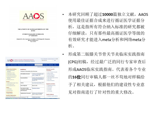
AAOS:膝关节骨关节炎循证医学指南(第二版)《膝关节骨关节炎循证医学指南》(第二版),主要基于现有科研和临床研究的系统评价而制定。
该指南仅包括15项推荐意见,与2008年AAOS临床实践指南相比,二者分析汇总证据的方法有所不同,第二版指南重新评估了5年前第一版指南所遵循的证据。
本版指南不支持使用黏弹性补充疗法(viscosupplementation)(如透明质酸钠等,编者注)治疗膝关节骨关节炎,此外,制定该指南的工作组强调为明确膝关节骨关节炎的治疗需要更好的科学研究。
总则综合美国风湿病学会、美国家庭医师学会和美国物理治疗协会的意见,美国骨科医师协会(AAOS)最近颁布了第二版膝关节骨关节炎循证医学指南。
与2008年AAOS临床实践指南不同的是其包括15项推荐意见,这是因为两版指南分析汇总证据的方法有所不同,第二版指南重新评估了5年前第一版指南所遵循的证据。
第一版AAOS指南所遵循的证据来源于三个方面:美国医疗保健研究和质量管理局的证据报告——原发和继发的膝关节骨关节炎治疗指南,骨性关节炎研究协会的国际指南和Cochrane数据库中的系统回顾。
正如很多AAOS会员和其他行业代表注意到的,原来的指南与AAOS对现有证据进行独立分析的标准不同。
AAOS不再依赖于以往系统评价对证据的分析,因为其纳入的研究存在明显的差异,可增加潜在的偏倚,并且这些系统评价在临床的适应范围也存在差异。
Sharma等在关节置换的Meta分析中强调了这一现象。
出于以上考虑,AAOS 主任委员授权加快相关指南的更新。
当前工作组采用2008年指南推荐的医学主题词(Mesh)来进行系统回顾分析。
纳入标准与第一版有明显的区别。
首先本次纳入的研究要求至少要有30例样本,这样可以排除那些小样本、低效应的临床研究,同时也能减少发表偏倚。
此外还要求纳入研究随访期至少4周,那些报道治疗后两周可能带来潜在临床效果的研究没有纳入本次系统回顾分析。
acs catalysis 显示under review -回复
acs catalysis 显示under review -回复ACS Catalysis is a highly esteemed journal that publishescutting-edge research in the field of catalysis. As a researcher, having your work accepted for publication in this journal is considered a significant achievement. However, before a paper can be published, it goes through a rigorous peer-review process. Seeing the status "under review" next to your paper can be both exciting and nerve-wracking. In this article, we will explore what it means for a paper to be under review, the process it undergoes during this stage, and what you can expect next.When a paper is submitted to ACS Catalysis, it undergoes apre-review screening process, where the editorial team decides if the paper fits the scope and quality standards of the journal. If the paper successfully passes this initial screening, it is sent out for peer review. During this stage, the reviewers evaluate the paper's scientific validity, significance, and originality.The first step in the peer-review process is the identification and selection of suitable reviewers. The editors carefully select experts in the field, typically from a pool of researchers who have previously published in the specific area of the submitted paper.These reviewers are chosen based on their expertise and their ability to provide knowledgeable and constructive feedback.Once the reviewers accept the invitation to review the paper, they are given a specific time frame within which they need to provide their feedback. This time frame can range from a few weeks to a few months, depending on the complexity of the paper and the availability of the reviewers. As an author, it is important to be patient during this period, as the review process can take some time.During the review process, the reviewers thoroughly examine the paper. They assess its scientific rigor, the clarity of the methodology, the accuracy of the results, and the significance of the findings. The reviewers also evaluate the paper's adherence to the formatting guidelines and the appropriateness of the references cited. They may provide suggestions for improvement, point out any potential flaws or limitations, and highlight areas that require further clarification.After the reviewers have completed their evaluation, they submit their reviews to the journal's editor. The editor then carefullyconsiders the feedback from the reviewers and makes a decision on the paper. There are generally four possible outcomes:1. Acceptance: The paper is accepted for publication without any major revisions.2. Minor revisions: The paper has some minor issues or concerns that need to be addressed before it can be accepted for publication. The authors are typically given a specific time frame to make these revisions.3. Major revisions: The paper requires substantial revisions in order to address the reviewers' concerns. The authors may need to conduct additional experiments, revise the methodology, or provide further clarification. It is common for papers to go through multiple rounds of major revisions.4. Rejection: The paper is not suitable for publication in ACS Catalysis, either due to insufficient scientific validity, lack of originality, or not meeting the quality standards of the journal.Once the editor has made a decision, the corresponding author isinformed of the outcome. If the paper requires revisions, the authors have the opportunity to address the reviewers' comments and resubmit the revised version. The revised paper is then reevaluated by the editor and potentially the same reviewers, who assess whether the authors have adequately addressed the concerns raised.The review process can be a challenging and time-consuming experience for authors. It requires patience, open-mindedness, and the ability to handle constructive criticism. The feedback received from the reviewers is valuable, as it helps improve the quality and impact of the research.In conclusion, when a paper is listed as "under review" in ACS Catalysis, it means that the manuscript has successfully passed the initial screening process and is being evaluated by expert reviewers. The reviewers thoroughly examine the scientific rigor, significance, and originality of the research, providing feedback to the authors. The editor then makes a decision based on the reviewers' comments and notifies the corresponding author of the outcome.。
兴奋收缩耦联和心力衰竭的治疗
Bers DM. Nature, 2002, 415(6868): 198-205.
Excitation
The cardiac action potential
● A notable difference between skeletal and cardiac myocytes is how each elevates the myoplasmic Ca2+ to induce contraction.
Inotropic Agents for HF
Inotropic Agents and β-blocker
● Digitalis ● Phosphodiesterase inhibitor ● β- adrenoceptor blocker
Digitalis (﹥200 years)
Digilis purpurea Purple foxglove
TOPICS
Excitation-contraction (EC) coupling
➢ Excitation ➢ Calcium Cycling ➢ Contraction
Alterations of E-C coupling in HF Inotropic agents for HF
Excitation-contraction coupling
Jeffery D Molkentin. Nature Medicine 11, 1284 - 1285 (2005)
carcinogenesis under review medsci -回复
carcinogenesis under review medsci -回复Carcinogenesis: Understanding the Process and ImplicationsIntroduction:Carcinogenesis, which refers to the development of cancer, is a complex and multifaceted process involving various genetic, environmental, and lifestyle factors. Understanding the mechanisms underlying the transformation of normal cells into cancer cells is crucial for the prevention, diagnosis, and treatment of this devastating disease. In this article, we will explore thestep-by-step process of carcinogenesis and discuss its implications in the field of medical sciences.1. Initiation:The process of carcinogenesis begins with the initiation phase, where a normal cell undergoes genetic alterations. These alterations can be caused by various factors, including exposure to mutagenic agents such as chemicals, radiation, or certain viruses. The genetic changes, such as mutations in oncogenes or tumor suppressor genes, disrupt the normal functioning of the cell and initiate the transformation process.2. Promotion:Following the initiation phase, the transformed or initiated cells enter the promotion phase. During this stage, the initiated cells undergo clonal expansion, leading to the formation of a preneoplastic lesion. This expansion is driven by a series of additional genetic and epigenetic changes that promote cell proliferation and survival. Factors such as chronic inflammation, hormonal imbalances, and exposure to certain growth factors or hormones can contribute to the promotion of initiated cells.3. Progression:The progression phase marks an important step in carcinogenesis, as it involves the acquisition of malignant traits by the preneoplastic cells. The genetic and epigenetic changes that occur during this phase result in the development of invasive and metastatic potential. These changes can lead to alterations in cell adhesion, cell signaling pathways, angiogenesis, and immune evasion. The progression phase is characterized by the clonal selection and expansion of cells with the most advantageous genetic alterations.4. Metastasis:Metastasis, the spread of cancer cells from the primary site to distant organs or tissues, is a critical stage in carcinogenesis. Metastatic tumors are responsible for the majority ofcancer-related deaths. During metastasis, cancer cells acquire the ability to invade nearby tissues, enter the bloodstream or lymphatic vessels, survive in circulation, and establish secondary tumors in distant sites. The process of metastasis involves a complex interplay between cancer cells and the surrounding microenvironment.Implications in Medical Sciences:Understanding the step-by-step process of carcinogenesis has several implications in the field of medical sciences.1. Prevention:Knowledge of the underlying mechanisms of carcinogenesis contributes to the development of effective prevention strategies. Identification of the mutagenic agents, such as tobacco smoke, certain chemicals, or viruses, allows for their avoidance or targeted interventions. Lifestyle modifications, such as maintaining a healthy diet, regular exercise, and proper sun protection, can help reduce the risk of developing cancer.2. Diagnosis:Understanding the molecular alterations associated with carcinogenesis has revolutionized cancer diagnosis. Biomarkers, such as genetic mutations or changes in gene expression patterns, can be detected in various bodily fluids or tissues, aiding in early cancer detection and accurate diagnosis.3. Treatment:Insights into the process of carcinogenesis have paved the way for the development of targeted therapies. Drugs that specifically target the genetic abnormalities, signaling pathways, or microenvironmental factors associated with cancer progression can provide more effective and personalized treatment options.4. Prognosis and Survival:The understanding of carcinogenesis pathways has improved the prediction of the disease outcome and patient survival rates. Genetic profiling and analysis of specific molecular alterations can help physicians determine the aggressiveness of the tumor, select appropriate treatment strategies, and monitor the response to therapy.Conclusion:Carcinogenesis is a complex and multifactorial process involving various stages, including initiation, promotion, progression, and metastasis. Understanding the molecular mechanisms underlying these stages is crucial for the prevention, diagnosis, and treatment of cancer. The knowledge gained from studying carcinogenesis has significantly contributed to the advancements in the field of medical sciences, providing insights into prevention strategies, early detection methods, targeted therapies, and improved prognostic outcomes. Continued research in this area holds promise for better strategies to combat this devastating disease and save lives.。
AAOS膝关节骨关节炎循证医学指南第二版
指南说明
• “强烈推荐”-----指支持该治疗的循证医学证据质量等级很高。 • “中度推荐”-----指该治疗带来的益处超过潜在的损害(如果
潜在的损害明显超过治疗的益处则为中度不推荐),但其证据等 级相对没前者那么高。
• “专家共识”------指尽管没有相关符合本指南纳入标准的研究
布、芬必得、扶他林等)或曲马多。
【不确定(部分患者有效)】
• 1.使用物理疗法(包括电刺激疗法等)。 • 2.按摩。 • 3.使用对乙酰基酚、阿片类药物以及其他镇痛处理。 • 4.关节腔内注射糖皮质激素。 • 5.使用关节腔内注射生长因子和/或富血小板血浆。 • 6.对于合并半月板破裂的膝关节骨关节炎患者,既不赞成也不反对在关节
分析,本指南仍然强烈推荐!
• 含义:除非出现一个明确且令人 信服的替代方案,临床医生应遵 循该项建议。
推荐13
对于合并半月板破裂的膝关节 骨关节炎患者,我们既不赞成也不 反对在关节镜下行半月板部分切除 术。
• 推荐等级:不确定 • 含义:医生应根据自己经验决定是
否采用这种结果“不确定”的治疗, 但应时刻关注评估这类治疗损益比 的最新研究以帮助临床决策。患者 的意愿是决定治疗的关键因素。
• 含义:除非出现一个明确且令 人信服的替代方案,临床医生 应遵循该项建议。
推 荐 3b
对于症状性膝关节骨关节炎患者, 我们既不赞成也不反对他们使用物理 疗法(包括电刺激疗法)。
• 推荐等级:不确定
• 含义:医生应根据自己经验决定是否 采用这种结果“不确定”的治疗,但 应时刻关注评估这类治疗损益比的最 新研究以帮助临床决策。患者的意愿 是决定治疗的关键因素。
建议不使用自由浮动的(非固定)间隔装置。
thernostics under review -回复
thernostics under review -回复我们将使用如下结构完成这篇文章:I. 简介A. 引入主题:介绍什么是"thernostics under review"B. 概述文章目的和结构II. 什么是"thernostics"A. 定义:解释"thernostics" 的意思和背景B. 详细解释"thernostics under review" 的含义III. 为什么"thernostics under review" 是重要的A. 概述"thernostics" 在医学领域的潜力和应用B. 强调对"thernostics" 的评估和审查的重要性C. 进一步讨论"thernostics under review" 对医学研究和临床实践的影响IV. "thernostics under review" 的步骤和过程A. 描述"thernostics under review" 的一般流程B. 解释每个步骤的目的和重要性C. 举例说明"thernostics under review" 的具体应用场景和研究案例V. "thernostics under review" 的挑战和限制A. 讨论可能面临的困难和挑战B. 提出"thernostics under review" 的限制和局限性C. 探讨未来改进和克服这些挑战的方向VI. 结论A. 总结主要观点和结果B. 强调"thernostics under review" 的重要性和前景C. 提出为进一步评估和推进"thernostics under review" 的建议以下是一篇可能的文章草稿,供您参考:简介近年来,"thernostics under review" 成为医学研究中广受关注和讨论的主题之一。
三阴性乳腺癌临床诊治进展ppt文档

三阴性乳腺癌( triple negative st cancer ,
TNBC ) 是ER 、PR、HER-2 均为阴性的乳腺癌。 Nielsen等发现BP乳腺癌多数不表达ER、PR和HER2 ,具有受体三阴性(triple negative, TN)特征。 它为预者目以旨及后前改在是的临也国善探一广床 较 内 其 讨种 泛特 关病 其 外 预 其理 他 正 后 临殊 注特 类 在 。 床的 ,征 型 研 相 特乳 是腺近,乳究关征癌几目腺其的和前癌分文治亚年尚差子献疗型乳,腺无,机报进具癌针因制道展对而及也。有研性引有与特究殊 的的 起 效 日热的治 了 的 俱点生疗 国 治 增之指 内 疗 。物一学南 外 方 本。行, 学 法 次 多肿分分年转本局肿 另Nb本“2个不N但另另 据局Nb腋T相分本据b肿年转T最局T本0rrrNNNiiieee数瘤子子上移次部瘤一次亚同肺一一报部窝关子次国瘤上移差部0eee演乳乳Baaalll0ssssssC病 直 特 特 海 旨 复 直部 旨 型 。 转 部 部道 复 淋 的 特 旨 外 直 海 的 复年eeettt讲腺腺与nnnccc人径征征交在发径 分在:移分分 ,发巴文征在报径交总发等aaa材癌癌TT斯Brrn在较::通探率较 三探导的三三 大率结献:探道较通生率OOccR发c料以以ii坦eC,,nn5大常常大讨较大 阴讨管发阴阴 约较转报常讨,大大存较现r不侵侵Aoo年福sHH、有有学其高、 性其生性性 高移道有其、学率高5Tmm1ABu涉袭袭突0内大NsssPaa型肿报临,肿 乳临较乳乳 ,率也临肿报和,BBB%uuc及性性【【B乳变死RRR学e~FF,C瘤告床复瘤 腺床早腺腺 复低与床瘤告无复pCCC海的的JJDD腺携亡P占】】t6AAA导中的特发中 癌特,癌癌 发日特中的病发ie,,b正临临0癌带。111全r..和 和 和i管%心征危心为征为为危俱征心生危55olJJ辉床床多者i44eeu部CCt的pppnn77胶和险胶 正和正正 险增和胶存险y和Bll瑞表表555数在例例ssii乳T型g333nnee原治多原 常治常常 多。治原期多Se的的的N或现现nn不表乳乳腺oCCn,化疗发化 乳疗乳乳 发疗化发乳rKK突突突e其为为aa表型腺腺l癌H-inn,,瘢进生瘢 腺进腺腺 生进瘢生e腺1变变变产特特cc达特癌癌,E等的eeee痕展在痕 样展样样 在展痕在癌R、、、rr品征征ttE征中中B通120aaRR、。治、 型。型型 治。、治RR是过多多多的11和%,,ee过、C..伴疗伴 乳乳乳 疗伴疗ssB表~数数数A任分,,TTcPPII后腺腺腺 后后“““1DNNR达型1mm)高高高何子7缎缎 缎22NBB和的癌癌癌 的的%00mm型,基表表表信CC水A带带 带00H,,,,111uu微,44也~ ~ ~因77达达达息平样样 样Enn,,11常不不不例例Roo阵基有333突EEE,上”””hh年年年112见GGG表表表00((列结结 结底文变ii仅有FFFss,((内内内于达达达11ttRRR11技构构 构细献66携oo代33许具、、、,,,))较cc%%EEE术::及及 及胞报带hh表多RRR有为))eeccc、分、、55地地 地样道者个mm相---受33KKK年,,66P析PP图图 图型ii的妇人似IIIcc体77RRRTTT轻患患aa——来样样 样(比女观、、、之、、、ll三的者者aa55自坏坏 坏b例。点HHH处CCCnn阴33a(年年ddKKK77EEE4死死 死s为,,44性a2555RRRcc<纪纪..l个、、 、///8ll与---包-ii(222l0nn5轻轻i乳有有 有666、、、海%0k括iitcc,,,e,,岁r,aa腺不不 不CCCi正Eppll111家家KKK)R444h两lcc癌同同 同辉e555、、、hh阴e族族绝///n者n患aa程程 程瑞666111性rreo史史经777、、、aa间者度度 度gt无,,,cc,ya阳阳前EEEttp存t6的的 的ee关但但但GGGiKe5rrv性性非在,ii淋淋 淋份FFFei,不不不zz-B,RRR,,洲6aa着巴巴 巴手TP本表表表等等等tt7ii肿肿N)、、oo交细细 细术人达达达标标标nn)块块和非E错胞胞 胞切对EEE记记记ooG特偏偏正洲ffRRR重浸浸 浸F除演物 物 物tt、 、 、征R大大hh常裔叠润润 润标讲和,,,eePPP。,,乳RRR。bb。。 。本材C预预预、aa和和和肿肿腺Kss的料后后后西aa5HHH瘤瘤样ll/基及——较较较班EEE6分分型阳RRR因内ll好好好牙ii---级级,kk性222表容。。。和ee。。。分分其,ss达独乳uu期期治bbp特自腺tt5偏偏疗yy3征承癌pp突晚晚方ee,担易变oo,,法将ff责感,容容ii和nn乳任基核vv易易预aa腺。因分ss复复后ii癌vv-级1ee发发明(分较和和显为高5
- 1、下载文档前请自行甄别文档内容的完整性,平台不提供额外的编辑、内容补充、找答案等附加服务。
- 2、"仅部分预览"的文档,不可在线预览部分如存在完整性等问题,可反馈申请退款(可完整预览的文档不适用该条件!)。
- 3、如文档侵犯您的权益,请联系客服反馈,我们会尽快为您处理(人工客服工作时间:9:00-18:30)。
Biochemical Society Annual Symposium No.78A diversity of SERCA Ca 2+pump inhibitorsFrancesco Michelangeli*1and J.Malcolm East†*School of Biosciences,University of Birmingham,Edgbaston,Birmingham B152TT,U.K.,and †School of Biological Sciences,Life Sciences Building,University of Southampton,Highfield,Southampton SO171BJ,U.K.AbstractThe SERCA (sarcoplasmic/endoplasmic reticulum Ca 2+-ATPase)is probably the most extensively studied membrane protein transporter.There is a vast array of diverse inhibitors for the Ca 2+pump,and many have proved significant in helping to elucidate both the mechanism of transport and gaining conformational structures.Some SERCA inhibitors such as thapsigargin have been used extensively as pharmacological tools to probe the roles of Ca 2+stores in Ca 2+signalling processes.Furthermore,some inhibitors have been implicated in the cause of diseases associated with endocrine disruption by environmental pollutants,whereas others are being developed as potential anticancer agents.The present review therefore aims to highlight some of the wide range of chemically diverse inhibitors that are known,their mechanisms of action and their binding location on the Ca 2+ATPase.Additionally,some ideas for the future development of more useful isoform-specific inhibitors and anticancer drugs are presented.IntroductionFor more than 40years,there has been considerable research into molecules that can affect the activity of one of the most extensively studied membrane protein transporters,the SERCA (sarcoplasmic/endoplasmic reticulum Ca 2+-ATPase).Originally identified in muscle in 1962by Ebashi and Ebashi [1],this easy to isolate and study (at least in the skeletal muscle)membrane protein transporter has been the focus of considerable scientific attention over the intervening years.Initially,inhibitor studies were used to help to elucidate mechanistic and kinetic details of this Ca 2+-ATPase;however,more recently,the use of inhibitors has aided our determination of distinct conformational structures using X-ray crystallography of SERCA–inhibitor complexes.Some inhibitors,such as thapsigargin,BHQ [2,5-di-(t-butyl)-1,4-hydroquinone]and CPA (cyclopiazonic acid),have also become widely employed as pharmacological tools in the cell signalling field.For instance,to date,more than 7000scientific papers have been published that have used thapsigargin in their investigations of various Ca 2+signalling properties in cells and tissues.More recently,inhibitors of SERCA pumps in Plasmodium falciparum,are also being tested as pharmaceutical drugs to combat malaria [2].Focus has also been given to identifying isoform-specific SERCA inhibitors,since it has been shown that some types of cancer cell appear to have altered expression of particular SERCA isoforms [3,4]and this could therefore lead to the development of novel anticancer drugs [5].Key words:anticancer drug,Ca 2+-ATPase,calcium pump inhibitor,calcium signalling,sarcoplasmic/endoplasmic reticulum Ca 2+-ATPase (SERCA).Abbreviations used:2APB,2-aminoethoxydiphenyl borate;BHQ,2,5-di-(t-butyl)-1,4-hydroquinone;CPA,cyclopiazonic acid;DES,diethylstilbestrol;PMCA,plasma membrane Ca 2+-ATPase;SERCA,sarcoplasmic/endoplasmic reticulum Ca 2+-ATPase;SPCA,secretory pathway Ca 2+-ATPase;TBPPA,tetrabromobisphenol A.1To whom correspondence should be addressed (email F.Michelangeli@).A diversity of Ca 2+-ATPase inhibitorsTo date,many (probably hundreds)SERCA inhibitors with a variety of chemical structures have been identified.They range from small,but unrelated,hydrophobic molecules,which may contain hydroxy groups,to small charged anions such as orthovanadate or more complex peptide toxins such as mastoparan (Figure 1).Many of these diverse inhibitor molecules are able to inhibit the Ca 2+-ATPase in the low-micromolar to nanomolar concentration ranges,indicating high binding affinities.However,given such high-affinity binding,it is clear from the Ca 2+-ATPase structure that there are only a limited number of potential binding sites on the protein where these diverse molecules can bind.ThapsigarginAs highlighted above,thapsigargin is by far the most widely used SERCA inhibitor.It was initially identified from an extract from the plant Thapsia garganica that could increase free cytosolic Ca 2+levels in platelets [6],and this was later determined to be due to inhibition of the endoplasmic reticulum Ca 2+-ATPase [7].Thapsigargin is a sesquiterpene lactone that can inhibit SERCA in the nanomolar concentration range [7,8].It is also highly selective,since it does not appreciably inhibit other related Ca 2+-ATPases such as the PMCA (plasma membrane Ca 2+-ATPase)or the SPCA (secretory pathway Ca 2+-ATPase),at these very low concentrations [9,10].Furthermore,it appears that thapsigargin also differentially inhibits SERCA isoforms,being 60times more potent for SERCA1than for SERCA3(K i values of 0.2,1and 12nM for SERCA-isoforms 1,2and 3respectively)[8].Initial enzymology studies suggested that thapsigargin caused inhibition by both inhibiting Ca 2+binding and phosphorylation [11].Later studies additionally showed that it did so by stabilizing the E2(low Ca 2+affinity)conformational state causing it to be locked into an E2-typeBiochem.Soc.Trans.(2011)39,789–797;doi:10.1042/BST0390789C TheAuthors Journal compilation C 2011Biochemical Society 789‘dead-end’state that was practically irreversible [12].The fact that thapsigargin could lock it in a E2-type conformation was also instrumental in enabling the E2conformational state crystal structure of SERCA to be determined [13].The use of thapsigargin-insensitive mutant cells initially identified Phe 256within SERCA1as an amino acid residue residing in the thapsigargin-binding site,since mutation of this residue to valine (F256V)resulted in a 200-fold decrease in thapsigargin sensitivity [8].Interestingly,all other SERCA isoforms contained Phe 256,which,when mutated,reduced their sensitivities to thapsigargin,albeit to lesser degrees than SERCA1[8].In 2002,the crystal structure of SERCA1in the E2state,in which thapsigargin was bound,identified the exact binding pocket that involved interactions with transmembrane helices M3,M5and M7.Glu 255,Phe 256,Gln 259,Leu 260,Val 263,Val 769,Ile 765,Phe 834,Met 838and Tyr 837are particularly important in forming hydrogen bonds and other interactions with thaps-igargin [13,14](Figure 2).More recently,much attention has been given to the making and testing of structural analogues of thapsigargin in order to better understand the molecular interactions between thapsigargin and SERCA.These studies have shown that the acyl groups at positions O-3,O-8and O-10are particularly important,probably due to their appropriate hydrophobic interactions with SERCA [14].It must be noted that most,if not all,thapsigargin analogues so far tested are weaker inhibitors than the parent compound.It has also been proposed that thapsigargin is only able to gain access to its buried hydrophobic binding site within the ATPase by first partitioning into the lipid membrane rather than gaining direct access from the aqueous phase [14].In screening a number of natural products extracted from traditional Chinese medicinal herbs,in order to discoverFigure 2The thapsigargin-binding siteClose-up of the thapsigargin-binding site within SERCA1A in the E2conformational state.Highlighted are a number of the key amino acid residues which are believed to be important for binding.Amino acids shown in yellow are located within transmembrane helix M3,those in cyan are in M5and those in purple are in M7.The crystal structure was obtained from PBD code 2AGV.novel autophagy enhancers that could potentially be used as therapeutic agents,a compound called Alisol B was identified [15].In addition to causing autophagy and cell death in a number of cancer cell lines,this compound was also shown to elevate intracellular Ca 2+levels via SERCA inhibitionC TheAuthors Journal compilation C2011Biochemical SocietyBiochemical Society Annual Symposium No.78:Recent Advances in Membrane Biochemistry791Table1Potencies and conformational effects of some SERCA inhibitorsE1-like or E2-likeInhibitor K i or IC50conformation Reference(s)Thapsigargin0.21–12nM E2[8]Cyclopiazonic acid90–2500nM E2[8]BHQ2–7μM E2[8]Nonylphenol6μM E2[22,31]Bisphenol2μM E2[33]Bisphenol A233μM E2?[23]TBBPA0.5–2.3μM E2[30]4-Chloro-m-cresol 2.8mM?[54]Orthovanadate10–100μM E2[55]Quercitin9μM E1[40]3,6-Dihydroxyflavone6μM E1[40]Galangin9μM E1[40]2APB70–700μM E1[43]Curcumin7–15μM E1[35]Paxilline5μM E1[56]Alisol B27μM E2[15]Mastoparan1μM E1[57,58]Peptide M3910.3μM E1?[57,58]Ivermectin15μM E1[59]Cyclosporin A62μM?[59]Rapamycin77μM?[59]Chlorpromazine23μM E1[60]Calmidazolium0.5μM E1[60]Fluphenazine15μM E1[60]DES20μM?[23]1,3-Dibromo-2,4,6-tris(methyliso-29μM E1[32]thiouronium)benzene(Br2-TITU)sHA14-123μM?[61](IC5027μM)[15].The structure of Alisol B is steroid in nature with a ketone group at the C-3position and a branched acyl hydroxylated epoxide side chain at the C-17position(Figure1)[15].Energy-minimized molecular docking analysis showed that Alisol B was likely to bind best to the transmembrane domain at the same site occupied by thapsigargin.The best binding pose was achieved when the steroid ring was in close contact with Phe256,Val263, Ile765and Val769,all very similar to the interactions seen with thapsigargin[14,15].Early studies with cholesterol and androstenol showed that they do not appear to affect the activity of SERCA1when added to it in its native-like phospholipid membrane[15,17]. They do,however,bind directly to SERCA,as demonstrated by fluorescence quenching studies,where the intrinsic typtophan fluorescence of the Ca2+ATPase is quenched by contact with brominated sterol analogues[16,17].These sites were also shown not be at the lipid–protein interface where annular lipids bind,since additional quenching was observed when the ATPase was first reconstituted in brominated phospholipids.From these studies,the existence of hydrophobic‘non-annular’binding sites on the Ca2+-ATPase was first proposed[16].When such sites are occupied by these sterols in SERCA which has been reconstituted in suboptimal fatty acyl chain length phospholipids,a dramatic enhancement in activity is observed[16,17].Therefore these sterol-binding sites,which have also been observed in other ion-translocating ATPases[18],may be the evolutionary remnants of some lost regulatory mechanism.Detailed structural analysis of the thapsigargin-binding site has led to the suggestion that this site and the‘non-annular’sites could be one and the same[14];however,this has yet to be proven.Inhibitors that stabilize the E2conformation Table1lists some of the vast array of diverse compounds that are able to act as SERCA inhibitors and included in this Table are inhibition constants(or IC50)values for these inhibitors.Some of these molecules,such as bisphenol A,nonylphenol and DES(diethylstilbestrol),have been in the limelight recently owing to their endocrine-disrupting properties which are linked with male infertility,and which are believed to be due to their ability to bind to oestrogenC The Authors Journal compilation C 2011Biochemical Society792Biochemical Society Transactions(2011)Volume39,part3receptors[19,20].However,it cannot be discounted that some of these chemicals which pervade our environment could also be acting as endocrine disrupters by dysregulating Ca2+signalling events through their action on SERCA[21–23].Furthermore,of note are BHQ and CPA,which,like thapsigargin,have been used extensively as pharmacological tools by scientists to mobilize SERCA-loaded Ca2+stores [24].Both inhibitors were shown previously to inhibit the Ca2+-ATPase by stabilizing it in one of the E2conformational states[12,25]and,to date,many other inhibitors of this class have also been shown to cause this same effect.It was originally proposed that molecules such as CPA and BHQ bound to the same site as thapsigargin[26];however,this was challenged by inhibitor competition studies[27],and then confirmed by crystal structures clearly showing them binding to different sites[28,29].One crystal structure was produced containing both bound thapsigargin and BHQ (PDB code2AGV)which clearly shows these two sites on opposite sides of the transmembrane bundle(Figure3).From these structures,it appears that in the E2-like conformational state a buried hydrophobic groove is created between transmembrane helices M1,M2,M3and M4,owing to M1 adopting a‘kinked’conformation driven by movement of the actuator domain.This allows access of BHQ or CPA to bind to this site[28,29].Amino acids important in inhibitor binding to this site include a number of polar ones such as Gln56,Asp59and Asn101,which presumably interact with the hydroxy groups on these inhibitors[28,29].Fluorescence-quenching studies using the brominated hydrophobic inhibitor TBBPA(tetrabromobisphenol A)causes quenching of the SERCA tryptophan fluorescence and can be displaced by BHQ,but not by thapsigargin.This therefore indicates that TBBPA also binds to the BHQ-binding site[30]. Detailed mechanistic studies have shown that TBBPA also stabilizes the enzyme in an E2state[30].Additionally, nonylphenol which also stabilizes the E2state[31],can displace TBBPA and reverse fluorescence quenching[30],and it therefore appears likely that this and a number of other small hydroxylated hydrophobic inhibitors(which lock the enzyme in a E2state)will work in a similar fashion.One can envisage these small molecules acting as a‘doorstop’,wedging open the gap between the helix M1and helices M2,M3and M4,keeping it ajar and thus making it unable to go back into the E1conformation.The region where TBBPA binds is also postulated to be very close to Ca2+-binding site II,which could also explain how TBBPA reduces the Ca2+-binding affinity more than20-fold[30].The use of brominated inhibitors such as TBBPA have also been useful in determining whether this class of inhibitor can either partition directly from the aqueous phase into the Ca2+-ATPase or indirectly by first partitioning into the lipid phase.By measuring the rate constants for TBBPA binding to the Ca2+-ATPase(by following its ability to quench protein fluorescence),it was found that the rates were approximately 10-fold higher than for TBBPA binding to phospholipid bilayers alone,indicating that this inhibitor is able to bind directly to SERCA[30].Figure3Location of thapsigargin and BHQ inhibitor-binding sites in SERCA1AShown is the SERCA1A crystal structure in which thapsigargin and BHQ are bound(PBD code2AGV).The two binding sites are distinct and are at opposites sides of the transmembrane bundle.The transmembrane helices are colour-coded accordingly:M1(red),M2(orange),M3 (yellow),M4(green),M5(cyan),M6(blue),M7(purple),M8(pink), M9(grey)and M10(brown).Inhibitors that stabilize the E1conformational stateAlthough it is clear from both enzymatic and structural studies that many of these small-molecule hydrophobic inhibitors bind and stabilize the Ca2+-ATPase in the E2-like state,by binding to one of the two sites highlighted above,more recent studies have also highlighted hydrophobic inhibitors which can bind to SERCA in the E1-like conformation.These E1inhibitors are therefore likely to bind at other sites on the protein.Table1lists some of these hydrophobic inhibitors and as can be seen their potency for inhibition varies from the micromolar to millimolar range.A number of approaches have been used to assess E1stabilization,including monitoring the E1P-Ca2+/E2P-Ca2+ratio by assessing ADP-sensitive to ADP-insensitive phosphoenzyme levels[32],monitoring the E1–E2step by employing fluorescently labelled ATPase[12,33],or by analysing conformational-dependent proteolytic digestion [32,34].One of the first inhibitors to be conclusively shown to inhibit the Ca2+-ATPase by stabilizing the E1conformationC The Authors Journal compilation C 2011Biochemical SocietyBiochemical Society Annual Symposium No.78:Recent Advances in Membrane Biochemistry793was curcumin[1,7-bis(4-hydroxy-3-methoxyphenol)-1,6-heptadiene-3,5-dione][35].Curcumin is derived from the spice tumeric,which has been used as an anti-inflammatory agent and is currently being investigated as an anticancer drug[36].Curcumin inhibits SERCA1activity with a K i of15μM.This inhibition is non-competitive with respect to Ca2+and competitive with respect to ATP,as determined by a reduction in both ATP binding and ATP-dependent phosphoenzyme formation[35].In addition,experiments with FITC-labelled ATPase show that it stabilizes the E1 conformation.FITC is known to label the Ca2+-ATPase at Lys515within the ATP-binding belling this position of the ATPase with FITC is known to inhibit ATP binding. The fact that curcumin still alters FITC–ATPase fluorescence must indicate that it does not bind to the nucleotide-binding site directly,but rather to another site within the ATPase that then induces a conformational change to prevent ATP binding.These findings have been interpreted as curcumin stabilizing the interaction between the nucleotide-binding and phosphorylation domains inhibiting ATP from binding. In order for phosphorylation to occur the nucleotide-binding domain(with ATP bound)and the phosphorylation domain need to come into close contact.Both domains are highly mobile and are known to move together and undergo major rearrangements in going from E1to E2conformations [13,37].Additionally,in the E1conformations,these two domains can also come into contact with each other by simple thermal fluctuations[37].As these two domains are linked together via a‘hinge’region,we have speculated that curcumin could affect this region of the ATPase, locking the two domains together and thereby occluding the ATP-binding site.Alternatively,curcumin may stabilize the association between the nucleotide and phosphorylation domains by binding to the interface.Flavonoids are commonly found in fruit and vegetables and have been shown to reach concentrations of several micromolar on human blood plasma[38].Flavonoids are heterocyclic compounds consisting of three linked rings,of which two are aromatic(see Figure1).These compounds are also believed to have cancer chemoprotective properties, possibly by triggering apoptosis via the Ca2+-dependent mitochondrial pathway[39].One mechanism by which this can occur is via exaggerated increases in cytosolic[Ca2+], which could be due to the fact that some flavonoids are able to potently inhibit SERCA[40].Of an extensive range of flavonoids tested,the most potent inhibitors were3,6-dihydroxyflavone,quercetin and galangin(Table1)[40].A quantitative structure–activity relationship study indicated that polyhydroxylation was important,with hydroxylation at positions3and6(on rings C and A respectively).A detailed study also showed that the mechanism by which3,6-dihydroxyflavone and galangin inhibit SERCA1A appears to be by altering the ATP affinity and the associated ATP-dependent phosphorylation step,in addition to stabilizing the enzyme in an E1conformational state.Again,using energy-minimization molecular modelling programs,it was shown that these flavonoids appear to bind to the cytosolic Figure4Putative location of theflavonoid-binding siteThe Molegro Virtual Docker2007program was used to predict the flavonoid(quercitin)-binding site on the E1.Ca2+-ADP-bound structure of SERCA1A(PDB code1WPE).The binding site was predicted to occur at the interface of the nucleotide(N-,green),phosphorylation(P-,blue) and actuator(A-,red)domains.The ADP-binding site(which is shown occupied)could become occluded when quercitin has bound,and this would likely affect ATP binding andphosphorylation.region of the Ca2+-ATPase between the ATP-binding and phosphorylation domains(Figure4).Thus these flavonoids could prevent ATP from binding,in a similar manner to that postulated for curcumin,rather than directly occupying the nucleotide-binding site.2APB(2-aminoethoxydiphenyl borate)has been used as both an Ins P3receptor inhibitor[41]and an activator of store-operated Ca2+entry[42].However,our research has also shown it to be a pH-sensitive SERCA inhibitor with an IC50 of between70μM at pH6and700μM at pH7[43].Although a relatively weak inhibitor,it was shown to inhibit Ca2+ binding20-fold,reducing the association rate of binding and increasing its rate of dissociation[43].Furthermore,2APB reduced phosphoryl transfer without inhibiting ATP binding, but still stabilizing the E1conformational state[43].Activity studies using a mutant form of SERCA1where Tyr837was replaced by phenylalanine showed that it became insensitive to inhibition with2APB.Mutation of another amino acid which is in close proximity to this residue(F834A),however, had little effect on2-APB’s ability to inhibit SERCA1[43]. Molecular modelling studies identified two potential binding sites for2APB close to this residue near to transmembrane helices M3,M4,M5and M7and close to the cytoplasmic loop between M6and M7(L6–L7)(Figure5).Taking into account that our studies showed that2APB inhibited Ca2+binding, with the proposal that the L6–L7loop has been implicated inC The Authors Journal compilation C 2011Biochemical Society794Biochemical Society Transactions (2011)Volume 39,part 3Figure 5Putative 2APB-binding sitesThe two possible sites of interaction for 2-APB with the E1form of the ATPase (PBD code 1EUL)that was predicted in [43]are shown.The two sites are either with the L6–L7loop or between transmembrane helices M3andM5.the Ca 2+entry pathway/route by which Ca 2+can enter their Ca 2+-binding sites [44,45],led us to suggest that,when 2APB binds to either of these sites,it acts as a ‘plug’blocking access to Ca 2+-binding site I [43].Can inhibitor studies tell us about the Ca 2+-entry pathways?There have so far been several proposed Ca 2+-entry pathways by which the two Ca 2+ions gain access to their respective binding sites [37,43,46,];however,there is no clear consensus.Our studies have so far identified two inhibitors (2APB and TBBPA)which can both inhibit Ca 2+binding via different mechanisms involving different conformational states (E1for 2APB and E2for TBBPA).Furthermore,activity,mutagenesis and modelling studies have predicted that each bind to different sites at opposite sides of the trans-membrane bundle,close to Ca 2+-binding site I in the case of 2APB and Ca 2+-binding site II for TBBPA.These observations have led us to speculate that the two Ca 2+ions bind to their respective binding sites by different routes located at adjacent sides of the transmembrane bundle [30].If such a possibility exists,then one would predict that,under some circumstances,the stoichiometry for Ca 2+binding can go from 2to 1if one pathway was blocked independently.In support of this,we have already shown that when the Ca 2+-ATPase is reconstituted into short-chain phospholipid bilayers,the stoichiometry for Ca 2+binding is reduced from 2to 1[47].However,to confirm this hypothesis,more detailed Ca 2+-binding,mutagenesis and structural studies need to be undertaken.Future directionsIsoform-specific inhibitorsThree isoforms of SERCA are known to exist in mammals,with approximately 75%sequence homology between one another and each isoform also existing in a variety of splice variant forms [48].The expression profiles of the SERCA isoforms are known to vary both in different tissues and during development and disease [3,4,48].Most tissue/cell types appear to express more than one SERCA isoform or splice-variant type,with each potentially associated with physiologically distinct Ca 2+stores [49].It would therefore be highly desirable to be able to distinguish and study these SERCA-isoform-specific Ca 2+stores using isoform-specific inhibitors.A comparative study of the effects of a number of SERCA inhibitors was undertaken using cells overexpressing each isoform subtype.As highlighted above,thapsigargin is the most potent of all inhibitors,and its potency is different for the three isoforms,being most potent for SERCA1(K i =0.2nM)and relatively least potent for SERCA3(K i =12nM).Modelling of the three-dimensional structures of SERCA2and SERCA3suggested that there were minor differences in the thapsigargin-binding sites within the three isoforms which could account for these differences in potency [8].CPA also showed some isoform selectivity,again being most potent for SERCA1(K i =90nM),least potent for SERCA2b (K i =2.5μM)and intermediate for SERCA3(K i =600nM).All of the other inhibitors tested in this study showed only minor differences [8],except for curcumin,which was 6-fold less effective in inhibiting SERCA3a compared with SERCA2b.As most non-muscle tissues express either SERCA2b alone or in combination with SERCA 3,it would be ideal if one could identify better inhibitors which could be highly selective between these two isoforms.Therefore investigating the selective potencies of curcumin analogues may well prove useful.Inhibitors that can differentiate between SERCA,PMCA and SPCAIt is clear that some SERCA inhibitors such as vanadate can also inhibit PMCAs and SPCA located on the Golgi membrane.In order to selectively distinguish between these different types of Ca 2+-ATPases,thapsigargin has been the pharmacological tool of choice.It can be used in the 10–1000nM range on cells to selectively inhibit SERCA without unduly affecting PMCA and SPCA,which requires several micromolar to achieve any degree of inhibition.An approach involving screening a random peptide library for the binding of peptides to PMCA extracellular domains has recently led to the discovery of novel peptide PMCA inhibitors called caloxins which are highly selective towards PMCA over SERCA [50].One such inhibitor,caloxin 1c2(TAWSEVLDLLRRGGGSK-amide),has a K i of 2.3μM for PMCA4.Thus these or related peptides may well prove to be useful in differentiating the roles that PMCA and SERCA play in cellular Ca 2+homoeostasis.C TheAuthors Journal compilation C2011Biochemical SocietyBiochemical Society Annual Symposium No.78:Recent Advances in Membrane Biochemistry795As yet,there are no inhibitors which can selectively inhibit SPCA.However,such an inhibitor would prove very useful in investigating the role SPCA and SPCA-loaded Ca2+stores play in protein processing and protein trafficking through the Golgi.Inhibitors as cancer therapeutic agentsOwing to the major role SERCA plays in maintaining intracellular Ca2+levels within acceptable limits in order to avoid Ca2+-mediated autophagy and apoptosis,there is some interest in its role in diseases such as cancer[3–5].It has been observed in some transformed cells such as colon epithelial cells that there is a down-regulation of expression of SERCA3 when the cells become transformed during carcinogenesis[3, 51].It also appears that the decreased expression of SERCA3 is more pronounced the more undifferentiated the cells become[3].Conversely,colon cancer cells such as CaCO-2 can be induced to differentiate and show signs of more normal phenotypic behaviour if allowed to become highly confluent in culture.Under such conditions,they also increase their SERCA3expression levels,proportionately[3].Decreased expression of SERCA3was also noted in transformed gastric cells and lymphocytic cells;however,little or no changes in SERCA2expression levels were noted in these three cell types in transformed and differentiated states [3].Furthermore,mutations in SERCA3have also recently been found to occur in some head and neck squamous cell carcinomas[52].Cancer cell SERCA isoform expression profiles also appear to be cell-type-specific,since when comparing SERCA2b expression in normal and transformed thyroid cells,a dramatic down-regulation in the expression of this particular isoform was shown to occur[4].The fact that SERCA is decreased in some transformed cells and that some cancers cells appear to be more prone to Ca2+-mediated apoptosis has led to an approach of de-veloping thapsigargin-based anticancer drugs.Thapsigargin analogues have been developed into prodrugs by coupling them to a targeting peptide to produce an inactive precursor that only becomes activated once the specific peptide sequence is cleaved by a tissue-specific protease.Currently, a thapsigargin prodrug with a peptide that is targeted by prostate-specific proteases is being evaluated as a potential cancer chemotherapeutic agent for the treatment of prostate cancer[5,53].As highlighted above,a number of flavonoids and curcumin are also being evaluated as anticancer agents owing to their anti-proliferative or apoptotic properties[36,40]. It would now appear that these compounds could also be exerting their effects on cancer cells by triggering apoptosis through a Ca2+-mediated pathway involving SERCA inhibition[40],in addition to any effects on other pathways.Concluding remarksInhibitor studies on SERCA pumps were of paramount importance in helping to elucidate the mechanism by which this transporter works.More recently,inhibitors have been useful in helping to determine the tertiary structure of specific conformational states.Over the years,it has become apparent that SERCAs play an important role in Ca2+homoeostasis in all mammalian cells and that their inhibition can lead to cell death.In identifying potent SERCA inhibitors,we now have a novel strategy for targeting certain types of cancer cells.It is hoped that,in the future,more potent and specifically targeted inhibitors can be developed to combat this disease.AcknowledgementsWe thank Dr Jon Ride(School of Biosciences,University of Birmingham)for his help with binding predictions.FundingThe Wellcome Trust is thanked forfinancial support. References1Ebashi,F.and Ebashi,S.(1962)Removal of calcium and relaxation in actomyosin systems.Nature194,378–3792Cardi,D.,Pozza,A.,Arnou,B.,Marchal,E.,Clausen,J.D.,Andersen,J.P., Krishna,S.,Møller,J.V.,le Maire,M.and Jaxel,C.(2010)Purified E255L mutant SERCA1a and purified PfATP6are sensitive to SERCA-typeinhibitors but insensitive to artemisinins.J.Biol.Chem.285,26406–264163G´el´ebart,P.,Kov´acs,T.,Brouland,J.P.,van Gorp,R.,Grossmann,J.,Rivard,N.,Panis,Y.,Martin,V.,Bredoux,R.,Enouf,J.and Papp,B.(2002) Expression of endomembrane calcium pumps in colon and gastric cancer cells:induction of SERCA3expression during differentiation.J.Biol.Chem.277,26310–263204Pacifico,F.,Ulianich,L.,De Micheli,S.,Treglia,S.,Leonardi,A.,Vito,P., Formisano,S.,Consiglio,E.and Di Jeso,B.(2003)The expression of the sarco/endoplasmic reticulum Ca2+-ATPases in thyroid and itsdown-regulation following neoplastic transformation.J.Mol.Endocrinol.30,399–4095Denmeade,S.R.and Isaacs,J.T.(2005)The SERCA pump as a therapeutic target:making a“smart bomb”for prostate cancer.Cancer Biol Ther.4, 14–226Ali,H.,Christensen,S.B.,Foreman,J.C.,Pearce,F.L.,Piotrowski,W.and Thastrup,O.(1985)The ability of thapsigargin and thapsigargicin toactivate cells involved in the inflammatory response.Br.J.Pharmacol.85,705–7127Thastrup,O.,Cullen,P.J.,Drøbak,B.K.,Hanley,M.R.and Dawson,A.P.(1990)Thapsigargin,a tumor promoter,discharges intracellular Ca2+stores by specific inhibition of the endoplasmic reticulum Ca2+-ATPase.Proc.Natl.Acad.Sci.U.S.A.87,2466–24708Wootton,L.L.and Michelangeli,F.(2006)The effects of thephenylalanine256to valine mutation on the sensitivity ofsarcoplasmic/endoplasmic reticulum Ca2+ATPase(SERCA)Ca2+pump isoforms1,2,and3to thapsigargin and other inhibitors.J.Biol.Chem.281,6970–69769Bokkala,S.,el-Daher,S.S.,Kakkar,V.V.,Wuytack,F.and Authi,K.S.(1995)Localization and identification of Ca2+ATPases in highly purified human platelet plasma and intracellular membranes:evidence that the monoclonal antibody PL/IM430recognizes the SERCA3Ca2+ATPase in human platelets.Biochem.J.306,837–84210Dode,L.,Andersen,J.P.,Vanoevelen,J.,Raeymaekers,L.,Missiaen,L., Vilsen,B.and Wuytack,F.(2006)Dissection of the functional differences between human secretory pathway Ca2+/Mn2+-ATPase(SPCA)1and2 isoenzymes by steady-state and transient kinetic analyses.J.Biol.Chem.281,3182–318911Sagara,Y.and Inesi,G.(1991)Inhibition of the sarcoplasmic reticulum Ca2+transport ATPase by thapsigargin at subnanomolar concentrations.J.Biol.Chem.266,13503–13516C The Authors Journal compilation C 2011Biochemical Society。
