Expression of Notch signal in rat model of chronic periodontitis
Jagged-1抑制小鼠胚胎干细胞分化为造血干祖细胞

January 2021Vol.41 No.12021年 1月 第41卷第1期基础医学与临床Basic & Clinical Medicine文章编号:1001-6325 ( 2021 ) 01-0055-07研究论文Jagged-1抑制小鼠胚胎干细胞分化为造血干/祖细胞陈日玲1,郑伟荣2,谭 霖3,刘东强3,史 惠3,陈启康1*收稿日期:2019-05-16 修回日期:2020-03-30基金项目:湛江市科技竞争性项目(2014A01022)*通信作者(corresponding author ) : chen.qikang@ (1.广东医科大学附属顺德妇女儿童医院儿科,广东顺德528300; 2.泉州市第一医院儿科,福建泉州362000;3.广东医科大学附属医院儿童医学中心,广东湛江524023)摘要:目的探讨Notch 信号通路在小鼠胚胎干细胞(ESC )分化为造血干/祖细胞(HSC/HPC )中的作用。
方法1)体外培养小鼠拟胚体细胞(EBs ),使用Jagged-1蛋白活化Notch 信号通路及DAPT (7-分泌酶抑制剂)抑制Notch信号通路。
实验分为拟胚体细胞复苏组(EB 组)、对照组、Jagged-1组、DAPT 组和Jagged-1-DAPT 组。
2)流式细胞计量术检测ESC 特异性表型和HSC/HPC 特异性表型的表达。
3)实时定量PCR 检测各组Notch 信号通路基因、小 鼠ESC 表型基因和HSC/HPC 表型基因的表达。
结果1) Jagged-1组细胞胚胎干细胞数较对照组和Jagged-1-DAPT组明显增多(P <0. 05) , 2)Jagged-1-DAPT 组分化的 HSC/HPC 数较 control 组及 Jagged-1 组明显增多(P <0. 05);3)Jagged-1 组 Notch1、Notch2、Notch4 mRNA 表达量较对照组明显升高(P <0. 05) ;4) DAPT 组和 Jagged-1-DAPT 组中 Notch1、Notch4 mRNA 表达量较对照组降低(P <0.05)。
C++出错英文对照表

C++出错提示英汉对照表Ambiguous operators need parentheses -----------不明确的运算需要用括号括起Ambiguous symbol ''xxx'' ----------------不明确的符号Argument list syntax error ----------------参数表语法错误Array bounds missing ------------------丢失数组界限符Array size toolarge -----------------数组尺寸太大Bad character in paramenters ------------------参数中有不适当的字符Bad file name format in include directive --------------------包含命令中文件名格式不正确Bad ifdef directive synatax ------------------------------编译预处理ifdef有语法错Bad undef directive syntax ---------------------------编译预处理undef有语法错Bit field too large ----------------位字段太长Call of non-function -----------------调用未定义的函数Call to function with no prototype ---------------调用函数时没有函数的说明Cannot modify a const object ---------------不允许修改常量对象Case outside of switch ----------------漏掉了case 语句Case syntax error ------------------ Case 语法错误Code has no effect -----------------代码不可述不可能执行到Compound statement missing{ --------------------分程序漏掉"{"Conflicting type modifiers ------------------不明确的类型说明符Constant expression required ----------------要求常量表达式Constant out of range in comparison -----------------在比较中常量超出范围Conversion may lose significant digits -----------------转换时会丢失意义的数字Conversion of near pointer not allowed -----------------不允许转换近指针Could not find file ''xxx'' -----------------------找不到XXX文件Declaration missing ; ----------------说明缺少";"Declaration syntax error -----------------说明中出现语法错误Default outside of switch ------------------ Default 出现在switch语句之外Define directive needs an identifier ------------------定义编译预处理需要标识符Division by zero ------------------用零作除数Do statement must have while ------------------ Do-while语句中缺少while部分Enum syntax error ---------------------枚举类型语法错误Enumeration constant syntax error -----------------枚举常数语法错误Error directive :xxx ------------------------错误的编译预处理命令Error writing output file ---------------------写输出文件错误Expression syntax error -----------------------表达式语法错误Extra parameter in call ------------------------调用时出现多余错误File name too long ----------------文件名太长Function call missing -----------------函数调用缺少右括号Fuction definition out of place ------------------函数定义位置错误Fuction should return a value ------------------函数必需返回一个值Goto statement missing label ------------------ Goto语句没有标号Hexadecimal or octal constant too large ------------------16进制或8进制常数太大Illegal character ''x'' ------------------非法字符xIllegal initialization ------------------非法的初始化Illegal octal digit ------------------非法的8进制数字Illegal pointer subtraction ------------------非法的指针相减Illegal structure operation ------------------非法的结构体操作Illegal use of floating point -----------------非法的浮点运算Illegal use of pointer --------------------指针使用非法Improper use of a typedefsymbol ----------------类型定义符号使用不恰当In-line assembly not allowed -----------------不允许使用行间汇编Incompatible storage class -----------------存储类别不相容Incompatible type conversion --------------------不相容的类型转换Incorrect number format -----------------------错误的数据格式Incorrect use of default --------------------- Default使用不当Invalid indirection ---------------------无效的间接运算Invalid pointer addition ------------------指针相加无效Irreducible expression tree -----------------------无法执行的表达式运算Lvalue required ---------------------------需要逻辑值0或非0值Macro argument syntax error -------------------宏参数语法错误Macro expansion too long ----------------------宏的扩展以后太长Mismatched number of parameters in definition ---------------------定义中参数个数不匹配Misplaced break ---------------------此处不应出现break语句Misplaced continue ------------------------此处不应出现continue语句Misplaced decimal point --------------------此处不应出现小数点Misplaced elif directive --------------------不应编译预处理elifMisplaced else ----------------------此处不应出现elseMisplaced else directive ------------------此处不应出现编译预处理elseMisplaced endif directive -------------------此处不应出现编译预处理endifMust be addressable ----------------------必须是可以编址的Must take address of memory location ------------------必须存储定位的地址No declaration for function ''xxx'' -------------------没有函数xxx的说明No stack ---------------缺少堆栈No type information ------------------没有类型信息Non-portable pointer assignment --------------------不可移动的指针(地址常数)赋值Non-portable pointer comparison --------------------不可移动的指针(地址常数)比较Non-portable pointer conversion ----------------------不可移动的指针(地址常数)转换Not a valid expression format type ---------------------不合法的表达式格式Not an allowed type ---------------------不允许使用的类型Numeric constant too large -------------------数值常太大Out of memory -------------------内存不够用Parameter ''xxx'' is never used ------------------能数xxx没有用到Pointer required on left side of -> -----------------------符号->的左边必须是指针Possible use of ''xxx'' before definition -------------------在定义之前就使用了xxx(警告)Possibly incorrect assignment ----------------赋值可能不正确Redeclaration of ''xxx'' -------------------重复定义了xxxRedefinition of ''xxx'' is not identical ------------------- xxx的两次定义不一致Register allocation failure ------------------寄存器定址失败Repeat count needs an lvalue ------------------重复计数需要逻辑值Size of structure or array not known ------------------结构体或数给大小不确定Statement missing ; ------------------语句后缺少";"Structure or union syntax error --------------结构体或联合体语法错误Structure size too large ----------------结构体尺寸太大Sub scripting missing ] ----------------下标缺少右方括号Superfluous & with function or array ------------------函数或数组中有多余的"&" Suspicious pointer conversion ---------------------可疑的指针转换Symbol limit exceeded ---------------符号超限Too few parameters in call -----------------函数调用时的实参少于函数的参数不Too many default cases ------------------- Default太多(switch语句中一个)Too many error or warning messages --------------------错误或警告信息太多Too many type in declaration -----------------说明中类型太多Too much auto memory in function -----------------函数用到的局部存储太多Too much global data defined in file ------------------文件中全局数据太多Two consecutive dots -----------------两个连续的句点Type mismatch in parameter xxx ----------------参数xxx类型不匹配Type mismatch in redeclaration of ''xxx'' ---------------- xxx重定义的类型不匹配Unable to create output file ''xxx'' ----------------无法建立输出文件xxxUnable to open include file ''xxx'' ---------------无法打开被包含的文件xxxUnable to open input file ''xxx'' ----------------无法打开输入文件xxxUndefined label ''xxx'' -------------------没有定义的标号xxxUndefined structure ''xxx'' -----------------没有定义的结构xxxUndefined symbol ''xxx'' -----------------没有定义的符号xxxUnexpected end of file in comment started on line xxx ----------从xxx行开始的注解尚未结束文件不能结束Unexpected end of file in conditional started on line xxx ----从xxx 开始的条件语句尚未结束文件不能结束Unknown assemble instruction ----------------未知的汇编结构Unknown option ---------------未知的操作Unknown preprocessor directive: ''xxx'' -----------------不认识的预处理命令xxx Unreachable code ------------------无路可达的代码Unterminated string or character constant -----------------字符串缺少引号User break ----------------用户强行中断了程序Void functions may not return a value ----------------- Void类型的函数不应有返回值Wrong number of arguments -----------------调用函数的参数数目错''xxx'' not an argument ----------------- xxx不是参数''xxx'' not part of structure -------------------- xxx不是结构体的一部分xxx statement missing ( -------------------- xxx语句缺少左括号xxx statement missing ) ------------------ xxx语句缺少右括号xxx statement missing ; -------------------- xxx缺少分号xxx'' declared but never used -------------------说明了xxx但没有使用xxx'' is assigned a value which is never used ----------------------给xxx赋了值但未用过Zero length structure ------------------结构体的长度为零。
C++出错提示英汉对照表
Ambiguous operators need parentheses -----------不明确的运算需要用括号括起Ambiguous symbol ''xxx'' ----------------不明确的符号Argument list syntax error ----------------参数表语法错误Array bounds missing ------------------丢失数组界限符Array size toolarge -----------------数组尺寸太大Bad character in paramenters ------------------参数中有不适当的字符Bad file name format in include directive --------------------包含命令中文件名格式不正确Bad ifdef directive synatax ------------------------------编译预处理ifdef有语法错Bad undef directive syntax ---------------------------编译预处理undef有语法错Bit field too large ----------------位字段太长Call of non-function -----------------调用未定义的函数Call to function with no prototype ---------------调用函数时没有函数的说明Cannot modify a const object ---------------不允许修改常量对象Case outside of switch ----------------漏掉了case 语句Case syntax error ------------------ Case 语法错误Code has no effect -----------------代码不可述不可能执行到Compound statement missing{ --------------------分程序漏掉"{"Conflicting type modifiers ------------------不明确的类型说明符Constant expression required ----------------要求常量表达式Constant out of range in comparison -----------------在比较中常量超出范围Conversion may lose significant digits -----------------转换时会丢失意义的数字Conversion of near pointer not allowed -----------------不允许转换近指针Could not find file ''xxx'' -----------------------找不到XXX 文件Declaration missing ; ----------------说明缺少";" Declaration syntax error -----------------说明中出现语法错误Default outside of switch ------------------ Default 出现在switch语句之外Define directive needs an identifier ------------------定义编译预处理需要标识符Division by zero ------------------用零作除数Do statement must have while ------------------ Do-while语句中缺少while部分Enum syntax error ---------------------枚举类型语法错误Enumeration constant syntax error -----------------枚举常数语法错误Error directive :xxx ------------------------错误的编译预处理命令Error writing output file ---------------------写输出文件错误Expression syntax error -----------------------表达式语法错误Extra parameter in call ------------------------调用时出现多余错误File name too long ----------------文件名太长Function call missing -----------------函数调用缺少右括号Fuction definition out of place ------------------函数定义位置错误Fuction should return a value ------------------函数必需返回一个值Goto statement missing label ------------------ Goto语句没有标号Hexadecimal or octal constant too large ------------------16进制或8进制常数太大Illegal character ''x'' ------------------非法字符x Illegal initialization ------------------非法的初始化Illegal octal digit ------------------非法的8进制数字houjiumingIllegal pointer subtraction ------------------非法的指针相减Illegal structure operation ------------------非法的结构体操作Illegal use of floating point -----------------非法的浮点运算Illegal use of pointer --------------------指针使用非法Improper use of a typedefsymbol ----------------类型定义符号使用不恰当In-line assembly not allowed -----------------不允许使用行间汇编Incompatible storage class -----------------存储类别不相容Incompatible type conversion --------------------不相容的类型转换Incorrect number format -----------------------错误的数据格式Incorrect use of default --------------------- Default使用不当Invalid indirection ---------------------无效的间接运算Invalid pointer addition ------------------指针相加无效Irreducible expression tree -----------------------无法执行的表达式运算Lvalue required ---------------------------需要逻辑值0或非0值Macro argument syntax error -------------------宏参数语法错误Macro expansion too long ----------------------宏的扩展以后太长Mismatched number of parameters in definition---------------------定义中参数个数不匹配Misplaced break ---------------------此处不应出现break语句Misplaced continue ------------------------此处不应出现continue语句Misplaced decimal point --------------------此处不应出现小数点Misplaced elif directive --------------------不应编译预处理elifMisplaced else ----------------------此处不应出现else Misplaced else directive ------------------此处不应出现编译预处理elseMisplaced endif directive -------------------此处不应出现编译预处理endifMust be addressable ----------------------必须是可以编址的Must take address of memory location ------------------必须存储定位的地址No declaration for function ''xxx'' -------------------没有函数xxx的说明No stack ---------------缺少堆栈No type information ------------------没有类型信息Non-portable pointer assignment --------------------不可移动的指针(地址常数)赋值Non-portable pointer comparison --------------------不可移动的指针(地址常数)比较Non-portable pointer conversion ----------------------不可移动的指针(地址常数)转换Not a valid expression format type ---------------------不合法的表达式格式Not an allowed type ---------------------不允许使用的类型Numeric constant too large -------------------数值常太大Out of memory -------------------内存不够用Parameter ''xxx'' is never used ------------------能数xxx没有用到Pointer required on left side of -> -----------------------符号->的左边必须是指针Possible use of ''xxx'' before definition -------------------在定义之前就使用了xxx(警告)Possibly incorrect assignment ----------------赋值可能不正确Redeclaration of ''xxx'' -------------------重复定义了xxx Redefinition of ''xxx'' is not identical ------------------- xxx的两次定义不一致Register allocation failure ------------------寄存器定址失败Repeat count needs an lvalue ------------------重复计数需要逻辑值Size of structure or array not known ------------------结构体或数给大小不确定Statement missing ; ------------------语句后缺少";" Structure or union syntax error --------------结构体或联合体语法错误Structure size too large ----------------结构体尺寸太大Sub scripting missing ] ----------------下标缺少右方括号Superfluous & with function or array ------------------函数或数组中有多余的"&"Suspicious pointer conversion ---------------------可疑的指针转换Symbol limit exceeded ---------------符号超限Too few parameters in call -----------------函数调用时的实参少于函数的参数不Too many default cases ------------------- Default太多(switch 语句中一个)Too many error or warning messages --------------------错误或警告信息太多Too many type in declaration -----------------说明中类型太多Too much auto memory in function -----------------函数用到的局部存储太多Too much global data defined in file ------------------文件中全局数据太多Two consecutive dots -----------------两个连续的句点Type mismatch in parameter xxx ----------------参数xxx类型不匹配Type mismatch in redeclaration of ''xxx'' ---------------- xxx 重定义的类型不匹配Unable to create output file ''xxx'' ----------------无法建立输出文件xxxUnable to open include file ''xxx'' ---------------无法打开被包含的文件xxxUnable to open input file ''xxx'' ----------------无法打开输入文件xxxUndefined label ''xxx'' -------------------没有定义的标号xxx Undefined structure ''xxx'' -----------------没有定义的结构xxxUndefined symbol ''xxx'' -----------------没有定义的符号xxx Unexpected end of file in comment started on line xxx----------从xxx行开始的注解尚未结束文件不能结束Unexpected end of file in conditional started on line xxx ----从xxx 开始的条件语句尚未结束文件不能结束Unknown assemble instruction ----------------未知的汇编结构Unknown option ---------------未知的操作Unknown preprocessor directive: ''xxx'' -----------------不认识的预处理命令xxxUnreachable code ------------------无路可达的代码Unterminated string or character constant -----------------字符串缺少引号User break ----------------用户强行中断了程序Void functions may not return a value ----------------- Void类型的函数不应有返回值Wrong number of arguments -----------------调用函数的参数数目错''xxx'' not an argument ----------------- xxx不是参数''xxx'' not part of structure -------------------- xxx不是结构体的一部分xxx statement missing ( -------------------- xxx语句缺少左括号xxx statement missing ) ------------------ xxx语句缺少右括号xxx statement missing ; -------------------- xxx缺少分号xxx'' declared but never used -------------------说明了xxx 但没有使用xxx'' is assigned a value which is never used----------------------给xxx赋了值但未用过Zero length structure ------------------结构体的长度为零。
常见汇编错误中英对照
empty (null) string 没有字符串
nondigit in number 没有总数
invalid type expression 无效的类型表达式
distance invalid for word size of current segment 当前区、段的大小命令无效
PROC, MACRO, or macro repeat directive must precede LOCAL PROC, MACRO, 或 macro repeat指令必须在LOCAL之前
cannot find cvpack.exe 找不到cvpack.exe
SEVERE 严重的错误
memory operand not allowed in context 内存操作数无法载入上下文环境
[ELSE]IF2/.ERR2 not allowed : single-pass assembler [ELSE]IF2/.ERR2不允许单独汇编
expression too complex for .UNTILCXZ .UNTILCXZ表达式太复杂
can ALIGN only to power of 2 仅能对齐到2的幂
forced error 强制错误
forced error : value equal to 0 标准等于零
forced error : value not equal to 0 标准不等于零
statement too complex 声明太复杂
identifier too long 标识符太长
河北医科大学第四医院2019年度

河北医科大学第四医院2019年度河北省医学科技奖推荐报奖项目公示根据《河北省卫生健康委办公室关于做好2020年度河北省科学技术奖组织推荐工作的通知》(冀卫办科教〔2019〕9号)文件精神,现将我院申报2019年度河北省医学科技奖项目的基本情况予以公示。
以上项目公示7天,有异议者请与科研处联系。
联系人:施靖赵阳联系电话:86095237邮箱:kyc@河北医科大学第四医院二零一九年十二月三十一日项目名称:关于单边加速技术在无线医疗中的应用研究主要完成单位:河北医科大学第四医院全部完成人:蔡蓬勃申报奖种:河北省医学科技奖论文专著专利等知识产权情况:本项目由河北医科大学第四医院承担,为第一完成单位,知识产权归属河北医科大学第四医院。
发表相关论文如下:注意:论文需写明全部作者1. 蔡蓬勃,无线网络结合单边加速技术在移动医疗应用中的探索,中国科技核心期刊,自动化与仪器仪表,2018,09:169-172.ISSN 1001-9227(2018)10-14016.2. 蔡蓬勃,荆鑫,基于单边加速技术的无线网络在临床查房中的应用,中国科技核心期刊,中国数字医学,2018,10:112-114.ISSN 1673-7571(2018)10-3969.3. 蔡蓬勃,荆鑫,严萍,基于单边加速技术的无线移动医护信号自动采集装置设计,中国科技核心期刊,自动化与仪器仪表,2019,10:47-51.ISSN 1001-9227(2019)10-10047.4. 蔡蓬勃,单边加速技术提升移动查房效率的分析,河北省科学院学报,2017,34(4):13-16.ISSN 1001-9383(2017)04-0013-04.5. 蔡蓬勃,单边加速技术在无线网络医疗中的应用,全国中文核心期刊,计算机与网络,2016,21:69-71.1008-1739(2016)21-69-3.6. 蔡蓬勃,荆鑫,基于医疗行业新需求的网络基础管理系统的分析,河北省科学院学报,2015,32(4):10-13.ISSN 1001-9383(2015)04-0010-04.7. 荆鑫,蔡蓬勃,基于医疗环境的无线网络建设安全问题研究,全国中文核心期刊,计算机与网络,2016,06:65-67.1008-1739(2016)06-65-3.项目名称:长链非编码RNA-CARLo-5在子宫内膜癌中的表达及意义主要完成单位:河北医科大学第四医院全部完成人:赵喜娃、张海波、赵炜、史丽、魏许瑞、张辉、康山、程建新申报奖种:河北省医学科技奖论文专著专利等知识产权情况:本项目由河北医科大学第四医院承担,为第一完成单位,知识产权归属河北医科大学第四医院。
DAPT_DataSheet_MedChemExpress

Inhibitors, Agonists, Screening Libraries Data SheetBIOLOGICAL ACTIVITY:DAPT is a γ–secretase inhibitor, reduces the total beta–amyloid peptide (Aβ) production with IC 50 of 20 nM in HEK 293 cells.IC50 & Target: IC50: 20 nM (Aβ, in HEK 293 cells)[1]In Vitro: DAPT inhibits Aβ production over 90%, effects only a modest reduction in APPβ in the culture media. Although APPβ is reduced by about 30% by DAPT treatment, this effect is not concentration–dependent and is reversed by the removal of DAPT [1].CNE–2 cells are treated with increasing concentrations of DAPT (0, 25, 50 and 75 μM), and the γ–secretase–generated Notch 1fragment Val1744–NICD is decreased after 48 h in a dose–dependent manner (P<0.01). The activation of γ–secretase is almost completely inhibited by DAPT at the concentration of 50 μM [2]. In Vivo: DAPT is administered to PDAPP mice (100 mg/kg s.c.) and the levels of DAPT and Aβ are examined in the brain cortex. Peak DAPT levels of 490 ng/g are achieved in the brain 3 h after treatment, and levels greater than 100 ng/g (~200 nM) are sustained throughout the first 18 h. These brain concentrations of DAPT are in excess of its IC 50 for lowering Aβ in neuronal cultures (115 nM),and results in a robust and sustains pharmacodynamic effect [1]. DAPT protects brain against cerebral ischemia by down–regulating the expression of Notch 1 and Nuclear factor kappa B in rats. Western blot analyses also show a significant decrease of Notch 1 andNF–κB expression in DAPT (0.03 mg/kg) group (P<0.05 vs. MCAO group)[3]. PROTOCOL (Extracted from published papers and Only for reference)Cell Assay: DAPT is dissolved in DMSO and stored, and then diluted with appropriate media (DMSO 0.1%) before use [1].[1]Human embryonic kidney cells, transfected with the gene for APP 751 (HEK 293) are used for routine Aβ reduction assays. Cells are plated in 96–well plates and allowed to adhere overnight in Dulbecco's modified Eagle medium (DMEM) supplemented with 10%heat–inactivated fetal bovine serum. Cells are pre–treated for 2 h at 37°C with DAPT (0, 0.4, 2, 10, 50 and 250 nM), media are aspirated off and fresh compound solutions applied. After an additional 2–h treatment period, conditioned media are drawn off and analyzed by a sandwich ELISA (266–3D6) specific for total Aβ. Reduction of Aβ production is measured relative to control cells treated with 0.1% DMSO and expressed as a percentage inhibition. Data from at least six doses in duplicate are fitted to afour–parameter logistical model using XLfit software in order to determine potency [1].Animal Administration: DAPT is formulated in corn oil, 5% (v/v) ethanol (Mice)[1].DAPT is dissolved in 0.01 M PBS including 5% DMSO to prepare concentrations of 8.3 mg/mL (Rat)[3].[1][3]Mice [1]The three– to four–month–old heterozygous PDAPP transgenic mice overexpressing the APP V717F mutant form of the amyloidprecursor protein. Each treatment group (n=10) consists of equal numbers of age–matched male and female animals that are fasted overnight prior to treatment. Both treatment and control groups are dosed at a volume of 10 mL/kg with DAPT or vehicle alone.Tissues are processed and all Aβ and APP measurements are made. After removal of the brain, the cortex from one hemisphere is homogenized, extracted with 5 M guanidine, 50 mM Tris–pH 8.0, centrifuged, and the supernatant is used for Aβ measurements.Cortex from the other hemisphere is snap frozen for analysis of compound levels. Aβ levels are expressed as ng/g of wet tissue weight,Product Name:DAPT Cat. No.:HY-13027CAS No.:208255-80-5Molecular Formula:C 23H 26F 2N 2O 4Molecular Weight:432.46Target:γ–secretase; γ–secretase; Autophagy Pathway:Stem Cell/Wnt; Neuronal Signaling; Autophagy Solubility:DMSO: ≥ 215 mg/mLand percentage reductions are calculated relative to the mean Aβ level of tissue from vehicle–treated control animals. Data are analyzed with Mann–Whitney non–parametric statistics to assess significance.Rat[3]Male Sprague–Dawley rats (260–290 g) are used. DAPT solution is stereotactically injected into the lateral cerebral ventricle (LV) immediately after MCAO. The stereotactic injections into the LVs are performed at coordinates -0.8 mm anteroposterior, ±1.5 mm mediolateral and -4.5 mm dorsoventral from the bregma. 30 rats are randomly assigned to three operating groups (10 rats in each group): sham–operated group that receive equal volume of PBS without MCAO operation; MCAO group that receive equal volume PBS after MCAO (MCAO); and DAPT group that receive DAPT as 0.03 mg/kg after MCAO. 24 h after operation the first neurological function is assessed and then 48 h after operation the second neurological function is assessed. Meanwhile, brain water content and infarction volume are measured and compared among different groups.References:[1]. Dovey HF, et al. Functional gamma–secretase inhibitors reduce beta–amyloid peptide levels in brain. J Neurochem. 2001 Jan;76(1):173–81.[2]. Zhou JX, et al. γ–secretase inhibition combined with cisplatin enhances apoptosis of nasopharyngeal carcinoma cells.Exp Ther Med. 2012 Feb;3(2):357–361.[3]. Li S, et al. DAPT protects brain against cerebral ischemia by down–regulating the expression of Notch 1 and nuclear factor κB in rats. Neurol Sci. 2012 Dec;33(6):1257–64.Caution: Product has not been fully validated for medical applications. For research use only.Tel: 609-228-6898 Fax: 609-228-5909 E-mail: tech@Address: 1 Deer Park Dr, Suite Q, Monmouth Junction, NJ 08852, USA。
BU_61580寄存器说明中文版
目录
1 SOFTWARE INTERFACE 软件接口 ....................................................................................................................... 1 1.1. POWER TURN-ON/INITIALIZATION STATE 上电/初始化状态 .................................................................... 1 1.2. OVERALL ADDRESS MAPPING: WORDS VS. BYTES 整体地址映射:字 和 位 ........................................ 2 1.3. SOFTWARE INTERFACE: INTERNAL RAM 软件接口:内部 RAM .............................................................. 3 1.4. INTERNAL REGISTERS ADDRESS AND BIT MAPPING 内部寄存器地址和位映射 ..................................... 3 1.5. INTERRUPT MASK REGISTER 中断屏蔽寄存器 ........................................................................................ 6 1.5.1. RAM PARITY ERROR RAM 校验错误..................................
陈景元,男,1962年6月出生,辽宁宽甸人,医学博士
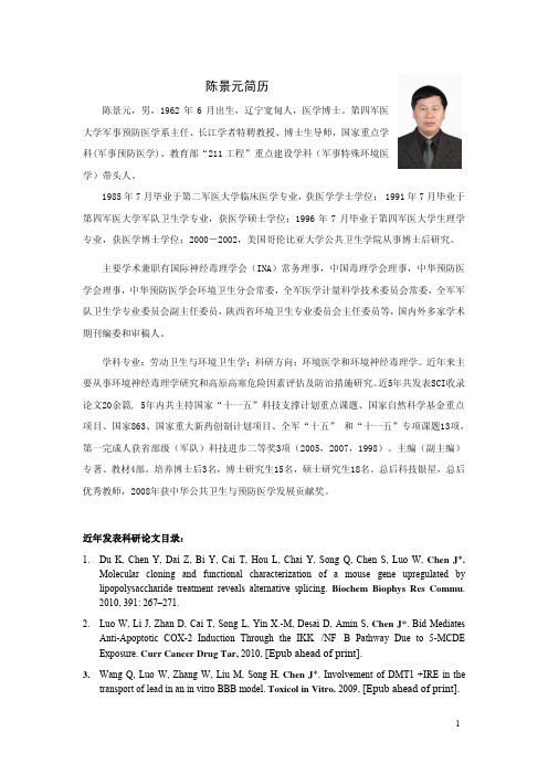
陈景元简历陈景元,男,1962年6月出生,辽宁宽甸人,医学博士。
第四军医大学军事预防医学系主任、长江学者特聘教授、博士生导师,国家重点学科(军事预防医学)、教育部“211工程”重点建设学科(军事特殊环境医学)带头人。
1985年7月毕业于第二军医大学临床医学专业,获医学学士学位; 1991年7月毕业于第四军医大学军队卫生学专业,获医学硕士学位;1996年7月毕业于第四军医大学生理学专业,获医学博士学位;2000-2002,美国哥伦比亚大学公共卫生学院从事博士后研究。
主要学术兼职有国际神经毒理学会(INA)常务理事,中国毒理学会理事,中华预防医学会理事,中华预防医学会环境卫生分会常委,全军医学计量科学技术委员会常委,全军军队卫生学专业委员会副主任委员,陕西省环境卫生专业委员会主任委员等,国内外多家学术期刊编委和审稿人。
学科专业:劳动卫生与环境卫生学;科研方向:环境医学和环境神经毒理学。
近年来主要从事环境神经毒理学研究和高原高寒危险因素评估及防治措施研究。
近5年共发表SCI收录论文20余篇, 5年内共主持国家“十一五”科技支撑计划重点课题、国家自然科学基金重点项目、国家863、国家重大新药创制计划项目、全军“十五” 和“十一五”专项课题13项,第一完成人获省部级(军队)科技进步二等奖3项(2005,2007,1998)。
主编(副主编)专著、教材4部。
培养博士后3名,博士研究生15名,硕士研究生18名。
总后科技银星,总后优秀教师,2008年获中华公共卫生与预防医学发展贡献奖。
近年发表科研论文目录:1.Du K, Chen Y, Dai Z, Bi Y, Cai T, Hou L, Chai Y, Song Q, Chen S, Luo W, Chen J*.Molecular cloning and functional characterization of a mouse gene upregulated by lipopolysaccharide treatment reveals alternative splicing. Biochem Biophys Res Commu.2010, 391: 267–271.2.Luo W, Li J, Zhan D, Cai T, Song L, Yin X.-M, Desai D, Amin S, Chen J*. Bid MediatesAnti-Apoptotic COX-2 Induction Through the IKK/NF B Pathway Due to 5-MCDE Exposure. Curr Cancer Drug Tar, 2010, [Epub ahead of print].3.Wang Q, Luo W, Zhang W, Liu M, Song H, Chen J*. Involvement of DMT1 +IRE in thetransport of lead in an in vitro BBB model. Toxicol in Vitro. 2009, [Epub ahead of print].4.Liu M, Cai T, Zhao F, Zheng G, Wang Q, Chen Y, Huang C, Luo W, Chen J*. Effect ofMicroglia Activation on Dopaminergic Neuronal Injury Induced by Manganese, and Its Possible Mechanism. Neurotox Res. 2009, 16(1):42-9.5.Zheng G, Zhang W, Zhang Y, Chen Y, Liu M, Yao T, Yang Y, Zhao F, Li J, Huang C, LuoW, Chen J*. γ-aminobutyric acid A (GABA A) receptor regulates ERK1/2 phosphorylation in rat hippocampus in high doses of Methyl Tert-Butyl Ether (MTBE)-induced impairment of spatial memory. Toxicol and Appl Pharmacol. 2009; 236( 2): 239-245.6.Yang RH, Wang WT, Chen JY, Xie RG, Hu SJ. Gabapentin selectively reduces persistentsodium current in injured type-A dorsal root ganglion neurons. Pain. 2009; 143(1-2):48-55.7.Zhao F, Cai T, Liu M, Zheng G, Luo W, Chen J*. Manganese induces dopaminergicneurodegeneration via microglial activation in a rat model of manganism. Toxicol Sci. 2009;107(1):156–164.8.Luo W, Chen Y, Liu M, Chen J*. EB1089 induces Skp2-dependent p27 accumulation,leading to cell growth inhibition and cell cycle G1 phase arrest in human hepatoma cells.Cancer Invest. 2009; 27(1):29-37.9.Jin C, Bai L, Wu H, Teng Z, Guo G, Chen J*. Cellular uptake and radiosensitization ofSR-2508 loaded PLGA nanoparticles. J Nanopart Res. 2008; 10 (6):1045-105210.Zheng G, Chen Y, Zhang X, Cai T, Liu M, Zhao F, Luo W, Chen J*. Acute cold exposureand rewarming enhanced spatial memory and activated the MAPK cascades in the rat brain.Brain Res. 2008;1239:171-180.11.Yang RH, Hu SJ, Wang Y, Zhang WB, Luo WJ, Chen JY*. Paradoxical sleep deprivationimpairs spatial learning and affects membrane excitability and mitochondrial protein in the hippocampus. Brain Res. 2008;1230:224-32.12.Jin C, Bai L, Wu H, Liu J, Guo G, Chen J*. Paclitaxel-loaded poly(D,L-lactide-co- glycolide)nanoparticles for radiotherapy in hypoxic human tumor cells in vitro. Cancer Biol Ther.2008;7(6):911-6.13.Luo W, Liu J, Li J, Zhang D, Liu M, Addo JA, Patil S, Zhang L, Yu J, Buolamwini JK, ChenJ*, Huang C. Anti-cancer Effects of JKA97 are Associated with its Induction of Cell Apoptosis via a Bax-dependent, and p53-independent Pathway. J Biol Chem. 2008, 283(13):8624-33.14.Ouyang W, Luo W, Zhang D, Jian J, Ma Q, Li J, Shi X, Chen J, Gao J, Huang C. PI-3K/AktPathway-Dependent Cyclin D1 Expression Is Responsible for Arsenite-Induced Human Keratinocyte Transformation. Environ Health Persp. 2008, 116(1): 1-6.15.Du K, Chai Y, Hou L, Chang W, Chen S, Luo W, Cai T, Zhang X, Chen N, Chen Y, Chen J*.Over-expression and siRNA of a novel Environmental lipopolysaccharide responding gene on the cell cycle of the human hepatoma derived cell line HepG2. Toxicology, 2008, 243:303-310.16.Zhang XP, Zheng G, Zou L, Liu HL, Hou LH, Zhou P, Yin DD, Zheng QJ, Liang L, ZhangSZ, Feng L, Yao LB, Yang AG, Han H, Chen JY*. Notch activation promotes cell proliferation and the formation of neural stem cell-like colonies in human glioma cells. Mol Cell Biochem. 2008, 307(1-2):101-8.17.Cai T, Yao T, Li Y, Chen Y, Du K, Chen J*, Luo W. Proteasome inhibition is associatedwith manganese-induced oxidative injury in PC12 cells. Brain Res. 2007, 1185: 359-65.18.Zheng G, Zhang X, Chen Y, Zhang Y, Luo W, Chen J*. Evidence for a role of GABAAreceptor in the acute restraint stress-induced enhancement of spatial memory. Brain Res.2007, 1181:61-73.19.Wang Q, Luo W, Zheng W, Liu Y, Xu H, Zheng G, Dai Z, Zhang W, Chen Y, Chen J*. Ironsupplement prevents lead-induced disruption of the blood-brain barrier during rat development. Toxicol and Appl Pharmacol. 2007, 219(1):33-41.20.Wang Q, Luo W, Zhang W, Dai Z, Chen Y, Chen J*. Iron supplementation protects againstlead-induced apoptosis through MAPK pathway in weanling rat cortex. NeuroToxicology.2007, 28(4):850-859.21.Liu YL, Bi H, Chi SM, Fan R, Wang YM, Ma XL, Chen YM, Luo WJ, Pei JM, Chen JY*.The Effect of Compound Nutrients on Stress-induced Changes in Serum IL-2, IL-6 and TNF-a Levels in Rats. Cytokine. 2007, 37:14-21.22.Ding J, Li J, Chen J, Chen H, Ouyang W, Zhang R, Xue C, Zhang D, Amin S, Desai D,Huang C.. Effects of Polycyclic Aromatic Hydrocarbons (PAHs) on Vascular Endothelial Growth Factor Induction through Phosphatidylinositol 3-Kinase/AP-1-dependent, HIF-1{alpha}-independent Pathway. J Biol Chem. 2006, 281(14):9093-100. (Co-first author)23.Chen J, Yan Y, Li J, Ma Q, Stoner GD, Ye J, Huang C. Differential requirement of signalpathways for benzo[a]pyrene (B[a]P)-induced nitricoxide synthase (iNOS) in rat esophageal epithelial cells. Carcinogenesis 2005, 26(6):1035-104324.Meller E, Shen C, Nikolao TA, Jensen C, Tsimberg Y, Chen J, Gruen RJ. Region-specificeffects of acute and repeated restraint stress on the phosphorylation of mitogen-activated protein kinases.Brain Res. 2003, 979: 57-64.25.Chen J, Shen C, Meller E. 5-HT1A receptor-mediated regulation of mitogen- activated proteinkinase phosphorylation in rat brain.Eur J Pharmacol 2002, 452;155-162.26.Louis ED, Zheng W, Jurewicz EC, Watner D, Chen J, Factor-Litvak P, Parides M. Elevationof blood carboline alkaloids in essential tremor. Neurology. 2002, 59:1940–1944.27.Chen JY, Tsao GC, Zhao Q, Zheng W. Differential Cytotoxicity of Mn(II) and Mn(III):Special Reference to Mitochondrial [Fe-S] Containing Enzymes.Toxicol and Appl Pharmacol. 2001, 175, 160–168.。
Notch3在大鼠肝癌中的表达及其意义

第32卷第1期2012年2月健康研究Health Research Vol.32No.1Feb.2012收稿日期:2011-10-16作者简介:邱丽香(1982-),女,浙江龙泉人,本科,护师。
DOI :10.3969/j.issn.1674-6449.2012.01.002基础医学Notch3在大鼠肝癌中的表达及其意义邱丽香1,翟淑亭2,秦建芬1(1.浙江大学医学院附属邵逸夫医院护理部,浙江杭州310016;2.杭州市下沙医院普外科,浙江杭州310006)摘要:目的研究Notch3在大鼠肝癌发生发展中的动态变化。
方法二乙基亚硝胺诱导大鼠肝细胞癌的发生,于4周、8周、12周、16周时取肝脏标本,免疫组化和Western blot 法检测标本中Notch3的表达。
结果随着肝组织损伤加剧,Notch3表达逐渐升高。
与正常组相比,肝损伤组、肝硬化组和肝癌组的Notch3蛋白明显表达增加(P <0.05)。
结论Notch3参与了大鼠肝癌发生的全过程,并且随着肝损伤的不断加剧,其表达也逐渐上升。
Notch3信号的激活可能是肝癌发生的一项重要机制。
关键词:Notch3蛋白;肝癌中图分类号:R735.7文献标识码:A 文章编号:1674-6449(2012)01-0004-03Expression of Notch 3in Rat Liver Cancer and Its EffectQIU Li-xiang 1,ZHAI Shu-ting 2,QIN Jian-fen 1(1.Department of Nursing ,The Sir Run Run Shaw Hospital ,School of Medicine ,Zhejiang University ,Hangzhou ,Zhejiang310016,China ;2.Department of General Surgery ,Hangzhou Xiasha Hospital ,Hangzhou ,Zhejiang 310006,China )Abstract :Objective Objective To investigate the dynamic changes of Notch 3in the development and progression ofhepatic carcinoma in rats.MethodsHepatocarcinoma was induced by diethylinitrosamine.Liver specimens were taken at week 4,8,12and 16for detection of Notch3expression using immunohistochemical and Western blot methods.ResultsNotch3expression increased gradually with the worsening of hepatic injury.There were statistically significant increasesof Notch3protein expression in hepatic injury group ,hepatic cirrhosis group and hepatocarcinoma group as compared to thenormal control group (P <0.05).Conclusions Notch3participates in the whole process of hepatocarcima developmentand progression in rats.Increased expression of Notch3is associated with the worse of hepatic injury.The activation ofNotch3may be an important mechanism for the development of hepatocarcinoma.Key words :Notch3protein ;hepatocarcinomaNotch信号通路在哺乳动物的胚胎形成中扮演重要的角色,作为一种进化上相对保守的细胞相互作用机制,维持着细胞增殖、分化、凋亡之间的平衡,对细胞分化命运起决定性作用。
组蛋白赖氨酸甲基转移酶在糖尿病中的研究进展
组蛋白赖氨酸甲基转移酶在糖尿病中的研究进展包志伟; 杨天竹; 刘洋; 王志刚【期刊名称】《《中国医药导报》》【年(卷),期】2019(016)025【总页数】4页(P45-48)【关键词】组蛋白; 组蛋白甲基化; 赖氨酸甲基转移酶; 糖尿病【作者】包志伟; 杨天竹; 刘洋; 王志刚【作者单位】哈尔滨医科大学(大庆)医学检验与技术学院生物化学教研室黑龙江大庆 163319【正文语种】中文【中图分类】R587.1糖尿病是在遗传因素与环境因素相互作用下,以胰岛素的分泌和利用异常引发的高血糖为特征的代谢性疾病,目前尚无根治方法。
糖尿病分为两型,1 型糖尿病主要与免疫系统缺陷和遗传因素有关,2 型糖尿病主要与胰岛素抵抗和肥胖有关,在我国糖尿病患者中有90%是2 型糖尿病患者。
胰岛素的分泌和利用是糖尿病发生发展的关键因素,胰岛素分泌和发挥功能的过程又受到许多因素的调节,如胰岛素受体和胰岛素样生长因子1 的分子含量变化影响胰岛素亲和能力[1]。
许多组蛋白修饰酶在代谢疾病中起着至关重要的作用,包括糖尿病及其并发症。
组蛋白甲基化调节酶在代谢疾病中的作用也引起了人们的关注,多种组蛋白赖氨酸甲基转移酶与糖尿病及其并发症的发生发展有关,因此本文对组蛋白赖氨酸甲基转移酶在糖尿病及其并发症中的研究进展作一简要综述。
1 影响组蛋白甲基化的相关因素组蛋白甲基化是表观遗传学中组蛋白修饰的主要形式之一。
组蛋白甲基转移酶和组蛋白脱甲基酶共同完成组蛋白甲基化修饰这一动态过程。
组蛋白的甲基化主要发生在H3 和H4 的赖氨酸(lysine,K)和精氨酸(arginine,R)上,组蛋白赖氨酸甲基转移酶主要由SET(Su var 3-9,E z,Trithorax)结构域家族和非SET 结构域家族的甲基转移酶完成。
目前,对SET 结构域家族研究相对充分。
H3K4、H3K36、H3K79 的甲基化主要参与基因激活,而H3K9、H3K20、H3K27 的甲基化主要参与基因沉默。
局灶性缺血性脑卒中大鼠海马Notch信号通路的动态表达.
生堡医堂苤壶垫!生!Q旦垫旦筮塑鲞筮12塑盟型塑型』堡塾i坠:Q!垫坠竺垫:塑!:y些婴:壁垒!!局灶性缺血性脑卒中大鼠海马Notch信号通路的动态表达郭怡菁张志琚王少华隋毓秀孙奕滕皋军【摘要】目的观察局灶性缺血性脑卒中大鼠海马Notch信号通路的表达变化,初步探讨该通路在局灶性缺血性脑卒中后神经再生的调控作用。
方法大脑中动脉阻塞(MCAO)法建立大鼠局灶性脑缺血模型,通过Western印迹法和实时RT.PCR法分别于卒中后19d和28d检测各实验组及对照组大鼠海马Notch,及其配体Jagged。
的蛋白、基因表达。
结果卒中后19d,缺血组Notchl蛋白和基因表达均分别高于对照组(均P<0.05),缺血组28d的Notch。
蛋白和基因表达显著低于19d的蛋白和基因表达(均P<0.01)。
缺血组19d和28d的Jagged。
蛋白表达(P<0.05)和基因表达(P<0.05)高于对照组,但28d的Jagged.蛋白和基因表达与19d相比差异无统计学意义(均P>0.05)。
结论局灶性缺血性卒中大鼠Notch信号通路呈现动态表达变化,提示该通路可能参与了对卒中后内源性神经前体细胞增殖、分化的调控。
【关键词】脑梗死;信号传导;神经再生DynamicchangesoftheexpressionofNotchlandJaggedlinthehippocampusafterfocalischemia:experimentwithratsGUOYi-jing’.ZHANGZhi-jun,WANGShao—hua,SUIYu—xiu,SUNYi,TENGCnw-jurL’DepartmentofNeurology,ZhongdaHospitalAffiliatedtOSoutheastUniversity;InstitutionofCerebralVascularD诂easeofSoutheastUniversity,Nanjing210009,ChinaCorrespondingauthor:ZHANGZhi-jun。
notch信号通路参与龙生蛭胶囊的抗动脉粥样硬化作用

doi:10.3969/j.issn.1000⁃484X.2019.23.009Notch 信号通路参与龙生蛭胶囊的抗动脉粥样硬化作用秦 咏 王 莹① (山东省胸科医院药剂科,济南250013) 中图分类号 R285.5 文献标志码 A 文章编号 1000⁃484X (2019)23⁃2858⁃06①山东大学第二医院手术室,济南250033㊂作者简介:秦 咏,男,主管药师,主要从事中药药理研究㊂通讯作者及指导教师:王 莹,女,主管护师,主要从事围术期患者安全方面的研究,E⁃mail:wangy_sd@㊂[摘 要] 目的:研究龙生蛭胶囊抗动脉粥样硬化(AS)的作用,并对Notch 信号通路在其中的调控机制进行初步探讨㊂方法:60只SD 大鼠随机分为正常组(N)㊁模型组(AS)㊁龙生蛭胶囊低剂量组(LSZ⁃LD,0.75g /kg)㊁龙生蛭胶囊中剂量组(LSZ⁃MD,1.5g /kg)㊁龙生蛭胶囊高剂量组(LSZ⁃HD,3.0g /kg)和辛伐他汀组(Simvastatin,5.0mg /kg),每组10只㊂除正常组大鼠使用普通饲料喂养外,其他组均使用高脂饲料外加注射维生素D3构建AS 大鼠模型㊂4周后,进行给药干预㊂8周后,采血检测总胆固醇(TC)㊁三酰甘油(TG)㊁低密度脂蛋白胆固醇(LDL⁃C)㊁高密度脂蛋白胆固醇(HDL⁃C)㊁一氧化氮(NO)㊁TNF⁃α和IL⁃6含量;流式细胞仪检测外周血中循环内皮细胞数量;苏木素⁃伊红(HE)染色观察动脉组织形态变化;Western blot 检测动脉组织Notch 胞内结构域(NICD)和转录因子Hes1蛋白表达水平㊂结果:与正常组相比,模型组主动脉内皮损伤严重,血脂和炎症因子水平显著升高(P <0.05)㊂与模型组比较,龙生蛭胶囊各组的主动脉内皮损伤得到改善,血清中TC㊁TG㊁LDL⁃C㊁TNF⁃α和IL⁃6水平降低,而HDL⁃C 升高;血清NO 含量也得到明显升高(P <0.05),循环内皮细胞数量减少(P <0.05);NICD 和Hes1蛋白表达水平显著升高(P <0.05)㊂结论:龙生蛭胶囊可明显抑制AS 大鼠模型的血脂水平及炎症反应,还可改善血管内皮功能,其抗AS 作用机制可能与抑制Notch 信号通路有关㊂[关键词] 龙生蛭胶囊;动脉粥样硬化;Notch 信号通路;血脂;内皮细胞功能Experimental study of Longshengzhi capsule on anti⁃atherosclerosis by regula⁃ting Notch signaling pathwayQIN Yong ,WANG Ying .Department of Pharmacy ,the Thoracic Hospital of Shandong Province ,Jinan 250013,China[Abstract ] Objective :To investigate the anti⁃atherosclerosis effect of Longshengzhi capsule,and to explore the regulationmechanism of Notch signaling pathway.Methods :Sixty SD rats were randomly divided into normal group,model group,low dose ofLong⁃shengzhi capsule group(0.75g /kg),middle dose of Longshengzhi capsule group(1.5g /kg),high dose of Longshengzhi capsule group (3.0g /kg)and Simvastatin group(5.0mg /kg).In addition to normal group which was fed with normal diet,other five groups were fed with high⁃fat diet plus Vitamin D injection to establish the atherosclerosis(AS)rat model.Four weeks later,the treatment group received drug intervention.Eight weeks later,contents of total cholesterol(TC),triglyceride(TG),low⁃density lipoprotein cholesterol(LDL⁃C),high⁃density lipoprotein cholesterol(HDL⁃C),nitric oxide(NO),Tumor necrosis factor α(TNF⁃α)and interleukin⁃6(IL⁃6)were meas⁃ured.The circulating endothelial cells in peripheral blood were detected by flow cytometry.The morphological changes of arterial tissues were observation by HE staining.The expression of Notch intracellular domain(NICD)and transcription factor Hes1in arterial tissues were detected by Western blot.Results :Compared with the normal group,the aortic endothelium was damaged seriously,and the levels of blood lipids and inflammatory factors were significantly increased in the model pared with the model group,the aortic en⁃dothelial injury was improved in each group of Longshengzhi capsule.Longshengzhi capsule decreased the serum levels of TC,TG,LDL⁃C,IL⁃6and TNF⁃α,increased the HDL⁃C level.We also found that Longshengzhi capsule obviously increased the serum NO content(P <0.05),and the number of circulating endothelial cells was significantly decreased (P <0.05).In addition,Longshengzhi capsule significantly increased the expression of NICD and Hes1(P <0.05).Conclusion :Longshengzhi capsule can significantly inhibit the blood lipid level and inflammation in AS rat model,and improve vascular endothelial function.Its anti⁃AS mechanism may be related to the inhibition of Notch signaling pathway.[Key words] Longshengzhi capsule;Atherosclerosis;Notch signaling pathway;Blood lipid;Vascular endothelial function 动脉粥样硬化(Atherosclerosis,AS)是心脑血管疾病的重要病理基础,是一种导致斑块发展和冠状动脉进行性狭窄的炎症性疾病[1]㊂当人体内脂质代谢发生紊乱,引起局部炎症反应,从而导致AS发生[2]㊂已有研究发现Notch信号通路可调控血管新生,且阻断Notch配体可减轻代谢紊乱和动脉粥样硬化[3,4]㊂龙生蛭胶囊是由黄芪㊁水蛭㊁川芎等12味药组成的复合制剂,具有补气活血㊁化瘀通络的功效,在临床上联合其他药物对脑梗及气虚血瘀型脑梗死患者有较好的治疗效果[5,6]㊂龙生蛭胶囊可降低血脂,抑制炎症反应,但其作用的具体机制尚未阐述清楚[5,7]㊂因此,本次研究通过高脂饲料喂养构建AS大鼠模型,探讨龙生蛭胶囊是否通过调节Notch信号通路对AS大鼠产生治疗作用㊂1 材料与方法1.1 材料1.1.1 动物 无特定病原体(Specific pathogen Free,SPF)级SD大鼠60只,体质量(180±20)g,购自长沙市天勤生物技术有限公司,动物许可证号: SCXK(湘)2014⁃0010㊂1.1.2 主要试剂 总胆固醇(Total cholesterol, TC)㊁三酰甘油(Triglyceride,TG)㊁低密度脂蛋白胆固醇(Low⁃density lipoprotein cholesterol,LDL⁃C)㊁高密度脂蛋白胆固醇(High⁃density lipoprotein cholesterol,HDL⁃C)及一氧化氮(NO)试剂盒均购自南京建成生物工程研究所;TNF⁃α和IL⁃6ELISA试剂盒均购自武汉华美生物有限公司;CD31抗体购自BD公司;兔抗活性Notch胞内结构域(Notch in⁃tracellular domain,NICD)多克隆抗体和兔抗Hes1多克隆抗体均购自Abcam公司;GAPDH购自Sigma公司;PVDF膜和化学发光试剂均购自Millipore公司㊂1.2 方法1.2.1 动物分组及模型构建[8] SD大鼠60只随机分为6组:正常组(N)㊁模型组(AS)㊁龙生蛭胶囊低剂量组(LSZ⁃LD)㊁龙生蛭胶囊中剂量组(LSZ⁃MD)㊁龙生蛭胶囊高剂量组(LSZ⁃HD)和辛伐他汀组(Simvastatin),每组10只㊂除正常组大鼠给予普通饲料喂养外,其他五组大鼠均给予高脂饲料喂养和一次性腹腔注射维生素D3(70万U/kg)㊂高脂饲料配方:80.8%基础饲料㊁3.5%胆固醇㊁10%炼制猪油㊁0.2%丙基硫氧嘧啶㊁0.5%胆酸钠㊁5%白糖㊂4周后开始灌胃给药处理,正常组和模型组给予等量生理盐水灌胃,低剂量组大鼠给予0.75g/kg龙生蛭胶囊灌胃,中剂量组给予1.5g/kg龙生蛭胶囊灌胃,高剂量组给予3.0g/kg龙生蛭胶囊灌胃,辛伐他汀组给予5.0mg/kg辛伐他汀灌胃,1d/次㊂8周后,开始取材并检测㊂1.2.2 取材 给药结束后,各组大鼠禁食过夜㊂用戊巴比妥(30mg/kg)对大鼠进行麻醉,打开大鼠腹腔进行腹主动脉取血,于-80℃中保存㊂分离并剪下1cm的胸主动脉,置于4%多聚甲醛中固定,待检㊂1.2.3 生化及ELISA检测指标 将取出的腹主动脉血静置后离心分离出血清,然后根据试剂盒说明操作,采用比色法测定大鼠血清中TC㊁TG㊁LDL⁃C㊁HDL⁃C及NO水平㊂采用ELISA试剂盒检测血清中的TNF⁃α和IL⁃6含量㊂1.2.4 流式细胞仪检测循环内皮细胞数量 取出外周血并制成单细胞悬液,调整细胞密度后将细胞接种于96孔板中,1.5×103细胞/孔㊂加入5μl CD31抗体,混匀后避光孵育30min㊂PBS洗涤3次,离心收集细胞㊂PBS重悬细胞后进行流式细胞仪检测㊂1.2.5 苏木素⁃伊红(HE)染色观察形态学变化 取出固定的胸主动脉组织于包埋盒中进行脱水浸蜡包埋,然后切成厚度为4~5μm的切片㊂脱蜡后行苏木素⁃伊红染色,梯度乙醇进行脱水,再用二甲苯透明,最后中性树脂封片,于显微镜下观察主动脉的形态变化㊂1.2.6 Western blot检测通路相关蛋白表达 提取主动脉组织总蛋白,BCA法蛋白定量后,用10% SDS⁃PAGE凝胶电泳分离目的蛋白㊂湿转法将蛋白转印至PVDF膜上,5%脱脂奶粉室温封闭2h㊂加入稀释后的Ⅰ抗(NICD稀释比:1∶500;Hes1稀释比:1∶2000),室温孵育1h,TBST洗涤3次㊂加入经HRP标记的Ⅱ抗后,室温孵育1h,TBST洗涤3次后,加入化学发光试剂,置于暗室中曝光显影,全自动化学发光分析仪分析蛋白条带㊂1.3 统计学处理 采用SPSS22.0统计软件进行统计学分析,实验数据以x±s表示㊂多组间比较采用单因素方差分析,各组均数间两两比较采用LSD检验㊂P<0.05表示差异具有统计学意义㊂2 结果2.1 主动脉形态学观察 正常组胸主动脉内皮表面光滑,结构完整㊂模型组血管内皮细胞排列紊乱,内膜出现严重增生,有大量的泡沫细胞堆积,血管腔狭窄㊂经过不同剂量龙生蛭胶囊处理后主动脉内皮损伤得到改善,血管腔扩大,且高剂量组最明显,呈现一定的剂量依赖性(图1)㊂2.2 龙生蛭胶囊对血脂水平的影响 生化实验检测各组大鼠血脂水平,结果如表1所示㊂与正常组比较,模型组血清中的TC㊁TG 和LDL⁃C 含量显著升高(P <0.01),HDL⁃C 含量显著降低(P <0.01)㊂与模型组相比较,龙生蛭胶囊各剂量处理组的TC㊁TG 和LDL⁃C 含量降低,HDL⁃C 含量升高,尤其是中剂量和高剂量组的改善效果更显著(P <0.01)㊂高剂量组与辛伐他汀组之间无明显差异(P >0.05)㊂2.3 龙生蛭胶囊对血清炎症因子的影响 模型组大鼠血清中的TNF⁃α和IL⁃6含量均高于正常组,差异具有统计学意义(P <0.01)㊂而与模型组比较,各剂量给药组大鼠血清中的TNF⁃α和IL⁃6含量显著降低(P <0.01),效果最佳的高剂量组与阳性药物图1 主动脉HE 染色(×100)Fig.1 HE staining of aorta (×100)辛伐他汀组间的差异无统计学意义(P >0.05)㊂结果见表2㊂2.4 龙生蛭胶囊对血清NO 水平及循环内皮细胞数量的影响 结果如图2和表3所示,与正常组相比,模型组大鼠血清中NO 含量显著降低(P <0.01),外周血中循环内皮细胞数量显著增多(P <0.01)㊂与模型组相比,给予龙生蛭胶囊治疗的大鼠血清中NO 含量出现明显升高(P <0.05),循环内皮细胞数量降低(P <0.05)㊂高剂量组和辛伐他汀组间无明显差异(P >0.05)㊂2.5 龙生蛭胶囊对Notch 通路蛋白表达的影响 模型组大鼠的NICD 和Hes1蛋白相对表达量显著高与正常组(P <0.01)㊂而经过不同剂量龙生蛭胶囊治疗后,AS 大鼠模型的NICD 和Hes1蛋白相对表达量降低,差异具有显著性(P <0.05或P <0.01)㊂NICD 和Hes1在高剂量组和辛伐他汀组中表达量无明显差异㊂结果见图3㊂表2 龙生蛭胶囊对大鼠血清TNF⁃α和IL⁃6水平的影响(n =10,x ±s )Tab.2 Effect of Longshengzhi capsule on TNF⁃αand IL⁃6levels in serum of rats (n =10,x ±s )Groups TNF⁃αIL⁃6N15.24±0.8242.53±2.26AS 37.08±1.471)80.25±4.121)LSZ⁃LD26.55±1.382)64.16±3.222)LSZ⁃MD 21.31±1.942)57.93±1.772)LSZ⁃HD16.14±0.682)43.31±1.532)Simvastatin 17.08±0.962)44.82±2.372)Note:N.Normal pared with the normal group,1)P <0.01;compared with the AS group,2)P <0.01.表1 龙生蛭胶囊对大鼠血脂水平的影响(n =10,x ±s )Tab.1 Effect of Longshengzhi capsule on blood lipid levels in rats (n =10,x ±s )Groups TCTGLDL⁃CHDL⁃CN1.37±0.220.47±0.140.56±0.170.98±0.23AS2.04±0.371)0.82±0.201)0.81±0.111)0.64±0.151)LSZ⁃LD1.96±0.150.76±0.110.74±0.140.81±0.132)LSZ⁃MD 1.63±0.183)0.63±0.173)0.65±0.083)0.93±0.213)LSZ⁃HD1.45±0.213)0.56±0.153)0.60±0.123)1.16±0.193)Simvastatin 1.26±0.243)0.49±0.133)0.57±0.153)1.19±0.223)Note:N.Normal pared with the normal group,1)P <0.01;compared with the AS group,2)P <0.05,3)P <0.01.图2 流式检测循环内皮细胞数量Fig.2 Number of circulating endothelial cells was measu⁃red by flow cytometry表3 龙生蛭胶囊对血清NO 水平及循环内皮细胞数量的影响(n =10,x ±s )Tab.3 Effect of Longshengzhi capsule on NO level andnumber of circulating endothelial cells (n =10,x ±s )Groups NO(μmol /L)Number of circulation endothelia(ml -1)N48.53±3.75108.15±23.52AS 31.47±1.521)183.34±62.411)LSZ⁃LD37.24±1.942)165.29±37.542)LSZ⁃MD 43.21±2.333)142.50±44.243)LSZ⁃HD52.63±3.423)115.54±33.173)Simvastatin 51.76±2.843)112.81±34.633)Note:N.Normal pared with the normal group,1)P <0.01;compared with the AS group,2)P <0.05,3)P <0.01.图3 龙生蛭胶囊对NICD ㊁Hes1蛋白表达的影响Fig.3 Effect of Longshengzhi capsule on proteins expression of NICD and Hes1Note:N.Normal pared with the normal group,**.P <0.01;compared with the AS group,#.P <0.05,##.P <0.01.3 讨论AS 的典型特征是动脉中形成动脉粥样硬化病变,即斑块,而斑块是在脂质代谢和免疫应答紊乱的背景下,由炎症反应支持而成,具有限制血流㊁血栓闭塞及破裂的倾向[9]㊂大量研究表明,血脂异常㊁高血压及肥胖是AS 的早期干预危险因素[10,11]㊂且LDL⁃C㊁TC 和TG 水平升高易导致血管壁脂质沉积而形成斑块㊂因此,调节血脂是治疗AS 的主要方法之一㊂而HDL 具有抗动脉粥样硬化的功能,可抑制巨噬细胞释放炎症因子,并促进内皮细胞的自我修复[12]㊂在本研究中,龙生蛭胶囊可显著降低AS 大鼠模型血清中的TC㊁TG 及LDL⁃C 含量,并升高HDL⁃C 水平,发挥了明显的降脂作用㊂进一步检测了血清中TNF⁃α和IL⁃6含量,结果显示龙生蛭胶囊可显著减低TNF⁃α和IL⁃6水平㊂提示龙生蛭胶囊具有降低血脂,抗血管炎症的功能㊂血管内皮细胞损伤及功能障碍在AS 的发生发展过程中起重要作用,与早期AS 发病的机制相关[13]㊂Gimbrone 等[14]也阐述了内皮细胞功能障碍会影响凝血和血栓形成㊁局部血管张力和氧化还原平衡以及急性和慢性炎症反应之间的协调,是AS 病变起始和进展中的关键因素[14]㊂NO 由血管内皮细胞释放,具有扩张血管,抑制血小板黏附㊁聚集等功能,当内皮细胞功能发生障碍时导致NO 合成减少或通路障碍,从而促进了AS 的发病过程[15]㊂正常人体内的循环内皮细胞数量是极少的,当内皮细胞受损时,循环内皮细胞数量便会增多,是反映血管内皮损伤的标志物[16,17]㊂本次研究结果也显示,在AS 大鼠模型血清中的NO 含量降低,外周血中的循环内皮细胞数量增加,而经过龙生蛭胶囊给药处理后,NO 含量升高,循环内皮细胞数量趋于正常,提示龙生蛭胶囊可改善AS 大鼠的内皮细胞功能㊂Notch 信号通路是在进化过程中高度保守的一类通路,由受体㊁配体及效应物组成,不同的配体与不同的受体结合后转导的Notch 信号所产生的生物学效用不同[18]㊂当受体胞外区和配体结合被激活后,胞内区释放出具有活性的NICD,NICD 与重组信号蛋白结合后进一步激活下游靶基因Hes [19⁃21]㊂据报道,Notch 信号通路参与了心血管疾病的炎症反应,从而推动了心血管疾病的病理过程[22]㊂Binesh等[19]的研究结果显示抑制NICD 和下游靶基因Hes 表达,即抑制了Notch 信号通路可发挥抗AS 的作用㊂鉴于Notch 信号通路在炎症反应及AS 病理中的作用,本研究中对AS 大鼠模型主动脉血管组织中的NICD和Hes1蛋白表达水平进行检测,发现龙生蛭胶囊可有效地抑制AS大鼠模型的NICD和Hes1蛋白表达㊂综上所述,龙生蛭胶囊可降低AS大鼠模型的血脂水平,发挥抗血管炎症反应㊁保护内皮细胞功能的作用,其抗AS的作用机制可能与抑制Notch信号通路有关㊂另外,在本次研究的HE染色结果中,龙生蛭胶囊高剂量组的作用效果稍差于辛伐他汀组,而Notch信号通路蛋白表达㊁炎性细胞因子及NO 的水平与辛伐他汀组的差异并不明显,推测阳性药物辛伐他汀治疗AS的主要作用机制可能不是通过调控炎性细胞因子㊁NO等水平实现的㊂参考文献:[1] 肖 滨,黄小波.衰老与动脉粥样硬化关系的研究进展[J].中华老年多器官疾病杂志,2018,17(11):866⁃869.Xiao B,Huang XB.Atherosclerosis in relation to aging[J].Chin J Mult Organ Dis Elderly,2018,17(11):866⁃869.[2] Nahrendorf M,Swirski FK.Lifestyle Effects on Hematopoiesis andAtherosclerosis[J].Circ Res,2015,116(5):884⁃894. [3] 温艺超,陈伟燕,谢富华,等.DAPT阻断Notch通路对动脉粥样硬化型缺血性脑卒中大鼠的保护作用[J].中国病理生理杂志,2018,34(11):2031⁃2036.Wen YC,Chen WY,Xie FH,et al.Protective effects of DAPT on rat model with atherosclerotic ischemic brain stroke by blocking Notch pathway[J].Chin J Pathophysiol,2018,34(11): 2031⁃2036.[4] Fukuda D,Aikawa E,Swirski FK,et al.PNAS Plus:Notch ligandDelta⁃like4blockade attenuates atherosclerosis and metabolic disorders[J].Proc Natl Acad Sci U S A,2012,109(27): 10763⁃10764.[5] 刘小英.龙生蛭胶囊联合丹红注射液治疗脑梗死疗效及对hs⁃CRP的影响[J].现代中西医结合杂志,2011,20(22): 2742⁃2743.Liu XY.Curative effects of Longshengzhi capsule combined with Danhong injection on cerebral infarction and its influence on hs⁃CRP[J].Modern J Int Tradit Chin Western Med,2011,20(22): 2742⁃2743.[6] 周 敏,彭丹亚.龙生蛭胶囊联合尼莫地平治疗急性脑梗死的临床研究[J].现代药物与临床,2018,33(11):2836⁃2839.Zhou M,Peng DY.Clinical study on Longshengzhi capsule combined with nimodipine in treatment of acute cerebral infarction [J].Drugs Clin,2018,33(11):2836⁃2839.[7] 方欢乐,陈衍斌,张飒乐,等.龙生蛭胶囊抑制大鼠动脉粥样硬化形成保护血管内皮功能的分子机制研究[J].中药新药与临床药理,2017,28(1):6⁃13.Fang HL,Chen YB,Zhang SL,et al.Protective effects and mechanism of Longshengzhi capsules on inhibiting atherosclerosis formation and improving vascular dysfunction in rats[J].Tradit Chin Drug Res Clin Pharmacol,2017,28(1):6⁃13. [8] 赵 娟,李相军,孙 波,等.维生素D3联合高脂饲料建立大鼠动脉粥样硬化模型[J].实用医学杂志,2009,25(21): 3569⁃3571.Zhao J,Li XJ,Sun B,et al.Establishment of a rat model of athero⁃sclerosis using vitamin D3and high⁃fat diet[J].J Pract Med, 2009,25(21):3569⁃3571.[9] Scherer DJ,Psaltis PJ.Future imaging of atherosclerosis:molecularimaging of coronary atherosclerosis with(18)F positron emission tomography[J].Cardiovasc Diagn Ther,2016,6(4):354⁃367.[10] 安冬青,吴宗贵.动脉粥样硬化中西医结合诊疗专家共识[J].中国全科医学,2017,20(5):507⁃511.An DQ,Wu ZG.Expert consensus on the diagnosis and treatmentof atherosclerosis by combination of traditional chinese andwestern medicine[J].Chin General Pract,2017,20(5):507⁃511.[11] O′neal WT,Soliman EZ,Qureshi W,et al.Sustained pre⁃hypertensive blood pressure and incident atrial fibrillation:themulti⁃ethnic study of atherosclerosis[J].J Am Soc Hypertens,2015,9(3):191⁃196.[12] 邓 艳,许晓婷,雷霆雯,等.phiC31整合酶介导的低密度脂蛋白受体基因治疗对小鼠动脉粥样硬化病变的影响[J].中国动脉硬化杂志,2018,26(6):572⁃576.Deng Y,Xu XT,Lei TW,et al.Effect of phiC31integrase⁃mediated low density lipoprotein receptor gene therapy genetherapy on atherosclerosis in mice[J].Chin J Arteriosclerosis,2018,26(6):572⁃576.[13] 王竟悟,于 杨,王大新,等.血管平滑肌细胞与动脉粥样硬化关系的研究新进展[J].实用医学杂志,2017,33(21):3661⁃3663.Wang JW,Yu Y,Wang DX,et al.Recent advances in therelationship between vascular smooth muscle cells andatherosclerosis[J].J Pract Med,2017,33(21):3661⁃3663. [14] Gimbrone MA,Guillermo GC.Endothelial cell dysfunction andthe pathobiology of atherosclerosis[J].Circ Res,2016,118(4):620⁃636.[15] 王秀芬,崔姝雅,叶子芯,等.一氧化氮在动脉粥样硬化发病机制中作用的研究进展[J].中国老年学杂志,2016,36(21):5459⁃5462.Wang XF,Cui SY,Ye ZX,et al.Research progress on the role ofnitric oxide in the pathogenesis of atherosclerosis[J].Chin JGerontol,2016,36(21):5459⁃5462.[16] 马贤德,李雪晶,李大勇,等.桃红四物汤调节血管内皮细胞功能及治疗动脉硬化闭塞症的实验研究[J].中国中西医结合杂志,2014,34(2):191⁃196.Ma XD,Li XJ,Li DY,et al.TaohongSiwu decoction regulatedfunctions of endothelial cells and treated arteriosclerosisobliterans:an experimental study[J].Chin J Integrat TraditWestern Med,2014,34(2):191⁃196.[17] Eleftheriou D,Ganesan V,Hong Y,et al.Endothelial injury inchildhood stroke with cerebral arteriopathy:a cross⁃sectionalstudy[J].Neurology,2012,79(21):2089⁃2096. [18] 杨敬宁,张 素,罗羽莎,等.DAPT调节Treg/Th17细胞免疫平衡抑制动脉粥样硬化[J].中国免疫学杂志,2017,33(3):343⁃346.Yang JN,Zhang S,Luo YS,et al.DAPT regulates Treg/Th17cellsimmune balance to inhibit atherosclerosis[J].Chin J Immunol,2017,33(3):343⁃346.[19] Binesh A,Devaraj SN,Devaraj H.Inhibition of nucleartranslocation of notch intracellular domain(NICD)by diosgeninprevented atherosclerotic[J].Biochimie,2018,148:63⁃71. [20] 陶 敏,康品方,张 恒.Notch信号通路和心血管疾病研究进展[J].中国老年学杂志,2017,37(24):6251⁃6253.Tao M,Kang PF,Zhang H.Advances in notch signaling pathwayand cardiovascular diseases[J].Chin J Gerontol,2017,37(24):6251⁃6253.[21] Schwanbeck R,Martini S,Bernoth K,et al.The Notch signalingpathway:molecular basis of cell context dependency[J].Eur JCell Biol,2011,90(6⁃7):572⁃581.[22] Quillard T,Charreau B.Impact of notch signaling on inflammatoryresponses in cardiovascular disorders[J].Int J Mol Sci,2013,14(4):6863⁃6888.[收稿2019⁃02⁃20 修回2019⁃05⁃07](编辑 刘格格)(上接第2857页)[13] Yu HX,Wang XL,Zhang LN,et al.Involvement of the TLR4/NF⁃κB signaling pathway in the repair of esophageal mucosa injury inrats with gastroesophageal reflux disease[J].Cell PhysiolBiochem,2018,51(4):1645⁃1657.[14] Xu J,Lu C,Liu Z,et al.Schizandrin B protects LPS⁃inducedsepsis via TLR4/NF⁃κB/MyD88signaling pathway[J].Am JTransl Res,2018,10(4):1155⁃1163.[15] Jia X,Liang Y,Zhang C,et al.Polysaccharide PRM3fromRhynchosia minima root enhances immune function throughTLR4⁃NF⁃κB pathway[J].Biochim Biophys Acta Gen Sub J,2018,1862(8):1751⁃1759.[16] Teng JF,Wang K,Jia ZM,et al.Lentivirus⁃mediated silencing ofsrc homology2domain⁃containing protein tyrosine phosphatase2inhibits release of inflammatory cytokines and apoptosis in renaltubular epithelial cells via inhibition of the TLR4/NF⁃κB pathwayin renal ischemia⁃reperfusion injury[J].Kidney Blood Press Res,2018,43(4):1084⁃1103.[17] Li Q,Ye T,Long T,et al.Ginkgetin exerts anti⁃inflammatoryeffects on cerebral ischemia/reperfusion⁃induced injury in a ratmodel via the TLR4/NF⁃κB signaling pathway[J].BiosciBiotechnol Biochem,2019,83(4):675⁃683.[18] Pan LF,Yu L,Wang LM,et al.Augmenter of liver regeneration(ALR)regulates acute pancreatitis via inhibiting HMGB1/TLR4/NF⁃κB signaling pathway[J].Am J Transl Res,2018,10(2):402⁃410.[19] 李 敏,郭志松,邵换璋,等.橘皮苷对大鼠重症急性胰腺炎的治疗作用及机制[J].中华危重病急救医学,2017,29(10):921⁃925.Li M,Guo ZS,Shao HZ,et al.Therapeutic effect and mechanismof tangerine on severe acute pancreatitis in rates[J].Chin CritCare Med Chin Crit Care Med,2017,29(10):921⁃925. [20] Bi Y,Zhu Y,Zhang M,et al.Effect of Shikonin on spinal cordinjury in rats via regulation of HMGB1/TLR4/NF⁃κB signalingpathway[J].Cell Physiol Biochem,2017,43(2):481⁃491. [21] Lin MX,Yi YX,Fang PP,et al.Shikonin protects against D⁃Galactosamine and lipopolysaccharide⁃induced acute hepaticinjury by inhibiting TLR4signaling pathway[J].Oncotarget,2017,8(53):91542⁃91550.[22] Wang L,Li Z,Zhang X,et al.Protective effect of shikonin in ex⁃perimental ischemic stroke:attenuated TLR4,p⁃p38MAPK,NF⁃κB,TNF⁃αand MMP⁃9expression,up⁃regulated claudin⁃5expression,ameliorated BBB permeability[J].Neurochem Res,2014,39(1):97⁃106.[收稿2019⁃04⁃29 修回2019⁃05⁃13](编辑 刘格格)欢迎订阅和投稿‘中国免疫学杂志“。
DAPT 阻断 Notch信号通路抑制去势大鼠骨髓间充质干细胞增殖及诱导其成骨分化的作用研究

DAPT 阻断 Notch信号通路抑制去势大鼠骨髓间充质干细胞增殖及诱导其成骨分化的作用研究郑介柏;刘旭良;陈文雄;方雄明;刘贻运【摘要】Objective:To observe the effect of blocking notch signaling pathway on bone marrow mesen-chymal stem cell ( BMSC) proliferation and differentiation .Method:Established postmenopausal osteoporosis rat model first , and then BMSC were isolated for identification and cultivation .The rats were divided into shame group and DAPTgroup .observed BMSC proliferation and osteogenic differentiation of with MTT assay and the marker of ALP .Result:BMSC proliferation of both groups reached a peak value at the 9th day.OD values of DAPT group were lower than that of the control group from the 3rd day(n=6,P<0.05).The ALP expression of both groups reached a peak value at the 7th day.And at the 7th and 11th day, the ALP expres-sion of DAPT group were higher than control group (n=6,P <0.01).Conclusion:After blocking the notch pathway with DAPT, the BMSC proliferation of osteoporosis rat was inhibited , but the osteoblasts differentia-tion of BMSC was enhanced .%目的:观察阻断Notch通路对于大鼠骨髓间充质干细胞( Bone mesenchymal stem cells;BMSC )增殖及成骨分化的影响。
蛇床子素调节Mincle

复㊂Notch信号通路中通过相邻细胞的受体和配体相互作用,激活下游基因如Hes1,参与T细胞的分化㊁增殖和凋亡[14]㊂Notch信号在复杂的免疫反应尤其是Th1和Th2细胞的分化中发挥着重要作用,研究报道, Notch信号可直接调节GATA-3的表达参与Th2细胞分化,阻断Notch信号传导可恢复Th1/Th2失衡[15]㊂本实验中,IMQ组Notch1㊁Hes1蛋白水平上升,D-松醇可降低Notch1㊁Hes1蛋白水平㊂由此可见D-松醇可能通过抑制Notch1/Hes1信号通路激活,调节Th细胞转录因子的水平,从而促进IMQ大鼠中Th1/Th2平衡的恢复㊂综上所述,D-松醇可能通过抑制Notch1/Hes1信号通路调节Th1/Th2平衡,降低炎性因子产生,减轻IMQ大鼠银屑病皮损㊂此外,D-松醇还可作用于核转录因子kappaB(nuclearfactor-kappa B,NF-κB)[3]通路等其他通路,而这些通路能否调节Th1/Th2平衡,还需进一步研究㊂ʌ参考文献ɔ[1]㊀Pradhan M,Alexander A,Singh MR,et al.Understanding theprospective of nano-formulations towards the treatment of psoriasis[J].Biomed Pharmacother,2018,107(1):447-463.[2]㊀Sun P,Vu R,Dragan M,et al.OVOL1regulates psoriasis-like skin inflammation and epidermal hyperplasia[J].InvestDermatol,2021,141(6):1542-1552.[3]㊀Ma J,Feng S,Ai D,et al.D-pinitol ameliorates imiquimod-induced psoriasis like skin inflammation in a mouse model via the NF-κB pathway[J].Environ Pathol Toxicol Oncol, 2019,38(3):285-295.[4]㊀Gratton R,Tricarico PM,Moltrasio C,et al.Pleiotropic role ofnotch signaling in human skin diseases[J].Int Mol Sci, 2020,21(12):4214.[5]㊀Lin YW,Li XX,Fu FH,et al.Notch1/Hes1-PTEN/AKT/IL-17A feedback loop regulates Th17cell differentiation inmouse psoriasis-like skin inflammation[J].Mol Med Rep, 2022,26(1):223.[6]㊀Fukuda A,Kano S,Nakamaru Y,et al.Notch signaling in ac-quired middle ear cholesteatoma[J].Otol Neurotol,2021,42(9):e1389-e1395.[7]㊀You X,Sun X,Kong J,et al.D-pinitol attenuated ovalbumin-induced allergic rhinitis in experimental mice via balancingTh1/Th2response[J].Iran Allergy Asthma Immunol,2021, 20(6):672-683.[8]㊀Parmar KM,Jagtap CS,Katare NT,et al.Development of apsoriatic-like skin inflammation rat model using imiquimodas an inducing agent[J].Indian Pharmacol,2021,53(2):125-131.[9]㊀安月鹏,杨素清,周妍妍.基于miR-155调控SOCS1-JAK2/STAT3通路研究蜈蚣败毒饮治疗银屑病模型鼠的机制[J].中国皮肤性病学杂志,2021,35(4):405-412. [10]㊀Rendon A,Schakel K.Psoriasis pathogenesis and treatment[J].Int Mol Sci,2019,20(6):1475.[11]㊀Sharif K,Watad A,Bragazzi NL,et al.Physical activity andautoimmune diseases:get moving and manage the disease[J].Autoimmun Rev,2018,17(1):53-72. [12]㊀Butcher MJ,Zhu J.Recent advances in understanding theTh1/Th2effector choice[J].Fac Rev,2021,10(1):30.[13]㊀罗雅琴,黄伟.芪黄益气摄血方对ITP模型小鼠Th1/Th2细胞因子及转录因子T-bet/GATA3表达的影响[J].时珍国医国药,2022,33(1):48-51.[14]㊀杨鑫娜,刘晓菊,赵兰婷,等.Notch信号通路在慢性阻塞性肺疾病免疫失衡中的作用及机制研究[J].中华结核和呼吸杂志,2016,39(11):881-885.[15]㊀Li Q,Zhang H,Yu L,et al.Down-regulation of Notch signa-ling pathway reverses the Th1/Th2imbalance in tuberculo-sis patients[J].Int Immunopharmacol,2018,54(1):24-32.ʌ文章编号ɔ1006-6233(2023)03-0380-08蛇床子素调节Mincle/Syk/NF-κB信号通路对肾病综合征大鼠的治疗作用何晓梅,㊀丁㊀洁,㊀王艳芳,㊀程群芳,㊀封宝红(湖北省武汉市第三医院肾内科,㊀湖北㊀武汉㊀430070)ʌ摘㊀要ɔ目的:探讨蛇床子素(OST)调节巨噬细胞诱导性C型凝集素样受体(Mincle)/脾脏酪氨酸激酶(Syk)/核因子κB(NF-κB)信号通路对肾病综合征(NS)大鼠的治疗作用㊂方法:随机选择10只大鼠记为N组,其它大鼠构建NS模型,将建模成功的50只大鼠随机平分为NS组(模型组)㊁L-OST组㊃083㊃ʌ基金项目ɔ湖北省武汉市卫生健康委医疗卫生科研项目,(编号:WX19C20)(10mg/kg OST)㊁M-OST组(20mg/kg OST)㊁H-OST组(40mg/kg OST)㊁H-OST+TDB组(40mg/kg OST+ 50μg TDB/周),OST每天注射1次,连续治疗4周㊂ELISA法检测血清中炎性因子(IL-1β㊁IL-6㊁TNF-α)㊁氧化应激指标(SOD㊁MDA㊁LDH)㊁24h尿液中UP水平;通过全自动化学分析仪检测BUN㊁Scr水平; HE染色以及Masson染色检测肾脏病理变化;TUNEL染色检测肾脏细胞凋亡;Western blot检测collagen I㊁Ly6g㊁Ki-67㊁Mincle/Syk/NF-κB通路蛋白表达㊂结果:N组肾组织结构正常,染色清晰,NS组出现大量间质炎性细胞浸润㊁肾小管萎缩㊁大量空泡的现象,NS组较N组24h UP㊁Scr㊁BUN含量㊁IL-1β㊁IL-6㊁TNF-α㊁MDA㊁LDH水平㊁胶原容积分数㊁凋亡率㊁collagen I㊁Ly6g㊁Ki-67㊁Mincle㊁Syk㊁p-NF-κB/NF-κB 蛋白水平均显著升高(P<0.05),SOD水平显著下降(P<0.05);与NS组相比,L-OST组㊁M-OST组㊁H-OST组均改善炎性细胞浸润㊁肾小管萎缩现象,24h UP㊁Scr㊁BUN含量㊁IL-1β㊁IL-6㊁TNF-α㊁MDA㊁LDH 水平㊁胶原容积分数㊁凋亡率㊁collagen I㊁Ly6g㊁Ki-67㊁Mincle㊁Syk㊁p-NF-κB/NF-κB蛋白水平均显著下降(P<0.05),SOD水平显著升高(P<0.05),且H-OST组治疗效果最佳;TDB消除了OST对NS大鼠肾损伤的改善作用㊂结论:OST可能通过抑制Mincle/Syk/NF-κB信号通路减轻OST大鼠的肾损伤㊂ʌ关键词ɔ㊀蛇床子素;㊀肾病综合征;㊀巨噬细胞诱导性C型凝集素样受体;㊀脾脏酪氨酸激酶;㊀核因子κBʌ文献标识码ɔ㊀A㊀㊀㊀㊀㊀ʌdoiɔ10.3969/j.issn.1006-6233.2023.03.06Therapeutic Effects of Osthol in Modulating Mincle/Syk/NF-κB Signaling Pathway in Rats with Nephrotic SyndromeHE Xiaomei,DING Jie,WANG Yanfang,et al(Wuhan Third Hospital,Hubei Wuhan430070,China)ʌAbstractɔObjective:To investigate the therapeutic effect of osthol(OST)on nephrotic syndrome (NS)in rats by regulating macrophage-inducible C-type lectin(Mincle)/spleen tyrosine kinase(Syk)/nu-clear factor-κB(NF-κB)signal pathway.Methods:Ten rats were randomly selected and recorded as group N,and other rats were used to construct NS model.The50successfully modeled rats were randomly divided into NS group(model group),L-OST group(10mg/kg OST),M-OST group(20mg/kg OST),H-OST group(40mg/kg OST),H-OST+TDB group(40mg/kg OST+50μg TDB/week).OST was injected once a day for4consecutive weeks.The levels of inflammatory factors(IL-1β,IL-6,TNF-α),oxidative stress in-dicators(SOD,MDA,LDH)in serum and UP in24h urine were detected by ELISA;the levels of BUN and Scr was measured by automatic chemical analyzer;HE staining and Masson staining were used to detect the pathological changes of kidney;TUNEL staining was used to detect renal cell apoptosis;Western blot was used to detect the expression of collagen I,Ly6g,Ki-67,and Mincle/Syk/NF-κB pathway proteins.Results:In group N,the renal tissue structure was normal and the staining was clear.In NS group,a large number of in-terstitial inflammatory cells infiltrated,renal tubules atrophied,and a large number of vacuoles were observed, compared with group N,the contents of24h UP,Scr,BUN,levels of IL-1β,IL-6,TNF-α,MDA,LDH, the collagen volume fraction,apoptosis rate,the levels of collagen I,Ly6g,Ki-67,Mincle,Syk,p-NF-κB/ NF-κB proteins in NS group increased significantly(P<0.05),the level of SOD decreased significantly(P< 0.05);compared with NS group,the inflammatory cell infiltration and renal tubule atrophy in the L-OST group,M-OST group and H-OST group were improved,the contents of24h UP,Scr,BUN,levels of IL-1β,IL-6,TNF-α,MDA,LDH,the collagen volume fraction,apoptosis rate,the levels of collagen I, Ly6g,Ki-67,Mincle,Syk,p-NF-κB/NF-κB proteins decreased significantly(P<0.05),the level of SOD increased significantly(P<0.05),the treatment effect of H-OST group was the best;TDB eliminated the a-meliorative effect of OST on renal injury in NS rats.Conclusion:OST may alleviate renal injury in OST rats by inhibiting the Mincle/Syk/NF-κB signal pathway.ʌKey wordsɔ㊀Osthol;㊀Nephrotic syndrome;㊀Mincle;㊀Syk;㊀NF-κB㊃183㊃㊀㊀肾病综合征(NS)是一种临床综合征,通常以大量蛋白尿为特征㊂其发生通常是由多种原因引起的,包括针对特定肾小球抗原的自身抗体㊁基因突变㊁感染㊁代谢紊乱㊁足细胞病或副肿瘤综合征[1]㊂据数据统计,NS的发生率占所有肾活检的40%,其中原发性NS 占75%[2]㊂目前,NS的发病机制尚不明确,治疗手段有限㊂因此,迫切需要一种更安全㊁更有效的抗NS药物㊂蛇床子素(OST)是从蛇床子果实中提取的天然香豆素衍生物,已有大量研究发现OST对于缓解肾损伤具有积极作用,例如OST可以通过抑制成纤维细胞活化和上皮间质转化改善小鼠肾纤维化㊂OST还可以保护脓毒症引起的急性肾损伤[3]㊂但是关于OST对NS 的研究及其作用机制却尚未见人报道㊂研究发现巨噬细胞诱导性C型凝集素样受体(Mincle)及其下游脾脏酪氨酸激酶(Syk)可以激活许多信号通路以增加炎性细胞因子的产生,例如IL-1β㊁TNF-α㊁IL-6㊂Syk是一种转录因子,充当Mincle和核因子κB(NF-κB)信号传导之间的桥梁,Mincle信号传导先前已被证明与NF-κB相关,NF-κB可激活促炎因子和炎症小体,从而增强神经炎症反应[4]㊂最近人们发现调控Mincle/ Syk/NF-κB信号通路可缓解肾损伤[5]㊂那是否OST 可能通过抑制Mincle/Syk/NF-κB信号通路减轻OST 大鼠的肾损伤,我们进行了研究㊂1㊀材料与方法1.1㊀动物:SPF级雄性SD大鼠(200ʃ20)g购自珠海百试通生物科技有限公司,生产许可证号:SCXK(粤) 2020-0051,本实验严格遵守3R原则㊂1.2㊀主要材料:OST(货号:J35858)购自上海金穗生物科技有限公司;Mincle/Syk/NF-κB通路激活剂海藻糖-6,6-二苯甲酸酯(TDB)(货号:tlrl-tdb)购自北京阿斯雷尔生物技术有限公司;试剂盒:血肌酐(Scr) (货号:ZK-7579)㊁血尿素氮(BUN)(货号:ZK-6621)上海臻科生物科技有限公司;白介素-1β(IL-1β)(货号:ER1094)㊁白介素-6(IL-6)(货号:ER0042)㊁肿瘤坏死因子-α(TNF-α)(货号:ER1393)㊁超氧化物歧化酶(SOD)(货号:ER1347)㊁丙二醛(MDA)(货号: ER1878)购自武汉菲恩生物科技有限公司;尿蛋白(UP)试剂盒(货号:BY-PD6156S)购自上海白益生物科技有限公司;乳酸脱氢酶(LDH)试剂盒(货号: B6475)购自上海名劲生物科技有限公司;TUNEL细胞凋亡试剂盒(货号:YC-B22025)购自无锡云萃生物科技有限公司;抗体:Mincle(货号:sc-390806)购自San-ta Cruz Biotechnology;Syk(货号:13198)㊁p-NF-κB(货号:3033)㊁NF-κB(货号:4764)购自Cell Signaling Technology;Ly6g(货号:ab25377)㊁Ki-67(货号: ab16667)㊁collagen I(货号:ab90395)购自Abcam公司㊂1.3㊀建模及分组:随机选择10只大鼠作为对照组(N 组),其余大鼠参考前人文献通过一次性注射6.5mg/ kg盐酸阿霉素构建NS模型,若24h UP>100mg,则建模成功[6]㊂将建模成功的50只大鼠随机平分为NS 组㊁L-OST组㊁M-OST组㊁H-OST组㊁H-OST+TDB组, L-OST组㊁M-OST组㊁H-OST组分别按10㊁20㊁40mg/ kg的剂量腹腔注射OST,每天1次,H-OST+TDB组在腹腔注射40mg/kg OST时每周注射50μg TDB,连续治疗4周㊂每组均10只大鼠㊂NS组和N组注射等量生理盐水㊂1.4㊀样本采集:治疗结束24h后,收集第1天24h尿液,将大鼠麻醉然后通过腹主动脉采取大鼠血液3mL,然后取大鼠两侧肾脏,左侧固定在4%多聚甲醛,用于HE染色㊁Masson染色以及TUNEL染色,右侧用于Western blot检测㊂1.5㊀ELISA试剂盒检测炎性因子和氧化应激指标:将保存的血液经离心机3500转/分,离心10min后取上清液,参照ELISA试剂盒步骤检测血清中炎性因子(IL-1β㊁IL-6㊁TNF-α)水平以及氧化应激指标(SOD㊁MDA㊁LDH)水平㊂1.6㊀肾功能分析:采用ELISA试剂盒检测24h尿液UP水平,通过全自动化学分析仪检测血清Scr㊁BUN 水平㊂1.7㊀HE染色以及Masson染色检测肾脏病理变化: HE染色:将固定在4%多聚甲醛的肾脏用PBS冲洗后,脱水㊁石蜡包埋㊁然后制成5μm石蜡切片,切片水化后,用苏木精㊁伊红染色,中性树胶封片,显微镜下拍照后分析病理变化㊂Masson染色:上述石蜡切片经苏木精染色,冲洗后用丽春红染色5min,磷钼酸处理5min,苯胺蓝染色5min,1%CH3COOH水化后,脱水封片,在显微镜下观察肾纤维化情况,胶原容积分数(%)=(蓝染面积/视野总面积)ˑ100%㊂1.8㊀TUNEL染色检测肾脏细胞凋亡率:将石蜡切片脱蜡后,用PBS洗涤,并用平衡缓冲液孵育,切片用脱氧核糖核苷酸末端转移酶(TdT)处理,PBS冲洗后用H2O2淬灭内源性过氧化物酶,随后用DAB染色,用苏木精复染细胞核后,将切片脱水,二甲苯透化,树胶封片㊂细胞凋亡率=阳性细胞/总细胞核ˑ100%㊂1.9㊀Western blot检测肾损伤相关蛋白㊁Mincle/Syk/ NF-κB通路蛋白表达:将各组保存的肾脏组织经裂解提取总蛋白,通过BCA法测量蛋白质含量,并将蛋白㊃283㊃质在SDS-PAGE中分离转移到PVDF膜上,之后用5%BSA将膜封闭1h加入一抗collagen I(1ʒ500)㊁Ly6g(1ʒ500)㊁Ki-67(1ʒ500)㊁Mincle(1ʒ1000)㊁Syk(1ʒ1000)㊁p-NF-κB(1ʒ1000)㊁NF-κB(1ʒ1000)和GAPDH抗体(1ʒ1000),4ħ孵育一夜;在室温下按1ʒ1000加入二抗孵育4h,用ECL试剂染色后,Image Lab TM软件分析目标蛋白的灰度值㊂1.10㊀统计学分析:SPSS21.0软件用于分析数据,数据经正态分布检验后表示为平均值ʃ标准差( xʃs),单因素方差分析用于比较多组间差异,两组之间比较使用SNK-q检验㊂P<0.05表示差异有统计学意义㊂2㊀结㊀果2.1㊀各组大鼠炎性因子和氧化应激水平:NS组较N 组IL-1β㊁IL-6㊁TNF-α㊁MDA㊁LDH水平显著升高(P< 0.05),SOD水平显著下降(P<0.05);与NS组相比,L -OST组㊁M-OST组㊁H-OST组IL-1β㊁IL-6㊁TNF-α㊁MDA㊁LDH水平依次显著下降(P<0.05),SOD水平依次显著上升(P<0.05);H-OST+TDB组IL-1β㊁IL-6㊁TNF-α㊁MDA㊁LDH水平较H-OST组显著增加(P<0.05),SOD水平显著减少,见表1~2㊂表1㊀OST对大鼠炎性因子的影响( xʃs,n=10)组别IL-1β(pg/mL)IL-6(pg/mL)TNF-α(pg/mL)N组53.43ʃ5.1822.29ʃ2.0950.89ʃ5.32NS组150.40ʃ15.34a89.29ʃ9.74a230.30ʃ23.03aL-OST组127.34ʃ14.03b67.48ʃ7.21b174.25ʃ19.62bM-OST组102.28ʃ10.03bc55.32ʃ5.35bc128.21ʃ11.92bcH-OST组70.22ʃ7.02bcd33.16ʃ3.32bcd72.16ʃ8.21bcdH-OST+TDB组133.39ʃ14.04e78.12ʃ7.23e224.24ʃ19.23e㊀㊀注:与N组比较,aP<0.05;与NS组比较,bP<0.05;与L-OST组比较,cP<0.05,与M-OST组比较,dP<0.05,与H-OST组比较,eP<0.05表2㊀OST对大鼠氧化应激水平的影响( xʃs,n=10)组别SOD(U/mL)MDA(mmoL/mL)LDH(U/mL)N组 1.71ʃ0.1910.29ʃ1.210.89ʃ0.09NS组0.53ʃ0.05a59.85ʃ6.92a 3.30ʃ0.31aL-OST组0.84ʃ0.09b47.97ʃ5.77b 2.55ʃ0.22bM-OST组 1.19ʃ0.11bc35.65ʃ3.49bc 1.71ʃ0.21bcH-OST组 1.55ʃ0.16bcd23.23ʃ2.36bcd 1.03ʃ0.11bcdH-OST+TDB组0.72ʃ0.07e48.85ʃ4.82e 2.29ʃ0.23e㊀㊀注:与N组比较,aP<0.05;与NS组比较,bP<0.05;与L-OST组比较,cP<0.05,与M-OST组比较,dP<0.05,与H-OST组比较,eP<0.052.2㊀各组大鼠肾功能比较:与N组相比,NS组24h UP㊁Scr㊁BUN含量均显著升高(P<0.05);与NS组相比,L-OST组㊁M-OST组㊁H-OST组24h UP㊁Scr㊁BUN 含量均显著减少(P<0.05),且H-OST组治疗效果最佳;H-OST+TDB组24h UP㊁Scr㊁BUN含量较H-OST 组显著增加(P<0.05),见表3㊂2.3㊀各组大鼠肾组织病理学变化:HE染色显示N组肾组织结构正常,染色清晰;NS组出现大量间质炎性细胞浸润㊁肾小管萎缩㊁大量空泡的现象;L-OST组㊁M -OST组㊁H-OST组均改善炎性细胞浸润㊁肾小管萎缩现象;H-OST+TDB组肾组织结构与NS组相似,见图1㊂Masson染色显示NS组较N组胶原容积分数显著升高(P<0.05);与NS组相比,L-OST组㊁M-OST组㊁H-OST组胶原容积分数依次显著下降(P<0.05);与H -OST组相比,H-OST+TDB组胶原容积分数显著升高(P<0.05),见表4,图2㊂㊃383㊃表3㊀各组大鼠肾功能比较( xʃs ,n =10)组别UP (mg /24h )Scr (mmoL /L )BUN (mmoL /L )N 组20.00ʃ2.3442.40ʃ4.21 3.21ʃ0.32NS 组132.93ʃ13.38a 142.23ʃ14.21a 8.54ʃ0.84a L -OST 组102.15ʃ10.23b 98.78ʃ9.65b 6.89ʃ0.95b M -OST 组71.21ʃ7.11bc 71.83ʃ7.54bc 5.57ʃ0.63bc H -OST 组40.32ʃ4.07bcd 54.54ʃ5.46bcd 4.02ʃ0.44bcd H -OST +TDB 组112.17ʃ12.24e106.32ʃ10.03e7.21ʃ0.73e㊀㊀注:与N 组比较,aP<0.05;与NS 组比较,bP<0.05;与L -OST 组比较,cP<0.05,与M -OST 组比较,dP <0.05,与H -OST 组比较,eP<0.05图1㊀各组大鼠肾组织病理变化(HE ,ˑ200)表4㊀OST 对大鼠肾组织胶原容积分数的影响( xʃs ,n =10)组别胶原容积分数(%)N 组0.65ʃ0.09NS 组34.65ʃ3.67a L -OST 组25.54ʃ2.57b M -OST 组14.76ʃ1.59bc H -OST 组 5.32ʃ0.52bcd H -OST +TDB 组29.26ʃ3.01e㊀㊀注:与N 组比较,aP<0.05;与NS 组比较,bP <0.05;与L -OST 组比较,cP<0.05,与M -OST 组比较,dP <0.05,与H -OST组比较,eP<0.052.4㊀各组大鼠细胞凋亡情况:与N 组相比,NS 组凋亡率显著升高(P <0.05);与NS 组相比,L -OST 组㊁M -OST 组㊁H -OST 组凋亡率显著下降(P <0.05);与H -OST 组相比,H -OST +TDB 组凋亡率显著上升(P <0.05),图3,见表5㊂图2㊀各组大鼠肾组织纤维化程度(Masson ,ˑ200)图3㊀TUNEL 染色评估各组大鼠肾细胞凋亡2.5㊀各组大鼠肾损伤相关蛋白的比较:与N 组相比,NS 组collagen I ㊁Ly6g ㊁Ki -67蛋白水平显著上调(P<0.05);与NS 组相比,L -OST 组㊁M -OST 组㊁H -OST 组collagen I ㊁Ly6g ㊁Ki -67蛋白水平显著下降(P <0.05);与H -OST 组相比,H -OST +TDB 组collagen I ㊁Ly6g ㊁Ki-67蛋白水平显著上升(P<0.05),图4,见表6㊂㊃483㊃表5㊀各组大鼠肾细胞凋亡率的比较( xʃs,n=10)组别凋亡率(%)N组 6.34ʃ0.66NS组59.21ʃ6.93aL-OST组43.67ʃ4.46bM-OST组28.04ʃ2.86bcH-OST组15.21ʃ1.63bcdH-OST+TDB组44.23ʃ4.44e㊀㊀注:与N组比较,aP<0.05;与NS组比较,bP<0.05;与L-OST组比较,cP<0.05,与M-OST组比较,dP<0.05,与H-OST组比较,eP<0.05表6㊀OST对大鼠肾损伤相关蛋白的影响( xʃs,n=10)组别collagen I/GAPDH Ly6g/GAPDH Ki-67/GAPDHN组0.31ʃ0.02 1.03ʃ0.110.22ʃ0.02NS组 1.21ʃ0.16a 2.11ʃ0.22a0.94ʃ0.12aL-OST组0.93ʃ0.12b 1.73ʃ0.19b0.70ʃ0.09bM-OST组0.63ʃ0.07bc 1.42ʃ0.18bc0.51ʃ0.06bcH-OST组0.36ʃ0.05bcd 1.10ʃ0.13bcd0.32ʃ0.03bcdH-OST+TDB组 1.01ʃ0.11e 1.82ʃ0.19e0.81ʃ0.08e㊀㊀注:与N组比较,aP<0.05;与NS组比较,bP<0.05;与L-OST组比较,cP<0.05,与M-OST组比较,dP<0.05,与H-OST组比较,eP<0.052.6㊀各组大鼠Mincle/Syk/NF-κB通路蛋白的比较:与N组相比,NS组Mincle㊁Syk㊁p-NF-κB/NF-κB蛋白水平显著上升(P<0.05);与NS组相比,L-OST组㊁M-OST组㊁H-OST组Mincle㊁Syk㊁p-NF-κB/NF-κB 蛋白水平显著降低(P<0.05);与H-OST组相比,H-OST+TDB组Mincle㊁Syk㊁p-NF-κB/NF-κB蛋白水平显著升高(P<0.05),图5,见表7㊂表7㊀OST对大鼠肾组织Mincle/Syk/NF-κB通路蛋白的影响( xʃs,n=10)组别Mincle/GAPDH Syk/GAPDH p-NF-κB/NF-κBN组0.42ʃ0.040.61ʃ0.060.21ʃ0.02NS组 1.87ʃ0.21a 1.91ʃ0.23a0.93ʃ0.06aL-OST组 1.48ʃ0.16b 1.53ʃ0.19b0.74ʃ0.08bM-OST组 1.15ʃ0.12bc 1.12ʃ0.18bc0.56ʃ0.07bcH-OST组0.79ʃ0.08bcd0.73ʃ0.07bcd0.35ʃ0.03bcdH-OST+TDB组 1.51ʃ0.17e 1.62ʃ0.17e0.79ʃ0.09e㊀㊀注:与N组比较,aP<0.05;与NS组比较,bP<0.05;与L-OST组比较,cP<0.05,与M-OST组比较,dP<0.05,与H-OST组比较,eP<0.05㊃583㊃图4㊀Western blot 检测肾组织collagen I ㊁Ly6g ㊁Ki -67蛋白表达图5㊀Western blot 检测肾组织Mincle /Syk /NF -κB 通路蛋白表达3㊀讨㊀论NS 是一种高度流行的肾病,目前,大多数治疗NS的药物主要针对NS 相关的继发症状,但并不能防治NS 的发展[7]㊂因此,更有效的NS 治疗药物和策略仍需进一步研究㊂OST 在肾脏方面的研究已有多篇报道,例如,Garcia -Arroyo 等[8]研究发现OST 可改善由高脂肪/高糖饮食引起的肾脏损伤和代谢综合征㊂Huang 等[9]发现OST 减轻慢性肾功能衰竭大鼠模型中的炎症损伤,表明OST 可能改善肾损伤㊂本研究发现建模大鼠24h UP>100mg ,表明建模成功㊂HE 染色发现N 组肾组织结构正常,染色清晰,NS 组出现大量间质炎性细胞浸润㊁肾小管萎缩㊁大量空泡的现象,进一步证实NS 造模成功㊂NS 组较N 组Scr ㊁BUN 含量均显著升高,也暗示NS 大鼠肾损伤严重,而OST 治疗后24h UP ㊁Scr ㊁BUN 含量显著下降且病理损伤改善,证实OST 确实可以对肾损伤起到保护作用㊂持续性炎症和进行性纤维化是NS 的重要特征,而氧化应激是评估肾病恶化的另一个重要指标,研究发现,可以通过减轻炎症反应㊁氧化应激来缓解肾损伤[10]㊂collagen I ㊁Ly6g ㊁Ki -67是检测慢性肾病(CKD )的标志性蛋白,在CKD 大鼠体内高表达[11]㊂本研究发现NS 组较N 组IL -1β㊁IL -6㊁TNF -α㊁MDA ㊁LDH 水平㊁胶原容积分数㊁凋亡率㊁collagen I ㊁Ly6g ㊁Ki -67蛋白水平均显著升高,SOD 水平显著下降,而OST 处理后炎性水平㊁氧化应激水平㊁细胞凋亡率以及肾损伤标志蛋白均显著下降,表明OST 可能减轻NS 大鼠炎症损伤㊁氧化应激损伤以及纤维化㊂Mincle /Syk /NF -κB 通路在保护肾损伤方面也有研究证实,Tan 等[5]发现通过抑制Mincle /Syk /NF -κB信号传导减轻炎症来防止顺铂诱导的急性肾损伤㊂抑制Mincle /Syk /NF -κB 信号通路还可以保护肾脏免受急性缺血/再灌注损伤㊂本研究结果显示,NS 组较N 组Mincle ㊁Syk ㊁p -NF -κB /NF -κB 蛋白水平均显著升高,进一步证实Mincle /Syk /NF -κB 信号通路在NS 起到重要作用,OST 治疗后,Mincle ㊁Syk ㊁p -NF -κB /NF -κB 蛋白水平显著下调,暗示OST 可能通过下调Min-cle /Syk /NF -κB 通路发挥NS 保护作用㊂为了进一步证实我们的结论,我们在腹腔注射40mg /kg OST 基础上每周注射50μg Mincle /Syk /NF -κB 通路激活剂TDB ,结果发现,H -OST +TDB 组通路蛋白水平显著升高与L -OST 组结果相似,TDB 减弱了OST 对NS 大鼠肾损伤的治疗效果㊂OST 可能通过下调Mincle /Syk /NF -κB 通路抑制促炎因子和炎症小体,从而抑制炎症反应,改善肾损伤㊂综上所述,OST 可能通过下调Mincle /Syk /NF -κB 通路发挥对NS 大鼠的治疗效果㊂本研究为治疗NS 提供数据参考以及潜在治疗靶点,但是本研究尚未进行临床研究,这将是我们下步的研究重点㊂ʌ参考文献ɔ[1]㊀Wang CS ,Greenbaum LA.Nephrotic syndrome [J ].PediatrClin North Am ,2019,66(1):73-85.[2]㊀Downie ML ,Gallibois C ,Parekh RS ,et al.Nephrotic syn-drome in infants and children :pathophysiology and manage-ment [J ].Paediatr Int Child Health ,2017,37(4):248-258.[3]㊀Yu C ,Li P ,Qi D ,et al.Osthole protects sepsis -induced acutekidney injury via down -regulating NF -κB signal pathway [J ].Oncotarget ,2017,8(3):4796-4813.[4]㊀Lv LL ,Tang PM ,Li CJ ,et al.The pattern recognition recep-tor ,Mincle ,is essential for maintaining the M1macrophagephenotype in acute renal inflammation [J ].Kidney Int ,2017,91(3):587-602.[5]㊀Tan RZ ,Wang C ,Deng C ,et al.Quercetin protects againstcisplatin -induced acute kidney injury by inhibiting Mincle /㊃683㊃Syk /NF -κB signaling maintained macrophage inflammation[J ].Phytother Res ,2020,34(1):139-152.[6]㊀王新斌,戴恩来,薛国忠,等.基于RhoA /ROCK 通路探讨淫羊藿苷对肾病综合征大鼠的保护机制[J ].中国实验方剂学杂志,2020,26(11):78-84.[7]㊀Yao H ,Cai ZY ,Sheng ZX.NAC attenuates adriamycin -in-duced nephrotic syndrome in rats through regulating TLR4signaling pathway [J ].Eur Rev Med Pharmacol Sci ,2017,21(8):1938-1943.[8]㊀Garcia -Arroyo FE ,Gonzaga -Sánchez G ,Tapia E ,et al.Os-thol ameliorates kidney damage and metabolic syndrome in-duced by a high -fat /high -sugar diet [J ].Int Mol Sci ,2021,22(5):2431-2447.[9]㊀Huang T ,Dong Z.Osthole protects against inflammation in arat model of chronic kidney failure via suppression of nuclearfactor -κB ,transforming growth factor -β1and activation ofphosphoinositide 3-kinase /protein kinase B /nuclear factor (erythroid -derived 2)-like 2signaling [J ].Mol Med Rep ,2017,16(4):4915-4921.[10]㊀Lu C ,Zheng SF ,Liu J.Forsythiaside A alleviates renal dam-age in adriamycin -induced nephropathy [J ].Front Biosci (Landmark Ed ),2020,25(3):526-535.[11]㊀Dong Y ,Zhang Q ,Wen J ,et al.Ischemic duration and fre-quency determines AKI -to -CKD progression monitored by dynamic changes of tubular biomarkers in IRI mice [J ].Front Physiol ,2019,10(1):153-167.ʌ文章编号ɔ1006-6233(2023)03-0387-06CD68-TAMs 和CD105-MVD 在喉鳞状细胞癌组织中的表达及临床意义郭益玮1,㊀郭荣昌1,㊀李㊀萍2,㊀远㊀洋2,㊀季㊀民2,㊀孙树军2(1.潍坊医学院临床医学院,㊀山东㊀潍坊㊀2610002.潍坊医学院附属医院耳鼻咽喉科,㊀山东㊀潍坊㊀261030)ʌ摘㊀要ɔ目的:探讨以CD68标记的肿瘤相关巨噬细胞(CD68-TAMS )及CD105标记的微血管密度(CD105-MVD )在喉鳞癌组织的表达及临床意义㊂方法:回顾性研究经手术切除及病理检查确诊的喉鳞癌石蜡标本40例,并以20例癌旁正常组织(未被喉癌细胞浸润)作为对照组㊂采用免疫组织化学SP 法检测CD68-TAMs 和CD105蛋白表达情况和微血管密度MVD ,采用Keplan -meier 法分析CD68-TAMs 和CD105-MVD 表达与患者预后的关系㊂结果:喉癌组CD68-TAMS 阳性表达率77.5%(31/40)明显高于对照组20.00%(2/20),两者比较差异有统计学意义(P <0.05),CD105-MVD 喉癌组阳性表达率72.50%(31/40)明显高于对照组30.00%(6/20),两者比较差异有统计学意义(P <0.05)㊂CD68-TAMs 表达与淋巴结转移㊁肿瘤分化程度㊁TNM 分期有关(P <0.05),CD105-MVD 表达与淋巴结转移㊁肿瘤分化程度有关(P <0.05)㊂CD68-TAMs 和CD105-MVD 呈正相关关系(r =0.528);CD68-TAMs 表达阳性患者术后3年生存率低于低表达者(P <0.05);CD105-MVD 表达阳性患者术后3年生存率低于低表达者(P <0.05)㊂Cox 回归分析显示,CD68-TAMs 阳性㊁CD105-MVD 阳性㊁肿瘤分期㊁淋巴结转移为喉癌预后不良的独立影响因素(P <0.05)㊂结论:CD68-TAMs 和CD105-MVD 在喉癌组织的表达情况说明两者促进肿瘤的血管形成和肿瘤发展,可作为喉鳞状细胞癌诊断的标志物,喉癌组织中CD68-TAMs 阳性㊁CD105-MVD 阳性㊁肿瘤分期㊁淋巴结转移与预后密切相关,可作为喉癌预后不良的独立评估指标㊂ʌ关键词ɔ㊀喉鳞癌;㊀肿瘤相关巨噬细胞;㊀微血管密度;㊀CD68;㊀CD105ʌ文献标识码ɔ㊀A㊀㊀㊀㊀㊀ʌdoi ɔ10.3969/j.issn.1006-6233.2023.03.07The Expression and Clinical Significance of CD68-TAMs andCD105-MVD in Laryngeal Squamous Cell CarcinomaGUO Yiwei ,GUO Rongchang ,et al(Weifang Medical University ,Shandong Weifang 261000,China )㊃783㊃ʌ基金项目ɔ山东省潍坊市科技局计划,(编号:2019GX020)ʌ通讯作者ɔ孙树军。
姜黄素联合5-氨基水杨酸介导Notch_信号通路治疗溃疡性结肠炎大鼠的分子机制研究

[收稿日期]2023-01-01 [修回日期]2023-03-30[基金项目]蚌埠医学院自然科学研究重点项目(2020byzd074)[作者单位]蚌埠医学院第一附属医院1.消化科;2.放射科,安徽蚌埠233004[作者简介]薛永举(1982-),男,硕士,副主任医师,副教授.[通信作者]杨 丽,副主任医师,讲师.E⁃mail:byyli@[文章编号]1000⁃2200(2023)09⁃1185⁃08㊃基础医学㊃姜黄素联合5⁃氨基水杨酸介导Notch 信号通路治疗溃疡性结肠炎大鼠的分子机制研究薛永举1,杨 丽2,柯希权1,朱 玉1,王 猛1[摘要]目的:探讨Notch 信号通路在溃疡性结肠炎(UC)大鼠发病中的可能作用及姜黄素通过该信号通路治疗UC 大鼠的分子机制㊂方法:按照将2mL /kg 三硝基苯磺酸注入结肠的方法制备大鼠UC 模型㊂实验设对照组㊁UC 模型组㊁5⁃氨基水杨酸(5⁃ASA)组㊁姜黄素组㊁姜黄素+5⁃氨基水杨酸(5⁃ASA)组,每组各6只㊂后3组每天灌胃1次,连续10d㊂同时每日观察大鼠一般情况,并且记录其每天的疾病活动指数评分,在连续10d 给予不同处理方式后,取大鼠结肠组织进行结肠黏膜损伤指数评分㊁HE 染色分析,观察统计大鼠结肠黏膜损伤情况,用免疫组织化学法㊁Western blotting 法检测大鼠结肠黏膜内Notch1㊁jaggled1㊁Hes1蛋白表达,RT⁃PCR 检测大鼠结肠黏膜内Notch1㊁jaggled1㊁Hes1mRNA 表达㊂结果:HE 染色结果表明模型组与对照组相比,出现了显著性病理损伤,5⁃ASA 和姜黄素治疗组病理损伤有所减缓(P <0.01);姜黄素+5⁃ASA 联合治疗组病理损伤减缓效果最佳(P <0.01)㊂免疫组织化学结果㊁Western blotting 结果及RT⁃PCR 结果显示与对照组比较,模型组Notch1㊁jaggledl㊁Hes1mRNA 表达水平均上升(P <0.01);与模型组相比,5⁃ASA 和姜黄素治疗组大鼠黏膜组织中Notch1㊁jaggledl㊁Hes1表达下降(P <0.01),而姜黄素+5⁃ASA 联合治疗组Notch1㊁jaggledl㊁Hes1下降趋势最明显(P <0.01)㊂结论:姜黄素对UC 有一定的治疗作用,可能与参与介导Notch 信号通路有关㊂[关键词]溃疡性结肠炎;5⁃氨基水杨酸;姜黄素;Notch 信号通路;大鼠[中图法分类号]R 574.62 [文献标志码]A DOI :10.13898/ki.issn.1000⁃2200.2023.09.002Molecular mechanism of curcumin combined with 5⁃aminosalicylic acid⁃mediatedNotch signaling pathway in the treatment of ulcerative colitis ratsXUE Yong⁃ju 1,YANG Li 2,KE Xi⁃quan 1,ZHU⁃Yu 1,WANG Meng 1(1.Department of Gastroenterology ,2.Department of Radiology ,The First Affiliated Hospitalof Bengbu Medical College ,Bengbu Anhui 2333004,China )[Abstract ]Objective :To explore the possible role of Notch signaling pathway in the pathogenesis of ulcerative colitis (UC)in ratsand the molecular mechanism of curcumin treating UC in rats through Notch signaling pathway.Methods :A rat UC model was prepared by injecting 2mL /kg of trinitrobenzenesulfonic acid into the colon.There were five groups including control group,UC model group,5⁃aminosalicylic acid (5⁃ASA)group,curcumin group,and curcumin +5⁃ASA group,with 6rats in each group.The last three groups were applied intragastrically once a day for 10consecutive days.Meanwhile,the general condition of rats was observed daily,and the diseaseactivity index was recorded.After 10days of treatment,the colonic tissues of rats was taken to carry out the colonic mucosa damage?index score and HE staining for observing the colonic mucosal injury of rats;immunohistochemistry and Western blotting were used todetect the protein expression of Notch1,jaggled1,and Hes1in rat colonic mucosa;RT⁃PCR was applied to analyze the mRNA expression of Notch1,jaggled1,and Hes1in rat colonic mucosa.Results :The HE staining results showed that there appeared significant pathological damage in the model group compared to the control group,and the pathological damage in the 5⁃ASA group and curcumin group was alleviated (P <0.01);the alleviated pathological damage in the curcumin +5⁃ASA group was the most obvious (P <0.01).The results of immunohistochemistry,Western blotting,and RT⁃PCR showed that compared with the control group,the mRNA expression levels of Notch1,jaggledl,and Hes1in the model group increased (P <0.01);compared with the model group,the expression of Notch1,jaggledl,and Hes1in the mucosal tissue of rats in the 5⁃ASA group and curcumin group decreased (P <0.01),and the decrease level in Notch1,jaggledl,and Hes1was most significant in the curcumin +5⁃ASA group (P <0.01).Conclusions : Curcumin has a certain therapeutic effect on UC,which may berelated to its involvement in mediating the Notch signaling pathway.[Key words ]ulcerative colitis;5⁃aminosalicylic acid; curcumin;Notch signaling pathway;rats 溃疡性结肠炎(ulcerative colitis,UC)是以非特异性炎症病变为主的肠道疾病,病变位置可累及直肠和结肠黏膜层[1],其病因主要为遗传倾向㊁环境触发因素㊁肠道微生物作用和免疫失调[2]㊂UC的临床特征为持续性或复发性腹泻㊁大便伴黏液血或化脓㊁腹痛和各种全身症状[3]㊂这种疾病很难治愈,目前治疗主要针对症状缓解[1]㊂UC的并发症包括贫血㊁低蛋白血症㊁中毒性巨结肠㊁肠穿孔和下消化道出血,严重威胁病人的健康[4]㊂近年来,随着饮食习惯的改变,UC的患病率急剧上升㊂5⁃氨基水杨酸(5⁃aminosalicylic acid,5⁃ASA)在治疗轻中度UC的临床试验中起重要作用[5],主要以抑制环氧合酶和脂加氧酶的活性在临床治疗中起作用[6]㊂但近段时间内也有研究[7]报道,5⁃ASA可以影响肠道微生物群的生长环境,从而改变肠道微生物的黏附力和持久性[7],导致UC病人在治疗后肠道黏膜细菌减少㊂因此,需要一种更可靠㊁风险更低有抗炎治疗作用的药用植物或其活性成分,尤其是含食物驱动成分㊂姜黄素(二阿魏酰甲烷)就是这样一种具有广泛治疗应用的化合物㊂已证明具有广泛的生物和药理活性,大量研究证据支持姜黄素的抗炎㊁抗氧化㊁抗肿瘤㊁抗菌和伤口愈合作用[8-12]㊂有Meta分析显示,姜黄素辅助疗法可有效缓解UC病人的临床病情,而不会引起严重不良反应[13]㊂由于其溶解性和生物利用度较差,天然姜黄素无法发挥其治疗功效的全部潜力㊂因此,为了提高姜黄素的生物利用度,将其与5⁃ASA进行复合㊂本研究将探究姜黄素与5⁃ASA联合使用是否能够在三硝基苯磺酸(TNBS)诱导UC大鼠中发挥更显著的治疗作用㊂1 材料和方法1.1 实验动物 本实验所使用的动物均为SD大鼠,采购于湖南斯莱克景达实验动物有限公司(许可证号:SCXK(湘)2019⁃0004)㊂饲养和繁殖均在湖南斯莱克景达实验动物有限公司医药实验动物中心恒温室(24±2)℃中,每日光照/黑暗循环各12h,用常规标准饲料喂养,自由获取食物和水㊂SD大鼠为雄性,体质量180~220g,饲养于温度20~26℃,湿度40%~70%的条件下㊂所有动物实验严格遵照动物护理和使用委员会的指南进行㊂1.2 实验试剂 姜黄素(货号:C7090,厂家:索来宝);5⁃ASA(货号:14055,厂家:MCE);TNBS(货号: SLCK4178,厂家:Sigma);苏木素染液(货号:ZLI⁃9610,厂家:中衫金桥);伊红染色液(货号:G1100,厂家:索来宝);Mouse Monoclonal Anti⁃Actin(货号: TA⁃09,厂家:中杉金桥);辣根酶标记山羊抗鼠IgG (H+L)(货号:ZB⁃2305,厂家:中杉金桥);目的一抗:Rabbit Anti Notch1(货号:af5037,厂家:affinify);目的一抗:Rabbit Anti HESL(货号:df7569,厂家: affinify);目的一抗:Rabbit Anti Jagged1(货号:bs⁃1448R,厂家:Bioss);Trizon Reagent(货号: CW0580S)和超纯RNA提取试剂盒(货号: CW0581M)北京康为世纪生物科技有限公司; HiScriptⅡQ RT SuperMix for qPCR(+gDNA wiper)(货号:R223⁃01)和ChamQ Universal SYBR qPCR Master Mix(货号:Q711⁃02)采购于南京诺唯赞生物科技股份有限公司㊂1.3 实验仪器 显微镜(型号:CX43)采购于奥林巴斯(中国)有限公司;切片机(型号:2235)采购于徕卡显微系统(上海)贸易有限公司;全自动酶标仪(型号:WD⁃2102B)和蛋白垂直电泳仪(型号:DYY⁃6C)采购于北京市六一仪器厂;高速台式冷冻离心机(型号:H1750R)采购于湖南湘仪实验室仪器开发有限公司;荧光PCR仪(型号:CFX Connect实时)采购于伯乐生命医学产品(上海)有限公司㊂1.4 实验方法 1.4.1 大鼠UC模型制备 大鼠经禁食不禁水48h,麻醉后由肛门轻缓插入直径为2mm的橡胶输液管,深度为7cm,输液管内装有TNBS(按照2mL/ kg溶入0.25mL的50%乙醇中)㊂使用注射器来推动橡胶输液管中的TNBS,使之注入大鼠结肠,随后将大鼠持续倒置60s㊂随后让大鼠平躺在笼中,等待自然清醒,任其自由饮食㊂48h后取2只模型组大鼠的结肠组织观察及HE染色,肉眼可见模型组结肠黏膜充血水肿,伴坏死及溃疡形成,HE染色见结肠组织炎细胞浸润及隐窝炎甚至隐窝脓肿形成,证明模型制备成功(见图1)㊂1.4.2 药物治疗处理 将18只UC大鼠随机分组进行药物治疗,分为5⁃ASA组㊁姜黄素中剂量组和姜黄素中剂量+5⁃ASA组,每组各6只㊂对模型组UC大鼠每天以5mL/kg的5⁃ASA进行灌胃,对姜黄素中剂量组UC大鼠每天以5mL/kg的姜黄素溶液进行灌胃,对姜黄素中剂量+5⁃ASA联用组UC 大鼠每天以5mL/kg的姜黄素溶液和5⁃ASA进行灌胃,模型组和正常组大鼠以溶剂对照按相同方式灌胃处理,共灌胃10d㊂1.4.3 疾病活动指数(DAI)评分 各组大鼠注药后每日观察它们的一般情况,如精神状态㊁进食㊁活动和大便情况等,并每天使用联苯胺法测大便隐血,根据观察结果进行DAI评价㊂DAI评分标准见表1㊂表1 DAI评分标准体质量下降/%大便性状大便隐血/肉眼血便计分/分0正常大便:成形大便无01~5松散大便:不黏附于肛门的糊状OB+15~10稀便:可黏附于肛门的稀水样便肉眼血便210~15稀便:可黏附于肛门的稀水样便肉眼血便3>15稀便:可黏附于肛门的稀水样便肉眼血便4 注:DAI=(体质量下降分数+大便性状分数+便血分数)/31.4.4 结肠黏膜损伤指数(CMDI) 处死动物观察大体标本并评价CMDI㊂CMDI评分标准见表2㊂表2 CMDI评分标准结肠情况计分/分无损伤0轻度充血㊁水肿㊁表面光滑,无糜烂或溃疡1充血水肿㊁黏膜粗糙呈颗粒状,有糜烂或肠粘连2高度充血水肿,粘膜表面有坏死及溃疡形成,溃疡最大纵径<1cm,肠壁增厚或表面有坏死及炎症3在3分基础上溃疡最大纵径>1cm,或全肠壁坏死41.4.5 HE染色和组织学评分 大鼠结肠黏膜石蜡切片,先进行烤片㊁脱蜡㊁水化,再用苏木素染液染色3~5min,经流水冲洗后,用1%盐酸乙醇分化,反蓝液反蓝,伊红染色3~5min后,切片脱水,封片,在显微镜(BX43,Olympus)下观察,进行组织学评分㊂组织学指标包括溃疡㊁炎症㊁肉芽肿㊁病变深度及纤维化,按有无及轻重分别记0㊁1㊁2分,病变深度达黏膜下层㊁肌层㊁浆膜层分别计1㊁2㊁3分,各项相加得总分㊂1.4.6 免疫组织化学 免疫组织化学可以检测大鼠结肠黏膜内Notch信号通路中Notch1㊁jaggled1㊁Hes1的蛋白表达情况㊂大鼠结肠黏膜切片,烤片㊁脱蜡㊁水化后,用柠檬酸缓冲液进行抗原修复㊂经5%BSA抗原封闭2h后,用Notch1(1∶100,兔抗)㊁jaggled1(1∶100兔抗)㊁Hesl(1∶100兔抗)进行一抗孵育,4℃过夜后,进行辣根过氧化物酶标记的山羊抗兔二抗(1∶100)孵育,经DAB适当时间显色,再用苏木精复染反蓝,用梯度乙醇和二甲苯脱水透明后,用中性树脂进行封片,最后在显微镜(BX43,Olympus)下进行观察,拍片记录㊂1.4.7 Western blotting 取大鼠结肠黏膜组织适当重量,加入磷酸酶蛋白酶抑制剂和RIPA裂解液的混合物,用组织研磨仪进行研磨,提取组织总蛋白(对于细胞:取细胞,弃去培养基,用RIPA裂解液提取总蛋白)㊂在4℃㊁16000g的设定条件下,高速离心机离心15min,仔细吸取上清液避免吸取到EP管底沉淀物质,用BCA蛋白测定法来进行蛋白定量㊂对蛋白样品进行变性后,进行十二烷基苯磺酸钠凝胶电泳(SDS⁃PAGE),后用300mA恒流转膜㊂将蛋白转移至PVDF膜(millipore)后用脱脂奶粉室温摇床上封闭2h后,用TBST清洗3×10min,随后在4℃条件下过夜孵育一抗,次日用TBST清洗3×10min,室温摇床上孵育二抗2h,漂洗3×10min㊂用ECL显影液浸湿PVDF膜,置于超高灵敏度化学发光成像系统(Chemi DocTM XRS+,伯乐生命医学产品(上海)有限公司)中进行显影㊂1.4.8 RT⁃PCR 采用Trizol试剂提取结肠黏膜组织中的总RNA,采用RNA超纯提取试剂盒提取mRNA,利用紫外可见分光光度计测定mRNA的浓度和纯度(OD260/OD280),通过RNA逆转录试剂盒合成cDNA,采用荧光PCR仪进行荧光定量PCR㊂反应步骤如下:预变性95℃,10min;变性95℃,10s;退火58℃,30s;延伸72℃,30s;40个循环㊂以GAPDH作为内参,基因的相对表达量根据2-△△Ct法计算㊂引物序列见表3㊂1.5 统计学方法 采用方差分析和q检验㊂表3 引物序列引物名称引物序列(5'-3') GAPDH F GAC AAC TTT GGC ATC GTG GAGAPDH R ATG CAG GGA TGA TGT TCT GGNotch1F TCT CAC GCT GAT GTC AAT GCTNotch1R AGT CTC CTC CTT GTT GTT CTG CAT jaggled1F TCC CCT TGT GCC TTT GG jaggled1R AGG TGT TAC AGT CGT CAT CCCHes1F AAA AAT TCC TCG TCC CCG GTHes1R GGA ATG CCG GGA GCT ATC TT2 结果2.1 姜黄素对UC大鼠疾病活动和结肠黏膜损伤的影响 各组大鼠结肠组织对比,可以看出除对照组外,其余各组结肠黏膜均有不同程度损伤,模型组损伤最严重,药物干预后略有下降(见图2A)㊂在造模后第1天与对照组相比,其他4组大鼠出现松散大便和稀便,大便肉眼可见隐血,且DAI显著高于对照组㊂随着时间的推移,5⁃ASA组㊁姜黄素中剂量组和联合给药组大便隐血消退,消退所需要的时间短于模型组(见图2B)㊂与对照组相比,模型组CMDI评分明显上升(P<0.01),其他给药组较模型组CMDI下降(P<0.05~P<0.01),且联合给药治疗组CMDI评分下降最为明显(P<0.01)(见表4)㊂2.2 姜黄素对UC大鼠结肠黏膜病理的影响 与对照组比较,模型组的结肠黏膜组织肉眼明显可观察到出血糜烂和溃疡,显微镜下观察发S现杯状细胞消失,肠壁增厚,黏膜下层有大量的炎性细胞深入浸润(见图3)㊂与模型组相比,5⁃ASA组㊁姜黄素中剂量组和联合用药组结肠组织学损伤评分明显下降(P<0.01);且联合用药组结肠组织学损伤评分下降更为明显(P<0.01)(见表5)㊂表4 各组CMDI评分比较(x±s;分)分组n CMDI评分F P MS组内对照组60.00±0.00模型组6 2.83±0.75**5⁃ASA组6 1.67±0.52#18.28<0.050.340姜黄素中剂量组6 1.83±0.75#联合给药组6 1.50±0.55## q检验:与对照组比较**P<0.01;与模型组比较#P<0.05,##P<0.01表5 各组大鼠结肠黏膜病理情况(x±s;分)分组n组织学损伤评分F P MS组内对照组3 1.00±0.00模型组311.67±1.55**5⁃ASA组3 6.67±1.53##45.93<0.011.000姜黄素中剂量组37.00±1.00##联合给药组3 4.33±0.58## q检验:与对组照比较**P<0.01;与模型组比较##P<0.012.3 姜黄素对UC大鼠结肠黏膜组织中Notch1通路蛋白表达的影响 与对照组相比,模型组Notch1㊁jaggledl㊁Hes1蛋白水平表达量明显升高(P<0.01);与模型组相比,5⁃ASA组和姜黄素中剂量组㊁联合用药组Notch1㊁jaggledl㊁Hes1蛋白均明显表达下降(P<0.01)(见表6~8)㊂大鼠结肠黏膜中Hes1㊁jaggledl㊁Notch1免疫组织化学示意图见图4㊂2.4 姜黄素对UC大鼠结肠黏膜Hes1㊁jaggledl㊁Notch1蛋白表达的影响 与对照组比较,模型组Hes1㊁jaggledl㊁Notch1蛋白表达明显上升(P<0.01);与模型组相比,5⁃ASA组和姜黄素中剂量组㊁联合用药组Hes1㊁jaggledl㊁Notch1蛋白表达水平均明显下降(P<0.01),下降幅度:联合用药组>姜黄素中剂量组>5⁃ASA组>模型组(见表9~11㊁图5)㊂2.5 姜黄素对UC大鼠结肠黏膜Notch1㊁jaggledl㊁Hes1mRNA表达的影响 与对照组比较,模型组Notch1㊁jaggled1㊁Hes1mRNA表达水平明显升高(P<0.01);与模型组相比,5⁃ASA组和姜黄素中剂量组Notch1㊁jaggled1㊁Hes1mRNA表达水平明显下降(P<0.01)(见表12~14)㊂表6 各组大鼠结肠黏膜中Hes1表达情况(x±s)分组n IOD/Area F P MS组内对照组30.0307±0.0035 模型组30.0628±0.0097**5⁃ASA组30.0408±0.0022##23.10<0.010.001姜黄素中剂量组30.0411±0.0046##联合给药组30.0253±0.0012## q检验:与对照组比较**P<0.01;与模型组比较##P<0.01表7 各组大鼠结肠黏膜中jaggledl表达情况(x±s)分组n IOD/Area F P MS组内对照组30.0327±0.0056 模型组30.0708±0.0080**5⁃ASA组30.0419±0.0037## 2.76<0.010.0003姜黄素中剂量组30.0409±0.0050##联合给药组30.0259±0.0052## q检验:与对照组比较**P<0.01;与模型组比较##P<0.01表8 各组大鼠结肠黏膜中Notch1表达情况(x±s)分组n IOD/Area F P MS组内对照组30.0172±0.0031 模型组30.0328±0.0069**5⁃ASA组30.0122±0.0023##13.57<0.010.0001姜黄素中剂量组30.0163±0.0023##联合给药组30.0137±0.0028## q检验:与对照组比较**P<0.01;与模型组比较##P<0.01表9 大鼠结肠黏膜中Hes1蛋白表达情况(x±s)分组n Hes1/Actin F P MS组内对照组30.32±0.13模型组3 1.39±0.25**5⁃ASA组30.96±0.12#22.07<0.050.025姜黄素中剂量组30.88±0.13#联合给药组30.69±0.10## q检验:与对照组比较**P<0.01;与模型组比较#P<0.05,##P<0.01表10 大鼠结肠黏膜中jaggledl 蛋白表达情况(x ±s )分组n jaggledl /Actin FPMS 组内对照组30.65±0.21模型组3 1.22±0.07**5⁃ASA 组30.88±0.1926.91<0.050.018姜黄素中剂量组30.75±0.07#联合给药组30.58±0.04## q 检验:与对照组比较**P <0.01;与模型组比较#P <0.05,##P <0.01表11 大鼠结肠黏膜中Notch1蛋白表达情况(x ±s )分组n Notch1/Actin FPMS 组内对照组30.73±0.15 模型组3 1.10±0.20*5⁃ASA 组30.92±0.14 4.72<0.050.018姜黄素中剂量组30.89±0.03联合给药组30.67±0.10# q 检验:与对照组比较*P <0.05;与模型组比较#P <0.05表12 大鼠结肠黏膜中Hes1的mRNA 表达情况(x ±s )分组n Hes1/GAPDH F P MS 组内对照组3 1.00±0.04 模型组3 2.18±0.23**5⁃ASA 组3 1.61±0.16#19.08<0.050.032姜黄素中剂量组3 1.56±0.14#联合给药组31.21±0.24## q 检验:与对照组比较**P <0.01;与模型组比较#P <0.05,##P <0.01表13 大鼠结肠黏膜中jaggledl 的mRNA 表达情况(x ±s )分组n jaggledl /GAPDH FPMS 组内对照组3 1.00±0.20 模型组3 4.55±0.95**5⁃ASA 组3 2.92±0.33#20.63<0.050.269姜黄素中剂量组3 2.50±0.53##联合给药组31.62±0.09## q 检验:与对照组比较**P <0.01;与模型组比较#P <0.05,##P <0.01表14 大鼠结肠黏膜中Notch1的mRNA 表达情况(x ±s )分组n Notch1/GAPDH FPMS 组内对照组3 1.00±0.17 模型组3 4.04±0.81**5⁃ASA 组3 2.75±0.29#23.82<0.050.184姜黄素中剂量组3 2.79±0.27#联合给药组31.44±0.29## q 检验:与对照组比较**P <0.01;与模型组比较#P <0.05,##P <0.013 讨论 UC 是一种慢性及复发性炎症[14],结肠黏膜的炎性反应是其主要病理特征[15],且病因复杂,临床发现可能与遗传因素㊁免疫缺陷㊁环境刺激等多种因素有关[16-19],其最突出的表现是血性腹泻,其他症状有腹痛㊁消瘦㊁直肠紧迫感㊁里急后重等㊂UC 发病机制较复杂,研究[20]发现肠黏膜屏障功能障碍在UC 发病中起极重要作用[20],而结肠黏膜屏障损伤的主要原因是结肠上皮细胞增殖㊁分化失常[21]㊂已知构成结肠上皮的主要细胞是杯状细胞,而当杯状细胞缺失时,黏膜会发生一定的缺损㊁肠道的通透性也会升高㊁免疫功能发生改变以及肠源性感染也时有发生㊂本研究通过将100mg /kg TNBS 肛门给药构建大鼠UC 模型[22],HE 染色结果表明UC 模型组的大鼠结肠黏膜部位发生出血,糜烂和溃疡,杯状细胞消失,肠壁增厚,黏膜下层炎性细胞浸润㊂姜黄素是一种源自姜黄的多酚类化合物,具有抗氧化㊁抗炎和免疫抑制作用,并下调肿瘤坏死因子⁃α(TNF⁃α)㊁干扰素⁃γ㊁转化生长因子⁃β㊁白细胞介素(IL )⁃1β㊁IL⁃2㊁IL⁃6㊁IL⁃12和IL⁃17的表达[23-25]㊂姜黄素还具有抗菌和抗肿瘤的潜力,可以改善与UC 相关的症状[26-27]㊂本研究按照姜黄素(100mg /kg)和5⁃ASA(100mg /kg)单独和联合对UC 大鼠给药进行灌胃治疗,结果显示,姜黄素和5⁃ASA 单独或联合使用均能够显著降低TNBS 诱导的UC 大鼠CMDI 评分和DAI 评分㊂HE 病理染色结果显示姜黄素和5⁃ASA 单独给药组炎性细胞有所减少,结肠黏膜出血糜烂溃疡情况缓解,且姜黄素⁃5⁃ASA 联合给药组不见出血溃疡情况,只有少量炎性细胞,效果最显著㊂说明姜黄素联合5⁃ASA 给药可有效缓解肠黏膜的损伤,发挥治疗UC 的作用㊂研究人员最初是在果蝇中发现的Notch 信号通路㊂随后有大量的研究证明,在许多哺乳动物中Notch 信号通路也广泛存在㊂胚胎干细胞㊁造血干细胞㊁淋巴细胞㊁肠上皮细胞和各种肿瘤细胞是Notch信号通路主要表达的位置㊂此通路在跨膜受体蛋白家族进化中是相对保守的信号通路㊂有研究[28]称,Notch通路靶基因Hes1和Math1以及配体Dll1可能是治疗UC的新的治疗靶点,其作用可能为进一步研究提供理论基础㊂事实上,Notch信号通路是肠内稳态过程中细胞命运决定的主要调节器,在维持结肠上皮细胞增殖和分化中起着重要作用,治疗UC最关键的信号通路之一[29]㊂据报道[29-30],DSS结肠炎小鼠炎症肠细胞的增生性隐窝中存在Notch信号通路基因的过度表达㊂Notch受体㊁Notch配体㊁CSLDNA结合蛋白㊁靶基因㊁效应分子等是Notch信号通路的主要组成成分㊂而在哺乳动物中,Notch受体之一的Notch1在肠道中存在[31],当Notch受体与配体结合时,就会使Notch信号通路被激活,从而使下游靶基因Hesl等发生转录调节,随后使肠上皮细胞增殖分化,靶基因Hesl的表达促使结肠上皮细胞向吸收系细胞分化[32]㊂研究[33]表明,当把小鼠的Hesl基因敲除后,发现杯状细胞等分泌细胞在小鼠结肠增多,而肠细胞却发生减少[34]㊂而一些实验结果发现,在UC小鼠结肠上皮组织中,Notch1㊁Hesl mRNA水平和蛋白水平表达量均较正常小鼠有显著升高的趋势,在经过特定药物干预后,可以显著降低小鼠结肠上皮组织Notch1㊁Hesl mRNA和蛋白水平的表达㊂值得注意的是,Notch信号通路基因的异常表达和失调与几种疾病的发病机制密切相关,Notch1㊁Notch2㊁Hes1和Jagged1等Notch基因的表达在隐窝中富集[35]㊂在研究人员[36]对UC研究过程中发现,人类结肠细胞系中,Notch信号通路的异常表达导致了转录因子HES1的表达也随之增加,并抑制肠上皮细胞分化为杯状细胞从而减弱黏液屏障㊂那么在姜黄素治疗大鼠UC中是否有Notch信号通路的调控呢? DEY等[37]研究发现姜黄素可以抑制Notch1/Hes1信号通路进而减轻氧化应激对内皮细胞的损伤㊂也有研究发现姜黄素可下调Notch1通路从而改善气道炎症[38]㊂在本研究通过免疫组织化学㊁Western blotting㊁qPCR检测姜黄素㊁5⁃ASA单独和联合对UC 大鼠给药后的结肠黏膜中Notch1㊁jaggledl㊁Hes1蛋白和mRNA表达发现,UC模型组中Notch1㊁jaggled1㊁Hes1表达上升,而与模型组相比,姜黄素中剂量组和5⁃ASA Notch1㊁jaggled1㊁Hes1表达下降,且姜黄素中剂量+5⁃ASA联合用药组Notch1㊁jaggled1㊁Hes1基因下降趋势最明显㊂综上所述,姜黄素与5⁃ASA联合应用能有效抑制UC大鼠结肠黏膜炎性反应的病理改变,并且加快恢复肠黏膜组织损伤的修复,而本研究中发现下调结肠黏膜组织中Notch1㊁jaggled1㊁Hes1蛋白和mRNA表达水平是其可能存在的作用机制㊂因此,可以通过抑制Notch信号通路的过度活化来抑制其发挥作用,并且促进结肠黏膜上皮细胞正常分化,黏液层结构和功能完整来最终实现对UC的治疗作用㊂[参考文献][1] GHOSH S,SHAND A,FERGUSON A.Ulcerative colitis[J].BMJ2000,320(7242):1119.[2] ASLAM N,SHENG WL,BARNES T,et al.A review of thetherapeutic management of ulcerative colitis[J].Therap AdvGastroenterol,2022,15:17562848221138160.[3] YANG X,YAO W,LIU W,et al.Clinical manifestations andoutcomes in severe ulcerative colitis[J].Front Med China,2007,1(2):192.[4] 朱月,仪栩辰,刘同亭.中国人民解放军联勤保障部队第960医院近十年溃疡性结肠炎并发症的临床分析[J].胃肠病学和肝病学杂志,2022,31(6):666.[5] MEHTA SJ,SILVER AR,LINDSAY JO.Review article:strategiesfor the management of chronic unremitting ulcerative colitis[J].Aliment Pharmacol,Ther,2013,38(2):77.[6] HAUSO Y,MARTINSEN TC,WALDUM H.5⁃Aminosalicylicacid,a specific drug for ulcerative colitis[J].Scand JGastroenterol,2015,50(8):933.[7] DAHL JU,GRAY MJ,BAZOPOULOU D,et al.The anti⁃inflammatory drug mesalamine targets bacterial polyphosphateaccumulation[J].Nat Microbio,2017,2(4):16267. [8] TEJADA S,MANAYI A,DAGLIA M,et al.Wound healing effectsof curcumin:a short review[J].Curr Pharm Biotechnol,2016,17(11):1002.[9] AC D SILVA,SANTOS P,SILVA J,et al.Impact of curcuminnanoformulation on its antimicrobial activity[J].Trends in FoodSci Technol,2018,72:74.[10] CHANG CH,MEIKLE TG,SU YJ,et al.Encapsulation in eggwhite protein nanoparticles protects anti⁃oxidant activity ofcurcumin[J].Food Chem,2019,280:65.[11] FARHOOD B,MORTEZAEE,K,NASSER HASHEMI G,et al.Curcumin as an anti⁃inflammatory agent:Implications toradiotherapy and chemotherapy[J].J Cell Physiol,2019,234(5):5728.[12] ABRAHAMS S,HAYLETT WL,JOHNSON G,et al.Antioxidanteffects of curcumin in models of neurodegeneration,ageing,oxidative and nitrosative stress:a review[J].Neuroscience,2019,406:1.[13] YIN J,WEI LS,WANG NQ,et al.Efficacy and safety of adjuvantcurcumin therapy in ulcerative colitis:a systematic review andmeta⁃analysis[J].J Ethnopharmacol,2022,289:115041. [14] 赵圆圆,朱云,高树娟,等.熊去氧胆酸联合双歧三联活菌治疗小鼠实验性结肠炎的机制研究[J].徐州医科大学学报,2021,41(1):51.[15] LIU Q,ZUO R,WANG K,et al.Oroxindin inhibits macrophageNLRP3inflammasome activation in DSS⁃induced ulcerative colitisin mice via suppressing TXNIP⁃dependent NF⁃κB pathway[J].Acta Pharmacol Sin,2020,41(6):771.[16] LI MY,ZHANG ZH,WANG Z,et al.Convallatoxin protectsagainst dextran sulfate sodium⁃induced experimental colitis inmice by inhibiting NF⁃κB signaling through activation of PPARγ[J].Pharmacol Res,2019,147:104355.[17] YADAV V,VARUM F,BRAVO R,et al.Inflammatory boweldisease:exploring gut pathophysiology for novel therapeutic targets[J].Transl Res,2016,176:38.[18] UNGARO R,MEHANDRU S,ALLEN PB,et al.Ulcerative colitis[J].Lancet,2017,389(10080):1756.[19] GLICK LR,CIFU AS,FELD L.Ulcerative colitis in adults[J].JAMA,2020,324(12):1205.[20] 韩丰,冀子中.观察美沙拉嗪肠溶片联合加味芍药汤对溃疡性结肠炎患者肠黏膜修复作用[J].新中医,2016,48(4):56.[21] 闫曙光,惠毅,李京涛,等.Notch信号通路与溃疡性结肠炎的相关研究进展[J].重庆医学,2015,44(35):5033. [22] XU B,LI YL,XU M,et al.Geniposide ameliorates TNBS⁃inducedexperimental colitis in rats via reducing inflammatory cytokinerelease and restoring impaired intestinal barrier function[J].Acta Pharmacol Sin,2017,38(5):688.[23] MAZIEIRO R,FRIZON RR,BARBALHO SM,et al.Is curcumina possibility to treat inflammatory bowel diseases?[J].J MedFood,2018,21(11):1077.[24] HOLLERAN G,SCALDAFERRI F,GASBARRINI A,et al.Herbal medicinal products for inflammatory bowel disease:a focuson those assessed in double⁃blind randomised controlled trials[J].Phytother Res,2020,34(1):77.[25] PICARDO S,ALTUWAIJRI M,DEVLIN SM,et al.Complementary and alternative medications in the management ofinflammatory bowel disease[J].Therap Adv Gastroenterol,2020,13:175628482092755.[26] CUNHA NETO F,MARTON LT,DE MARQUI SV,et al.Curcuminoids from CurcumaLonga:new adjuvants for thetreatment of crohn′s disease and ulcerative colitis?[J].Crit RevFood Sci Nutr,2019,59(13):2136.[27] ZHANG X,ZHANG C,REN Z,et al.Curcumin affects gastriccancer cell migration,invasion and cytoskeletal remodelingthrough Gli1⁃β⁃catenin[J].Cancer Manag Res,2020,12:3795.[28] KWAK SY,SHIM S,PARK S,et al.Ghrelin reverts intestinalstem cell loss associated with radiation⁃induced enteropathy byactivating Notch signaling[J].Phytomedicine,2021,81:153424.[29] VOOIJS M,LIU Z,KOPAN R.Notch:architect,landscaper,andguardian of the intestine[J].Gastroenterology,2011,141(2):448.[30] NOAH TK,SHROYER NF.Notch in the intestine:regulation ofhomeostasis and pathogenesis[J].Ann Rev Physiol,2013,75(1):263.[31] 秦冰杰,李余星,郑军,等.Notch信号通路对相关疾病调控作用的研究进展[J].生命的化学,2019,39(2):308. [32] 杜楠楠,汪引芳,张鹏,等.中药调控干细胞Notch信号通路的研究进展[J].河北中医,2020,42(3):476.[33] SUZUKI K,FUKUI H,KAYAHARA T,et al.Hes1⁃deficient miceshow precocious differentiation of Paneth cells in the smallintestine[J].Biochem Biophys Res Commun,2005,328(1):348.[34] 惠毅,闫曙光,王倩,等.6⁃姜烯酚对溃疡性结肠炎小鼠结肠上皮细胞Notch信号通路的影响[J].中国应用生理学杂志,2020,36(1):90.[35] MURANO T,OKAMOTO R,ITO G,et al.Hes1promotes the IL⁃22⁃mediated antimicrobial response by enhancing STAT3⁃dependent transcription in human intestinal epithelial cells[J].Biochem Biophys Res Commun,2014,443(3):840. [36] ZHENG X,TSUCHIYA K,OKAMOTO R,et al.Suppression ofhath1gene expression directly regulated by hes1via notchsignaling is associated with goblet cell depletion in ulcerativecolitis[J].Inflamm Bowel Dis,2011,17(11):2251. [37] DEY P,LIAO WT,LAN ZD,et al.Genomic deletion of malicenzyme2confers collateral lethality in pancreatic cancer[J].Nature,2017,542(7639):119.[38] ZHANG Z,LEE JH,HANG R,et al.Transcriptional landscapeand clinical utility of enhancer RNAs for eRNA⁃targeted therapyin cancer[J].Nat Commun,2019,10(1):4562.(本文编辑 刘畅)。
复方丹参片对高脂血症模型大鼠血脂水平的改善及肾功能保护作用机制研究

复方丹参片对高脂血症模型大鼠血脂水平的改善及肾功能保护作用机制研究作者:张明昊高一盈董文霞王珍薛鹏坤马伟洋来源:《中国药房》2022年第07期中图分类号 R972;R285.5 文献标志码 A 文章编号 1001-0408(2022)07-0818-07DOI 10.6039/j.issn.1001-0408.2022.07.09摘要目的探讨复方丹参片改善高脂血症大鼠血脂水平的作用及保护其肾功能的作用机制。
方法 60只雄性SD大鼠分为正常组、模型组、辛伐他汀联合(2S)-N-[N-(3,5-二氟苯乙酰基)-L-丙氨酰]-2-苯基甘氨酸叔丁酯(DAPT)组和复方丹参片低、中、高剂量组,每组10只。
正常组大鼠常规饲养;其余5组大鼠按10 mL/kg腹腔注射75%蛋黄乳液,禁食、自由饮水,16 h后饲喂高脂饲料,连续喂养4周造模。
辛伐他汀联合DAPT组在造模同时灌胃辛伐他汀0.002 g/kg和DAPT 0.012 g/kg,复方丹参片低、中、高剂量组在造模同时分别灌胃复方丹参片0.25、0.5、1 g/kg,正常组、模型组灌胃等体积蒸馏水,每日1次,连续4周。
采用生化法检测大鼠血清中总胆固醇(TC)、三酰甘油(TG)、肌酐(Cr)、尿素氮(BUN)水平;计算大鼠肾脏系数;采用苏木精-伊红染色法观察大鼠肾脏组织病理形态学变化,根据肾皮质区肾小管损伤和肾小球硬化程度进行肾损伤评分;采用免疫组化法检测肾脏组织中Notch信号受体1(Notch1)、Notch信号配体1(Jagged1)、发状分裂相关增强子1(Hes1)蛋白表达水平;采用实时荧光定量聚合酶链反应检测肾脏组织中Notch1、Jagged1、Hes1 mRNA表达水平。
结果与正常组比较,模型组大鼠血清中TG、TC、Cr、BUN水平均显著升高(P关键词复方丹参片;高脂血症;肾功能;Notch信号受体1/Notch信号配体1信号通路;血脂ABSTRACT OBJECTIVE To investigate the effect of Compound danshen tablets on improving the blood lipid levels and the mechanism of protecting renal functions in hyperlipidemia model rats. METHODS Sixty male SD rats were divided into normal group, model group, simvastatin combined with (2S)-N-[N-(3,5-difluorophenylacetyl)-L-alanyl]-2-phenylglycine tert butyl (DAPT) group and low-dose, medium-dose and high-dose groups of Compound danshen tablets, with 10 rats in each group. Rats in the normal group received routine diet. The other 5 groups were intraperitoneally injected with 75% yolk emulsion 10 mL/kg, fasting and drinking freely. After 16 h, they were fed high-fat diet for 4 weeks. Simvastatin combined with DAPT group was given simvastatin 0.002 g/kg and DAPT 0.012 g/kg at the same time of modeling. The low-dose, medium-dose and high-dose groups of Compound danshen tablets were given Compound danshen tablets 0.25, 0.5 and 1 g/kg respectively at the same time of modeling, the normal group and model group were given equal volume of distilled water, once a day, for 4 weeks. The serum levels of total cholesterol (TC), triglyceride (TG), creatinine (Cr) and urea nitrogen (BUN) in serum were detected by biochemical method; kidney coefficient of rats was calculated; histopathological changes of rat kidney were observed by HE staining, and the renal injury was scored according to the degree of renal tubular injury and glomerular sclerosis in renal cortex; expression levels of Notch signal receptor 1 (Notch 1), Notch signal ligand 1 (Jagged1) and hairy division associated enhancer 1 (Hes1) in kidney were detected by immunohistochemistry; mRNA expressions of Notch1, Jagged1 and Hes1 in renal tissue were detected by real-time fluorescence quantitative polymerase chain reaction. RESULTS Compared with normal group, the serum levels of TG, TC, Cr and BUN were increased significantly in model group (PKEYWORDS Compound danshen tablets; hyperlipidemia; renal function; Notch1/Jagged1 signal pathway; blood lipid高脂血症是由脂肪代谢紊乱引起的一种血浆脂质失衡性疾病[1],以血液中总胆固醇(total cholesterol,TC)、三酰甘油(triglyceride,TG)、低密度脂蛋白胆固醇(low density lipoprotein-cholesterol,LDL-C)水平升高或高密度脂蛋白胆固醇(high density lipoprotein-cholesterol,HDL-C)水平降低为主要临床表现[2-3],是心脑血管疾病的主要诱发因素。
骨碎补总黄酮干预Notch信号通路影响骨重建过程中成血管-成骨耦联

5116 |中国组织工程研究|第25卷|第32期|2021年11月骨碎补总黄酮干预Notch 信号通路影响骨重建过程中成血管-成骨耦联黄敏玲1,卢赵琦2,申 震3,林海雄1,冯骏杰1,黄 枫4,姜自伟4,蔡群斌4文题释义:骨缺损:是指骨质缺损到达一定程度使其不能在自然状态下达到骨愈合。
由于创伤、骨肿瘤、肢体畸形及骨感染等导致的大段骨缺损不仅会造成严重的社会、经济负担,也是骨科临床面临的重大挑战,成为近年来骨科领域的研究热点。
Notch 信号通路:Notch 信号通路由受体(Notch1、Notch2、Notch3、Notch4)和配体(GLL1、DLL3、DLL4、Jagged1、Jagged2)、细胞内效应分子(Hes1、Hes5)以及相关的酶组成。
在新生血管形成过程中,Notch 信号通路是目前已知的最重要信号通路之一。
摘要背景:骨缺损是骨科临床的难题,骨碎补总黄酮对骨缺损治疗有确切疗效,但是其具体机制尚不明确。
目的:基于Notch 信号通路探讨骨碎补总黄酮在骨重建过程中对血管生成和骨形成的影响。
方法:48只SD 大鼠随机分为模型组、中药组、中药+DAPT 组、DAPT 组,每组12只。
构建大鼠股骨缺损模型,分别给予中药骨碎补总黄酮和Notch 通路阻滞剂DAPT 干预,干预12周后通过X 射线片、Micro-CT 和苏木精-伊红染色观察骨缺损的成骨情况,免疫组化检测骨组织中血管内皮生长因子、CD31、Hes1、骨形态发生蛋白2、Notch1蛋白的表达。
结果与结论:①X 射线片、Micro-CT 和苏木精-伊红染色显示中药组的成骨效果明显优于其他3组;②免疫组化检测显示中药组骨形态发生蛋白2、血管内皮生长因子、CD31、Hes1、Notch1平均吸光度值较模型组明显增多(P < 0.05);中药+DAPT 组血管内皮生长因子、Hes1、骨形态发生蛋白2的平均吸光度值大于DAPT 组(P < 0.05),但CD31和Notch1表达组间差异无显著性意义;③结果表明,骨碎补总黄酮通过激活Notch 通路促进骨重建过程中成血管-成骨耦联,从而促进骨修复。
阿魏酸钠通过Notch1

论著㊃基础研究 d o i :10.3969/j.i s s n .1671-8348.2023.13.005网络首发 h t t ps ://k n s .c n k i .n e t /k c m s 2/d e t a i l //50.1097.r .20230223.1030.002.h t m l (2023-02-23)阿魏酸钠通过N o t c h 1/H e s 信号通路对抗A β所致大鼠海马神经元损伤的机制研究*邹珊珊1,2,闫恩志1ә(1.锦州医科大学基础医学院药理学教研室,辽宁锦州121001;2.赤峰学院附属医院药品供应科,内蒙古赤峰024005) [摘要] 目的 研究阿魏酸钠(S F )对β淀粉样蛋白1-42(A β1-42)所致大鼠海马神经元损伤的保护作用,并阐明其相关机制㊂方法 将S D 大鼠40只随机分为对照组㊁A β1-42模型组㊁S F 治疗组和S F +N o t c h 1信号通路阻断剂(D A P T )组,每组10只㊂S F 治疗组连续灌胃给药3周(100m g /k g ),对照组㊁A β1-42模型组灌胃等量蒸馏水,S F +D A P T 组连续腹腔注射D A P T (100m g /k g )7d ,采用脑室内注射A β1-42(5μL ,2mm o l /L )制备痴呆动物模型,对照组注射等量生理盐水,脑室注射后第2天进行M o r r i s 水迷宫实验,检测大鼠的学习记忆能力,采用N i s s l 染色观察海马C A 1区锥体神经元损伤情况,免疫组织化学染色观察胶质纤维酸蛋白(G F A P )表达水平,W e s t e r n b l o t 检测海马C A 1区N o t c h 1㊁N I C D ㊁H e s 1蛋白表达㊂结果 与对照组比较,A β1-42模型组大鼠出现明显的空间学习记忆障碍,表现为逃避潜伏期较对照组明显延长,在原平台象限游泳时间占总时间百分比明显降低,同时伴有海马C A 1区尼氏体碎裂,锥体神经元数目明显减少,差异均有统计学意义(P <0.05);G F A P 免疫阳性细胞数明显增多,染色增强,胞体增大,突起增多并延长㊂N o t c h 1㊁N I C D ㊁H e s 1蛋白表达明显下降,差异均有统计学意义(P <0.05)㊂与A β1-42模型组比较,S F 给药3周能明显改善大鼠的学习记忆障碍,并减轻A β所致的上述神经细胞损伤及改变蛋白表达水平,D A P T 可对抗S F 对学习记忆的改善作用,下调N I C D ㊁H e s 1蛋白表达,差异均有统计学意义(P <0.05),并部分对抗S F 对海马神经元的保护作用及星形胶质细胞的激活㊂结论 S F 通过激活N o t c h 1/H e s 信号通路对抗A β1-42所致大鼠海马神经元损伤及星形胶质细胞的活化㊂[关键词] 阿魏酸钠;β淀粉样蛋白;海马神经元;N o t c h 1/H e s 信号通路[中图法分类号] R 742;R 285.5[文献标识码] A [文章编号] 1671-8348(2023)13-1943-06S t u d y o n m e c h a n i s m o f s o d i u m f e r u l a t e a n t a g o n i z i n g A βam y l o i d 1-42-i n d u c e d r a t h i p p o c a m p a l n e u r o n d a m a g e b y N o t c h 1/H e s s i g n a l i n g p a t h w a y*Z O U S h a n s h a n 1,2,Y A N E n z h i1ә(1.T e a c h i n g a n d R e s e a r c h i n g S e c t i o n o f P h a r m a c o l o g y ,C o l l e g e o f B a s i c M e d i c i n e ,J i n z h o u M e d i c a l U n i v e r s i t y ,J i n z h o u ,L i a o n i n g 121001,C h i n a ;2.D e p a r t m e n t o f P h a r m a c e u t i c a l S u p p l y ,A f f i l i a t e d H o s p i t a l o f C h i f e n g C o l l e g e ,C h i f e n g ,I n n e r M e n g o l i a 024005,C h i n a ) [A b s t r a c t ] O b j e c t i v e T o i n v e s t i g a t e t h e p r o t e c t i v e e f f e c t o f s o d i u m f e r u l a t e (S F )o n A βam y l o i d 1-42(A β1-42)-i n d u c e d r a t h i p p o c a m p a l n e u r o n d a m a g e ,a n d t o c l a r i f y i t s r e l a t e m e c h a n i s m.M e t h o d s F o r t y S D r a t s w e r e r a n d o m l y d i v i d e d i n t o t h e c o n t r o l g r o u p ,A β1-42m o d e l g r o u p ,S F t r e a t m e n t g r o u p a n d S F +N o t c h 1s i g n a l p a t h w a y i n h i b i t o r (D A P T )g r o u p ,10c a s e s i n e a c h g r o u p .T h e S F t r e a t m e n t g r o u p wa s g i v e n t h e m e d i c a t i o n (100m g ㊃k g -1㊃d -1,i n t r a v e n o u s i n j e c t i o n )f o r 3w e e k s c o n t i n u o u s i n t r a ga s t r i c a d m i n i s t r a t i o n .T h e c o n t r o l g r o u p a n d A β1-42m o d e l g r o u p w e r e g i v e n t h e s a m e a m o u n t o f d i s t i l l e d w a t e r b y g a s t r i c g a v a g e .T h e S F+D A P T g r o u p w a s g i v e n D A P T (100m g /k g )b y c o n t i n u o u s i n t r a p e r i t o n e a l i n je c t i o nf o r 7d .T h e d e m e n t i a a n i -m a l m o d e l w a s p r e p a r e d b y t h e i n t r a c e r e b r o v e n t r i c u l a r i n j e c t i o n o f A β1-42(5μL ,2mm o l /L ).T h e c o n t r o lg r o u p w a s i n j e c t e d b y th e s a m e a m o u n t o f n o r m a l s a li n e .T h e M o r r i s w a t e r m a z e e x pe r i m e n t w a s c o n d u c t e d t o d e t e c t t h e r a t l e a r n i n g a n d m e m o r y a b i l i t y o n t h e s e c o n d d a y af t e r c e r e b r a l v e n t r i c l e i n j e c t i o n .T h e N i s s l s t a i n i ng wa s u s e d t o ob s e r v e t h e i n j u r y o f p y r a m i d a l n e u r o n s i n h i p p oc a m p a l C A 1r e gi o n .T h e i mm u n o h i s t o c h e m i c a l t e c h -n i q u e w a s u s e d t o o b s e r v e t h e e x p r e s s i o n l e v e l o f g l i a l f i b r i l l a r y a c i d i c p r o t e i n (G F A P ).T h e e x pr e s s i o n s o f N o t c h 1,3491重庆医学2023年7月第52卷第13期*基金项目:国家自然科学基金项目(81400875);辽宁省科技厅计划项目(2019-Z D -0823);锦州科学技术计划项目(J Z 2022B 040)㊂作者简介:邹珊珊(1990-),主管中药师,硕士,主要从事神经药理学研究㊂ ә 通信作者,E -m a i l :y a n e n z h i @jz m u .e d u .c n ㊂Copyright ©博看网. All Rights Reserved.N o t c h1i n t r a c e l l u l a r d o m a i n(N I C D),H a i r y a n d e n h a n c e r o f s p l i t1(H e s1)p r o t e i n w e r e d e t e r m i n e d b y t h e W e s t e r n b l o t m e t h o d.R e s u l t s C o m p a r e d w i t h t h e c o n t r o l g r o u p,t h e Aβ1-42m o d e l g r o u p a p p e a r e d t h e s i g n i f i-c a n t t h e s p a t i a l l e a r n i n g a n d m e m o r y i m p a i r m e n t s,s h o w i n g t h a t t h e e s c a p e l a t e n c y s t a g e w a s s i g n i f i c a n t l y p r o l o n g e d c o m p a r e d w i t h t h e c o n t r o l g r o u p.T h e p e r c e n t a g e o f s w i mm i n g t i m e i n t h e o r i g i n a l p l a t f o r m q u a d-r a n t a c c o u n t i n g f o r t h e t o t a l t i m e w a s d e c r e a s e d s i g n i f i c a n t l y,m e a n w h i l e t h i s w a s a c c o m p a n i e d b y f r a g m e n t a-t i o n o f N i s s e l l i t e i n t h e C A1r e g i o n o f h i p p o c a m p u s,t h e n u m b e r o f p y r a m i d a l n e u r o n s w a s d e c r e a s e d s i g n i f i-c a n t l y,a n d t h e d i f f e r e n c e s w e r e s t a t i s t i c a l l y s i g n i f i c a n t(P<0.05).T h e n u m b e r o f G F A P i mm u n o p o s i t i v e c e l l s w a s i n c r e a s e d s i g n i f i c a n t l y,t h e s t a i n i n g w a s e n h a n c e d,t h e c e l l b o d y w a s e n l a r g e d,a n d t h e p r o t r u s i o n s w e r e i n c r e a s e d a n d p r o l o n g e d.T h e e x p r e s s i o n l e v e l s o f N o t c h1,N I C D a n d H e s1p r o t e i n w e r e d e c r e a s e d s i g n i f i-c a n t l y,a n d t h e d i f f e r e n c e s w e r e s t a t i s t i c a l l y s i g n i f i c a n t(P<0.05).C o m p a r e d w i t h t h e Aβ1-42m o d e l g r o u p,t h e S F a d m i n i s t r a t i o n f o r3w e e k s c o u l d m a r k e d l y i m p r o v e t h e l e a r n i n g a n d m e m o r y i m p a i r m e n t,a t t e n u a t e d t h e Aβi n d u c e d a b o v e n e u r a l c e l l s i n j u r y,c h a n g e d t h e p r o t e i n e x p r e s s i o n l e v e l,D A R T c o u l d c o n f r o n t t h e i m p r o v e-m e n t e f f e c t o f S F i n l e a r n i n g a n d m e m o r y,d o w n-r e g u l a t e d t h e e x p r e s s i o n o f N I C D a n d H e s1p r o t e i n,a n d t h e d i f f e r e n c e s w e r e s t a t i s t i c a l l y s i g n i f i c a n t(P<0.05).C o n c l u s i o n S F c o n f r o n t s t h e Aβ1-42i n d u c e d r a t h i p p o c a m-p a l n e u r o n d a m a g e a n d a c t i v a t i o n o f a s t r o c y t e s v i a a c t i v a t i n g N o t c h1/H e s s i g n a l i n g p a s s w a y.[K e y w o r d s]s o d i u m f e r u l a t e;β-a m y l o i d p r o t e i n;h i p p o c a m p a l n e u r o n s;N o t c h1/H e s s i g n a l i n g p a t h w a y阿尔茨海默病(A D)为最常见的痴呆类型,目前,全球有近5亿患者,预计到2050年患病人数将增加3倍[1]㊂虽然目前可以对疾病做出早期诊断,但尚无有效防治药物[2]㊂越来越多的资料表明,星形胶质细胞激活引发的炎性反应可能是A D的主要发病机制, A D最早期的神经病理学改变为在沉积的β淀粉样蛋白(Aβ)周围聚集大量活化的星形胶质细胞㊂有研究证实,大脑星形胶质细胞的胶质化程度与认知功能损伤程度呈正相关[3-4]㊂海马是学习㊁记忆的关键部位, A D患者及动物模型均证实海马C A1区神经元容量和数量严重缺失,并伴有明显的C A1区星形胶质细胞的增殖和严重的胶质化[5]㊂因此,促进海马神经细胞的再生㊁抑制星形胶质细胞的激活可能对改善A D 患者学习㊁记忆功能具有重要意义㊂阿魏酸具有抗氧化㊁抗自由基㊁抗炎等药理作用,是很有前景的A D防治药物[6]㊂有研究表明,阿魏酸钠(S F)能改善A D模型大鼠及老年大鼠的学习记忆障碍[7-8],但其作用机制尚未阐明㊂近年来,有研究表明,N o t c h信号传导通路在A D发病调控中具有关键作用[9-10]㊂N o t c h1/H e s信号通路与神经元受损后修复密切相关,该通路的激活可以促进神经干细胞/前体细胞增殖,抑制前体细胞分化为神经元,进而参与神经系统增殖和分化㊂有研究表明,激活N o t c h1/ H e s信号通路可对抗Aβ所致的海马神经细胞的损伤,并促进神经细胞再生,抑制星形胶质细胞的激活和胶质化[11]㊂一项最近的离体实验证实,阿魏酸通过N o t c h信号通路对抗Aβ1-42诱导海马神经元凋亡[12]㊂本研究通过脑室注射Aβ1-42建立动物模型,进一步观察了S F的保护作用并探讨了其机制,旨在为其治疗A D提供实验依据㊂1材料与方法1.1实验动物S D大鼠40只(雄性㊁体重250~350g)由锦州医科大学科学实验动物中心提供[动物合格证号:S C X K (辽)2017-0004]㊂1.2实验药品S F(纯度大于99%)购自苏州布莱恩斯(畅通化学)有限公司,使用时用蒸馏水溶解㊂Aβ1-42㊁焦油紫试剂㊁二甲基亚砜(D M S O)均购自美国S i g m a公司, Aβ1-42用前用无菌生理盐水稀释为2mm o l/L,放置于37ħ温箱内孵育24h,使之变为凝聚态㊂N o t c h1信号通路阻断剂(D A P T,临用前用D M S O溶解)㊁胶质纤维酸蛋白(G F A P)兔多克隆抗体㊁山羊抗兔F I T C/ C y3标记的荧光二抗均购自美国S a n t a C r u z公司㊂兔抗大鼠N o t c h1㊁兔抗大鼠N I C D㊁兔抗大鼠H e s1均购自美国A b c a m公司㊂1.3方法1.3.1动物分组、给药及模型制备将S D大鼠40只随机分为对照组㊁Aβ1-42模型组㊁S F治疗组和S F+D A P T组,每组10只㊂S F给药剂量及模型制备参照文献[7,13]的方法,S F连续灌胃给药3周(100m g/k g),对照组㊁Aβ1-42模型组灌胃等量蒸馏水,S F+D A P T组在脑室注射前连续腹腔注射D A P T(100m g/k g)7d㊂通过脑立体定位仪在无菌条件下将Aβ1-42(5μL,2mm o l/L)恒速注入左侧脑室(前囟后0.8mm㊁前囟左1.3mm㊁深3.5mm)建立A D动物模型,对照组注射等量生理盐水㊂1.3.2 M o r r i s水迷宫实验参照文献[8,13]的方法进行M o r r i s水迷宫实验㊂脑室注射后第2天进行定位航行实验,连续训练5d,记录大鼠爬上平台所需的逃避潜伏期,第6天撤出平台进行空间探索实验,记录60s内大鼠在原平台象限游泳时间占总游泳时间百分比㊂1.3.3尼氏染色4491重庆医学2023年7月第52卷第13期Copyright©博看网. All Rights Reserved.用10%水合氯醛腹腔麻醉大鼠后取脑组织制备冰冻切片进行焦油紫染色,去掉多余的染色液后中性树胶封片,晾干后镜下观察拍照㊂海马锥体神经元计数方法为在低倍镜(40ˑ)下确定海马C A 1区位置,然后在高倍镜(400ˑ)下连续计数2个视野的海马锥体神经元数目,取平均值,每只动物计数4张切片,取平均值,即为该只大鼠海马C A 1区锥体神经元数㊂1.3.4 免疫组织化学荧光染色将制备完成的切片贴片并晾干放入0.1%T r i -t o n X -100(含5%山羊血清)封闭液中室温封闭1h ,封闭后用含5%山羊血清的一抗杂交液㊁4ħ杂交过夜,用三羟甲基氨基甲烷缓冲液(T B S )洗3次后加入二抗避光孵育1h ,用T B S 避光漂洗3次后将抗荧光淬灭封片剂[含4,6联脒-2-苯基吲哚(D A P I)]滴在载玻片上,用眼科弯镊子夹取盖玻片进行封片,使用L e i c a4000B 荧光显微镜观察㊁照相及分析㊂1.3.5 W e s t e r n b l o t 检测大鼠海马组织N o t c h 1㊁N I C D ㊁H e s 1蛋白表达 提取总蛋白,采用蛋白质定量(B C A )法测蛋白浓度,用10%十二烷基硫酸钠-聚丙烯酰胺凝胶电泳法分离蛋白质,每个泳道蛋白上样量为30μg,然后转移至聚偏氟乙烯膜,以含1%牛蛋白血清(B S A )的T B S T (T B S+T w e e n )室温封闭2h ;加入一抗[N o t c h 1(1ʒ1000)㊁N I C D (1ʒ1000)㊁H e s 1(1ʒ1000)㊁β-a c t i n (1ʒ10000)]4ħ孵育过夜;用T B S T 洗涤4次,每次5m i n ;二抗室温2h ,用T B S T 洗涤4次,每次5m i n ㊂增强化学发光(E C L )显影,采用I m -a ge J 软件分析灰度值㊂1.4 统计学处理采用S P S S 17.0统计软件进行数据分析,计量资料以x ʃs 表示,采用o n e -w a y A N O V A ㊁L S D s p o s t h o c t e s t 等㊂以P <0.05为差异有统计学意义㊂2 结 果2.1 学习记忆能力与对照组比较,A β1-42模型组大鼠第4~5天定位航行实验中逃避潜伏期明显延长,第6天空间探索实验中在原平台象限游泳时间百分比明显降低,差异均有统计学意义(P <0.05)㊂与A β1-42模型组比较,S F 组大鼠第4~5天的逃避潜伏期缩短,原平台象限游泳时间占总游泳时间的百分比增大,D A P T 可对抗S F 对学习记忆的改善作用,差异均有统计学意义(P <0.05),见图1㊂2.2 神经元形态和数量与对照组比较,A β1-42模型组大鼠海马C A 1区锥体神经元数量明显减少,差异有统计学意义(P <0.05);细胞肿胀,细胞质内尼氏体减少,突触回缩,神经元不整齐㊂与A β1-42模型组比较,S F 组大鼠海马C A 1区锥体神经元排列整齐,细胞数量明显增多,D A P T 可对抗S F 对神经元损伤的保护作用,差异均有统计学意义(P <0.05),见图2㊂2.3 G F A P 表达与对照组比较,A β1-42模型组大鼠海马C A 1区星形胶质细胞胞体增大,突起增多㊁延长,染色增强㊂与A β1-42模型组比较,S F 组大鼠海马C A 1区G F A P 阳性细胞染色减弱,胞体变小,突起短细,D A P T 可对抗S F 对星形胶质细胞激活的抑制作用,见图3㊂ *:P <0.05,与对照组比较;#:P <0.05,与A β1-42模型组比较;ә:P <0.05,与S F 组比较㊂图1 各组大鼠典型游泳轨迹图及实验结果比较5491重庆医学2023年7月第52卷第13期Copyright ©博看网. All Rights Reserved.A :对照组(ˑ40);B :对照组;C :A β1-42模型组;D :S F 组;E :S F +D A P T 组(B -E N i s s l 染色,ˑ400);*:P <0.05,与对照组比较;#:P <0.05,与A β1-42模型组比较;ә:P <0.05,与S F 组比较㊂图2 各组大鼠海马C A 1区尼氏染色及计数比较图3 各组大鼠海马C A 1区G F A P 免疫荧光染色影像图2.4 N o t c h 1㊁N I C D ㊁H e s 1蛋白表达与对照组比较,A β1-42模型组大鼠海马C A 1区N o t c h 1㊁N I C D ㊁H e s 1蛋白表达均明显减少,差异均有统计学意义(P <0.05)㊂与A β1-42模型组比较,S F 组大鼠海马C A 1区N o t c h 1㊁N I C D ㊁H e s 1蛋白表达均明显增加,差异均有统计学意义(P <0.05);D A P T 可抑制S F 升高N I C D ㊁H e s 1蛋白表达的作用,但对N o t c h 1蛋白表达无影响,见图4㊂6491重庆医学2023年7月第52卷第13期Copyright ©博看网. All Rights Reserved.1:对照组;2:Aβ1-42模型组;3:S F组;4.S F+D A P T组;*:P< 0.05,与对照组比较;#:P<0.05,与Aβ1-42模型组比较;ә:P<0.05,与S F组比较㊂图4各组大鼠海马C A1区N o t c h1㊁N I C D㊁H e s1蛋白表达比较3讨论A D病因复杂,发病机制尚不清楚,目前尚未开发出有效的治疗药物㊂阿魏酸是蔬菜㊁水果㊁谷物中广泛存在的一种酚类化合物,具有抗氧化㊁抗炎㊁神经保护㊁抑制Aβ形成㊁T a u蛋白磷酸化等药理作用[14]㊂已有研究表明,长期小剂量使用阿魏酸能延缓A D的发生㊁发展[6,15-17],但其机制目前还不清楚㊂有研究表明,S F可通过调节c-J u n氨基末端激酶(J N K)/蛋白激酶(E R K)等多种信号通路发挥其保护作用[7,13]㊂N o t c h信号通路与J N K㊁E R K通路关联紧密,激活N o t c h通路不仅可直接抑制J N K通路[18],N o t c h受体与配体结合后还经过γ-分泌酶裂解,释放胞内段,参与了基因转录调控,E R K是γ-分泌酶活性的重要调节因子[19],参与了N o t c h信号通路的生物学效应㊂因此,可以推测激活N o t c h信号通路可能是S F发挥其保护作用的主要机制之一,研究并阐明S F作用机制可能在探寻A D新的治疗策略方面具有重要意义㊂海马C A1区是长时程记忆相关的重要脑区,该区损伤与A D患者典型的情节记忆和空间记忆障碍密切相关㊂有研究表明,C A1区神经元更脆弱,损伤程度远高于其他脑区,A D患者及动物模型均证实海马C A1区神经元容量和数量严重缺失,伴有明显的C A1区星形胶质细胞的增殖和严重的胶质化㊂因此,促进海马神经细胞的再生㊁抑制星形胶质细胞的激活可能对改善A D患者学习记忆功能具有重要意义㊂N o t c h信号通路也是脑内调控星形胶质细胞活化和神经元再生的重要通路[20-21]㊂N o t c h为一个保守的细胞表面受体,通过与周围细胞的配体相结合而被激活㊂目前发现N o t c h共有4个受体,其中N o t c h1受体主要分布于神经元㊁星形胶质细胞㊁祖细胞㊁室管膜细胞㊁内皮细胞中㊂有研究表明,A D发病早期海马C A1区N o t c h1信号传导通路活性增强,伴随大量被激活的星形胶质细胞[22]㊂星形胶质细胞在脑内具有双重作用,一方面可以维持脑的许多重要功能,与神经元进行信息传递;另一方面过度激活的星形胶质细胞形成胶质瘢痕,释放炎性细胞因子和活化的氧自由基㊂有研究证实,Aβ可刺激星形胶质细胞释放谷氨酸[23]和肿瘤坏死因子-α㊁一氧化氮合酶(i N O S)㊁白细胞介素-1等炎性细胞因子[24],损伤海马神经元㊂如果可以减轻或延缓星形胶质细胞的激活,使其尽可能地发挥对神经元的保护作用就可以减轻或延缓Aβ对神经元的损伤,从而减轻因Aβ造成的大脑功能的损害㊂本研究结果显示,脑室注射Aβ1-42后出现了明显的神经元损伤及海马C A1区胶质化,S F可对抗Aβ引起的神经元损伤及星形胶质细胞的激活,可能是其改善大鼠学习记忆障碍的主要机制㊂N o t c h信号通路还参与了神经再生过程,与轴突和树突生长密切相关,成年动物脑内N o t c h1信号通路对脑缺血损伤后的神经修复具有重要作用㊂有研究发现,A D晚期出现N o t c h信号通路下调,并伴随海马神经元的丢失㊂本研究结果显示,脑室注射Aβ1-42后N o t c h通路相关蛋白表达减少,与Z H A N G等[11]研究结果一致㊂为阐明S F的保护作用是否与激活N o t c h通路有关,本研究应用该通路的特异性抑制剂 D A P T,检测了各组大鼠海马海马C A1区N o t c h通路相关蛋白 N o t c h1㊁N I C D㊁H e s1的相对表达情况㊂由于N o t c h1被激活后在跨膜区近胞膜内位点经γ-分泌酶水解释放胞内段N I C D[25-26]㊂所以,本研究结果显示,与S F组比较,S F+D A P T组大鼠经D A P T处理后海马C A1区N o t c h1蛋白表达无改变,但N I C D㊁H e s1蛋白表达均明显降低㊂D A P T在抑制N I C D㊁H e s1蛋白表达的同时也对抗了S F的上述保护作用,说明激活N o t c h通路是S F的主要作用机制,但本研究进行的蛋白表达结果也显示,与Aβ1-42模型组比较,S F+ D A P T组仍高一些,说明S F的保护作用可能还与其他通路相关㊂综上所述,S F对脑室注射Aβ1-42所致大鼠海马神经元具有明显的保护作用,其机制可能与其激活N o t c h1/H e s信号通路有关,提示S F可能有望成为防治A D的潜在有效药物㊂但成年大鼠脑内N o t c h1/ H e s通路调控机制比较复杂,该通路激活是否对其他信号通路具有调节作用尚需进一步研究㊂参考文献[1]T I WA R I S,A T L U R I V,K A U S H I K A,e t a l.A l z h e i m e r s d i s e a s e:p a t h o g e n e s i s,d i a g n o s t i c s,a n d t h e r a p e u t i c s[J].I n t J N a n o m e d i c i n e,2019,14:5541-5554.[2]HU S N A,I B R A H I M N,Y A H A Y A M F,e t a l.P h a r m a c o t h e r a p y o f A l z h e i m e r s d i s e a s e:s e e-k i n g c l a r i t y i n a t i m e o f u n c e r t a i n t y[J].F r o n tP h a r m a c o l,2020,11:261.[3]T A K A H A S H I R H,N A G A O T,G O U R A S G7491重庆医学2023年7月第52卷第13期Copyright©博看网. All Rights Reserved.K.P l a q u e f o r m a t i o n a n d t h e i n t r a n e u r o n a l a c-c u m u l a t i o n o fβ-a m y l o i d i n A l z h e i m e r s d i s e a s e[J].P a t h o l I n t,2017,67(4):185-193. [4]K A S HO N M L,R O S S G W,O C A L L A G H A N J P,e t a l.A s s o c i a t i o n s o f c o r t i c a l a s t r o g l i o s i sw i t h c o g n i t i v e p e r f o r m a n c e a n d d e m e n t i a s t a t u s[J].J A l z h e i m e r s D i s,2004,6(6):595-604.[5]L A N A D,U G O L I N I F,G I O V A N N I N I M G.S p a c e-D e p e n d e n t G l i a-N e u r o n I n t e r p l a y i n t h e h i p p o c a m-p u s o f t r a n s g e n i c m o d e l s o fβ-a m y l o i d d e p o s i t i o n[J].I n t J M o l S c i,2020,21(24):9441.[6]S G A R B O S S A A,G I A C O M A Z Z A D,D I C A R L OM.F e r u l i c a c i d:a h o p e f o r a l z h e i m e r s d i s e a s e t h e r a p y f r o m p l a n t s[J].N u t r i e n t s,2015,7(7): 5764-5782.[7]闫恩志,范莹,隋海娟,等.阿魏酸钠通过J N K信号传导通路对抗Aβ_(1-42)引起的海马神经元凋亡[J].中国药理学通报,2013,12(7):112-116.[8]J I N Y,Y A N E Z,L I X M,e t a l.N e u r o p r o t e c-t i v e e f f e c t o f s o d i u m f e r u l a t e a n d s i g n a l t r a n s-d u c t i o n m e c h a n i s m s i n t h e a g e d r a t h i p p o c a m-p u s[J].A c t a P h a r m a c o l S i n,2008,29(12): 1399-1408.[9]E T H E L L D W.A n a m y l o i d-n o t c h h y p o t h e s i s f o rA l z h e i m e r s d i s e a s e[J].N e u r o s c i e n t i s t,2010,16(6):614-617.[10]S A L A Z A R J L,Y A N G S A,Y AMAMO T O S.P o s t-d e v e l o p m e n t a l r o l e s o f n o t c h s i g n a l i n g i n t h e n e r v o u s s y s t e m[J].B i o m o l e c u l e s,2020,10(7):985.[11]Z H A N G S,WA N G P,R E N L,e t a l.P r o t e c t i v ee f f e c t o f m e l a t o n i n o n s o l u b l e Aβ1-42-i n d u c e dm e m o r y i m p a i r m e n t,a s t r o g l i o s i s,a n d s y n a p t i cd y s f u n c t i o n v i a t he M u s a s h i1/N o t c h1/H e s1 s i g n a l i n g p a t h w a y i n t h e r a t h i p p o c a m p u s[J].A l z h e i m e r s R e s T h e r,2016,8(1):40.[12]郭晓伟,闫恩志.阿魏酸钠通过N o t c h1/H e s信号通路对抗Aβ(1-42)引起的S D胎鼠海马神经元凋亡[J].解剖科学进展,2022,28(3):351-354. [13]J I N Y,Y A N E Z,F A N Y,e t a l.S o d i u m f e r u-l a t e p r e v e n t s a m y l o i d-b e t a-i n d u c e d n e u r o t o x i c i-t y t h r o u g h s u p p r e s s i o n o f p38MA P K a n d u p-r e g u l a t i o n o f E R K-1/2a n d A k t/p r o t e i n k i n a s eB i n r a t h i p p o c a m p u s[J].A c t a P h a r m a c o l S i n, 2005,26(8):943-951.[14]S I N G H Y P,R A I H,S I N G H G,e t a l.A r e v i e wo n f e r u l i c a c i d a n d a n a l o g s b a s e d s c a f f o l d s f o r t h e m a n a g e m e n t o f A l z h e i m e r s d i s e a s e[J].E u r J M e d C h e m,2021,215:113278.[15]D I G I A C OMO S,P E R C A C C I O E,G U L LÍM,e t a l.R e c e n t a d v a n c e s i n t h e n e u r o p r o t e c t i v ep r o p e r t i e s o f f e r u l i c a c i d i n A l z h e i m e r s d i s-e a s e:a n a r r a t i v e r e v i e w[J].N u t r i e n t s,2022,14(18):3709.[16]WA N G E J,WU M Y,L U J H.F e r u l i c a c i d i na n i m a l m o d e l s o f A l z h e i m e r s d i s e a s e:a s y s-t e m a t i c r e v i e w o f p r e c l i n i c a l s t u d i e s[J].C e l l s, 2021,10(10):2653.[17]T U R K E Z H,A R S L A N M E,B A R B O Z A J N,e t a l.T h e r a p e u t i c p o t e n t i a l of f e r u l i c a c i d i nA l z h e i m e r s d i s e a s e[J].C u r r D r u g D e l i v,2022, 19(8):860-873.[18]C H E N G Q,W A N G Q M,Y U M,e t a l.N o t c h s i g-n a l i n g i s i n v o l v e d i n r e g u l a t i o n o f L P S-i n d u c e dm a c r o p h a g e a p o p t o s i s t h r o u g h J N K/N F-k B s i g n a l i n g p a t h w a y[J].J B i o l R e g u l H o m e o s tA g e n t s,2020,34(1):10-16.[19]K I M S K,P A R K H J,H O N G H S,e t a l.E R K1/2i s a n e n d o g e n o u s n e g a t i v e r e g u l a t o r o f t h e g a m-m a-s e c r e t a s e a c t i v i t y[J].F A S E B J,2006,20(1):157-159.[20]A B L E S J L,B R E U N I G J J,E I S C H A J,e t a l.N o t(c h)j u s t d e v e l o p m e n t:N o t c h s i g n a l l i n g i n t h e a d u l t b r a i n[J].N a t R e v N e u r o s c,2011,12(5):269-283.[21]A L B E R I L,H O E Y S E,B R A I E,e t a l.N o t c h s i g-n a l i n g i n t h e b r a i n:i n g o o d a n d b a d t i m e s[J].A g e i n g R e s R e v,2013,12(3):801-814.[22]KWA K Y D,MA R U T L E A,D A N T UMA E,e ta l.I n v o l v e m e n t o f n o t c h s i g n a l i n g p a t h w a y i n a m y l o i d p r e c u r s o r p r o t e i n i n d u c e d g l i a l d i f f e r-e n t i a t i o n[J].E u r J P h a r m a c o l,2011,650(1): 18-27.[23]刘琪,张显晨,李香雨,等,氯化锂对Aβ1-42引起的星形胶质细胞谷氨酸释放的抑制作用及其对海马神经元损伤的保护作用[J].吉林大学学报(医学版),2021,47(1):35-43.[24]刘晓程,苗永慧,张博,等.阿魏酸钠通过抑制β淀粉样蛋白1-42所致的星形胶质细胞炎症因子释放保护海马神经元机制研究[J].中药药理与临床,2014,30(5):35-38.[25]I S O T,K E D E S L,H AMAMO R I Y.H E S a n dH E R P f a m i l i e s:m u l t i p l e e f f e c t o r s o f t h e N o t c hs i g n a l i n g p a t h w a y[J].J C e l l P h y s i o l,2003,194(3):237-255.[26]K O P A N R,I L A G A N M X.T h e c a n o n i c a l N o t c hs i g n a l i n g p a t h w a y:u n f o l d i n g t h e a c t i v a t i o n m e c h a-n i s m[J].C e l l,2009,137(2):216-233.(收稿日期:2022-10-23修回日期:2023-02-26)(编辑:刘绍兴)8491重庆医学2023年7月第52卷第13期Copyright©博看网. All Rights Reserved.。
miR-34a在低氧诱导的大鼠肺动脉平滑肌细胞增殖中的作用

miR-34a在低氧诱导的大鼠肺动脉平滑肌细胞增殖中的作用马兰;刘川川;杨全余;马燕【摘要】[目的]观察miR-34a在低氧诱导的大鼠肺动脉平滑肌细胞增殖中的作用并探讨其可能的机制.[方法]原代分离和培养大鼠肺动脉平滑肌细胞(PASMC),并给予3%低氧处理后,用Real-time PCR法检测miR-34a和Notch1mRNA在大鼠PASMC中的表达;低氧条件下用细胞转染法过表达和抑制miR-34a、沉默Notch1基因的表达后用EDU法观察细胞增殖情况,并用Real-time PCR和Western blot法检测细胞核增殖抗原PCNA的表达.[结果]分离和培养大鼠PASMC并给予3%低氧处理后,大鼠PASMC中miR-34a表达明显降低,且低氧48 h降低较明显,组间差异有统计学意义(P<0.05).而Notch1的表达明显增高,且48 h增高较明显,组间差异有统计学意义(P<0.05).此外,过表达miR-34a和沉默Notch1后会显著抑制低氧引起的细胞增殖,抑制miR-34a的表达后则会促进细胞增殖,且组间差异有统计学意义(P<0.05).[结论]在低氧诱导的PASMC细胞增殖过程中miR-34a参与了PASMC的增殖过程,且可能是通过上调Notch1引起了细胞增殖.【期刊名称】《中山大学学报(医学科学版)》【年(卷),期】2019(040)004【总页数】7页(P525-531)【关键词】miR-34a;肺动脉平滑肌细胞;低氧【作者】马兰;刘川川;杨全余;马燕【作者单位】青海大学医学院高原医学研究中心,青海西宁810001;青海省高原医学应用基础重点实验室,青海西宁810001;青海大学医学院高原医学研究中心,青海西宁810001;青海大学医学院高原医学研究中心,青海西宁810001;青海省高原医学应用基础重点实验室,青海西宁810001;青海大学医学院高原医学研究中心,青海西宁810001;青海省高原医学应用基础重点实验室,青海西宁810001【正文语种】中文【中图分类】R363低氧性肺动脉高压(hypoxic pulmonary artery hypertension,HPH)是由于长期低氧引起肺血管收缩和肺血管重建从而导致肺动脉压力持续升高的一种常见的慢性高原病[1]。
过表达Notch1胞内域对c-Kit+骨髓间充质干细胞分化的影响

过表达Notch1胞内域对c-Kit+骨髓间充质干细胞分化的影响哈艳平;王振良;雷洪;丁然然;蒋晓帆;王可可;申志华;揭伟【期刊名称】《中国组织工程研究》【年(卷),期】2016(020)006【摘要】BACKGROUND:Activation of Notch signaling plays a critical role in stem cel differentiation, and this effect seems to be cel-type dependent. Little is reported on the role of activation of Notch1 signaling in the differentiation of c-Kit+ bone marrow mesenchymal stem cels. OBJECTIVE:To analyze the influence of activation of Notch1 signaling on the differentiation of c-Kit+ bone marrow mesenchymal stem cels. METHODS:The Notch1 intracelular domain (N1-ICD) was obtained from the cDNA library by PCR and cloned intoBamHI/AgeI digested adenoviral GV314 plasmid to construct N1-ICD overexpressing shuttle plasmid, and the positive clones were verified by sequencing. N1-ICD shuttle plasmid and helper plasmids pBHGloxΔE1,3 Cre were used to co-transfect HEK293T cels to obtain N1-ICD overexpressing adenoviral particles (N1-ICD-Ad). The c-Kit+ subpopulation were isolated from bone marrow mesenchymal stem cels of the Sprague-Dawley rat femurviamagnetic activated cel sorting. After transfection of the c-Kit+ BMSCs with N1-ICD-Ad adenovirus, we assessed the activation of Notch1 signaling and differentiation in c-Kit+ bone marrow mesenchymal stem cels by quantitative RT-PCR andimmunofluorescent staining. RESULTS AND CONCLUSION:N1-ICD coding sequence was successfuly generated from the cDNA library, and then was cloned into the linearized adenoviral vectors GV314. The resistant clones were verified by sequencing. With the assistance of packaging plasmids, recombinant N1-ICD-Ad adenovirus plasmids were successful packaged in HEK293T cels, and its title was 2×10¹² PFU/L. c-Kit+ bone marrow mesenchymal stem cels with the purity of 91.6% were successfuly isolated from the bone marrow mesenchymal stem cels of the Sprague-Dawley rat femur. Compared with the blank and negative controls, N1-ICD-Ad infection in the c-Kit+ bone marrow mesenchymal stem cels led to substantial accumulation of N1-ICD in the cytoplasm and nuclei, significantly unregulated expressions of Hes1 (a downstream gene of Notch) and cardiomyocyte differentiation genes Nkx2.5 and cTnT, significantly increased the expression of von Wilebrand factor, an endothelial cel differentiation gene, and mildly increased the expression of smooth muscle22α, a smooth muscle cel differentiation gene. These experimental results indicate that the activation of Notch1 signaling contributes to multi-lineages differentiation of c-Kit+ bone marrow mesenchymal stem cels, and the construction of N1-ICD overexpressing adenoviral vector makes the foundation for further research on the role of Notch1 signaling in stem cel biology.%背景:Notch信号活化对干细胞分化有重要的影响,其效应具有细胞特异性,但有关Notch信号与c-Kit+骨髓间充质干细胞分化的相关报道较少。
- 1、下载文档前请自行甄别文档内容的完整性,平台不提供额外的编辑、内容补充、找答案等附加服务。
- 2、"仅部分预览"的文档,不可在线预览部分如存在完整性等问题,可反馈申请退款(可完整预览的文档不适用该条件!)。
- 3、如文档侵犯您的权益,请联系客服反馈,我们会尽快为您处理(人工客服工作时间:9:00-18:30)。
1Journal of Hainan Medical University 2019; 25(6): 1-5Journal of Hainan Medical UniversityCorresponding author: Bao-Liang Wang, Department of Stomatology, Linyi PeopleHospital, Linyi, 276000, Shandong Province, China.Fund Project: 1. Project supported by the National Natural Science Foundation of Hainan Province; 2. Science develop Foundation of Hainan Medical University. Project No: 1. 20158318; 2. HY2014-009.1. IntroductionChronic periodontitis is a commonly occurring oral infectiousdisease in dental sustentacular tissues such as gingiva, parodontium and alveolar bones [1]. With the further research of its pathogenesis,gradually it is found that the progressive loss of periodontal support tissues in chronic periodontitis is not directly caused by microbial infections, but indirectly caused by the host in the immune response to infection microorganisms and their toxic products [2]. In the immune response, CD4+ T cells are a kind of important cells involved in [3]. They can differentiate into many subtypes underdifferent microenvironment. Th17 cells and regulatory T cells (Tregs)play an important role in promoting or inhibiting the inflammatory reaction [4]. The differentiation of Th subsets is regulated by multiple signal transduction pathways. Notch signaling pathway is currently recognized as an important signal transduction system regulating cell differentiation [5], including cell proliferation, differentiation and apoptosis [6].To establish a chronic periodontitis model of SD rats and detect the expression of Notch in peripheral blood mononuclear cells (PBMCs) of chronic periodontitis model was detected by real-time quantitative polymerase chain reaction (RT-PCR), intracellular staining and flow cytometry. It is hoped that we can find out the potential significance of Notch related molecules in the development of chronic periodontitis and provide a theoretical basis for seeking new methods of treating chronic periodontitis.Li Zhang et al./ Journal of Hainan Medical University 2019; 25(6): 1-522. Materials and methods2.1 Experimental animalsSPF-grade 6-8-week-old male SD rats, weighing about 20–24 g, were purchased from Shanghai Stryker Laboratory Animal Co.,Ltd.. The feeding environment was temperature range 20-25 ℃ and relative humidity range 40%-70%. Rats were maintained with free access to water and food (SPF solid feed) for six days prior to the experiment.2.2 Gingivalis cultureDissolved the Porphyromonas gingivalis lyophilized powderin 0.2 mL culture medium and inoculated on blood agar plate at 37 ℃instrict anaerobic for 7-10 d. Picked up a loop of gingivalis from the plate and seeded it into a fresh liquid TSA medium containing 5% defibrinated sheep’s blood. Strictly anaerobic at 37 ℃ and 100 r/min Shaker training 3-5 d. Using 1 mL pipette to absorb 0.5 mL bacterial liquid and slowly poured into a sterile tube containing4.5 mL physiological saline and mixed on a homogenizer to makea 1:10 sample homogenate. Take another 1 mL pipette, according to the above sequence of operations, 10-fold increasing sample homogenization was made. According to the estimation of the sample concentration, 2-3 samples were selected and 10-fold incremental dilution was used to extract 0.5 mL bacteria solution in sterile dishes, each dilution made two dishes. At the same time, 0.5 mL blank diluents were added to two aseptic plates as blank control. Poured 15-20 mL 46 ℃ -blood agar medium into a plate and turned the flat to make it mixed evenly. This process will be completed in 15 min. After the agar solidified, the plate was turned over and anaerobically cultured for 7-10 d, and all the colonies on the plate were counted.Main reagents: tryptone (Sinopharm Chemical Reagent Co., Ltd.); Porphyromonas gingivalis lyophilized powder, soy peptone (Hangzhou Microbial Reagent Co., Ltd.); sodium chloride (Sinopharm Chemical Reagent Co., Ltd.); agar powder, (Qingdao Haibo); sterile defibrinated sheep blood (Nanjing Mao Jie Microbial Technology Co., Ltd.).2.3 Experimental grouping and modelingTwenty-seven adult male SD rats were randomly divided into two groups: normal control group (group N, 12) and experiment group (group M, 15). In addition to normal feeding group N, group M: 10% high glucose diet + ligation of left maxillary second molar neck + gingivalis; rats in experiment group were anesthetized by 10% chloral hydrate, fixed, the assistant used a cloth towel forceps to hook up the rats’ tongues and jaws and open their mouths, exposed molars of both sides. First, the left maxillary second molar was separated from the root surface, and then the second molar neck was ligated with 6-0 mousse wire, and 10% sugar water was drinking from the day of the experiment. Eight weeks after operation, 100 µg of gingivalis fluid was injected into the sulcus once a week for 4 weeks. The concentration was 1ˑ108/mL. The rats in the normal control group were not treated. 12 weeks after model establishment, taken blood from the abdominal aorta and EDTA anticoagulant was used to separate the PBMC, and the left side was fixed with 4% formaldehyde in the upper jaw molar for preparation. By the end of the experiment, no animal died.2.4 Observation projectThe rats' behavior, status, signs, ingestion, water intake, stool, urine and body weight were observed. The gingival index, tooth mobility and periodontal pocket depth were recorded. Twelve weeks after experiment, the animals were sacrificed, their maxillas were separated and the molars segments were harvested. 100 mL/L formaldehyde fixed, mixed acid decalcification, gradient ethanol dehydration, paraffin embedding, slice thickness 5 μm, conventional HE staining, light microscopy.2.5 Notch protein expression(1) Mixed 2.5 mL anticoagulant and 2.5 mL sample diluents; (2) Took acentrifuge tube, added the same amount of separation fluid and sample, 2 500 r/min centrifugation 15 min. At this time, the liquid in the centrifuge tube was divided into 4 layers from the top to the bottom; (3) Collected the second layer cells into tubes containing 15 mL PBS, after fully mixed, centrifuged at 2 000 r/min for 10 min; (4) washed the precipitation twice with PBS, the cell sedimentation was collected,namely PBMC; (5) fixed the cells and washed with 3 mL stain buffer (pH 7.4 phosphate buffer + 2% fetal bovine serum + 0.1% stack Sodium nitrate) twice; (6) Resuspended the above cell precipitation with PBS to 105-108/mL; (7) Took the above suspension 4 mL, collected cells in each group, blank (without antibody), IgG control Group, notch antibody group (notch 2, notch 3, notch 4) were set up respectively. After one hour of incubation, the fluorescent secondary antibody was added and protected from light and incubated at 4 ℃ for 30 min. FCM was performed on the machine and notch 2/3/4 was detected on the FL-1A channel. 2.6 Total RNA extraction and reverse transcription of whole blood in ratsTook 3 mL anticoagulated whole blood, diluted with an equal volume of saline and separated mononuclear cells by 5 mL lymphocyte separation solution. Centrifuged at 1 000 g for 30 min, collected mononuclear cell layer. The solution was centrifuged at 350 g for 10 min to wash the cells twice (pH 7.2, PBS), and the cell sedimentation was collected. Total ribonucleic acid (RNA)Li Zhang et al./ Journal of Hainan Medical University 2019; 25(6): 1-53was extracted using RNA extraction kit. The complementary deoxyribonucleic acid (cDNA) was synthesized from 1 µg total RNA of each sample using the PrimeScriptTM RT Master Mix reverse transcriptase kit (Takara Bio, USA).2.7 Quantitative PCRThe expression of the related genes in the fluorescence quantitative PCR analysis was carried out by the reverse transcriptase cDNA template. Relative quantificationof transcript levels was performed by the 2-ΔΔCt method.Table 1.Fluorescence was performed using reverse transcribed cDNA as a template, as a specific primer for the four Notch receptors and the internal reference in2.8 Statistical methodsThe data was expressed as Mean ± sd. The software SPSS 17.0 was used for statistical analyses. The t-test was used for intergroup data comparison. P<0.05 denote statistical significance.3. Results3.1 Macroscopic observations group MDue to the influence of general anesthesia and local modeling, animals in Group M all showed appetite loss, loose fur disorder, dark yellow, weight loss and other manifestations. Group M: Swelling and congestion, ulcers, erosion, touch bleeding of the left maxillary first molar gingival tissue; alveolar bone absorption, visible root exposure, combined with epithelial proliferation to the root, deep periodontal pocket formation. At the same time, three rats were infected with different degrees of interspace infection due to the diffusion of endotoxin in the modeling process. Group N: normal appetite, weight gain, no abnormal spirit, normal periodontal tissue.3.2 Histological observationExperiment group: junctional epithelium root proliferation, mesh-like, deep periodontal pocket formation. Telangiectasia, hyperemia and infiltration of lymphocytes and leukocytes were seen in the connective tissue. The crest of the alveolar ridge and the deep alveolar bone were absorbed, showing obvious osteoclasts and bone resorption lacuna (Figure 1). Control group: no telangiectasia and hyperemia in the connective tissue; the periodontal ligament cells were arranged in a consistent manner, and the surface of the alveolar bone was smooth with no osteoclasts and bone resorption lacuna(Figure 2).(HE×100) (HE×200)Figure 1. Experiment group (M) histomorphology.(HE×100)(HE×200)Figure 2. Control group (N) histomorphology.3.3 Gingivalis culture resultsGram-negative bacilli, which was colonies when they grew in the frozen hemolytic medium (Figure 3).Figure 3. P. gingivalis microscopic examination.Li Zhang et al./ Journal of Hainan Medical University 2019; 25(6): 1-543.4 Changes of Notch signaling pathway protein expressionThe flow test results showed the Notch receptor protein expression in Table 2. The expression of Notch 1, Notch 3 and Notch 4 protein in the experimental group were significantly lower than that in the control group (P<0.01). but Notch 2 protein was significantly higher (P<0.05).Table 2.3.5 Notch receptor gene expression changesThe expression of Notch1, Notch2, Notch3 and Notch4 mRNA in the experiment group was significantly lower than that in the control group (P<0.01) (Table 3).Notch 2 mRNA were significantly higher (P<0.05).Table 3.Expression of Notch gene.4. DiscussionPeriodontitis is a kind of disease of connective tissue attachment loss and alveolar bone damage caused by bacteria of dental plaque biofilm[7] and their products. The main link in the process of periodontitis is the indirect immune response mediated by the bacterial infection of the host T cells[8]. The Notch signaling pathway is a pathway that controls cell fate through local cell interactions[9]. It consists of Notch receptors, ligands, and downstream signaling molecules and nuclear response factors. Experiments showed that Notch1 signal was activated during Th17 differentiation in mice and humans[10]. Blocking Notch signaling can significantly decrease the level of Th17-associated inflammatory cytokines[11], suggesting that Notch signaling plays a key role in Th17 differentiation. Using chromatin immunoprecipitation, study found that IL-17 and retinoic acid-related orphan receptor γt (RORγt) in Th17 cells are directly regulated by Notch signaling[12]. Blocking Notch signaling can decrease IL-17 expression and delay the progression of Th17-stimulated immune diseases in EAE animals, further demonstrated the important role of Notch signaling in Th17 cell differentiation[13]. On the other hand, in vitro experiments showed that parathyroid hormone-related peptide (PTHrP) can stimulate the expression of Notch signalingligand-Jagged 1 in periodontal ligament cells, participate in the regulation of RANKL-induced osteoclastogenesis and differentiation[14], while the bone formation factor can improve the Notch of periodontal ligament cells, the expression level of signal ligand DII-1[15,16]. Jagged 1 inhibits osteoclast differentiation by binding to Notch 1 receptor; whereas DII-1[17] and Notch 2 receptor binding promotes the process of osteoclast precursor cells into osteoclasts[18]. Compared with the Jagged/Notch 1 negative regulation, positive regulation effect of DII-1/Notch2 was the dominant effect[19,20].The success of modeling was preliminarily explained, and the expression of four Notch receptors in mouse BMC was detected by RT-PCR and flow cytometry respectively from the mRNA and protein levels. The results showed that the expression of Notch 1, Notch 3 and Notch 4 protein in peripheral blood of mice with chronic periodontitis was significantly lower than that of the control group (P<0.01); the expression of Notch 1, Notch 3 and Notch 4 mRNA of the experiment group was significantly lower than that of the control group (P<0.01). However, Notch 2 mRNA and percentage of cell expression were significantly higher(P<0.05). The results show that Notch gene in chronic periodontitis model mouse at mRNA and protein levels were obviously changed, suggesting that Notch gene may be involved in the pathogenesis of chronic periodontitis, and through the signal path that affect the differentiation function in vivo cells, immuno-inflammatory responses in the rats with chronic periodontitis are induced and different Notch gene different trends may have a different impact on the development of chronic periodontitis mechanism, which provides ideas for the further study of chronic periodontitis mechanism and to seek treatment at the molecular level. However, the specific functions and mutual influence mechanism of different Notch genes in the development of chronic periodontitis remains to be further studied.Conflict of interest statementWe declare that we have no conflict of interest.References[1] C osgarea R, Juncar R, Heumann C. Non-surgical periodontal treatmentin conjunction with 3 or 7 days systemic administration of amoxicillin and metronidazole in severe chronic periodontitis patients. A placebo-controlled randomized clinical study. J Clin Periodontol 2016; 43(9): 767-777.[2] P etrovi MS, Kannosh IY, Milašin JM. Clinical, microbiological andLi Zhang et al./ Journal of Hainan Medical University 2019; 25(6): 1-55cytomorphometric evaluation of low-level laser therapy as an adjunct to periodontal therapy in patients with chronic periodontitis. Int J Dent Hyg 2018; 16(2): 120-127.[3] W atanabe R, Hosgur E, Zhang H, Wen Z. Pro-inflammatory and anti-inflammatory T cells in giant cell arteritis. Joint Bone Spine 2017; 84(4): 421-426.[4] L uchting B, Rachinger-Adam B, Heyn J, Hinske L, Kreth S, Azad S.Anti-inflammatory T-cell shift in neuropathic pain. J Neuroinflamm 2015; 12(1): 12-13.[5] Z hao B. TNF and bone remodeling. Curr Osteoporos Rep 2017; 15(3):126-134.[6] K eerthivasan S, Suleiman R, Lawlor R. Notch signaling regulates mouseand human Th17 differentiation. J Immunol 2011; 187(2): 692-701.[7] M anji F, Dahlen G, Fejerskov O. Caries and periodontitis: Contesting theconventional wisdom on their aetiology. Caries Res 2018; 52(6): 548-564.[8] S abarish R, Rao SR, Lavu V. Natural T regulatory cells (n Treg) inthe peripheral blood of healthy subjects and subjects with chronic periodontitis - A pilot study. J Clin Diagn Res 2016; 10(3): 36-39.[9] P latonova N , Lesma E , Basile A , Bignotto M. Targeting notch as atherapeutic approach for human malignancies. Curr Pharm Des 2017; 23(1): 108-134.[10] K eerthivasan S, Suleiman R, Lawlor R, Roderick J. Notch signalingregulates mouse and human Th17 differentiation. J Immunol 201l; 187(2): 692-701.[11] N aufel AO, Aguiar MCF, Madeira FM, Abreu LG. Treg and Th17 cellsin inflammatory periapical disease: a systematic review. Braz Oral Res2017; 31: e103.[12] Y ang S, Zhu L, Xiao L. Imbalance of interleukin-17+ T cell and Foxp3+regulatory T cell dynamics in rat periapical lesions. J Endod 2014; 40(1): 56-62.[13] C outaz M, Hurrell BP, Auderset F, Wang H. Notch regulates Th17differentiation and controls trafficking of IL-17 and metabolic regulators within Th17 cells in a context-dependent manne. Sci Rep 2016; 6: 39117.[14] N akao A, Kajiya H, Fukushima H. PTHrP induces Notch signaling inperiodontal ligament cells. J Dent Res 2009; 88(6): 551-556.[15] W ang L, Ning G, Jin Y , Xiaoping L, Gao H. Subcutaneous vaccinationwith Porphyromonas gingivalis ameliorates periodontitis by modulating Th17/Treg in a murine model. Int Immunopharmacol 2015; 1(17): 1567-1569.[16] S errano-Coll H, Acevedo-Saenz L, Cardona-Castro N. A hypotheticalrole for Notch signaling pathway in immunopathogenesis of leprosy. Med Hypotheses 2017; 109: 162-169.[17] S ekine C, Koyanagi A, Koyama N. Differential regulation ofosteoclastogenesis by Notch2/Delta -like 1 and Notch1/Jag-ged1 axes. Arthritis Res Ther 2012; 14(2): 45.[18] L iu L, Ling J, Wei X. Stem cell regulatory gene expression in humanadult dental pulp periodontal ligament cells under-going odontogenic/osteogenic differentiation. J Endod 2009: 35(10):1368-1376.[19] S ekine C, Koyanagi A, Koyama N. Differential regulation ofosteoclastogenesis by Notch2/Delta -like 1 and Notch1/Jag-ged1 axes. Arthritis Res Ther 2012; 14(2): 45.。
