分离与分析细胞壁组成重要
人教版高中生物必修1分子与细胞课件知识点-植物细胞的质壁分离和复原

4.质壁分离实验的拓展应用:
1)判断细胞的死活
待测细胞+蔗糖溶液
镜检
Hale Waihona Puke 发生质壁分离和复原——活细胞
不发生质壁分离——死细胞
2)测定细胞液浓度范围
待测细胞+一系列浓度梯度的蔗糖溶液 细胞液浓度范围等于未发 生质壁分离和刚刚发生质壁分离的外界溶液的浓度范围
分别镜检
3)比较不同植物细胞的细胞液浓度(农业生产中的水肥管 理)(生活中杀菌、防腐、腌制食品)
答案 C 解析 发生质壁分离的细胞必须是具有大液泡的植物细胞, 根尖生长点细胞、干种子细胞由于没有大液泡,所以不能 发生质壁分离;而蛔虫属于动物,其卵细胞和人体细胞均 不存在细胞壁,因此也不能发生质壁分离。
3.利用紫色的洋葱外表皮细胞和不同浓度的蔗糖溶 液,可以探究细胞质壁分离和复原。下列有关该实 验的叙述正确的是( ) A.该实验只是观察了质壁分离和复原现象,没有设 计对照实验 B.在逐渐发生质壁分离的过程中,细胞的吸水能力 逐渐增强 C.该实验需要染色才能观察到质壁分离现象 D.不同浓度蔗糖溶液下发生质壁分离的细胞,滴加 清水后都能复原
②原生质层与细胞壁逐渐分离
5. 清水 吸水纸吸引
6.低倍镜观察: ①中央液泡逐渐胀大 ②原生质层逐渐贴近细胞壁
• 外界溶液浓度﹥ 细胞液浓度时, 细胞渗透失水,发生质壁分离
重庆市2020_2021学年高一生物上学期期末考试试题(含答案)
重庆市2020-2021学年高一生物上学期期末考试试题注意:本试卷包含Ⅰ、Ⅱ两卷。
第Ⅰ卷为选择题,所有答案必须用2B铅笔涂在答题卡中相应的位置。
第Ⅱ卷为非选择题,所有答案必须填在答题卷的相应位置。
答案写在试卷上均无效,不予记分。
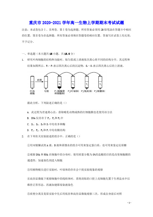
一、单选题(本大题共15小题,共15.0分)1.研究叶肉细胞的结构和功能时,取匀浆或上清液依次离心将不同的结构分开,其过程和结果如图所示,P1~P4表示四次离心后的沉淀物,S1~S4表示四次离心后的上清液。
据此分析,下列叙述正确的是()A. 此过程为差速离心法,获取哺乳动物成熟的红细胞膜也是使用该方法B. DNA仅存在于P1、P2和P3中C. S1、S2、S3和S4中均有多种酶D. P1、P2、P3和P4中均有膜结构2.在下列有关实验叙述的组合中,正确的是()①用双缩脲试剂A液、B液和蒸馏水的组合可用来鉴定蛋白质,也可用来鉴定还原糖②观察DNA和RNA在细胞中的分布时,使用质量分数为8%的盐酸的目的是改变细胞膜的通透性,加速染色剂进入细胞③用植物根尖进行实验时,叶绿体的存在会干扰实验现象的观察④高倍显微镜下观察细胞中的线粒体时,需将刮取的口腔上皮细胞先置于生理盐水中以维持正常形态,再滴加健那绿染液染色⑤质壁分离及复原实验中先后用低倍和高倍显微镜观察三次,形成自身前后对照⑥科学家通过伞藻的嫁接实验证明了细胞核控制着伞藻帽的性状A. ①②③④⑤⑥B. ①②③④⑥C. ②④D. ①②③④3.下表为甲同学用某浓度KNO3溶液进行质壁分离实验时所测得的数据。
下图为乙同学用另一浓度的KNO3溶液进行质壁分离实验时所绘制的曲线图。
下列分析正确的是()2分钟4分钟6分钟8分钟10分钟原生质体相对90% 60% 30% 30% 30%大小A. 甲同学实验进行到8分钟时质壁分离达到平衡,滴加清水后发生质壁分离复原B. 甲同学所用溶液浓度要大于乙同学C. 乙同学在T1时可观察到质壁分离现象,此时细胞液浓度一定小于外界溶液浓度D. 乙同学在T2时观察不到质壁分离现象,此时细胞液浓度一定等于外界溶液浓度4.核糖体由大亚基和小亚基组成。
北京市房山区2023-2024学年度高三上学期期末检测生物试题(word版含解析)

房山区2023-2024学年度第一学期期末检测试卷高三生物学本试卷共10页,共100分。
考试时长90分钟。
考生务必将答案答在答题卡上,在试卷上作答无效。
考试结束后,将答题卡交回,试卷自行保存。
第一部分本部分共15题,每题2分,共30分。
在每题列出的四个选项中,选出最符合题目要求的一项。
1.蛋白质是生命活动的主要承担者,其功能具有多样性。
下列不属于...膜蛋白功能的是()A.催化化学反应B.协助物质运输C.储存遗传信息D.参与信息传递【答案】C【解析】【分析】蛋白质是生命活动的主要承担者,有的蛋白质是细胞和生物体的重要组成成分,有的蛋白质具有催化功能,有的蛋白质具有运输功能,有的蛋白质具有调节机体生命活动的功能,有的蛋白质具有免疫功能等。
【详解】A、催化功能是蛋白质的功能之一,例如主动运输中的蛋白质是ATP酶,可以催化ATP的水解,A正确;B、运输功能是蛋白质的功能之一,例如主动运输和协助运输中起作用的蛋白质,B正确;C、具有储存遗传信息功能的物质是核酸,不是蛋白质,C错误;D、传递某种信息,调节机体的新陈代谢,是蛋白质的功能之一,D正确。
故选C。
2.单细胞生物肺炎支原体可引起支原体肺炎,其结构模式图如下所示。
相关叙述正确的是()A.细胞膜以磷脂双分子层为基本支架B.肺炎支原体以有丝分裂方式繁殖C.肺炎支原体在核糖体上加工蛋白质D.抑制细胞壁形成的药物可治疗支原体肺炎【答案】A【解析】【分析】原核生物与真核生物的区别:原核生物没有以核膜为界限的细胞核,原核生物只有核糖体一种细胞器。
【详解】A、细胞膜以磷脂双分子层为基本支架,A正确;B、支原体属于原核生物,通过二分裂增殖,不能进行有丝分裂,B错误;C、核糖体是蛋白质的合成场所,并非加工场所,C错误;D、支原体没有细胞壁,抑制细胞壁形成的药物对支原体无效,D错误。
故选A。
3.为确定某品种樱桃番茄的适宜贮藏温度,工作人员做了相关实验,结果如下图。
下列说法错误..的是()A.随着贮藏时间的增加,不同温度下樱桃番茄的腐烂率都不断增加B.10℃组前6d出现的腐烂率异常需进一步实验确认产生原因C.8℃及以下的零上低温对于此樱桃番茄果实的防腐效果最佳D.适度低温可降低樱桃番茄的细胞呼吸速率,减少有机物消耗【答案】A【解析】【分析】樱桃番茄的贮藏,既要使呼吸作用降到最低,以减少有机物的消耗,同时液要保证腐烂率低。
2023届重庆市高三第一次冲刺卷生物试题(含答案解析)

2023届重庆市高三第一次冲刺卷生物试题学校:___________姓名:___________班级:___________考号:___________一、单选题1.如图是豚鼠胰腺腺泡细胞中分泌蛋白形成过程的部分图解,①~④表示细胞结构,该细胞可合成并分泌胰蛋白酶等多种消化酶。
据图分析,下列叙述错误的是()A.③可以对进入其腔内的多肽链进行加工、折叠B.葡萄糖在②内被氧化分解,为该细胞供能C.由蛋白质纤维组成的细胞骨架与①的移动有关D.与豚鼠胰腺表皮细胞相比,该细胞中④的体积较大【答案】B【分析】由题图信息分析可知,①是囊泡,②是线粒体,③是内质网,④是核仁。
【详解】A、③是内质网,内质网可以对进入其腔内的多肽链进行加工、折叠,A正确;B、②是线粒体,葡萄糖不能在线粒体中被氧化分解,葡萄糖只能利用细胞质基质产生的丙酮酸,B错误;C、真核细胞中的细胞骨架是由蛋白质纤维组成的,细胞骨架与①囊泡的移动有关,C 正确;D、④是核仁,核仁与核糖体的形成及RNA的合成有关,图示为豚鼠胰腺腺泡细胞,可以产生分泌蛋白,与豚鼠胰腺表皮细胞相比,该细胞中④的体积较大,D正确。
故选B。
A.Mg是构成各种光合色素必需的元素,参与光能的吸收、传递和转化B.P是构成ATP的元素,ATP是将光能转化为有机物中稳定化学能的桥梁C.N是构成多种酶的元素,在叶绿体基质中的酶能催化CO2的固定和C3的还原D.N和P都是构成NADPH的元素,NADPH能将C3还原形成糖类和C5【答案】A【分析】光合作用的场所是叶绿体,其中的能量转化:光能转化为ATP、NADPH中活跃的化学能再转化为有机物中稳定的化学能。
【详解】A、光合色素包括叶绿素和类胡萝卜素,Mg是构成叶绿素所必需的元素,并不是所有的光合色素都含有镁元素,A错误;B、ATP由于含有3个磷酸基团,故含有P元素,光合作用是将光能转化为ATP和NADPH 中活跃的化学能,进而转变为有机物中稳定化学能,故ATP是能量转化的桥梁,B正确;C、酶是具有催化作用的有机物,大部分是蛋白质,少部分是RNA,均含有N元素,叶绿体基质是暗反应的场所,其中含有的酶能催化CO2的固定和还原,C正确;D、NADPH是还原型辅酶Ⅱ,含有N和P元素,NADPH参与暗反应,能将C3还原形成糖类(CH2O)和C5,D正确。
某大学生物工程学院《细胞生物学》考试试卷(1772)

某大学生物工程学院《细胞生物学》课程试卷(含答案)__________学年第___学期考试类型:(闭卷)考试考试时间:90 分钟年级专业_____________学号_____________ 姓名_____________1、判断题(50分,每题5分)1. 核仁是细胞核的一种结构,任何时候都可以看到。
()答案:错误解析:核仁只能在细胞间期观察到。
在分裂的前期、中期、后期消失,不易观察。
2. 中心粒的复制是在细胞周期S期全部完成的。
()答案:错误解析:中心粒在G1期末复制,在S期由一对中心体连接在一起,到G2期一对中心体开始分离并逐渐向细胞两极移动。
3. 核小体并不是一个静止的结构,它始终处于装配与去装配的动态变化中。
()答案:正确解析:和细胞其他部分一样,染色质包括核小体都处在动态变化之中,并不是一成不变的。
4. 生长因子通过不依赖于CDK的途径促进细胞增殖。
()答案:错误解析:生长因子通过与其受体结合后,激活下游许多酶的活性,包括MPF等CDK酶,从而促进细胞的增殖。
5. 秀丽新小杆线虫的ces1、ces2基因是死亡执行基因。
()答案:错误解析:ces1、ces2是决定死亡的基因,ced3、ced4是细胞死亡的执行基因。
6. 从能量转换的角度来看,线粒体的内膜起主要作用。
()答案:正确解析:通过线粒体内膜上的电子传递和ATP合成酶的磷酸化的作用,将NADH中的能量转化为ATP中的活跃的化学能。
7. 原核生物和真核生物核糖体的亚基虽然不同,但两者形成的杂交核糖体仍能进行蛋白质合成。
()答案:错误解析:杂交重组后的核糖体没有合成蛋白质的功能。
8. 细胞凋亡时,核小体间DNA断裂是由物理,化学因素或病理性刺激引起的。
()[中国科学院2005研]答案:错误解析:细胞凋亡是一种主动的由基因决定的细胞自我破坏的过程,而坏死则是极端的物理,化学因素或严重的病理性刺激引起的细胞损伤和死亡。
引起细胞凋亡和细胞坏死的因素不同。
纤维素降解微生物的分离与鉴定方法解析
纤维素降解微生物的分离与鉴定方法解析纤维素是植物细胞壁的主要成分之一,它是一种由大量葡萄糖分子通过β-1,4-糖苷键连接而成的多糖。
纤维素的降解对于生物能源开发、废弃物处理和环境保护具有重要意义。
而纤维素降解微生物则扮演着关键的角色。
因此,分离和鉴定纤维素降解微生物的方法显得尤为重要。
本文将介绍几种常用的纤维素降解微生物的分离与鉴定方法。
一、平板法平板法是最为常用的纤维素降解微生物分离方法之一。
具体操作如下:1. 准备培养基:将适合纤维素降解微生物生长的培养基高温固化。
常用的培养基包括CMC培养基和Avicel培养基。
2. 稀释样品:将待分离的纤维素降解微生物样品进行适当稀释,通常采用百倍至千倍的稀释倍数。
3. 倒平板:将稀释后的样品均匀倒在高温固化的培养基上,并利用均衡板将其平均分布。
4. 培养:将平板培养在适当的温度下,一般为30-37℃,孵育时间根据需要而定。
5. 分离:观察培养基上的菌落情况,挑取个别菌落进行分离纯化。
二、液体培养法液体培养法是另一种常用的纤维素降解微生物分离方法。
主要包括以下步骤:1. 准备液体培养基:选取适合纤维素降解微生物生长的液体培养基,如液体CMC培养基、液体Avicel培养基等。
2. 接种:将待分离的纤维素降解微生物样品接种到含有相关培养基的试管中。
3. 培养:将试管放置于摇床或恒温培养箱中,在适当的温度和转速条件下培养一定时间。
4. 分离: 通过稀释方法,将培养液中的微生物进行分离纯化,得到单菌株。
三、生理生化特性分析对于分离的纤维素降解微生物,进一步进行鉴定需要进行生理生化特性分析。
常见的特性分析包括以下内容:1. 糖类利用能力:在各种糖类培养基上观察微生物的菌落形态和生长情况。
2. pH和温度适应性:分析微生物在不同pH和温度条件下的生长状况。
3. 酶活性检测:测定微生物产酶的能力,如纤维素酶、β-葡萄糖苷酶等。
4. 生理代谢产物分析:通过气相色谱-质谱联用技术或其他适当的方法,分析微生物在纤维素降解过程中产生的代谢产物。
重庆市巴蜀中学校2023-2024学年高三上学期月考卷生物试题(一)(解析版)

巴蜀中学2024届高考适应性月考卷(一)生物注意事项:1.答题前,考生务必用黑色碳素笔将自己的姓名、准考证号、考场号、座位号在答题卡上填写清楚。
2.每小题选出答案后,用2B铅笔把答题卡上对应题目的答案标号涂黑。
如需改动,用橡皮擦干净后,再选涂其他答案标号。
在试题卷上作答无效。
3.考试结束后,请将本试卷和答题卡一并交回。
满分100分,考试用时75分钟。
一、选择题:本题共15小题,每小题3分。
在每小题给出的四个选项中,只有项是符合题目要求的。
1.沙眼衣原体是--类严格的细胞内寄生原核生物,有较复杂的、能进行-定代谢活动的酶系统,但不能合成高能化合物。
下列相关说法正确的是A.沙眼衣原体细胞内的某些蛋白质可通过核孔进入细胞核B.沙眼衣原体无丝分裂的过程中不会出现染色体和纺锤体C.沙眼衣原体不能合成高能化合物的原因可能是无相关的酶D.沙眼衣原体利用宿主细胞的核糖体合成自身蛋白质2.水和无机盐对动植物的生命活动影响非常大。
下列相关说法正确的是A.水分子易与带正电荷或负电荷的分子结合,因此可作为维生素D等物质的良好溶剂B.活性蛋白失去结合水后会改变空间结构,重新得到结合水后能恢复其活性C.无机盐是构成细胞内某些重要化合物的组成成分,如Fe2+参与血靛蛋白的组成D.人体内Na+缺乏会引起神经、肌肉细胞的兴奋性降低,最终引发肌肉酸痛、无力等3.图1为牛胰岛素结构图及其局部放大示意图,该物质由51个氨基酸组成,其中-S-S-是由两个-SH脱去两个H形成的。
下列叙述正确的是A.该胰岛索至少含有的氧原子数和氮原子数分别是53、52B.将图示牛胰岛素彻底水解为氨基酸后,相对分子质量增加888C.该胰岛索能催化葡萄糖合成肝糖原从而降低血糖浓度D.人和牛的胰岛素分子不同的根本原因是组成它们的氨基酸种类、数量、排列顺序及空间结构不同4.图2为一个DNA分子片段,下列有关叙述正确的是A.DNA分子中的每个五碳糖都同时连接2个磷酸基团B.图中有4种核苷酸,烟草花叶病毒的核酸中只含其中3种核苷酸C.DNA发生初步水解的过程中,发生断裂的健是②④D.DNA分子中的氮元索,分布在其基本骨架上5.哺乳动物的细胞膜具有不同类型的磷脂(SM、PC、PE、PS和PI),磷脂均由亲水性的头部和疏水性的尾部组成。
一中202届高三生物开学质量检测试题含解析

【解析】
【分析】
理解核苷酸的分类依据,以及在DNA和RNA两种核酸中碱基的种类。DNA中含有A、T、G、C四种碱基,RNA中含有A、U、C、G四种碱基,共同含有的碱基A、G、C三种碱基,T是DNA特有碱基,U是RNA特有碱基。
【详解】核酸基本单位核苷酸有一分子含氮碱基、一分子五碳糖和一分子磷酸组成: ;其中五碳糖分为两类:脱氧核糖、核糖(如下图)
B.由于钾离子和硝酸钾离子会被细胞主动运输进入细胞,所以原生质体最终体积 b 与溶液初始浓度有关,B错误。
C.由于植物细胞在甘油溶液中可以自由进出,若在一定浓度的甘油溶液中可能发生植物细胞的质壁分离及复原的,此时与曲线①相似,若甘油浓度过大,会一直失水而不能复原,此时与曲线②相似,C正确。
D.根据前面的分析可知,①曲线中的细胞在a 时刻已经开始吸水,细胞液浓度在下降,所以此时细胞液浓度不是最大,D错误。
依据五碳糖的不同,将核苷酸分为脱氧核苷酸和核糖核苷酸;
在脱氧核苷酸中,依据四种碱基:A、T、G、C,将脱氧核苷酸分为四类:
在核糖核苷酸中,依据四种碱基:A、U、G、C,同样将核糖核苷酸分为四类:
在菊花叶肉细胞中所含的核酸有DNA和RNA两种,所以含有核苷酸8种、由于碱基中A、C、G是共有的碱基,所以碱基5种。
故选C。
【点睛】在菊花叶肉细胞中所含的核酸有DNA和RNA两种,所以有脱氧核苷酸和核糖核苷酸两大类,由于碱基有共有相同的,所以碱基只有5中.
5。 将相同的植物根毛区细胞置于一定浓度的蔗糖溶液或 KNO3溶液中后(溶液体积远大于细胞),原生质体的体积随时间的变化如右上图所示(最终不再变化),下列分析正确的是
A. ①是蔗糖溶液中的变化,②是 KNO3溶液中
B。 原生质体最终体积 b 与溶液初始浓度无关的变化
河南省淅川县第一高级中学2024学年生物高二第二学期期末检测试题(含解析)

河南省淅川县第一高级中学2024学年生物高二第二学期期末检测试题注意事项1.考生要认真填写考场号和座位序号。
2.试题所有答案必须填涂或书写在答题卡上,在试卷上作答无效。
第一部分必须用2B 铅笔作答;第二部分必须用黑色字迹的签字笔作答。
3.考试结束后,考生须将试卷和答题卡放在桌面上,待监考员收回。
一、选择题:(共6小题,每小题6分,共36分。
每小题只有一个选项符合题目要求)1.下列关于基因与染色体关系的描述,正确的是A.基因与染色体存在一一对应的关系B.基因和染色体的组成成分完全相同C.一个基因由一条染色体构成D.基因在染色体上呈线性排列2.制作无色洋葱鳞片叶表皮细胞的临时装片用于“观察植物细胞吸水与失水”实验,用高浓度(质量浓度为0.075g/mL)的胭脂红溶液(一种水溶性的大分子食用色素,呈红色)作为外界溶液进行引流处理后,观察细胞的变化。
下列有关实验操作和结果的叙述,正确的是A.表皮细胞的液泡为红色时,细胞仍具正常生理功能B.发生细胞质壁分离现象时,表皮细胞内的红色区域变小C.发生细胞质壁分离复原现象时,表皮细胞内的无色区域变大D.用不同浓度的胭脂红溶液处理细胞后,均能观察到质壁分离和复原现象3.下列关于ATP的叙述中,正确的是()A.细胞内的吸能反应一般与ATP水解的反应相联系B.光合作用形成的ATP也可用于各项生命活动C.ATP分子中的“A”代表腺苷,是由腺嘌呤和脱氧核糖结合而成D.ATP的第三个高能磷酸键很容易断裂和再形成4.下列关于酶的表述,全面而准确的是()A.酶不能脱离生物体起作用B.酶是蛋白质C.酶与无机催化剂都能降低活化能,所以没有本质区别D.酶是活细胞产生的有催化作用的有机物5.用显微镜观察洋葱鳞片叶表皮的同一部位,应选择下列哪种目镜和物镜的组合,才能使视野内所看到的细胞数目最多()A.目镜5×物镜10×B.目镜10×物镜100×C.目镜15×物镜40×D.目镜10×物镜40×6.高尔基体是真核细胞中的物质转运系统的组成部分,下列叙述错误的是A.高尔基体是由双层单位膜构成的细胞器B.髙尔基体可将蛋白质运输到细胞内或细胞外C.高尔基体断裂形成的溶酶体中有大量水解酶D.高尔基体参与细胞膜的更新二、综合题:本大题共4小题7.(9分)果蝇的性别决定方式是XY型,下图表示某种果蝇纯合亲本杂交产生的1355只F2代的个体,体色由基因A/a控制、眼色由基因B/b控制,两对等位基因均属于细胞核基因。
观察植物细胞的质壁分离和复原的实验设计(教案)
观察植物细胞的质壁分离和复原的实验设计(教案)一、实验原理1. 植物细胞的结构:细胞壁、细胞膜、细胞质、细胞核、液泡、叶绿体等。
2. 质壁分离:当细胞液的浓度小于外界溶液的浓度时,细胞会失水,细胞壁和原生质层收缩,导致质壁分离。
3. 复原:当细胞液的浓度大于外界溶液的浓度时,细胞会吸水,细胞壁和原生质层恢复原状,实现质壁复原。
二、实验材料与仪器1. 材料:紫色洋葱鳞片叶、蔗糖、清水、显微镜等。
2. 仪器:显微镜、载玻片、盖玻片、滴管、镊子、解剖针等。
三、实验步骤1. 制作洋葱鳞片叶表皮细胞的临时装片。
2. 在盖玻片的一侧滴加蔗糖溶液,另一侧用吸水纸吸引,重复几次,使细胞失水,观察质壁分离现象。
3. 观察并记录细胞质壁分离的程度。
4. 更换装片,重复步骤2和3,观察细胞在不同的蔗糖浓度下的质壁分离现象。
5. 滴加清水于盖玻片的一侧,用吸水纸吸引,使细胞吸水,观察质壁复原现象。
四、实验注意事项1. 制作装片时,要确保洋葱鳞片叶表皮细胞均匀分布在载玻片上。
2. 滴加蔗糖溶液和清水时,要控制好滴加速度,避免细胞过度失水或吸水。
3. 观察质壁分离和复原现象时,要仔细观察细胞壁、细胞膜、液泡等结构的变化。
五、实验拓展1. 探讨不同浓度蔗糖溶液对细胞质壁分离的影响。
2. 探讨不同植物细胞对质壁分离和复原的敏感性。
3. 结合生物学知识,分析质壁分离和复原在植物生长发育、逆境响应等方面的意义。
六、实验结果与分析1. 实验结果:在一定浓度的蔗糖溶液中,洋葱鳞片叶表皮细胞发生质壁分离,随着蔗糖浓度的增加,质壁分离程度加大;在清水中,细胞发生质壁复原,复原程度与失水程度相对应。
2. 结果分析:细胞壁的伸缩性小于原生质层的伸缩性,导致质壁分离。
随着蔗糖浓度的增加,细胞失水越多,质壁分离程度越大。
在清水中,细胞吸水,原生质层恢复原状,实现质壁复原。
1. 报告内容:实验目的、原理、材料与仪器、步骤、结果与分析、结论等。
2. 报告要求:语言简练、条理清晰、数据准确、图表规范、结论明确。
2022-2023学年陕西省安康市高三上学期第一次质量联考(高考一模)生物试卷带讲解

B.台盼蓝将胚乳细胞染成蓝色,可判定胚乳细胞是死细胞
C.观察洋葱表皮细胞有丝分裂过程需要用醋酸洋红液染色
D.酒精可与溴麝香草酚蓝水溶液反应呈黄绿色,可判断酵母菌的呼吸方式
B
【分析】斐林试剂可用于鉴定还原糖,在水浴加热的条件下,溶液的颜色变化为砖红色(沉淀)。斐林试剂只能检验生物组织中还原糖(如葡萄糖、麦芽糖、果糖)存在与否,而不能鉴定非还原性糖(如淀粉)。【详解】A、某种糖类与斐林试剂反应呈砖红色,可确定其是还原糖,但还原糖不一定是葡萄糖,A错误;
故选B。
9.“米酒”因其口感好、酒精度低而获得老年人的青睐。下列关于其酿制过程中涉及的生物学知识的叙述,正确的是()
A.酿酒时用到的酵母菌为兼性厌氧型生物,无细胞核但合有核糖体
B.酵母菌进行无氧呼吸过程时,在细胞质基质中将丙酮酸转化为乙醇
C.酵母菌有氧呼吸和无氧呼吸产物不完全相同,根本原因是酶的种类不同
D、供氧充足时酵母菌进行有氧呼吸,在线粒体内膜上进行有氧呼吸的第三阶段,氧化前两个阶段产生的[H]产生大量ATP和水,D错误。
故选B。
10.植物叶肉细胞能进行光合作用亦能进行呼吸作用,两代谢过程中存在多种相似物质的转化,如水的分解与合成、二氧化碳的产生与消耗等。下列相关叙述正确的是()A.若给植株提供H218O,一段时间后能在体内检测出C18O2
A.两类转运蛋白的运输过程主要体现了生物膜的流动性
B.两类转运蛋白运输物质的速率均受到其数量的制约
C.载体蛋白介导运输的速率较慢是因为其需要消耗能量
D.人红细胞吸收葡萄糖和K+的方式均为协助扩散
B
【分析】转运蛋白可以分为载体蛋白和通道蛋白两种类型。载体蛋白只容许与自身结合部位相适应的分子或离子通过,而且每次转运时都会发生自身构象的变化;通道蛋白只容许与自身通道的直径和形状相适配、大小和电荷相适宜的分子或离子通过。分子或离子通过通道蛋白时,不需要与通道蛋白结合。
广东省部分中学2023高中生物第3章细胞的基本结构知识点总结(超全)

广东省部分中学2023高中生物第3章细胞的基本结构知识点总结(超全)单选题1、下列有关细胞膜的叙述,正确的是()A.制备细胞膜常用哺乳动物成熟红细胞是因为显微镜下容易观察到B.肝细胞和甲状腺细胞功能的根本差异是因为细胞膜蛋白质种类不同C.相邻两细胞的接触实现细胞间的信息交流是因为膜上糖蛋白的作用D.癌细胞的恶性增殖和转移是因为细胞膜的成分产生肌动蛋白的改变答案:C分析:1 .有关细胞分化:(1)细胞分化概念:在个体发育中,由一个或多个细胞增殖产生的后代,在形态、结构和生理功能上发生一系列稳定性差异的过程;(2)特征:具有持久性、稳定性和不可逆性;(3)意义:是生物个体发育的基础;(4)原因:基因选择性表达的结果,遗传物质没有改变。
2 .细胞膜的功能:(1)将细胞与外界环境分开;(2)控制物质进出细胞;(3)进行细胞间的信息交流。
3 .癌细胞的特征:(1)具有无限增殖的能力;(2)细胞的形态发生改变;(3)细胞膜表面的糖蛋白减少,容易扩散和转移。
4 .选哺乳动物成熟的红细胞作实验材料的原因:(1)动物细胞没有细胞壁,不但省去了去除细胞壁的麻烦,而且无细胞壁的支持、保护,细胞易吸水涨破;(2)哺乳动物和人成熟的红细胞,没有细胞核和具有膜结构的细胞器,易用离心分离法得到不掺杂细胞内膜系统的纯净的细胞膜。
科学家常用哺乳动物成熟的红细胞作材料来制备细胞膜,是因为:动物细胞放入清水中会吸水胀破,且哺乳动物成熟的红细胞没有细胞器和细胞核,没有细胞器膜和核膜的干扰,因此能制备较纯净的细胞膜,A错误;肝细胞和甲状腺细胞功能的根本差异是因为细胞内表达的基因的种类不同,B错误;细胞膜具有进行细胞间的信息交流的功能,两个相邻细胞的细胞膜接触可实现细胞间的信息传递,如精细胞和卵细胞的直接接触,C正确;癌细胞的恶性增殖和转移是因为细胞膜的成分发生改变,细胞膜上的糖蛋白减少,D错误。
2、进行生物学实验离不开科学的方法。
下列有关生物学实验及其对应科学方法不匹配的是()A.AB.BC.CD.D答案:B分析:模型构建法:模型是人们为了某种特定的目的而对认识对象所做的一种简化的概括性的描述,这种描述可以是定性的,也可以是定量的,有的借助具体的实物或其它形象化的手段,有的则抽象的形式来表达。
高考生物一轮复习微专题小练习专练100教材实验和实验设计分析综合练

专练100 教材实验和实验设计、分析综合练1.[2023·陕西西安一中期中]在下列有关实验叙述的组合中,不正确的有几项( )①探究酵母菌细胞呼吸的方式实验中,可用溴麝香草酚蓝溶液检测酒精的产生②观察根尖分生组织细胞的有丝分裂实验中,解离和漂洗的目的都是使细胞分散③叶绿体色素的提取和分离实验中可用体积为95%的乙醇加入适量无水碳酸钠来代替无水乙醇④若用硝酸钾溶液代替蔗糖进行质壁分离及复原实验,当细胞体积不变时,细胞内外渗透压相等⑤科学家通过伞藻的嫁接实验证明了细胞核控制着伞藻帽的性状A.五项B.四项C.三项D.两项2.下列有关实验设计的叙述中,正确的是( )A.洋葱鳞片叶外表皮的细胞质壁分离和质壁分离复原实验为对比实验B.实验不一定有空白对照组,但应该有相应的对照C.探究促进插条生根的最适生长素浓度的预实验设置空白对照可减小实验误差D.在实验之前针对提出问题所作的假说往往都是正确的3.以下高中生物学实验中,操作不正确的是( )A.在制作果酒的实验中,将葡萄汁液装满整个发酵装置B.鉴定DNA时,将粗提产物与二苯胺混合后进行沸水浴C.用苏丹Ⅲ染液染色,观察花生子叶细胞中的脂肪滴(颗粒)D.用甲紫染液染色,观察洋葱根尖分生区细胞中的染色体4.在进行“观察叶绿体”的活动中,先将黑藻放在光照、温度等适宜条件下预处理培养,然后进行观察。
下列叙述正确的是( )A.制作临时装片时,实验材料不需要染色B.黑藻是一种单细胞藻类,制作临时装片时不需切片C.预处理可减少黑藻细胞中叶绿体的数量,便于观察D.在高倍镜下可观察到叶绿体中的基粒由类囊体堆叠而成5.黑藻是一种叶片薄且叶绿体较大的水生植物,分布广泛、易于取材,可用作生物学实验材料。
下列说法错误的是( )A.在高倍光学显微镜下,观察不到黑藻叶绿体的双层膜结构B.观察植物细胞的有丝分裂不宜选用黑藻成熟叶片C.质壁分离过程中,黑藻细胞绿色加深、吸水能力减小D.探究黑藻叶片中光合色素的种类时,可用无水乙醇作提取液6.下列物质的鉴定与所用试剂、实验手段、实验现象搭配,正确的是( )A.脂肪—苏丹Ⅲ染液—显微镜观察—染成红色的脂肪颗粒B.葡萄糖—斐林试剂—直接观察—砖红色沉淀C.蛋白质—双缩脲试剂—直接观察—紫色反应D.酒精—溴麝香草酚蓝溶液—观察—灰绿色反应7.[2023·重庆八中模拟]耐甲氧西林金黄色葡萄球菌(MRSA)是临床上常见的毒性较强的“超级细菌”,一般的青霉素对该细菌的杀伤力不强。
观察植物细胞的质壁分离和复原的实验设计(教案)
一、实验背景与目的1.1 实验背景植物细胞的质壁分离和复原是植物细胞学中的一个重要现象,也是生物学实验教学中的经典实验。
通过观察植物细胞的质壁分离和复原,可以帮助学生理解和掌握植物细胞结构、细胞壁的功能以及渗透作用的原理。
1.2 实验目的通过本实验,让学生了解植物细胞质壁分离和复原的原理及过程,培养学生的实验操作能力和观察分析能力,提高学生对生物学知识的理解和运用能力。
二、实验原理2.1 植物细胞结构植物细胞具有细胞壁、细胞膜、细胞质、液泡、叶绿体等结构。
细胞壁由纤维素等物质构成,具有支持和保护细胞的作用。
细胞膜是细胞内外物质交换的关键界面,具有选择性透过性。
2.2 质壁分离原理当细胞外界溶液浓度高于细胞内部浓度时,细胞会失水,细胞壁与细胞质膜分离。
这是因为细胞壁的伸缩性小于细胞质膜,当细胞失水时,细胞质膜向内收缩,与细胞壁分离。
2.3 复原原理当细胞外界溶液浓度低于细胞内部浓度时,细胞会吸水,细胞质膜与细胞壁复原。
这是因为细胞吸水时,细胞质膜向外膨胀,与细胞壁重新贴合。
三、实验材料与仪器3.1 实验材料植物细胞样本(如洋葱表皮细胞)生理盐水蔗糖溶液显微镜3.2 实验仪器显微镜载玻片盖玻片滴管解剖针四、实验步骤4.1 制作装片取一片植物细胞样本,放在载玻片上。
用滴管滴入适量的生理盐水或蔗糖溶液。
用盖玻片盖住样本,避免气泡产生。
4.2 观察质壁分离将装片放在显微镜下,调整镜头至适当倍数。
观察细胞在生理盐水或蔗糖溶液中的变化,记录质壁分离的过程。
4.3 观察复原过程将装片转移到含有生理盐水的载玻片上。
观察细胞在生理盐水中的变化,记录复原的过程。
五、实验结果与分析5.1 实验结果观察到植物细胞在蔗糖溶液中发生质壁分离,细胞失水,细胞壁与细胞质膜分离。
观察到植物细胞在生理盐水中发生复原,细胞吸水,细胞质膜与细胞壁复原。
5.2 实验分析分析质壁分离和复原的原因,探讨细胞壁的伸缩性和细胞膜的选择性透过性在实验过程中的作用。
2023届陕西省宝鸡市高三上学期高考模拟检测(一)理综生物试题(解析版)

B、艾弗里体外转化实验证明了DNA是遗传物质,同时证明了蛋白质不是遗传物质,B错误;
C、噬菌体是病毒,不能直接用培养基培养,故应用35S或32P标记大肠杆菌→用被35S或32P标记的大肠杆菌培养噬菌体,获得被35S或32P标记的噬菌体,C错误;
【答案】(1)①.原癌基因②.抑癌基因
(2)①.防御②.胞吞
(3)①.否②.应补充一组未用转运抑制剂处理的癌细胞作为对照组,比较两组察细胞的生长情况
(4)①.是否注射E酶②.E酶通过提高CD8+细胞数量,抑制肿瘤生长(或抑制注射瘤和非注射瘤生长)
【解析】
【分析】免疫系统的基本功能:
①免疫防御:机体排除外来抗原性异物的一种免疫防护作用。这是免疫系统最基本的功能。该功能正常时,机体能抵抗病原体的入侵;异常时,免疫反应过强、过弱或缺失,可能会导致组织损伤或易被病原体感染等问题。
A.尿苷与腺苷、鸟苷、胸苷可在细胞中连接形成RNA
B.用H标记的尿苷饲喂动物,可以在细胞核、细胞质、组织液中检测到放射性
C.衰老的细胞内酶的活性降低,细胞核体积缩小,染色质收缩
D.尿苷促进组织再生修复有可能是切除了端粒DNA序列
【答案】B
【解析】
【分析】细胞衰老在形态学上表现为细胞结构的退行性变,如在细胞核,核膜凹陷,最终导致核膜崩解,染色质结构变化,超二倍体和异常多倍体的细胞数目增加;细胞膜脆性增加选择性通透能力下降,膜受体种类、数目和对配体的敏感性等发生变化;脂褐素在细胞内堆积,多种细胞器和细胞内结构发生退行性变。细胞衰老在生理学上的表现为功能衰退与代谢低下,如细胞周期停滞,细胞复制能力丧失,对促有丝分裂刺激的反应性减弱,对促凋亡因素的反应性改变;细胞内酶活性中心被氧化,酶活性降低,蛋白质合成下降等。
化学物质分离和分析的原理
化学物质分离和分析的原理化学物质分离和分析是化学中重要的基础实验技术。
它们的目的是研究物质的组成、结构和性质,进而探究物质的变化规律,为化学研究提供重要的实验依据。
化学物质的分离和分析方法较多,其中最主要的方法包括色谱法、光谱法、电化学法、质谱法、薄层层析法和溶液析出、蒸馏、提取等方法。
本文将介绍其中几种主要的原理和实现方法。
一、色谱法色谱法是一种将混合物进行分离和分析的重要方法。
根据不同的物质分子特性,利用溶剂将物质分子和不同的吸附或分离介质相互作用的程度不同,使得物质分子分离、分解,从而进行分析。
目前,色谱法的种类很多,最常用的是气相色谱法和液相色谱法。
气相色谱法是利用气态载气将样品中的化合物从进样口引入进化器,再通过毛细管柱的分离作用,将化合物分离后以各自峰的时间荷尔滨方法进行检定。
液相色谱法则是将溶解于液体载液中的化合物引入进样口,在柱中通过不同的相互作用来完成分离。
二、光谱法光谱法是一种研究物质的化学结构和性质的方法,它是利用物质在不同波长下吸收和发射光线的特性来进行分析的。
光谱分析的种类较多,包括原子光谱分析、分子光谱分析、拉曼光谱分析等。
原子光谱分析是指利用原子的吸收或发射光谱,对元素进行分析的方法。
分子光谱分析是指利用分子在吸收或发射辐射时的特性对分子进行分析的方法。
拉曼光谱分析则是利用分子分布震动的模式对样品进行分析。
光谱法对于分析多种化合物的成分具有广泛的应用。
三、电化学法电化学法是利用化学反应或物理过程所伴随的电荷转移现象来进行分析的方法。
电化学法的种类较多,包括溶液电化学,电化学荧光光谱,极谱法,电导度法等。
溶液电化学是指利用电极电势的变化来测定样品中离子的含量。
电导度法是指利用电极的电导性来测定样品的电阻率。
极谱法是指利用电极电位来控制电化学反应的进程,对应的反应产物进行分析的方法。
四、质谱法质谱法是一种将大分子化合物分离并分析其结构和分子量的方法。
它将物质分子分解成小分子离子后,通过质谱分析得到分析结果。
某种细菌外膜蛋白组分离与纯化分析

某种细菌外膜蛋白组分离与纯化分析细菌是一类十分普遍的微生物,它们存在于我们周围的环境中,并且在我们身体内的某些部位也扮演重要角色。
而细菌的外膜蛋白(OMP)也十分重要,它们不仅存在于细菌的表面,同时也是细菌与宿主细胞相互作用的一个重要接口。
因此,对于细菌OMP的研究具有很重要的意义。
本文将介绍某种细菌OMP的离子、纯化和分析过程。
一、细菌菌株的选取首先需要选取一种合适的细菌菌株。
本实验中选取的是大肠杆菌(Escherichia coli),它是一种常见且易于培养的细菌,是许多研究的重要模型生物。
二、细菌同时存在其他蛋白物质的去除由于细菌中存在着许多其他蛋白物质,所以需要去除这些物质,以便更好地分离、纯化和分析OMP。
本实验中选用了亲和层析法,印记纤维素层析法和凝胶过滤法,这些方法都可以有效地去除其他蛋白物质。
三、 OMP的提取提取OMP需要破坏细菌的细胞壁,并使OMP获得充分的暴露。
本实验中选择了孢子杆菌素(n-丙基十六烷基胺),破坏细菌细胞壁,让OMP暴露在外。
四、离子交换层析纯化提取后的OMP并不是纯度很高的,因此需要进行纯化。
离子交换层析纯化是较为常用的纯化方法之一。
在离子交换层析纯化中,将清洗后的OMP经过负载离子的反应,可以使得细菌中的细胞壁杂质和蛋白质分离开来。
本实验中采用盐度逐渐升高的方法进行离子交换层析纯化。
五、凝胶过滤法分离纯化凝胶过滤法分离纯化是一种常用的分离纯化方法,主要有亲和色谱、毛细管电泳、SDS-PAGE等方法,本文采用SDS-PAGE方法进行分析。
SDS-PAGE分离法是一种现代分离和定量细胞蛋白质的重要手段,利用聚丙烯酰胺凝胶与SDS缓冲体系电泳可以显著地分离出不同蛋白质。
在此基础上我们可用肽质谱等方法确认蛋白质的鉴定。
六、细菌OMP的功能研究OMP对于生命体的存活和生长发育是至关重要的,因此研究OMP的功能具有很高的意义。
细菌OMP在感染人体时,可以使得病原体更易进入细胞内、更快繁殖、更难被免疫系统消灭。
细胞壁分层结构
细胞壁分层结构
细胞壁是分为三层的结构,从外到内依次是胞间层、初生壁和次生壁。
1. 胞间层:又称为中胶层,是细胞壁的最外层。
它是由相邻细胞的细胞膜之间插入的纤维素和果胶质组成的,主要起到粘连相邻细胞的作用,并有助于细胞间的物质运输和信息交流。
此外,它还具有缓冲细胞间挤压和保护细胞的功能。
2. 初生壁:位于胞间层的内侧,是在细胞分裂末期由原生质体分泌形成的。
初生壁的主要成分是纤维素、半纤维素和果胶质等,它们以纤维状交织在一起,形成网状结构,为细胞提供支撑和保护。
初生壁具有弹性和可塑性,能够随着细胞的生长而不断增加面积,并允许水分和溶质通过。
3. 次生壁:是细胞壁的最内层,位于初生壁的内侧。
它是在细胞停止生长后,由原生质体继续分泌纤维素和其他物质形成的。
次生壁的主要成分是纤维素、半纤维素和木质素等,它们以更紧密的方式排列在一起,使细胞壁更加坚硬和厚实。
次生壁能够增强细胞的机械强度和抗压能力,并防止水分和溶质的自由通过。
在植物细胞中,不是所有的细胞都具有完整的三层细胞壁结构。
例如,一些幼嫩的、正在生长的细胞可能只有胞间层和初生壁两层结构;而一些成熟的、特化的细胞则可能具有完整的三层结构或更复杂的细胞壁组成。
以上信息仅供参考,如有需要,建议查阅相关书籍或咨询专业人士。
遗传物质DNA的分离与分析技术
遗传物质DNA的分离与分析技术DNA(脱氧核糖核酸)是生物体遗传信息的携带者,它的分离与分析技术对于生物学、医学、犯罪侦查等领域都具有重要意义。
在此,我们将重点介绍DNA的分离与分析技术。
一、DNA的分离技术DNA的分离技术是指将生物体的DNA从复杂的细胞组分中抽取出来,分离出纯净的DNA供后续实验使用。
1. 机械法分离机械法是最早用于DNA分离的方法之一。
机械法分离是通过对生物组织进行机械切割、压碎等操作,将细胞膜和细胞壁破坏,然后以高速离心等方法分离出DNA。
2. 化学法分离化学法分离是将生物体中的DNA化学溶解,利用一定的化学方法与其他杂质分离,最终得到纯净的DNA。
常用的化学分离方法有酚-氯仿法、硅胶柱层析法等。
3. 磁性微珠法分离磁性微珠法是一种新型的DNA分离技术。
该技术利用带有磁性的微珠,通过对DNA的靶标反应,使DNA与磁性微珠结合。
然后,利用磁性微珠的磁性将DNA直接分离,得到纯净的DNA。
二、DNA的分析技术DNA的分析技术是指将DNA分子按照一定的方法进行解析、比较、判断和识别的方法。
DNA分析涉及到的方法和技术非常多,下面我们将介绍几种常用的DNA分析技术。
1. 聚合酶链式反应(PCR)PCR是一种利用特定引物在体外扩增DNA分子的技术。
PCR可以从少量的DNA复制出足够的数量用于后续实验,广泛用于DNA检测、基因分型、疾病诊断等领域。
2. 电泳电泳是一种用电场力使DNA分子在凝胶中运动,实现DNA分子的分离和检测的方法。
常用的电泳包括琼脂糖凝胶电泳、聚丙烯酰胺凝胶电泳等。
电泳技术广泛应用于DNA分析、基因型分析、基因突变检测等领域。
3. 单倍型分析单倍型分析是通过分析DNA序列中的多个位点变异,确定单倍型的方法。
单倍型分析可以用于基因型定性和定量分析、疾病筛查、人类遗传家谱建立等领域。
最后,DNA的分离与分析技术在生物学、医学、犯罪侦查等领域均有广泛应用。
随着科技的不断进步,DNA的分离与分析技术也不断得到新的发展和应用。
- 1、下载文档前请自行甄别文档内容的完整性,平台不提供额外的编辑、内容补充、找答案等附加服务。
- 2、"仅部分预览"的文档,不可在线预览部分如存在完整性等问题,可反馈申请退款(可完整预览的文档不适用该条件!)。
- 3、如文档侵犯您的权益,请联系客服反馈,我们会尽快为您处理(人工客服工作时间:9:00-18:30)。
Isolation and analysis of cell wall components from Streptococcus pneumoniaeNhat Khai Bui a ,1,Alice Eberhardt a ,1,2,Daniela Vollmer a ,Thomas Kern b ,3,Catherine Bougault b ,Alexander Tomasz c ,Jean-Pierre Simorre b ,Waldemar Vollmer a ,⇑aCentre for Bacterial Cell Biology,Institute for Cell and Molecular Biosciences,Newcastle University,Newcastle upon Tyne NE24AX,UK bInstitut de Biologie Structurale,UMR5075(CEA/CNRS/UJF),38027Grenoble,France cLaboratory of Microbiology,Rockefeller University,New York,NY 10021,USAa r t i c l e i n f o Article history:Received 23September 2011Received in revised form 14November 2011Accepted 22November 2011Available online 1December 2011Keywords:Streptococcus pneumoniae Peptidoglycan Teichoic acid Muropeptides Solid-state NMRa b s t r a c tThe complex and heterogeneous cell wall of the pathogenic bacterium Streptococcus pneumoniae is com-posed of peptidoglycan and a covalently attached wall teichoic acid.The net-like peptidoglycan is formed by glycan chains that are crosslinked by short peptides.We have developed a method to purify the glycan chains,and we show that they are longer than approximately 25disaccharide units.From purified pep-tidoglycan,we released 50muropeptides that differ in the length of their peptides (tri-,tetra-,or penta-peptides with or without mono-or dipeptide branch),the degree of peptide crosslinking (monomer,dimer,or trimer),and the presence of modifications in the glycan chains (N-deacetylation,O-acetylation,or lack of GlcNAc or GlcNAc-MurNAc)or peptides (glutamic acid instead of glutamine).We also estab-lished a method to isolate wall teichoic acid chains and show that the most abundant chains have 6or 7repeating units.Finally,we obtained solid-state nuclear magnetic resonance spectra of whole insoluble cell walls.These novel tools will help to characterize mutant strains,cell wall-modifying enzymes,and protein–cell wall interactions.Ó2011Elsevier Inc.All rights reserved.The gram-positive bacterium Streptococcus pneumoniae is a ma-jor pathogen capable of causing otitis media,pneumonia,meningi-tis,and bloodstream infections [1].The cell wall protects the bacterial cell from bursting by the internal turgor and is required to maintain cell shape.The main constituents of pneumococcal cell wall are peptidoglycan and the covalently attached wall teichoic acid (WTA),4capsular polysaccharides,and cell surface proteins.More than 90serotypes with different structures in their polysac-charide capsule [2]and a strain-dependent suite of more than 30cell surface proteins [3]allow the versatile S.pneumoniae to colonize the human nasopharynx and evade the immune system.The focus of this work was on the peptidoglycan–teichoic acid cell wall complex (Fig.1A).Peptidoglycan forms a network ofglycan chains of alternating N -acetylmuramic acid (MurNAc)and N -acetylglucosamine (GlcNAc)residues that are connected by short peptides containing L -and D -amino acids and unusual amide bonds [4].In S.pneumoniae ,the newly made peptides with the se-quence L -Ala-D (c )Gln-L -Lys-D -Ala-D -Ala are efficiently used either to form crosslinks with neighboring peptides by DD -transpeptida-tion,forming dimeric,trimeric,or tetrameric structures,and/or to be trimmed to tetra-or tripeptides by carboxypeptidases [5,6].Some peptides contain L –Ser-L -Ala or L -Ala-L -Ala dipeptides linked to the e -amino group of L -Lys,and this modification is abundant in certain b -lactam-resistant strains [7,8].The glycan chains in pneu-mococcal peptidoglycan are subject to secondary modification by various degrees of deacetylation of the GlcNAc [9]residues and O-acetylation of MurNAc residues [10];both modifications con-tribute to the resistance of the cell to lysozyme,an important bac-teriolytic enzyme of the innate immune system.Although previous work has analyzed the peptides and lactoyl peptides from pneu-mococcal peptidoglycan [5,8,11],the composition of the disaccha-ride peptide subunits,the muropeptides,is not yet known.WTA is a major constituent of the cell wall in gram-positive bacteria,accounting for approximately 40to 50%of its dry weight [12].The WTA of S.pneumoniae is unusual for several reasons [13].First,it has the same structure in its repeating unit as the membrane-anchored lipoteichoic acid (LTA);in most bacteria,the WTA and LTA repeats have different structures.Second,pneumo-coccal WTA has an unusually complex primary structure,0003-2697/$-see front matter Ó2011Elsevier Inc.All rights reserved.doi:10.1016/j.ab.2011.11.026⇑Corresponding author.Fax:+441912083205.E-mail address:w.vollmer@ (W.Vollmer).1These authors contributed equally to this work.2Current address:Department of Microbiology,Tumor,and Cell Biology,Karo-linska Institutet,17177Stockholm,Sweden.3Current address:Institute of Structural Biology,Helmholtz Zentrum München,85764,Neuherberg,Germany.4Abbreviations used:WTA,wall teichoic acid;MurNAc,N -acetylmuramic acid;GlcNAc,N -acetylglucosamine;LTA,lipoteichoic acid;MS,mass spectrometry;NMR,nuclear magnetic resonance;SDS,sodium dodecyl sulfate;dH 2O,deionized water;HF,hydrofluoric acid;HPLC,high-performance liquid chromatography;ESI–MS/MS,electrospray ionization–tandem mass spectrometry;MALDI–TOF,matrix-assisted laser desorption/ionization–time-of-flight.containing the rare amino sugar 2-acetamido-4-amino-2,4,6-tride-oxy-galactose,glucose,ribitol-phosphate,N -acetylgalactosamine,and phosphocholine.Third,the presence of phosphocholine is also remarkable because it is rarely found in bacteria and pneumococci are the only bacteria known to require choline for growth [14,15],presumably due to the specificity of a teichoic acid flippase [16].Pneumococci use their WTA phosphocholine residues to bind a set of cell surface proteins,the choline-binding proteins,some of which have peptidoglycan hydrolase activity such as LytA and LytB [17].Complete LTA chains have been previously isolated and ana-lyzed by mass spectrometry (MS)[18],but WTA chains have not yet been isolated.The aim of this work was to establish novel protocols for the isolation and analysis of various pneumococcal cell wall components.We found that pneumococcal peptidoglycan glycan chains cannot be isolated by the standard cation exchange chroma-tography method due to their partial N-deacetylation.We isolated the glycan chains via size exclusion chromatography and show that they are significantly longer than those from Staphylococcus aureus or Escherichia coli .We also developed a method to determine the muropeptide composition and detected 50muropeptide structures by MS.Muropeptide analysis (but not peptide or lactoylpeptide analysis)allows one to quantify secondary glycan modifications in monomeric and crosslinked muropeptides.We also established a novel purification protocol for WTA chains and show that some chains are not fully loaded with phosphocholine residues.Finally,we established 13C labeling and 13C and 31P solid-state nuclear magnetic resonance (NMR)spectra for pneumococcal cellwall.components in S.pneumoniae and their isolation.(A)Scheme of the cell wall components and the cleavage sites of cellosyl and chains of alternating N -acetylmuramic acid (MurNAc,M)and N -acetylglucosamine (GlcNAc,G)residues.Some of the GlcNAc residues of the MurNAc residues become O-acetylated (OAc).The peptides are designated in single-letter code,with an asterisk (Ã)indicating chains are linked to the peptidoglycan via an unknown linkage unit (X),and their repeating units contain 2-acetamido-4-amino-2,4,6-trideoxy-galactose (Glc),ribitol-phosphate (ribitol-P ),N -acetylgalactosamine (GalNAc),and phosphocholine (Cho-P ).(B)Scheme for the purification indicates N-deacetylation)and wall teichoic acid (WTA)chains from pneumococcal cell wall.These novel tools will help to characterize mutant strains and the enzymology of cell wall modifications and will aid in investigating the biophysical properties and interactions of the pneumococcal cell wall.Materials and methodsMaterials,bacterial strains,and growth conditionsAll chemicals were purchased from Sigma(Dorset,UK,and Mu-nich,Germany)unless mentioned otherwise.The muramidase cel-losyl was kindly provided by Hoechst(Frankfurt,Germany).A recombinant His-tagged version of the cell wall amidase LytA was expressed in E.coli BL21(DE3)pHis-LytA and affinity purified first over Ni2+-nitrilotriacetic acid sepharose using a standard pro-tocol(Qiagen,Hilden,Germany)and then in a second purification step over DEAE sepharose as described previously[19].S.pneumo-niae laboratory strain R6[20],the penicillin-resistant strain Pen6 [21],and the mutant strains CS1[6]and R36A::pgdA[9]were grown at37°C in complex C+Y medium containing1mg/ml yeast extract[22].Strain R6was grown at37°C in C+Y medium supple-mented with2mg/L[13C]glucose for NMR analysis of the cell wall. Isolation of pneumococcal cell wallCell wall from S.pneumoniae was purified as described previ-ously[5,23,24]with modifications.Cells of a2-L culture were har-vested at an OD620of0.5by centrifugation for30min at4°C and 7500g.The cell pellet was resuspended in40ml of ice-cold 50mM Tris–HCl(pH7.0).The cell suspension was introduced dropwise into aflask with150ml of boiling5%sodium dodecyl sul-fate(SDS)solution.The solution was boiled for another30min.The cell suspension was pelleted by ultracentrifugation at25°C for 45min at130,000g.The pellet was washed twice with30ml of 1M NaCl and repeatedly with deionized water(dH2O)until it was free of SDS,confirmed by a previously published assay[25]. The pellet was resuspended in2to4ml of dH2O,1/3volume of acid-washed glass beads(diameter of0.17–0.18mm,Sigma,Mün-chen,Germany)was added,and cells were disrupted in a FastPrep machine(FP120,Thermo Scientific,Hemel Hempstead,UK).For cell disruption,2Â6pulses at maximal speed were applied for 20s,with5-min cooling periods on ice between the pulses.The glass beads were then separated from the cell lysate with a grade 4glassfilter(Winzer,Wertheim,Germany).Thefiltrate was centri-fuged at1000g for5min,and the supernatant,which contains the disrupted cell wall,was centrifuged at130,000g for45min at 25°C.The pellet was resuspended in20ml of100mM Tris–HCl (pH7.5)containing20mM MgSO4.DNase A and RNase I were added tofinal concentrations of10and50l g/ml,respectively, and the sample was stirred for2h at37°C.CaCl2(10mM)and trypsin(100l g/ml)were added,and the sample was stirred for 18h at37°C.Then,1%SDS(final concentration)was added,and the sample was incubated for15min at80°C to inactivate the en-zymes.The cell wall was recovered by centrifugation for45min at 130,000g at25°C,resuspended with20ml of8M LiCl,and incu-bated for15min at37°C.After another centrifugation(see above), the pellet was resuspended in10mM ethylenediaminetetraacetic acid(EDTA,pH7.0)and incubated at37°C for15min.The cell wall was washed with dH2O,acetone,and again with water beforefinal-ly being resuspended in2to4ml of dH2O prior to lyophilization. Cell wall samples(typically$40–60mg from1-L culture)were stored atÀ20°C.Cell walls were labeled by the addition of5l g/ml and1l Ci/ml [methyl-3H]choline to the growth medium of exponentially grow-ing cells as described previously[23].For labeling of glycan strands with[3H]GlcNAc(Amersham Pharmacia Biotech),we followed a published procedure[26].Cell walls were isolated as described above.The specific activities were 6.82Â106cpm/mg for [methyl-3H]choline-labeled cell walls and3.38Â106cpm/mg for [3H]GlcNAc-labeled cell walls(counting efficiency>90%).Preparation of peptidoglycanNext,5mg of cell wall was stirred with48%hydrofluoric acid (HF)for48h at4°C in an ultracentrifuge tube made of polyallomer tightly closed by Parafilm.The volatile,aggressive,and toxic HF was handled with great caution to avoid any contact with the skin and spillage.The peptidoglycan was recovered by centrifugation (45min,130,000g,4°C)and washed with ice-cold dH2O,100mM Tris–HCl(pH7.0),and then twice with dH2O.The pellet containing 2to2.5mg of peptidoglycan was resuspended in750l l of0.02% NaN3and stored at4°C.Preparation of peptidoglycan glycan strands[3H]GlcNAc-labeled or unlabeled peptidoglycan($0.5mg)was digested in a total volume of600l l at37°C in50mM Tris–HCl (pH7.0)with a total of90l g of recombinant and His-tagged LytA amidase for46h,whereby30-l g aliquots of the enzyme were added at time points of0,18,and28h.The sample was boiled for5min,and the clear supernatant was taken.A150-l l aliquot of LytA-digested[3H]GlcNAc-labeled peptido-glycan was N-acetylated at a pH of8.0by the addition of55l l of 100mM freshly prepared N-hydroxysuccinimido acetate(NHS ace-tate)and incubation for1h at ambient temperature.Then,Tris–HCl(pH7.5)was added to afinal concentration of100mM,and the sample was further incubated for1h.Aliquots of the N-acety-lated glycan chains were shortened by partial digestion with lyso-zyme(0.4or4l g/ml)for20min at37°C.The samples were boiled for5min and cleared by brief centrifugation.Purification of peptidoglycan glycan strandsN-acetylated glycan strands were purified on a Keystone GFS-150size exclusion column connected to a Shimadzu high-perfor-mance liquid chromatography(HPLC)system and operating at 30°C at aflow of1ml/min with dH2O.Detection occurred either with a scintillationflow-through detector for radioactive labeled material or at205nm for unlabeled glycan chains.The glycan strands eluting in the void volume were collected,and aliquots were incompletely digested with lysozyme as described above. HPLC analysis of glycan strandsGlycan strands were reduced with sodium borohydride[27]and separated by HPLC as described previously[28].Radioactive glycan strands were detected with a Canberra Packard scintillationflow-through detector.Unlabeled glycan strands were detected at 205nm.Preparation of muropeptidesMuropeptides were released from peptidoglycan with cellosyl, reduced,and analyzed by HPLC similar to the previously published procedure for E.coli muropeptides[27].Peptidoglycan suspension ($0.5mg in100l l)was incubated with10l g of cellosyl in20mM sodium phosphate(pH4.8)in a total volume of200l l for24h at 37°C while stirring.The reaction was stopped by heating the sam-ple for10min at100°C.The sample was cleared by centrifugation for10min at13,000g.For reduction,1sample volume of500mM sodium borate(pH9.0)and a few crystals of NaBH4were added,Cell wall components from S.pneumoniae/N.K.Bui et al./Anal.Biochem.421(2012)657–666659followed by30min of incubation at20°C.The reaction was stopped by adjusting the pH to4.0to5.0with20%H3PO4.For on-line electrospray–MS analysis,peptidoglycan was digested with cellosyl in20mM ammonium acetate buffer(pH 4.8)and the resulting muropeptides were not reduced[29].Separation of muropeptides by HPLCMuropeptides were separated on a reversed-phase column (Prontosil120-3-C18-AQ,250Â4.6mm,3l M,Bischoff,Germany) using an Agilent1100system operating ChemStation software. HPLC was performed with a column temperature of55°C using a linear135-min gradient from100%buffer A(10mM sodium phos-phate[pH6.0]and20l M NaN3)to100%buffer B(10mM sodium phosphate[pH6.0]and30%methanol)(Fisher Scientific,Lough-borough,UK).Muropeptide fractions detected at205nm were col-lected for electrospray ionization–tandem mass spectrometry (ESI–MS/MS)analysis.The muropeptides were quantified by inte-gration of their peak area using Laura4software(LabLogic,Shef-field,UK).The total peak area from all fractions,excluding the salt fractions(eluting before8min),was normalized to100%,and the relative area of each fraction was determined.Online and offline electrospray–MS of muropeptidesMuropeptide fractions were analyzed at the Newcastle Univer-sity Pinnacle facility by offline electrospray–MS on a Finnigan LTQ-FT FT mass spectrometer as described previously[29].Mixtures of nonreduced muropeptides were analyzed with a Waters nanoA-quity HPLC system with a self-packed column(0.5Â150mm)of Reprosil-Pur C18-AQ3l m medium(Maisch,Ammerbuch,Ger-many)with aflow rate of12l l/min connected to the same mass spectrometer.The elution occurred with0.5%acetonitrile and 0.1%formic acid for1min,followed by a60-min gradient from 0.5%acetonitrile to20%acetonitrile in0.1%formic acid.Preparation of WTA chains and their analysis by MALDI–TOF MSCell wall from S.pneumoniae R6(120mg)was stirred at37°C in 12ml of50mM Tris–HCl(pH7.0)with1.2mg of recombinant and His-tagged LytA amidase added in three aliquots(0.4mg each) after0,24,and48h for a total period of incubation of72h.The pH was adjusted with50mM sodium hydroxide to5.0,and1mg of cellosyl was added.The sample was incubated for24h at 37°C,boiled for5min,and cleared by centrifugation at25,000g for15min,and then the supernatant was lyophilized.WTA was separated from the smaller peptidoglycan fragments by size exclusion chromatography.Here,5mg of the lyophilized cell wall digest was dissolved in200l l of dH2O and applied to a BioBasic SEC-120column operated at30°C with aflow of1ml/ min of100%dH2O(300Â7.8mm,pore size of120Å,Thermo Sci-entific)using a Waters HPLC system.WTA eluted at the void vol-ume and was collected for matrix-assisted laser desorption/ ionization–time-of-flight(MALDI–TOF)MS using an ABI Voyager DE STR MALDI–TOF mass spectrometer operated in the positive ion mode.Solid-state NMR analysis of pneumococcal cell wallFor solid-state NMR analysis,a suspension of13C-labeled pneu-mococcal cell wall in50mM Hepes(pH7.5)was packed into the rotor by centrifugation[30,31].NMR experiments were performed at9.4T(1H NMR frequency at400.13MHz)and14.1T(1H NMR frequency at600MHz)using a Varian BioMAS spectrometer equipped with a3.2-mm triple resonance probe.The13C and31P experiments were done with a MAS spinning speed set to 12.5kHz and a sample temperature of285K.31P one-dimensional NMR spectra were obtained by direct31P excitation.Acquisition time was set to50ms,and SPINAL decoupling was used at a proton rffield strength of45kHz.The experimental time was5min,with an interscan delay of5s.The parameters set for recovery time and magneticfield were allowed to correlate the integral of each31P signal peak with the abundance of each phosphate group. ResultsIsolation of pneumococcal cell wall componentsWe purified the cell wall(i.e.,the peptidoglycan–WTA complex) from S.pneumoniae R6and isolated the components according to the scheme shown in Fig.1B.We then established novel methods to isolate and analyze the glycan chains from peptidoglycan and the WTA from cell wall,and we improved the method of muropep-tide analysis.We also present13C and31P solid-state NMR spectra of high-molecular-weight pneumococcal cell wall.To purify peptidoglycan from cell wall,the WTA can be frag-mented by HF,which hydrolyzes phospho mono-and diester bonds.Previous publications have used7or48%HF to remove WTA[5,9,32].To determine the efficiency of WTA removal by HF, we measured the release of radioactivity from cell wall with the la-bel either in choline,present only in WTA,or in amino sugars,pres-ent in both peptidoglycan and WTA.In addition,we measured the release of phosphate,which is present only in WTA(see Supple-mentary Table1in supplementary material).Treatment with48% HF nearly quantitatively removed choline and phosphate,which is present only in WTA,whereas only approximately70%of the radioactivity was solubilized from amino-sugar-labeled cell wall, indicating that the remaining30%of the radioactivity was present in the peptidoglycan.By contrast,treatment with7%HF was not efficient and solubilized only approximately19%of the total cho-line and5%of the total phosphate from the cell wall.Thus,treat-ment with48%HF is required to quantitatively remove WTA from pneumococcal cell walls to obtain pure peptidoglycan.In the following experiments,the peptidoglycan was digested either with cellosyl to obtain the muropeptides or with LytA amidase to isolate the glycan strands(Fig.1).Glycan chains in peptidoglycanPeptidoglycan isolated from cells grown in the presence of radioactive GlcNAc was digested with LytA amidase,and the resulting mixture of radioactive glycan strands and unlabeled pep-tides was analyzed by an HPLC system connected to a radioactivity detector.Unlike the glycan chains from E.coli or S.aureus,which separate according to their degree of oligomerization,giving rise to typical hedgehog-like HPLC elution profiles[28,33],the glycan chains from S.pneumoniae eluted in a hump of unresolved glycan material(Fig.2A,chromatogram I).We reasoned that partial deacetylation of GlcNAc residues by the enzyme PgdA[9],which occurs in S.pneumoniae but not E.coli or S.aureus,could cause het-erogeneity and short retention times of the pneumococcal glycan strands.Indeed,on chemical acetylation,the retention time of all the radioactive material shifted to the end of the chromatogram, where a methanol step elutes the high-molecular-weight glycan fraction(Fig.2A,chromatogram II)[28],indicating that virtually all glycan strand material consists of strands that are longer than approximately25disaccharide units.Consistent with thisfinding, partial or more complete digestions of the high-molecular-weight, N-acetylated glycan material with lysozyme produced the ex-pected regular pattern of oligosaccharides with1to9disaccharide units(Fig.2A,chromatograms III and IV).Thus,the peptidoglycan660Cell wall components from S.pneumoniae/N.K.Bui et al./Anal.Biochem.421(2012)657–666from S.pneumoniae contains significantly longer glycan chains than that from E.coli or S.aureus.The presence of deacetylated amino sugars prevented the puri-fication of pneumococcal glycan strands by the established method of MonoS cation exchange chromatography(data not shown)[28]. Due to their high molecular weight,we could separate the glycan strands from peptides and acetylation reagent by size exclusion chromatography(Fig.2B).The glycan strand fraction was collected and analyzed by reversed-phase chromatography with or without prior digestion with lysozyme(Fig.2C).Detection at205nm con-firmed the presence of pure high-molecular-weight glycan mate-rial,eluting in the methanol step,and the long glycan strands were digested to shorter ones with lysozyme.There were no major peaks of contaminating components such as peptides.Hence,the long pneumococcal glycan chains can be purified by size exclusion chromatography.Separation and analysis of muropeptidesPneumococcal peptidoglycan has previously been analyzed in different ways.LytA amidase or alkaline treatment released pep-tides or lactoylpeptides that were separated by HPLC[5,11].These methods fail to detect known modifications in the glycan strands such as N-deacetylation and O-acetylation.Muropeptides are the disaccharide peptide fragments of peptidoglycan;hence,their analysis provides information on both the glycan strandanalysis of peptidoglycan glycan chains.(A)Radioactively labeled glycan chains were analyzed by reversed-phase HPLC before chromatogram I indicates the partially deacetylated glycan chains that shift to the methanol step at long retention timesindicating that all glycan chains are longer than approximately20disaccharide units.Partial(III)or more complete(IV)digestion chains from1to9disaccharide units that elute between10and50min.(B)Separation of radioactively labeled,chemicallypeptides and acetylation reagent(dashed arrow)by size exclusion chromatography.Upper chromatogram:UV profile(205 Analysis of purified nonlabeled acetylated glycan chains by reversed-phase chromatography.Chromatogram I showsthe methanol step at approximately100min(arrow).Partial(II)or more complete(III)digestion with lysozyme generatesprofile of S.pneumoniae R6obtained by reversed-phase HPLC.The collected fractions(numbers)were analyzed by ESI–MS/MS(Supplementary S1shows the muropeptide profiles of different strains.The proposed muropeptide structures are shown in Supplementary Fig.S2.modifications and peptide composition.Therefore,we optimized the conditions for the analysis of pneumococcal muropeptide com-position by HPLC and LTQ–MS(Fig.3).Cell wall from pneumococ-cal strains was treated with48%HF to quantitatively remove WTA. The resulting peptidoglycan was solubilized with the muramidase cellosyl,and the muropeptides generated were reduced with so-dium borohydride prior to reversed-phase HPLC analysis.Fig.3 shows a representative muropeptide profile of strain R6.ESI–MS/ MS analysis of37fractions collected from HPLC allowed us to pro-pose50different muropeptide structures that account for71.4%of the total peak area in the muropeptide profile(Supplementary Ta-ble2).Some fractions,especially those with long retention times, contained more than one muropeptide.The large structural heter-ogeneity of pneumococcal peptidoglycan is due to combinations of (i)uncrosslinked monomeric and crosslinked dimeric and trimeric muropeptides,(ii)the different lengths of peptides(tri-,tetra-,or pentapeptides with or without an Ser-Ala or Ala-Ala branch),and (iii)the different modifications(deacetylation of GlcNAc,O-acety-lation of MurNAc,the lack of amidation of iGln,and the loss of Glc-NAc or GlcNAc-MurNAc).To aid structure assignment,we analyzed the muropeptide profiles of strains with known alterations in pep-tidoglycan structures,R36A::pgdA lacking deacetylated muropep-tides,Pen6with a high percentage of branched peptides,and CS1 with a high percentage of pentapeptides(see Supplementary Fig.S1in supplementary material).The predominant muropeptides in peptidoglycan from strain R6 were Tri(fraction3,13.0%peak area),Tri(SA)(fraction12,6.2%), TetraTri(fraction19,5.3%),and Tetra(SA)Tri(fraction23,6.6%) (Supplementary Table2);these four muropeptides contribute to 31.1%of the total peptidoglycan.35.9%of all muropeptides were monomers,25.9%were dimers and9.6%were trimers(Table1). Most peptides were efficiently trimmed to tetra-and tripeptides, as indicated by the low fraction of pentapeptides,confirming pre-vious peptide analysis[5].Modifications were not equally distrib-uted among monomers,dimers,and trimers;deacetylation of GlcNAc was most abundant in dimers(45.1%)and least abundant in trimers(14.2%).The presence of unamidated glutamate was most prevalent in monomers(12.6%)and was of low abundance in dimers and trimers.Despite the acidic treatment with HF,the acid-labile O-acetyl modification was detected in8.1%of the tri-mers.Missing GlcNAc or GlcNAc-MurNAc residues indicative of peptidoglycan hydrolase cleavage events were detected mainly in trimers(11.9or8.1%,respectively).Purification and analysis of WTA chainsIn S.pneumoniae,the membrane-anchored LTA and peptidogly-can-attached WTA have the same unusually complex repeating unit structure(Fig.1A).Recent MS analysis has led to a new model of the LTA structure[18],but WTA chains have not yet been iso-lated for analysis.Therefore,we established a method to isolate the WTA chains from cell wall after digestion of the peptidoglycan with cellosyl and LytA.The resulting WTA chains were separated from the smaller peptidoglycan fragments(peptides and disaccha-rides)on a BioBasic120size exclusion column(Fig.4A).The fraction containing the WTA was collected and analyzed by MALDI–TOF MS in the positive mode(Fig.4B).We observed a reg-ular pattern of singly charged molecular ions from WTA chains containing1to8repeating units(Fig.4B,signals I–VIII),whereby the apparently most abundant chains with6and7repeating units were also observed as double charged ions(signals VI⁄and VII⁄). Masses corresponding to peptidoglycan fragments were not de-tected,indicating that the WTA fraction contained little,if any,of these fragments.We also observed masses corresponding to WTA chains with losses of up to four phosphocholine residues per chain, indicating that some of the repeating units contain fewer than two phosphocholine residues(Fig.4B and Table2).Solid-state NMR of cell wall and peptidoglycanRecent work has used solid-state NMR spectroscopy to analyze whole peptidoglycan sacculi from E.coli[31]or the insoluble,high-molecular-weight peptidoglycan–WTA complex from Bacillus sub-tilis and S.aureus[30].Here,we established the method to13C label pneumococcal cell wall and obtained the13C–13C correlation spec-trum that offers detailed qualitative and quantitative chemical information.[13C]glucose added in the C+Y growth medium was integrated into the glycosidic parts of peptidoglycan and WTA as observed by solid-state NMR recorded on the harvested cell wall. Except for intense alanine correlations,the peptide parts of pepti-doglycan were barely observed due to the presence of unlabeled 12C carbons in the growth medium.Fig.5shows the spectrum of a typical13C–13C correlation experiment.We assigned the signals detected in the sugar C1region(90–110ppm)and in the region containing all other sugar-type carbon signals(50–80ppm).We also recorded the31P solid-state NMR spectrum of pneumococcal cell wall and found it to be similar to the published liquid state NMR spectrum of LTA[34],confirming the similarity in the chem-ical structures of the repeating units of WTA and LTA.Spectra re-corded with a long recycle delay could be used to quantify the relative proportion of the different phosphate groups.The intensity of the ribitol phosphate residues within the WTA chain was6.2-fold higher than the intensity of the terminal ribitol phosphate at the linkage unit,and the two phosphocholine signals wereTable1Modifications in monomeric,dimeric,and trimeric muropeptides from strain R6.Monomers/ oligomers (%of total)Muropeptide ormodificationRelativepercentage a(%)Monomers Tri79.9(35.9)Tetra11.8Penta8.4Unmodified43.7Deacetylated18.5Glutamate12.6Deacetylated/glutamate 5.0O-Acetylated n.d.-GlcNAc0.3-GlcNAc-MurNAc n.d.Ala-Ala8.1Ser-Ala29.7Dimers TetraTri100.0(25.9)Unmodified19.0Deacetylated45.1Glutamate 1.8Deacetylated/glutamate 1.8O-Acetylated n.d.Missing GlcNAc n.d.Missing(GlcNAc-MurNAc)n.d.Ala-Ala18.5Ser-Ala74.8Trimers TetraTetraTri97.9(9.6)TetraTetraTetra 2.1Unmodified 2.1Deacetylated14.2Glutamate n.d.Deacetylated/glutamate n.d.O-Acetylated8.1Missing GlcNAc11.9Missing(GlcNAc-MurNAc)8.1Ala-Ala31.7Ser-Ala45.6Note:n.d.,not detected.a Percentage of muropeptides or modifications in monomers,dimers,or trimers.662Cell wall components from S.pneumoniae/N.K.Bui et al./Anal.Biochem.421(2012)657–666。
