Casimir torque between corrugated metallic plates
神经病学考博试题
一.人类的大脑皮质极大地发展,它的结构特征及功能定位已被人们所认识。
试问:1. 第二躯体运动区的定位?2. 古、旧、新皮质是如何区分的(举例)。
3. 如何理解columnar organization。
4. 海马结构的subiculum指什麽?5. 苍白球、齿状回、Broca氏区的血液供应。
二.脑干由延髓、脑桥、中脑组成。
它与脊髓、间脑、小脑相连,结构复杂。
试问:1. 脑干网状结构的概念?2. 前庭神经核的纤维联系。
3. 上丘是如何分层的?4. 何谓locus caeruleus?5. Substantia nigra的纤维联系。
6. 脑干的Weber氏征候群的解剖学基础。
三.内脏运动神经又称自主神经系,根据形态、功能和药理特点,内脏运动神经可分为交感神经和副交感神经两部分。
试问:1. 副交感神经的低级中枢是什麽?2. Communicative branches含义?3. 上肢皮肤汗腺是如何支配的?4. Iris的神经支配?5. 肾上腺的神经支配?四.小脑是中枢神经系统中最大的运动结构,在随意运动控制中起重要作用。
试问:1. 小脑皮质的分层及有哪两类传入纤维?2. 何谓rosette?3. 如何理解microzones?4. 小脑皮质内五种神经细胞是什麽?5. 小脑皮质内的纤维和神经细胞是如何联系的?一.名词解释:1. 蓝斑2.海绵窦3.鼓索4.玫瑰结5.纹状体6.终纹6. 灰被 8.下托 9.边缘叶 10.翼腭神经节二.简述下列结构的神经支配及相关的起始核和终止核1. 肱桡肌2.梨状肌3.镫骨肌4.鱼际肌5.小指立毛肌6.竖脊肌7. 舌 8.泪腺 9.颈动脉窦 10.鼓膜张肌三.试述小脑皮质、大脑皮质的分层、结构特征及纤维联系?四.何谓上运动神经元?何谓下运动神经元?哪些部分的损伤可出现右下肢单瘫?五.综合分析中枢神经系是如何调控躯体运动的?同济1998博士试题一 1 west syndrome 2 transient global amnesia 3 argyll-robertosn's pupil4 lambert-eaton syndrome5 oculogyric-crisis二何谓脑分水岭梗塞?其常见病因及临床表现是什么?三试述肝豆状核变性的诊断依据和治疗措施四癫痫的诊断步骤和鉴别诊断五试述运动神经元病的常见类型及临床特点河北医科医大2001年神经病学试题一.周围神经疾病基本病理变化有几种?并加以说明二.简述蛛网膜下腔出血的症状,体征和治疗原则,并举出三种常见病因?三、重症肌无力发病原理?何谓肌无力危象及胆碱能危象?如发生上述两种危象如何处理?四、何谓脑膜刺激症?举出除脑膜炎症以外的三种疾病?五.简述桥脑小脑脚综合症临床表现?六、何谓闭锁综合症?七.上矢状窦血栓形成病因及临床表现?。
咪唑类驱虫药致脱髓鞘脑病21 例临床分析
・论著・咪唑类驱虫药致脱髓鞘脑病21例临床分析杨平生(湖南省永州市人民医院神经内科,湖南永州425000)[摘要]【目的】探讨咪唑类驱虫药致脱髓鞘脑病的临床特点。
【方法】回顾性分析21例咪唑类驱虫药致脱髓鞘脑病患者的临床表现、实验室检查及治疗。
【结果】21例主要临床表现为:精神异常(87.2%)、意识障碍(68.1%)、运动障碍(66.7%)等,脑脊液呈轻度炎性改变(68.3%)、脑电图异常(80.0%)、MRI异常(100%)。
病灶主要分布在大脑皮质下、侧脑室周围白质内;肾上腺皮质激素治疗有效,但甲基强的松龙冲击组总显效率高于常规地塞米松治疗组(P<0.05)。
【结论】咪唑类驱虫药致脱髓鞘脑病主要表现为脑部弥漫性受损的症状和体征;服用驱虫药史及MRI资料是诊断的关键;肾上腺皮质激素尤其是甲基强的松龙疗效显著。
[关键词]驱虫药/副作用;脑疾病;脱髓鞘疾病/化学诱导;咪唑类/副作用Clinical Analysis of Twenty-one Cases with Demyelinating Encephalopathy Induced by Ant-helmintic Imidazoles YANG Ping-sheng(Department of Neurology,Yongzhou Municipal PeopleˊsHospital,Yongzhou425000,Hunan,China)[Abstract]【Objective】To study the clinical features of demyelinating encephalopathy induced by anthelminticimidazoles(DEIAI).【Methods】The clinical manifestation,laboratory examination and treatment of21cases with DE-IAI were analyzed retrospectively.【Results】The main clinical manifestations in these patients were mental symptoms(87.2%),disorders of consciousness(68.1%),motor disorder(66.7%),etc.The examinations of CSF showedmild inflammatory changes(68.3%).Abnormalities in electroencephalography(80%)and MRI(100%)were ob-served.The MRI of21cases displayed multiple white matter lesions scattered in the subcortical and periventricularregions.The therapeutic efficacy of methylprednisolone impact therapy was apparently better than that of routine dexa-methasone treatment(P<0.05).【Conclusion】The main manifestations of DEIAI are symptoms and signs causedby acute or subacute diffuse cerebral lesions.Previous history of anthelmintic administration and MRI data are thekeys for its diagnosis.Corticosteroids,especially methylprednisolone,exerts prominent therapeutic efficacy to thisdisease.[Key words]insect repellents/AE;brain disease;demyelinating diseases/CI;imidazoles/AE[中图分类号]R744.5 [文献标识码] A [文章编号]1671-7171(2004)07-0748-03驱虫药所致脱髓鞘脑病也称迟发性脑病,是一种以脑部炎性脱髓鞘为主要特征的脱髓鞘性疾病,也是近十余年发现的中枢神经系统的药源性变态反应性疾病。
参考文献——精选推荐
参考⽂献[1] Meng W., Wei J., Luo X., et al. Separation of β-agonists in pork on a weak cation exchange column by HPLC with fluorescence detection. Analytical Methods,2012, 4(4): 1163.[2] 聂建荣, 朱铭⽴, 连槿, 等. ⾼效液相⾊谱-串联质谱法检测动物尿液中的15 种β-受体激动剂. ⾊谱,2010, 28(8): 759-764.[3] Traynor I., Crooks S., Bowers J., et al. Detection of multi-β-agonist residues in liver matrix by use of a surface plasma resonance biosensor. Analytica Chimica Acta,2003, 483(1): 187-191. [4] Kuiper H., Noordam M., van Dooren-Flipsen M., et al. Illegal use of beta-adrenergic agonists: European Community. Journal of Animal Science,1998, 76(1): 195-207.[5] Watkins L., Jones D., Mowrey D., et al. The effect of various levels of ractopamine hydrochloride on the performance and carcass characteristics of finishing swine. Journal of Animal Science,1990, 68(11): 3588-3595.[6] Parr M. K., Opfermann G., Sch?nzer W. Analytical methods for the detection of clenbuterol. Bioanalysis,2009, 1(2): 437-450.[7] López-Mu?oz F., Alamo C., Rubio G., et al. Half a century since the clinical introduction of chlorpromazine and the birth of modern psychopharmacology. Prog Neuropsychopharmacol Biol Psychiatry,2004, 28(1): 205-208.[8] Goodman L., Gilman A. The pharmacological basis of therapeutics, 7th edn Macmillan. New York,1980: 1054-1105.[9] 王春燕. ⽑细管电泳—电化学发光检测吩噻嗪类药物的研究. 长春理⼯⼤学, 2006.[10] 孙雷, 张骊, 徐倩, et al. 超⾼效液相⾊谱-串联质谱法检测猪⾁和猪肾中残留的10 种镇静剂类药物. ⾊谱,2010, 28(1): 38-42.[11] 顾华兵, 谢洁, 彭涛, et al. 鸡⾁组织中氯丙嗪残留的HPLC-MS/MS 检测⽅法的建⽴. 中国家禽,2014, 36(15): 33-36.[12] Mitchell G., Dunnavan G. Illegal use of beta-adrenergic agonists in the United States. Journal of Animal Science,1998, 76(1): 208-211.[13] Directive C. Council Directive 96/23/EC of 29 April 1996 on measures to monitor certain substances and residues thereof in live animals and animal products and repealing Directives 85/358/EEC and 86/469/EEC and Decisions89/187/EEC and 91/664/EEC. Official Journal L125,1996, 23(5): 10-32.[14] 农业部, 卫⽣部. 禁⽌在饲料和动物饮⽤⽔中使⽤的药物品种⽬录[Z] 农业部公告[2002] 176 号. 2002.[15] Damasceno L., Ventura R., Cardoso J., et al. Diagnostic evidence for the presence of β-agonists using two consecutive derivatization procedures and gas chromatography–mass spectrometric analysis. Journal of Chromatography B,2002,780(1): 61-71.[16] 王培龙. β-受体激动剂及其检测技术研究. 农产品质量与安全,2014, 1): 44-52.[17] Wang L.-Q., Zeng Z.-L., Su Y.-J., et al. Matrix effects in analysis of β-agonists with LC-MS/MS: influence of analyte concentration, sample source, and SPE type. Journal of Agricultural and Food Chemistry,2012, 60(25): 6359-6363.[18] Shao B., Jia X., Zhang J., et al. Multi-residual analysis of 16 β-agonists in pig liver, kidney and muscle by ultra performance liquid chromatography tandem mass spectrometry. Food Chemistry,2009, 114(3): 1115-1121.[19] Josefsson M., Sabanovic A. Sample preparation on polymeric solid phase extraction sorbents for liquid chromatographic-tandem mass spectrometric analysis of human whole blood--a study on a number of beta-agonists and beta-antagonists. Journal of Chromatography A 2006, 1120(1-2):1-12.[20] Zhang Z., Yan H., Cui F., et al. Analysis of Multiple β-Agonist and β-Blocker Residues in Porcine Muscle Using Improved QuEChERS Method and UHPLC-LTQ Orbitrap Mass Spectrometry. Food Analytical Methods,2015: 1-10. [21] Wang P., Liu X., Su X., et al. Sensitive detection of β-agonists in pork tissue with novel molecularly imprinted polymer extraction followed liquid chromatography coupled tandem mass spectrometry detection. Food chemistry,2015, 184(72-79.[22] Li T., Cao J., Li Z., et al. Broad screening and identification of beta-agonists in feed and animal body fluid and tissues using ultra-high performance liquid chromatography-quadrupole-orbitrap high resolution mass spectrometry combined with spectra library search. Food Chem,2016, 192(188-196.[23] Xiong L., Gao Y.-Q., Li W.-H., et al. A method for multiple identification of four β2-Agonists in goat muscle and beefmuscle meats using LC-MS/MS based on deproteinization by adjusting pH and SPE for sample cleanup. Food Science and Biotechnology,2015, 24(5): 1629-1635.[24] Zhang Y., Zhang Z., Sun Y., et al. Development of an Analytical Method for the Determination of β2-Agonist Residues in Animal Tissues by High-Performance Liquid Chromatography with On-line Electrogenerated [Cu (HIO6) 2] 5--Luminol Chemiluminescence Detection. Journal of Agricultural and Food chemistry,2007, 55(13): 4949-4956.[25] Liu W., Zhang L., Wei Z., et al. Analysis of beta-agonists and beta-blockers in urine using hollow fibre-protected liquid-phase microextraction with in situ derivatization followed by gas chromatography/mass spectrometry. Journal of Chromatography A 2009, 1216(28): 5340-5346. [26] Caban M., Mioduszewska K., Stepnowski P., et al. Dimethyl(3,3,3-trifluoropropyl)silyldiethylamine--a new silylating agent for the derivatization of beta-blockers and beta-agonists in environmental samples. Analytica Chimica Acta,2013, 782(75-88.[27] Caban M., Stepnowski P., Kwiatkowski M., et al. Comparison of the Usefulness of SPE Cartridges for the Determination of β-Blockers and β-Agonists (Basic Drugs) in Environmental Aqueous Samples. Journal of Chemistry,2015, 2015([28] Zhang Y., Wang F., Fang L., et al. Rapid determination of ractopamine residues in edible animal products by enzyme-linked immunosorbent assay: development and investigation of matrix effects. J Biomed Biotechnol,2009, 2009(579175.[29] Roda A., Manetta A. C., Piazza F., et al. A rapid and sensitive 384-microtiter wells format chemiluminescent enzyme immunoassay for clenbuterol. Talanta,2000, 52(2): 311-318.[30] Bacigalupo M., Meroni G., Secundo F., et al. Antibodies conjugated with new highly luminescent Eu 3+ and Tb 3+ chelates as markers for time resolved immunoassays. Application to simultaneous determination of clenbuterol and free cortisol in horse urine. Talanta,2009, 80(2): 954-958.[31] He Y., Li X., Tong P., et al. An online field-amplification sample stacking method for the determination of β 2-agonists in human urine by CE-ESI/MS. Talanta,2013, 104(97-102.[32] Li Y., Niu W., Lu J. Sensitive determination of phenothiazines in pharmaceutical preparation and biological fluid by flow injection chemiluminescence method using luminol–KMnO 4 system. Talanta,2007, 71(3): 1124-1129.[33] Saar E., Beyer J., Gerostamoulos D., et al. The analysis of antipsychotic drugs in humanmatrices using LC‐MS (/MS). Drug testing and analysis,2012, 4(6): 376-394.[34] Mallet E., Bounoure F., Skiba M., et al. Pharmacokinetic study of metopimazine by oral route in children. Pharmacol Res Perspect,2015, 3(3): e00130.[35] Thakkar R., Saravaia H., Shah A. Determination of Antipsychotic Drugs Known for Narcotic Action by Ultra Performance Liquid Chromatography. Analytical Chemistry Letters,2015, 5(1): 1-11.[36] Kumazawa T., Hasegawa C., Uchigasaki S., et al. Quantitative determination of phenothiazine derivatives in human plasma using monolithic silica solid-phase extraction tips and gas chromatography–mass spectrometry. Journal of Chromatography A,2011, 1218(18): 2521-2527.[37] Flieger J., Swieboda R. Application of chaotropic effect in reversed-phase liquid chromatography of structurally related phenothiazine and thioxanthene derivatives. J Chromatogr A,2008, 1192(2): 218-224.[38] Tu Y. Y., Hsieh M. M., Chang S. Y. Sensitive detection of piperazinyl phenothiazine drugs by field‐amplified sample stacking in capillary electrophoresis with dispersive liquid–liquid microextraction. Electrophoresis,2015, 36(21-22): 2828-2836.[39] Geiser L., Veuthey J. L. Nonaqueous capillary electrophoresis in pharmaceutical analysis. Electrophoresis,2007, 28(1‐2): 45-57.[40] Lara F. J., García‐Campa?a A. M., Gámiz‐Gracia L., et al. Determination of phenothiazines in pharmaceutical formulations and human urine using capillary electrophoresis with chemiluminescence detection. Electrophoresis,2006,27(12): 2348-2359.[41] Lee H. B., Sarafin K., Peart T. E. Determination of beta-blockers and beta2-agonists in sewage by solid-phase extraction and liquid chromatography-tandem mass spectrometry. J Chromatogr A,2007, 1148(2): 158-167.[42] Meng W., Wei J., Luo X., et al. Separation of β-agonists in pork on a weak cation exchange column by HPLC with fluorescence detection. Analytical Methods,2012, 4(4): 1163-1167. [43] Yang F., Liu Z., Lin Y., et al. Development an UHPLC-MS/MS Method for Detection of β-Agonist Residues in Milk. Food Analytical Methods,2011, 5(1): 138-147.[44] Quintana M., Blanco M., Lacal J., et al. Analysis of promazines in bovine livers by high performance liquid chromatography with ultraviolet and fluorimetric detection. Talanta,2003, 59(2): 417-422.[45] Tanaka E., Nakamura T., Terada M., et al. Simple and simultaneous determination for 12 phenothiazines in human serum by reversed-phase high-performance liquid chromatography. J Chromatogr B Analyt Technol Biomed Life Sci,2007, 854(1-2): 116-120.[46] Kumazawa T., Hasegawa C., Uchigasaki S., et al. Quantitative determination of phenothiazine derivatives in human plasma using monolithic silica solid-phase extraction tips and gas chromatography-mass spectrometry. J ChromatogrA,2011, 1218(18): 2521-2527.[47] Qian J. X., Chen Z. G. A novel electromagnetic induction detector with a coaxial coil for capillary electrophoresis. Chinese Chemical Letters,2012, 23(2): 201-204.[48] Baciu T., Botello I., Borrull F., et al. Capillary electrophoresis and related techniques in the determination of drugs of abuse and their metabolites. TrAC Trends in Analytical Chemistry,2015, 74(89-108.[49] Sirichai S., Khanatharana P. Rapid analysis of clenbuterol, salbutamol, procaterol, and fenoterol in pharmaceuticals and human urine by capillary electrophoresis. Talanta,2008, 76(5):1194-1198.[50] Toussaint B., Palmer M., Chiap P., et al. On‐line coupling of partial filling‐capillary zone electrophoresis with mass spectrometry for the separation of clenbuterol enantiomers. Electrophoresis,2001, 22(7): 1363-1372.[51] Redman E. A., Mellors J. S., Starkey J. A., et al. Characterization of Intact Antibody Drug Conjugate Variants using Microfluidic CE-MS. Analytical chemistry,2016.[52] Ji X., He Z., Ai X., et al. Determination of clenbuterol by capillary electrophoresis immunoassay with chemiluminescence detection. Talanta,2006, 70(2): 353-357.[53] Li L., Du H., Yu H., et al. Application of ionic liquid as additive in determination of three beta-agonists by capillary electrophoresis with amperometric detection. Electrophoresis,2013, 34(2): 277-283.[54] 张维冰. ⽑细管电⾊谱理论基础. 北京:科学出版社,2006.[55] Anurukvorakun O., Suntornsuk W., Suntornsuk L. Factorial design applied to a non-aqueous capillary electrophoresis method for the separation of beta-agonists. J Chromatogr A,2006, 1134(1-2): 326-332.[56] Shi Y., Huang Y., Duan J., et al. Field-amplified on-line sample stacking for separation and determination of cimaterol, clenbuterol and salbutamol using capillary electrophoresis. J Chromatogr A,2006, 1125(1): 124-128.[57] Chevolleau S., Tulliez J. Optimization of the separation of β-agonists by capillary electrophoresis on untreated and C 18 bonded silica capillaries. Journal of Chromatography A,1995, 715(2): 345-354.[58] Wang W., Zhang Y., Wang J., et al. Determination of beta-agonists in pig feed, pig urine and pig liver using capillary electrophoresis with electrochemical detection. Meat Sci,2010, 85(2): 302-305.[59] Lin C. E., Liao W. S., Chen K. H., et al. Influence of pH on electrophoretic behavior of phenothiazines and determination of pKa values by capillary zone electrophoresis. Electrophoresis,2003, 24(18): 3154-3159.[60] Muijselaar P., Claessens H., Cramers C. Determination of structurally related phenothiazines by capillary zone electrophoresis and micellar electrokinetic chromatography. Journal of Chromatography A,1996, 735(1): 395-402.[61] Wang R., Lu X., Xin H., et al. Separation of phenothiazines in aqueous and non-aqueous capillary electrophoresis. Chromatographia,2000, 51(1-2): 29-36.[62] Chen K.-H., Lin C.-E., Liao W.-S., et al. Separation and migration behavior of structurally related phenothiazines in cyclodextrin-modified capillary zone electrophoresis. Journal of Chromatography A,2002, 979(1): 399-408.[63] Lara F. J., Garcia-Campana A. M., Ales-Barrero F., et al. Development and validation of a capillary electrophoresis method for the determination of phenothiazines in human urine in the low nanogram per milliliter concentration range using field-amplified sample injection. Electrophoresis,2005, 26(12): 2418-2429.[64] Lara F. J., Garcia-Campana A. M., Gamiz-Gracia L., et al. Determination of phenothiazines in pharmaceutical formulations and human urine using capillary electrophoresis with chemiluminescence detection. Electrophoresis,2006,27(12): 2348-2359.[65] Yu P. L., Tu Y. Y., Hsieh M. M. Combination of poly(diallyldimethylammonium chloride) and hydroxypropyl-gamma-cyclodextrin for high-speed enantioseparation of phenothiazines bycapillary electrophoresis. Talanta,2015, 131(330-334.[66] Kakiuchi T. Mutual solubility of hydrophobic ionic liquids and water in liquid-liquid two-phase systems for analytical chemistry. Analytical Sciences,2008, 24(10): 1221-1230.[67] 陈志涛. 基于离⼦液体相互作⽤⽑细管电泳新⽅法. 万⽅数据资源系统, 2011.[68] Liu J.-f., Jiang G.-b., J?nsson J. ?. Application of ionic liquids in analytical chemistry. TrAC Trends in Analytical Chemistry,2005, 24(1): 20-27.[69] YauáLi S. F. Electrophoresis of DNA in ionic liquid coated capillary. Analyst,2003, 128(1): 37-41.[70] Kaljurand M. Ionic liquids as electrolytes for nonaqueous capillary electrophoresis. Electrophoresis,2002, 23(426-430.[71] Xu Y., Gao Y., Li T., et al. Highly Efficient Electrochemiluminescence of Functionalized Tris (2, 2′‐bipyridyl) ruthenium (II) and Selective Concentration Enrichment of Its Coreactants. Advanced Functional Materials,2007, 17(6): 1003-1009.[72] Pandey S. Analytical applications of room-temperature ionic liquids: a review of recent efforts. Anal Chim Acta,2006, 556(1): 38-45.[73] Koel M. Ionic Liquids in Chemical Analysis. Critical Reviews in Analytical Chemistry,2005, 35(3): 177-192.[74] Yanes E. G., Gratz S. R., Baldwin M. J., et al. Capillary electrophoretic application of 1-alkyl-3-methylimidazolium-based ionic liquids. Analytical chemistry,2001, 73(16): 3838-3844.[75] Qi S., Cui S., Chen X., et al. Rapid and sensitive determination of anthraquinones in Chinese herb using 1-butyl-3-methylimidazolium-based ionic liquid with β-cyclodextrin as modifier in capillary zone electrophoresis. Journal of Chromatography A,2004, 1059(1-2): 191-198.[76] Jiang T.-F., Gu Y.-L., Liang B., et al. Dynamically coating the capillary with 1-alkyl-3-methylimidazolium-based ionic liquids for separation of basic proteins by capillary electrophoresis. Analytica Chimica Acta,2003, 479(2): 249-254.[77] Jiang T. F., Wang Y. H., Lv Z. H. Dynamic coating of a capillary with room-temperature ionic liquids for the separation of amino acids and acid drugs by capillary electrophoresis. Journal of Analytical Chemistry,2006, 61(11): 1108-1112.[78] Qi S., Cui S., Cheng Y., et al. Rapid separation and determination of aconitine alkaloids in traditional Chinese herbs by capillary electrophoresis using 1-butyl-3-methylimidazoium-based ionic liquid as running electrolyte. Biomed Chromatogr,2006, 20(3): 294-300.[79] Wu X., Wei W., Su Q., et al. Simultaneous separation of basic and acidic proteins using 1-butyl-3-methylimidazolium-based ion liquid as dynamic coating and background electrolyte in capillary electrophoresis. Electrophoresis,2008, 29(11): 2356-2362.[80] Guo X. F., Chen H. Y., Zhou X. H., et al. N-methyl-2-pyrrolidonium methyl sulfonate acidic ionic liquid as a new dynamic coating for separation of basic proteins by capillary electrophoresis. Electrophoresis,2013, 34(24): 3287-3292.[81] Mo H., Zhu L., Xu W. Use of 1-alkyl-3-methylimidazolium-based ionic liquids as background electrolytes in capillary electrophoresis for the analysis of inorganic anions. J Sep Sci,2008, 31(13): 2470-2475.[82] Yu L., Qin W., Li S. F. Y. Ionic liquids as additives for separation of benzoic acid and chlorophenoxy acid herbicides by capillary electrophoresis. Analytica Chimica Acta,2005, 547(2): 165-171.[83] Marszall M. P., Markuszewski M. J., Kaliszan R. Separation of nicotinic acid and itsstructural isomers using 1-ethyl-3-methylimidazolium ionic liquid as a buffer additive by capillary electrophoresis. J Pharm Biomed Anal,2006, 41(1): 329-332.[84] Gao Y., Xu Y., Han B., et al. Sensitive determination of verticine and verticinone in Bulbus Fritillariae by ionic liquid assisted capillary electrophoresis-electrochemiluminescence system. Talanta,2009, 80(2): 448-453.[85] Li J., Han H., Wang Q., et al. Polymeric ionic liquid as a dynamic coating additive for separation of basic proteins by capillary electrophoresis. Anal Chim Acta,2010, 674(2): 243-248.[86] Su H. L., Kao W. C., Lin K. W., et al. 1-Butyl-3-methylimidazolium-based ionic liquids and an anionic surfactant: excellentbackground electrolyte modifiers for the analysis of benzodiazepines through capillary electrophoresis. J ChromatogrA,2010, 1217(17): 2973-2979.[87] Huang L., Lin J. M., Yu L., et al. Improved simultaneous enantioseparation of beta-agonists in CE using beta-CD and ionic liquids. Electrophoresis,2009, 30(6): 1030-1036.[88] Laamanen P. L., Busi S., Lahtinen M., et al. A new ionic liquid dimethyldinonylammonium bromide as a flow modifier for the simultaneous determination of eight carboxylates by capillary electrophoresis. J Chromatogr A,2005, 1095(1-2): 164-171.[89] Yue M.-E., Shi Y.-P. Application of 1-alkyl-3-methylimidazolium-based ionic liquids in separation of bioactive flavonoids by capillary zone electrophoresis. Journal of Separation Science,2006, 29(2): 272-276.[90] Liu C.-Y., Ho Y.-W., Pai Y.-F. Preparation and evaluation of an imidazole-coated capillary column for the electrophoretic separation of aromatic acids. Journal of Chromatography A,2000, 897(1): 383-392.[91] Qin W., Li S. F. An ionic liquid coating for determination of sildenafil and UK‐103,320 in human serum by capillary zone electrophoresis‐ion trap mass spectrometry. Electrophoresis,2002, 23(24): 4110-4116.[92] Qin W., Li S. F. Y. Determination of ammonium and metal ions by capillary electrophoresis–potential gradient detection using ionic liquid as background electrolyte and covalent coating reagent. Journal of Chromatography A,2004, 1048(2): 253-256.[93] Borissova M., Vaher M., Koel M., et al. Capillary zone electrophoresis on chemically bonded imidazolium based salts. J Chromatogr A,2007, 1160(1-2): 320-325.[94] Vaher M., Koel M., Kaljurand M. Non-aqueous capillary electrophoresis in acetonitrile using lonic-liquid buffer electrolytes. Chromatographia,2000, 53(1): S302-S306.[95] Vaher M., Koel M., Kaljurand M. Ionic liquids as electrolytes for nonaqueous capillary electrophoresis. Electrophoresis,2002, 23(3): 426.[96] Vaher M., Koel M. Separation of polyphenolic compounds extracted from plant matrices using capillary electrophoresis. Journal of Chromatography A,2003, 990(1-2): 225-230.[97] Francois Y., Varenne A., Juillerat E., et al. Nonaqueous capillary electrophoretic behavior of 2-aryl propionic acids in the presence of an achiral ionic liquid. A chemometric approach. J Chromatogr A,2007, 1138(1-2): 268-275.[98] Lamoree M., Reinhoud N., Tjaden U., et al. On‐capillary isotachophoresis for loadability enhancement in capillary zone electrophoresis/mass spectrometry of β‐agonists. Biological mass spectrometry,1994, 23(6): 339-345.[99] Huang P., Jin X., Chen Y., et al. Use of a mixed-mode packing and voltage tuning for peptide mixture separation in pressurized capillary electrochromatography with an ion trap storage/reflectron time-of-flight mass spectrometer detector. Analytical chemistry,1999, 71(9):1786-1791.[100] Le D. C., Morin C. J., Beljean M., et al. Electrophoretic separations of twelve phenothiazines and N-demethyl derivatives by using capillary zone electrophoresis and micellar electrokinetic chromatography with non ionic surfactant. Journal of Chromatography A,2005, 1063(1-2): 235-240.。
医学影像学中枢神经系统的英语名词(有音标)及重点知识、考点

第二章中枢神经系统颅内非病理性钙化:1、松果体与缰联合钙化2、大脑镰钙化3、床突间韧带钙化(前后床突)4、侧脑室脉络丛钙化(侧脑室三角区对称出现)5、基底节区局限性钙化6、小脑齿状核局限性钙化7、颈内动脉虹吸段钙化8、小脑幕和岩床韧带的局限性钙化基底节钙化在年轻人中出现,考虑甲状旁腺功能低下可能。
颈总动脉在C4椎体水平分为颈内和颈外神经胶质瘤(glioma)[glaɪ'əʊmə]星形细胞肿瘤(astrocytic tumors)[,æstrə'sɪtɪk] 占颅内原发肿瘤60%I级毛细胞型星形细胞瘤(pilocytic astrocytoma,PA)[pɪlə'saɪtɪk] [,æstrəsai'təumə]好发小脑,囊变时囊壁轻度或不强化II级弥漫性星形细胞瘤(diffuse astrocytoma,DA)[,æstrəsai'təumə]III级间变性星形细胞瘤(anaplastic astrocytoma,AA)[,ænə'plæstɪk]Ⅳ级胶质母细胞瘤或多形性胶质母细胞瘤(glioblastoma multiform,GBM)['ɡlaiəu,blæs'təumə] ['mʌltɪfɔːm]表观扩散系数值(apparent diffusion coefficient,ADC)少突胶质细胞瘤(oligodendroglioma)['ɔliɡəu,dendrəɡli'əumə]间变性少突胶质细胞瘤(anaplastic oligodendroglioma)[,ænə'plæstɪk] ['ɔliɡəu,dendrəɡli'əumə]室管膜瘤(ependymoma)[e,pendi'məumə]间变性室管膜瘤(anaplastic ependymoma)[,ænə'plæstɪk] [e,pendi'məumə]髓母细胞瘤(medulloblastoma)[mə'dʌləu,blæs'təumə]常位于小脑蚓部,突入第四脑室,边界清楚75%见于15岁以下,4-8岁为发病高峰儿童颅后窝中线区实体性肿块,增强明显均一强化,多为髓母脑膜瘤(meningioma)[mi,nindʒi'əumə] 来源蛛网膜粒帽细胞无正常神经元故NAA峰缺乏非典型表现:1、全瘤囊性为主;2、肿瘤内密度不均匀;3、环形强化;4、壁结节;5、全瘤低密度并不均匀强化;6、瘤内有高密度出血灶;7、肿瘤完全钙化;8、骨化性脑膜瘤;9、瘤周脑脊液样低密度区;10、酷似脑内的肿瘤;11、多发性脑膜瘤。
全身运动不安运动阶段质量评估对婴幼儿神经系统疾病预测价值的Meta分析
全身运动不安运动阶段质量评估对婴幼儿神经系统疾病预测价值的Meta分析门光国;王凤敏;崔英波【摘要】目的探讨婴幼儿早期(出生后20周内)全身运动(GMs)不安运动阶段质量评估对婴幼儿神经系统疾病的预测价值.方法利用数据库检索到2015年12月前发表的相关文献,共有16篇文献纳入研究并进行Meta分析.结果 16篇文献QUADAS评分≥10的有8篇,临床特征等信息差异均无统计学意义(P>0.05).GMs 不安运动阶段质量评估对神经系统发育不良结局(包括脑性瘫痪)的预测分析显示,灵敏度、特异度、阳性似然比(PLR)、阴性似然比(NLR)和诊断比值比(DOR)分别为0.78、0.93、11.26、0.24和55.43;SROC曲线表明灵敏度和特异度最佳结合点的Q值为0.852 2,AUC值为0.919 0.GMs不安运动阶段质量评估对脑性瘫痪的预测分析显示,灵敏度、特异度、PLR、NLR和DOR分别为0.91、0.94、12.91、0.12和133.66,SROC曲线表明灵敏度和特异度最佳结合点的Q值为0.918 5,AUC值为0.969 2.结论 GMs不安运动阶段质量评估是预测婴幼儿神经系统疾病的一种有效方法,但不推荐单独使用.【期刊名称】《浙江医学》【年(卷),期】2016(038)014【总页数】5页(P1161-1165)【关键词】全身运动;不安运动阶段;婴幼儿;神经系统疾病;脑性瘫痪;Meta分析【作者】门光国;王凤敏;崔英波【作者单位】315012 宁波市妇女儿童医院新生儿科;315012 宁波市妇女儿童医院新生儿科;315012 宁波市妇女儿童医院新生儿科【正文语种】中文全身运动(general movements,GMs)是一种复杂的动作,包括头部、躯干、手臂和腿的运动,出现于胎儿早期并持续到出生后3~4个月。
近年来,GMs质量评估对婴幼儿脑性瘫痪(CP)等神经系统疾病的预测价值得到越来越多证据支持[1-2]。
中药治疗胃食管反流病的研究进展

- 151 -[28] MUBDER M,AZAB M,JAYARAJ M,et al. Autoimmunehepatitis in patients with human immunodeficiency virus infection: a systematic review of the published literature[J/OL]. Medicine (Baltimore),2019,98(37):e17094.https:///31517833/.[29] STEFANOU M I,KRUMBHOLZ M,ZIEMANN U,et al.Human immunodeficiency virus and multiple sclerosis: a review of the literature[J]. Neurol Res Pract,2019,1:24.[30] KARAMPOOR S,ZAHEDNASAB H,BOKHARAEI-SALIM F,et al. HIV-1 Tat protein attenuates the clinical course of experimental autoimmune encephalomyelitis (EAE)[J]. Int Immunopharmacol,2020,78:105943.[31] FERNANDEZ-GUTIERREZ B. COVID-19 with pulmonaryinvolvement. an autoimmune disease of known cause[J]. Reumatol Clin (Engl Ed),2020,16(4):253-254.[32] ZHOU Y,HAN T,CHEN J,et al. Clinical and autoimmunecharacteristics of severe and critical cases of COVID-19[J]. Clin Transl Sci,2020,13(6):1077-1086.[33] CASO F,COSTA L,RUSCITTI P,et al. Could Sars-coronavirus-2 trigger autoimmune and/or autoinflammatory mechanisms in genetically predisposed subjects?[J]. Autoimmun Rev,2020,19(5):102524.[34] GAGIANNIS D,STEINESTEL J,HACKENBROCH C,et al.Clinical,serological,and histopathological similarities between severe COVID-19 and acute exacerbation of connective tissue disease-associated interstitial lung disease (CTD-ILD)[J]. Front Immunol,2020,11:587517.[35] TSAO H S,CHASON H M,FEARON D M. Immunethrombocytopenia (ITP) in a pediatric patient positive for SARS-CoV-2[J/OL]. Pediatrics,2020,146(2):e20201419.https:///32439817/.[36] SZEKANECZ Z,BALOG A,CONSTANTIN T,et al.COVID-19: autoimmunity, multisystemic inflammation and autoimmune rheumatic patients[J/OL]. Expert Rev Mol Med,2022,24:e13.https:///35311631/.(收稿日期:2023-08-24)*基金项目:国家自然科学基金项目(81774243);江苏省中医药管理局科技项目(JD2022SZ03)①南京中医药大学第一临床医学院 江苏 南京 210023②南京中医药大学附属医院通信作者:赵崧中药治疗胃食管反流病的研究进展*李牛牛① 赵崧② 【摘要】 胃食管反流病是消化内科的常见病,目前西医治疗主要以抑酸为主,但对于气体反流、非酸反流、内脏高敏感患者的疗效欠佳,抑酸不能调节反流本身,并可能因其强力抑酸作用造成不良反应。
嗅成鞘细胞海马内注射对阿尔茨海默病模型大鼠的作用

阿 尔 茨 海 默 病 ( z e rSdsae AD) A1h i ies , me 是
一
Hale Waihona Puke 探讨 O C 移 植 对 阿 尔 茨海 默 病 模 型 大 鼠 的治 疗 E s
效果 。
组 以学 习记忆 减 退 和认 知 障 碍 为 主 要 特 点 的 临
床 综合 征 , 发病 机 制与 神经 元 中的线 粒体 结 构受 其 损 和能 量代 谢 障碍关 系密 切 ; 细胞 色素 氧 化酶 而 (yo h o xd s , OX) ctc rmeo iae C 又是 线 粒 体 呼 吸 链 的
4 O只 随 机 分 为 4组 : 康 对 照 组 、 健 AD 模 型 组 、 C 移 植 OE s 组、 S MC F注 射 组 , 组 1 每 0只 12 建 立 大 鼠 A . D模 型 将 A . 于无 菌 生 理 盐 水 (  ̄ / , 人 3 ℃ 温 B . 溶 5/ mI) 放 g 7
郧 阳 医学 院 ( 4 0 0 420) 姚 淞 元 姚 伟 史 丹 青 王金 勇 王 军 姚 柏 春
【 摘 要 】 目 的 探 讨 嗅 成 鞘 细 胞 ( E s海 马 内注 射 对 阿 尔茨 海 默 病模 型 大 鼠 的作 用 。 方 法 S 大 鼠双 O C) D 侧 海 马注 射 Aa , 立 AD 大 鼠模 型 。 实 验 动 物 分 为 4组 : 康 对 照 组 、 l 。 建 健 AD模 型 组 、 C 移 植 组 、 工 脑 脊 液 OE s 人 注射组 , 组 l 每 O只 。 体外 原 代 培 养 嗅 成 鞘 细 胞 并 将 其 移 植 至 A 大 鼠海 马 内 。运 用 行 为 学 测 试 、 织 化 学 、 位 D 组 原 杂 交 , 合 图 像 分 析 以 及 电镜 等 技 术 , 察 、 结 观 比较 各 组 大 鼠学 习 记 忆 能 力 、 马 C 区线 粒 体 细 胞 色 素 氧 化 酶 海 A
库什曼螺旋体英文介绍

库什曼螺旋体英文介绍《库什曼螺旋体:一种奇特的微生物》The "Kushman Spirillum: An Extraordinary Microorganism"Introduction:Microorganisms constitute a diverse range of life forms on Earth, including bacteria, viruses, fungi, and protozoa. One intriguing microbe is the Kushman spirillum, also known as the spirochete bacterium. This microorganism exhibits distinctive physical characteristics and a fascinating mode of life that has captivated the attention of scientists worldwide.Physical Description:The Kushman spirillum is a helical-shaped bacterium, resembling a tightly coiled spring or a corkscrew. Its spiral shape is the result of their unique cell structure and motility machinery, which includes periplasmic flagella. These flagella allow it to twist and rotate its body, propelling itself through the surrounding environment. With an average length of 10 to 20 micrometers, the Kushman spirillum appears as a long, thin filament under a microscope.Habitat and Distribution:These microorganisms inhabit a variety of environments, including freshwater bodies, marine ecosystems, and even gastrointestinal tracts of animals. They are often found in environments with high organic content, such as sewage and decaying matter. Kushman spirilla are known to form biofilms – communities of microorganisms attached to surfaces – in order to protect themselves and efficiently access nutrients.Metabolism:Kushman spirilla are chemoorganotrophic bacteria, meaning they obtain energy by breaking down organic compounds through respiration. They are capable of using a wide range of organic substances, including sugars, amino acids, and fatty acids, as energy sources. These microorganisms also play critical roles in various biochemical cycles, such as nitrogen and sulfur cycles, contributing to the recycling of essential elements in the ecosystem.Role in Disease:While the majority of Kushman spirilla are harmless, certain strains have been associated with diseases in humans and other animals. For example, some species of spirochetes are responsible for causing Lyme disease and syphilis. Understanding the pathogenic characteristics and mechanisms of these bacteria is essential for the development of effective treatments and preventive measures. Scientific Research:Research on Kushman spirilla is multidisciplinary, involving microbiology, genetics, molecular biology, and ecology. Scientists are keen to decipher the mechanisms of its unique helical structure, motility, and its ability to adapt to various environments. Furthermore, investigations into the genetics and metabolism of this microorganism have contributed to advancements in biotechnology, such as the production of useful enzymes and biofuels.Conclusion:The Kushman spirillum is an extraordinary microorganism that continues to fascinate scientists due to its distinct helical shape and unique biological characteristics. Its ability to adapt to diverse environments and perform various essential functions highlights its ecological importance. Furthermore, research on this microorganism has paved the way for numerous applications in biotechnology and medical science. As we delve deeper into the world of microorganisms, the Kushman spirillum remains a remarkable subject of study, unveiling the mysteries of the microbial world.。
小鼠大脑中动脉近端及远端阻塞脑缺模型比较
小鼠大脑中动脉近端及远端阻塞脑缺模型比较目的建立两种稳定的小鼠大脑中动脉近端和远端闭塞的脑缺血模型,比较两种方法在脑梗死体积/行为学等方面的差异。
方法48只C57BL/6J雄性小鼠随机分为线栓组(传统的MCAO模型制作,n=18),大脑中动脉远端永久性闭塞组(p-MCAO,电凝左侧大脑中动脉远端及其两侧分支,n=18),对照组(未予任何手术处理,n=12)。
模型制作后进行行为学检测并计算脑梗死体积,比较各组间差异。
结果线栓组脑梗死体积较p-MCAO组脑梗死体积明显增大,差异有统计学意义(P<0.05);行为学方面,线栓组小鼠上提肢体摇摆试验评分明显低于P-MCAO组,差异有统计学意义(P<0.05),而圆柱筒试验评分明显高于p-MCAO,差异有统计学意义(P<0.05)。
结果相对于线栓法小鼠模型,p-MCAO 模型易制作,成功率高,梗死体积较小、以皮质区损伤为主,对运动功能的影响较线栓法脑缺模型小,相对于脑大小的梗死体积与大多数人类中风相似,更接近人类脑卒中,存活时间长,适合涉及为期较长的行为学测验的实验。
标签:脑缺血;模型;脑梗死体积;行为学人类的缺血性脑卒中是一种具有复杂的病理生理学机制的异质性疾病,所以想要在一个动物模型中模仿人类脑卒中的各个方面是不可行的。
目前大部分卒中人群的梗死区主要在大脑中动脉(middle cerebral artery,MCA)供血区内,所以大部分卒中模型选择阻断MCA或者它的一个分支。
传统的MCAO模型是利用線栓阻塞大脑中动脉近端制作的脑缺血模型,简称线栓MCAO。
但该方法对术者技术要求高,模型制作难度较大,脑损伤体积大,动物不易长期存活,不利于对脑缺血的长期研究。
大脑中动脉永久性闭塞(p-MCAO)模型采取电凝阻断小鼠一侧大脑中动脉血流方法实现同侧相应供血区皮层发生缺血性损伤,即所谓的“凝血模型”,该模型尚未像线栓法MCAO模型那样在各实验室普及。
本研究通过比较p-MCAO模型与传统的MCAO模型在模型制作成功率、模型制作成功后的死亡率、脑梗死体积、神经行为学等方面的差异,探讨两种MCAO模型的优劣性和应用范围。
用检查母体血液的非侵入性方法发现唐氏综合症的方法和设备[发明专利]
![用检查母体血液的非侵入性方法发现唐氏综合症的方法和设备[发明专利]](https://img.taocdn.com/s3/m/eaceb002551810a6f42486ac.png)
专利名称:用检查母体血液的非侵入性方法发现唐氏综合症的方法和设备
专利类型:发明专利
发明人:詹姆斯·尼古拉斯·马克里
申请号:CN90100994.6
申请日:19900117
公开号:CN1047390A
公开日:
19901128
专利内容由知识产权出版社提供
摘要:一种用于确定孕妇是否处于怀有患唐氏综合症胎儿明显危险的方法和设备。
该方法包括测量孕妇母体血液中人绒毛膜促性腺激素的游离β亚单位的含量。
将人绒毛膜促性腺激素的游离β亚单位含量单独地或与其它指标一起地与一组基准数据进行比较。
一个计算机化的设备、最好用由一组标准数据通过线性判别分析处理得到的一个概率密度函数、来进行判断。
申请人:詹姆斯·尼古拉斯·马克里
地址:美国纽约州
国籍:US
代理机构:中国专利代理有限公司
代理人:曹恒兴
更多信息请下载全文后查看。
用于诊断和治疗的改善的药代动力学和胆囊收缩素-2受体(CCK2R)靶向[
专利名称:用于诊断和治疗的改善的药代动力学和胆囊收缩素-2受体(CCK2R)靶向
专利类型:发明专利
发明人:伊丽莎白·翁·古根贝格·祖·里德霍芬,麦克西米利安·克林格勒
申请号:CN201880046794.1
申请日:20180608
公开号:CN110891589A
公开日:
20200317
专利内容由知识产权出版社提供
摘要:本发明提供了用于治疗和诊断目的的有价值的肽模拟物,以及基于这些肽模拟物的组合物、方法、用途和试剂盒。
特别地,本发明的肽模拟物被表达CCK2R的细胞摄取,例如被癌细胞摄取。
这允许例如选择性地破坏癌细胞或选择性地对表达CCK2R的癌细胞进行成像。
申请人:因斯布鲁克医科大学
地址:奥地利因斯布鲁克
国籍:AT
代理机构:成都超凡明远知识产权代理有限公司
更多信息请下载全文后查看。
雷公藤内酯醇对阿尔茨海默病模型大鼠海马突触素表达及突触超微结构的影响

雷公藤内酯醇对阿尔茨海默病模型大鼠海马突触素表达及突触超微结构的影响万斌;吕诚;胡小令;杨宝林;聂菁;桂婷;黄涛波【期刊名称】《解剖学杂志》【年(卷),期】2010(033)006【摘要】目的:探讨雷公藤内酯醇对阿尔茨海默病模型大鼠海马突触素表达及突触超微结构的影响.方法:大鼠随机分成对照组、模型组、治疗组.模型组给予双侧海马各一次性注射凝聚态Aβ1-4010μg,治疗组在海马注射凝聚态Aβ1-40后,每日腹腔注射雷公藤内酯醇0 4mg/kg, 15d后用免疫组织化学方法和蛋白免疫印迹技术检测海马突触素表达情况,透射电镜观察突触结构的变化.结果:与模型组相比,治疗组海马区突触素免疫反应阳性产物数量(152 80±15 76)及平均光密度(0 3180±0 0278)均增加;突触素表达总量(1917 71±41 02)及密度比值(0 87±0 03)亦增加;突触结构较清晰,界面增长,突触后电子致密物增厚.结论:雷公藤内酯醇可以增加阿尔茨海默病模型大鼠海马突触素的表达,减轻阿尔茨海默病模型大鼠海马突触损伤程度.【总页数】4页(P716-719)【作者】万斌;吕诚;胡小令;杨宝林;聂菁;桂婷;黄涛波【作者单位】南昌大学医学院解剖学教研室,南昌,330006;南昌大学医学院解剖学教研室,南昌,330006;南昌大学医学院解剖学教研室,南昌,330006;南昌大学医学院解剖学教研室,南昌,330006;南昌大学医学院解剖学教研室,南昌,330006;南昌大学医学院解剖学教研室,南昌,330006;南昌大学医学院解剖学教研室,南昌,330006【正文语种】中文【相关文献】1.雷公藤内酯醇对阿尔茨海默病模型大鼠突触后致密物蛋白N-甲基-D-天门冬氨酸受体亚基2B和突触后致密物质95的影响 [J], 桂婷;胡小令;吕诚;黄涛波2.雷公藤内酯醇对阿尔茨海默病模型大鼠海马iNOS表达和突触超微结构的影响[J], 胡小令;桂婷;黄涛波;吕诚;李耀斌;薛国勇;温蔚;石嘉庆3.雷公藤内酯醇对慢性脑缺血模型大鼠海马突触素和突触后致密物95的影响 [J], 潘发福;梁明春;刘卉芳;吕诚;胡小令;万斌;杨宝林;聂菁4.雷公藤内酯醇对阿尔茨海默病模型大鼠海马白细胞介素-1β mRNA和蛋白表达的影响 [J], 吕诚;胡小令;杨宝林;万斌;聂菁;薛国勇5.苦参碱对阿尔茨海默病模型大鼠海马突触超微结构的影响 [J], 周争道; 刘建明; 杨宇秀; 叶锡勇; 高建华因版权原因,仅展示原文概要,查看原文内容请购买。
侵袭性牙周炎龈沟液中有机酸与牙龈卟啉单胞菌和齿垢密螺旋体的关系_路瑞芳

·论著·侵袭性牙周炎龈沟液中有机酸与牙龈卟啉单胞菌和齿垢密螺旋体的关系路瑞芳1,冯琳2,高学军2,孟焕新1△,冯向辉1(北京大学口腔医学院·口腔医院1.牙周科,2.牙体牙髓科,北京100081)[摘要]目的:分析侵袭性牙周炎(aggressive periodontitis ,AgP )患者龈沟液中牙龈卟啉单胞菌和齿垢密螺旋体对有机酸浓度的影响,初步探讨有机酸在AgP 疾病中的作用。
方法:共20例AgP 患者和14例健康对照者纳入本研究,每位研究对象每象限选一个位点采集龈沟液,分离上清液采用高效毛细管电泳仪检测琥珀酸、乙酸、丙酸、丁酸和异戊酸,分离沉淀物采用PCR 技术检测牙龈卟啉单胞菌和齿垢密螺旋体,并分析其电泳条带的灰度值作为该微生物的PCR 产物量。
结果:AgP 组龈沟液中琥珀酸、乙酸、丙酸、丁酸和异戊酸的浓度,牙龈卟啉单胞菌、齿垢密螺旋体的检出率和PCR 产物量均显著高于健康对照组,其中在牙龈卟啉单胞菌检出组中丁酸浓度显著高于未检出组[2.87(0.99,4.36)mmol /L vs.0.33(0.00,1.44)mmol /L ,P <0.05],琥珀酸、乙酸、丙酸、丁酸和异戊酸浓度均与牙龈卟啉单胞菌产物量呈正相关(r 值分别为0.334,0.548,0.411,0.493,0.273,P <0.05)。
齿垢密螺旋体检出组中有机酸浓度均高于未检出组,琥珀酸1.67(1.15,2.11)mmol /L vs.0.80(0.48,1.06)mmol /L ,乙酸31.95(23.77,43.13)mmol /L vs.12.51(7.57,15.69)mmol /L ,丙酸11.86(6.55,14.98)mmol /L vs.2.82(1.71,7.03)mmol /L ,丁酸3.45(2.41,4.78)mmol /L vs.0.54(0.00,1.56)mmol /L ,异戊酸2.23(1.05,3.85)mmol /L vs.0.62(0.00,2.33)mmol /L ,琥珀酸、乙酸、丙酸和丁酸与齿垢密螺旋体产物量呈显著正相关(r 值为0.443,0.702,0.625,0.557,P <0.05)。
脑神经系统-脊髓
第十六页,共三十页。
长纤维组成纤维束(或叫传导束) 一般具有共同起点、行程、止点和执行同一机能的
第十九页,共三十页。
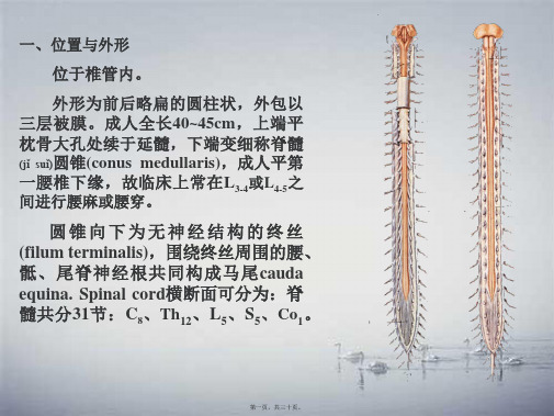
功能:传递整个肢体运动和姿势的信息, 主要传递来自下肢的冲动。
损伤表现:脊小脑前束、后束损伤,出现 症状不明显,由薄、楔束代偿,如小脑同时 病变,则出现小脑共济失调,与感觉性共济 失调主要区别,不能为视觉(shìjué)所纠正。
与脊髓(jǐ suǐ)小脑后束相当,传导上肢的同类冲动至小脑称楔小 脑束,起自延髓楔束副核,径小脑下脚→小脑。
第十三页,共三十页。
Ⅷ层:具有中间神经元,接受邻近层及脑干下行纤维,并发出轴突至
同侧和对侧Ⅸ层的神经元,起兴奋作用。
Ⅸ层:主要有、运动神经元,还有中间神经元Renshaw's cell, 接受神经元的侧支,又发出轴突止于神经元(同一个或其它的神经 元),形成一负反馈环路(huán lù),对神经元起抑制作用。
第九页,共三十页。
Ⅵ层:仅见脊髓膨大 处,也相当于后角基部。 也分为内、外侧两部。
Ⅶ层:相当于前、 后角之间的中间带, 包 括 (bāokuò) 侧 角 , 此 层 细胞分布均匀,但也 有集中成神经核(柱)者。
胸核(背核) Th1-L2 (位于后角底部内侧)
中间外侧(wài cè)核
Th1-L3 S2-4 相当于侧角部位,
功能:传递粗触觉,损伤后对触觉功能影响不大。 侧束 lateral spinothalamic tract
位置:外侧索,脊髓丘脑前束的后外方,脊髓小脑 (xiǎonǎo)前束的内侧。
西罗莫司洗脱支架或紫杉醇洗脱支架与血管内球囊扩张术用于预防冠状动脉支架内再狭窄复发的随机对照试验
西罗莫司洗脱支架或紫杉醇洗脱支架与血管内球囊扩张术用于预防冠状动脉支架内再狭窄复发的随机对照试验Kastrati A.;Mehilli J.;Von Beckerath N.;腾增辉【期刊名称】《世界核心医学期刊文摘:心脏病学分册》【年(卷),期】2005(000)006【摘要】Context: In patients with de novo coronary lesions, drugeluting stents have drastically reduced restenosis risk compared with bare metal stents and conventional balloon angioplasty. It is less clear whether drug-eluting stents are superior to conventional balloon angioplasty for the treatment of patients with in-stent restenosis. Objectives: To assess ifdrug-eluting stents are a more effective treatment of instent restenosis than conventional balloon angioplasty, and to assess the relative merits of 2 drug-eluting stents, a sirolimus-eluting stent and a paclitaxel-eluting stent. Design, Setting, and Participants: Randomized, open-label, active-controlled trial conducted among 300 patients with angiographically significant in-stent restenosis in 2 tertiary German centers from June 1, 2003, to October 20, 2003. Interventions: After pretreatment with 600 mg of clopidogrel for at least 2 hours before intervention, all patients were randomly assigned to 1 of 3 treatment groups: sirolimus stent, paclitaxel stent, or balloon angioplasty(100 patients in each group). Main Outcome Measures: Primary end point: angiographic restenosis(diameter stenosis≥50%) at 6-month follow-up angiography based on “insegment”analysis.Primary analysis was comparison between stent groups and balloon angioplasty groups; a secondary analysis compared sirolimus and paclitaxel stents. Results: Follow-up angiography was performed in275(92%) of 300 patients. The incidence of angiographic restenosis was 44.6%(41/92) in the balloon angioplasty group, 14.3%(13/91) in the sirolimus stent group(P< .001 vs balloon angioplasty), and 21.7%(20/92) in the paclitaxel stent group(P=.001 vs balloon angioplasty). When compared with balloon angioplasty, receiving a sirolimus stent had a relative risk(RR) of angiographic restenosis of 0.32(95%confidence interval [CI], 0.18-0.56);a paclitaxel stent had an RR of 0.49(95%CI, 0.31-0.76). The incidence of target vessel revascularization was 33.0%(33/100) in the balloon angioplasty group, 8.0%(8/100) in the sirolimus stent group(P< .001 vs balloon angioplasty), and 19.0%(19/100) in the paclitaxel stentgroup(P=.02 vs balloon angioplasty). The secondary analysis showed a trend toward a lower rate of angiographic restenosis(P=.19) and a significantly lower rate of target vessel revascularization(P=.02) among sirolimus stent patients compared with paclitaxel stent patients. Conclusions: In patients with in-stent restenosis, a strategy based on sirolimus-or paclitaxel-eluting stents is superior to conventional balloon angioplasty for the prevention of recurrent restenosis. Sirolimus-eluting stents may be superior to paclitaxel-eluting stents for treatment of this disorder.【总页数】2页(P19-20)【作者】Kastrati A.;Mehilli J.;Von Beckerath N.;腾增辉【作者单位】Dr.Deutsches Herzzentrum;Lazarettstr-asse 36;80636 Munich;Germany【正文语种】中文【中图分类】R543【相关文献】1.国产西罗莫司洗脱支架与紫杉醇洗脱支架抑制血管内膜增生的血管内超声比较研究 [J], 杜润;张瑞岩;朱政斌;张奇;胡健;张建盛;沈卫峰2.西罗莫司洗脱支架与紫杉醇洗脱支架抑制血管内膜增生的血管内超声比较 [J], 杜润;张瑞岩;朱政斌;张奇;胡健;张建盛;沈卫峰3.西罗莫司洗脱支架和紫杉醇洗脱支架之间支架内再狭窄的血管造影影像比较 [J], Park;C.-B.;Kim;Y.-H.;王亭忠4.西罗莫司洗脱支架与紫杉醇洗脱支架对冠状动脉病变的影响REALITY试验:随机对照试验 [J], Marie-Claude Morice;Antonio Colombo;Bernhard Meier;孙艺红5.关于西罗莫司和紫杉醇洗脱支架用于新发冠状动脉病变的随机对照试验:REALITY试验 [J], Morice; M.; -C.; Colombo; A.; Meier; B.; 任付先(译); 杜媛(校)因版权原因,仅展示原文概要,查看原文内容请购买。
改良“轴保持短缩法”单人操作结肠镜在结肠术后患者检查中的临床应用

·临床研究·doi :10.3969/j.issn.1006-5725.2011.02.016基金项目:解放军总医院苗圃基金项目(编号:09MP05)作者单位:100853北京市,解放军总医院南楼消化内镜诊疗科通信作者:王志强E-mail :yxmnc1956@163.com结肠镜是检查大肠疾病可靠、有效的检查方法[1],自从日本消化内镜专家工藤进英提出“轴保持短缩法”及“Jiggling 手技法”[2]以来,结肠镜单人操作技术日趋完美。
因其操作安全、简便、患者痛苦小、成功率高、患者反应良好、又节省人力,单人操作结肠镜已经逐渐成为国际上结肠镜插入法的主流技术。
结肠术后患者由于肠管部分切除、吻合手术造成结肠正常生理走形改变,肠管之间、肠管与腹壁之间可能存在粘连,给结肠镜的插入带来不便[3]。
我们改良了工藤进英教授提出的单人操作结肠镜“轴保持短缩法”,对结肠术后患者进行检查,与传统单人操作结肠镜方法进行对比,探讨该方法对结肠术后患者检查的临床作用。
1资料与方法1.1一般资料2009年4-12月来我院消化内镜中心进行随访的结肠术后患者512例,其中男317改良“轴保持短缩法”单人操作结肠镜在结肠术后患者检查中的临床应用李明阳王志强令狐恩强卢忠生黄启阳摘要目的:探讨改良“轴保持短缩法”单人操作结肠镜在结肠术后患者检查中的临床应用。
方法:对2009年4-12月来我院消化内镜中心进行随访的512例结肠术后患者,分别应用改良“轴保持短缩法”单人操作法以及常规单人操作结肠镜进行检查,对两种方法的成功率、进镜时间、疼痛评分等指标进行比较。
结果:常规单人操作结肠镜和改良“轴保持短缩法”单人操作结肠镜到达回盲部(或结肠-小肠吻合口)的成功率分别为93.8%和99.1%。
常规单人操作结肠镜检查和改良“轴保持短缩法”单人操作结肠镜的平均进镜时间分别为7.6min 和3.5min (P <0.05)。
常规单人操作结肠镜和改良“轴保持短缩法”单人操作结肠镜检查后,采用数字评定量表(NRS )评定疼痛程度的平均分数分别为6.7分和3.8分(P <0.05)。
- 1、下载文档前请自行甄别文档内容的完整性,平台不提供额外的编辑、内容补充、找答案等附加服务。
- 2、"仅部分预览"的文档,不可在线预览部分如存在完整性等问题,可反馈申请退款(可完整预览的文档不适用该条件!)。
- 3、如文档侵犯您的权益,请联系客服反馈,我们会尽快为您处理(人工客服工作时间:9:00-18:30)。
a rXiv:071.5679v1[qua nt-ph]3O ct27Casimir torque between corrugated metallic plates Robson B.Rodrigues 1,Paulo A.Maia Neto 1,Astrid Lambrecht 2and Serge Reynaud 21Instituto de F´ısica,UFRJ,CP 68528,Rio de Janeiro,RJ,21941-972,Brazil 2Laboratoire Kastler Brossel,CNRS,ENS,Universit´e Pierre et Marie Curie case 74,Campus Jussieu,F-75252Paris Cedex 05,France Abstract.We consider two parallel corrugated plates and show that a Casimir torque arises when the corrugation directions are not aligned.We follow the scattering approach and calculate the Casimir energy up to second order in the corrugation amplitudes,taking into account nonspecular reflections,polarization mixing and the finite conductivity of the metals.We compare our results with the proximity force approximation,which overestimates the torque by a factor 2when taking the conditions that optimize the effect.We argue that the Casimir torque could be measured for separation distances as large as 1µm .1.IntroductionThe relevance of the Casimir effect[1]in connection with micro and nano-electromechanical systems(MEMS and NEMS)has been recently highlighted[2,3,4, 5,6].The attractive Casimir force can lead to permanent adhesion of the movable parts of MEMS and NEMS when they are close enough,a phenomenon known as‘stiction’, resulting in malfunctioning of these devices.On the other hand,the Casimir effect may provide novel actuation schemes[7,8]with promising potential applications.Besides the usual normal Casimir force between metallic or dielectric plates,the lateral Casimir force between corrugated plates[9,10]can also be used for micro-mechanical control.Very recently,two devices based on the lateral Casimir force were theoretically proposed:a rack and pinion device[11],which is actuated by the lateral Casimir force between a cylinder with a corrugated surface and a corrugated plane plate, and a Casimir ratchet[12],driven by the lateral Casimir force between a plate with a symmetric corrugation and a plate with an asymmetric corrugation.The experimental results for the lateral Casimir force werefirst compared with a theoretical analysis[9,10]based on the proximity-force approximation(PFA),or Derjaguin approximation[13,14].Within this approximation,the Casimir energy for non-planar surfaces is obtained by simply averaging the energy for parallel planes over the local separation distance.This approximation holds when the corrugation periodλC is much larger than the average separation distance L,so that the surfaces are nearly plane in the scale of L[15].It is extremely important to check the accuracy of this approximation,since it was widely employed for comparison with experimental results for the(normal)Casimir force between curved surfaces[16,17,18,19](see Ref.[20]for a more detailed discussion and a review on recent theoretical advances).We have computed the lateral force beyond the PFA[21,22]by employing the scattering approach[23],which takes into account thefinite conductivity of the metallic plates as well as diffraction and polarization mixing.The corrugation is treated as a small perturbation of the plane geometry,and the lateral force is computed up to second order in the corrugation amplitudes a1and a2for each plate.The perturbation expansion holds as long as a1,a2≪λC,L,but arbitrary relative values of L andλC are allowed,with the PFA regime corresponding to the limit L/λC→0.This formalism was also employed to compute the roughness correction to the Casimir force[24]and more recently the lateral Casimir-Polder force[25,26].Beyond-PFA theories for uni-axial corrugation on perfect reflecting plates werefirst reported by Emig et al for both perturbative[27]and nonperturbative[28]regimes.The lateral Casimir force results from breaking the translational symmetry along directions parallel to the plates[29].A more general situation occurs when the corrugations are not aligned,so that the Casimir energy depends on the relative orientation between the two plates,and a Casimir torque arises[30](seefigure1).In this paper,we review the main physical properties of this effect.Whereas the formalism developed in Refs.[27,28]requires the existence of a direction of translational symmetryFigure1.Periodic corrugations(periodλC,amplitudes a1and a2)are imprinted onboth plates.L is the average separation distance andθthe rotation angle.We assumethat a1,a2≪L,λC.(so as to allow for a convenient definition offield polarizations which are not coupled by the nonspecular reflection in this case),the scattering approach[23,24]allows for a more general situation since it explicitly takes the coupling between different polarizations into account.Thus,the scattering approach allows one to consider the geometry with rotated corrugated plates,as long as the corrugation amplitudes remain the smallest length scale as discussed above.Thanks to the high sensitivity of torsion balance techniques[31],the Casimir torque between corrugated plates provides an attractive way to measure nontrivial(i.e. beyond-PFA)geometry effects.The use of torsion balances has also been proposed to measure the Casimir torque between two(plane)birefringent dielectric plates[32](see also[33,34,35,36]).For the proposed separation distances around100nm[32],the Casimir torque for corrugated plates is up to three orders of magnitude larger than the torque between birefringent plates,for comparable values of the separation distance and plate area,and taking realistic values for the corrugation amplitudes and thefinite conductivity of the metallic plates.This could allow one to perform the experiment at larger separation distances,thus minimizing problems related to plate parallelism.2.Casimir energy for corrugated platesWe assume that both plates have sinusoidal corrugation profiles with the same period λC and amplitudes a1and a2.The corrugation lines of the bottom plate are along the y direction(ie,the surface profile depends only on x).The top plate position along the x axis is b(by symmetry the energy does not depend on the position along the y axis), andθis the rotation angle.b=0,θ=0corresponds to the configuration where the corrugation lines are aligned with the surface crests facing each other(seefigure1).This is the configuration corresponding to the global energy minimum as discussed below.The top plate lateral dimensions are L x and L y,and L is the average separation distance(along the z direction).Both plates are assumed to be very large compared to L(so that border effects are negligible)as well as compared toλC:L x∼L y≫λC.TheFigure2.Level curves for the Casimir energy(in arbitrary units)as a function of thelateral displacement b and of the rotation angleθ.Regions with lower energy valuesare darker.Casimir energy correction is then calculated to order a1a2[30]:δE PPG(k)cos(kb)sinc(kL yθ/2).(1)2where k=2π/λC,sinc(ξ)=sinξ/ξand G(k)is a response function which also depends on the separation distance L.Infigure2,we plot the level curves of the Casimir energy correction(in arbitrary units)as a function of b andθ.Since G(k)is always negative, the Casimir energy has global minima atθ=0and b=0,λC,2λC,...and local minima aroundθ≈1.43λC/L y(minimum of sinc(kL yθ/2))and b=λC/2,3λC/2,...If we start fromθ=b=0and rotate the top plate around its center,we follow the dashed line b=0shown infigure2.Forθ<λC/L y the plate is attracted back to the minimum atθ=b=0without sliding laterally.On the other hand,if the plate is released after a rotation ofθ>λC/L y its subsequent motion will be a combination of rotation and lateral displacement.In the next section,we compute the Casimir restoring torque for the case of pure rotations with small rotation angles.3.Casimir torqueThe Casimir torque,given by∂τ=−is maximum at θ=0.66λC /L y where it is given byτL x L y PF A=0.109a 1a 2ke ′′PP (L )L y .(3)According to eq.(3),the torque grows linearly with k in the PFA (dotted straight line in figure 3).Figure 3shows that the scattering curve is very close to the PFA straight line when k ≪1/L as expected (G (k )≈G (0)).However,the discrepancy increases with k,and at the peak value k =2.6/L =2.6µm −1the PFA overestimates the torque by 103%.A detailed discussion of the ratio G (k )/G (0)for several separation distances is presented in Ref.[22].4.ConclusionAs in the case of the lateral force,the Casimir torque between corrugated metallic plates might have potential applications in the design of MEMS and NEMS.We have studiedFigure3.Torque as a function of k=2π/λC for a separation distance L=1µm.Corrugation amplitudes:a1a2=200nm2.The plate length along the direction ofthe corrugation lines is L y=24µm.Results for gold-covered plates(λP=137nm)correspond to the solid(scattering)and dotted(PFA)lines;the dashed line correspondsto perfect reflectors.All results are computed up to second order in the corrugationamplitudes.The vertical dotted line indicates the optimal value k=2.6µm−1.the Casimir torque with the help of the scattering approach,which provides an exact result for the second-order energy correction.This torque is up to three orders of magnitude larger than the torque between birefringent dielectric plates for comparable separation distance and area.The measurement of the Casimir torque with corrugated plates would provide a direct demonstration of a non-trivial(beyond PFA)geometry dependence of the Casimir energy.RBR and PAMN thank FAPERJ,CAPES,CNPq and Institutos do Milˆe nio de Informa¸c˜a o Quˆa ntica e Nanociˆe ncias forfinancial support.AL acknowledges partial financial support by the European Contract STRP12142NANOCASE.References[1]Casimir H B G,1948Proc.K.Ned.Akad.Wet.51793.[2]Serry F M,Walliser D and Maclay G J,1998J.Appl.Phys.842501[3]Buks E and Roukes M L,2001Europhys.Lett.54220[4]Buks E and Roukes M L2001Phys.Rev.B63033402[5]Capasso F,Munday J N,Iannuzzi D and Chan H B,2007IEEE J.Selected Topics QuantumElectronics13400and references therein.[6]Esquivel-Sirvent R,Reyes L and B´a rcenas J,2006New J.Phys.8241and references therein.[7]Chan H B,Aksyuk V A,Kleiman R N,Bishop D J and Capasso F,2001Science2911941.[8]Chan H B,Aksyuk V A,Kleiman R N,Bishop D J and Capasso F,2001Phys.Rev.Lett.87211801[9]Chen F,Mohideen U,Klimchitskaya G L and Mostepanenko V M,2002Phys.Rev.Lett.88101801[10]Chen F,Mohideen U,Klimchitskaya G L and Mostepanenko V M,2002Phys.Rev.A66032113.[11]Ashourvan A,Miri M,Golestanian R,2007Phys.Rev.Lett.98,140801.[12]Emig T,2007Phys.Rev.Lett.98,160801.[13]Derjaguin B V,1934Kolloid Z.69155[14]Derjaguin B V and Abrikosova I I,1957Sov.Phys.JETP3819[15]Genet C,Lambrecht A,Maia Neto,P A and Reynaud S,2003Europhys.Lett.62484.[16]Ederth T,2000Phys.Rev.A62062104[17]Chen F et al2004Phys.Rev.A69022117[18]Decca RS et al,2005Annals Phys.(NY)31837[19]Brown-Hayes M,Dalvit DAR,Mazzitellli FD,Kim WJ and Onofrio R,2005Phys.Rev.A72052102[20]Reynaud S,Maia Neto PA and Lambrecht A,in the present issue.[21]Rodrigues R B,Maia Neto,P A,Lambrecht A and Reynaud S,2006Phys.Rev.Lett.96100402[22]Rodrigues R B,Maia Neto,P A,Lambrecht A and Reynaud S,2007Phys.Rev.A75,062108.[23]Lambrecht A,Maia Neto,P A and Reynaud S,2006New J.Phys.8243.[24]Maia Neto PA,Lambrecht A and Reynaud S,Europhys.Lett.69,924(2005);Phys.Rev.A72(2005)012115.[25]Dalvit DAR,Maia Neto PA,Lambrecht A and Reynaud S,2007arxiv:0709.2095[26]Dalvit DAR,Maia Neto PA,Lambrecht A and Reynaud S,in the present issue.[27]Emig T et al,2003Phys.Rev.A67022114[28]B¨u scher R and Emig T,2005Phys.Rev.Lett.94133901[29]Golestanian R and Kardar M,1997Phys.Rev.Lett.783421[30]Rodrigues R B,Maia Neto P A,Lambrecht A and Reynaud S,2006Europhys.Lett.76822[31]Gundlach JH,1999Meas.Sci.Technol.10454[32]Munday J N,Iannuzzi D,Barash Y and Capasso F,2005Phys.Rev.A71042102[33]Parsegian V A and Weiss G H,1972J.Adhes.3259[34]Barash Y,1978Izv.Vyssh.Uchebn.Zaved.,Radiofiz.121637[35]van Enk S J,1995Phys.Rev.A522569[36]Torres-Gusm´a n J C and Moch´a n W L,2006J.Phys.A:Math Gen.396791。
