不同来源的人脂肪干细胞体外成脂诱导分化能力的比较
干细胞成骨、成脂、成软骨诱导分化及检测

三个方向诱导分化诱导骨髓间充质干细胞成软骨分化一、诱导方法将细胞悬液置于50 mL聚苯乙烯培养瓶(Nunclon)中培养。
完全培养基中加入能诱导MSCs 向软骨细胞转化的诱导因子: (一致统一的诱导物质)转化生长因子β1 10ng/ml,(左旋)维生素C 50 mg/L(也有0.1mmol/L)地塞米松0.1nmol/L (也有10 nmol/L)ITS 50mg/ml丙酮酸钠1mmol/L亚油酸5.35ug/mg牛血清白蛋白1.25ng/ml。
倒置显微镜逐日(1-3 weeks)观察细胞生长情况。
细胞长成单层后进行传代。
细胞培养3周。
去除培养液,晾干,①细胞用通用Ⅱ型胶原检测试剂盒(晶美生物工程有限公司) 免疫组化,按试剂盒说明书操作;②用alcian blue 孵育5 min, 流水冲洗2 min ,麦氏苏木素复染5 min ,流水冲洗2 min ,晾干,显微镜下观察细胞的蛋白聚糖沉积。
或者用甲苯胺蓝染色,具体我也没做过,查到一篇文章中是这么说的:MSC成软骨诱导后的甲苯胺蓝染色检测:培养不同时间的MSC培养皿倒掉诱导培养液,10%甲醛固定lh,自来水冲洗15min,双蒸水冲洗1次,滴加1%甲苯胺蓝染液于培养皿内,染色3h,加人95%乙醇,洗去多余的染液,烘干,中性树胶封片。
诱导骨髓间充质干细胞成骨方向分化成骨诱导培养: 取第3代细胞, 接种入含体积分数为0.1 的新生牛血清、0.1 μmol/L 地塞米松、50 μmol/L 抗坏血酸、10 mmol/L β- 甘油磷酸钠的高糖DMEM培养基进行成骨诱导培养, 进行形态观察、功能检测。
间充质干细胞成骨特性检测:①碱性磷酸酶组织化学染色:取成骨诱导14 d 的细胞, 40 g/L 中性甲醛固定15 min, Gomori改良钙钴法染色。
取5 块玻片, 每片随机取2个视野, 采用网格计数法, 计算碱性磷酸酶染色阳性细胞的百分比。
:取第3 代细胞, 以1×10⁵/ 孔的密度接种于6 孔板中, 分别成骨诱导3, 5, 7, 10, 12, 14 d,按碱性磷酸酶活性检测试剂盒要求进行检测。
体外培养成人脂肪间充质干细胞生物学特性及向心肌样细胞的诱导分化
摘 要 目的 : 外 分 离 培 养 成 人脂 肪 组 织 来 源 的 间 充 质 干 细 胞 , 索 其 基 本 生 体 探 物 学 特 性 及 向心 肌 细 胞 诱 导 分 化 的 潜 能 ,为 心 肌 再 生 提 供 理 想 的 自体 干 细 胞 来 源 方 法 : 验 于 20 — 1 1 实 0 5 0 / 2在解 放 军 兰 州 军 区 兰 州 总 医 院 医 学 实 验 中心 完 成 取 本 院 微 创 外 科 中心 一 急 性 单 纯 性 阑 尾 炎患 者 大 网 膜 脂 肪 组 织
细胞周期及 C 4 C 3 D 4、D 4的 表 达 , 第 4代 细 胞 用 5 氮 胞 苷 诱 导 使 其 向 一 心肌细胞分化 , 免疫 细 胞化 学 鉴定 心 肌 样 细 胞 。 结 果 : 分 离 培 养 的 脂 肪 间 充 质 干 细 胞 经 传 代 纯 化 后 细胞 形 态 呈 均 一 显 , 染 色 质 多 , 染 电 核 核 常 异
・
基础 研 究 ・
体外培养成人脂肪问充质干细胞生物学特性及向心肌样细胞的诱导分化☆
张 卫 泽 , 陈跃 武 , 永 清 , J琴 , 陈 哈J\ 马 凌 -秦 , 勉 , 志 斌 z 洪
1 —7 hn l 4 { ia C
解 放 军 兰州 军 区 兰 州 总 医 院G 内科 ,甘 肃 省 兰州 市 7 o 5 ;兰 州 大 3 0 0 学 临床 医 学院 , 肃 省 兰 州 市 7 0 0 ;解 放 军 兰州 军 区 兰 州 总 医院 医 甘 3 0 0 学 实验 中, , 肃 省 兰 州 市 7 0 5 甘 300 张 卫 泽 ☆ , ,9 5年 生 , 肃 省和 政 县 人 , 族 ,o 2年 中 国 协 和 医 科 男 16 甘 汉 20 大 学毕 业 , 士 , 任 医师 , 博 主 主要 从 事心 血 管 内科 临 床 与 基 础 研 究
DFAT 细胞体外分化诱导实验及体内生物学特性综述
DFAT 细胞体外分化诱导实验及体内生物学特性综述李福海;王志;张陈匀【摘要】去分化脂肪( dedifferentiated fat ,DFAT)细胞,由于来源广泛、取材方便、增殖能力强、免疫原性低、表型稳定,很大程度上符合再生医学对理想种子细胞的要求,现已成为各个领域的研究热点。
本文对体外诱导DFAT细胞向多个方向分化的诱导方法及DFAT细胞在动物模型体内的多向分化和组织修复特性进行了综述。
目前对DFAT的研究仍滞留于寻求更高效的分化诱导因子的早期阶段,若欲将DFAT细胞作为再生医学的种子细胞投入临床应用,仍需大量的研究与探索。
%Dedifferentiated fat ( DFAT) cells, which to a large extent meet the demands for the ideal seed cells of the regenerative medicine research for its extensive sources , convenient drawing , strong proliferation ability , weak immunogenicity and stable phenotype , have become a research hotspot in many fields.This thesis conducts a review on the induction methods of the differentiation of induced DFAT cells in vitro into multiple directions , the multi-directional differentiation and the tissue repairing features of DFAT cells.At present , the study on DFAT cells still stagnates at the early stage of seeking for more efficient differentiation inducing factors .A large amount of research and exploration remains to be done before DFAT cells are put into clinical use as the seed cells in the research of regenerative medicine .【期刊名称】《北京生物医学工程》【年(卷),期】2015(000)003【总页数】6页(P310-315)【关键词】去分化脂肪细胞;分化;诱导;生物学特性【作者】李福海;王志;张陈匀【作者单位】贵州省心血管病研究所贵阳 550002;贵州省心血管病研究所贵阳550002;贵州省心血管病研究所贵阳 550002; 贵州省人民医院心内科贵阳550002【正文语种】中文【中图分类】R31820世纪80年代后期,组织学工程概念的提出,把再生医学带入了一个充满机遇与挑战的新时期。
人脂肪干细胞及其外泌体的分离与鉴定

人脂肪干细胞及其外泌体的分离与鉴定李洪超;金银鹏;王皙;李莉;王晓今;周荣;陈成伟;傅青春;程明亮【摘要】背景:间充质干细胞如今在科研领域被广泛地研究和应用,许多研究认为其发挥作用的机制很大程度上依赖于其旁分泌的外泌体.目的:分离纯化人脂肪干细胞来源的外泌体,并鉴定其生物学特性.方法:采用胶原酶消化法获得人脂肪干细胞,进行细胞表面分子标志和成骨、成脂分化能力鉴定.运用超滤法从人脂肪干细胞条件培养基中提取外泌体,应用扫描电子显微镜、粒度仪观察所获外泌体的形态和大小,采用抗体芯片检测外泌体所含蛋白质表达.结果与结论:①人脂肪干细胞呈梭形、漩涡状生长,细胞表面表达CD73、CD44、CD90、CD105分子,具备成脂、成骨等多向分化潜能,可证实为人脂肪干细胞;②大多数外泌体直径均在30-150 nm范围内;扫描电镜显示外泌体呈均一大小的圆杯形态;③抗体芯片显示外泌体含 FLOT1、ICAM、ALIX、CD81、CD63、ANXA5、TSG101等多种特殊蛋白;④以上结果表明实验成功分离得到人脂肪干细胞外泌体.%BACKGROUND: Currently, mesenchymal stem cells have been widely explored and applied in scientific research field, and many studies suggest that the underlying mechanism of mesenchymal stem cells mainly relies on its exosomes. OBJECTIVE: To isolate and identify human adipose-derived stem cells and its exosomes, and to identify their biological characteristics. METHODS: Human adipose tissue was digested with collagenase l, and adipose-derived stem cells were isolated and purified. Immunophenotype, osteogenic and adipogenic abilities of adipose-derived stem cells were identified. Exosomes were isolated by using ultrafiltration method. Morphology of exosomes was observed by Nanosight and electronmicroscope. Expression of proteins in exosomes was detected by antibody array method. RESULTS AND CONCLUSION: Adipose-derived stem cells exhibited long spindle-like or fibroblast-like appearance, expressed CD73, CD44, CD90, CD105 and had the potential to differentiate into many tissues, including bone and adipose tissues. The exosomes had the similar size, with the diameter of 30-150 nm. They possessed many proteins including FLOT1, ICAM, ALIX, CD81, CD63, ANXA5, TSG101, and so on. Findings from the present study indicate the successful isolation of exosomes from human adipose-derived stem cells.【期刊名称】《中国组织工程研究》【年(卷),期】2018(022)013【总页数】6页(P2033-2038)【关键词】外泌体;干细胞;间充质干细胞;脂肪干细胞【作者】李洪超;金银鹏;王皙;李莉;王晓今;周荣;陈成伟;傅青春;程明亮【作者单位】贵州医科大学临床医学院,贵州省贵阳市 550004;解放军第八五医院,上海肝病研究中心,上海市 200235;贵州医科大学临床医学院,贵州省贵阳市550004;解放军第八五医院,上海肝病研究中心,上海市 200235;解放军第八五医院,上海肝病研究中心,上海市 200235;解放军第八五医院,上海肝病研究中心,上海市200235;解放军第八五医院,上海肝病研究中心,上海市 200235;解放军第八五医院,上海肝病研究中心,上海市 200235;贵州医科大学附属医院感染科,贵州省贵阳市550004【正文语种】中文【中图分类】R394.20 引言 Introduction间充质干细胞是一类具有自我更新能力和多向分化潜能的成体干细胞,不仅能够从骨髓、脐带、脂肪中分离,也可以从脾脏、肝脏、肾脏、肺、胰腺中得到,尽管其组织来源不同,但是都拥有相似的表型特征[1-4]。
脂肪来源的间充质干细胞及外囊泡促成骨分化的研究进展
1672V ol.40 No.12 Dec. 2020上海交通大学学报(医学版)JOURNAL OF SHANGHAI JIAO TONG UNIVERSITY (MEDICAL SCIENCE )综述近年来,我国骨质疏松症患病率逐渐攀升。
一项最新研究[1]显示,我国骨质疏松症的总患病率为13%,总人数已超过1.78亿。
老年人是骨质疏松症的重点人群,自1982年起,我国65岁以上老年人占人口比重不断升高,2019年我国65岁以上老年人占人口比重已达到12.6%[2]。
预计到2050年,我国骨质疏松症或骨密度低的患者将达到2.12亿[1]。
骨质疏松症使得骨质脆性增加、易于骨折,导致患者的生活水平急剧下降。
利用脂肪来源的间充质干细胞(adipose-derived mesenchymal stem cells ,ADMSCs )诱导成为成骨细胞治疗骨质疏松症是医学研究的新方向[1]。
ADMSCs 可以通过旁分泌功能,分泌一些生物活性分子,为组织修复建立良好的微环境,促进新生血管的形成和伤口愈合,并且减少组织的炎症反应。
ADMSCs 也可分泌促进血管生成和抗凋亡潜能的生长因子,如转化生长因子(transforming growth factor ,TGF )、胰岛素样生长因子(insulin growth factor ,IGF )、血管内皮生长因子(vascular endothelial growth factor ,VEGF )、肝细胞生长因子(hepatocyte growth factor ,HGF )[3]和骨形态发生蛋白(bone morphogenetic protein ,BMP )家族BMP-2、BMP-7等[4]。
ADMSCs 来源丰富,通过脂肪抽吸术易于获得,无免疫排斥。
平均每300 mL 脂肪组织可获得108个 细胞,每克动物脂肪可获得5 000个成纤维集落形成单位(colony forming unit-fibroblast ,CFU-F )[5]。
人皮下脂肪干细胞的成骨、成脂分化诱导及鉴定
人皮下脂肪干细胞的成骨、成脂分化诱导及鉴定肖建红;张阳春;张常然;杨兴【摘要】背景:脂肪干细胞是从脂肪组织中分离得到的一种具有多向分化潜能的间充质干细胞,在组织工程中比骨髓间充质干细胞具有更多优势,是当今的研究热点之一。
<br> 目的:在体外建立一种分离纯化人皮下脂肪来源脂肪干细胞的方法,进行体外培养扩增及诱导其向成骨、成脂细胞分化。
<br> 方法:通过密度梯度离心和贴壁筛选法相结合,对人脂肪干细胞进行分离、体外培养及扩增。
倒置显微镜下观察人脂肪干细胞的细胞形态和生长特性。
选取第2代及第5代细胞,用CCK-8法检测细胞活性,绘制细胞生长曲线,用流式细胞仪检测细胞表面抗原标记。
选取第5代细胞,分别进行成脂及成骨诱导分化,以确定其多向分化潜能。
<br> 结果与结论:①采用密度梯度离心和贴壁培养相结合方法,可成功从人脂肪组织中获得纯度较高的人脂肪干细胞。
②人脂肪干细胞经历了生长滞缓期、对数增殖期和生长平台期,符合正常细胞的生长规律,且其生长具有对数生长期,其倍增时间较短。
③人脂肪干细胞表达干细胞表面抗原标记,具有低免疫原性,且具有成脂成骨多向分化潜能。
④DAPI是一种简单、有效的标记人脂肪干细胞的方法,DAPI不损伤细胞活性,对细胞标记率高。
%BACKGROUND:Adipose-derived stem cels are a kind of mesenchyam stem cels with multipotent differentiation capacity, which have more advantages than bone marrow mesenchymal stem cels in tissue engineering research. OBJECTIVE: To establish a method to isolate and purify adipose-derived stem cels from human subcutaneous adipose tissues folowed byin vitro amplification and osteoblastic/adipogenic differentiation. <br> METHODS: Adipose-derived stem cels were isolated from human subcutaneous adipose tissue andcultured by density gradient centrifugation and adherent culture. Cel morphology and growth features were observed under inverted microscope. Adipose-derived stem cels at passages 2 and 5 were selected for viability measurement using cel counting kit-8 method, and then cel growth curves were drawn. The immunophenotype identification was analyzed by flow cytometry. Passage 5 cels underwentosteoblastic/adipogenic induction to confirm the multi-differentiation potential. <br> RESULTS AND CONCLUSION: (1) Using density gradient centrifugation and adherent culture method, high-purity human adipose-derived stem cels can be successfuly isolated from human adipose tissues.(2) The growth process of human adipose-derived stem cels includes stagnant phase, logarithmic phase and plateau phase, which meets the growth rhythm of normal cels. Moreover, the population doubling time is shorter. (3). Human adipose-derived stem cels are positive for stem cel-related antigens, with low immunogenicity and the multi-differentiation potential. (4) Labeling human adipose-derived stem cels with DAPI is a simple efficient labeled method, and the labeling rate is high but the cytotoxicity is low【期刊名称】《中国组织工程研究》【年(卷),期】2015(000)032【总页数】7页(P5155-5161)【关键词】干细胞;脂肪干细胞;人来源的脂肪干细胞;细胞分化;脂肪组织;细胞培养;细胞鉴定;国家自然科学基金【作者】肖建红;张阳春;张常然;杨兴【作者单位】中山大学附属第一医院东院普通内科,广东省广州市 510700;中山大学附属第一医院东院下肢骨科,广东省广州市 510700;中山大学附属第一医院东院普通内科,广东省广州市 510700;中山大学附属第一医院东院下肢骨科,广东省广州市 510700【正文语种】中文【中图分类】R394.2文章亮点:1 实验通过密度梯度离心和贴壁筛选法相结合,对人脂肪干细胞进行分离、体外培养及扩增。
人脂肪间充质干细胞的原代培养及体外成骨成脂诱导分化
人脂肪间充质干细胞的原代培养及体外成骨成脂诱导分化闵敏;张雪静;马红;许辉;李遇梅【摘要】目的:建立体外分离培养获得人脂肪间充质干细胞(human adipose-derived mesenchymal stem cells,hADSCs)的方法,并观察其形态、免疫表型、生物学特性.初步探讨其体外成骨过程中出现成脂现象的原因及机制.方法:剖宫产手术获得腹部皮下脂肪,0.15%Ⅰ型胶原酶消化法获得人脂肪间充质干细胞并进行体外培养.行MTT细胞增殖实验绘制增殖曲线,使用流式细胞及细胞免疫荧光技术检测细胞表面抗原,行成骨成脂分化鉴定其分化潜能.通过RT-PCR技术检测成骨成脂过程中过氧化物酶体增殖物激活受体γ-2 (peroxisome proliferator activated receptorγ-2,PPARγ-2)和骨桥蛋白mRNA表达情况.结果:通过分离培养,获得了大量旋涡状生长的人脂肪间充质干细胞.MTT法显示细胞增殖能力强.流式细胞鉴定结果显示,CD29、CD73、CD105、CD166高表达,CD31、CD34、CD45、HLA-DR 低表达.细胞免疫荧光结果与之相符.hADSCs在特定诱导条件下,具有成骨成脂分化潜能.在成骨分化过程中同时伴有成脂发生.RT-PCR结果显示,与对照组相比,成脂诱导组PPARγ-2 mRNA有时间依赖性递增表达,差异具有统计学意义(P<0.01).与对照组相比,成骨诱导组骨桥蛋白,PPARγ-2 mRNA均有时间依赖性递增表达,差异具有统计学意义(P<0.01).结论:建立了一种分离培养hADSCs简单可靠的方法.获得的细胞具有贴壁生长、增殖活性强、干细胞表型以及多向分化等特征.hADSCs体外成骨过程中,三酰甘油的形成对成骨具有一定的促进作用.%Objective:To establish the isolation and culture method of human adipose-derived mesenchymal stem cells (hADSCs) derived from human adipose in vitro,so as to explore their morphology,identify cell surface markers,observe biological properties,and discuss the possible causes and mechanism ofthe phenomenon of hADSCs' osteogenic differentiation accompanying with synthesis of triglycerides.Methods:Human adipose tissue were obtained from abdominal operation.The hADSCs were isolated from human adipose tissue by 0.15% collagenase digesting.The cells were applied to do the experiments:MTT method,flowcytometry,immunofluorescence.Its differentiation potential was proved by osteogenic and adipogenic differentiation.The osteogenic and adipogenic related genes:PPARγ-2,osteopontin expression were detected by real-time fluorescent quantitative PCR technique.Results:After isolation and culture,we obtained a large amount of hADSCs,which grew like swirls.MTT revealed high capability for and proliferation.The flow cytometry showed CD29 +,CD31-,CD34-,CD45-,CD73 +,CD105 +,CD166 +,HLA-DR-,which fit the results of immuno fluorescence.Moreover,these cells could be functionally induced into adipocytes and osteoblasts in the presence of appropriate conditioned media.During osteogenic differentiation,we found it accompanying with the synthesis of triglycerides.RT-PCR results proved that during the differention process,osteogenic and adipogenic related genes began to be expressed gradually,which had statistically significant(P <0.01).Conclusion:Highly efficient isolation and cultivation methods for hADSCs have been developed.They are a kind of mesenchymal cells with great application prospect,which characterized with adherent growth,high proliferation,stem cell phenotype and multipotent differentiation.During vitro osteogenic differentiation,the triglyceride formation has a certain role in promoting osteogenesis.【期刊名称】《江苏大学学报(医学版)》【年(卷),期】2013(023)003【总页数】6页(P185-190)【关键词】人脂肪间充质干细胞;鉴定;成骨分化;成脂分化【作者】闵敏;张雪静;马红;许辉;李遇梅【作者单位】江苏大学附属医院皮肤科,江苏镇江212001;镇江市第二人民医院皮肤科,江苏镇江212002;江苏大学附属医院皮肤科,江苏镇江212001;江苏大学附属医院皮肤科,江苏镇江212001;江苏大学附属医院皮肤科,江苏镇江212001【正文语种】中文【中图分类】R329.2对于间充质干细胞的研究,最初的对象是骨髓间充质干细胞,并且已经形成了成熟的分离培养方法[1]。
诱导脂肪干细胞成软骨分化的研究进展

诱导脂肪干细胞成软骨分化的研究进展作者:赖仲宏钟佳宁徐房添来源:《右江医学》2020年第10期【关键词】脂肪干细胞;生长因子;成软骨分化;软骨缺损;组织工程中图分类号:R68 ; 文献标志码:A ; DOI:10.3969/j.issn.1003-1383.2020.10.012由于创伤、肿瘤、骨关节炎等原因常导致软骨组织缺损,软骨组织缺损是骨科领域最具挑战性的难题之一[1]。
软骨组织由于缺乏营养血管、神经和淋巴回流,常导致软骨自我修复能力差、增殖活性和再生能力低[2]。
近年来,软骨组织工程的发展为软骨组织缺损修复提供了新的思路,软骨组织工程主要包括三方面:种子细胞、生长因子、生物支架[3]。
脂肪干细胞(Adipose-derived stem cells,ADSCs)作为理想的种子细胞具有组织来源丰富、取材容易、免疫原性低、增殖速度快和具有多向分化潜能等优点,已成为软骨组织工程研究的热点。
ADSCs在不同的诱导条件下可向脂肪细胞、成骨细胞、软骨细胞等方向分化[4]。
基于ADSCs 具有成软骨分化特性,软骨组织工程为软骨缺损修复提供了新的治疗方法。
本文将从脂肪干细胞的特性、ADSCs成软骨分化的主要影响因素和当前现状及展望等方面进行综述。
1 脂肪干细胞的起源和特点ADSCs起源于细胞的中胚层,是一种具有多向分化潜能的间充质干细胞。
2001年,ZUK 等[5]从脂肪组织中分离出成纤维母样细胞,并发现在不同的诱导条件下可向脂肪细胞、成骨细胞、软骨细胞等方向分化。
在后续的研究中,通过对 PLA的克隆形成能力研究和多谱系分化能力的鉴定,发现PLA具有干细胞的特性,并首次将分离的细胞命名为ADSCs。
ADSCs与骨髓间充质干细胞(bone marrow stem cells,BMSCs)同属于间充质干细胞,生物学特性基本相似,在分子表型上,都能够阳性表达CD29、CD44、CD90及CD105,阴性表达CD31、CD45、HLA-DR,同时2种细胞都能阳性表达Nanog、Oct-4、SOX-2等干细胞相关转录因子[6]。
Choukroun's PRF对体外培养人脂肪干细胞增殖及成骨分化的影响
Choukroun's PRF对体外培养人脂肪干细胞增殖及成骨分化的影响作者:刘元媛柳大烈南华黄佳诚单磊来源:《中国美容医学》2013年第01期[摘要]目的:观察自体富血小板纤维蛋白Choukroun's PRF(Choukroun's platelet-rich fibrin)对体外培养人脂肪来源干细胞(adipose-derived stem cells,ADSCs)增殖及成骨分化能力的影响。
方法:取吸脂术者自愿捐献的脂肪组织分离培养ADSCs,采用Choukroun法制备自体PRF备用,观察细胞生长情况。
取第3代ADSCs分别向骨细胞、脂肪细胞、神经球细胞定向诱导分化鉴定,并行细胞表面抗原CD29、CD45、CD90流式检测鉴定。
将第3代ADSCs 分别采用含PRF的普通培养基(PRF组Ⅰ)和不含PRF的普通培养基(对照组Ⅰ)进行培养,观察细胞生长情况,培养1天、3天、5天、7天后采用CCK-8试剂检测细胞增殖活性。
另外分别采用含PRF的成骨诱导培养基(PRF组Ⅱ)、不含PRF的成骨诱导培养基(对照组Ⅱ)及不含PRF的普通培养基(空白组)进行培养,第7天、14天、21天、28 天行碱性磷酸酶活性(ALP活性)检测;诱导细胞培养后第7天、14天天各组分别行von Kossa染色观察钙结节形成情况。
结果:第3代ADSCs倒置显微镜下观察大多呈梭形,向骨细胞、脂肪细胞、神经干细胞定向诱导鉴定均为阳性,流式检测鉴定CD29、CD90为阳性,CD45为阴性。
CCK-8法示PRF组Ⅰ的OD值均大于对照组Ⅰ,两组比较差异均有统计学意义(P[关键词]自体PRF;人脂肪来源干细胞(ADSCs);增殖;成骨分化[中图分类号]Q813.1 [文献标识码]A [文章编号]1008-6455(2013)01-0040-03The effect of Choukroun's PRF on the proliferation and osteogenetic differentiation of human adipose-derived stem cells in vitroLIU Yuan-yuan,LIU Da-lie,NAN Hua,HUANG Jia-cheng,SHAN Lei(Department of Plastic and Aesthetic,Zhujiang Hospita of Nanfang Medical University,Guangzhou 510282,Guangdong,China)Abstract: Objective To observe the effect of Choukroun's PRF (Choukroun's platelet-rich fibrin) on the proliferation and osteogenetic differentiation of human adipose-derived stem cells in vitro. Methods Human ADSCs (adipose-derived stem cells) were isolated from adipose tissue obtained from donor undergoing liposuction and were cultured,and autologous PRF was prepared by Choukroun method,and growth condition of ADSCs was observed.ADSCs at passage 3 were cultured in adipogenic,osteogenetic and neurospheres differentiation medium and underwent identification,flow cytometric analysis for cell surface antigen CD29,CD45 and CD90 wereperformed. ADSCs at passage 3 cultured by common culture medium containing PRF (PRFgroupⅠ), and cultured by common culture medium without PRF (control groupⅠ). Growth condition of the cells was observed by inverted microscope. CCK-8 method was used to observe cell proliferation activity 1,3,5,7 days after culture. Then ADSCs cultured by osteogenic induction culture medium containing PRF (PRF groupⅡ), cultured by osteogenic induction culture medium without PRF (control groupⅡ) and cultured by common culture medium without PRF (blank group).ALP activity detection was conducted 7,14,21 and 28 days after culture. Von Kossa staining was performed on the three groups 7 and 14 days after culture to detect the formation of calcium nodule. Results Most ADSCs at passage 3 were spindle-shaped under the inverted microscope. Adipogenic , osteogenetic and neurospheres differentiation were positive, and flow cytometric analysis of CD29 and CD90 were positive,CD45 were negative. CCK-8 method revealed the Optical Density value of PRF groupⅠwere all greater than control groupⅠ,and there were significant diferences between two groups (PKey words:Autologous PRF; human adipose-derived stem cells; cell proliferation;osteogenetic differentiation国内外许多研究已经证明血小板浓缩物被激活后可释放出多种细胞生长因子,1984年,Assion从人血浆提取富血小板血浆(platelet-rich plasma,PRP),大量研究表明PRP内富含血小板,血小板脱颗粒后,可以释放大量生长因子,可促进软硬组织愈合再生,已应用于整形美容和创伤外科等领域[1-2]。
人脂肪来源干细胞的分离、培养及成骨分化的研究

骨 诱 导分 化 , 为 临 床 骨 组 织 重 建 提 供 种 子 细 胞 。方 法
组 织 中提 取 h AS C s 。 MT S法 绘 制 h A S C s 生长 曲线 ; 利用 流式细胞 术检 测 h AS C s 表 面标 志 ; 以低糖 D ME M含 1 O
F B S及 1 青/ 链 霉 素 为基 础 培 养 基 , 含l n M 地塞米松 , 1 0 mM 磷 酸 甘 油 , 0 . 0 5 mM 维 生 素 C的 条 件 培 养 基 连 续 培 养
b r o s i s [ J ] . Am J P h y s i o l R e n a l P h y s i o l , 2 0 0 2 , 2 8 2 ( 3 ) : 4 3 1 . [ 1 3 ] P a t e l S , D o b l e B, Wo o d g e t t J R . Gl y c o g e n s y n t h a s e k i n a s e 一 3 i n i n —
文章编 号 : 1 0 0 7 —4 2 8 7 ( 2 0 1 3 ) 0 4 —0 6 3 9 —0 5
人 脂 肪 来 源 干 细胞 的分 离 、 培 养 及 成 骨 分 化 的研 究
赵 强 , 吕 爽 , 翟 颖仙。 , 高振 平 。 , 石 英 爱。
( 吉林 大 学 l _ 第一临床医 院 儿外科 , 吉林长春 1 3 0 0 2 1 ; 2 . 白求 恩 医 学 院 病 理 生 物 学 教 育部 重 点 研究 室 ; 3 . 白求恩医学院 解剖教研室 )
[ 1 2 ] S u r e n d r a n K, Mc C a u l S P, S i mo n T C . A r o l e f o r Wn t 4 i n r e n a l f i —
人脂肪干细胞的提取和鉴定

人脂肪干细胞的提取和鉴定吴尉;梁芳;宋小琴;胡平安;刘敏【摘要】BACKGROUND:Adipose-derived stem cel s are totipotent stem cel s in the adipose tissue, and have the function of self-renewal and multi-directional differentiation. Human adipose-derived stem cel s are ideal seed cel s with stable genetic milieu and few rejections. <br> OBJECTIVE:To extract human adipose-derived stem cel s from human omental adipose tissue and to identify the cel s by adipogenic and osteogenic induction. <br> METHODS:Omental adipose tissues were col ected from surgical patients to isolate and culture adipose-derived stem cel s using type I col agenase digestion, filtration and centrifugation. Cel growth was observed and proliferative curve of human adipose-derived stem cel s were drawn by cel counting method to calculate the doubling time at logarithmic growth phase. After adipogenic and osteogenic induction, induced cel s were identified using oil red O and alizarin red staining, respectively. <br> RESULTS AND CONCLUSION:Human adipose-derived stem cel s were successful y isolated from the omentum tissues of surgical patients. Adherent cel s were fusiform-shaped and like fibroblasts. The growth curve of passage 3 cel s was in S shape, and the doubling time was 45.90 hours. After adipogenic and osteogenic induction for 2 and 3 hours, respectively, oil red O staining showed unequal-sized orange fat droplets, and alizarin red staining showed typical calcified nodules that were in orange. These findings indicate that adipose-derived stemcel s have the adipogenic and osteogenic capacity.%背景:脂肪干细胞是存在于脂肪中的全能干细胞,具备自我更新能力与多向分化潜能,遗传背景相当稳定,体内植入后免疫排斥少,是一种比较理想的种子细胞。
人脂肪干细胞的培养及鉴定
人脂肪干细胞的培养及鉴定尚志刚;郭琼;李甜;白生宾;钟近洁;秦纹;冯树梅【摘要】Objective To establish an effective method on isolating and identifying Human adipose stem cells(hADSCs). Methods Collagenase digestion was adopted to isolate stem cells from human adipose tissue.These isolated cells were subcultured and passaged in vitro,the morphology and cell growth curve were characterized subsequently.Moreover,flow cytometry and multi-lineage differentia⁃tion ability were identified. Results The obtained cells with spindle-shape were adherent monolayer cells,and were arranged in whirlpool or radiation.The cell growth curve was a typical“S”shape ,indi⁃cating a 26h of doubling generation time.These cells are positive for CD29,CD105,CD44,negative for CD45 showed by Flow cytometry.The positive results of oil red and alizarin red staining also indicated that the obtained cells were pluripotent. Conclusion HADSCs could be obtained from human abode⁃men adipose tissue through a series of isolation and identified methods.%目的:探讨人脂肪干细胞的分离、培养并鉴定。
小鼠骨髓和脂肪间充质干细胞定向分化能力的比较研究

实验研究小鼠骨髓和脂肪间充质干细胞定向分化能力的比较研究钟家帅,冯玉梅△摘要:目的探讨小鼠骨髓源性间充质干细胞(BM-MSCs)和脂肪源性间充质干细胞(AD-MSCs)的定向分化能力。
方法从C57BL/6J小鼠股骨骨髓和腹股沟白色脂肪组织中分别分离和培养BM-MSCs和AD-MSCs,分别使用成骨、成软骨和成脂诱导分化培养基诱导两种细胞定向分化。
采用茜素红、阿利新蓝和油红O染色检测成骨、成软骨和成脂分化程度;实时荧光定量PCR(qPCR)鉴定MSCs并检测定向分化相关基因Runx2、Sp7(成骨),Sox9、Col2a1(成软骨),Pparg和Cebpa(成脂)表达水平,确定细胞的定向分化能力。
基于GEO数据库中GSE43804和GSE122778数据集的小鼠和人类BM-MSCs和AD-MSCs基因表达谱数据,分析差异表达基因及其富集的信号通路。
结果分离培养得到的BM-MSCs和AD-MSCs细胞形态不同,AD-MSCs梭形形态更明显;两种细胞均表达CD29、CD44和CD90,不表达CD34和CD45。
定向诱导后AD-MSCs的成骨和成脂分化程度高于BM-MSCs,而成软骨分化程度低于BM-MSCs (P<0.05);定向诱导后AD-MSCs中Runx2、Pparg和Cebpa mRNA表达水平高于BM-MSCs,Sox9mRNA表达水平低于BM-MSCs(P<0.05)。
小鼠和人的AD-MSCs高表达的基因富集于PPAR和WNT信号通路,BM-MSCs高表达的基因富集于软骨和骨发育信号通路。
结论小鼠AD-MSCs成骨和成脂分化能力强于BM-MSCs,而成软骨分化能力弱于BM-MSCs,PPAR、WNT、软骨和骨发育信号通路的活化状态在决定BM-MSCs和AD-MSCs不同定向分化潜能中起重要调节作用。
关键词:间质干细胞;骨髓;脂肪类;PPARγ;Wnt信号通路;成骨分化;成软骨分化;成脂分化中图分类号:R329.24文献标志码:A DOI:10.11958/20230437Comparative study on the directed differentiation ability of mouse bone marrow andadipose-derived mesenchymal stem cellsZHONG Jiashuai,FENG Yumei△Tianjin Medical University Cancer Institute and Hospital,National Clinical Research Center for Cancer;Tianjin's ClinicalResearch Center for Cancer;Key Laboratory of Cancer Prevention and Therapy,Tianjin;Department of Biochemistry and Molecular Biology,Tianjin Medical University Cancer Institute and Hospital,Tianjin300060,China△Corresponding Author E-mail:**************.cnAbstract:Objective To investigate the targeted differentiation ability of mouse bone marrow derived mesenchymal stem cells(BM-MSCs)and adipose-derived mesenchymal stem cells(AD-MSCs).Methods BM-MSCs and AD-MSCs were isolated and cultured from bone marrow of femur and white adipose tissue of groin of C57BL/6J mice respectively,and the two types of cells were induced by osteogenic,chondrogenic and adipogenic differentiation medium respectively.Alizarin red,alcian blue and oil red O staining were used to detect the differentiated degree of osteogenic,chondrogenic and lipogenic differentiation.Real-time fluorescence quantitative PCR(qPCR)was used to identify MSCs and detected expression levels of directed differentiation-related genes Runx2,Sp7(osteoblast),Sox9,Col2a1(chondroblast),Pparg and Cebpa(lipogenesis)to determine the directed differentiation ability of cells.Based on gene expression profiles of mouse and human BM-MSCs and AD-MSCs in GEO database GSE43804and GSE122778,the differentially expressed genes and their enrichment signal pathways were analyzed.Results The cell morphology of BM-MSCs and AD-MSCs obtained by isolation and culture was different,and spindle-shaped morphology was more obvious in AD-MSCs.Both cells expressed CD29,CD44and CD90,but did not express CD34and CD45.AD-MSCs showed higher osteogenic and lipogenic differentiation than those of BM-MSCs after directed induction,while chondrogenic differentiation was lower in AD-MSCs than that of BM-MSCs(P<0.05).After directional induction,expression levels of Runx2,Pparg and Cebpa mRNA were higher in AD-MSCs than those in BM-MSCs,and Sox9mRNA expression levels were lower than those in BM-MSCs(P<0.05).Highly expressed genes of AD-MSCs in mice and human were enriched in PPAR and WNT signaling pathways.Highly expressed genes of BM-MSCs were基金项目:天津市医学重点学科(专科)建设项目(TJYXZDXK-009A)作者单位:天津医科大学肿瘤医院,国家恶性肿瘤临床医学研究中心,天津市恶性肿瘤临床医学研究中心,天津市肿瘤防治重点实验室,天津医科大学肿瘤医院肿瘤研究所生物化学与分子生物学研究室(邮编300060)作者简介:钟家帅(1999),男,硕士在读,主要从事肿瘤分子生物学方面研究。
长链非编码RNA调控不同来源干细胞成骨分化机制的研究进展
2021年 06月第18卷第3期生物骨科材料与临床研究O rthobaedic B iomechanics M aterials A nd C linical S tudy .78.doi: 10.3969/i.issn. 1672-5972.2021.03.015 文章编号:swgk2019-l 1-00243长链非编码RNA 调控不同来源干细胞成骨分化机制的研究进展**基金项目:广东省医学科学技术研究基金项目(编号:A2018262);深圳市卫生健康委员会学科建设能力提升项目(编号:SZXJ2018065);深圳市三名工程 (编号:SZSM201612086)作者单位:1深圳市第二人民医院,广东深圳,518035;2汕头大学医学院,广 东汕头,515000林梓聪▽刘建全I 赵誌I 李文翠I 熊建义I *[摘要]肌腱病是世界范围内的常见疾病,无论手术与否,肌腱钙化均是其最常见的并发症之一。
然而,肌腱病导致肌腱钙化的发病机制尚不清楚,有证据表明此病理改变可能与肌腱组织中外源性或内源性的干细胞异常成骨分化有关。
近年来,长链非编码RNA (lncRNA )参与调控不同来源干细胞成骨分化成为研究热点,这也为解决相应的临床问题提供了一个新方法。
本文从lncRNA 调控不同来源干细胞成骨分化机制的角度作一综述,为治 疗肌腱组织异常成骨分化的发病寻找新的研究方向和靶点。
[关键词]长链非编码RNA ;干细胞;成骨分化[中图分类号]R329.2 [文献标识码]AResearch progress of long non-coding RNA regulation in osteogenic differentiation of stem-cellLili Zicong Liu Jianquan ] Zhao Zhe 1, Li Wencui', Xiong Jianyi'. 1 Shenzhen Second P eople's Hospital, ShenzhenGuangdong, 518035; 2 Shantou University Medical College, Shantou Guangdong, 515000, China[Abstract] Tendinopathy is a common disease among the world, and whether repair operation or not, calcification oftendon, is one of the most common complications. Although the mechanism of tendon calcification is unclear, some evidences demonstrated that this pathological change may be related to the abnonnal osteogenic differentiation of exogenous or endogenous stem cells in tendon tissue. Recently, IncRNA has become a research hotspot of r egulating the osteogenic differentiation of s tem-cell in tendon tissue, which also provides a new method to solve the corresponding clinical problems.This article reviews the mechanism of I ncRNA regulating the osteogenic differentiation of stem cells from different sources, in order to find a new research direction and target for the treatment of a bnormal osteogenic differentiation of t endon tissue.Osteogenic differentiation[Key words ] Long non-coding RNA (IncRNA ); Stem cell;肌肉骨骼疾病是全世界都面临的重大医疗问题,而有研 究指出肌腱疾病约占肌肉骨骼疾病的30%;甚至在美国,仅2008 一年便有约200万人因为肌腱问题咨询医生111 o 即便 如此,经过治疗后的损伤肌腱也并不能保证恢复如初,极易遗留各种各样的后遗症,令患者痛苦不堪。
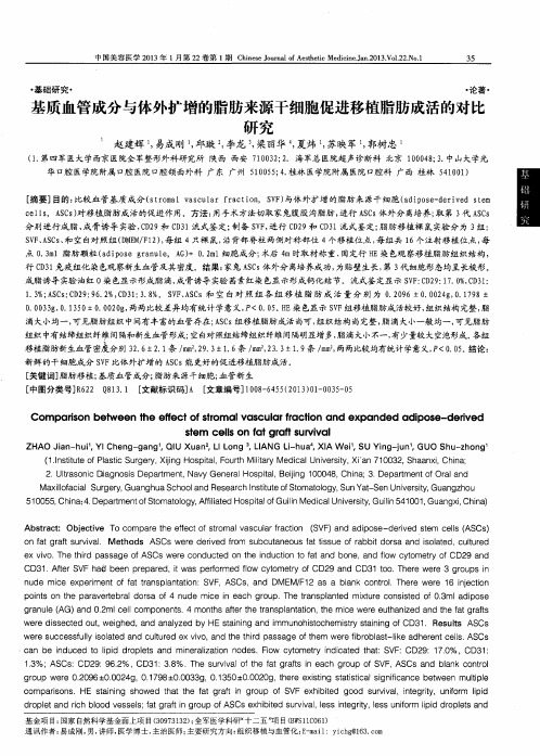
基质血管成分与体外扩增的脂肪来源干细胞促进移植脂肪成活的对比研究
・
基础 研究・
・ 论著 ・
基质血管成分 与体外扩增的脂肪来源干细胞促进移植脂肪成活的对比 研 究
赵建辉 , 易成 刚 , 邱璇 : , 李龙 , , 梁丽华 , 夏炜 , 苏映军 , 郭树忠
( 1 . 第 四军 医大学西京 医院全军整形外科研究所 陕西 西安 7 1 0 0 3 2 ; 2 .海军总医院超声诊断科 北京 1 0 0 0 4 8 ; 3 . 中山大学光 华1 : 2 腔 医学院附属 口腔 医院 口腔颌 面外科 广 东 广 州 5 1 0 0 5 5 ; 4 . 桂林 医学院附属 医院口腔科 广西 桂 林 5 4 1 0 0 1 ) [ 摘要] 目的: 比较血 管基质成 分 ( s t r o m a l v a s c u l a r f r a c t i o n ,S V F ) 与体外扩增 的脂肪 来源千细胞 ( a d i p o s e - d e r i r e d s t e m
中国美容医学 2 0 1 3年 1月第 2 2卷第 1期 C h i n e s e J o u r n a l o f A e s t h e t i c Me d i c i n e . J a n . 2 0 1 3 . V o 1 . 2 2 . N o . 1
3 5
0 . 0 0 3 3 g , 0 . 1 3 5 0 ±0 . 0 0 2 0 g , 两两比较差异均有 统计学意义, P <0 . 0 5 。 H E染 色显示 S V F 组移植脂肪成活较好, 组织结构完整, 脂 滴大小均一 , 可见脂肪组 织中间有丰 富的血管存在 ; A S C s组移植脂肪成 活尚可 , 组织结构 尚完整 , 脂 滴大小一般 均一, 可见脂肪 组织 中有结缔组织纤维 间隔和新生血 管形成; 空白对照组结缔组织纤维间隔明显增 多, 脂 滴大小不一, 有少量较大空泡形成 。 各组 移植脂肪新生血 管密度分别 3 2 . 6 ±2 . 1 条/ m m , 2 9 . 3 土1 . 6 条/ m m , 2 3 . 3 ±1 _ 9条 / m m , 两两比较 均有统计 学意义, P <0 . 0 5 。 结论 :
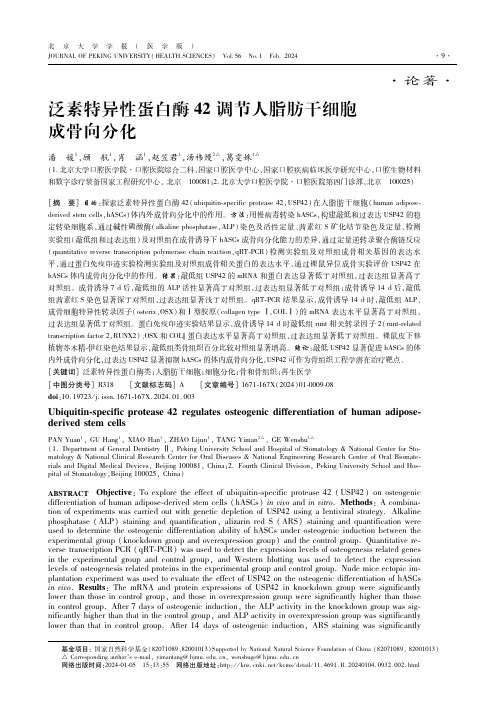
泛素特异性蛋白酶42调节人脂肪干细胞成骨向分化
""#"
北 京大学学报! 医学版$ QRS%A(TREUMVLAWSALXM%6LYZ! [M(TY[6GLMAGM6$ !X9C'5)!A9'"!E<>',#,-
=<<8<:24 ;@<K49DK=9e4 H:978 ;@B4 24 ;@<D94;:9CH:978# B4= 32H42F2DB4;C?C2H@;<:24 9c<:<d8:<33294 H:978 ;@B4 24 ;@<D94;:9CH:978'Y@<:<37C;39Ff%Y*UG%3@9e<= ;@B;;@<I%A(<d8:<33294 C<c<C39F(TU# 93* ;<:2d! R6+$ B4= D9CCBH<4 ;?8<# ! GRT#$ 24 ;@<K49DK=9e4 H:978 e<:<32H42F2DB4;C?@2H@<:;@B4 ;@93< 24 ;@<D94;:9CH:978 BF;<:"- =B?39F93;<9H<42D24=7D;294# B4= ;@93<24 9c<:<d8:<33294 H:978 e<:<32H42F2* DB4;C?C9e<:;@B4 ;@93<24 D94;:9CH:978'Y@<:<37C;39Fa<3;<:4 >C9;;24H3@9e<= ;@B;;@<<d8:<33294 C<c<C3 9F:74;*:<CB;<= ;:B43D:28;294 FBD;9:, ! %SA+,$ # R6+B4= GRT# 24 ;@<K49DK97;H:978 e<:<32H42F2DB4;* C?@2H@<:;@B4 ;@93<24 ;@<D94;:9CH:978 B;"- =B?3BF;<:93;<9H<42D24=7D;294# e@2C<;@<<d8:<33294 C<c<C3 9F%SA+,# R6+B4= GRT# 24 ;@<9c<:<d8:<33294 H:978 e<:<32H42F2DB4;C?C9e<:;@B4 ;@93<24 ;@<D94;:9C H:978'[<IB;9d?C24*<9324 3;B2424H9F37>D7;B4<973H:BF;324 47=<I2D<3@9e<= ;@B;;@<8<:D<4;BH<9F 93;<92= B:<B24 ;@<K49DK=9e4 H:978 eB332H42F2DB4;C?@2H@<:;@B4 ;@B;24 ;@<D94;:9CH:978'I"+,47.#"+& V49DK=9e4 9FS6U-, DB4 32H42F2DB4;C?8:9I9;<;@<93;<9H<42D=2FF<:<4;2B;294 9F@(6G3$" '$%,+B4= $" '$'+# B4= 9c<:<d8:<33294 9FS6U-, 32H42F2DB4;C?24@2>2;3$" '$'+93;<9H<42D=2FF<:<4;2B;294 9F@(6G3# B4= S6U-, DB4 8:9c2=<B89;<4;2BC;@<:B8<7;2D;B:H<;F9:>94<;2337<<4H24<<:24H' @A?BC>D:! S>2f72;24*38<D2F2D8:9;<B3<3% [7IB4 B=2893<*=<:2c<= 3;<I D<CC3% G<CC=2FF<:<4;2B;294% \94<B4= >94<3% %<H<4<:B;2c<I<=2D24<
《山羊胎肺间充质干细胞的体外分离培养与诱导分化》范文

《山羊胎肺间充质干细胞的体外分离培养与诱导分化》篇一一、引言近年来,干细胞研究已成为生命科学领域的前沿课题。
其中,间充质干细胞(MSCs)因其具有自我更新能力和多向分化潜能,在再生医学和临床治疗中具有广阔的应用前景。
山羊作为一种重要的经济动物,其胎肺间充质干细胞(Goat Fetal Lung Mesenchymal Stem Cells,GFL-MSCs)的研究对于推动动物医学和人类医学的发展具有重要意义。
本文旨在研究GFL-MSCs的体外分离培养及诱导分化过程,为进一步应用提供理论依据。
二、材料与方法1. 材料本实验所需材料包括:山羊胎儿肺组织、培养基、胰蛋白酶、胎牛血清、抗凝剂等。
2. 方法(1)分离培养:选取健康山羊胎儿肺组织,经胰蛋白酶消化、离心等方法,获得间充质干细胞。
将细胞接种于培养皿中,加入含胎牛血清的培养基,置于恒温培养箱中培养。
(2)诱导分化:当细胞达到一定密度后,将细胞分为若干组,分别加入不同诱导剂进行诱导分化,如成骨诱导剂、成脂诱导剂等。
(3)观察与记录:采用显微镜观察细胞形态变化,记录细胞生长情况及分化情况。
三、实验结果1. 分离培养通过胰蛋白酶消化法成功分离出GFL-MSCs,细胞呈纺锤形或多角形,生长旺盛,具有良好的增殖能力。
在培养过程中,细胞逐渐形成集落,形成单层细胞层。
2. 诱导分化(1)成骨诱导:经过成骨诱导剂处理后,GFL-MSCs逐渐形成矿化结节,ALP活性显著提高,表明细胞成功向成骨方向分化。
(2)成脂诱导:经过成脂诱导剂处理后,GFL-MSCs逐渐形成脂滴,并表达脂肪相关基因,表明细胞成功向脂肪方向分化。
四、讨论本实验成功分离培养出GFL-MSCs,并对其进行了成骨和成脂方向的诱导分化。
结果表明,GFL-MSCs具有多向分化潜能,为进一步研究其生物学特性和应用提供了基础。
此外,山羊作为一种经济动物,其胎肺间充质干细胞的研究对于推动动物医学和人类医学的发展具有重要意义。
脂肪干细胞成脂诱导及鉴定程序

1.0 L(SH30021.01B, Hyclone) 2. 胎牛血清( Fetal Bovine Serum ,FBS)10%1%1 pmo/ L10 mol / L黄嘌呤0.5 mmol/L200 mol / L (ES-009-B, Millipore)(TMS-AB2C, CHEMICON ) 分子量:392.46(D4902-25MG, Sigma)分子量:5808(91077C—1g, Sigma)分子量:222.24( I5879-100MG, Sigma)分子量:357.79(I7378 —5G, Sigma)脂肪干细胞成脂诱导及鉴定程序、试剂准备一)成脂分化诱导液(Adipogenic Medium, AM )配方【1】:试剂名称浓度商品信息1. 极限必须培养基(Dulbecco 'S Modified Eagle Medium,DMEM)二)成脂分化诱导液浓储液配制试剂名称f=p 曰.质量浓缩倍数配制方法1. Stock A 分装1ml/管X100胎牛血清1ml/ 管1X( liquid )保存:-20C2. Stock B 分装0.1ml/管X100青霉素/ 链霉素0.1ml/ 管100X( liquid )保存:-20C3. Stock C 溶于30ml 无水乙醇(0.1%)地塞米松0.0117738 g 1000X 分装0.1ml/管X300保存:-20C4. Stock D 溶于10ml Hcl(0.1 mol/L ,PH2.0)胰岛素0.05808 g 100X 分装0.1ml/管X100保存:4C5. Stock E 溶于 2.5ml DMSO(0. 5%)3-异丁基-1 -甲基黄嘌呤0.05556 g 200X 分装0.05ml/EP 管X50保存:-20C6. Stock F 溶于2ml 无水乙醇(0.2%)吲哚美辛0.07155 g 500X分装0.02ml/ 管X100 保存:-20C (三)成脂分化诱导液工作液配制(10ml)1. 取DMEM(L)8.72ml 加入15ml 离心管(BD )2. 加1 管Stock A(1ml);3. 加1 管Stock B(0.1ml);4. 取1 管Stock C (0.1ml)溶解,加入0.01ml ;5. 加1 管Stock E(0.05ml);6. 加1 管Stock F (0.02ml);7. 加1 管Stock D (0.1ml);8. 测渗透压,调pH7.2-7.4;9. 0.22 m微孔过滤,4C贮存,一周内使用。
- 1、下载文档前请自行甄别文档内容的完整性,平台不提供额外的编辑、内容补充、找答案等附加服务。
- 2、"仅部分预览"的文档,不可在线预览部分如存在完整性等问题,可反馈申请退款(可完整预览的文档不适用该条件!)。
- 3、如文档侵犯您的权益,请联系客服反馈,我们会尽快为您处理(人工客服工作时间:9:00-18:30)。
Received: 2006- 02- 11 Accepted: 2006- 02- 16
Abstr act AIM: To compare the adipogenic differentiation ability in vitro of induced human adipose stem cells from different sites of the same body, so as to provide experimental foundation for further application of adipose stem cells (ASCs). METHODS: The experiment was carried out in the Research Center of Plastic Surgery Hospital, Peking Union Medical College from November 2004 to May 2005. The mesenchymal stem cells in lipoaspirates were extracted from 8 sites (anterior thigh, upper buttocks\ upper buttocks, upper abdomen\ iliolumbalis\ upper abdomen\ back, upper arm) of 5 female donors (mean age: 32 years old) undergoing the liposuction in clinic, then these cells were induced to differentiate to the adipogenic cells in vitro. ASCs at passage 1, cultured in the control media (DMEM, supplemented with 10% FBS) were plated at a density of 3 ×10⁴ cells every dish into 35 mm culture dishes, and the control media was replaced with adipogenic media (AM). The media was changed twice weekly and the culture dish added with the control media served as negative control. Two weeks later, the induced cells and negative cells were assessed with an Oil Red-O stain to identify whether there was lipid in cells or not. Then through the Oil Red O stain and positive cell counting, the adipogenic differentiation ability of induced human adipose stem cells from 2 different sites of 3 patients were assessed with qualitative and semi-quantitative analysis. The adipogenic differentiation rates of different cells were determined with chi- square test. RESULTS: ①The measured results of adipogenic differentiation ability of ASCs: The mean number of cells isolated from per 100 mL of lipoaspirates of different donors were quite different, the mean cell yields were ranged from 2.45×10⁸ /100 mL to 1.25×10⁹ /100 mL\ but the mean number from different sites of the same body were quite similar\ the mean adipogenic differentiation rate measured from 8 sites of 5 donors was 36.2% calculated according to the adipogenic differentiation rates of every kind of cell. The statistics analysis confirmed that adipogenic differentiation ability of induced human adipose stem cells from different sites of the same body was different, and there was no correlation between the mean numbers of stem cells and the age of donor (r=0.098, P=0.817), while the adipogenic differentiation ability was significant negatively correlated with the age of donor (r=- 0.568, P < 0.01). ②The ASCs morphological change and the qualitative analysis results of Oil Red-O stain: The ASCs exhibited an expanded morphology and differentiated to the adipogenic lineage after adipogenic induced. The cells contained multiple intracellular orange lipid-filled droplets, which accounted for 80 - 90% volume of the cells. The nuclei shrank or located on one side of cells, and
中国临床康复 第 10 卷 第 29 期 2006- 08- 10 出版
44
Chinese Journal of Clinical Rehabilitation,August 10 2006 Vol. 10 No. 29
·基础研究·
不同来源的人脂肪干细胞体外成脂诱导分化能力的比较★
严 笠 1, 马桂娥 2, 曹 蕊 1, 贾春实 1, 吕晓岩 1, 王春梅 1
